The CaV3.3 calcium channel is the major sleep spindle ... · The CaV3.3 calcium channel is the...
6
The Ca V 3.3 calcium channel is the major sleep spindle pacemaker in thalamus Simone Astori a , Ralf D. Wimmer a,b , Haydn M. Prosser c,1 , Corrado Corti d,e , Mauro Corsi d,e , Nicolas Liaudet a , Andrea Volterra a , Paul Franken b , John P. Adelman f , and Anita Lüthi a,2 a Department of Cell Biology and Morphology, University of Lausanne, CH-1005 Lausanne, Switzerland; b Center for Integrative Genomics, Génopode, University of Lausanne, CH-1015 Lausanne-Dorigny, Switzerland; c Genetics Research, GlaxoSmithKline Pharmaceuticals, New Frontiers Science Park, Harlow, Essex CM19 5AW, United Kingdom; d GlaxoSmithKline Neuroscience Centre for Excellence for Drug Discovery, 37129 Verona, Italy; e Aptuit Medicine Research Centre, 37129 Verona, Italy; and f Vollum Institute, Oregon Health and Science University, Portland, OR 97239 Edited by Rodolfo R. Llinas, New York University Medical Center, New York, NY, and approved July 13, 2011 (received for review April 18, 2011) Low-threshold (T-type) Ca 2+ channels encoded by the Ca V 3 genes endow neurons with oscillatory properties that underlie slow waves characteristic of the non-rapid eye movement (NREM) sleep EEG. Three Ca V 3 channel subtypes are expressed in the thalamocort- ical (TC) system, but their respective roles for the sleep EEG are unclear. Ca V 3.3 protein is expressed abundantly in the nucleus retic- ularis thalami (nRt), an essential oscillatory burst generator. We re- port the characterization of a transgenic Ca V 3.3 -/- mouse line and demonstrate that Ca V 3.3 channels are indispensable for nRt func- tion and for sleep spindles, a hallmark of natural sleep. The absence of Ca V 3.3 channels prevented oscillatory bursting in the low- frequency (4–10 Hz) range in nRt cells but spared tonic discharge. In contrast, adjacent TC neurons expressing Ca V 3.1 channels re- tained low-threshold bursts. Nevertheless, the generation of syn- chronized thalamic network oscillations underlying sleep-spindle waves was weakened markedly because of the reduced inhibition of TC neurons via nRt cells. T currents in Ca V 3.3 -/- mice were <30% compared with those in WT mice, and the remaining current, car- ried by Ca V 3.2 channels, generated dendritic [Ca 2+ ] i signals insuffi- cient to provoke oscillatory bursting that arises from interplay with Ca 2+ -dependent small conductance-type 2 K + channels. Finally, nat- urally sleeping Ca V 3.3 -/- mice showed a selective reduction in the power density of the σ frequency band (10–12 Hz) at transitions from NREM to REM sleep, with other EEG waves remaining unal- tered. Together, these data identify a central role for Ca V 3.3 chan- nels in the rhythmogenic properties of the sleep-spindle generator and provide a molecular target to elucidate the roles of sleep spin- dles for brain function and development. inhibition | ion channel | Cacna1i | alpha1i | synchrony T he Ca 2+ channels encoded by the Ca V 3 genes activate near resting membrane potentials and generate low-threshold Ca 2+ spikes leading to burst firing and low-frequency oscillatory dis- charge that are prominent in some thalamic, olivary, and cerebellar neurons (1). Among the low-threshold Ca 2+ currents carried by Ca V 3 channels, those mediated by Ca V 3.3 channels are unique in that they display the slowest time course, the fastest recovery from inactivation, and often the most depolarized activation voltages (2, 3). Moreover, Ca V 3.3 mRNA is expressed predominantly in brain and shows highest regional specificity (3–5). To date, identification of specific physiological roles for Ca V 3.3 channels has been ham- pered for several reasons. First, these channels typically are coexpressed with Ca V 3.1 and/or Ca V 3.2 channels (4, 5), and spe- cific pharmacological tools are not available (1). Second, Ca V 3.3 channels often are found in distal dendrites, limiting accessibility for electrophysiological characterization (6, 7). Finally, Ca V 3.3 −/− mice have not been reported, whereas Ca V 3.1 −/− and Ca V 3.2 knockdown mice have helped address the roles of Ca V 3.1 and Ca V 3.2 channels in sleep and pain, respectively (8–10). In the thalamus, Ca V 3.3 mRNA and protein are abundant in GABAergic cells of the surrounding nucleus reticularis thalami (nRt), the principal source of inhibition to relay nuclei, whereas excitatory thalamocortical (TC) neurons are not immunopositive for Ca V 3.3 (4, 5, 11). The slow decay and the depolarized acti- vation range of low-threshold Ca 2+ currents in the nRt (12) are thought to reflect the dominant mRNA expression of Ca V 3.3 over Ca V 3.2 channels (5, 7, 13). Vigorous bursting properties in nRt underlie its well-documented role in pacemaking sleep spindles (14) and its proposed involvement in attentional gating mecha- nisms (15). Therefore, the nRt is ideally suited to explore the cellular and circuit roles of Ca V 3.3 channels. Here, we describe the Ca V 3.3 −/− mouse and report that Ca V 3.3 channels are required for nRt cell bursting and for synchronized rhythmicity in intrathalamic circuits. Ca V 3.3 −/− mice show selec- tively weakened spindle wave generation during spontaneous sleep. These results establish a specific role for Ca V 3.3 channels in a sleep EEG hallmark and, more generally, an animal model to provide insight into sleep’s role for the brain. Results Generation of Ca V 3.3 -/- Mice. Interruption of the Ca V 3.3 gene was achieved through homologous recombination in 129Ola ES cells. The targeting construct deleted a 6.8-kb genomic region (National Center for Biotechnology Information m37 15:80,198, 299-80,205,160) encoding exons 11–21, replacing them with a β-galactosidase–neomycin cassette (Fig. 1A). The engineered allele deletes the coding sequence between the predicted IIS2 and IIIS4 transmembrane regions, including the third predicted in- tracellular domain (3). Additionally, exons 10 and 22 are out of the translational reading frame; therefore, any truncated protein product will not be functional. Ca V 3.3 gene deletion was con- firmed by RT-PCR experiments using RNA extracted from cer- ebellum, olfactory bulb, cortex, and thalamus (Fig. 1B). The deletion of the Ca V 3.3 gene did not affect transcription of the Ca V 3.2 gene in thalamus, as assessed by quantitative RT-PCR (Fig. 1C). For all experiments described below, mouse genotype was determined by PCR (Fig. 1D), and homozygous Ca V 3.3 −/− and WT littermates were used. nRt Oscillatory Burst Discharges Are Impaired in Ca V 3.3 -/- Mice. Whole-cell current-clamp recordings were obtained from nRt cells in acute brain slices of 3- to 4-wk-old mice. The oscillatory bursting characteristic for nRt cells was elicited at the offset of brief membrane hyperpolarizations, whereas tonic discharges Author contributions: S.A., R.D.W., P.F., and A.L. designed research; S.A., R.D.W., H.M.P., C.C., and M.C. performed research; N.L. and A.V. contributed new reagents/analytic tools; S.A. and R.D.W. analyzed data; and S.A., R.D.W., J.P.A., and A.L. wrote the paper. The authors declare no conflict of interest. This article is a PNAS Direct Submission. 1 Present address: The Wellcome Trust Sanger Institute, Wellcome Trust Genome Campus, Hinxton, Cambridge CB10 1SA, United Kingdom. 2 To whom correspondence should be addressed. E-mail: [email protected]. This article contains supporting information online at www.pnas.org/lookup/suppl/doi:10. 1073/pnas.1105115108/-/DCSupplemental. www.pnas.org/cgi/doi/10.1073/pnas.1105115108 PNAS | August 16, 2011 | vol. 108 | no. 33 | 13823–13828 NEUROSCIENCE
Transcript of The CaV3.3 calcium channel is the major sleep spindle ... · The CaV3.3 calcium channel is the...

The CaV3.3 calcium channel is the major sleep spindlepacemaker in thalamusSimone Astoria, Ralf D. Wimmera,b, Haydn M. Prosserc,1, Corrado Cortid,e, Mauro Corsid,e, Nicolas Liaudeta,Andrea Volterraa, Paul Frankenb, John P. Adelmanf, and Anita Lüthia,2
aDepartment of Cell Biology and Morphology, University of Lausanne, CH-1005 Lausanne, Switzerland; bCenter for Integrative Genomics, Génopode,University of Lausanne, CH-1015 Lausanne-Dorigny, Switzerland; cGenetics Research, GlaxoSmithKline Pharmaceuticals, New Frontiers Science Park,Harlow, Essex CM19 5AW, United Kingdom; dGlaxoSmithKline Neuroscience Centre for Excellence for Drug Discovery, 37129 Verona, Italy;eAptuit Medicine Research Centre, 37129 Verona, Italy; and fVollum Institute, Oregon Health and Science University, Portland, OR 97239
Edited by Rodolfo R. Llinas, New York University Medical Center, New York, NY, and approved July 13, 2011 (received for review April 18, 2011)
Low-threshold (T-type) Ca2+ channels encoded by the CaV3 genesendow neurons with oscillatory properties that underlie slowwaves characteristic of the non-rapid eye movement (NREM) sleepEEG. Three CaV3 channel subtypes are expressed in the thalamocort-ical (TC) system, but their respective roles for the sleep EEG areunclear. CaV3.3 protein is expressed abundantly in the nucleus retic-ularis thalami (nRt), an essential oscillatory burst generator. We re-port the characterization of a transgenic CaV3.3
−/− mouse line anddemonstrate that CaV3.3 channels are indispensable for nRt func-tion and for sleep spindles, a hallmark of natural sleep. The absenceof CaV3.3 channels prevented oscillatory bursting in the low-frequency (4–10 Hz) range in nRt cells but spared tonic discharge.In contrast, adjacent TC neurons expressing CaV3.1 channels re-tained low-threshold bursts. Nevertheless, the generation of syn-chronized thalamic network oscillations underlying sleep-spindlewaves was weakened markedly because of the reduced inhibitionof TC neurons via nRt cells. T currents in CaV3.3
−/− mice were<30%compared with those in WT mice, and the remaining current, car-ried by CaV3.2 channels, generated dendritic [Ca2+]i signals insuffi-cient to provoke oscillatory bursting that arises from interplaywithCa2+-dependent small conductance-type 2 K+ channels. Finally, nat-urally sleeping CaV3.3
−/− mice showed a selective reduction in thepower density of the σ frequency band (10–12 Hz) at transitionsfrom NREM to REM sleep, with other EEG waves remaining unal-tered. Together, these data identify a central role for CaV3.3 chan-nels in the rhythmogenic properties of the sleep-spindle generatorand provide a molecular target to elucidate the roles of sleep spin-dles for brain function and development.
inhibition | ion channel | Cacna1i | alpha1i | synchrony
The Ca2+ channels encoded by the CaV3 genes activate nearresting membrane potentials and generate low-threshold Ca2+
spikes leading to burst firing and low-frequency oscillatory dis-charge that are prominent in some thalamic, olivary, and cerebellarneurons (1). Among the low-threshold Ca2+ currents carried byCaV3 channels, those mediated by CaV3.3 channels are unique inthat they display the slowest time course, the fastest recovery frominactivation, and often the most depolarized activation voltages (2,3). Moreover, CaV3.3 mRNA is expressed predominantly in brainand shows highest regional specificity (3–5). To date, identificationof specific physiological roles for CaV3.3 channels has been ham-pered for several reasons. First, these channels typically arecoexpressed with CaV3.1 and/or CaV3.2 channels (4, 5), and spe-cific pharmacological tools are not available (1). Second, CaV3.3channels often are found in distal dendrites, limiting accessibilityfor electrophysiological characterization (6, 7). Finally, CaV3.3
−/−
mice have not been reported, whereas CaV3.1−/− and CaV3.2
knockdown mice have helped address the roles of CaV3.1 andCaV3.2 channels in sleep and pain, respectively (8–10).In the thalamus, CaV3.3 mRNA and protein are abundant in
GABAergic cells of the surrounding nucleus reticularis thalami(nRt), the principal source of inhibition to relay nuclei, whereas
excitatory thalamocortical (TC) neurons are not immunopositivefor CaV3.3 (4, 5, 11). The slow decay and the depolarized acti-vation range of low-threshold Ca2+ currents in the nRt (12) arethought to reflect the dominant mRNA expression of CaV3.3 overCaV3.2 channels (5, 7, 13). Vigorous bursting properties in nRtunderlie its well-documented role in pacemaking sleep spindles(14) and its proposed involvement in attentional gating mecha-nisms (15). Therefore, the nRt is ideally suited to explore thecellular and circuit roles of CaV3.3 channels.Here, we describe the CaV3.3
−/− mouse and report that CaV3.3channels are required for nRt cell bursting and for synchronizedrhythmicity in intrathalamic circuits. CaV3.3
−/− mice show selec-tively weakened spindle wave generation during spontaneoussleep. These results establish a specific role for CaV3.3 channels ina sleep EEG hallmark and, more generally, an animal model toprovide insight into sleep’s role for the brain.
ResultsGeneration of CaV3.3
−/− Mice. Interruption of the CaV3.3 genewas achieved through homologous recombination in 129Ola EScells. The targeting construct deleted a 6.8-kb genomic region(National Center for Biotechnology Information m37 15:80,198,299-80,205,160) encoding exons 11–21, replacing them with aβ-galactosidase–neomycin cassette (Fig. 1A). The engineeredallele deletes the coding sequence between the predicted IIS2 andIIIS4 transmembrane regions, including the third predicted in-tracellular domain (3). Additionally, exons 10 and 22 are out ofthe translational reading frame; therefore, any truncated proteinproduct will not be functional. CaV3.3 gene deletion was con-firmed by RT-PCR experiments using RNA extracted from cer-ebellum, olfactory bulb, cortex, and thalamus (Fig. 1B). Thedeletion of the CaV3.3 gene did not affect transcription of theCaV3.2 gene in thalamus, as assessed by quantitative RT-PCR(Fig. 1C). For all experiments described below, mouse genotypewas determined by PCR (Fig. 1D), and homozygous CaV3.3
−/−
and WT littermates were used.
nRt Oscillatory Burst Discharges Are Impaired in CaV3.3−/− Mice.
Whole-cell current-clamp recordings were obtained from nRtcells in acute brain slices of 3- to 4-wk-old mice. The oscillatorybursting characteristic for nRt cells was elicited at the offset ofbrief membrane hyperpolarizations, whereas tonic discharges
Author contributions: S.A., R.D.W., P.F., and A.L. designed research; S.A., R.D.W., H.M.P.,C.C., and M.C. performed research; N.L. and A.V. contributed new reagents/analytic tools;S.A. and R.D.W. analyzed data; and S.A., R.D.W., J.P.A., and A.L. wrote the paper.
The authors declare no conflict of interest.
This article is a PNAS Direct Submission.1Present address: The Wellcome Trust Sanger Institute, Wellcome Trust Genome Campus,Hinxton, Cambridge CB10 1SA, United Kingdom.
2To whom correspondence should be addressed. E-mail: [email protected].
This article contains supporting information online at www.pnas.org/lookup/suppl/doi:10.1073/pnas.1105115108/-/DCSupplemental.
www.pnas.org/cgi/doi/10.1073/pnas.1105115108 PNAS | August 16, 2011 | vol. 108 | no. 33 | 13823–13828
NEU
ROSC
IENCE
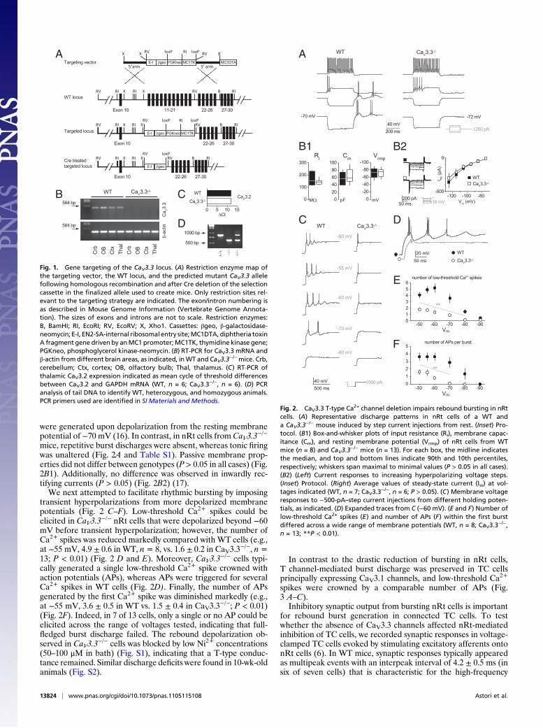
were generated upon depolarization from the resting membranepotential of −70 mV (16). In contrast, in nRt cells from CaV3.3
−/−
mice, repetitive burst discharges were absent, whereas tonic firingwas unaltered (Fig. 2A and Table S1). Passive membrane prop-erties did not differ between genotypes (P > 0.05 in all cases) (Fig.2B1). Additionally, no difference was observed in inwardly rec-tifying currents (P > 0.05) (Fig. 2B2) (17).We next attempted to facilitate rhythmic bursting by imposing
transient hyperpolarizations from more depolarized membranepotentials (Fig. 2 C–F). Low-threshold Ca2+ spikes could beelicited in CaV3.3
−/− nRt cells that were depolarized beyond −60mV before transient hyperpolarization; however, the number ofCa2+ spikes was reduced markedly compared with WT cells (e.g.,at −55 mV, 4.9 ± 0.6 in WT, n= 8, vs. 1.6 ± 0.2 in CaV3.3
−/−, n=13; P < 0.01) (Fig. 2 D and E). Moreover, CaV3.3
−/− cells typi-cally generated a single low-threshold Ca2+ spike crowned withaction potentials (APs), whereas APs were triggered for severalCa2+ spikes in WT cells (Fig. 2D). Finally, the number of APsgenerated by the first Ca2+ spike was diminished markedly (e.g.,at −55 mV, 3.6 ± 0.5 in WT vs. 1.5 ± 0.4 in CaV3.3
−/−; P < 0.01)(Fig. 2F). Indeed, in 7 of 13 cells, only a single or no AP could beelicited across the range of voltages tested, indicating that full-fledged burst discharge failed. The rebound depolarization ob-served in CaV3.3
−/− cells was blocked by low Ni2+ concentrations(50–100 μM in bath) (Fig. S1), indicating that a T-type conduc-tance remained. Similar discharge deficits were found in 10-wk-oldanimals (Fig. S2).
In contrast to the drastic reduction of bursting in nRt cells,T channel-mediated burst discharge was preserved in TC cellsprincipally expressing CaV3.1 channels, and low-threshold Ca2+
spikes were crowned by a comparable number of APs (Fig.3 A–C).Inhibitory synaptic output from bursting nRt cells is important
for rebound burst generation in connected TC cells. To testwhether the absence of CaV3.3 channels affected nRt-mediatedinhibition of TC cells, we recorded synaptic responses in voltage-clamped TC cells evoked by stimulating excitatory afferents ontonRt cells (6). In WT mice, synaptic responses typically appearedas multipeak events with an interpeak interval of 4.2 ± 0.5 ms (insix of seven cells) that is characteristic for the high-frequency
WT locus
Targeted locus
Exon 10 11-21
RIRV
22-26 27-30
X RVX BRI
Targeting vector MC1DTA
loxPRV loxPRI
RIRV X RVX BRIloxPRV loxPRI
RI
X X RV B
5’arm 3’ arm
A
B
Crb
OB
Ctx
Thal
Crb
OB
Ctx
Thal
WT CaV3.3-/-
564 bp
564 bp
β-ac
tinC
a V3.
3
C
Exon 10 22-26 27-30
E-I βgeo PGKneo MC1TK
−/−
+/−
+/+
1000 bp
500 bp
E-I βgeo PGKneo MC1TK
Cre treatedtargeted locus
RI
RIRV X RVX BRIloxPRV
Exon 10 22-26 27-30
E-I βgeo
RI
D
WT
CaV3.3-/-
0 5 10 15ΔCt
CaV3.2
Fig. 1. Gene targeting of the CaV3.3 locus. (A) Restriction enzyme map ofthe targeting vector, the WT locus, and the predicted mutant CaV3.3 allelefollowing homologous recombination and after Cre deletion of the selectioncassette in the finalized allele used to create mice. Only restriction sites rel-evant to the targeting strategy are indicated. The exon/intron numbering isas described in Mouse Genome Information (Vertebrate Genome Annota-tion). The sizes of exons and introns are not to scale. Restriction enzymes:B, BamHI; RI, EcoRI; RV, EcoRV; X, Xho1. Cassettes: βgeo, β-galactosidase-neomycin; E-I, EN2-SA-internal ribosomal entry site;MC1DTA, diphtheria toxinA fragment gene driven by anMC1 promoter;MC1TK, thymidine kinase gene;PGKneo, phosphoglycerol kinase-neomycin. (B) RT-PCR for CaV3.3 mRNA andβ-actin from different brain areas, as indicated, inWT and CaV3.3
−/−mice. Crb,cerebellum; Ctx, cortex; OB, olfactory bulb; Thal, thalamus. (C) RT-PCR ofthalamic CaV3.2 expression indicated as mean cycle of threshold differencesbetween CaV3.2 and GAPDH mRNA (WT, n = 6; CaV3.3
−/−, n = 6). (D) PCRanalysis of tail DNA to identify WT, heterozygous, and homozygous animals.PCR primers used are identified in SI Materials and Methods.
WT CaV3.3-/-
200 ms40 mV
-70 mV
A
250 pA
-72 mV
E
F
Vm
012345
-90-80-70-60-50
**
number of APs per burst
-90Vm
0123456
-80-70-60-50
**
number of low-threshold Ca2+ spikes
D
50 ms20 mV WT
CaV3.3-/-
500 ms40 mV
-50 mV
-55 mV
-60 mV
-70 mV
-80 mV
C
500 pA
B1
MΩ
Ri
mV
Vrmp300
200
100
0
100806040200
Cm
pF
-100-80-60-40-20
050 ms
200 pA
B2
10 mV
WTCaV3.3-/-
-500
0
-120 -100 -80Vm (mV)
I ss (p
A)
WT CaV3.3-/-
Fig. 2. CaV3.3 T-type Ca2+ channel deletion impairs rebound bursting in nRtcells. (A) Representative discharge patterns in nRt cells of a WT anda CaV3.3
−/− mouse induced by step current injections from rest. (Inset) Pro-tocol. (B1) Box-and-whisker plots of input resistance (Ri), membrane capac-itance (Cm), and resting membrane potential (Vrmp) of nRt cells from WTmice (n = 8) and CaV3.3
−/− mice (n = 13). For each box, the midline indicatesthe median, and top and bottom lines indicate 90th and 10th percentiles,respectively; whiskers span maximal to minimal values (P > 0.05 in all cases).(B2) (Left) Current responses to increasing hyperpolarizing voltage steps.(Inset) Protocol. (Right) Average values of steady-state current (Iss) at vol-tages indicated (WT, n = 7; CaV3.3
−/−, n = 6; P > 0.05). (C) Membrane voltageresponses to −500-pA–step current injections from different holding poten-tials, as indicated. (D) Expanded traces from C (−60 mV). (E and F) Number oflow-threshold Ca2+ spikes (E) and number of APs (F) within the first burstdiffered across a wide range of membrane potentials (WT, n = 8; CaV3.3
−/−,n = 13; **P < 0.01).
13824 | www.pnas.org/cgi/doi/10.1073/pnas.1105115108 Astori et al.

bursts of nRt cells (Fig. 3D1). In contrast, in CaV3.3−/− mice,
multiple peaks appeared rarely (in two of six cells). As a result,the inhibitory driving force onto TC cells was significantlysmaller in CaV3.3
−/− mice, as determined from the integral ofsynaptic currents evoked by increasing stimulus intensities (e.g.,with 250-μA stimulus intensity: 26 ± 5 pC in WT mice vs. 10 ± 4pC in CaV3.3
−/− mice; P < 0.05) (Fig. 3D2).To investigate the effects of reduced nRt inhibitory drive on
intrathalamic circuitry involved in sleep spindles (18), we ex-amined synchronized multiunit discharges in nRt elicited viabipolar electrical stimulation of excitatory afferents in the adja-cent internal capsule and perpetuated via reciprocal synapticinteractions with TC cells. To facilitate network activity, we firstperfused slices with artificial CSF (ACSF) containing increasedCa2+ (3 mM) and decreased Mg2+ (0.5 mM). In WT mice, ro-bust reverberating activity at 7–9 Hz lasting ∼1.5 s was elicited(Fig. 3E). Similar activity was detected in slices from CaV3.3
−/−
mice, but with weaker oscillatory strength (P < 0.05) and shorterduration (P = 0.07) (Fig. 3 E and F). Notably, after returning tonormal divalent cation concentrations (2 mM Ca2+/1.2 mM
Mg2+), synchronized oscillations disappeared in CaV3.3−/− mice
in most cases (four of six slices) but remained in WT slices (six ofseven slices). Thus, the absence of CaV3.3 channels impairs thecapability of the nRt–TC network to generate spindle-like dis-charge, but rhythmic discharge can be restored partially by in-creasing cellular excitability.
Dominant Contribution of CaV3.3 Channels to T Currents in nRtNeurons. Whole-cell T currents were elicited by increasingly de-polarized voltage steps (1 s) from −100 mV using a fluoride-containing patch pipette solution (Fig. 4 and SI Materials andMethods). Compared with WT cells, T-current density in CaV3.3
−/−
nRt cells was reduced throughout the activation range (e.g., 78%reduction at −70 mV; P < 0.01, and 54% reduction at −50 mV;P < 0.01; n = 10 for WT and n = 8 for CaV3.3
−/−) (Fig. 4A2).Moreover, the activation curve was shifted toward more depo-larized potentials in CaV3.3
−/− mice (P < 0.01) (Fig. 4A3). Strik-ingly, T currents from CaV3.3
−/− cells had a 2.5-fold faster decaythan WT cells (e.g., at −50 mV τw, decay = 96 ± 10 ms for WTvs. τw, decay = 38 ± 6 ms for CaV3.3
−/−; n = 10 and 8, respectively;P < 0.01) (Fig. 4B). Similar alterations were found at near-physiological temperature (Fig. S3). Notably, recovery from steady-state inactivation was retarded in CaV3.3
−/− cells (time constant ofrecovery: τw, rec = 1.5 ± 0.3 s for WT vs. τw, rec = 7.9 ± 2.3 s forCaV3.3
−/−; n = 10 and 8, respectively; P < 0.05) (Fig. 4C).Pharmacological properties of T currents in WT and CaV3.3
−/−
cells were compared for current block by Ni2+, which differsbetween CaV3.2 and CaV3.3 channels (Fig. 4D) (19). In CaV3.3
−/−
cells, T currents were strongly reduced with 50 μM Ni2+ (by 74 ±7%; n = 6; P < 0.01), but 200 μM Ni2+ blocked T currents onlypartially in WT cells (by 40 ± 5%; n= 6; P < 0.01), and a residualcurrent remained even with 500 μM Ni2+ (peak reduction: 60 ±4%; n = 6; P < 0.01). Similar blockade was found with an in-tracellular solution that preserves cellular discharge properties(Fig. S4). Additionally, low-threshold Ca2+ currents evoked inCaV3.3
−/− cells were invariant against the R-type channel blockerSNX-482 (change by −1.0 ± 2.9%; n = 4, P > 0.05) (Fig. S4) atdoses that reduce R currents in nRt slices (20). Around restingmembrane potentials, low-threshold Ca2+ currents thus are car-ried dominantly by T channels in WT nRt cells and contain twocomponents with different Ni2+ sensitivity (13).
Elimination of Burst-Mediated Dendritic Ca2+ Signaling in CaV3.3−/−
Neurons. CaV3.3 channels have been implicated previously inintracellular [Ca2+]i dynamics important for cellular rhythmicity(16, 21). We performed fluorescent imaging in cells infused withthe Ca2+-sensitive dye bis-fura-2 (1 mM) and focused on tran-sients in dye-bound intracellular Ca2+ concentrations (Δ[Ca2+]i)in nRt dendrites, which are essential for oscillatory bursting(16). Fluorescent signals were collected from portions of den-dritic arbors up to ∼200 μm from the cell body (Fig. 5 A and B).In cells from WT mice, repetitive burst firing induced by briefhyperpolarizing steps evoked large Δ[Ca2+]i (Fig. 5A). In con-trast, in cells from CaV3.3
−/− mice, rebound firing, even whenfacilitated by prior membrane depolarization, produced onlya minor Δ[Ca2+]i, with peak amplitudes of Δ[Ca2+]i reducedby ∼80% compared with WT (Fig. 5B). Tonic firing elicited bydepolarizing steps evoked comparable Δ[Ca2+]i in both geno-types, indicating intact function of high-voltage–activated (HVA)Ca2+ channels.Coupling between T channels and Ca2+-dependent small con-
ductance-type 2 K+ (SK2) channels in nRt dendrites underliesoscillatory discharge of nRt cells (16). We used the SK channelblocker apamin to assess whether the absence of CaV3.3 channelsdisrupted this coupling, which manifests as a biphasic currentcontaining inward T- and outward SK2-current components (Fig.5C) (16). Apamin-sensitive SK currents, obtained via digital sub-traction of whole-cell currents in apamin from those in control
-70 mV
A
100 pA
-71 mV
WTCaV3.3-/-
200 ms40 mV
010203040
0 100 200 300stimulus intensity (μA)ch
arge
tran
sfer
(pC
)50 ms100 pA
C
BWT CaV3.3-/-
D1
50 ms20 mV
current injected (pA)
num
ber o
f APs
pe
r bur
st
0123
-300-200-1000
10 μV1 s
E1
F13 mM Ca2+/0.5 mM Mg2+
2 mM Ca2+/1.2 mM Mg2+
12
00.10.20.30.40.5
1 20
1
2
3
1 2
dura
tion
(s)
oscil
lato
ry s
treng
th
E2
F2
1
0-1s 0 1
1
21
0
1
0
1
21
0
nRtthalamus
D2
-1s 0 1
-1s 0 1
-1s 0 110 μV
1 s
00.10.20.30.40.5
1 20
1
2
3
1 2
dura
tion
(s)
oscil
lato
ry s
treng
th
WTCaV3.3-/-
WT
Ca V3
.3-/-
Fig. 3. CaV3.3 deletion does not affect rebound discharge in TC cells butreduces intrathalamic synchronized network activity. (A) Representativetraces of rebound bursting in TC cells. (Inset) Protocol. (B) Expanded overlayof traces shown in A (−250-pA steps). (C) The number of APs within the burstdid not differ across the whole range of current injections tested (WT, n = 8;CaV3.3
−/−, n = 7; P > 0.05). (D1) Synaptic responses evoked in TC cells byelectrical stimulation in nRt. (Inset) Recording configuration. (D2) Synapticresponses in TC cells show a larger charge transfer in WT cells at the stimulusintensities at which responses could be evoked reliably (WT, n = 7; CaV3.3
−/−,n = 6; P < 0.05). (E1) Representative multiunit discharges in nRt from WTmice. Circled numbers indicate Ca2+/Mg2+ in ACSF. Traces are aligned tostimulus artifacts. (E2) Oscillatory strength and duration of spindles werecalculated from autocorrelograms as ratio of second to first peak and as timebetween 100% and 5% of the maximum, respectively. Values from singleexperiments (circles) and average values (horizontals bars) are displayed forthe two ionic conditions (n = 7). (F1 and F2) As in E1 and E2 for CaV3.3
−/−.In four of six cases, change in Ca2+/Mg2+ completely abolished oscillations.
Astori et al. PNAS | August 16, 2011 | vol. 108 | no. 33 | 13825
NEU
ROSC
IENCE

(16), were strongly reduced when evoked via T currents (265 ± 26pA inWT vs. 50± 4 pA in CaV3.3
−/−; n=7 and 9, respectively; P<0.01) but not when activated via HVA Ca2+ currents (188 ± 16 pAinWT vs. 185 ± 32 pA in CaV3.3
−/−; n= 7 and 9, respectively; P >0.05). Notably, the reduction (∼80%) in SK2 current was not inproportion to the T-current amplitudes remaining in theCaV3.3
−/−
nRt cells (∼40%) recorded for this series (average peak amplitude:250 ± 117 pA in CaV3.3
−/− vs. 591 ± 81 pA in WT). Therefore, toobtain a coupling index, we quantified the ratio between the apa-min-sensitive SK2 current and the net T current for every cell. Thisratio approached zero inCaV3.3
−/− cells (0.5± 0.1 inWT vs. 0.03±0.17 in CaV3.3
−/−; P < 0.05). Thus, CaV3.3 channels are in-
dispensable for the coupling betweenT channels and SK2 channelsin nRt cells.
Impaired Spindle Generation During Natural Sleep in CaV3.3−/− Mice.
Repetitive nRt low-threshold bursting is vigorous during naturalsleep (14, 18). To test how the impaired activity of nRt cells inCaV3.3
−/− mice affected sleep physiology, we performed poly-somnographic recordings in freely moving mice with chronicallyimplanted EEG and electromyography (EMG) electrodes (WT,n = 8; CaV3.3
−/−, n = 7). These recordings yielded the typicalnon-rapid eye movement (NREM) sleep power spectrum dom-inated by a peak in the δ range (0.75–4 Hz) and a small contri-bution in the σ range (10–12 Hz), both of which were unaffectedby the absence of CaV3.3 channels (Fig. 6A). Because sleepspindles contribute dominantly to the σ range at NREM-to-REM sleep transitions but represent a minor component in thespectral profile of total time spent in NREM sleep (22), we nextfocused on the surge of σ power (10–12 Hz) peaking ∼30 s beforeREM sleep onset. This analysis has proven useful in assessingdeficits related to nRt bursting (16, 23). Indeed, the surge in σpower was reduced significantly in CaV3.3
−/− mice (at peak: WT,
D
A1
200 pA100 ms
020406080
100120
0 5 10 15 20 25 30
50 μM 500 μM200 μM
min
peak
T-c
urre
nt (%
) Ni2+
50 ms200 pA
50 μM
500 μM200 μM
50 μM
200 μM
50 ms200 pA
-100 mV
-45 mV
****
B
A2
potential (mV)
T-cu
rrent
den
sity
(pA/
pF) **25
20
15
10
5
0-90 -80 -70 -60 -50 -40
% G
/Gm
ax
potential (mV)
100
75
50
25
0-90 -80 -70 -60 -50 -40
C 1
0.8
0.6
0.4
0.2
0108642
Δt (s)
norm
. T-c
urre
nt
-100 mV
-50 mV
Δt
100 ms
τ w, d
ecay
(ms)
**
potential (mV)-70 -60 -500
50
100
τw, decay= 84 msτw, decay= 42 ms
τw, rec= 1.4 s
τw, rec= 4.9 s
WTCaV3.3-/-
A3
0
WTCaV3.3-/-
200 pA2 s
Fig. 4. CaV3.3 channels determine T-current characteristics in nRt cells. (A1)Families of isolated T currents evoked in WT and CaV3.3
−/− cells. (Inset)Protocol. (A2) T-current density calculated by normalizing peak currents tocell capacitance (WT, n = 10; CaV3.3
−/−, n = 8). (A3) Activation curve ofT currents (estimated Vhalf = −70.4 ± 1.0 mV for WT vs. Vhalf = −63.6 ± 1.9 mVfor CaV3.3
−/−; n = 10 and 8, respectively). (B) Scaled-to-peak traces revealfaster decay kinetics in CaV3.3
−/− mice. Plot displays average values ofτw, decay, calculated from double-exponential fit (WT, n = 10; CaV3.3
−/−, n =8). (C) (Left) Example of a recording of recovery from steady-state in-activation in a WT nRt cell, with the voltage-clamp protocol indicated below.(Right) Time course of recovery from inactivation, with T-current peaksnormalized to that obtained for Δt = 10 s. Recovery time constants (τw, rec)were obtained from double-exponential fits (WT, n = 10; CaV3.3
−/−, n = 8).(D) (Left) Representative traces of T currents with increasing Ni2+. (Right)Average time course shows higher sensitivity of CaV3.3
−/− cells to Ni2+ (WT,n = 6; CaV3.3
−/−, n = 6). For all plots in this figure, **P < 0.01.
A
B
C1 C2
Fig. 5. Impaired Ca2+ signaling in CaV3.3−/− mice. (A) Example of [Ca2+]i
transients (black) evoked in a WT nRt cell mediated by rebound bursting ortonic firing (gray). Fluorescent signals were collected in a dendritic region(enlarged image). The photograph of the fluorescent cell was taken at theend of the recording session and with increased illumination intensity. Bargraphs are aligned to raw traces and show summary data (n = 8) for eachpeak Δ[Ca2+]i for rebound bursting and tonic firing. (Scale bars: 5 μm.) (B) Asin A, for a CaV3.3
−/− mouse (n = 8). In CaV3.3−/− mice, only cells displaying
rebound spiking were considered. **P < 0.01 compared with correspondingWT signal. (C1) SK2 currents evoked by voltage protocols shown in Insets.The last portions of a 125-ms voltage step and the following 500 ms aredisplayed. Control, black; apamin (Apa), gray; apamin-sensitive currents(blue) were generated by digital subtraction. (C2) Summary data (WT, n = 7;CaV3.3
−/−, n = 9; **P < 0.01).
13826 | www.pnas.org/cgi/doi/10.1073/pnas.1105115108 Astori et al.

143 ± 7% vs. CaV3.3−/−, 120 ± 4%; P < 0.05) (Fig. 6 B and C),
but absolute σ power baseline values did not differ (WT, 6.1 ±0.9 μV2 vs. CaV3.3
−/−, 4.8 ± 0.8 μV2, P > 0.05). The decrement ofδ power at the transition was unaffected (P > 0.05) (Fig. 6D), andabsolute δ power baseline values were similar (WT, 24.6 ± 2.0μV2 vs. CaV3.3
−/−, 25.4 ± 5.2 μV2, P > 0.05). Deficits in nRtbursting thus manifest specifically at the level of a single sleepEEG frequency band.
DiscussionThe present results demonstrate an obligatory role for CaV3.3channels in burst discharge of cells in the nRt, a long-establishedpacemaker element in the genesis of the EEG characteristics forslow-wave sleep. We also identify CaV3.3 channels as the Ca2+
source necessary for dendritic Ca2+ transients and for the cou-pling to SK2 channels that underlies the oscillatory bursting ofnRt cells. Finally, CaV3.3 channels are essential for spindlegeneration during natural sleep, with other frequency bandsremaining untouched. Together, the CaV3.3 channel representsa specific molecular link between a special type of neuronaldischarge and an EEG hallmark of sleep.The unique kinetic properties of CaV3.3-mediated currents (2,
3) made these channels candidates to underlie the strong burstdischarge properties of nRt cells (24). Several of our findingsnow show directly that CaV3.3 channels in nRt mediate nativecurrent characteristics. First, the absence of CaV3.3 channelsaccelerated the decay of the remaining current, indicating thatthe characteristic slow decay of whole-cell T currents in nRt isdependent on CaV3.3 channels. Also, in CaV3.2
−/− nRt cells
recorded with similar intracellular recording solutions (13), decaykinetics were comparable to the ones we obtained in WT cells.Second, recovery from inactivation of the T current was slowed inthe absence of CaV3.3 channels, consistent with the rapid recoveryof cloned CaV3.3 channels (2). Finally, Ni2+ sensitivity was low inthe WT cells but increased dramatically in the absence of CaV3.3channels, suggesting that a weakly Ni2+-sensitive Ca2+ currentdominates in WT, whereas a highly Ni2+-sensitive Ca2+ currentremained in the CaV3.3
−/− nRt cells. This pharmacological profilepoints to a T-current component carried by CaV3.2 channels,consistent with previous observations in CaV3.2
−/− mice (13). Wefound that CaV3.3 currents were largely responsible for cellularbursting, for dendritic Ca2+ signals, and for the coupling toSK2 channels, consistent with their predominant dendritic ex-pression (11). However, network activity could be sustained inCaV3.3
−/− nRt cells, and whether CaV3.2 channel activity in nRtcontributes to and plays a role in the regulation of natural sleep isnot yet known.Since Morison and Bassett’s original observation that spindle
oscillations in thalamus resist decortication (25), decades of re-search have established the recurrent thalamic circuits as theirsite of origin (14). In contrast, how thalamic oscillators shape thetwo other major NREM sleep frequency bands, the slow rhythm(<1 Hz) and the δ waves (0.5–4 Hz), remains unknown, althoughnRt bursting has been implicated (14, 26). In particular, δ wavesare suppressed in mice lacking either SK2- or KV3.1/3.3-type K+
channels, both of which are important for rhythmic bursting innRt (16, 27). The present results help clarify the intertwinedroles of cortical and thalamic oscillators in the sleep EEG. First,they establish a specific role for nRt bursting as the core mech-anism for spindle waves, thus providing a genetic basis for thewidely acknowledged unique standing of nRt in this character-istic sleep rhythm. The presence of CaV3.3 protein close toasymmetric synapses (11) also underscores the fact that corti-cothalamic excitatory input can trigger local nRt bursting (6) andrecruit thalamus into large-scale brain oscillations (14). Second,they demonstrate a minor role, if any, for nRt bursting in low-frequency (<4 Hz) EEG waves of natural sleep, suggesting amajor cortical contribution to these rhythms. This result is con-sistent with the established cortical origin of slow rhythms (14)but remains intriguing in the case of δ waves, for which nRt actsas an intrathalamic synchronizing element (14). Monitoring in-trathalamic and cortical activity during deep sleep will be requiredto resolve the relation between thalamic and cortical contributionsto δ waves. It also must be considered that CaV3.2 and CaV3.3channels are both expressed in cortex (4, 11), and the absence ofCaV3.3 channels from birth might alter cortical network activitychronically. This alteration, in turn, can promote low-frequencyburst discharge in pyramidal neurons, perhaps by up-regulation ofCaV3.2 expression (28). Moreover, marked adaptive plasticityoccurs in nRt in response to cortical injury (17), leading to aug-mented excitatory input in uninjured corticothalamic fibers. Thecurrent results show that thalamic cellular properties, in particularCaV3.2 channels in nRt, are largely spared from compensatoryalterations when CaV3.3 is removed, but compensations at syn-aptic and circuit levels remain to be explored.Sleep spindles contribute to brainplasticity both inadulthoodand
during development (29, 30). Spindle waves are implicated in sleep-dependent memory consolidation by coordinating informationtransfer from hippocampus to cortex (29). Moreover, mimickingspindle activity in cortical cells promotes associative synaptic plas-ticity (31). In the developing brain, local cortical spindles probablyinvolving thalamus are the first indications of synchronized networkactivity (30). Further analysis of the CaV3.3
−/− mouse undoubtedlywill be essential in delineating the role of thalamically generatedspindles in these diverse brain processes.
A
C
-75 -30 300
NREM REM
100 μV100 μV
time (s)
B
0
10
20
30
40
0 5 10 15 20
EEG
pow
er (μ
V2 per
0.2
5 H
z) δ
EEG frequency (Hz)
WTCaV3.3-/-
WT
CaV3.3-/-
0
10
20
30
EEG
pow
er (μ
V2 )
σ
δ σ
D
0 40 80 120160 time relative to REM sleep onset (min)
EE
G fr
eque
ncy
(Hz)
NREM REM
50 μV1 s
-3 -2 -1 0 10
5
10
15
20
25
-3 -2 -1 0 1
WT CaV3.3-/-
3 s around max σ
time (min)
σ po
wer
(% b
asel
ine)
-2 0 10
20
40
60
80
100
120
140
160 *
1-3
125
150
100
σ pe
ak (%
) *
WTCaV3.3-/-
Fig. 6. Selective reduction in EEG σ power in naturally sleeping CaV3.3−/−
mice. (A) Spectral analysis of the absolute EEG power between 0.75 and 20Hz for NREM sleep. Dotted lines delineate δ (0.75–4 Hz) and σ (10–12 Hz)bands. (Inset) Mean absolute δ and σ power. (B) (Left) Example of traces ofband pass-filtered (10–12 Hz) EEG recordings illustrating the surge of spindlepower at NREM-to-REM sleep transitions. (Right) Zoom-in on the maximal σactivity before the transition. (C) Time course of mean EEG activity in theσ frequency band at NREM-to-REM sleep transitions. Data were normalizedto the average σ power in the time window −3 to −1 min. Gray box indicatesdata points with significant difference (*P < 0.05) between groups. (Inset)Peak values of σ power at the surge before REM sleep onset. (D) Color-codedheat map of percent EEG power between 0.75–25 Hz (0.25-Hz bins) duringthe NREM-to-REM sleep transition. Contour lines connect levels of similarrelative power in nine color–coded 20% increments. White dashed lines attime 0 indicate NREM/REM sleep border.
Astori et al. PNAS | August 16, 2011 | vol. 108 | no. 33 | 13827
NEU
ROSC
IENCE

Materials and MethodsGeneration of CaV3.3
−/− Mice. Gene targeting was performed in E14.1 129O1aES cells, replacing exons 11–21 of the CaV3.3 gene with the expression/se-lection cassette indicated in Fig. 1. For target construction, the 5′ and 3′homology arms (∼5.1 kb XhoI and ∼4.5 kb EcoRV/BamHI restriction frag-ments, respectively) were cloned from a 129SVJ genomic BAC library andplaced on either side of the expression/selection cassette shown in Fig. 1.Homologous recombination in G418-resistant ES cells at the 5′ and 3′ endsof the target locus was determined by Southern blot of EcoRV (WT 17 kb/mutant 10 kb) and EcoRI (WT 17 kb/mutant 8 kb) digested ES cell genomicDNA, respectively, using external probes. The PGK Neo MC1 tk selectioncassette was removed by transient pCAG-Cre transfection of targeted cellsfollowed by selection with gancyclovir (2 μM). Targeted ES cell clones wereinjected into C57BL/6J-derived blastocysts. Male chimaeras were crossed withC57BL/6J females to produce N1F0 offspring, which were backcrossed intothe C57BL/6J line for seven generations and further intercrossed to producehomozygous mutant mice (CaV3.3
−/−). Details of genotyping and RT-PCR aregiven in SI Materials and Methods.
Electrophysiology and Fluorescent Imaging. For whole-cell patch clamprecordings and fluorescent Ca2+ imaging in nRt cells, horizontal slices 300 μmthick were prepared from 3- to 4-wk-old and 8- to 10-wk-old CaV3.3
−/− miceand WT littermates, as previously described (16). For extracellular multiunitrecordings, slices 400 μm thick were prepared from 6- to 8-wk-old mice andmaintained in an interface-style recording chamber at near-physiological tem-perature (30–32 °C). Details of electrophysiological recording conditions, fluo-rescence imaging, and data acquisition and analysis are given in SI Materialsand Methods.
EEG/EMG Recordings. Female 8- to 9-wk-old CaV3.3−/− mice and WT litter-
mates maintained under 12:12-h light/dark schedule (lights on at 8:00 AM)
were implanted with EEG and EMG electrodes according to standardprocedures (16). CaV3.3
−/− mice showed comparable locomotor activityacross the 24-h light-dark cycle (Fig. S5). Under deep ketamine/xylazineanesthesia (i.p., 100 mg/kg and 10 mg/kg, respectively, at a volume of 8 μL/g), six gold-plated miniature screws (1.1-mm diameter) were implanted intothe skull. The two screws placed over the right frontal and right parietalcortex served as EEG electrodes. For EMG measurements, two semirigidgold wires were inserted into the neck muscles, and all electrodes weresoldered to a connector. The construct was fixed to the skull using the fouradditional screws and sealed with dental cement. After 4–7 d of recoveryfrom surgery, animals were tethered to the recording leads and a commu-tator (Dragonfly). Four or five additional days were allowed for habitua-tion. Undisturbed sleep–wake behavior was recorded for 48 h. Furtherdetails of recording, scoring, and data analysis are given in SI Materialsand Methods.
Statistical Analysis. Data are presented in all figures as mean ± SEM. A pairedor unpaired Student’s t test was used as appropriate with significance ac-cepted for P < 0.05.
ACKNOWLEDGMENTS. We are indebted to Dr. A. Feltz for first alerting us tothe CaV3.3
−/− mouse. We are grateful to Drs. M. Geppert and A. Randall forsupervisory advice during knockout mouse generation; to Drs. I. Gloger,S. Harrison, and J. Latcham for their support; and to Dr. C. Davies for actingas a GlaxoSmithKline referent. We thank E. Grau and T. Hamilton for per-forming blastocyst microinjection, F. Faggioni for carrying out RNA expres-sion analysis, and M. Trenkoska-Olmo for excellent animal caretaking. Wethank Dr. S. Maret and all laboratory members for critical reading of themanuscript. This work was supported by Synapsis Foundation (S.A.), bythe National Institutes of Health (J.P.A.), and by the Swiss National ScienceFoundation (A.L.).
1. Perez-Reyes E (2003) Molecular physiology of low-voltage-activated T-type calciumchannels. Physiol Rev 83:117–161.
2. Klöckner U, et al. (1999) Comparison of the Ca2+ currents induced by expression ofthree cloned α1 subunits, α1G, α1H and α1I, of low-voltage-activated T-type Ca2+
channels. Eur J Neurosci 11:4171–4178.3. Lee JH, et al. (1999) Cloning and expression of a novel member of the low voltage-
activated T-type calcium channel family. J Neurosci 19:1912–1921.4. Talley EM, et al. (1999) Differential distribution of three members of a gene family
encoding low voltage-activated (T-type) calcium channels. J Neurosci 19:1895–1911.5. Broicher T, Kanyshkova T, Meuth P, Pape HC, Budde T (2008) Correlation of T-channel
coding gene expression, IT, and the low threshold Ca2+ spike in the thalamus of a ratmodel of absence epilepsy. Mol Cell Neurosci 39:384–399.
6. Crandall SR, Govindaiah G, Cox CL (2010) Low-threshold Ca2+ current amplifies distaldendritic signaling in thalamic reticular neurons. J Neurosci 30:15419–15429.
7. Joksovic PM, Bayliss DA, Todorovic SM (2005) Different kinetic properties of twoT-type Ca2+ currents of rat reticular thalamic neurones and their modulation by en-flurane. J Physiol 566:125–142.
8. Anderson MP, et al. (2005) Thalamic Cav3.1 T-type Ca2+ channel plays a crucial role instabilizing sleep. Proc Natl Acad Sci USA 102:1743–1748.
9. Lee J, Kim D, Shin HS (2004) Lack of delta waves and sleep disturbances during non-rapid eye movement sleep in mice lacking α1G-subunit of T-type calcium channels.Proc Natl Acad Sci USA 101:18195–18199.
10. Bourinet E, et al. (2005) Silencing of the Cav3.2 T-type calcium channel gene in sensoryneurons demonstrates its major role in nociception. EMBO J 24:315–324.
11. Liu XB, Murray KD, Jones EG (2011) Low-threshold calcium channel subunit Cav3.3is specifically localized in GABAergic neurons of rodent thalamus and cerebral cortex.J Comp Neurol 519:1181–1195.
12. Huguenard JR, Prince DA (1992) A novel T-type current underlies prolonged Ca2+-dependent burst firing in GABAergic neurons of rat thalamic reticular nucleus.J Neurosci 12:3804–3817.
13. Joksovic PM, et al. (2006) CaV3.2 is the major molecular substrate for redox regulationof T-type Ca2+ channels in the rat and mouse thalamus. J Physiol 574:415–430.
14. Steriade M (2006) Grouping of brain rhythms in corticothalamic systems. Neurosci-ence 137:1087–1106.
15. Guillery RW, Feig SL, Lozsádi DA (1998) Paying attention to the thalamic reticularnucleus. Trends Neurosci 21:28–32.
16. Cueni L, et al. (2008) T-type Ca2+ channels, SK2 channels and SERCAs gate sleep-related oscillations in thalamic dendrites. Nat Neurosci 11:683–692.
17. Paz JT, Christian CA, Parada I, Prince DA, Huguenard JR (2010) Focal cortical infarctsalter intrinsic excitability and synaptic excitation in the reticular thalamic nucleus.J Neurosci 30:5465–5479.
18. Beenhakker MP, Huguenard JR (2009) Neurons that fire together also conspire to-gether: Is normal sleep circuitry hijacked to generate epilepsy? Neuron 62:612–632.
19. Lee JH, Gomora JC, Cribbs LL, Perez-Reyes E (1999) Nickel block of three cloned T-typecalcium channels: Low concentrations selectively block α1H. Biophys J 77:3034–3042.
20. Zaman T, et al. (2011) Cav2.3 channels are critical for oscillatory burst discharges in thereticular thalamus and absence epilepsy. Neuron 70:95–108.
21. Chevalier M, Lory P, Mironneau C, Macrez N, Quignard JF (2006) T-type CaV3.3 calciumchannels produce spontaneous low-threshold action potentials and intracellular cal-cium oscillations. Eur J Neurosci 23:2321–2329.
22. Vyazovskiy VV, Achermann P, Borbély AA, Tobler I (2004) The dynamics of spindlesand EEG slow-wave activity in NREM sleep in mice. Arch Ital Biol 142:511–523.
23. Franken P, Malafosse A, Tafti M (1998) Genetic variation in EEG activity during sleepin inbred mice. Am J Physiol 275:R1127–R1137.
24. Cain SM, Snutch TP (2010) Contributions of T-type calcium channel isoforms to neu-ronal firing. Channels (Austin) 4:475–482.
25. Morison RS, Bassett DL (1945) Electrical activity of the thalamus and basal ganglia indecorticate cats. J Neurophysiol 8:309–314.
26. Crunelli V, Hughes SW (2010) The slow (<1 Hz) rhythm of non-REM sleep: A dialoguebetween three cardinal oscillators. Nat Neurosci 13:9–17.
27. Espinosa F, Torres-Vega MA, Marks GA, Joho RH (2008) Ablation of Kv3.1 and Kv3.3potassium channels disrupts thalamocortical oscillations in vitro and in vivo. J Neu-rosci 28:5570–5581.
28. Becker AJ, et al. (2008) Transcriptional upregulation of Cav3.2 mediates epilepto-genesis in the pilocarpine model of epilepsy. J Neurosci 28:13341–13353.
29. Fogel SM, Smith CT (2011) The function of the sleep spindle: A physiological index ofintelligence and a mechanism for sleep-dependent memory consolidation. NeurosciBiobehav Rev 35:1154–1165.
30. Khazipov R, Luhmann HJ (2006) Early patterns of electrical activity in the developingcerebral cortex of humans and rodents. Trends Neurosci 29:414–418.
31. Rosanova M, Ulrich D (2005) Pattern-specific associative long-term potentiation in-duced by a sleep spindle-related spike train. J Neurosci 25:9398–9405.
13828 | www.pnas.org/cgi/doi/10.1073/pnas.1105115108 Astori et al.