THE CARDIAC CYCLE - WordPress.comApr 06, 2019 · Less blood flows into the aorta & pulmonary...
39
THE CARDIAC CYCLE Dr. Ayisha Qureshi Professor MBBS, MPhil
Transcript of THE CARDIAC CYCLE - WordPress.comApr 06, 2019 · Less blood flows into the aorta & pulmonary...

THE CARDIAC CYCLE
Dr. Ayisha Qureshi Professor
MBBS, MPhil

The Cardiac Cycle The cardiac cycle represents the mechanical & pumping
action of the heart & is defined as:The cardiac events that occur from the beginning of one
heartbeat to the beginning of the next is called a Cardiac cycle.
OROne complete cycle of contraction (systole) & relaxation
(Diastole) of the heart.
Each cycle is initiated by the spontaneous generation of an action potential in the SA node.

Total duration of Cardiac cycle: 0.8 sec (800 msec)Systole: 0.3- 0.4 sec
Diastole: 0.5 sec

POINTS TO NOTE• SYSTOLE is the contraction of the cardiac muscle.• DIASTOLE is the relaxation of the cardiac muscle.• Both the atria contract together while the ventricles
contract together.• SYSTOLE OF BOTH ATRIA & VENTRICLES NEVER
OVERLAPS. • DIASTOLE OF BOTH ATRIA & VENTRICLES ALWAYS
OVERLAPS. • The atria act as the Primer Pumps for the ventricles
while the ventricles act as Power Pumps for the systemic and pulmonary circulation.

• Depolarization causes contraction of the cardiac muscle while relaxation of the cardiac muscle follows repolarization.
• Blood always flows from:Area of Higher Pressure → Area of Lower
Pressure Area of higher conc. / volume of blood → Area
of Lower conc. / volume of blood.

Heart Valves ensure ONE-WAY FLOW.

Veins
Atria
Valve
Ventricles
Valve
Arteries
Body

B/w veins & atria
Novalves
B/w Atria & Ventricles
AV Valves
2 in no.
B/w Ventricles &
vessels
Semilunar Valves
2 in no.


ATRIO-VENTRICULAR VALVES
• Present b/w Atria & Ventricles.
• Blood flows through them from the atria to the ventricles.
• Right & Left ventricular Valves.
• Right: tricuspid • Left: Bicuspid/ Mitral Valve • The valves attach to the
Papillary muscles by CHORDAE TENDINAE.
• Papillary muscles prevent the valves from bulging too far back into the atria & thus, leading to leakage.

SEMILUNAR VALVES
• Present b/w ventricles and Major arteries (AORTA & PULMONARY ARTERY).
• Right & left Semilunar Valves also called the AORTIC & PULMONARY VALVES.
• Due to high pressure of the arteries at the end of systole cause the semilunar valves to snap close, in contrast to the much softer closure of AV valves.
• Blood ejected with greater pressure as compared to the AV valves.
• Because of rapid closure and rapid ejection, the edges of the semilunar valves are subjected to much greater mechanical abrasion.
• Constructed with very strong and pliable fibrous tissue to withstand the extra physical stress.

Valves open due to: 1. Pressure changes in the chambers
2. Contraction of the chambers which forces the valves closed.

Heart is actually 2 separate pumps that work together: one in the Right half &
one in Left half.
Each Pump consists of PRIMER PUMPS (Atria) & POWER PUMP
(ventricles)

The Events of the Cardiac Cycle
(Only the events on the left side of the heart described, but keep in mind that identical events are occurring on the right
side of the heart, only the pressures are lower.)Our discussion will begin and end with ventricular diastole.

Phase 1: VENTRICULAR DIASTOLE
(Atrial diastole/ rapid ventricular filling)
There is continuous flow of blood from the PULMONARY VEIN into the Left atrium. The AV valves are closed so the atria are rapidly filling with blood.
↓Due to continuous flow of blood into the atria, Atrial pressure slightly exceeds
Ventricular pressure. (REMEMBER: the ventricles are still relaxing just having finished their contraction)
↓Due to the pressure difference, the AV (BICUSPID) valve opens.
↓All blood flows directly from the left atrium into the left ventricle throughout
ventricular diastole↓
MAXIMUM VENTRICULAR FILLING (75%) TAKES PLACE DURING THIS PHASE
REMEMBER: THE SEMILUNAR VALVES ARE CLOSED.

As most of the blood has already entered the ventricle, the ventricular filling decreases
↓AT THIS TIME: the atria & the ventricles are acting as a
single chamber. THUS,↓
Any blood reaching the atria immediately enters the ventricles. (Atria are acting as MERE PIPES)
↓This stage is also called DIASTASIS/ Also called the Slow
Ventricular Filling.

ATRIAL SYSTOLE
SA node depolarizes↓
Atria contract↓
Forces a smaller amount of additional blood into the ventricles↓
THE ATRIAL SYSTOLE IS RESPONSIBLE FOR 25% OF VENTRICULAR FILLING

What is the advantage of the Atrial Systole if maximum filling has already
taken place?

Question: What is the advantage of the atrial systole if maximum filling has already taken place?
• Answer:Arial systole provides a margin of safety when
heart rate decreases/ incresases OR if pathologies develop. E.g. stenosis of the AV valves.
(In the case of stenosis, as the blood flow through the impaired valves decreases, atrial contraction will force the blood through the narrowed opening.)

BLOOD FLOWS BACK INTO THE VEINS
Pressure increase that accompanies the atrial contraction also pushes a small amount of blood
backwards into the veins.
As there are no one-way valves to stop the backward flow, the retrograde flow into the veins may be
observed as a pulse in the Jugular vein in a normal person who is lying with the head & chest elevated
at about 30◦.

By the end of atrial contraction, ventricular filling is complete.
↓The volume of blood contained in the ventricles is
called END DIASTOLIC VOLUME.↓
END DIASTOLIC VOLUME (EDV)It is defined as the maximum amount of blood in the
ventricles at the end of the diastole. No more blood will be added to the ventricles during this cycle.
It is about 120 ml (up to 135 ml).

Phase 2: Isovolumetric Ventricular Contraction
Cardiac impulse from the atria reaches the AV node and then the ventricles↓
Ventricles are depolarized↓
Ventricles contract↓
Ventricular pressure rises & immediately exceeds the atrial pressure↓
The spiral ventricular muscles push the blood upwards towards the opening of the aorta ↓
This causes pressure to increase which forces the AV valve closed↓
First heart sound created by the vibrations following closure of Valve (LUB OF LUB-DUP)
↓As both the valves are CLOSED, no blood can enter or leave the ventricles during the this
timeThis is called ISOVOLUMETRIC (constant vol. & constant length) VENTRICULAR
CONTRACTION. Thus, during this phase:
1. Ventricular pressure continues to increase2. Volume remains constant

Phase 3: RAPID VENTRICULAR EJECTION PHASE
When left ventricular pressure finally becomes more than the aortic pressure ↓
Aortic (semilunar) valves open↓
Blood leaves the ventricles with great force & enters the aorta↓
High-pressure blood is forced into the arteries, displacing the low-pressure blood that fills them & pushing it further into the systemic circulation
↓Intra-ventricular pressure reaches its peak
↓ALMOST 70% OF VENTRICULAR EMPTYING TAKES PLACE
DURING RAPID VENTRICULAR EJECTION

Phase 4: SLOW VENTRICULAR EJECTION PHASE
After attaining the maximum ejection, the further contraction of ventricles is slow & weak
↓Ventricular pressure starts to fall
↓Less blood flows into the aorta & pulmonary arteries
↓THIS PHASE IS RESPONSIBLE FOR 30% OF THE VENTRICULAR
EMPTYING.
END-SYSTOLIC VOLUMEIt is the amount of blood remaining in the ventricles at the end of
systole when ejection is complete. It is about 50 ml (upto 65 ml). This is the least amount of blood the ventricle will contain during this
cycle.

STROKE VOLUME
The amount of blood pumped out of each ventricle with each contraction is called the
stroke volume.
SV= EDV-ESV= 120- 50 (OR 135-65) ml= 70 ml
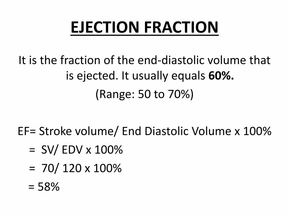
EJECTION FRACTION
It is the fraction of the end-diastolic volume that is ejected. It usually equals 60%.
(Range: 50 to 70%)
EF= Stroke volume/ End Diastolic Volume x 100%= SV/ EDV x 100%= 70/ 120 x 100% = 58%

Phase 5: ISOVOLUMETRIC RELAXATION PHASE
(Early Ventricular Diastole)Left Ventricle is relaxing after completing the contraction
↓Intra-ventricular pressure falls rapidly
↓Becomes lower than the aortic pressure
↓Blood tries to flow back into the ventricles again
↓Backflow of the blood fills the cuplike cusps of semilunar valves
↓Forces the Aortic valve to close
↓SECOND HEART SOUND PRODUCED (dup of lup-dup)
↓Ventricles become a sealed chamber as the AV valve is already closed.

ISOVOLUMETRIC RELAXATION PHASE
• Both the valves .i.e. semilunar & atrioventricularvalves are closed!
• Thus, the Left ventricle is a closed chamber.• When the pressure in the ventricles falls lower
than the atria, then the atrioventricular valves open.
• THE CYCLE RESTARTS!!!

CARDIAC OUTPUTThe volume of blood pumped by each ventricle per
minute is called the cardiac output (CO), usually expressed in litres per minute.
It is also the volume of blood flowing through either the systemic or the pulmonary circuit per minute.Cardiac Output= Stroke volume X Heart Rate
CO = SV x HR= 70 ml x 72 beats/min= 0.07 litres x 72 beats/ min= 5.0 litres/ min


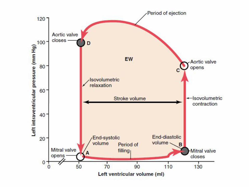
PRESSURE VOLUME CURVE

The relationship b/w the left ventricular pressure and left ventricular volume throughout one entire
cardiac cycle is called LEFT VENTRICULAR PRESSURE VOLUME LOOP (PV LOOP) or diagram.
It explains the normal function of the left ventricle.


PRELOADIt is the end-diastolic pressure just before it
starts contracting. OR
It represents the degree to which the myocardium is stretched & the pressure exerted on it, before it starts contracting.
• Thus, greater the ventricular filling, greater the preload (implying that preload is the volume of blood).

AFTERLOAD
The afterload of the ventricle is the pressure in the aorta against which the heart must contract.
ORIt is the resistance of the circulation against which the heart
must contract.↓
When the ventricles contract to open the semilunar valves, they must generate enough pressure to exceed the aortic or
pulmonary pressure. ↓
This aortic or pulmonary pressure is called the After load.

Source of Chemical Energy for Cardiac Contraction
1. 70-90 % from oxidative metabolism of FATTY ACIDS
2. 10-30 % from Lactate & Glucose