THE ARTERIES OF THE - umich.edu
16
THE ARTERIES OF THE PANCR#EAS RUSSELL T. WOODBURSE AND LLOYD L. OLSEN Departments of Anatomy a?&d Surgery, University o f Michigan XVedicnZ School, Ann Arbor FIVE FIGURES INTRODUCTION The present study was undertaken in response to the very considerable inadequacy in the description of the pancreatic blood supply in anatomical textbooks. Especially is the sur- geon currently conccrned with the feasibility of surgical attack on the pancreas and the success of snch operative procedures may depend in part on a knowledge of specific vessels reaching the gland. The authors were particularly interested in defining, if possible, a general, pattern in the blood supply of this organ as well as in determining the range of variation of the vessels concerned. REVIEW OF LITERATURE An exhaustivc review of the literaturc concerning investi- gations of the blood vessels will not he attempted here. A complete and informative review of the studies up to 1929 is iriclnded in the report by Petr6n ('29). Many studies ori- ented primarily toward the duodenum give only partial in- formation concerning the arteries of the pancreas. Other siudies suffer in that their statistical analysis is based on too few specimens. It will be profitable, however, to give empha- sis here to certain studies which materially advanced OW- linowledge of the arteries of the pancreas. The earliest description of certain important pancreatic arteries appears to have been that by Haller in 1742 who recognized 'double arterial arches or arcades anterior and 255
Transcript of THE ARTERIES OF THE - umich.edu

THE ARTERIES O F THE PANCR#EAS
RUSSELL T. WOODBURSE AND LLOYD L. OLSEN Departments o f Anatomy a?&d Surgery, University o f Michigan
XVedicnZ School, Ann Arbor
F I V E FIGURES
INTRODUCTION
The present study was undertaken in response to the very considerable inadequacy in the description of the pancreatic blood supply in anatomical textbooks. Especially is the sur- geon currently conccrned with the feasibility of surgical attack on the pancreas and the success of snch operative procedures may depend in part on a knowledge of specific vessels reaching the gland. The authors were particularly interested in defining, if possible, a general, pattern in the blood supply of this organ as well as in determining the range of variation of the vessels concerned.
REVIEW O F LITERATURE
An exhaustivc review of the literaturc concerning investi- gations of the blood vessels will not he attempted here. A complete and informative review of the studies up to 1929 is iriclnded in the report by Petr6n ('29). Many studies ori- ented primarily toward the duodenum give only partial in- formation concerning the arteries of the pancreas. Other siudies suffer in that their statistical analysis is based on too few specimens. It will be profitable, however, to give empha- sis here to certain studies which materially advanced OW-
linowledge of the arteries of the pancreas. The earliest description of certain important pancreatic
arteries appears to have been that by Haller in 1742 who recognized 'double arterial arches or arcades anterior and
255

256 R . T. JVOODBVRNE AXD L. L. OLSEN
posterior to the head of the pancreas and serving both duo- denum and pancreas. His was the first description of the posterior superior pancreaticoduodenal, branch of the gastro- duodenal artery to the posterior arcade. Haller also described the A. pancreatica magna suprema, a vessel which has a veq- frequent occurrence but is perhaps more commonly recog- nized at present as the dorsal pancreatic artery. With tho description of the A. transversa pancreatica as a continuation of the A. pancreatica magiia suprema Haller very iiearly rounded out a complete description of this important blood supply. Hallcr’s priority of description on all points may be open to question, however, since Winslow described in 1732 a duodenal or intestinal branch of the gastroduodenal artery which “sometimes is double,” and in speaking of the supcr:or niesenteric artery described ‘(a small branch near its origin which, dividing into two, goes to the lower side of the head of the pancreas and neighboring part of the duo- denum, communicating with the intestinalis by small arches. ” These early studies remained essentially unrecognized for about 150 years when Wiart (1899), writing on the form and relations of the pancreas, gave a good description of the pan- creaticoduodenal arcades. This included a description of a right superior pancreaticoduodenal artery as a proximal branch of the gastroduodenal completing a posterior arcade with a left superior pancreaticoduodenal artery. According to this report the terminal division of the gastroduodenal artery formed the right gastroepiploic and the right inferior pancreaticoduodenal arteries, the latter completing a second and lower posterior arcade with the left inferior panereatico- duodenal artery from the superior mesenteric system. In this description emphasis is placed on the right and left rela- tionships of the source vessels of the arcades and on the fact that both arcades are completed on the posterior aspect of the pancreas, one being higher or more cephalic than the other. Testut (1893) recognized two arches which he desig- nated “peripancreatic arterial circles” but his more notable
25'7 -4RTEltIES OF TEIE PANCIlEAS
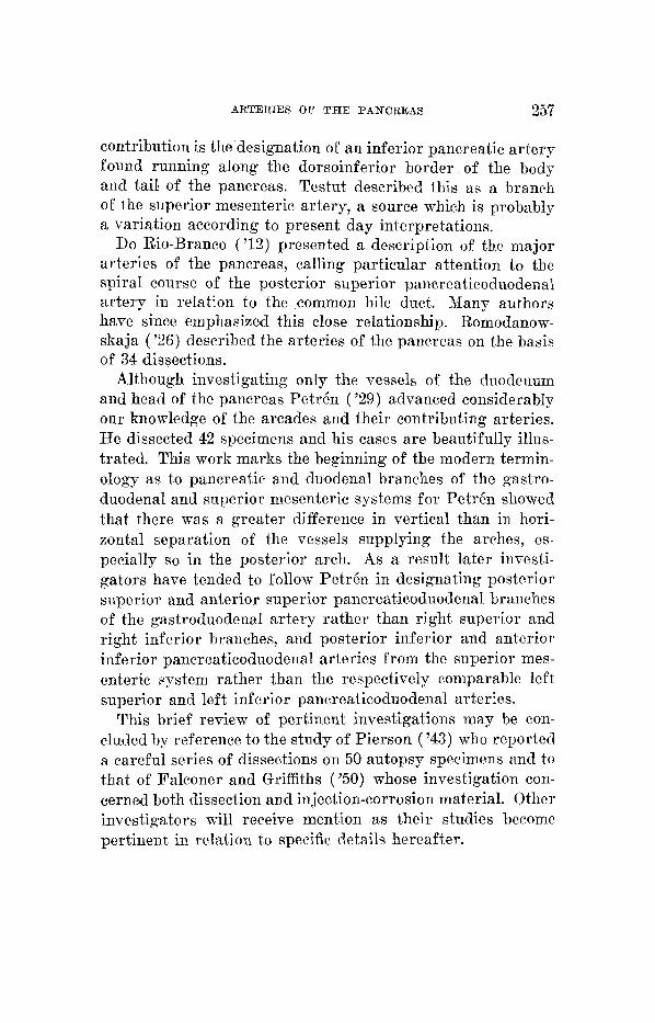
contribution is the designation of an inferior pancreatic artery found running along the dorsoinferior border of the body and tail of the pancreas. Testut described this as a branch of the superior mesenteric artery, a source which is probably a variation according to present day interpretations.
Do Rio-Branco ('12) presented a description of the major arteries of the pancreas, calling particular attention to the spiral course of the posterior superior pancreatieoduodenal artery in relat'on to the common bile duct. Xany authors have since emphasized this close relationship. Romodanow- skaja ('26) described the arteries of the pancreas on the basis of 34 dissections.
Although investigating only the vessels of the duodenum and head of the pancreas P e t r h ('29) advanced considerably our knowledge of the arcades and their contributing arteries. He dissected 42 specimens and his cases are beautifully illus- trated. This work marks the beginning of the modern termin- ology as to pancreatic and duodenal branches of the gastro- duodenal and superior mesenteric systems for P e t r h showed that there was a greater difference in vertical than in hori- zontal separation of the vessels supplying the arches, es- pecially so in the posterior arch. As a result later inmsti- gators have tended to follow Petrhn in designating posterior superior and anterior superior pancreaticoduodenal branches of the gastroduodenal artery rather than right superior an'd right inferior branches, and posterior inferior and anterior inferior pancreaticoduodenal arteries from the superior mes- enteric s p t e m rather than the respectively comparable left superior and left inferior pancreaticoduodenal arteries.
This brief review of pertinent investigations may be con- cluded by reference to the study of Pierson ( '43) who reported a careful series of dissections on 50 autopsy specimens and to that of Falconer and Griffiths ('50) whose investigation con- cerned both dissection and injection-corrosion material. Othcr investigators will receive mention as their studies become pertiiient in relation to specific details hereafter.

258 R. T. WOODBUltXE AND L. L. OMEN
PRESENTATION OF MATERIAL
The following description of the pattern of arterial blood supply to the pancreas and the statistical analysis of variants in this pattern is based on detailed dissection by the authors of 150 specimens. A preliminary survey was made of a con- siderable number of specimens after dissection by students but it was found that destruction and loss of some vessels together with incomplete dissection of others made observa- tions on this material so uncertain as to be worthless. Particu- larly was it true that vessels of apparently a certain source had a different origin than first appeared after a critical dis- section was made. The material finally used was consecutive cadavers placed in the anatomical laboratories over several years’ time but the authors made the dissection of the blood vessels in advance of student dissection in the region. Sketches of each specimen were prepared and the sketches were aug- mented by notations of origins and particular features.
1. Gemral pattern
The authors would like to stress that there is a general pattern of vasculature here (fig. 1) which is remarkably con- stant, much more constant than previous studies have indi- cated. There are two arterial arcades in relation to the head of the pancreas, one anterior and the other posterior; both serve to supply vasa recta to the duodenum and branches to the head of the pancreas. The anterior arcade occurred in every case in our series. The posterior arcade was lacking in only two cases, in one due to the absence of the posterior superior pancreaticoduodenal artery, in the other case due to the absence of the posterior inferior vessel.
There proved to be a constant terminal division of the gastrocluodenal artery into right gastroepiploic and anterior superior pancreaticoduodenal, arteries. The latter vessel de- scends on the anterior surface of the head of the pancreas, reaching the sulcus between the pancreas and duodenum at the junction of the secon’d and third parts of the duodenum
ARTERIES OF THE PANCREAS 259
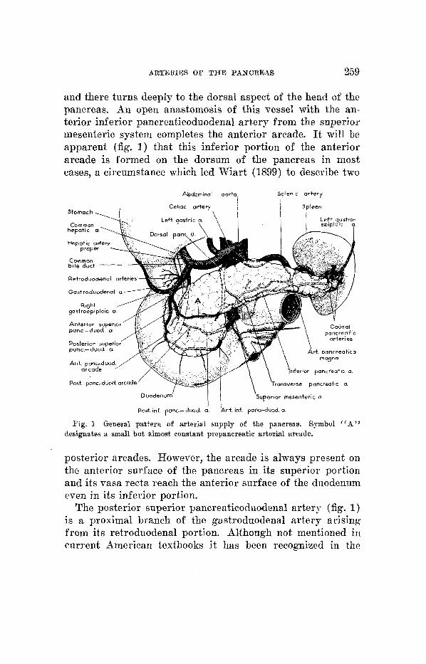
and there turns deeply to the dorsal aspect of the head of the pancreas. An open anastomosis of this vessel with the an- terior inferior pancreaticoduodenal artery from the superior. mesenteric system completes the anterior arcade. It will be apparent (fig. 1) that this inferior portion of the anterior arcade is formed on the dorsum of the pancreas in most cases, a circumstance which led Wiart (1899) to describe two
Abdominal aorta Splenic artery I I
Celiac artery Stomach
hepaiic a
Lef t gaotro- eplploic a
Retroduod~na l~or ter ies -1 Gastroduodenal
b i le duct __
- \ Caudal
- Superior mesenterx a
\
Ant inf ponc-duod a 1 1 i
Duodenum
Post nnf punc-duod a
Fig. 1 General pattern of arterial supply of the pancreas. Symbol ‘‘-4’’ designates a small but alnlost constant prcpancreatic arterial arcade.
posterior arcades. However, the arcade is always present on the anterior surface of the pancreas in its superior portion and its vasa recta reach the anterior surface of the duodenum even in its inferior portion.
The posterior superior pancreaticoduodenal arterr (fig. 1) is a proximal branch of the gastroduodenal artery arising from its retroduodenal portion. Alcthough not mentioned in current American textbooks it has been recognized in the

260 R. T. WOODBUItNE AND L. L. OLSEX
research literature since its first description by Haller in 1742 as the right superior pancreaticoduo'denal artery. Hal- lcr's mention of this vessel and his description of two arcades of vessels appear to have been lost to the notice of his con- temporaries and we find the next clear description of the ar- cades and their sources is that of Wiart in 1899. T'crneuil (1851) clearly stated that two arcades existed lout described both as formed from a descending branch of the hepatic and an ascending branch of the superior mesenteric artery. This inaccurate description delayed recognition of the important posterior superior pancreaticoduodenal artery and led ulti- mately to the investigations of do Rio-Branco ( ' la) , Romo- danowskaja ( '26), and Petren ( '29). Do Rio-Branco ( '12) emphasized that the artery in question (his right superior pancreaticoduodenal artery) arises 1-1.5 em from the origin of the gastroduodenal artery as the latter reaches the superior border of the pancreas. This origin was to the left of the com- mon bile duct in 42% of cases or along its left border in 38%. It was to the right of the duct in only 20% of cases. I n ac- cordance with these findings do Rio-Branco described a first part of the artery running transversely to the right to cross the anterior face of the common bile duct at the lmeevel of the upper border of the pancreas. I n its second se,ment the vessel turns thc right flank of the common bile duct and sinks clorsally between pancreas and duodenum. Reaching a. retro- pancreatic position it is directed inferiorly and to the left, crossing obliquely the posterior face of the retropancreatic. common bile duct and anastomosing with the posterior in- ferior pancreaticoduodenal artery. The posterior superior pancrcaticoduodenal artery has, contrary to the usage of all other authors, been designated the retroduodenal, artery by Edwards ('41) and by Michcls ('45 and '51). It is almost constant in occurrence (99.370) in our series and arises as the above described branch of thc gastroduodenal artery in 92.6% of its total representation.
The foregoing discussion of the arcades and their sources has anticipated to a certain extent the description of the in-
ARTERIES O F T H E PANCREAS 261
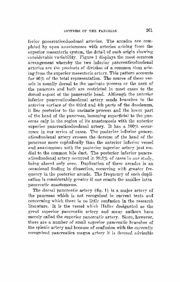
fcrior pancreaticoduodenal arteries. The arcades are com- pleted by open anastomoses with arteries arising from the superior mesenteric system, the detail of such origin showing considerable variability. Figure 1 displays the most common arrangement whereby the two iiif erior pancreaticoduodenal arteries are the products of division of a common stem aris- ing from the superior mesenteric artery. This pattern accounts for 4G% of the total representation. The source of these ves- sels is usuallv dorsal to the uncinate process or the neck of the pancreas and both are restricted in most cases to the dorsal aspect of thc pancreatic head. Although the anterior inferior pancreaticoduodenal artery sends branches to the anterior surface of the third and 4th parts of the duodenum, it lies posterior to the uncinate process and the lower part of the head of the pancreas, becoming superficial to the pan- creas only in the region of its anastomosis with the anterior superior pancreaticoduodenal artery. It has a 100% occur- rence in our series of cases. The posterior inferior pancre- aticoduodenal artery crosses the dorsum of the head of tlie pancreas more cephalical~ly than the anterior inferior vessel and anastomoses with the posterior superior artcrp just me- dial to the common bile duct. The posterior inferior pancre- aticoduodenal artery occurred in 99.3% of cases in our study, being absent only once. Dupl'ication of these arcades is an occasional finding in dissection, occurriiig with greater fre- quency in the posterior arcade. The frequency of such dupli- cation is considerably greater if one counts the smaller intra- pancreatic anastomoses.
The dorsal pancreatic artery (fig. 1) is a major artery of the pancreas which is not recognize'd in current texts and concerning which there is no little confusion in the research literature. It is the vessel wh:ch Haller designated as the great superior pancreatic artery and many authors have merely called the superior pancreatic artery. Since, however, there are a number of small superior pancreatic branches of the splenic artery and because of confusion with the currently recognized pancreatica m a p a artery it is deemed Rdvisable

262 R. T. WOODBURNE AND L. L. OLSEX
to adopt the name of dorsal pancratic artery, from Jfichels’ description. Do Rio-Branco used the term middle pancreatic arterq.. This vessel has a 90% occurrence in our series of dis- sections. It has traditionally been defined as a branch of the celiac, hepatic, or splenic artery arising within 1.5-2 em of the origin of such vessel. It became apparent in our dissec- tions that exactly the same artery with the same course and branches frequently arose from the superior mesenteric artcry. Not all authors have inchded the latter source which accounts f o r considerable difference in the statistics of occur- rence. The dorsal pancreatic artery descends dorsal to the neck or body of the pancreas, sometimes also being dorsal to the splenic vein, and toward the lower border of the pancreas divides into a right and a left branch. The right branch emerges on the surface of the pancreas at or near the junction of the uncinate process and the neck of the pancreas, sends a branch of supply down over the uncinate process and then turns across the head of the pancreas to make anastomosis with a small left branch of the anterior superior pancreatico- duodenal artery. This anastomosis forms a prepancreatic arterial arcade which supplies only pancreatic tissue and is labelled “A” in figure 1. It has a 93.35% occurrence in our series. In completing this anastomosis the left branch of the anterior superior pancreaticoduodenal may be substituted f o r by a branch of the adjacent right gastroepiploic artery or of the more proximal gastroduodenal artery. The left branch of the dorsal pancreatic vessel. fornis the inferior pancreatic artery next to be described.
The inferior pancreatic artery (fig. 1) is a vessel which runs along the inferior border of the body of the pancreas, fre- quently embedded in the dorsal aspect of that border and only partially visible from the surface of the pancreas. It is usually a continuation along the inferior edge of the pancreas of the left branch of the dorsal pancreatic artery. The name was first applied by Testut (1893) who described it as a loranch of the superior mesenteric artery, presumably the left branch of a dorsal pancreatic artery from the superior mesenteric,
ARTERIES O F THE PANCBEAS 263
as it would be analyzed under current concepts. The inferior pancreatic artery was designated the transverse pancreatic by Haller (1764). It usually anastomoses wit11 cantlal pan- creatic arteries and with radicles of the pancreatica magna artery and may communicate on the head of the gland with the prepancreatic arcade. It was a constantly occurring ressel in our series of dissections.
The pancreatica magna artery is described in current anato- my textbooks, usually as a large superior pancrcatic hranch of the splenic artery which enters the gland at approximately the junction of the middle and left thirds of the gland. I t penetrates immediately into the gland, sends branches to right and left which may be oriented along the main pancre- atic duct, and anastomoses with other vessels within the pan- creas. It had a 64.7% occurrence in our dissections. The earliest description of this artery available to the present writers is that in Quain's Elements of Anatomy of 1848 in which the vessel, after entering the gland, is described as passing from left to right along the main pancreatic duct.
Caudal pancreatic branches are to be found in most cases penetrating the tail of the pancreas to supply it and to form anastomoses with inferior pancreatic and pancreatica magma radicles. These vessels arise from the splenic artery before its division o r from products of its division, notably the left gastroepiploic artery. Caudal pancreatic branches from these various sources occurred in 78.7% of our dissected specimens. They have been noted previously by many workers, especially Winslow (1732), Haller (1764), Pigache and Worms ( ,09>, and hlichels ( '42).
2. Bari.ability of resse1.c
The very considerable regularity of the general pattern of the arterial supply to the pancreas is evidenced in the high percentage occurrence of the vessels described heretofore. It will be of additional interest to examine the degree of vari- ability in origin that these vessels exhibit.
264 R. T. WOODBURXE A N D L. L. OLSEX
The origin and relations of the anterior superior pancre- aticoduodenal’ artery are amazingly constant. Representing one of the terminal branches of the gastroduodenal artery, it had a 100% occurrence in our series arid there were no variations to report. Such has been the experience of almost all previous investigations also,
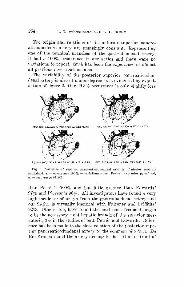
The variability of the posterior superior pancreaticoduo- denal artery is also of minor degree as is evidenced by exami- nation of figure 2. Our 99 3% occurrence is only slightly less
POST. SUP. PANLDUOD. A. FROM GASiRODUODEN 92.6% POST. SUP. PANC.DUOD. A. FROM HEPATIC A.-2.7%
P.S. PANC.WOD. FROM R. HEP. BR. OF SUP. MES. A,-3.4% POST. SUP. PANC.DUOD. A. FROM DORS. PANC. A.-I.3%
Fig. 2 Varieties of superior panereatieoduodenal arteries. Anterior superior pane.duod. a. - occurrence 100% - variations none. Posterior superior pane.duod. a. -occurrence 99.3%.
than P e t r h ’ s 100% and but little greater than Edwards’ 97% and Pierson’s 96%. All investigators have found H very high incideiice of origin froni the gastroduodenal artery and our 92.6% is virtually identical with Falconer and Griffiths’ 92%. Others, too, have found tlie next most frequent origin to be the accessory right hepatic branch of the superior mes- enteric, 3% in the studies of both Petrhn and Edwards. Refer- ence has been made to the close relation of tlic posterior supe- rior pancreaticoduodenal artcry to the common bile duct. Do Rio-Hranco found the artery arising to tlie left or in front of

ARTERIES OF THE PANCREAS 265
the duct in 80% and to its right in only ‘2054. The comparable figures of PetrQn were 86 and 14. Falconer and Qriffiths found thc artery “invariably in front of the coniinon bile duct.” Our investigations indicate that such was the case except when the origin of the vessel was bchind the head of the pancreas, as from the accessory right hepatic branch of the superior mesenteric or from tlie dorsal pancreatic artery.
Figure 3 illustrates tlie variability encountered in dissec- tions of the inferior pancreaticoduodenal arteries. The reports
COMMON STEM FROM SUPERIOR MESENTERK A -46% COMMON STEM FROM UPPER JEJUNAL A - 2 2 %
PANC.DVOD -16% POST. I PANCWD. A 14 6%
FROM WUSAL PANCREATIC A. FROM RT HEPATIC BR OFSUP. ME5 A
-7 INF. PAW.DUOD.A-BX-ANT. INF. PANC.WOD. A47%WST. INF PANCDWD A.-ZT%-ANT INF. PANC.mD. A -07%
Fig. 3 Varieties of inferior paiicrentieoduodenal arteries. Anterior inferior paic.duod. a. - occurrence 100%. Posterior inferior panc.duod. a. - occurrence 99.3%.
of other authors beai. out our results on general iiicidencc and that these vessels most commonly arise by a common stem from either the superior mesenteric artery or one of its upper jejunal branches. Thus Sliapiro and Robillard (’46) noted that a common stern was present in 70% of cases and assigned the upper jejunal origin f o r it in 20%. It will be noted in figure 3 that either as separate origiiis o r by a common stem the superior mesenteric system provides almost 99% of anterior infwior pancreaticoduodenal arteries and

266 R. T. WOODBURNE AND L. L. OMEX
almost 91 "/o of posterior inferior pancreaticoduodenal vessels. It has been emphasized that both inferior pancreaticoduodenal arteries lie dorsal to the head of the pancreas o r uncinate process. It should alsso be noted that they pass behind the superior mesenteric artery when arising from the upper jejunal arteries and (not illustrated) may take origin from the left side of the superior mesenteric or upper jejunal arteries.
The dorsal pancreatic artery (pancreatica magna suprema of Haller and superior pancreatic artery of certain authors) typically arises close to the aortic source of either the celiac or superior mesenteric systems. I ts total occurrence has been variously reported as lorn as 50% (do Rio-Branco) and as high as 90% (this investigation, fig. 4). Romodanowskaja ( '26) found an occurrence of 56% in 34 specimens following the traditional definition of the artery as an early branch of the celiac, hepatic, o r splenic artery. He observed, however, that the inferior pancreatic artery from the superior mesen- teric alternated in incidence with such a dorsal pancreatic and had the same type of branches. The fact that the inferior pancreatic artery is usually the left branch of the dorsal pan- creatic and that the latter does sometimes arise from the superior mesenteric gives a logical' explanation to this obser- vation. Adding Romodanowskaja's I1 cases of this type to his dorsal pancreatic incidence brings to 85% the total occur- rence of this artery in his series. Falconer and Griffiths ( '50) noted a 79% occurrence when all sources are included. The distribution of sources within the total varies in the reports of different observers. Do Rio-Branco assigned 50% to the splenic origin whereas Romodanowskaja found 50% from the celiac artery. Most earlier reports are somewhat unreliable because of the small number of specimens dissected and, wit11 respect to adequate numbers, perhaps the present investigation (fig. 4) will yield truer figwres of incidence from the various source vessels. It is probable, however, that those numerous vessels arising close to the division of the celiac artery pose

ARTEEIES O F THE PANCRESS 267
problems of interpretation that will militate against complete agreement among investigators.
The right branch of the dorsal pancreatic artery extends across the head of the pancreas and forms a prepancreatic arcade by means of an anastomosis with a left branch of the anterior superior pancreaticoduodenal artery or an immedi- ately related vessel. Do Rio-Branco ('12) stated that this arcade is almost constant, Pierson ( '43) that it is fairly con- stant. Falconer and Griffiths ('50) found it in 11 of 27 cases.
SPLENIC ORIGIN 37. I % CELIAC ORIGIN 33.3%
SUPERIOR MESENTERK: ORIGIN 21.5% HEPATIC ORIGIN 8.1%
Fig. 4 Varieties of dorsal pancreatic artery. Occurrence 90%.
Roniodanowskaja ('26) designated it as the anterior arcade (the pancreaticodiiodenal arcades being considered as poste- rior) and treated it as a regularly occurring structure. The occurrence of this arcade in OUT specimens was 93.3%. The middle colic artery is occasionally intimately related to the dorsal pancreatic artery. I n our series the middle colic or an accessory vessel took origin from this pancreatic artery in 5% of the cases dissected.
The inferior pancreatic artery (fig. 5) had a 100% occur- rence in our series of 150 dissections. Falconer and Griffiths ('50) found it in 89% in their series, while do Rio-Rranco

268 R. T. WOODBURSE AND L. L. OLSEN
('12) merely gave it the designation of almost constant. Its predominant source is the left branch of the dorsal pancre- atic artery which accounted for 84% of our specimens. When the dorsal pancreatic artery was missing the inferior pan- creatic artery usually existed as a continuation of the left branch of the anterior superior pancreaticoduodenal artery ; this occurred in 10% of our cases. Other minor sources for this vessel are listed in figure 5. We encountered the vessel as a separate branch of the superior mesenteric artery only rarely (1.3%) although it was this source which Testut (1893) listed in his early description and Falcoiier and Griffiths ( '50) cite this source f o r 37% of their specimens.
LEFT 6R.W DORSAL PANCREATIC A.-84% LEFT BR. OF ANT.SUP. PANC.WOD. A.- 10%
MINOR SOURCES- BR.CfGASTROWODENAL A.-2%; BR.WSUP. MES. A.-1.3%
BR. Of DORS. PANCR.tSUP. MES. A.-1.3%: BR.OF PANCREATICA MAGNA A.-1.3%
Fig. 5 parieties of inferior pancreatic artery. Occurrelice 100%.
Variability in the pancreatica magna artery exists in its occurrence (64.7% in our series) and also in its exact position, but to be so designated it should be a larger superior pancre- atic branch of the splenic artery entering the pancreas at approximately the junction of its middle and left thirds. A vessel in this position and frequently oriented along the duct system has been described f o r over a century (Quain, 1848). The term designating the artery is open to question and may have led to confusion previously since Sobotta, writing in BardeIeben's Handbuch der Anatomie des Menschen ( '14), stated that the arteria pancreatica magna of Hallcr is the first
ARTEBIES OF TRE PANCEEAS 269
pancreatic branch of the splenic artery and is usually the larger of such branches. He obviously refers to the 1-essel designated magna suprema by Haller (here called the dorsal pancreatic artery).
SUMNARY
This study of 150 dissections indicates that: 1. There is considerable regularity in the pattern of arte-
rial blood supply to the pancreas. 2. Two arterial arcades supply the duodenum and head of
the pancreas. 3. The anterior arcade is formed by the constant anterior
superior pancreaticoduodenal, artery from the gastroduodenal artery anastomosiiig with the almost constant anterior inferior pancreatieoduodenal artery from the superior mesenteric system. 4. The posterior arcade is contributed to from above by
the posterior superior pancreaticoduodenal artery, a proxi- mal branch of the gastroduodenal artery. This arcade is completed by an almost constantly occurring posterior in- ferior pancreaticoduodenai artery arising in over 90% of cases from the superior mesenteric system.
5. The inferior pancreaticoduodenal vessels are much more variable in the detail of their origin than the superior vessels.
6. A dorsal pancreatic artery occurs in 90% of cases. I t exhibits a typical course and pattern of branches but arises variously from the splenic, the celiac, the superior mesenteric, or the hepatic artery, I ts right branch crosses the head of the pancreas to form a prepancreatic arterial arcade (inci- dence 93.3%) by means of an anastomosis with the left branch of the anterior superior pancreaticoduodenal artery.
7. The constant inferior pancreatic artery is found along the dorsoinferior border of the pancreas. It is in the majority of cases (84%) the left branch of the dorsal pancreatic artery.
8. The paiicreatica magna artery is a superior pancreatic branch of the splenic artery entering the body of the pancreas at the junction of its middle and left thirds and has an occur- rence of 64.7 %.

270 R. T. WOODBURNE AND L. L. OMEN
9. Caudal pancreatic arteries reach the tail from predomi- nantly the splenic artery or its left gastroepiploic branch and occur in 78.7% of cases in this series.
REFERENCES
Do RIOBEANCO, P. 1912 Essai aur I’Anatomie et l a Mddecine operatoire du Tronc Coeliaque et de ses branches de 1’Arthre HBpatique en particu- lier. T h h e pour le Doetorat in MBdecine; Facult6 de M6decinc de Paris.
1941 The retroduodenal artery. Anat. Ree., 61.- 351-355. The anatomy of the blood vessels
EDWARDS, L. F. l?~mC$”, C. W. A., AND E. GRJFFITHS
HALLER, A. 1742 H. Boerhaave praelectiones acad., 1: Taurini.
M I ~ s , N. The variational anatomy of the spleen and splenic artery. Am. J. Anat., 70: 21-72.
-- 1945 Variations in blood supply of liver, gallbladder, stomach, duodenum and pancreas. J. Internat. Coll. Surg., 8: 502-50.4.
1951 The hepatic, cystic and retroduodeml arteries and their relations to the biliary ducts. Ann. Surg., 133: 503-524.
PETRAN, T. 1929 Die Arterien und Venen des Duodenums und des Pancre- askopfes beini Mensehen. Zeitsch. f. Anat. u. Entwicklungsgesch., 90 :
PIERSON, J. hf. 1943 The arterial blood supply of the pancreas. Burg., Gyn.,
PIQACIiE, R., AND G. WORMS 1909 Topographie du pedicule de la r,ate. Bull. et
QUAIN 1848 Elements of anatomy. v. 1, ed. by Richard Quain and William
RO~ODANOWSXAJA, Z. 1926 Die Arterien der Bsuchspeicheldriise. Zeitsch. f.
S-HAPIRO, A. L., AND G. L. ROBILLAED Morphology and variations of the
SOBOTTA, J. 1014 Anatomie der Bauchspeicheldriise. Handb. der Anat. des
TESTUT, L. 1893 Trsitd d’anatomie humaine. 3: 675-676. Pmis, 0. Doin. VERNEUIL, A. Memoire sur quelques points de l’anatomic du panerdas.
Gaz. Mdd. de Paris. pp. 398-401. W m T , P. 1899 Becherches sur la forme et les rapports du pancreas. J. de
1’Anat. et de la Physiol., 3 5 : 91-113. WINSLOW, J. 13. An anatomical exposition of the structure of the human
body. Translated from the French original by G. Douglas. London, Bettosworth, Hitch, Osborn, Longman, Ware, Birt. Davis and Astley.
1950 in the region of the pancreas. Brit. J. Surg., 37: 334-344.
1764 Elementa Physiologiae Corporis Humani. VI, Lib. XXII. 1942
234-277.
and Obs., 5’7: 486432.
Mem. de la Soe. Anat. de Paris. Ser. 6, 11: 589-605.
Sharpy. London, Walton and Maberly.
Anat. u. Entwicklungsgesch., 79 : 506-514.
duodenal vasculature. Arch. Surg., 52: 571-602.
Mensch. Ed. by Karl von Bardeleben. 6 (3) : 1-62.
1946
1851
1732