The Anatomy of the Infra-red Sense Organ in the Facial Pit of Pit Vipers
20
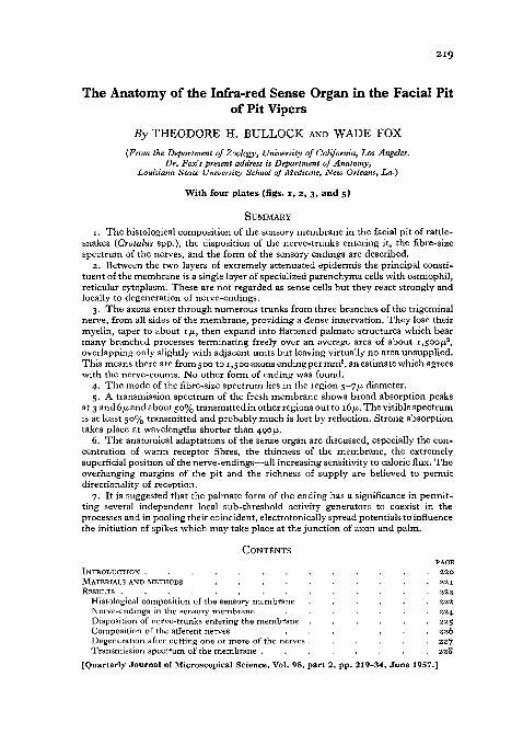
219 The Anatomy of the Infra-red Sense Organ in the Facial Pit of Pit Vipers By THEODORE H. BULLOCK AND WADE FOX (From the Department of Zoology, University of California, Los Angeles. Dr. Fox's present address is Department of Anatomy, Louisiana State University School of Medicine, New Orleans, La.) With four plates (figs, i, 2, 3, and 5) SUMMARY 1. The histological composition of the sensory membrane in the facial pit of rattle- snakes (Crotalus spp.), the disposition of the nerve-trunks entering it, the fibre-size spectrum of the nerves, and the form of the sensory endings are described. 2. Between the two layers of extremely attenuated epidermis the principal consti- tuent of the membrane is a single layer of specialized parenchyma cells with osmiophil, reticular cytoplasm. These are not regarded as sense cells but they react strongly and locally to degeneration of nerve-endings. 3. The axons enter through numerous trunks from three branches of the trigeminal nerve, from all sides of the membrane, providing a dense innervation. They lose their myelin, taper to about 1 JJ,, then expand into flattened palmate structures which bear many branched processes terminating freely over an average area of about I,SOOJU. 3 , overlapping only slightly with adjacent units but leaving virtually no area unsupplied. This means there are from 5 00 to 1,5 00 axons ending per mm 2 , an estimate which agrees with the nerve-counts. No other form of ending was found. 4. The mode of the fibre-size spectrum lies in the region 5-7/J, diameter. 5. A transmission spectrum of the fresh membrane shows broad absorption peaks at 3 and 6 /x and about 50% transmitted in other regions out to 16/n. The visible spectrum is at least 50% transmitted and probably much is lost by reflection. Strong absorption takes place at wavelengths shorter than 490 /A. 6. The anatomical adaptations of the sense organ are discussed, especially the con- centration of warm receptor fibres, the thinness of the membrane, the extremely superficial position of the nerve-endings—all increasing sensitivity to caloric flux. The overhanging margins of the pit and the richness of supply are believed to permit directionality of reception. 7. It is suggested that the palmate form of the ending has a significance in permit- ting several independent local sub-threshold activity generators to coexist in the processes and in pooling their coincident, electrotonically spread potentials to influence the initiation of spikes which may take place at the junction of axon and palm. CONTENTS PAGE I N T R O D U C T I O N . . . . . . . . . . . . . 220 M A T E R I A L S A N D M E T H O D S . . . . . . . . . . . 221 R E S U L T S . . . . . . . . . . . . . . 222 Histological composition of the sensory m e m b r a n e . . . . . . 222 Nerve-endings in the sensory m e m b r a n e . . . . . . . . 2 2 4 Disposition of nerve-trunks entering the m e m b r a n e . . . . . . 225 Composition of the afferent nerves . . . . . . . . 226 Degeneration after cutting o n e or m o r e of the nerves . . . . . . 227 Transmission s p e c t r u m of the m e m b r a n e . . . . . . . . 228 [ Q u a r t e r l y J o u r n a l of Microscopical Science, Vol. 98, p a r t 2, p p . 219-34, J u n e 1957.]
Transcript of The Anatomy of the Infra-red Sense Organ in the Facial Pit of Pit Vipers
219
The Anatomy of the Infra-red Sense Organ in the Facial Pitof Pit Vipers
By THEODORE H. BULLOCK AND WADE FOX(From the Department of Zoology, University of California, Los Angeles.
Dr. Fox's present address is Department of Anatomy,Louisiana State University School of Medicine, New Orleans, La.)
With four plates (figs, i, 2, 3, and 5)
SUMMARY1. The histological composition of the sensory membrane in the facial pit of rattle-
snakes (Crotalus spp.), the disposition of the nerve-trunks entering it, the fibre-sizespectrum of the nerves, and the form of the sensory endings are described.
2. Between the two layers of extremely attenuated epidermis the principal consti-tuent of the membrane is a single layer of specialized parenchyma cells with osmiophil,reticular cytoplasm. These are not regarded as sense cells but they react strongly andlocally to degeneration of nerve-endings.
3. The axons enter through numerous trunks from three branches of the trigeminalnerve, from all sides of the membrane, providing a dense innervation. They lose theirmyelin, taper to about 1 JJ,, then expand into flattened palmate structures which bearmany branched processes terminating freely over an average area of about I,SOOJU.3,overlapping only slightly with adjacent units but leaving virtually no area unsupplied.This means there are from 5 00 to 1,5 00 axons ending per mm2, an estimate which agreeswith the nerve-counts. No other form of ending was found.
4. The mode of the fibre-size spectrum lies in the region 5-7/J, diameter.5. A transmission spectrum of the fresh membrane shows broad absorption peaks
at 3 and 6 /x and about 50% transmitted in other regions out to 16/n. The visible spectrumis at least 50% transmitted and probably much is lost by reflection. Strong absorptiontakes place at wavelengths shorter than 490 /A.
6. The anatomical adaptations of the sense organ are discussed, especially the con-centration of warm receptor fibres, the thinness of the membrane, the extremelysuperficial position of the nerve-endings—all increasing sensitivity to caloric flux. Theoverhanging margins of the pit and the richness of supply are believed to permitdirectionality of reception.
7. It is suggested that the palmate form of the ending has a significance in permit-ting several independent local sub-threshold activity generators to coexist in theprocesses and in pooling their coincident, electrotonically spread potentials to influencethe initiation of spikes which may take place at the junction of axon and palm.
CONTENTSPAGE
I N T R O D U C T I O N . . . . . . . . . . . . . 2 2 0M A T E R I A L S A N D M E T H O D S . . . . . . . . . . . 2 2 1R E S U L T S . . . . . . . . . . . . . . 2 2 2
H i s t o l o g i c a l c o m p o s i t i o n o f t h e s e n s o r y m e m b r a n e . . . . . . 2 2 2N e r v e - e n d i n g s i n t h e s e n s o r y m e m b r a n e . . . . . . . . 2 2 4D i s p o s i t i o n o f n e r v e - t r u n k s e n t e r i n g t h e m e m b r a n e . . . . . . 2 2 5C o m p o s i t i o n o f t h e a f f e r e n t n e r v e s . . . . . . . . 2 2 6D e g e n e r a t i o n a f t e r c u t t i n g o n e o r m o r e o f t h e n e r v e s . . . . . . 2 2 7T r a n s m i s s i o n s p e c t r u m o f t h e m e m b r a n e . . . . . . . . 2 2 8
[ Q u a r t e r l y J o u r n a l o f M i c r o s c o p i c a l S c i e n c e , V o l . 9 8 , p a r t 2 , p p . 2 1 9 - 3 4 , J u n e 1 9 5 7 . ]

220 Bullock and Fox—Infra-red Sense Organ in Pit VipersD I S C U S S I O N . . . . . . . . . . . . . 2 3 0
T h e a n a t o m i c a l a d a p t a t i o n s o f t h e s e n s e o r g a n . . . . . . . 2 3 0C o m p a r i s o n w i t h o t h e r r e p t i l i a n r e c e p t o r s . . . . . . . . 2 3 2P o s s i b l e s i g n i f i c a n c e o f t h e p a l m a t e - e n d i n g . . . j . . . 2 3 2
R E F E R E N C E S . . . . . . . . . . . . . 2 3 3
INTRODUCTION
IN a recent communication the properties of the sense organ in the remark-able facial pit of pit vipers have been analysed physiologically (Bullock
and Diecke, 1956). It is shown that this structure is highly sensitive to inter-mediate and long infra-red radiation to or from the snake. The conclusionsare reached that the organ is specialized for the detection of solid objects ofslightly different surface temperature from background objects, that theobject can be rather small, that there is a certain degree of directional sensi-tivity, and that the special features are not so much measured by a hightemperature sensitivity as a caloric flux sensitivity. The receptors themselvesare regarded as warm receptors, a type poorly known heretofore, functionallyand anatomically. The three sizeable nerve-branches supplying it are foundto be nearly pure populations of warm fibres.
These results, together with the unsatisfactory state of the descriptionsin the literature, have prompted us to re-examine the facial pit organ histo-logically.
The facial pit is peculiar to and characteristic of the crotalid snakes and hashence aroused attention for a long time. Lynn (1931) gives an account of theearly studies and lists seven theories of its function that have been proposed.We may confine our notice here to the five workers who have undertakenmicroscopical investigation. Desmoulins (1824) was apparently the first to seethe rich innervation and concluded that it was sensory, probably olfactory infunction. Ley dig (1868) treated it, together with other unfamiliar structuresin lower vertebrates, in a memoir on organs of a sixth sense. He provided arather good description of its histology but described the nerve-fibres asoriginating in 'terminal ganglion cells' in the sensory membrane in spite ofrecognizing that they are trigeminal nerve-elements, presumably with cell-bodies in the Gasserian ganglion. As we shall see, the strange form of the freenerve-endings does in fact resemble a nerve-cell with several branching pro-cesses and it is possible that Leydig saw some of them, only erring in thinkinghe saw a nucleus. In any case he came closer than later workers for the next80 years.
West (1900) provided a more complete account and may also have seensome actual endings. But he also believed they were cells and identified asnerve-terminations in sections of embryos what must have been epidermalcells and in sections of adults what we call the parenchymatous cells of themembrane. It is remarkable that these workers made out as much as they did.when we recall that they had only a few, poorly fixed, mostly spirit specimensof these snakes. Not until Lynn (1931) took up this object did a worker with
Bullock and Fox—Infra-red Sense Organ in Pit Vipers 221access to fresh material exploit this advantage. With respect to the micro-scopic structure of the membrane, however, little could be added and thesame mistake was perpetuated concerning fifth cranial nerve-fibres with cell-bodies in the periphery. This mistake was corrected by Noble (1934) andNoble and Schmidt (1937), who identified the cells of West and Lynn asepidermal cells preparing for shedding and with silver impregnation describedfree nerve-endings. As we now see, they still did not see the endings. Theirimpregnations, like many of ours, were incomplete. There is also some reasonto doubt their identification of parenchyma as outer epidermis.
MATERIALS AND METHODS
Several species of rattlesnake were used, Crotalus atrox, C. cerastes, C. ruber,C. horridus, C. adamanteus, but mostly C. viridis. No consistent differencesbetween the species were noted in respect to the characters under study; itis probable, from incidental observations, that there are differences in thedegree of pigmentation of the pit and especially the sensory membrane. Thisis likely to have little functional significance.
The membrane at the bottom of the facial pit was studied microscopicallyin its natural position by reflected and by transmitted light. It was removedand observed while fresh. Intravitam methylene blue staining was attemptedbut without significant success. Overfixing in 1% buffered osmium tetroxide(Palade, 1952) was found to be useful for visualizing the disposition of thenerve-branches in the membrane and following individual fibres to the pointwhere myelin stops abruptly. Alcohol/formaldehyde/acetic, Carnoy, Bouin,and the fixatives required by special silver methods were used to reveal fibre-endings in both whole mounts and sections. Sections were cut in low viscositynitrocellulose or in paraffin or double embedded and stained with Masson'strichrome stain or silver-on-the-slide methods (Holmes, 1943; Romanes,1950). A large number of whole mounts of the membrane were treated accord-ing to these as well as the silver procedures of Palmgren (1951), Weddell andZander (1950), and Bodian (1936), and variations of these. A small number ofthese showed nerve-fibres in various degrees of completeness and a stillsmaller number showed the palmate expansions beyond the end of the myelinsheath, with their rich aborization of branching processes. We believe areasonable argument can be made that these preparations show the endingsvirtually completely, but of course we have no assurance of this.
Snakes in which a nerve or two nerves had been cut were kept for variousperiods and then the pit membrane was removed and over-fixed in osmiumtetroxide to show the pattern of degeneration from that nerve.
One very large specimen yielded a membrane so large as to permit mount-ing, fresh, in specially made adapters. The transmission spectrum between2 and 16/j, wavelength was recorded with a Baird Associates automatic infra-red spectrophotometer, and transmission through the visible into the nearultra-violet was recorded with a Beckman DU and a Beckman IR2.
In order to estimate the fibre-size spectrum in the nerves supplying the pit,
222 Bullock and Fox—Infra-red Sense Organ in Pit Vipersthese were fixed in Flemming's fluid (Lillie, 1948), embedded in paraffin, cut,and mounted without staining.
RESULTSHistological composition of the sensory membrane
A cutaway drawing of the pit showing the sensory membrane and its relationto the rest of the head is given in the physiological paper (Bullock and Diecke,1956). Further details of the gross anatomy are provided by the earlier authorsmentioned above. The portion of concern here is the thin, richly vascularizedand innervated, slackly suspended, dry membrane which forms the floor ofthe pit. It separates the pit or outer chamber from an air-filled inner chamberwhich has communication with the outside through a special duct openinginto the adnexa of the eye. This means that the membrane is in contact withair on both sides. Its thickness in recently-shed adult rattlesnakes is IOJU. orslightly less except for local thickenings, especially where nerve-bundles lie.As other authors have shown, it is thicker in developmental stages and webelieve it may be thicker in some of the other genera of the family. At 10^, itseems to have reached a kind of limit dictated by the size of the erythrocytes.Added to this thickness there will normally be a few microns of the multi-layered cornified epidermis preparing to be shed.
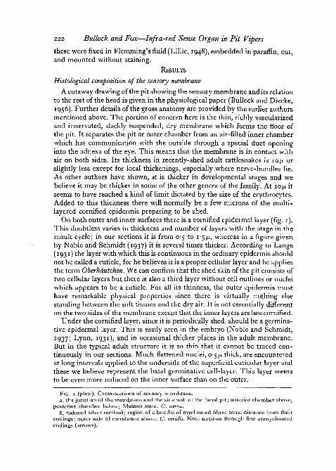
On both outer and inner surfaces there is a cornified epidermal layer (fig. 1).This doubtless varies in thickness and number of layers with the stage in themoult cycle; in our sections it is from 0-5 to i-5/x, whereas in a figure givenby Noble and Schmidt (1937) it is several times thicker. According to Lange(1931) the layer with which this is continuous in the ordinary epidermis shouldnot be called a cuticle, for he believes it is a proper cellular layer and he appliesthe term Oberhdutchen. We can confirm that the shed skin of the pit consists oftwo cellular layers but there is also a third layer without cell outlines or nucleiwhich appears to be a cuticle. For all its thinness, the outer epidermis musthave remarkable physical properties since there is virtually nothing elsestanding between the soft tissues and the dry air. It is not essentially differenton the two sides of the membrane except that the inner layers are less cornified.
Under the cornified layer, since it is periodically shed, should be a germina-tive epidermal layer. This is easily seen in the embryo (Noble and Schmidt,1937; Lynn, 1931), and in occasional thicker places in the adult membrane.But in the typical adult structure it is so thin that it cannot be traced con-tinuously in our sections. Much flattened- nuclei, 0-5 ̂ thick, are encounteredat long intervals applied to the underside of the superficial cuticular layer andthese we believe represent the basal germinative cell-layer. This layer seemsto be even more reduced on the inner surface than on the outer.
FIG. 1 (plate). Cross-sections of sensory membrane.A, the junction of the membrane and the side wall of the facial pit; anterior chamber above,
posterior chamber below; Masson stain. C. atrox.B, reduced silver method; region of a bundle of myelinated fibres some distance from their
endings; outer side of membrane above. C viridis. Note sections through fine unmyelinatedendings (arrows).

B"1O>JL
Fie. i
T. H. BULLOCK «»«/ WADK FOX

Bullock and Fox—Infra-red Sense Organ in Pit Vipers 223Under the epidermal layers on both surfaces is a distinct stratum staining
green with the Masson trichrome in contrast to the layers above and below it.This is generally 0-5 to i-$fi thick or even vanishingly thin on the outer sidebut 1 to 2-5 fj. or even much thicker in local regions on the inner side of themembrane. We believe these layers to represent connective tissue of thedermis or corium, contrary to Noble and Schmidt (1937), who identify every-thing from the outer surface through the next and deepest layer as outerepidermis and thus recognize but a single connective tissue-layer, just beneaththe inner epidermis. To be sure, the outer layer of connective tissue is so thinin most of the membrane that our only reason for so identifying it is its stain-ing affinity. But the inner has discernible collagenous connective tissue fibres.Both can be followed into thicker regions where they become continuous withtypical connective tissue. Fibres which appear to be collagenous connectivetissue-fibres are yisible in some silver impregnations of whole mounts wherethey reveal a sparse array of thin, straight fibres running in all directions forlong distances. Both outer and inner layers of connective tissue show nucleithough in the thin parts of the membrane these are far apart and very muchflattened (fig. 1).
This brings us to the middle and thickest layer which may be called theparenchyma. This is a single-cell layer, generally between the limits of 4 and12 jj. thick. The cells are quite specialized in having, unlike any others in themembrane, an abundant cytoplasm which is coarsely reticular, almost granular,and stains darkly. In Masson's stain it takes a dull red, distinct from thebrighter red of the epidermis in the side walls of the pit. In osmium prepara-tions and in some silver impregnations this cytoplasm takes up considerablecolour. The cell-type is not easily identified with any in ordinary skin. It ishere regarded as part of the dermis since it lies between connective tissue-layers and is cut up into lobules by the capillary bed which occupies the samelevel of the cross-section. The nuclei are rich in chromatin and often show twonucleoli, but are mainly distinguished from the epidermal nuclei by beingcommonly a little irregular in outline rather than smoothly oval as are those ofthe epidermis of the sensory membrane.
The vascular bed has been figured by Noble and Schmidt (1937) and wecan confirm the general character of the blood-supply as described. Being ina single plane, it is easy to visualize in the living membrane in its naturalposition, and also in prepared whole mounts. The richness of the supplyvaries considerably and can be suggested by stating that the maximumdistance from any point to the nearest capillary is between limits of about30 and 6oju..
Aside from the nerve-supply, the only other element in the membrane isa scattering of chromatophores. In the species of Crotalus examined melano-phores are very few; they are much more abundant in Agkistrodon. However,in silver whole mounts we often see extensive patches of what appear to bepigmentless chromatophores which have taken up a granular metallic im-pregnation. These have occasionally been recognized in cross-sections as very
224 Bullock and Fox—Infra-red Sense Organ in Pit Vipers
thin, dark-staining masses immediately beneath the outer-most epidermallayer. They may send processes toward both surfaces.
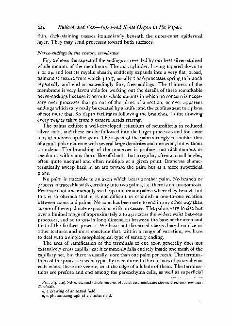
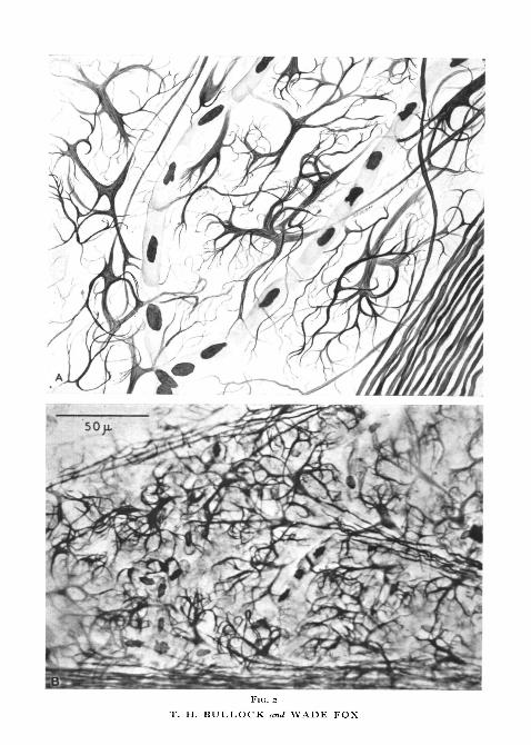
Nerve-endings in the sensory membraneFig. 2 shows the aspect of the endings as revealed by our best silver-stained
whole mounts of the membrane. The axis cylinder, having tapered down toi or 2(i and lost its myelin sheath, suddenly expands into a very flat, broad,palmate structure from which 3 to 7, usually 5 or 6 processes spring to branchrepeatedly and end as exceedingly fine, free endings. The thinness of themembrane is very favourable for working out the details of these remarkablenerve-endings because it permits whole mounts in which no concern is neces-sary over processes that go out of the plane of a section, or over apparentendings which may really be created by a knife; and the confinement to a planeof not more than 8fi depth facilitates following the branches. In the drawingevery twig is taken from a camera lucida tracing.
The palms exhibit a well-developed reticulum of neurofibrils in reducedsilver stain, and these can be followed into the larger processes and for sometens of microns up the axon. The aspect of the palm strongly resembles thatof a multipolar neurone with several large dendrites and one axon, but withouta nucleus. The branching of the processes is profuse, not dichotomous orregular or with many thorn-like offshoots, but irregular, often at small angles,often quite unequal and often multiple at a given point. Branches charac-teristically sweep back in an arc toward the palm but at a more superficialplane.
No palm is traceable to an axon which bears another palm. No branch orprocess is traceable with certainty into two palms, i.e. there is no anastomosis.Processes not uncommonly swell up into minor palms where they branch butthis is so obvious that it is not difficult to establish a one-to-one relationbetween axons and palms. No axon has been seen to end in any other way thanas one of these palmate expansions with processes. The palms vary in size butover a limited range of approximately 2 to 4/x across the widest waist betweenprocesses, and 20 to 30/x in long dimension between the base of the axon andthat of the farthest process. We have not discerned classes based on size orother features and must conclude that, within a range of variation, we haveto deal with a single morphological type of sensory ending.
The area of ramification of the terminals of one axon generally does notextensively cross capillaries; it commonly falls entirely inside one mesh of thecapillary net, but there is usually more than one palm per mesh. The termina-tions of the processes seem typically to conform to the outlines of parenchymacells where these are visible, as at the edge of a lobule of them. The termina-tions are profuse and end among the parenchyma cells, as well as superficial
FIG. 2 (plate). Silver stained whole mounts of facial pit membrane showing sensory endings.C. viridis.
A, a drawing of an actual field.B, a photomicrograph of a similar field.
FlC. 2
T. H. BULLOCK and WADE FOX

Bullock and Fox—Infra-red Sense Organ in Pit Vipers 225to them and, in smaller numbers, in the layers below them. Whether there arenerve-terjninals among the epithelial cells we cannot say, but certainly mostof them are not in that layer which is so exceedingly thin, while the terminalsare found through 6 or 7/x of thickness of the membrane. The palms lie at thesame depth as the capillaries, generally about 5 to 7/A below the outer surface,but are superficial to the capillaries when they cross them. Over the large nerve-trunks the palms are superficial to these. The palms are only about 2-5 JU. thickat maximum. The processes mostly proceed toward terminations in a moresuperficial plane, a large part of them terminating 4 to 5 JJ, above the palms,therefore very close to the surface. A few processes pass downwards 2 to 3 ptowards the inner surface.
There is not a great deal of overlap of area supplied by adjacent palms.Some interdigitation of terminal branches is seen in every field in good im-pregnations, but it is, broadly speaking, not very extensive and most of thearea of branching of the processes of one palm is not invaded by others. Thisarea is not simple in outline but rather is indented and produced. It is there-fore not easy to trace and hence to measure. An approximation of the typicalarea within which the branches of one axon ramify would lie between 1,000and 2,000/x2, but these are not outer limits. Diameters of 30 and of 50/x arenot rare. An estimate of the average area, not including overlap, can beobtained by counting the number of palms in a microscopic field of knownsize. In 18 fields chosen at random and including various parts of the mem-brane, an average of 520 palms per mm2 or i,o,2O/x2 per palm was found. Thisfigure can be regarded as slightly overestimating the area per palm and under-estimating the number of palms since it is difficult to allow for the palms whichare only partly in the measured field.
Disposition of nerve-trunks entering the membraneThe pit membrane is supplied by three divisions of the Vth cranial nerve,
as has been fully described by Lynn (1931). The smallest supply comes fromthe ophthalmic division which approaches the pit from above and divides intoa small number of branches which enter the dorsal and posterior quadrant ofthe membrane. The deep branch of the supramaxillary division which ap-proaches from below medially through the floor of the orbit and divides into5 or 6 trunks, entering the anterior dorsal side of the membrane, is sometimesthe largest nerve. The superficial branch of the supramaxillary division whichapproaches from behind and divides into 5 or 6 trunks entering the posteriorand ventral aspects is perhaps more commonly the largest. Each of thesenerves sends fibres also to the skin of the head, lips or roof of the mouth,according to Lynn, but Bullock and Diecke found exceedingly little evidenceof action potentials in the last two nerves in response to tactile stimulation ofthe skin outside the pit and the activity characteristic of them was completelysilenced by a drop of cool water placed in the pit. Our own gross dissectionsand cleared heads with the nerves stained with haematoxylin revealed veryfew twigs from the three nerves going elsewhere than into the pit membrane.

226 Bullock and Fox—Infra-red Sense Organ in Pit Vipers
Certainly we can agree with Lynn's statement that these nerves are over-whelmingly concerned with this organ.
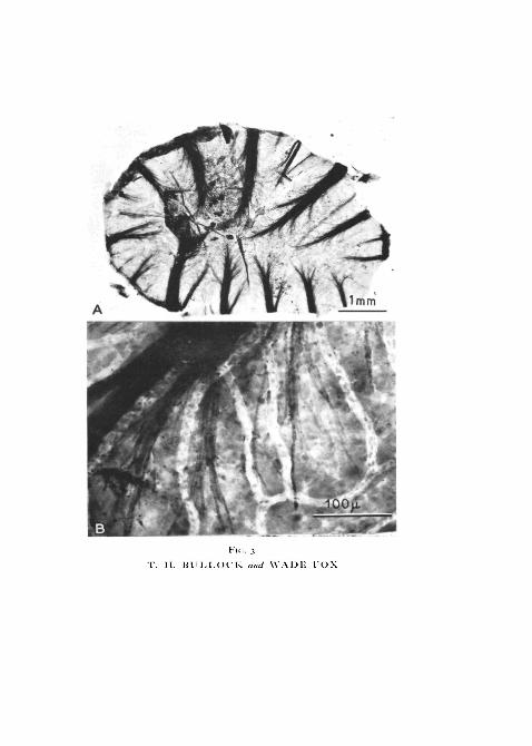
The nerve-trunks as they enter the membrane are of quite unequal size,10 or 12 in number and rather evenly distributed around its periphery(fig. 3, A). They immediately begin to splay out, losing size fibre by fibre andsending off a small number of branches. The larger trunks are discerniblealmost to the centre of the membrane. They travel on the inner side of thecross-section, between the parenchymatous layer and the inner epidermis andunder the capillaries.
As they fan out, the fibres travel short distances and then lose their myelinsheaths (fig. 3, B). Shortly thereafter they expand into the terminal palms butan unmyelinated segment of about 20 to 50 ju, intervenes. The unmyelinatedsegments come to lie superficially to the capillaries.
The fibres from adjacent trunks do not considerably interdigitate butsupply areas of the membrane with sharp, non-overlapping boundaries. Thiscan be seen by tracing from a silver-stained whole mount the palms in theregion of a boundary and noting the directions of the axons. These fall cleanlyinto two groups with a wavy line between the palms belonging to the two. Itcan be seen most strikingly in the preparations where one nerve has degene-rated (see p. 227).
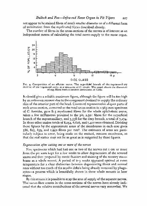
Composition of the afferent nervesThe fibre-size spectrum in a typical case is given in fig. 4. There appeared
to be no significant difference between specimens examined of Crotalus atrox,C. horridus, and C. viridis of snout-vent length from 701 to 965 mm. Theremay be some tapering between a level 10-15 m m from the membrane and thesizes close to the membrane, but if so, it is very slight as reflected in the mode.In three ophthalmic nerves the broad mode lay between 4 and 6/u. (uncorrecteddiameter of myelinated fibres), in one nerve between 3 and 5-5; the largestaxons in these small nerves were usually between 7 and 8JLI. In 5 deep branchesof the supramaxillary, the modes lay from 3-5-7-5 to 5-9-5 and the largestfibres reached 17/x, although only a few per cent, of the fibres are above 9/̂ ..Four superficial branches of the supramaxillary division lay in the same rangeand, for comparison, three mandibular nerves supplying ordinary skin gavecurves which overlapped completely with these.
In the pit membrane shortly before the myelin sheath stops, the diametersappear to have fallen somewhat but are still typically 3 to 5 /x.
We have not attempted to estimate the population of unmyelinated fibresin these nerves. In the silver-stained preparations of the membrane there do
FIG. 3 (plate). Osmium preparations (whole mounts) of facial pit membrane showingmyelinated nerve fibres. C. rtiber.
A, the whole membrane. Note capillary bed.B, higher power to show individual fibres. Note myelin sheaths abruptly ending; this is a
few tens of microns from the palmate-ending.
v
T. H. BULLOCK ami WADE FOX
Bullock and Fox—Infra-red Sense Organ in Pit Vipers 227not appear to be stained fibres of much smaller diameter or of a different formof termination from the myelinated fibres described already.
The number of fibres in the cross-sections of the nerves is of interest as anindependent means of calculating the total nerve-supply to the sense organ.
9 12
SIZE CLASSFIG. 4. Composition of an afferent nerve. The superficial branch of the supramaxillarydivision of the trigeminal nerve in a specimen of C. viridis. The graph shows the diameters
of 295 fibres from a random assortment of fields.
It should give a reliable maximum figure, although the figure will be too highby an unknown amount due to the component destined to supply the ordinaryskin of the anterior part of the head. Counts of representative aliquot parts ofeach cross-section, corrected to the total cross-section in a 965-mm specimenof C. horridus, gave 815 myelinated fibres for the whole ophthalmic nerve,taken a few millimetres proximal to the pit, 2,921 fibres for the superficialbranch of the supramaxillary, and 3,538 for the deep branch, a total of 7,274.In three other snakes totals of 8,254, 6,626, and 7,410 were obtained. Dividingthese figures by the approximate areas of the membranes in each case gives586, 825, 839, and 1,950 fibres per mm2. The estimates of areas are parti-cularly subject to error, being made on the excised, concave membrane, sothat the real scatter may not be as great as is suggested by these figures.
Degeneration after cutting one or more of the nervesFive specimens which had had one or two of the nerves cut 1 cm or more
from the pit were kept for a few weeks to allow degeneration of the severedaxons and then prepared by osmic fixation and staining of the sensory mem-brane as a whole mount. A period of 2-3 weeks appeared optimal at roomtemperature for a clear distinction between degenerating fibres and normalones without too much of the myelin debris being already removed by phago-cytes—a process which is beautifully shown in these whole mounts in laterstages.
By this means it is possible to map the area of supply of the separate nerves.The nerve-fibre counts in the cross-sections of the nerves have already indi-cated that the relative contributions of the several nerves vary somewhat. We
228 Bullock and Fox—Infra-red Sense Organ in Pit Vipers
cannot be sure, therefore, that in the few cases mapped a typical picture of thedistributions was obtained. The ophthalmic nerve has not been cut alone butafter cutting the other two a very small segment of the membrane, less thanone-sixth of its area, remains supplied by undegenerate fibres. After cuttingthe superficial nerve, a segment of about 60% of the area shows degeneratefibres. The deep nerve, by difference, supplies about half the membrane.These segments do not overlap in the slightest. We have not attempted topreserve accurately the orientation of these segments relative to the body axes.
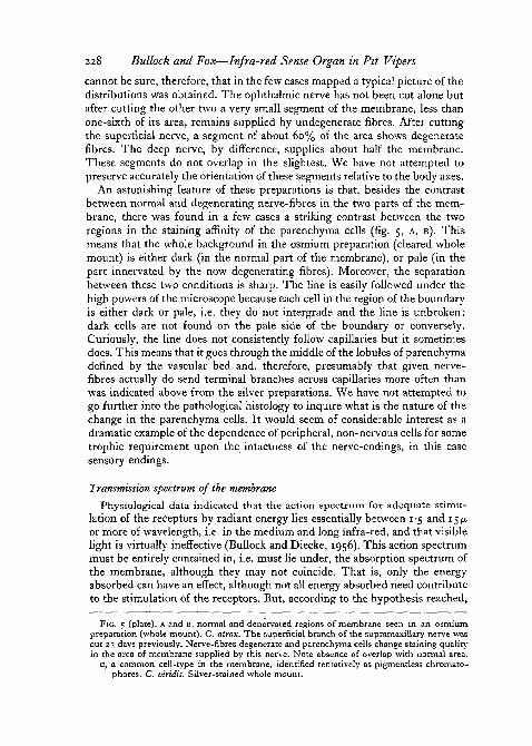
An astonishing feature of these preparations is that, besides the contrastbetween normal and degenerating nerve-fibres in the two parts of the mem-brane, there was found in a few cases a striking contrast between the tworegions in the staining affinity of the parenchyma cells (fig. 5, A, B). Thismeans that the whole background in the osmium preparation (cleared wholemount) is either dark (in the normal part of the membrane), or pale (in thepart innervated by the now degenerating fibres). Moreover, the separationbetween these two conditions is sharp. The line is easily followed under thehigh powers of the microscope because each cell in the region of the boundaryis either dark or pale, i.e. they do not intergrade and the line is unbroken:dark cells are not found on the pale side of the boundary or conversely.Curiously, the line does not consistently follow capillaries but it sometimesdoes. This means that it goes through the middle of the lobules of parenchymadefined by the vascular bed and, therefore, presumably that given nerve-fibres actually do send terminal branches across capillaries more often thanwas indicated above from the silver preparations. We have not attempted togo further into the pathological histology to inquire what is the nature of thechange in the parenchyma cells. It would seem of considerable interest as adramatic example of the dependence of peripheral, non-nervous cells for sometrophic requirement upon the intactness of the nerve-endings, in this casesensory endings.
Transmission spectrum of the membranePhysiological data indicated that the action spectrum for adequate stimu-
lation of the receptors by radiant energy lies essentially between 1-5 and 15^or more of wavelength, i.e. in the medium and long infra-red, and that visiblelight is virtually ineffective (Bullock and Diecke, 1956). This action spectrummust be entirely contained in, i.e. must lie under, the absorption spectrum ofthe membrane, although they may not coincide. That is, only the energyabsorbed can have an effect, although not all energy absorbed need contributeto the stimulation of the receptors. But, according to the hypothesis reached,
FIG. 5 (plate), A and B, normal and denervated regions of membrane seen in an osmiumpreparation (whole mount). C. atrox. The superficial branch of the supramaxillary nerve wascut 23 days previously. Nerve-fibres degenerate and parenchyma cells change staining qualityin the area of membrane supplied by this nerve. Note absence of overlap with normal area.
C, a common cell-type in the membrane, identified tentatively as pigmentless chromato-phores. C. viridis. Silver-stained whole mount.

Fie. 5
T. H. BULLOCK and WADE VOX

Bullock and Fox—Infra-red Sense Organ in Pit Vipers 229namely that the receptors are responding to the temperature changes of thetissue resulting from absorption of radiant energy, the absorption spectrumcan be expected to coincide with the action spectrum since the heating effect
400 600 1000 2000 5000 10000 15000WAVE LENGTH, MILLIMICRONS
FIG. 6. Transmission spectrum of fresh membrane. C. adamanteus.
will not discriminate between different wavelengths absorbed. A lack ofcoincidence could be caused by the absorbing structure being unable to heatthe receptors, because of distance between them or loss of heat by transport,e.g. in erythrocytes.
One specimen of Crotalus adamanteus became available which was so largethat its two pit-membranes together covered a window of usable dimensionsfor spectrophotometers, 2 by 15 mm. The freshly dissected membranes werewashed free of surface blood, dried for 10 min in a P2O5 desiccator underpartial vacuum, and mounted by Scotch tape while being held flat with a glassslide. The membrane is naturally curved in both directions, like an orangepeel, and cannot be perfectly flattened. Although the light beam consists ofparallel rays normal to the plane of the membrane, there is necessarily someloss of light due to reflection and scattering, and this is of unknown amount.For the Baird Associates IR spectrophotometer (NaCl prism), a singlemembrane in a window 2 by 7-5 mm was used. The results are shown in fig. 6.The main features of interest are the deep absorption maxima at 3 /u. and at 6p,,the gradual rise in transmission from 6-6 to 10 ft, and the rather high (approxi-mately 50%) transmission in the long infra-red out to at least I6JU.. The latteris not usually associated with tissue or organic substances, especially with highwater-content, and must be put down to the thinness of the membrane.Since the long infra-red is the region of most effective stimuli, these beingfrom objects of a temperature only a few degrees different from the snakes,this finding indicates a large percentage inefficiency.
The Beckman DU spectrophotometer showed no large absorption maximain the near infra-red or visible regions but at wavelengths shorter than 490 /x

230 Bullock and Fox—Infra-red Sense Organ in Pit Vipers
absorption rapidly increased and was virtually complete at 380JU,. The trans-mission through the visible was not high (between 40 and 50%), so that eitherthere were sizeable losses by scatter and reflection or there is a discrepancybetween the absorption and the action spectrum, since visible light was in-effective in stimulating unless at very high intensity.
DISCUSSION
The anatomical adaptations of the sense organIt was pointed out by Bullock and Diecke (1956) that the physiological
specializations of this organ were not profound. Sensitivity is much higherthan in other temperature receptors directly studied by nerve impulse record-ing, but not higher than that calculated for man. The steady state sensitivityfound in the other known temperature receptors is not present here. Theunusual forms of response to strong stimuli are not necessarily adaptations.Most of the adaptations of the organ are anatomical.
The primary one of these is the concentration of warm receptor elementsinto a pure population of that modality. The high density of these elementsmeans that full advantage can be taken of the phenomenon of central summa-tion (in which the threshold is lowered by simultaneous stimulation of manyreceptors), even though a very small area of the skin is stimulated. This meansthat the stimulation flux (calories per cm2 per sec) is small. Central summationis the phenomenon probably mainly responsible for behavioural threshold,assuring as it does that weak stimuli which give unreliable signals in unitreceptors give quite reliable signals in a group of parallel channels.
The thinness of the sensory membrane probably confers a higher sensitivityto caloric flux in a certain range of stimulus durations. If we separate fromthinness the closeness of the nerve-endings to the surface, that is, compare athick membrane with endings equally close to the surface, then thinness willpermit a given flux to warm the receptors more, provided that the stimulusis not allowed to act indefinitely and provided that it is allowed to act longerthan a very brief period. In the case of a very brief stimulus, the advantageof thinness decreases because there is proportionally less loss of heat tothe depths in the thick membrane than there is for longer stimuli. In thecase of very long stimuli warming approaches a steady state and, unlessthere is a large sink in the thick membrane into which heat flows, therewill be little advantage in a thin membrane. The range of periods forwhich the membrane as we see it offers an advantage cannot be accuratelycalculated without more knowledge of the loss of heat to the blood, butapparently it extends from a second or less to many seconds. This is basedon the consideration that in a thin membrane and neglecting losses byreradiation and blood-flow, the change in temperature for a given flux isproportional to the time, while in a thick structure which conducts heatpast the receptors, the change is proportional to the square root of the time(see Bullock and Diecke, 1956).

Bullock and Fox—Infra-red Sense Organ in Pit Vipers 231Perhaps more important than the thickness of the membrane is the super-
ficial location of the receptors. We could not give accurate measurements ofthe depth of the endings, but the average depth is probably no more than 5 pand possibly only 2/A from the outer surface. This not only increases thepromptness of the response and the ability to detect nickering and briefstimuli, but situates the detector at the most favourable place in the tempera-ture gradient. Actually we can say, from the transmission spectrum, that theadaptation has gone about as far as it is worth going, for the layer between thenerve-endings and the surface is already so thin that it is absorbing somethingless than half of the energy in the wavelengths chiefly available.
We need not discuss here the special features of the posterior chamber, itsoutlet and sphincter, the location on the head, the angle of view, the circula-tion, or the chromatophores (fig. 5, c). But one further feature of the anatomyof the accessories deserves mention. This is that the pit characteristically doesnot taper inward but actually overhangs, i.e. the mouth is smaller than themembrane at the bottom. This is obvious to the eye and in a small series ofrandomly chosen specimens of four species, the diameter of the pit openingof io pits was from 35% to 50% of that of the membrane. The consequenceof this, together with the small depth of the pit, is that most radiating objectsunless very large or very close will not illuminate the whole sensory membranebut will cast shadows of the pit margin. This confers the possibility of derivinginformation from the sense organ about the direction of small objects or ofedges of large objects, if the resolution of the sense organ is sufficient. We havealready seen that the innervation is rich. There are between 500 and 1,500axons ending per mm2. This compares with about 800 optic nerve fibres permm2 of the retina in man, averaging an estimate of 800,000 fibres into anestimate of 1,000 mm2 of retina. The membrane provides enough absolutearea and hence nerve-fibres to resolve even fairly poor shadow margins: insnakes between 350 and 1,000 mm snout-vent length the membrane diametersfall near a line connecting 2 and 4-4 mm, corresponding to areas somewhatover 3 and 15 mm2 (since the surface is concave). We may conclude that thecentral nervous system receives adequate information to analyse directionalitywith a degree of usefulness, especially if the snake scans or if the object ismoving.
A final point should be raised concerning the nature of the parenchymacells of the sensory membrane. These are the only non-nervous cells of thesense organ which are specialized, except for the attenuation of the epithelialand connective tissue-cells. They are not readily identified with any cell-typein ordinary skin. They are polygonal, about 6/u. thick, with an osmiophilcytoplasm of uniform, dense, reticular structure. Most significantly, theyreact with a change in staining quality to loss of the nerve-endings and evi-dently depend on the particular ending intimately located about them; forthere is no grading off of the reaction from the boundary between normal anddenervated regions (compare Hillarp, 1946). These facts might lead in termsof classical histology to regarding the cells as sense cells. We do not believe
232 Bullock and Fox—Infra-red Sense Organ in Pit Vipers
they should be so considered unless evidence is obtained that they actuallyrespond to the normal environmental stimulus and mediate the initiation ofnerve discharge. In the physiological study Bullock and Diecke were unableto obtain such evidence, although they could not eliminate this possibility.If, as we suppose, the parenchyma are not sense cells, they very probably dohave some essential accessory function in connexion with the transducing oftemperature change into neuronal activation.
Comparison with other reptilian receptorsIt has been suggested in the literature repeatedly that there may be an
analogy between the facial pits of pit vipers and the labial pits of some pythonsand boas. No other organ appears to resemble these in structure or functionas far as is known. The labial pits are simple depressions, without an innerchamber and hence with a floor receiving the nerve-supply but no membrane(Noble and Schmidt, 1937). Behavioural experiments clearly suggest a func-tion as receiver of warm radiation, like the facial pits of crotalids, although theauthors have apparently not realized this and speak of air temperature,measured by a mercury thermometer (Noble and Schmidt, 1937) or air move-ment resulting from the local heating of the air (Ros, 1935).
As far as we are aware the facial pits are not similar in anatomy to any othertemperature receptors among animals, or indeed to receptors of any modality.The nerve-endings themselves seem also to be unique, in the form of theterminal palmate expansion. It does correspond with the conclusions ofWeddell and his collaborators that temperature-endings should be freenerve-endings and not necessarily corpuscular. Free nerve-endings have beendescribed in reptilian skin (see Boeke, pp. 859-66), but none which give anyspecial suggestion of being forerunners of these. Bullock and Diecke (1956)could not find evidence of any temperature reception resembling that con-centrated in the pit, on searching through ordinary skin-nerves of rattlesnakesand even the homologous branches of the trigeminal in non-crotalid snakes.
Possible significance of the palmate-endingIf, as we suppose, the receptors are detecting and transducing small tem-
perature changes in the tissue, there is no special significance in the spatialaspect of the ramification of each axonal-ending, that is there is no particularadvantage in covering a wide territory as there would be if interceptingphotons or mechanical deformation were their function. But we may suggestthat the development of an extensive surface, subdivided into quasi-inde-pendent regions, enhances the probability of firing to a small change. This isbased on the supposition that these fine processes are prone to autorhythmic,sub-threshold, graded changes of state which do not propagate but spreaddecrementally, not interfering with each other but summating so that at somecritical point, perhaps at the emergence of the axon from the palm, all or noneimpulses are initiated whenever some sharp threshold is exceeded. We wouldvisualize the several processes of the palm each undergoing independently,
Bullock and Fox—Infra-red Sense Organ in Pit Vipers 233fluctuations in state, including membrane potential, and these may be quiterhythmic. As they happen to summate and therefore to spread farther theywould from time to time trigger impulses in the axon. This would explain, asfar as it goes, the non-rhythmic spontaneous background discharge. It alsomakes it possible to invoke the most exquisitely sensitive mechanism ofminute changes in the potential gradient between one part of the neuronesurface and another, recently shown by Terzuolo and Bullock (1956), to alterthe frequency of firing of already active neurones. This hypothesis of theactivity of sensory processes is not uniquely applicable to the present receptorsbut seems reasonable for many branching terminations of small diameter,especially those with a spontaneous or steady state discharge. It places afferentaxonal terminations in a class with dendrites, in not supporting impulses orpropagating a disturbance toward the axon, but influencing the initiation ofimpulses in the axon by small changes in potential gradient along the neuronalmembrane (compare Fessard, 1956).
It is a pleasure to thank Dr. Ralph Nusbaum of the University of Cali-fornia Atomic Energy Project at Los Angeles for running the infra-redtransmission spectra and for much valuable advice. We are also indebted toDr. R. B. Cowles of this department and Mr. Charles Shaw of the San DiegoZoo for generous provision of many animals. Financial support from theNational Science Foundation is gratefully acknowledged.
REFERENCESBODIAN, D., 1936. 'A new method for staining nerve fibers and nerve endings in mounted
paraffin sections.' Anat. Rec, 65, 89.BOEKE, J., 1934. 'Niedere Sinnesorgane.' In BOLK, L., GOPPERTT, E., KALLIUS, E., and
LUBOSCH, W., Handbuch der vergleichenden Anatomie der Wirbeltiere, Berlin (Urban &Schwarzenberg).
BULLOCK, T. H., 1954. Remarks and illustrations in discussion of Prof. Yngve Zotterman'spaper 'Sensory receptors' in Nerve impulse, Transactions of the Fourth Conference, 1953,edited by D. Nachmansohn, pp. 140-206. New York (Josiah Macy, Jr., Foundation).
MOULINS, A., 1824. Memoire sur le systeme nerveux et lappareil lacrymal des serpentsa sonnettes, des trigonocephales et de quelques autres serpents.' J. Phys. exp. Pathol.Magendie, 4, 264.
FESSARD, A., 1956. 'Formes et caracteres generaux de l'excitation neuronique.' XXe Congresinternational de physiologie, 1, 35.
HILLARP, N.-A., 1946. 'Structure of the synapse and the peripheral innervation apparatus ofthe autonomic system.' Acta Anat., Suppl. 4, 1.
HOLMES, W., 1943- 'Silver staining of nerve axons in paraffin sections.' Anat. Rec, 86, 157.and YOUNG, J. Z., 1942. 'Nerve regeneration after immediate and delayed suture.'
J. Anat. 77, 63.LANCE, B., 1931. 'Integument der Sauropsiden.' In BOLK and others, Handbuch der vergleich-
enden Anatomie der Wirbeltiere. Berlin (Urban & Schwarzenberg).LEYDIG, F., 1868. 'Uber Organe eines sechsten Sinnes.' Verh. d. Kaiserl. Leopold.-Carol.
Deutsch. Akad. Naturforscher, Novum Act., 34 (5), 1.LILLIE, R. D., 1948. Histopathologic technique. Philadelphia (Blakiston).LYNN, W. G., 1931. 'The structure and function of the facial pit of the pit vipers.' Am. J.
Anat., 49, 97.
234 Bullock and Fox—Infra-red Sense Organ in Pit VipersNOBLE, G. K., 1934. 'The structure of the facial pit of the pit vipers and its probable function.
Anat. Rec, 58 (suppl. to No. 2), 4.and SCHMIDT, A., 1937. 'Structure and function of the facial and labial pits of snakes.'
Proc. Amer. phil. Soc, 77, 263.PALADE, G. E., 1952. 'A study of fixation for electron microscopy.' J. exp. Med., 95, 285.PALMCREN, A., I95 I. 'A method for silver staining nerve fibres in very thick sections and in
suitable whole preparations.' Acta Zool., 32, 1.ROMANES, G. J., 1950. 'The staining of nerve fibres in paraffin sections with silver.' J. Anat.,
84, 104.Ros, M., 1935. 'Die Lippengruben der Pythonen als Temperaturorgane.' Jena. Zeit.
Naturw., 63, 1.TERZUOLO, C. A., and BULLOCK, T. H., 1956. 'Measurement of imposed voltage gradient
adequate to modulate neuronal firing.' Proc. Nat. Acad. Sci., 42, 687.WEDDELL, G., and SINCLAIR, D. C , 1951. 'Cutaneous sensibility in the pinna of the ear.'
J. Anat., 85, 424.and ZANDER, E., 1950. 'A critical evaluation of methods used to demonstrate tissue
neural elements, illustrated by reference to the cornea.' Ibid., 84, 168.WEST, G. S., 1900. 'On the sensory pit of the Crotalinae.' Quart. J. micr. Sci., 43, 49.