ARTICULO ORIGINAL Antibacterial activity of Eudesmanolides ...
1-2
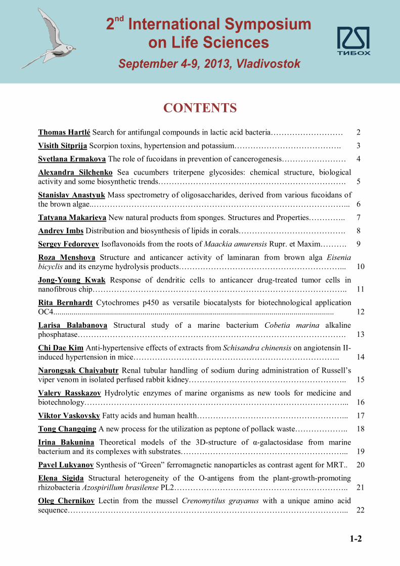
CONTENTS
Thomas Hartlé Search for antifungal compounds in lactic acid bacteria……………………… 2
Visith Sitprija Scorpion toxins, hypertension and potassium…………………………………. 3 Svetlana Ermakova The role of fucoidans in prevention of cancerogenesis…………………… 4
Alexandra Silchenko Sea cucumbers triterpene glycosides: chemical structure, biological activity and some biosynthetic trends……………………………………………………………. 5
Stanislav Anastyuk Mass spectrometry of oligosaccharides, derived from various fucoidans of the brown algae..…………………………………………………………………………………... 6
Tatyana Makarieva New natural products from sponges. Structures and Properties………….. 7 Andrey Imbs Distribution and biosynthesis of lipids in corals…………………………………. 8 Sergey Fedoreyev Isoflavonoids from the roots of Maackia amurensis Rupr. et Maxim………. 9
Roza Menshova Structure and anticancer activity of laminaran from brown alga Eisenia bicyclis and its enzyme hydrolysis products……………………………………………………... 10
Jong-Young Kwak Response of dendritic cells to anticancer drug-treated tumor cells in nanofibrous chip………………………………………………………………………………….. 11
Rita Bernhardt Cytochromes p450 as versatile biocatalysts for biotechnological application OC4........................................................................................................................................... 12
Larisa Balabanova Structural study of a marine bacterium Cobetia marina alkaline phosphatase………………………………………………………………………………………. 13
Chi Dae Kim Anti-hypertensive effects of extracts from Schisandra chinensis on angiotensin II-induced hypertension in mice………………………………………………………………….. 14
Narongsak Chaiyabutr Renal tubular handling of sodium during administration of Russell’s viper venom in isolated perfused rabbit kidney………………………………………………….. 15
Valery Rasskazov Hydrolytic enzymes of marine organisms as new tools for medicine and biotechnology……………………………………………………………………………………... 16
Viktor Vaskovsky Fatty acids and human health………………………………………………... 17 Tong Changqing A new process for the utilization as peptone of pollack waste……………….. 18
Irina Bakunina Theoretical models of the 3D-structure of α-galactosidase from marine bacterium and its complexes with substrates……………………………………………………... 19
Pavel Lukyanov Synthesis of “Green” ferromagnetic nanoparticles as contrast agent for MRT.. 20 Elena Sigida Structural heterogeneity of the O-antigens from the plant-growth-promoting rhizobacteria Azospirillum brasilense PL2……………………………………………………….. 21 Oleg Chernikov Lectin from the mussel Crenomytilus grayanus with a unique amino acid sequence…………………………………………………………………………………………... 22

2-2
Viktoria Davydova Some aspects of cleaning solution from endotoxin by zeolite modified with chitosan……………………………………………………………………………………… 23 Vladimir Muronetz Search for new inhibitors of pathological prion aggregation……………… 24
Olga Sidorova Mutant OmpF porins of Yersinia pseudotuberculosis: structure-functional and immunochemical properties………………………………………………………………………. 25
Wiesław Kaca The properties of chitosans and carrageenans complexes with smooth and rough forms of Proteus mirabilis lipopolysaccharides………………………………………………….. 26
Аndrey Byvalov Modern problems of microbial physiology……………………………………. 27 Natalia Plekhova The cells of innate immunity system in Tick-Borne Encephalitis……………. 28
Min Qu Antioxidant activity and protections of glycopeptides from giant salamander mucus on CCl4 acute live-injured in mice…………………………………………………………………… 29 Evgeniya Bystritskaya Transcriptional control of porin genes expression in Yersinia pseudotuberculosis………………………………………………………………………………... 30 Valentin Stonik New natural products from marine sources: structures and properties………… 31
Yuriy Sabutskii A new method of thiomethylation of hydroxy-1,4-naphthoquinones by N-acetyl- L-cysteine. First synthesis of fibrostatins B, C and D……………………………………. 32
Sergey Sarin Chitosan-based nanocomposite films formed through self-organization…………. 33 Darya Tarbeeva Polyphenolic compounds from Iris pseudacorus L. and its callus cultures…... 34
Anton Yurchenko Biologically active substances from marine-derived fungi from far eastern seas………………………………………………………………………………………………… 35
Marina Isaeva Microevolution of Yersinia porin genes: positive selection, recombination and pseudogenization…………………………………………………………………………………... 36
Vasily Golotin Biochemical and catalytic properties of the recombinant alkaline phosphatase from the marine bacterium Cobetia marina……………………………………………………….. 37
Ekaterina Sokolova Influence of red algal sulfated polysaccharides on blood coagulation and platelets in vitro……………………………………………………………………………………. 38
Alexey Belik Producing of active recombinant endo-1,3-β-D-glucanase from marine bacterium Formosa algae KMM 3553……………………………………………………………………….. 39
Alexander Rakin Molecular evolution of bacterial mobility vehicles…………………………… 40 Irina Yermak Structural peculiarities of sulfated polysaccharides-carrageenans from red alga families Tichocarpaceae and Gigartinaceae and aspects of their medical application…………… 41 Georgy Nevinsky HIV-1 integrase-hydrolyzing IgG and IgM antibodies from sera of HIV-infected patients…………………………………………………………………………………… 42 Irina Gileva Variola virus TNF-binding protein as a new type of TNF-antagonists…………….. 43
Maxim Kokoulin Structure of the O-specific polysaccharide from a marine bacterium Cellulophaga tyrosinoxydans……………………………………………………………………… 44
Alexey Ivanov Investigation of intermolecular interactions using surface plasmon resonance technology…………………………………………………………………………………………. 45
Jin Han Tetrahydrobiopterin is an essential cofactor for cardiac mitochondrial biogenesis and oxidative phosphorylation………………………………………………………………………… 46
In-Kyu Lee The importance of PDH flux on high fat induced obesity………………………….. 47

3-2
Irina Agafonova Development of experimental model of the acute impairment of blood brain circulation at wistar rats for the study of antioxidant properties of native and synthetic echinochromes. The verification of cerebral circulation with MRI………………………………. 48
Galina Likhatskaya Effects of chitosans and acylated chitooligosaccharides on LPS interactions with TLR4/MD2: in silico study……………………………………………………... 49
Vladimir Prassolov Screening of potential HIV-1 inhibitors using secure lentiviral in vitro system……………………………………………………………………………………………… 50
Elena Zelepuga Novel peptide toxins from the sea anemone Heteractis crispa: structural and functional aspects………………………………………………………………………………….. 51
Vladimir Apanasevich Local radio-sensibilisation of tumor tissue by using of aurum nanoparticles and iodine-containing compounds…………………………………………………. 52
Nguyen Manh Cuong Effects of Vietnamese selected plant extracts on L-type Ca2+ channel in rat ventricular myocytes…………………………………………………………………………… 53
Roman Popov Analysis of the Fraction of Asterosaponins and Related Compounds from the Far-Eastern Starfish Aphelasterias japonica by LC-ESI MS……………………………………... 55
Svetlana Bakholdina Outer membrane phospholipase A from Yersinia pseudotuberculosis: cloning, expression and function………………………………………………………………….. 56
Yulia Shtoda Medicinal properties of «Kourochitin» preparation at experimental modeling of various skin disorders……………………………………………………………………………… 57
Alexander Tsybulsky Experimental study of antitumor and cytokines-modulating activities of bisulfate luteolin and luteolin……………………………………………………………………… 58
Larisa Balabanova Expression of Pseudoalteromonas sp. alpha-galactosidase mutants………... 59 Nikolay Goncharov Construction and expression of YPM-TM fusion protein based on Yersinia pseudotuberculosis superantigen………………………………………………………………….. 60 Alexander Popov Biological activities of collagen peptides obtained by enzymic hydrolysis from far-eastern holothurians……………………………………………………………………… 61 Olga Krivoshapko Protective activity of redox-active compounds from marine organisms at modeling of hyperlipidemia and diabetes…………………………………………………………. 62 Pavel Spirin Associative down-regulation of C-KIT expression through shRNA mediated silencing of AML1-ETO oncogene in t(8;21) leukemic cells……………………………………… 63 Tran Thuy Chemical constituents of starfish Acanthaster planci and some synthetic polyhydroxylated steroids from the major sterol………………………………………………….. 64 Nguyen Nguyet First study on chemical constituents of starfish Anthenea aspera from Vietnamese northeast sea………………………………………………………………………….. 65 Stepan Logvinov Sulfoquinovosyldiacylglycerols from sea urchins…………………………….. 66
Ludmila Shevchenko Impact of media composition on antifungal activity of marine actinomycetes……………………………………………………………………………………… 67
Valery Glazunov The reinvestigation of the experimental IR-spectrum of o-vinylphenol………. 68 Valery Glazunov A conformational analysis of the dimerization products of o-vinylphenol…… 69
Valery Glazunov Assignment and forms of stretching vibrations of carbonyl groups of hydroxylated 1,4-naphthoquinones………………………………………………………………... 70

4-2
Valery Glazunov A DFT study of the antioxidant properties of Echinamines A and B the metabolites of the sea urchin Scaphechinus mirabilis…………………………………………….. 71 Dmitriy Berdyshev The B3LYP study of the keto-forms of acetylacetone……………………… 72
Dmitriy Berdyshev The DFT study of the influence of methanol solvent on the tautomeric equilibrium in b,b¢-triketones……………………………………………………………………... 73
Maria Sobolevskaya Bioactive metabolites of fungi Aspergillus and Penicillium genera………. 75 Margarita Monastyrnaya New multigene family of sea anemone Heteractis crispa pore-forming toxins (a-PFTs)…………………………………………………………………………… 76
Zeng Weimin Several polar compounds from the rhizoma of Dryopteris crassirhizoma Nakai... 77
Yulia Nabivailo Contrasting effects of elevated temperature on phlorotannin and oxidative responses in Fucalian and Laminarian seaweeds…………………………………………………. 78
Valentin Tabakmakher New recombinant Kunitz-type polypeptides of sea anemone Heteractis crispa………………………………………………………………………………………………. 79
Anna Kravchenko Composition of phycobiliproteins and polysaccharides from two forms of red alga Ahnfeltipsis flabelliformis………………………………………………………………... 80
Alexandra Kondrashina Physicochemical properties and biological activity of lectin from the mussel Mytilus trossulus…………………………………………………………………………... 81
Аlexandra Seitkalieva Acid and alkaline phosphatase of marine echinoderms and bivalve mollusks…………………………………………………………………………………………… 82
Natalya Mishchenko New polyhydroxynaphthoquinones from sea urchins……………………. 83 Darya Demidkova The comparative study of lipid class and fatty acid composition of cold-water and tropical soft corals……………………………………………………………………… 84 Temur Hasanov Expression of gene for murine TRAIL in E. coli cells and purification of recombinant protein……………………………………………………………………………….. 85 Natalie Utkina Antioxidant activity of zyzzyanones and makaluvamines from the marine sponge Zyzzya fuliginosa………………………………………………………………………….. 86 Yanlong Zhang Preparation of Auricularia auricula oligosaccharides and its antioxidant activity……………………………………………………………………………………………... 87
Feng Shan Yang Proteomics of Methyl Jasmonate response in Maize leaves…………………... 88 Wei Li Effects of lectins from marine invertebrates on physiological activity of microorganisms. 89
Tatyana I. Imbs Extraction method of fucose containing sulfated polysaccharide from the brown seaweed Fucus evanescens is important determinant for antioxidant activity development 90 Artem Silchenko Cloning and expression of fucoidanases from the marine bacterium Formosa algae KMM 3553………………………………………………………………………………….. 91 Alexander Popov Comparative Study of Adjuvant Properties of Triterpene Glycosides from Sea Cucumber and Minor Ginseng Glycoside Rh2………………………………………………. 92 Alexandra Volodko Soluble polyelectrolyte carrageenan: chitosan complexes and their gastroprotective activity…………………………………………………………………………… 93
Prospects to use supercritical fluid extraction method in bioorganic chem-istry...Sofia Razgonova

Oral Presentations

2
Search for antifungal compounds in lactic acid bacteria
Yanath Belguesmia, Yvan Choiset, Hanitra Rabesona, Jean-Marc Chobert and
Thomas Haertlé
Biopolymères Interactions Assemblages, Institut National de Recherche Agronomique, équipe Fonctions et Interactions des Protéines, rue de la Géraudière, BP 71627, 44316 Nantes Cedex 3,
France
The aim of our work was to identify antifungal agents produced by selected LAB and to study their action. In the first place the of antifungal properties of durancins isolated from Enterococcus durans A5-11 and of
their chemically synthesized fragments were investigated. Enterococcus durans A5-11 is a lactic acid bacteria (LAB) strain isolated from traditional Mongolian airag
cheese. This strain inhibits the growth of several fungi including Fusarium culmorum, Penicillium roqueforti and Debaryomyces hansenii. It produces two bacteriocins: durancin A5-11a and durancin A5-11b, which have similar antimicrobial properties. The whole durancins A5-11a and A5-11b, as well as their N- and C-terminal fragments were synthesized and their antifungal properties were studied. C-terminal fragments of both durancins showed stronger antifungal activities than other tested peptides. Treatment of Debaryomyces hansenii LMSA 2.11.003 strain with 2 mmol.l-1 of the synthetic peptides led to the loss of the membrane integrity and to several changes in the ultra-structure of the yeast cells.
Chemically synthesized durancins and their synthetic fragments showed different antimicrobial properties. C-ter peptides have specific activities against tested fungal strain and do not show antibacterial activity. However, they enhance the activity of the N-terminal fragment in the whole bacteriocins against bacteria.
This part of our investigations improved understanding of molecular causes of antimicrobial activities of bacteriocins and their fragments.
However, bacteriocins are not the unique antifungal agents produced by LAB.
Hence, in the next step antifungal compounds produced by Lactobacillus harbinensis K.V9.3N.1p isolated from raw cow milk were characterized. A fermentation process with immobilized cells of the protective culture of Lactobacillus harbinensis K.V9.3N.1p in enriched milk protein medium in the presence of Debaryomyces hansenii UBOCC-A-211003 and Kluyveromyces lactis UBOCC-A-212021 was developed. After seven days of fermentation, active cell-free culture supernatants with robust antifungal activities against Debaryomyces hansenii, Penicillium expansum and Penicillium roqueforti were observed. Active cell-free culture fractions collected during the batch fermentation process contained high amounts of organic acids such as lactic and acetic acids as well as hexanoic acid (9 mM), which revealed to be the most important antifungal component in these samples. The antifungal activity of hexanoic acid was related to the environmental conditions including the nature of the matrix and the pH. Even if organic acids are key antifungal agents, they are a part of complex mixtures of various molecules produced by Lb. harbinensis K.V9.3N.1p strain, acting in a synergic manner.
In result it can be stated that studied Lactic Acid Bacteria are using complex set of antifungal agents in their
fight against fungal competition.
Both types of the identified agents could practical application in the protection against fungal infections and spoilages.

3
Scorpion toxins, hypertension and potassium
Visith Sitprija
Queen Saovabha Memorial Institute The Thai Red Cross Society
Bangkok, Thailand
Symptomatology of scorpion envenoming is largely due to autonomic nervous system stimulation with the release of catecholamines and acetylcholine. In addition to catecholamines, renin angiotensin and aldosterone (RAA) axis is activated to generate hypertension and hemodynamic alteration with decreased renal blood flow. Renal function is compromised.
Scorpion toxins inhibit a number of K channels in renal tubules including voltage gated K channels, Ca activated K channels and ROMK resulting in decreased urinary K excretion and hyperkalemia. Depolarization of vascular smooth muscle cells due to inhibiton of Ca activated K channels also causes vasoconstriction and hypertension. The magnitude of decrease in urinary K excretion is determined by the degree and number of K channel inhibition. With counter effect of aldosterone in increasing urine K excretion, hyperkalemia, is therefore not consistent. The interaction between K channel inhibition, catecholamines, RAA axis activation and angiotensin converting enzyme inhibitor, present in some scorpion species, is of great interest and deserves discussion as an exercise in physiology.

4
Sea cucumbers triterpene glycosides: chemical structure, biological activity and some biosynthetic trends
Alexandra S. Silchenko, V.I. Kalinin, S.A. Avilov
G.B. Elyakov Pacific Institute of Bioorganic Chemistry, Far Eastern Branch, Russian Academy
of Sciences, 159 100-let Vladivostoku Pr., Vladivostok, 690022, Russia
Triterpene glycosides are characteristic metabolites of sea cucumbers (Holothurioidea), the class of marine invertebrates belonging to the phylum Echinodermata. The molecules of triterpene glycosides consist of the aglycone part and carbohydrate chain. Majority of the glycosides comprises the aglycones of so-called holostane type (lanostane derivatives with 18(20)-lactone), others have an aglycones with 18(16)-lactone or lack a lactone (lanostane type) and lack a lactone and a side chain (nor-lanostane type). Carbohydrate chains of these substances consist of two to six monosaccharide units and may include from one to three sulfate groups.
Most of triterpene glycosides have been isolated from sea cucumbers belonging to the orders Aspidochirotida and Dendrochirotida. Sea cucumber Synapta maculata is the only chemically studied representative of the order Apodida having very uncommon triterpene glycosides. During last years, main investigations were focused on holothurians of the order Dendrochirotida, which usually contain very complicated mixtures of the glycosides. Due to the modern approach in separation of mixtures of related substances appreciable number of new minor triterpene glycosides were isolated recently. Some of such minor glycosides have very interesting structural peculiarities. Thus, from the Far Eastern sea cucumber Eupentacta fraudtrix were isolated 27 new minor triterpene glycosides. The most uncommon structural features of some of these substances were: unique hydroxy group at C-18 of the aglycone having no lactone ring that may be considered as intermediate “hot” metabolite in biosynthetic transformations of triterpene glycosides; 27-nor-holostane glycoside that is representative of very rare among natural products 27-nor-derivatives; 23E,25-diene system in the aglycones side chain; unprecedented 16(22)-epoxy-group in the aglycone and unique trisaccharide carbohydrate chain. From Antarctic sea cucumbers Staurocucumis turqueti and S. liouvillei were isolated disulfated olygoglycosides having rare terminal 3-O-methyl-D-quinovose, that indicated this sugar is a chemotaxonomic character of the genus Staurocucumis.
So, triterpene glycosides are characterised by chemical variability in some structural features and peculiarities in combination with stable general plan of structure that allow to use them as chemotaxonomic markers.
Sea cucumber glycosides are natural products with mosaic type of biosynthesis since biosynthetic transformations of aglycones and carbohydrate chains assemblage proceed independently from each other and simultaneously. The analysis of chemical structure of novel glycoside cucumarioside A8 from E. fraudatrix having C-18 and C-20 hydroxyl groups in the aglycone istead of 18(20)-lactone allows to clarify some peculiarities of triterpene glycosides biosynthetic pathways. The aglycone with such low degree of oxidation may be considered as biosynthetic precursor of more oxidized holostane aglycones. The hypothetical scheme of biosynthesis of sea cucumbers glycosides is proposed, where direct formation of lanostane derivatives with a 7(8)-double bond from protosteroid cation is possible. The structures of new cucumariosides B1 and B2 having trisaccharide “non-developed” carbohydrate chains and holostane type “developed” aglycones is in good accordance with the mosaic type of biosynthesis. The prolongation of sugar chain passes through the consequent attachment of monosaccharide units to different positions of forming oligosaccharide chains and its sulfation probably is a final stage of biosynthesis.
Sea cucumbers triterpene glycosides demonstrate different kinds of biological activities caused by their membranolytic action. The cytotoxic activity against different cell lines and hemolytic activity of glycosides approximately correlate to each other because of common sterol-dependent mechanism and depend on the chemical structure of the glycosides. Some structural elements are signficantly decrease and others, conversely, increase bioactivity of triterpene glycosides.

5
Mass spectrometry of oligosaccharides, derived from various fucoidans of the brown algae
Stanislav D. Anastyuk, Natalia M. Shevchenko, Tatyana I. Imbs, Vladimir I. Gorbach, Pavel S.
Dmitrenok, Tatyana N. Zvyagintseva
G.B. Elyakov Pacific Institute of Bioorganic Chemistry, Far Eastern Branch, Russian Academy of
Sciences, 159 100-Let Vladivostoku Ave., Vladivostok 690022, Russian Federation; e-mail: [email protected]
Fucoidans are sulfated heteropolysaccharides, mainly built up of α-L-fucopyranose residues. Being non-
toxic, they possess wide variety of biological activities, including immunomodulating, anticoagulant, antiviral, antioxidant and antitumor activities. It is clear that the activity is related to the structural features of fucoidans: degree of sulfation, molecular weight, linkage pattern. Selected specie of alga may synthesize structurally different fucoidans, depending significantly on its age and less significantly on the environment. Furthermore, extraction conditions may affect the polysaccharide composition. Classic approach using 13C NMR and methods of carbohydrate chemistry for the analysis of such large amount of samples becomes time-consuming. Hence, new procedure for rapid analysis of material is required.
Mass spectrometry (MS) with matrix-assisted laser desorption/ionization (MALDIMS) and electrospray ionization (ESIMS) are important tools for the analysis of heterogeneous anionic carbohydrates. Its speed, sensitivity and accuracy fit the above-mentioned requirements. However, it is yet impossible to directly analyze large highly-charged molecules having molecular weight (MW) from tens to thousands kDa. Polysaccharide thus should be depolymerized to oligosaccharides, having MW 100 Da - 4 kDa. Since there are no widely available enzymes catalyzing specific transformation of fucoidans, acid hydrolysis is often performed. But, it also requires time-consuming experiments for optimal conditions selection. Recently, we have employed autohydrolysis* as an alternative strategy for fucoidan decomposition. The method was found to be reproducible for a preparation of multisulfated oligosaccharides, well reflecting the structure of the source polysaccharide. Using such an approach fragments of fucoidans from brown algae Saccharina cichorioides, Fucus evanescens, Silvetia babingtonii, Costaria costata and Coccophora langsdorfii were successfully analyzed by tandem ESI and MALDIMS techniques. Structural features of oligosaccharides matched with that observed previously for corresponding fucoidans using independent methods.
*‘Autohydrolysis’ is used here to denote acidic polysaccharide hydrolysis under very mild conditions using –SO3H groups of the compound as the source of acid

6
New natural products from sponges. Structures and Properties
Tatyana N. Makarieva
G.B. Elyakov Pacific Institute of Bioorganic Chemistry, Far Eastern Branch, Russian Academy
of Sciences, 159 100-let Vladivostoku Pr., Vladivostok, 690022, Russia e-mail: [email protected]
Sponges are widely recognized as the most prolific source of structurally unique and biologically active
marine natural products. They are also abundant benthic invertebrates of tropical, temperate and boreal waters and play an important role in the corresponding marine biocenoses. Our studies have been focused on the isolation and structure determination of novel compounds from these animals. Up to date about 200 compounds have been isolated and their biological activities have been studied by our group. The compounds isolated belong to a wide variety of biogenetic classes such as two-headed sphingolipids, isoprenoid sulfates, polyprenyl sulfates, pentacyclic, bicyclic and noncyclic guanidine alkaloids etc. Noticeable recent examples are monanchocidins and monanchomycalins from the Far-Eastern sponge Monanchora pulchra. These latter compounds were shown to be potent antileukemic agents possessing novel chemical structures that attract an attention from point of view their pharmaceutical properties and biosynthesis. More recently pulchranins A-C, first marine non-peptide inhibitors of TRPV-1 channels were isolated from the Far-Eastern marine sponge, Monanchora pulchra.
We are going to discuss herein the diversity of unusual sponge metabolites from the corresponding animals collected from the North-Western Pacific, problems concerning the isolation and structure elucidation of sponge metabolites, and some details of molecular mechanisms of their physiological actions.

7
Distribution and biosynthesis of lipids in corals
Andrey B. Imbs
A.V. Zhirmunsky Institute of Marine Biology, Far Eastern Branch, Russian Academy of Sciences,
Palchevskogo str., 17, 690059 Vladivostok, Russian Federation; e-mail: [email protected]
Reef-building corals, which have a hard exoskeleton, and soft corals widely distribute in a tropical zone of
the World Ocean. Hydrocorals, such as “fire coral” Millepora, are also members of coral reef ecosystem. The most of reef-building coral species, as well as Millepora, and a lot of soft corals species contain endocellular symbiotic dinoflagellates (SD) named zooxanthellae, which are the essential source of photosynthetic organic carbon for their host. Specific azooxanthellate soft corals and hydrocorals inhabit cold-water regions, such as the Bering Sea, the Okhotsk Sea, et ctr.
Lipids form up to 40% of coral dry biomass. Lipids are the main energy reserve of a coral colony and the base of coral cell membranes. Lipids are involved in a majority of biochemical and physiological processes in corals. Fatty acids (FA) are the main constituents of lipids. FA are used as biomarkers of food sources and symbionts of corals.
All coral species contain the same lipid classes. The lipid class composition of corals is related to their taxonomic position, presence of SD, and vary depending on the stage of development, coral health, and environmental factors. Among cnidarians, corals with hard exoskeleton have the highest content of lipids, at least half of which are reserve lipids (wax esters, sterol esters, and triglycerols). The percentage amount of the reserve lipids decreases in the sequence: scleractinians – soft corals – anemones and jellyfish. The significant energy expenditures that are required for the formation of the exoskeleton may be accompanied by an increased production of the reserve lipids. The ability to synthesize unusual phosphonolipids and monoalkyldiacylglycerols is the important characteristic of lipid metabolism in corals.
The FA composition of about 150 species of corals, as well as SD and the coral host, was determined using a uniform analytical protocol. Statistical analysis of the data obtained showed family- and genus-specific features of FA composition of hydrocorals, reef-building and soft corals. The application of FA as the markers for chemotaxonomy of corals was demonstrate. In soft corals and hydrocorals, several unique marker FA, which are absent in reef-building corals, and marker FA of SD were found.
To explain the observed peculiarities of the FA composition of corals we successfully apply the general principles of polyunsaturated FA (PUFA) biosynthesis. In symbiotic corals, the host is an animal and its SD are algae. The PUFA sets that the host and SD are able to synthesize significantly differ. The synthesis of the C18–22 PUFAs of the n-3 series occurs mainly in SD and the synthesis of the C20–22 PUFA of the n-6 series takes place in the tissues of coral host. Reef-building corals do not synthesize FAs with more than 22 carbon atoms in the carbon chain. Soft corals are able to synthesize tetracosapolyenoic acids (TPA), 24:5n-6 and 24:6n-3. TPA biosynthesis from C22 PUFA occurs in the tissues of polyps without the participation of SD. The synthesis of C16 PUFA in soft corals can be regarded as a result of the successive action of plant Δ6, Δ12 and Δ15 desaturases upon the acid 16:1n-7 in SD. Elongation of 16:2n-7 to 18:2n-7 occurs predominantly in the host.
Differences in the PUFA composition of reef-building corals, hydrocorals, and soft corals are very likely to be due to the absence of Δ4 desaturase in reef-building corals, the presence of this enzyme in Millepora, and the presence of both Δ4 desaturase and C22 → C24 elongase in soft corals.

8
Isoflavonoids from the roots of Maackia amurensis Rupr. et Maxim
Sergey A. Fedoreyev1, N.I. Kulesh1, M.V. Veselovaa1, N.P. Mischenko1, Y.F. Zverev2 and S.V. Zamyatina2
1G.B. Elyakov Pacific Institute of Bioorganic Chemistry, Far Eastern Branch, Russian Academy
of Sciences, 159 100-let Vladivostoku Pr., Vladivostok, 690022,Russia 2Altai State Medical University, 40 Lenin St, Barnaul, 656038, Russia,
e-mail: [email protected]
Natural phenolic metabolites such as isoflavonoids are found in plants of the Leguminosae family and in cell cultures derived from them both as free compounds and as various glycoconjugates. Some of these isoflavonoids play an important role in the prevention of cardiovascular and coronary heart diseases as well as breast and prostate cancers. The polyphenolic complex from the heartwood of M. amurensis (PHW), called the Maksar preparation, is registered in the Russian Federation as a hepatoprotective drug. The main components of this complex are isoflavones, pterocarpans, and monomeric and dimeric stilbenes. In addition to possessing a hepatoprotective effect, the Maksar preparation increases the activity of the antioxidant system, reduces lipid peroxidation, prevents increases in the total serum lipid content and prevents the development of alimentary hyperlipoproteinemia in experimental animals. The Maksar preparation increases the anti-aggregative activity of the vascular walls and potentiates endothelium-dependent vasodilatation in ovariectomized rats. It also possesses antithrombogenic, antiplatelet and anticancer properties.
Seven isoflavonoids were isolated from the roots of M. amurensis using repeated column chromatography on a Toyopearl HW-50F sorbent and identified by HPLC–PDA–MS, 1H NMR, 13C, 1H–1H COSY, HSQC NMR and HMBC NMR analyses as daidzin (1), genistein-7-O-gentiobioside (2), pseudobaptigenin-7-O-gentiobioside (3), formononetin-7-O-gentiobioside (4), (6aR,11aR)-maackiain-3-O-gentiobioside (5), (6aR,11aR)-medicarpin-3-O-gentiobioside (6), and 5-O-methylgenistein-7-O-gentiobioside (7). New gentiobiosides of isoflavones 3 and 7 as well as pterocarpans 5 and 6 were isolated from natural sources for the first time. This is the first report of the isolation of six isoflavone and pterocarpan gentiobiosides from a natural source. The total isoflavonoid content of the plant roots was approximately 0.77% dry weight (DW). Formononetin-7-O-gentiobioside (4) and pseudobaptigenin-7-O-gentiobioside (3) were the main isoflavonoids (0.39% and 0.12% DW of the plant root contents, respectively), and the other five compounds comprised a smaller contribution. Our results demonstrated that the chemical compositions of the heartwood, bark, and roots of M. amurensis significantly differ. The monomeric and dimeric stilbenes identified as the main components of the heartwood were not found in substantial quantities in the bark and roots of this tree. Moreover, the M. amurensis roots contain predominantly isoflavone and pterocarpan gentiobiosides. No isoflavone glycosides, which were extracted in the previous studies from the bark of this plant, were found in its roots in the present study. An exception is formononetin-7-O-gentiobioside (4), which has been isolated from both the bark and roots of M. amurensis.
In the model of oxidative stress induced by formalin injection, the isolated isoflavone and pterocarpan glucosides 1-7 were shown to reduce the formation of malondialdehyde and other thiobarbituric acid reactive substances as well as the glutathione peroxidase activity in rats. Notably, pretreating animals with isoflavonoids 1-7 at a dose of 25 mg/kg inhibited prooxidant activities more efficiently than did treatment with the PHW of M. amurensis (at a dose of 100 mg/kg). It has been reported that the isoflavones and pterocarpan glucosides 1-7 from the roots of this tree possessed a more pronounced hepatoprotective effect compared with those of the PHW of M. amurensis. The significant bioactivity of these compounds may be caused by their higher water solubility in comparison with that of aglycones from the M. amurensis core wood. Moreover, isoflavonoids 1-7 were observed to possess significant antioxidant activity, which may be responsible for their hepatoprotective property. Therefore, our results indicate that M. amurensis roots appear to be a new biological source of isoflavone and pterocarpan gentiobiosides with a potential for future pharmaceutical applications.
This research was supported by the Russian Foundation for Basic Research (grant 11-04-00770) and the Program of the Presidium of the Far Eastern Branch of the Russian Academy of Sciences (grant 12-I-P5-04).

9
Structure and anticancer activity of laminaran from brown alga Eisenia bicyclis, and its enzyme hydrolysis products
Roza V. Menshova1, Svetlana P.Ermakova1, Byung-Hun Um2, Tatiana Zvyagintseva1
1G.B. Elyakov Pasific Institute of Bioorganic Chemistry, Far-Eastern Branch, Russian Academy of Sciences, 159, 100-let Vladivostoku Pr., Vladivostok 690022, Russia;
e-mail: [email protected] 2Korea Institute of Science and Technology Gangneung Institute, Ga-17 Block, Gangneung
Techno Valley, Daejeon-dong, Gangneung 210-340, Republic of Korea;
Brown algae are a rich and easily renewable source of biologically active polysaccharides, including alginic acids, laminarans and fucoidans. These compounds exhibit a broad spectrum of biological activity and low toxicity in vitro and in vivo. Laminarans are water-soluble polysaccharides of brown algae, consisting of 1,3- and 1,6-linked -D-glucose residues. Laminarans from different species of algae are known to vary due to the ratio of 1,3:1,6 bonds and types of including of these bonds in the molecule of -D-glucan. The laminarans (-D-glucans) are of interest due to their anticancer, radioprotective and immunomodulatory activities. Laminarans usually have a molecular weight 4–5 kDa. The main chain of most laminarans consist of 1,3-linked -D-glucose residues with a small amount (≤ 10 %) of branches at С-6 as single -D-glucose residues. As a rule, these laminarans possess slight biological activity. Branched 1,3;1,6--D-glucans with more high molecular weight (8–10 kDa) and 1,6-linked -D-glucose residues in main chain of glucan have the highest immunomodulatory activity.
High molecular weight (19-27 kDa) laminaran was isolated from brown alga Eisenia bicyclis (EbL) by us. In present work the structure of laminaran was investigated by chemical (methylation and Smith degradation), enzymatic methods and instrumental analysis (NMR spectroscopy and mass spectrometry). As a result, the structure of laminaran can be represented as follows: laminaran EbL was found to be a branched high molecular weight 1,3;1,6-β-D-glucan, including structural fragments →3)-β-D-Glcp-(1→3)- and/or →3,6)-β-D-Glcp-(1→3)-; →6)-β-D-Glcp-(1→3)- and/or β-D-Glcp-(1→3)-; →3)-β-D-Glcp-(1→6)- and/or →3,6)-β-D-Glcp-(1→6)- and →6)-β-D-Glcp-(1→6)- and/or β-D-Glcp-(1→6)-, by NMR spectroscopic analysis. It was shown that laminaran EbL contained about 18% of the sites, including structural fragments →3)-β-D-Glcp-(1→6)-β-D-Glcp-(1→3)-β-D-Glcp-(1→.
The laminaran is characterized by a high content of 1,6-linked glucose residues (ratio of bonds 1,3:1,6 = 1.5:1), which are both in the branches and in the main chain of laminaran. The degree of polymerization of fragments, building from 1,3-linked glucose residues with single glucose branches at C-6 or without it, was no more than four glucose residues. The main part of the 1,3-linked glucose blocks were disaccharide fragments. It was suggested that 1,6-linked glucose residues were concentrated basically on non-reduced ends of molecules. The degree of polymerization of 1,6-linked blocks was no more three glucose residues. Based on the results of the investigation, laminaran can contain single glucose, laminarioligosaccharide residues, gentiobiose and gentiotriose residues in the branches at C-6.
The anticancer activity of native laminaran and its enzyme hydrolysis products were examined on SK-MEL-28 human melanoma and DLD-1 human colon cancer cells. It was shown that all of the samples inhibited cell transformation of both cell lines. The potency of inhibition of DLD-1 cell transformation with laminaran and laminarioligosaccharides was higher than SK-MEL-28 cells. Thus, some correlations of the anticancer effect of laminarioligo- and polysaccharides from E. bicyclis, and their structural characteristics were determined. Decreasing the molecular weight of native laminaran to a determined limit (degree of polymerization 9–23) and increasing the content of 1,6-linked glucose residues (ratio of bonds 1,3:1,6 = 1:1 increased the anticancer effect. Therefore, laminaran from E. bicyclis and its enzyme hydrolysis products may be effective antitumor agents.

10
Response of dendritic cells to anticancer drug-treated tumor cells in nanofibrous chip
Jong-Young Kwak
Department of Biochemistry, Dong-A University College of Medicine, Immune-network Pioneer Research Center, Dong-A University, Busan 602-714, Korea
Chemotherapy induces immunogenic tumor cell death, which could trigger T cell immunity. However, the
role of DCs in immunogenic cell death has not been investigated and an assay system to detect immune response against immunogenic cell death has not been developed. We developed compartmental coculture system consisting of bone marrow-derived dendritic cells (BM-DCs) and CT26 colon cancer cells on polycaprolactone scaffolds made by electrospinning process. The system was designed to assay immune response of DCs against damaged cancer cells which were treated with anticancer drugs, such as mitoxathrone and etoposide in a chip level. Cancer cells and BM-DCs were cocultured in two compartments on nanofibrous mats which were molded in PDMS-coated slide glass. BM-DCs migrated toward cancer cell compartment after 6 h coculture of BM-DCs and cancer cells which were pretreated with mitoxathrone or etoposide for 1 h in two different compartments. More BM-DCs migrated to mitoxanthrone-treated cancer cells compared to etoposide-treated or untreated cancer cells. Moreover, when primary immune cells isolated from secondary lymphoid tissues were cocultured with labeled BM-DCs in the same compartment, the primary immune cells also migrated to mithoxanthrone-treated cancer cells. Migration of DCs in respond to supernatants of cancer cells was further confirmed by micro-channel assay and trans-well chamber assay. Phenotypic activation of DCs was measured by immunofluorescence microscopic analysis of CD86 up-regulation. Dual-tissue nanofiber chip, containing immune cells and cancer cells was successfully created and migration of immune cells toward damaged cancer cells which were treated with anticancer drugs was detected on the chip level.

11
Cytochromes p450 as versatile biocatalysts for biotechnological application
Rita Bernhardt
Universität des Saarlandes, Lehrstuhl für Biochemie, Campus B2.2, D-66123 Saarbrücken, Germany
e-mail: [email protected]
Besides playing an important role in drug metabolism and the biosynthesis of steroid hormones, cytochromes P450 are crucial for the detoxification of xenobiotics, the biosyntheses of many secondary metabolites but also as drug targets. They catalyze a variety of chemical reactions and use an unbelievable diversity of substrates. Main applications of these enzymes at present include the pharmaceutical industry and the production of flowers with changed colors.
We investigated the structural basis for the regio- und stereo-selectivity of hydroxylation in human mitochondrial steroid hydroxylases using rational protein design. Moreover, CYP106A2 from Bacillus megaterium ATCC 13368, one of the few known bacterial steroid converting cytochromes P450 that hydroxylates many 3-oxo-4-steroids mainly in 15-position was used to study and change the selectivity of steroid hydroxylation. We report on the creation of mutants of this enzyme with improved activity and changed selectivity of hydroxylation. Basing on a computer model, amino acids in the putative substrate recognition site 6 of CYP106A2 were converted to the corresponding ones found in the human steroid 11-hydroxylase CYP11B1. In fact, the selectivity of hydroxylation was shifted from the 15 to the 11-position. In addition, screening of substrate libraries revealed novel substrates for this P450 and for P450s from the myxobacterium Sorangium cellulosum So ce56. The CYPome of this bacterium has been characterized using bioinformatics methods and a total of 21 different cytochrome P450 genes have been cloned and expressed. It was demonstrated that steroids as well as sesqui- and diterpenes are efficient substrates of myxobacterial P450s. The biotechnological impact of some of these reactions will be discussed.

12
Structural study of a marine bacterium Cobetia marina alkaline phosphatase
Larisa A. Balabanova, V.A. Golotin, G.N. Likhatskaya, V.A. Rasskazov
G.B. Elyakov Pacific Institute of Bioorganic Chemistry, Far Eastern Branch, Russian Academy of Sciences, 159 100-let Vladivostoku Pr., Vladivostok, 690022, Russia
e-mail: [email protected]
Marine bacterial phosphatases possess unique properties to contribute greatly to living and functioning the habitants of phosphorus-depleted oceanic waters. These enzymes appeare to be distinct from their terrestrial counterparts in substrate preference, metal-binding specificity, Pi-affinity, thermostability and catalytic efficiency. Cobetia marina alkaline phosphatase (CmAP) displays 10-100-fold higher kcat values than do all known eukaryotic and other bacterial APs.
In elucidation of the properties that defined this enzyme, CmAP 3-D structure was considered. The CmAP model building process was completed with the use of MOE package on the base of the crystal structure of Vibrio sp. G15-21 alkaline phosphatase (VAP) at 1.4 Å resolution (PDB code: 3E2D). VAP was the best template on the base of the alignment score (69,4% identity and 82% homology) and stereochemical quality of the model. The Cα root mean square deviation (RMSD) value between the CmAP and VAP models was calculated to be 0,43 Å. Although catalytic Ser65 and Arg129 and the residues bound with the metal ions Zn1, Zn2 and Mg were identical, the slight differences were observed within the secondary structure. CmAP has a higher content of α-helices and β-strand elements by 2%. The monomeric CmAP displays a three-layer β-sandwich fold and contains β-sheet of 9 β-strands surrounded by α-helices at both sides (α-β-α type). The CmAP structure is stabilized by 148 hydrogen bonds, 180 hydrophobic contacts and 20 ionic bonds with the absence of disulfide bonds.
CmAP intermolecular contact analysis revealed that all metal ions (Me2+) included in the hydrogen bond network penetrated through the entire structure. Estimates of binding strengths and specificities were carried out for different ion types. Knowledge-based potentials for interactions of all non-hydrogen protein atoms with inorganic ions were obtained from a training set of PDB structures using Monte Carlo reference state (MCRS). The substitution of Gly for the residues Asp12, Asp273, Asp315, His316, Tre118, Glu268, and W274 was experimentally carried out for signification of their role in the binding of Mg2+. The enzymatic activity in the mutants was completely lost, although the integrity of the structure remained and only one ore two bounds were broken in their hydrogen bond networks. The enzymatic activity of Asp64Gly and Asp443Gly mutants, where the breaking of non-essential for Mg2+-binding hydrogen bonds occurred in the periphery of the structure, was reduced sevenfold and one and half-fold, respectively.
A distinctive feature of the enzyme was also found by analysis of CmAP protonation at various pH. In the pH range of 10.0 the substrate-binding Arg129 and the residue Tyr441 directed towards Pi becomes neutral and negatively charged, respectively, which should contribute to rapid release negatively charged product of the reaction from the active center. This could explain such a high activity of CmAP. In addition, the calculation of the electrostatic surface potential of CmAP near the active site revealed the presence of a large extended region of the positively charged surface along the entrance to the enzymatic cave. It may conveyorize the substrate molecules towards the active center.
Molecular docking CmAP with AMP has shown that the residue W274 placed near the active site entrance forms stacking interaction with the substrate that may facilitate the correct orientation of the substrate to the active site, indicating specificity to DNA. Thus, the CmAP model of a high accuracy can be successfully used for structural study of the active site, metal-binding sites and building the structure of mutants.
Financial support was provided by grants from RFBR 12-04-00825-а and the RAS Presidium under the project “Molecular and Cellular Biology” 12-I-П6-10.

13
Anti-hypertensive effects of extracts from Schisandra chinensis on angiotensin II-induced
hypertension in mice
Chi Dae Kim,1 Byeong Hyeok Ye,1 Jin Ung Bae,1 Ji Young Park,1 Young Whan Choi2
1Department of Pharmacology, School of Medicine and MRC for Ischemic Tissue Regeneration, Pusan National University, Republic of Korea; 2College of Natural Resources & Life Sciences,
Pusan National University, Republic of Korea; e-mail: [email protected]
Schisandra chinensis (SC) is a well known medical herb to improve the vascular health of postmenopausal
women in Korea. Five dibenzocyclooctadiene lignans, schisandrin, gomisin A, schisandrin C, gomisin N and gomisin J have been extracted from SC. In our previous study, hexane extracts of SC caused a concentration-dependent relaxation in both endothelium-intact and -denuded aorta of rats. The relaxation in endothelium-intact aorta was greater than that in endothelium-denuded aorta, suggesting an important role of vascular endothelium in vasorelaxation induced by SC extracts. Among various lignans from SC, gomisin A and gomisin J showed a prominent vasorelaxant activity which was mediated through activation of endothelium-nitric oxide pathway. In addition, gomisin A and gomisin J induced vasorelaxation partially through dephosphorylation of myosin light chain in smooth muscle and scavenging oxygen free radicals in the vasculature, respectively. Thus, we investigated the preventive effects of gomisin A on angiotensin II (AII)-induced hypertension in mice. Using osmotic pump, both AII (1 and 2 g/kg/min) and gomisin A (2 and 10 μg/kg/min) was infused subcutaneously for 2 weeks. In our study, C57/BL6 mice infused subcutaneously with AII alone showed an increase in blood pressure, which was significantly attenuated by infusion of gomisin A in a dose-dependent manner. In line with these results, both endothelium-dependent vasorelaxation and nitric oxide production in aorta from gomisin A-treated mice were significantly higher than those in control. These results suggested that gomisin A had preventive effects on AII-induced hypertension via its direct effects on hypertensive vasculature. In addition, based on the vasorelaxant activity of gomisin J in our previous study, it is suggested that gomisin J might have beneficial effects on hypertension induced by various kinds of stimuli.

14
Renal tubular handling of sodium during administration of Russell’viper venom in isolated perfused rabbit kidney
Narongsak Chaiyabutr, L. Chanhome , T. Vasaruchapong, V. Sitprija
Queen Saovabha Memorial Institute, The Thai Red Cross Society, Bangkok10330, Thailand
e-mail: [email protected]
Envenoming by Russell's viper (Daboia siamensis) causes a broad spectrum of renal impairment, but its pathogenesis involved an alteration of kidney functions is still not well understood. The study of the pathophysiological mechanism of renal failure during envenomation requires the dissociation of extrarenal and intrarenal control mechanisms. It is not yet possible to evaluate the role played in the response to Russell's viper venom (RVV) by intrarenal mechanisms particularly, changes in segmental tubular handling in proximal and distal tubular sodium reabsorption. Such dissociation can be achieved by suppression of the messages transmitted to the kidneys by hormonal, hemodynamic or nervous pathways by means of progressive isolation of kidney organs. The behavior of the isolated kidney differs from the behavior of the kidney in situ, albeit the knowledge for the reasons of the differences in changes in segmental tubular handling in proximal and distal tubular sodium reabsorption in isolated kidney may be of significance. The present study was therefore designed to investigate the effect of RVV in isolated perfused rabbit kidney. The method, namely lithium clearance technique (CLi) was chosen to estimate the rate of renal proximal and distal tubular reabsorption of sodium and water during envenomation. The RVV was added to the perfusion system to obtain the final concentration of 10μg/ml. The immediate decreases in perfusion pressure (PP) and the renal vascular resistance (RVR) caused by the venom were significantly apparent (P<0.05) in the first 15 min after RVV administration. The gradual rise of both PP and RVR occurred 15 min after the initial reduction of the first phase, but its remained below pretreatment values. The glomerular filtration rate (GFR), the urinary flow (V) and osmolar clearance (Cosm) decreased significantly throughout experiments after venom perfusion (P<0.05). The total fractional sodium excretion (FENa) increased significantly after venom perfusion throughout experiments, while significant reductions (P<0.05) of renal tubular handling of sodium were apparent for proximal absolute reabsorption of sodium (PARNa) and proximal fractional reabsorption of sodium (PFRNa) including marked reductions of distal absolute reabsorption of sodium (DARNa) and distal fractional reabsorption of sodium (DFRNa) of the venom treated kidney. Optical microscopy of treated kidney tissue showed acute tubular necrosis at the end of experiment. The present results suggest that an administration of RVV in the isolated rabbit kidney causes direct acute nephrotoxicity and acute alterations of main functional parameters are probably mediated by either the direct action of venom components or indirect effect from vasoactive mediators release from renal cell of the treated kidney.

15
Hydrolytic enzymes of marine organisms as new tools for medicine and biotechnology
Valery A. Rasskazov , L.A. Balabanova, V.A. Golotin, V.B. Kozhemyako, S.N. Kovalchuk, G.N. Lichatskaya, N.I. Menzorova, A.V. Seytkalieva, Yu.T. Sibirtzev, N.A. Terentyeva, T.N.
Zvyagintseva
G.B. Elyakov Pacific Institute of Bioorganic Chemistry, Far-Eastern Branch of the Russian Academy of Sciences, 690022, Vladivostok-22, Russia
e-mail: [email protected]
The great interest of marine biochemists to hydrolytic enzymes of marine organisms is resulted from their exclusively important role in marine life. Their ability to digest the main cellular biopolymers such as polysaccharides, proteins, and nucleic acids determines not only their regulatory function in the cellular metabolism route, but the direct implication of these enzymes in the process of the utilization of exogenous biopolymers by the different assemblages of the marine organisms. Another reason of the great attention to such enzymes is connected with their possible application as novel catalysts in different biotechnological processes. In this case enzymes of marine origin must have some advantages in comparison with the well-known enzymes, for example, the unique properties and specificity, or superior resistance towards the hard factors (temperature or salinity). To be suitable for the practical application in medicine, these enzymes must be isolated from accessible and cheap sources. From this point of view marine organisms seem to be very promising source of enzymes due to their reproduction by aquaculture methods. The wastes of aquaculture and fishery industry can be one of the ecologically important sources of the valuable enzymes. By the reasons mentioned above we have started and carrying out the investigations of hydrolytic enzymes isolated from marine invertebrates and symbiotic microorganisms. The first step of these investigations are wide screening of the properties and specificity of nucleases and phosphatases, produced by these marine organisms. The data obtained have permitted to identify some new enzymes promising for biotechnology such Ca2+,Mg2+-dependent DNases isolated from the marine invertebrates and highly active alkaline phosphatases from the eggs of sea urchin Strongylocentrotus intermedius and the marine bacteria Cobetia marina. In this report we present some data on properties, mechanism of action, and specificity of these enzymes as well as some aspects of their practical application.

16
Fatty acids and human health
A.V.Esipov, T.A.Gorbach and Victor E.Vaskovsky G.B. Elyakov Pacific Institute of Bioorganic Chemistry, Far Eastern Branch, Russian Academy
of Sciences, 159 100-let Vladivostoku Pr., Vladivostok, 690022, Russia, A.V. Zhirmunsky Institute of Marine Biology , Medical Unit FABRASy
e-mail: [email protected] Three main components of human food – carbohydrates, proteins and lipids - first of all give energy and
precursors for biosynthesis. But besides they are biologically active substances which have positive or negative effects on human health. Fatty acids of lipids are the number one in this respect. The communication gives a short history of the investigation and today situation in this field. The history of food biological active components was begun from vitamins and essential amino acids. Up to end of the 20th of last century lipids were considered as noactive components. The situation was changed by G.O. and M.M.Burrrs who demonstrated than lipids of food have essential components – some fatty acids.
Burr G.O.; Burr M.M. A new deficiency disease produced by the rigid exclusion of fat from the diet. J. Biol. Cem. 1929, v. 82, pp. 345-367. 862 Cit.
Burr G.O.; Burr M.M. On the nature and role of the fatty acids essential in nutrition. J. Biol. Cem. 1930,v. 86 pp. 587- 621. 608 Cit.
In period from 30th to 60th years the main results in fatty acids investigations were connected with research of fatty acid biosynthesis and understanding of role of fatty acids as prostaglandin precursors. The period of interest to fatty acids as preparations for medicine was begun from works of Danish medical biochemists H.O.Bang and J.Dyerberg.
Bang H.O. Dyerberg J., Hjorne N. Composition of food consumed by Greenland eskimos. Acta Med. Scand. 1976, v.200, pp. 63-73. 636 Cit.
Bang H.O. Dyerberg J., Stoffersen, E. et al. Eicosapentanoic acid and prevention of thrombosis and atherosclerosis. Lancet 1978, v. 2, pp. 117-119. 1315 Cit.
Dyerberg J., Bang H.O. Hemostatic function and platelet poly-unsaturated fatty acids in Eskimos. Lancet 1979, v. 2, pp. 434-435. 714 Cit.
Up to now Web of Science collected more than 10 thousands publications and more 600 reviews on omega-3 fatty acids. There are a lot of highly cited among them.
Valagussa, F; Franzosi, MG; Geraci, E; et al. Dietary supplementation with n-3 polyunsaturated fatty acids and vitamin E after myocardial infarction: results of the GISSI-Prevenzione trial. Lancet 1999, v. 354, pp. 447-455. 1905 Cit.
Endres, S; Ghorbani, R; Kelley, V.E et al. The effect of dietary supplementation with n-3 poly-unsaturated fatty-acids on the synthesis of interleukin-1 and tumor necrosis factor by mononuclear-cells. New Engl.J. Med. 1989, v. 320, pp. 265-271. 1308 Cit.
Kris-Etherton P.M.; Harris W.S.; Appel L.J. Fish consumption, fish oil, omega-3 fatty acids, and cardiovascular disease. Crculation. 2002, v. 106, pp.: 2747-2757. 1190 Cit.
The communication delivers essential information on the today situation with omega-3 acids. Other fatty acids with attract attention of biomedicine in last two decades are trans fatty acids and monoenic acids. The first ones demonstrated negative effects to human health and the second one are very positive. But among trans fatty acids were found components which now are used in medicine. They are conjugated linoleic acids.
Ip C.; Chin S.F.; Scimeca J.A. et al. Mammary-cancer prevention by conjugated dienoic derivative of linoleic-acid. Cancer Res. 1991, v. 51, pp. 6118-6124 611 Cit.
Pariza. M.W.; Park Y.; Cook M.E. The biologically active isomers of conjugated linoleic acid Progr. Lipid Res. 2001, v. 40, pp. 283-298. 556 Cit. In conclusion we would like to attract attention to works of one of the leading scientist in fields of omega -3
and other fatty acids – Artemius Simonopoulos. Basing on her own results and deep knowledges of information on the problem of fatty acids she gives valuable spracticaladvises.
Simopoulos A.P. Omega-3-fatty-acids in health and disease and in growth and development Am. J. Clin. Nutrit. 1991, v. 54, pp. 438-463. 1141 Cit.
Simopoulos A.P. The importance of the ratio of omega-6/omega-3 essential fatty acids. Biomed. Pharmacother. 2002, v. 56, pp. 365-379. 589 Cit.
Simopoulos A.P. Omega-3 fatty acids in inflammation and autoimmune diseases. J. Am. Coll. Nutrit. v. 21, pp. 495-505. 509 Cit.

17
A new process for the utilization as peptone of pollack waste
Jin Qiao1, Tong Changqing1,2, Qu Min1, Li Wei1
1Key and Open Laboratory of Aquatic Products Processing and Utilization of Liaoning
Province, College of food science and engineering, Dalian Ocean University, Dalian 116023 China; 2Key Laboratory of Saline-alkali Vegetation Ecology Restoration in Oil Field, Ministry of
Education, Northeast Forestry University, Harbin 150040 China e-mail: [email protected]
A peptone for utilization in microbiological culture media can be produced from Alaska Pollack (Theragra
Chalcogramma). A major component of the fish is protein. Peptones are defined as protein hydrolysates. Alaska Pollack obtained from an aquatic products processing factory in Dalian, China were hydrolyzed by treatment with pepsin and acid (0.5N- acetic acid) and Alaska Pollack hydrolysate (APH) was obtained. The APH was evaporated and termed Alaska Pollack peptone (APP). APP was compared with a bacto-tryptone from casein and other peptones. The results show that APP can be utilized as a peptone and may be a valuable supplement in biotechnology.

18
Theoretical models of the 3D-structure of α-galactosidase from marine bacterium and its complexes with substrates
Irina Yu. Bakunina, G.N. Likhatskaya, V.A. Golotin, L.A. Balabanova, V. Rasskasov G.B. Elyakov Pacific Institute of Bioorganic Chemistry, Far Eastern Branch, Russian Academy
of Sciences, 159 100-let Vladivostoku Pr., Vladivostok, 690022, Russia; e-mail: [email protected]
α-Galactosidase (EC 3.2.1.22) isolated from marine Gammaproteobacterium Pseudoalteromonas sp. KMM 701 is a representative of the 36 families of glycoside hydrolases (GH) Clan-D in accordance with CAZy classification. The enzyme cleaves α-1,3-linked D-galactose residues from the non-reducing end of oligosaccharides and reduces serological activity in human B red blood cells. Enzymatic modification of serological properties of the human A, B and AB red blood cells has been used in biotechnology to obtain "universal blood." A theoretical model of the 3D structure of Pseudoalteromonas α-galactosidase was constructed using as prototype the crystal structure of GH36 α-galactosidase from probiotic bacterium L. acidophilus (RMSD 0.8 Å). The amino acid sequence of the prototype has 44% similarity with the sequence of α-galactosidase from marine bacteria.
The 3D structure of one subunit of Pseudoalteromonas α-galactosidase has three domains: N-terminal twist β-sandwich domain, central (β/α)8-barrel domain and C-terminal β-sheet domain. Two identical monomers form a dimmer with β-sheet of the N-terminal domains twist β-sandwiches.
Active center space identical residues whose function for α-galactosidase L. acidophilus installed were aligned. So it can be assumed that the active site of role α-galactosidase conjugate acid / base performs Asp516, and the role of nucleophilic residue - Asp451. The distance between the catalytic carboxyl groups of α-galactosidase from Pseudoalteromonas sp. KMM 701 is typical for GH36 α-galactosidases, which catalyze the hydrolysis of the glycoside on the persistence configuration anomeric center (6.6 Å). The functional importance of the α-galactosidase Trp 308 and Cys 494 which follows from the theoretical model of the 3D structure is in agreement with the experimental data of the inhibitor analysis performed earlier. Theoretical model of the structure of the complex α-galactosidase molecule with D-Gal and B-trisaccharide were constructed with the method of molecular docking. The theoretical model can be used in further studies of structurally functional properties of enzymes.
Nucleotide and amino acid sequences homology and similarity searches and alignments were carried out by using the BLAST and ClustalW, MUSCLE facilities. The Molecular Operating Environment version 2010.10 software (MOE) was used for 3D-structure modeling and visualization. The theoretical model of alpha-galactosidase 3D-structure was constructed with the use of Homology module of MOE package on the based of X-ray structure of Lactobacilus acidophilus alpha-galactosidase (PDB ID: 2XN2) as template. Molecular docking alpha-galactosidase with blood group B trisaccharide was performed with the use of Docking module of MOE.
The work was supported by the Russian Foundation for Basic Research (project no. 13-04-00806), the program ”Molecular and Cell Biology” of the Russian Academy of Sciences, and grants of the Far Eastern Branch of the Russian Academy of Sciences (project nos. 12-III-A-05-064).

19
Synthesis of “Green” ferromagnetic nanoparticles as contrast agent for MRT
Irina G. Agafonova, S.S. Kobelev, O.V. Chernikov, P.A. Lukyanov
G.B. Elyakov Pacific Institute of Bioorganic Chemistry, Far Eastern Branch, Russian Academy of Sciences, 159 100-let Vladivostoku Pr., Vladivostok, 690022, Russia
e-mail: [email protected] The ferromagnetics are long lived contrast compounds used in MRI. Vector delivery to a targeting tissue
will increase the contrast and decrease the pharmaceutical load. The search for new vector molecules is an urgent task today.
Recently, the use of plant extracts is becoming popular in the synthesis of contrasting nanoparticles (NP) in situ. The aim of our study was the use of carbohydrate-containing extracts of marine invertebrates in the preparation of ferromagnetic NP. Extracts of bivalve Patinopecten yessoensis, Crenomytilus grayanus and Mytilus trossulus, the sea anemone Metridium senile and sea ascidia Halocynthia aurantium were used for the NP synthesis. FT-IR spectra of initial extracts and correspondent nanoparticles obtained were identical. The size of the nanoparticles was from 12 nm to 80 nm. Biodistribution of contrast agents synthesized will be studied by MRI method.
The work was supported by FEB RAS grant # 09-I-P24-05.

20
Structural heterogeneity of the O-antigens from the plant-growth-promoting rhizobacteria Azospirillum brasilense
Elena Sigida1, Y. Fedonenko1, E. Zdorovenko2, S. Konnova1, V. Ignatov1
1Institute of Biochemistry and Physiology of Plants and Microorganisms, Russian Academy of Sciences, Prospekt Entuziastov 13, 410049 Saratov, Russia,
2 N. D. Zelinsky Institute of Organic Chemistry, Russian Academy of Sciences, Leninsky Prospekt 47, 119991 Moscow, Russia
e-mail: [email protected]
Bacteria of the genus Azospirillum are typical inhabitants of the plant rhizosphere, which possess plant-
growth-promoting activity [1]. It is significant that glycopolymers of Azospirillum cell surface, namely exo-, capsular and lipopolysaccharides (LPS), are engaged in all stages of the interaction between bacteria and plant roots [2]. Being presented on the bacterial surface LPSs are responsible for immunological properties. Structural variability of the LPS O-specific polysaccharides (O-antigens, OPSs) is the basis for bacterial sero- and chemotyping. Previous studies of the chemical structure and serological properties of the O-antigens allowed dividing Azospirillum into three serogroups [3-5]. Azospirillum serogroup II strains have heteropolysaccharide OPSs and demonstrate a cross-reaction with antibodies developed to the LPS of Azospirilum brasilense Sp7, whose OPS structure, however, remains unknown.
In this work, we used Azospirillum brasilense Sp7, Jm6B2, and SR80, isolated from the rhizosphere of different gramineous plants in Brazil, Ecuador, and Russia, respectively. Bacteria were cultivated in a selective malate medium till the end of the exponential growth phase. LPSs were extracted from dry biomass by Westphal procedure. Degradation of the LPSs under mild acid conditions resulted in OPSs, whose structures were established by composition and methylation (ethylation) analyses, Smith degradation, and 1D and 2D 1H and 13C NMR spectroscopy.
The OPSs of all strains under study were characterized by the presence of at least two polysaccharides. Structure 1 of the repeating unit was found to be major in the OPS of A. brasilense Jm6B2 and to be minor in the OPSs of A. brasilense Sp7 and SR80. Besides, the OPS of A. brasilense Jm6B2 was characterized by the non- stoichiometric presence of D-acofriose (3-OMe-Rha) instead of Rha, whereas the predominant OPSs of A. brasilense Sp7 and SR80 had tetrasaccharide repeating units of the structures 2 and 3, respectively (italics indicates a non-stoichiometric (~65 %) 2-O-methylation of L-Rha).
→4)-α-L-Fucp-(1→4)-β-D-Xylp-(1→ →3)-α-L-Rhap-(1→3)-β-D-Galp-(1→3)-β-D-GlcpNAc-(1→ 3 2 4
↑ │ ↑ 1 OMe 1 α-D-Rhap 1 2 α-L-Fucp
→3)-α-D-Galp-(1→3)-β-D-Galp-(1→3)-β-D-GalpNAc-(1→ 2 ↑ 1
α-L-Fucp 3 Therefore, the evidence for the serological cross-reactions between these strains was determined. Such a structural diversity of the azospirillar OPSs could be an adaptation mechanism that allows establishing successful symbiosis between these bacteria and plants.
This work was funded in part by the Russian Foundation for Basic Research (project 11-04-00533). [1] Fibach-Paldi, S. et al., FEMS Microbiol. Lett. 2012, 326, 99–108; [2] Skvortsov, I.M. and Ignatov, V.V. FEMS Microbiol. Lett. 1998, 165, 223–229; [3] Konnova, O.N. et al., Microbiologiya (Moscow). 2008, 77. 350–357; [4] Boiko, A.S. et al., Microbiologiya (Moscow). 2010, 79. 219–227; [5] Fedonenko, Y.P. et al., Biochemistry (Moscow) 2011, 76. 976–982.

21
Lectin from the mussel Crenomytilus grayanus with a unique amino acid sequence
Oleg V. Chernikov, I.V. Chikalovets, V.I. Molchanova, S.N. Kovalchuk, G.N. Likhatskaya, P.A.Lukyanov
G.B. Elyakov Pacific Institute of Bioorganic Chemistry, Far Eastern Branch, Russian Academy of Sciences, 159 100-let Vladivostoku Pr., Vladivostok, 690022, Russia
e-mail: [email protected]
Lectins are sugar-binding proteins which recognize specifically carbohydrate structures. Lectins are found in all taxa. Based on the structural similarity of carbohydrate recognition domain lectins are classified into a number of structurally distinct families. In recent years, many lectins from marine invertebrates have been identified.
Previously, Gal/GalNAc-specific lectin (CGL) with a molecular mass of 17 kDa was isolated by us from the mussel Crenomytilus grayanus. In the present study, the sequence of cDNA encoding CGL was determined for the first time. The obtained cDNA sequence of 750 bp contained an open reading frame of 450 bp encoding a polypeptide of 150 amino acid residues (GenBank ID:JQ314213). The predicted CGL amino acid sequence comprised peptides determined by Edman degradation and ESI-MS/MS earlier. The CGL calculated molecular weight was in agreement with those estimated by MALDI.
Results of NCBI-BLAST and WU-BLAST search revealed that CGL amino acid sequence had not similarity with lectins of known families. NCBI Conserved Domain Search program have not identified any conserved domain in CGL amino acid sequence. SMART server also could not predict in CGL any known domains, but revealed that CGL contained three internal repeats. Multiple alignments of the tandem-repeat amino acid sequences (which we designed as subdomains α, β and γ) have shown their high similarity. CD spectra of CGL and PSIPRED data showed that a characteristic feature of the structural organization of CGL is the predominance of β-structure. 3D-model of CGL built by PHYRE2 server was used for prediction of carbohydrate binding site with 3DLigandSite and fit docking protocol of MOE. It was found that CGL contained three putative binding sites.
Thus, analysis of the amino acid sequence of CGL revealed that this protein is a member of a novel lectin family which adopts a ß-trefoil fold. However, the elucidation of the three-dimensional structure of CGL and site-directed mutagenesis studies are necessary to verify prediction of CGL fold and sugar-binding sites.
This research was supported by the grant from the Far Eastern Branch of the Russian Academy of Sciences (no. 12-III-А-05-067).

22
Some Aspects of Cleaning Solution from Endotoxin by Zeolite Modified with Chitosan
Viktoria N. Davydova1, N. Shapkin2, V. Razov2, I. Yermak1
G.B. Elyakov Pacific Institute of Bioorganic Chemistry, Far Eastern Branch, Russian Academy of Sciences, 159 100-let Vladivostoku Pr., Vladivostok, 690022, Russia
e-mail: [email protected] Chitosan is a widely known non-toxic polysaccharide, with a variety range of physiological activities,
including antibacterial, antiviral, anti-tumor, etc, that provides its attraction for widespread use in medicine and pharmacy. The ability of chitosan to form specific complexes with polyanions opens up broad opportunities for its application as drugs and gene delivery vector as well as a constituent component of biospecific adsorbents and composites. We have previously demonstrated that chitosan had a high ability to bind endotoxins of gram-negative bacteria. Endotoxins (lipopolysaccharides - LPS), are the major component of cell walls of these bacteria. The penetration of LPS into the macroorganism often leads to extremely serious problems such as disseminated intravascular coagulation, shock, and multiple organ failure. Endotoxins also being presented in environments that creates serious difficulties for the food and pharmaceutical industries. Unlike bacteria, endotoxin can not be removed by standard methods such as autoclaving or sterile filtration. Hence development of highly efficient sorbents (e.g. chitosan-based) for clearing of biological fluids from endotoxins is of great important.
Various types of sorbents derived from natural mineral zeolite treated by chitosan have been developed and efficiency of adsorption of two different samples of LPS (Escherichia coli LPS and Yersinia enterocolitica LPS) with these sorbents has been found (table 1).
The sorption efficiency of the chitosan-modified zeolites was shown to enhancement in comparison with untreated zeolite for both types of LPS (table 1). This process depended on the incubation duration and type of sorbent and increased over time. Sorption of the both LPS with zeolites 4 was weakly depended from temperature, whereas the ability of zeolite № 2 to bind of Y. enterocolitica and E. coli LPS slightly increased at rising temperature to 37°C.
Table 1. Sorption efficiency (%).
№ Sorbent pore diameter,
nm Sorption efficiency, %
Y. enterocolitica E. coli 1 Untreated zeolite 42,47±6,02 52,50±5,75 2 Zeolite +chitosan 1.88 81,73±5,56 90,94±0,66 3 Zeolite +chitosan+Cu(Fe(CN)6 1.94 96,08±0,52 83,44±1,10
The amount of sorbent for effective elimination of E. coli LPS from solution was notable higher than that for Y. enterocolitica LPS. This fact is well in accord with our earlier data which showed that the affinity of LPS-chitosan binding are determined by the length of the О-specific polysaccharide and the fatty acid content of lipid A: the chitosan bond the least amount of the E. coli LPS in contrast to LPS of other structural types. Sorbent №2 was more suitable for elimination of E. coli LPS whereas the sample №4 was better favorable for effective sorption of Y.enterocolitica LPS.

23
Search for new inhibitors of pathological prion aggregaton
Vladimir Muronetz, Yu. Stroylova, I. Zanyatkin and V. Stroylov
Belozersky Institute of Physico-Chemical Biology and Faculty of Bioengineering and Bioinformatics, Lomonosov Moscow State University, Leninskie gory 119992, Moscow, Russia,
e-mail: [email protected]
Pathological prion aggregation leads to fatal neurodegenerative diseases such as scrapie, CJD, fatal familial insomnia, etc. These diseases are difficult for early diagnostics and one of the promising ways to inhibit pathological aggregation is using of non-toxic anti-aggregative compounds with preventive aim. Some plant polyphenols were shown as possible inhibitors of prion aggregation, as curcuminoids, flavonoids, stilbenes, etc. Based on the literature data, molecular dynamics approach was used to investigate interactions of known ligands such as curcumin (Hafner-Bratkovič et.al.,2008) and thiamine (Perez-Pineiro et.al., 2011) with prion in order to identify possible binding sites and find some new ligands using docking technics with further verification in vitro. A possible binding site for curcumin was identified using molecular docking with subsequent molecular dynamics simulation. An experimentally determined binding site for thiamine was reported previously (PDB ID 2LH8). Interestingly, both binding sites were located between helix H1 and helices H2 and H3. Disruption of the contact between H1 and H2/H3 is currently believed to be the trigger event that is followed by b-sheet formation and aggregation (Hafner-Bratkovič et.al., 2011).
Using molecular docking approach, we proposed two possible inhibitors of prion aggregation (one for each binding site): 3,4-dimetoxycinnamic acid (DMCA) and L-ascorbic acid. We have tested the proposed compounds for inhibition of the de novo formation of two types of amyloid aggregates – intermediate oligomers and fibrils. Using DLS method, DMCA and L-ascorbate were both found to lower prion particle diameter to a value of 10 nm while showing more marked effect compared to that of curcumin and thiamine.
The degree of influence exerted by the proposed ligands on pathological prion aggregation was evaluated using Congo red and Thioflavine T staining. L-ascorbate was shown to inhibit the formation of intermediate PrP oligomers better than thiamine, but neither of them had influence on the PrP fibril formation. DMCA was found to inhibit the formation of both types of PrP aggregates, but less effective than curcumin.
The authors acknowledge the support from the Russian Foundation for Basic Research (11-04-01350, 12-04-91330-NNIOM, 12-08-33063 mol_a_ved), and the Russian Federation President Grant for young scientists to YYS (МК-877.2012.4)

24
Mutant OmpF porins of Yersinia pseudotuberculosis: structure-functional and immunochemical properties
Olga V. Sidorova, O.Yu. Portnyagina, V.A. Khomenko, M.P. Isaeva, D.K. Chistyulin, G.N.
Likhatskaya, N.Yu. Kim, T.I. Vakorina, O.D. Novikova
G.B. Elyakov Pacific Institute of Bioorganic Chemistry, Far Eastern Branch, Russian Academy of Sciences, 159 100-let Vladivostoku Pr., Vladivostok, 690022, Russia
[email protected] Earlier in the Labotarory of the molecular basis of antibacterial immunity recombinant OmpF porin (RP) of
Yersinia pseudotuberculosis was obtained and characterized. In this work four mutant OmpF porins of Y.pseudotuberculosis outer membrane (OM) were obtained using site-directed mutagenesis of the recombinant plasmid including ompF gene. We used OmpF mutants where single extracellular loops (L1, L4, L6 and L8) were deleted one at time. Heterologeous expression of the mutant proteins was carried out in strain Rosetta of E. coli (Novagen, USA). The proteins were expressed at levels comparable to the full-structured recombinant porin (RP) and isolated from the inclusion bodies. Oligomers (trimers) of the mutant porins were obtained after dialysis and consequent ion-exchange chromatography. Spatial structure of the mutant proteins was found to have some special features in comparison with that of the full-structured OmpF porin on the level of both secondary and tertiary structure. As shown using bilayer lipid membrane (BLM) technique the absence of the loops L1, L4, L6 and L8 didn’t affect the conductivity of Y. pseudotuberculosis porin channel.
The mutant porins were immunogenic for BALB /c mice. Immune sera yielded a working dilution of 1/6400 were obtained as result of triple immunization with the full-structured RP and the mutant proteins (100 mg per mouse). As shown by ELISA, the absence of the loops mentioned above differently influenced the immunogenicity of the mutant porins. In addition, the regions of loops L1, L4, L6 and L8 were the part of antigenic structure of OmpF porin.

25
The properties of chitosans and carragieens complexes with smooth and rough forms of Proteus mirabilis lipopolysaccharides
Wiesław Kaca1, V.N. Davydova2, I.M. Yermak2, M. Arabski1
1Jan Kochanowski University, Department of Microbiology, 25-406 Kielce, Swietokrzyska 15 Poland,
e-mail: [email protected] 2 G.B. Elyakov Pacific Institute of Bioorganic Chemistry, Far Eastern Branch, Russian
Academy of Sciences, 159 100-let Vladivostoku Pr., Vladivostok, 690022, Russia
The negative and positive charges of LPS molecules make it capable of binding with varied heteropolimers .
In the presented work we analysed the interactions of chitosan and carragieens with LPS and biological effects of these complexes on eukaryotic cells. We observed that the presence of O-polysaccharide part of LPS or lack of fatty acids in lipid A increased binding affinity of endotoxin with chitosan. Chitosan reduced biological potencies of P. mirabilis lipid A structure and this effect depended on the presence of O-PS. The presence of two or one Ara4N residues in lipid A part of B. cepacia, P. mirabilis, respectively, promoted binding to chitosan and interactions with cell membrane resulted in DNA damage induction in CHO cells lines.
Carrageenan/iota induced in dose-dependent manner IL8 and TNF by HEK 293 and murine cell lines RAW 264.7, respectively. The presence of 4 ng/ml of carrageenan/iota reduce LPS 100 (ng/ml) potencies of TNF-alpha production. Similarly, in Limulus Amabocyte Lysate fluorescence test mixture of carrageenan/iota and P. mirabilis R110 (Ra type) and R45 (Re) were less active then LPSs alone. Laser interferometric methods reveals that R45 LPS and carrageenan/iota enhance the antibiotic colistine migration by cellulose membranes. In conclusion : chitosan and carrageenan modified biological properties of LPSs depends on their Lipid A and PSs structures.
Acknowledgements: This work was supported by grant BS125/S (2013 for WK).

26
Modern problems of microbial physiology
Аndrey А. Byvalov1,2
1Institute of physiology Komi Science center the Ural’s branch Russian Academy of sciences, Syktyvkar
2Vyatka State University, Kirov e-mail: [email protected]
Traditionally basic subsections of physiology are human physiology, zoophysiology and plant physiology. However, well-established understanding of microbe physiology as subsection of microbiology including investigation of morphology, physicochemical properties, chemical composition, nutrition, respiration, growth and reproduction, metabolism of bacteria only doesn’t already reflect all spectrum of accumulated knowledge in this field and in other ones of biology. In the last decades basic efforts by investigators in that direction were connected with decoding of molecular genetic mechanisms underlying the microbial cells functioning. Nevertheless, accumulated scientific data recommend strongly to interprete and systematize results of studying of vital functions of all living organisms in a new way, namely basing on integrative physiological principles. It has been clearly proved as yet that life of higher organisms is impossible without permanent interaction with microbial world which is the greatest part of wildlife both in size of species diversity and in total biomass. Therefore human physiology, zoophysiology and plant physiology inseparably associate with physiological processes in cells of human, animal and plant microsymbionts. Object domain, system of theoretical and practical connections, modern directions of investigations in the sphere of microbial physiology are analyzed in this report. It’s emphasized necessity to mark out this field of knowledge as independent section of physiology. The last one uses concepts and methods of classical physiology as well as cell physiology, cytology, genetics, biochemistry and microbiology.
27
The cells of innate immunity system in Tick-Borne Encephalitis
Natalia G. Plekhova1,2, L.M. Somova 1, E.V. Pustovalov 2, N.V. Krylova 1, G.N. Leonova1, I.N. Lyapun1
1Institute of Epidemiology and Microbiology, Siberian Branch of the Russian Academy of
Medical Sciences, 690087, Vladivostok, Russia, e-mail: [email protected]
2 Far Eastern National University, 690950, Vladivostok, Russia The cells of innate immunity system (neutrophils and monocytes/macrophages) are on a key position in
formation of adaptive immune responses of organism. These cells produce the reactive radicals of oxygen and nitrogen oxide which has antiviruses affect and are capable initiate the process of apoptosis. It was previously reported that human immunodeficiency virus, cytomegalovirus, and influenza virus can interact with phagocytes, resulting in a decrease in natural immunity. At the same time the reproduction of flaviviruses in monocytes/macrophages influence on the antigen-presents function for the stimulation of lymphocytes and cytokines adaptive reaction. The role of innate system cells in pathogenesis of Tick-Borne Encephalitis Virus infection has an especial meaning, escalated fact of the ubiquitous spreading of these cells in organism of the person. The purpose of the present study is the research of infection mechanisms of neutrophils and monocytes/macrophages by Tick-Borne Encephalitis Virus (TBEV).
The adhesion on the macrophages surface and penetration with reproduction in this cells of the Tick Born encephalitis virus from virus-included liquid was determined by virological and morphological methods. The multichannel spectrum analysis by fluorescent confocal microscopy was demonstrated that TBEV penetrated in cell not depend on caveolin-1 for endocytosis. The research by method electron microscopy determined that the TBEV after his adsorption on cell surface penetrated in macrophages in result local fusion of plasmolemma. Then, viral particles were observed within the cytoplasm and were localized mostly on the smooth granular endoplasmic reticulum vesicles of the infected cells. The viroplasts were defined in macrophages after 4 hours post-infection. The synthesis of nucleoproteins and first covers of viruses were localized in perinuclear area. The formation of TBEV was observed on the surface of viroplast. Thereby, TBEV, either as all of enveloped viruses, realizes entry and output from cell-dartboards not destroying its plazmolemma, that defines of this ability to long reproduction in macrophages without of cytopathical effect. Consequently, in the absence of denominated destruction changes mononuclear phagocytes can emerge in role of long source of virus and take certain part in process of virus dissemination in TBE.
It was established, that the TBEV infected neutrophlis and inducted them apoptosis. It confirmed the moderate increase of succinate dehydrogenase activity and presence in infected by TBEV neutrophils the direction to on anaerobe pathway of energy production, the increase of activity of lactate dehydrogenase about it were testified.
The research of macrophages metabolic activity infected by TBEV established the stimulation of cellular oxygen metabolism; increase of NADPH-oxidase complex activity, as well as the mitochondrial enzymes lactate dehydrogenase, succinate dehydrogenase, and cytochrome oxidase. Also the stimulation of nitric oxide production in TBEV infected of macrophages was proved. On background of the cells production NO metabolites, the activity of NADF diaphoraze, inducible NO synthase and cytochrome oxidase, heme include enzymes of mitochondria, indicated the NO generation on the nitric reductaze way, were determined. The activity of TNF α cytokine and nitric oxide production in TBEV infected macrophages was depended on strains of virus. The avirulent strain was considerably enhanced production of TNF α cytokine and nitric oxide by phagocytes for early period of infection. On the contrary, the cells infected by highly virulent strain TBEV were generated the small quantity of these components. Thus, the early increase in the activity of the cell enzymes indicates the activation of the macrophages, and the subsequent increase in their activity corresponds to the enhanced synthetic activity of the macrophages.

28
Antioxidant activity and protections of glycopeptides from giant salamander mucus on CCl4 acute live-injured in mice
Min Qu1, W. Li1, C.Tong 1, Qiao Jin1, P.A. Lukyanov2
1College of Food Science and Engineering, Dalian Ocean University, Dalian 116023, P. R. China
2 G.B. Elyakov Pacific Institute of Bioorganic Chemistry, Far Eastern Branch, Russian Academy of Sciences, 159 100-let Vladivostoku Pr., Vladivostok, 690022, Russia
e-mail: [email protected]
There are no scales but many skin glands on the body surface of giant salamander. These skin glands secrete a lot of mucus during the process of growth. Giant salamander has remarkable characteristics of longevity and self-repairs with a unique non-specific immune system presented as in their mucus. In this study, giant salamander glycopeptides (GSGPs) were obtained by using the Aspergillus sp. acid protease from the marine organisms to hydrolyze the mucus. Antioxidant activity of GSGPs and its protection of CCl4 acute live-injured in mice are studied. The free radical scavenging capacity is enhanced with the increase in concentration of GSGPs. When the concentration reached to 1.0 mg/mL, GSGPs exhibited 54.69% scavenging activity against hydroxyl radical, 92.25% against DPPH radical and 52% against superoxide radical, which indicates that GSGPs exerts good antioxidant activities.( )2 Acute liver-injuried protections of GSGPs are demonstrated by the fact that increases in activity of liver AST and ALT caused by CCl4 in mice treated with middle [400mg/(kg.d)] and high [800mg/(kg.d)] doses of GSGPs were significantly inhibited while the MDA content was significantly lowered and SOD activity enhanced as well. Microscopic observations show that in the CCl4 model group, liver cells are markedly swelled and deformed with inordinate liver cell cords and accompanied inflammatory cell infiltration. However, in the liver of mice treated with low, middle and high concentrations of GSGPs, liver cell cords were arranged in order, liver cells were regular arranged, and the damage was significantly reduced compared with the model group. The results indicate that the free radical can be well scavenged and CCl4 acute liver damages can be inhibited by GSGPs. The results are of theoretical and practical significance for the processing and utilization of this mucus for the benefits of human beings. The work was supported by grants of Natural Science Foundation of China (31071612).

29
Transcriptional control of porin genes expression in Yersinia pseudotuberculosis
Evgeniya P. Bystritskaya1, A.M. Stenkova 1,2, A.V. Rakin 3, M.P. Isaeva 1,2
1 G.B. Elyakov Pacific Institute of Bioorganic Chemistry, Far Eastern Branch, Russian Academy of Sciences, 159 100-let Vladivostoku Pr., Vladivostok, 690022, Russia
2Far Eastern Federal University, Vladivostok, 690950, Sukhanova str., 8, Russia, 3Max von Pettenkofer Institute for Hygiene and Clinical Microbiology, Munich, 80336,
Pettenkofer str. 9a, Munich, Germany, e-mail: [email protected]
Yersinia pseudotuberculosis is a gram-negative zoonotic bacterium, the causative agent of
pseudotuberculosis infection. In nature this organism can grow in significantly different conditions, so it has a unique ability to adapt to the external changes. It is known that porins, nonspecific pore-forming proteins, take part in the adaptation of bacteria to the environment. They regulate permeability of the outer membrane to low-molecular compounds, including nutrients, metabolites, toxins, antibiotics, by changing its composition and quantity. To date the impact of environmental conditions on porin genes expression in Y. pseudotuberculosis is still unclear.
In this work we investigated the transcriptional responses of porin genes (ompF, ompC, ompY) in Y. pseudotuberculosis exposed to different environmental factors (temperature, aeration, osmolarity) and antibiotics. The expression of the genes (ompF, ompC, ompY) was determined using Real Time-PCR method and genetically engineered fluorescent reporter constructions.
Our results indicate that temperature is the predominant factor determining porin genes expression in Y. pseudotuberculosis. In the cold conditions there was a significant increase in ompF and decrease in ompC, ompY transcription. However, certain regularity was found in transcriptional responses of porin genes exposed to other investigated factors. So high concentration of NaCl increased the level of ompC and decreased the level of ompF. Under anaerobic conditions ompF expression was decreased and ompC expression was increased. Interesting that under atypical conditions transcription of ompY significantly ranged in Y. pseudotuberculosis. These results indicate that a minor porin such as OmpY can also participate in bacterial adaptation. We also investigated effects of different antibiotics on transcription of the porin genes and several transcriptional factors (ompR, micF, marAs). We revealed that the regulation of the porins transcription in response to the antibiotics in Y. pseudotuberculosis may be strain- and/or drug-dependent. These finding highlights a potential role of transcriptional control of the porins in mechanisms of membrane permeability alteration.
This work was partly supported by FEBRAS grant № 13-III-B-05-017 and by the Ministry of education and science of Russian Federation, project 14.132.21.1326.

30
Some results and prospects of studies on marine natural products
Valentin A.Stonik
G.B. Elyakov Pacific Institute of Bioorganic Chemistry, Far-Eastern Branch of the Russian
Academy of Sciences, Vladivostok-22, Prospect 100 let Vladivostoku, 159, [email protected]
Marine organisms are known as a rich source of secondary metabolites. Totally, about 20,000 of new natural products were found and isolated from marine organisms by numerous scientific groups from different countries. During last three years about 200 new secondary metabolites were discovered at the G.B. Elyakov Pacific Institute of Bioorganic Chemistry. Compounds, found by us were isolated from algae, marine invertebrates, marine fungi, and marine bacteria. In fact, new polysaccharides from red and brown algae; alkaloids, steroids, terpenoids, glycosides, peptides, polypeptides, and unusual lipids from marine invertebrates; terpenoids and aromatic derivatives from marine fungi; peptides and lipopolysaccharides from marine bacteria were discovered.
Structure diversity of the compounds discovered is impressive and characterized by the presence of bromine and chlorine atoms in many secondary metabolites, including new alkaloids. Many glycosides and polar steroids contain sulfate groups and unusual sugars; bipolar lipids have unprecedented carbon skeleton systems. Peptides and polypeptides form big libraries of close related compounds, while often polysaccharides have phosphate groups and amino acid residues, acylated monosaccharide units.
Unique enzymes were also isolated from mollusks and marine microorganisms. They have unusual specificities and can be applied for the obtaining new polysaccharide biologically active preparations.
The studies on biological activity of new marine natural products gave a series of interesting findings. Some polysaccharides improve bacterial flora in intestine of humans and animals. Glycosides from sea cucumbers show immunostimulatory activities and defend animals against radiation. Guanidine alkaloids from sponges are highly toxic against leukemic tumor cells. Polar steroids from starfish induce the growth and differentiation of neurites. Some secondary metabolites from marine invertebrates and fungi were proved to be potential cancer preventive agents; polypeptides from sea anemones were extremely cytotoxic. New potent antioxidants were isolated from sea urchins and ascidians.
Several marine natural products as well as their analogs and derivatives were obtained using methods of organic synthesis.

31
A new method of thiomethylation of hydroxy-1,4-naphthoquinones by N-acetyl-L-cysteine.
First synthesis of fibrostatins B, C and D
Yuriy E. Sabutskii, S.G. Polonik
G.B. Elyakov Pacific Institute of Bioorganic Chemistry, Far Eastern Branch, Russian Academy of Sciences, 159 100-let Vladivostoku Pr., Vladivostok, 690022, Russia
e-mail: [email protected]
1,4-Naphthoquinones are widely distributed in Nature [1]. Some of them exhibited broad spectrum of physiological activities and are used in medical practice in Russia. The main goal of our study was to synthesize of new quinone N-acetyl-L-cystein-S-yl derivatives in order to improve solubility, bioavailability and enhancing biological activity of parent quinone compounds.
We have developed the reaction thiomethylation of substituted 2-hydroxy-1,4-naphthoquinones 1 a-g with N-acetyl-L-cysteine (NAC) as thiol and paraformaldehyde. A series of quinone N-acetyl-L-cystein-S-yl derivatives 2 a-g were prepared in good yields by treatment of hydroxyquinones 1 a-g with NAC in acetone-HCOOH solution under reflux. In these conditions 2-methoxyderivatives of quinones 1 a-g were unreactive.
1 a-g 2 a-g
R1, R3 =H, OH; R2= H, OH, Cl, Me, MeO (8 Examples)
The condensation of NAC with 2,7-dimethoxyflavioline derivate 4 no yielded the formation of expected fibrostatin C or any isomeric condensation products. When low-boiling acetone was replaced by 1,4-dioxane we obtained fibrostatin C in yield 60%. In these conditions the reaction of NAC with methoxymethylquinones 5, 6, prepared from flaviolin 3, led to fibrostatin B and fibrostatin D. The NMR, IR and MS spectra of all synthesized compounds 7-9 were identical to data, published previously for natural bio-active fibrostatins B, C and D, isolated previously from strain Streptomyces [2,3].
R2O
OH O
OOR1
HO2CAcNHCHCH2SCH2OH O
OOMe
R
R1O
R
3 R=R1=R2= H (Flaviolin) 7 R=H, R1= Me (Fibrostatin C)
4 R=H, R1=R2= Me 8 R= Me, R1= Me (Fibrostatin B)
5 R=R1=R2=Me 9 R= Me, R1=OH (Fibrostatin D)
6 R=R1= Me, R2= OH
1 Thomson R.H. (1997) Naturally occurring quinones, IV ed, Blackie Academic & Professional, London-Weinheim-NY-Tokyo-Melbourne-Madras. 2. T. Ishimary, T. Kanamaru, K. Ohta, H. Okazaki. 1987. J. Antibiotics 40.1231.
3. K. Ohta, F. Kasahara, T. Ishimary, Y. Wada, T. Kanamaru, H. Okazaki. 1987. J. Antibiotics 40.1239.

32
Chitosan-based nanocomposite films formed through self-organization
Sergey Sarin1, S. Kolesnikova2, I. Postnov3, Y. Shchipunov1
1Institute of Chemistry, Far East Department, RAS, 690022, Vladivostok, Russia, 2G.B. Elyakov Pacific Institute of Bioorganic Chemistry, Far Eastern Branch, Russian Academy
of Sciences, 159 100-let Vladivostoku Pr., Vladivostok, 690022, Russia 3Far Eastern Federal University, the School of Natural Sciences, Vladivostok, Russia;
e-mail: [email protected]
Chitosan - a natural polysaccharide, which has the second abundance in nature after cellulose. Chitosan is a film-forming material. Films based on of this polysaccharide have high mechanical strength at break and shear, due to properties of chitosan to form a three-dimensional network structure on nanoscopic level. Chitosan has antiseptic, healing, and sorption properties. Biocompatibility and lack of toxicity makes it possible to use materials based on chitosan in pharmacology, medicine, bioengineering, and cosmetics[1].
This work is devoted to formation of nanocomposite films of chitosan with saponite (Ch-Sap). Saponite belongs to the aluminosilicate clay mineral montmorillonite group consisting of flat nano-sized particles with charges on the surface [2]. The proposed method consists in the association of chitosan with charged particles of clay. Electrostatic interactions are regulated by the charging of macromolecules of chitosan by the gradual acidification of the aqueous solution. This approach allows achieving a homogeneous distribution of components, except for phase separation [3]. The films were obtained by prolonged drying of hydrogels prepared to constant weight.
The objective of this work was to study the mechanical properties and morphology of nanocomposite films based on chitosan saponite.
A study of swelling of the films in water, conducted at the initial stage, found the optimal concentration ratio of Ch-Sap and allowed to optimize the conditions for their formation. The criterion here is the minimum water absorption, which occurs at a stoichiometric ratio of oppositely charged groups in the polysaccharide and saponite.
Morphology of the films using a scanning electron microscope revealed the presence of lamellar structures up to 10 microns long and 0.2 microns thick. It has been established that stoichiometric concentrations of the components of the spatial packing becomes denser, and the plate are reduced in size. This is accompanied by the lowest degree of swelling of the films.
The study by small-angle X-ray scattering revealed the presence two type of crystallinity in associates: monolayers and bilayers of chitosan macromolecules into the inside gallery of saponite stack. It was assumed that this structural phenomenon is related to processes of PEG formation in aqueous solution: charged chitosan macromolecules adsorbed on a basal surface of saponite nanoparticle. Evaporation of water contributes to the convergence between the nanoparticles each other and their self-organization into crystalline lamellar structure associates.
This conclusion is in agreement with the results of the study of mechanical and thermal properties of nanocomposite films by means of dynamic thermo-mechanical analysis. Were found a well-defined low-temperature transitions associated with the motion of functional groups of chitosan bonded to the molecules of water or glycerol. Such thermal behavior of nanocomposite films is explained by existence of hydrated crystals of chitosan into the gallery of saponite. [1] N.V.R. Kumar, et. al. // Chem. Reviews. 2004. V.104. P. 6017-6084. [2] Yury Shchipunov, Nadya Ivanova and Vladimir Silant’ev // Green Chemistry. 2009. V11. P. 1758-1761. [3] Yury Shchipunov, Sergei Sarin, Il Kim and Chang-Sik Ha // Green Chemistry. 2010. V.12. P. 1187–1195.
Work has been supported by the grant of OPTEC LLC
33
Polyphenolic compounds from Iris pseudacorus L. and its callus cultures
Darya V. Tarbeeva, S.A. Fedoreyev, M.V. Veselova 1
G.B. Elyakov Pacific Institute of Bioorganic Chemistry, Far Eastern Branch, Russian Academy of Sciences, 159 100-let Vladivostoku Pr., Vladivostok, 690022, Russia;
e-mail: [email protected]
The plants of the genus Iris are known to produce various polyphenols, quinones and terpenoids. The major polyphenolic compounds found in Iridaceae are isoflavonoids. Isoflavonoids of Iridaceae are interesting with respect to their unusual structures, which are different from widely known isoflavonoids of leguminous plants. The objective of the present investigation was to compare metabolites produced by cell culture of I. pseudacacorus with those isolated from roots and stems of this plant. Polyphenols from cultured cells of I. pseudacorus have not been previously studied.
The most abundant polyphenolic compounds of the ethanolic extract from the callus culture and roots and stems of I. pseudacorus were isolated and identified by HPLC-PDA-MS, CD, 1H, 13C NMR data and COSY, HSQC, HMBC, NOESY experiments. The main polyphenolic compounds of the cell culture were lavandoside, dehydrodiconiferyl alcohol-4-O-β-D-glucoside, tectoridin, tectorigenin, iristectorigenin A, irilin A and B. The major polyphenolic compounds of roots and stems of I. pseudacorus were irilins A and B.
Lavandoside and dehydrodiconiferyl alcohol-4-O-β-D-glucoside were previously isolated from Lavandula spica and Euphrasia rostkoviana, respectively. This is the first time these compounds have been isolated from a cell culture of Iridacae species. The absolute configuration of dehydrodiconiferyl alcohol-4-O-β-D-glucoside was determined by comparing its CD spectrum to that reported in the literature. Tectorigenin and iristectorigenin A were previously isolated from I. pseudacorus leaves treated with cupric chloride. Irilin A, irilin B, tectorigenin and tectorigenin glucosides were found in I. crocea and I. carthaliniae.
The Ip-ISO callus line has been analyzed using HPLC and HPLC-PDA-MS. The total amounts of polyphenols produced by the calli were equal to 0.4 % of dry weight (DW). Tectorigenin was the main compound identified in the callus extracts (0.19% DW). A derivate of cinnamic acid, lavandoside, accumulated at a level up to 0.1 % DW; dehydrodiconiferyl alcohol-4-O-β-D-glucoside was produced at the smallest quantities (0.05% DW). These biosynthetic patterns of the callus line were stable for long-time cultivation (three years).
Addition of biosynthetic precursors is often used to activate biosynthesis of polyphenols. Because phenylalanine (Phe) is a precursor of phenylpropanoid metabolites, we tested whether the addition of Phe to nutrient media would increase the polyphenol content. The medium with Phe added at the concentration of 1 mM enabled the polyphenols to accumulate to 0.69% without decreasing callus growth. Therefore, this medium was selected as the basic medium for further cultivation of the Ip-ISO callus line.
Thus, the Ip-ISO callus line of I. pseudacorus represents a biotechnological source of valuable polyphenols, which may potentially be used to improve human health. The I. pseudacorus callus culture produced not only isoflavonoids, which are known to be common for this species, but it also produced neolignans and phenylpropanoids, which are uncharacteristic of intact plants.
Recent studies of tectorigenin in combination therapies have shown promising clinical effects in ovarian carcinoma. Tectorigenin is a promising chemotherapeutic and chemopreventive agent for the treatment of hepatocellular carcinoma. Our work represents a first example of high accumulation of tectorigenin (0.3% DW) in plant cell cultures.
This research was supported by the Russian Foundation for Basic Research (grant 11-04-00770) and the Program of the Presidium of the Far Eastern Branch of the Russian Academy of Sciences (grant 12-I-P5-04).

34
Biologically active substances from marine-derived fungi from far eastern seas
Anton N. Yurchenko, O.F. Smetanina, A.I. Kalinovsky, N.N. Kirichuk, Sh.Sh. Afiyatullov
G.B. Elyakov Pacific Institute of Bioorganic Chemistry, Far Eastern Branch, Russian Academy of Sciences, 159 100-let Vladivostoku Pr., Vladivostok, 690022, Russia
e-mail: [email protected] Marine-derived fungi are promising source of structurally diverse bioactive substances [1]. In our ongoing
search of novel and bioactive fungal metabolites we have investigated 7 strains of marine-sediment-derived fungi (Acremonium roseum, Aspergillus versicolor, Curvularia inaequalis, Isaria felina, Myceliophtora lutea, Penicillium citrinum, Wardomyces inflatus), 1 isolate of coral-derived fungus P. citrinum and 2 isolates of fungus from sea grass (P. glabrum, P, implicatum).
Each fungus was cultivated for 2-3 weeks in agar or rice medium and then extracted by EtOAc. The EtOAc extract was separated by column chromatography on silica gel and further by RP HPLC to yield individual compounds.
Six new highly oxygenated chromene derivatives, oxirapentyns B-H (1-7) and known oxirapentyn A were isolated from extract of the fungus Isaria felina KMM 4639 together with the new benzofuran iso-acremine D1 and known cyclodepsipeptides isariidin E and iso-isariin B. The new meroterpenoid asperdemin (8) and known diorcinol and viridicatol were isolated from the fungus Aspergillus versicolor. From Myceliophthora lutea were isolated new isoacremin D (9) and known acremin A. From the fungus Acremonium roseum were isolated two known sterols one of them was isolated also from Curvularia inaequalis beside known γ-lactones (+)-phomalactone, curvulapyrone, radicinin and anthraquinone cinodontin. Known isochromene and decaline derivative eujavanicol A were isolated from marine sediment-derived fungi Penicillium citrinum and Wardomyces inflatus respectively. Four known sesquiterpenes were isolated from the coral-derived fungus Penicillium citrinum Thom. From the both fungi P. glabrum and P. inplicatum were isolated the new styryl pyrone 4-methoxy-3-methyl goniothalamine (10) and known phenyl ketone sulochrine.
The structures of all compounds were determined based on spectroscopic methods. Relative structures of oxirapentines A and B, acremine A and goniothalamine derivative were proved by X-ray analysis. The absolute configuration of new compounds 1, 8 and 10 were established using modified Mosher’s method.
Oxirapentyn A showed weak cytotoxicity against several cancer cell lines. Oxirapentyns A and D (3), iso-acremine D (9) and diorcinol exhibited antimicrobial activity to gram-positive microorganisms. Diorcinol and sporogen AO-1 were active against yeast Candida albicans. Oxirapentyn G (6) induced of Hsp70 expression.
References [1] M.E. Rateb and R. Ebel, Nat. Prod. Rep. 2011, 28, 290-344.

35
Microevolution of Yersinia porin genes: positive selection, recombination and pseudogenization.
Marina P. Isaeva1,2, Stenkova A.M.1,2, Rakin A.V.3
G.B. Elyakov Pacific Institute of Bioorganic Chemistry, Far Eastern Branch, Russian Academy
of Sciences, 159 100-let Vladivostoku Pr., Vladivostok, 690022, Russia 1, 2Far Eastern Federal University, Vladivostok, 690950, Sukhanova str., 8, Russia,
3Max von Pettenkofer Institute for Hygiene and Clinical Microbiology, Munich, 80336, Pettenkofer str. 9a, Munich, Germany
e-mail: [email protected]
The Yersinia genus is one of the largest and heterogeneous genera in the Enterobacteriaceae. It comprises 17 species, three of them (Y. pestis, Y. pseudotuberculosis and Y. enterocolitica) are pathogenic for human. Y. pseudotuberculosis and Y. enterocolitica are both ubiquitous microorganisms in nature, comprising biochemically and serologically heterogeneous groups. They can produce infections range from a self-limiting gastroenteritis to more rarely a fatal sepsis. The ability to adapt to different environments may be determined by variations in amino acid sequence as well as in composition of outer membrane proteins (OMPs). Non-specific pore-forming porins encoded by four paralogous genes in Yersinia genome account for up to 70% of the OMPs. These proteins differ in their pore size, ionic selectivity and expression profiles.
From the whole genome sequence phylogeny of Yersinia species shown that all the environmental species are located between the pathogenic ones. It means that the pathogenic species evolved in a completely independent manner. So, for us the great interest was to know about the extent and mechanisms of molecular diversity of porin paralogous genes in Yersinia species, specifically in the pathogenic ones.
We found multiple evolutionary mechanisms in Yersinia porin genes including: 1) SNPs and indels mutations producing allele polymorphism; 2) inter- and intraspecies intragenic recombination producing chimeric genes; 3) gene inactivation (pseudogenizarion) most often resulted from IS element insertions. Comparative analysis of the paralogs highlights purifying selection in overall and co-localization of sequence variations with predicted external loops. The sequence analysis of the porin genes appears to be evolving at different rates. The most conserved gene is ompC, the most divergent is ompF. A hallmark of the porin genes is the prevalence of ompF chimeric genes in the Y. enterocolitica. Pseudogenization in the porin paralogs is limited to ompY in Y. pestis and rarely in Y. pseudotuberculosis.
Taken together, these results demonstrate that the patterns of evolution shape the molecular diversity of porin paralogs in Yersinia.

36
Biochemical and catalytic properties of the recombinant alkaline phosphatase from the marine bacterium Cobetia marina
Vasily A. Golotin, L.A. Balabanova, V.A. Rasskazov
G.B. Elyakov Pacific Institute of Bioorganic Chemistry, Far Eastern Branch, Russian Academy
of Sciences, 159 100-let Vladivostoku Pr., Vladivostok, 690022, Russia; e-mail: [email protected]
Alkaline phosphatase (AP) is widespread in nature enzyme. It catalyzes the removal of the 5'-phosphate groups of DNA or RNA. AP from the strain of marine bacterium Cobetia marina (CmAP) is a very promise enzyme for biotechnology and genetic engineering, demonstrating unusual biochemical and catalytic properties. The recombinant protein was purified with a specific activity of 12,700 U/mg protein, which is the highest activity reported of any alkaline phosphatases studied to date. Although CmAP has 82% of homology with the recently characterized psychrophilic Vibrio sp. G15-21 alkaline phosphatase (VAP), it possesses some unique properties.
The highly active recombinant CmAP was determined to be a monomer with the molecular weight of 55 kDa by gel permeation. CmAP was stable up to 45 ºC with a half-life of 15 min at 47 ºC and 120 min at 45 ºC in the presence of 2 mM Mg2+. The enzyme had two temperature optimums of 40 ºC and 50 ºC for the manifestation of the highest of its activity. The enzyme was stable in a wide range of pH (6.0-11.0) and different buffers with the exception of glycine buffer. The optimum pH of CmAP was 10.3 in 1.0 M DEA buffer with 15 mM p-nitrophenyl phosphate (pNPP) as a substrate. CmAP was activated by adding 0,1M of NaCl or KCl to the reaction twofold. The results suggest that increasing ionic strength weakens electrostatic interactions owing to more rapid dissociation of the phosphate noncovalently bound with the active center. However, NaCl began to inhibit the CmAP activity in concentrations more than 0.3M. The enzyme did not require the presence of divalent cations for activity. The presence of 1-5 mM EDTA in the incubation mixture did not appreciably inhibit CmAP. However, incubation CmAP with 100 mM EDTA required the presence of 2 mM Mg2+ to retain 100% of its activity when the recombinant protein was eluted from Ni-agarose column. Although the result indicates CmAP is metalloenzyme, it does not require an excess of metal ions for binding in the active site. It is evident that the structural features of the monomeric CmAP active site provide a barrier to prevent release of Mg2+, which essential for catalysis.
These structural features should be the reason to increase the efficacy for catalytic reactions. Under optimal conditions at 25 ºC, the kinetic parameters of the recombinant CmAP were 11.1 mM (Km) and 17340 s-1 (kkat) with catalytic efficiency (kkat/ Km ) of 1.6x10⁶ for pNPP. For comparison, the Km of VAP was 2.0 mM with a kkat of 1024 s-1, whereas the Km of bovine intestinal AP, including its catalytically improved mutants, ranged from 1.3-3.9 mM and the kkat ranged from 1800-6100 s-1, and the Km of E. coli AP was 0.06 mM with a kkat of 62 s-1.
It seems probable that the high working efficacy of a monomeric CmAP can be attributed to the cold-adapted mutualistic life style in phosphorus-depleted oceanic waters, when the intrinsically tight binding of catalytic and structural metal ions together with the flexibility of the intermolecular links may ease the structural rearrangement at low temperatures and thereby facilitating more rapid release of phosphate and thus catalytic turnover.
The work was supported by grants RFBR 12-04-00825-a, FEB RAS 13-III-В-05-022 and the RAS Presidium under the project “Molecular and Cellular Biology” 12-I-П6-10.

37
Influence of red algal sulfated polysaccharides on blood coagulation and platelets in vitro
Ekaterina V. Sokolova1, A.O. Byankina1, A.A. Kalitnik1, Y.H. Kim2, L.N. Bogdanovich3, T.F. Solov’eva1, I.M. Yermak1
1G.B. Elyakov Pacific Institute of Bioorganic Chemistry, Far Eastern Branch, Russian Academy of Sciences, 159 100-let Vladivostoku Pr., Vladivostok, 690022, Russia
2Department of Food Science & Biotechnology, Kyonggi University, Suwon 442-760, Republic of Korea.
3Medical association of the Far East Branch of the Russian Academy of Sciences, Vladivostok, st. Kirova, 95, 690022, Russia.
Red algae sulfated polysaccharides – carrageenans – are widely used in the medical and food fields due to
their physical and chemical properties and miscellaneous biological activity. Porphyra species contain sulfated polysaccharides called porphyran, as a complex galactan. Porphyran is a dietary fibre of high quality and chemically resembles agar. Such polyionic polysaccharides as carrageenans and porphyran are capable of multipoint interaction with blood cells and components of the coagulation cascade. However, there are few data on the influence of the polysaccharide structure and their molecular mass on hemostasis components to date. Carrageenans have been isolated from abundant alga family Gigartinaceae and Tichocarpaceae collected from the Pacific coast. Chemical structure of various carrageenan types (kappa- and lambda- carrageenans from Chondrus armatus, and hybrid kappa/beta-type carrageenan from Tichocarpus crinitus) were established earlier [1, 2, 3, 4]. The study aim concludes in investigating in vitro the influence of sulfated polysaccharides (λ-, κ- and κ/β-carrageenan and porphyran) on blood coagulation and platelet activation.
Carrageenans were much weaker inhibitors of a coagulation process in the thrombin time (TT) assay than heparin, while porphyran had not that effect. The mechanism of the anticoagulant activity of carrageenans can be shown via activated partial thromboplastin time (aPTT) and prothrombin time (PT) assays. Results of these assays suppose that carrageenans affected mostly intrinsic pathway of coagulation, while their effect on the extrinsic pathway is extremely low (λ and κ/β) or absent (κ, LMW derivative of κ-carrageenan). Of carrageenans, the most sulfated λ-type was the strongest inhibitor of coagulation in TT, aPTT, PT, and anti-factor Xa activity. On the other hand, it worth noting the fact that LMW derivative of κ-carrageenan preserved the effect of the native polysaccharide in aPTT supposing that molecular mass was not important for action of carrageenans in this assay.
On the contrary, results obtained by platelet aggregation in plasma (PRP) allow us to conclude that some plasma substances are not only involved in antithrombotic action of carrageenans but also significantly change it: heparin was not active at all, while λ-carrageenan had extremely high inhibitory effect on PRP aggregation induced by collagen, and the others were either inactive or moderately effective. Generally, the correlation of anticoagulant and antithrombotic action in PRP is preserved for carrageenans but not for heparin.

38
Producing of active recombinant endo-1,3-β-D-glucanase from marine bacterium Formosa algae KMM 3553
Alexsey A. Belik, S.N. Kovalchuk, V.A. Rasskazov
G.B. Elyakov Pacific Institute of Bioorganic Chemistry, Far Eastern Branch, Russian Academy of Sciences, 159 100-let Vladivostoku Pr., Vladivostok, 690022, Russia;
e-mail: [email protected]
The bacterial endo-1,3-β-D-glucanases belong to 16 and 81 glycoside hydrolase families and catalyze the hydrolysis of glycoside bonds in β-1,3-D-glucanes, that are used by bacteria as a source of energy. For today there are isolated and characterized about 20 bacterial 1,3-β-D-glucanases. Mainly, they play a role in the cell metabolism [1,2]. Endo-1,3-β-D-glucanase from actinobacterium Streptomyces sp. S27 inhibits proliferation of some a mycotoxin-producing fungi and can be used as a conservant in food industry and for protection of plants against fungic diseases [3]. Transglycosylating ability of bacterial endo-1,3-β-D-glucanases, for example, Agrobacterium faecalis, is widely used for synthesis of oligosaccharides with the structure of interest [4]. Due to their high thermostability, the recombinant 1,3-1,4-β-D-glucanases of 16 family from Bacillus subtilis and B. amyloliquefaciens are widely used in brewing industry [4]. Bacterial endo-1,3-β-D-glucanases molecular masses vary from 16 to 144 kDa, usually ranging from 30 to 50 kDa. Typical values of their pH-optimum are from 5.5 to 6.0; the temperature optimum is from 50 to 60 °C that is on the average 10 °C higher than for glucanases from other sources. The greatest value of a temperature optimum is distinctive for endo-1,3-β-D-glucanases from hot spring bacteria P. furiosus, R. marinus and T.neapolitana. Thus, for endo-1,3-β-D-glucanase from P. furiosus has the temperature optimum of 100-105 °C that coincides with the optimal temperature of cultivation of this strain. Unfortunately, many natural sources of these enzymes are quite difficult for cultivation, so the only way is producing their recombinant analogs in convenient expression systems; primarily, on the basis of E. coli. We designed genetic constructions for expression endo-1,3-β-D-glucanases from the marine Gram-negative bacteria Formosa algae KMM 3553, from thallus of brown alga Fucus evanescens. This glucanase is completely unexplored object, its gene sequence was obtained from the analysis of complete bacterial genome sequence. With the PCR method we obtained full and cut forms of the gene, designed genetic constructions on the basis of pET-40b(+) plasmid, made transformed strains of E. coli Rosetta™ (DE3) cells; tested endo-1,3-β-D-glucanase activity of each strain, and selected those with the greatest specific activity of the cell lysate. The work on isolation and purification of individual protein was begun. Further it is planned to assay main physicochemical and catalytic properties of this enzyme.
References: 1. Gueguen Y, Voorhorst WG, van der Oost J, de Vos WM. Molecular and biochemical characterization of an
endo-beta-1,3- glucanase of the hyperthermophilic archaeon Pyrococcus furiosus. J Biol Chem. 1997 Dec 12;272(50):31258-64.
2. Fuchs KP, Zverlov VV, Velikodvorskaya GA, Lottspeich F, Schwarz WH. Lic16A of Clostridium thermocellum, a non-cellulosomal, highly complex endo-beta-1,3-glucanase bound to the outer cell surface. Microbiology. 2003 Apr;149(Pt 4):1021-31.
3. Shi P, Yao G, Yang P, Li N, Luo H, Bai Y, Wang Y, Yao B. Cloning, characterization, and antifungal activity of an endo-1,3-beta-D-glucanase from Streptomyces sp. S27. Appl Microbiol Biotechnol. 2010 Feb;85(5):1483-90. doi: 10.1007/s00253-009-2187-1. Epub 2009 Aug 21.
4. Planas A. Bacterial 1,3-1,4-beta-glucanases: structure, function and protein engineering. Biochim Biophys Acta. 2000 Dec 29;1543(2):361-382. Review.

39
Molecular evolution of bacterial mobility vehicles
Alexander Rakin
Max von Pettenkofer-Institute, LMU, Pettenkofer str. 9a, 80336 Munich, Germany; e-mail: [email protected]
Genomic islands together with temperate bacteriophages, transposons, and integrons belong to Mobile
Genetic Elements. Some islands code for resistance to antibiotics, other for metabolic or fitness functions. A single step acquisition of the accessory genetic information that has “pre-evolved” in other bacteria might result in creation of a new pathogen with scarcely predictable behavior. The last Escherichia coli HUSEC041 outbreak in Germany in 2011 (Frank C et al., 2011) clearly demonstrates a tremendous impact of the mobile genetic elements on “quantum leaps’ evolution” in pathogenic bacteria. Mobility elements are built of two parts, a functional part and a mobility vehicle responsible for site-specific recombination with the host DNA to protect the invading DNA from degradation by host nucleases. The vast majority of the integrases of genomic islands demonstrates high similarity to the P4 phage-like integrase and is grouped with the P4-like recombinases (van der Meer et al., 2001). At least three main structures are involved in recombination of the invading island with the resident genome, the integrase and two recombination sites, attB and attP. The highly conserved transport RNA (tRNA) genes are usually utilized as attB recombination sites, while more complex attP sites are not conserved outside the recombination “core” and might suffer multiple nucleotide substitutions. We have tried to follow the molecular co-evolution of the Asn tDNA-associated P4-like site-specific recombinases with their cognitive attP recognition sites to delineate the critical residues involved in protein - DNA interactions and to understand fine mechanisms behind acquisition and homing of novel resistance genes and pathogenicity clusters. Such knowledge of the mechanisms of mobility of the Asn tDNA-associated genomic islands will make it possible to understand the dissemination potential of the undesirable genes and functions but also to control the “stepwise” evolution of the virulent and antibiotic resistant bacteria.

40
Structural peculiarities of sulfated polysaccharides-carrageenans from red alga families Tichocarpaceae and Gigartinaceae and aspects of their medical application
Irina M. Yermak, A.O. Byankina, E.V.Sokolova
G.B. Elyakov Pacific Institute of Bioorganic Chemistry, Far Eastern Branch, Russian Academy of Sciences, 159 100-let Vladivostoku Pr., Vladivostok, 690022, Russia
e-mail: [email protected]
Sea algae have been used in food industry and medical practice for more than six hundred years. Cell walls of red algae are mostly composed of sulfated galactans such as agars or carrageenans which are not found in land plants, and have a wide application in practice due to their excellent physical functional properties, such as thickening, gelling and stabilizing abilities. The importance of carrageenans in pharmaceutical development in last years has been shown. Carrageenan-yielding species are abundant in the Far Eastern seas and more than 10 potential source of hydrocolloids have been identified among the algae from the Sea of Japan.
The variations in carrageenan yield and composition under the environmental conditions, such as temperature of water, photon irradiance and of life history stage of algae families of Gigartinaceae and Thicocarpaceae were studied. The carrageenans with hybrid kappa/iota and kappa/beta structures and also new type- carrageenan that composed of alternating 1,3-linked β-D-galactopyranosyl-2,4-disulfates and 1,4-linked 3,6-anhydro-α-D-galactopyranosyl residues were obtained. The physico-chemical properties of polysaccharides were studied by methods of viscosimetry, reolometry, analytical ultracentrifuge. The dependence of the reological properties and macromolecular organization of carrageenans on the features of their structure was shown by viscosimetry, reolometry, electron and atomic force microscopy. Biological activities of different types of carrageenan «in vivo» and «in vitro» were investigated. The antivirus activities of high molecular weight carrageenans was higher than that of their low molecular weight derivatives. The immunomodulatory activity of carrageenans depends on the molecular weight of the polysaccharides, monosaccharide composition, sites of sulfation and the distribution of sulfated units along the galactan chain. All polysaccharides have stimulated the production of cytokines in the dose-dependent fashion in the human cells. The carrageenans were able to increase the synthesis of anti-inflammatory interleukin-10 (IL-10) which is increased with increasing concentration. Since cytokines play a critical role in regulating inflammatory and immunological processes of the host, in vivo administration of carrageenan may influence antibacterial host-defense systems and the infection process through modulation of cytokine production. The protective effect of carrageenans against damaging effect of endotoxin (LPS) is shown in vivo. Pretreatment with carrageenans significantly prolonged survival of mice against E. coli and Y. pseudotuberculosis LPS-challenge. The degree of polysaccharide protection depends on the structure of carrageenans, polysaccharide concentration and administration time and route. The activation of cells by carrageenan occurs through specific for LPS TLR4 receptors and the ability of carrageen to induce cytokine production plays important role in the modification of the toxicity of LPS. The conversion of the ultra structure and sizes of LPS was explored as the result of action of carrageenans
“Carrageenan –DV” food supplement on the basis of two structural types of carrageenan was developed and its effects were studied in hospital. The estimation of therapeutic action of carrageenans in complex therapy of patients with intestinal infections of Salmonella etiology is given. Carrageenans restore the system of hemostasis, correct some biochemical indices and parameters of the immune system of patient organisms more actively in comparison with a control. The results of our investigation showed that application of “Carrageenan-DV” as additive food fiber source during complex therapy of patients with cardio vascular disorders contributed normalization of indices of lipid metabolism and chronic inflammatory process.

41
HIV-1 integrase-hydrolyzing IgG and IgM antibodies from sera of HIV-infected patients
Georgy A. Nevinsky1, Svetlana V. Baranova1, Valentina N. Buneva1, Pavel S. Dmitrenok2 1Institute of Chemical Biology and Fundamental Medicine, Siberian Division of the Russian
Academy of Sciences, 8 Lavrentiev Ave., Novosibirsk 630090, Russia; e-mail: [email protected]
2 G.B. Elyakov Pacific Institute of Bioorganic Chemistry, Far Eastern Branch, Russian Academy of Sciences, 159 100-let Vladivostoku Pr., Vladivostok, 690022, Russia
HIV-1 is the etiologic agent of an extremely dangerous human disease, AIDS. HIV-1 integrase (IN) catalyzes integration of a DNA copy of the viral genome into the host genome. Electropho-retically and immunologically homogeneous IgGs and IgMs were isolated from the sera of AIDS patients by chromatography on several affinity sorbents. Several rigid criteria have been applied to show that the IN-hydrolyzing activity is an intrinsic property of IgGs and IgMs from HIV-infected patients but not from healthy donors. It was shown, that 90-92% IgGs and IgMs purified from the sera of HIV-infected patients specifically hydrolyze only HIV IN but not many other tested proteins. Usually proteolytic antibodies of autoimmune patients are serine protease-like or metal-dependent. Only 20-30% of IN-hydrolyzing abzymes were inhibited by specific inhibitors of serine proteases and 50-60%, by inhibitors of metal-dependent proteases. Unusually, a significant reduction of the activity by specific inhibitors of acidic (in 10-20% of abzymes) and thiol proteases (in 95-100% of IgG and IgM preparations) was observed. Forty sites of IN cleavage determined by MALDI mass spectrometry were localized mainly within seven known immunodominant regions of IN. Thin layer chromatography analysis has shown that the abzymes could also cleave 17 to 22-mer oligopeptides (OPs) corresponding to the immunodominant regions of IN sequence with a much higher rate than non-specific long peptides or three- and tetrapeptides of various sequence. Therefore, a prolonged incubation of IN with AIDS IgGs and IgMs having high catalytic activity usually produces many oligopeptides of different length.
The active sites of all anti-protein abzymes are localised on their light chains, while heavy chain is responsible for the affinity of protein substrates. Interactions of intact globular proteins with both light and heavy chains of Abzs provide the specificity of protein hydrolysis. The affinity of anti-IN and anti-MBP abzymes for intact IN and MBP is ~102–105-fold higher than for short and long specific and nonspecific OPs. The data suggest that all short OPs interact mainly with the light chain of different Abs, which possesses a lower affinity for substrates, and therefore, depending on the OP sequences, their hydrolysis may be less specific or completely nonspecific.
Although HIV-infection leads to formation of Abs to many viral and human antigens, possible biological roles for most of them are unknown. Since anti-IN IgGs and IgMs can efficiently hydrolyze IN, a positive role of the Abzs in counteracting the infection is possible. In addition, detection of IN-hydrolyzing activity can be useful for diagnostic purposes and for assessment of the immune status in AIDS patients.
References: Biochimie 2009, 91: 1081-1086; Int. Immunol., 2010, 22: 671-680; Int. Immunol., 2011, 23: 601-612; J. Mol. Recognit., 2012, 25: 193-207; J. Mol. Recognit., 2013, 26: 121-135.
42
Variola virus TNF-binding protein as a new type of TNF-antagonists
Irina P. Gileva1., Shchelkunov S.N. 1, Topokova L.B.2, Vyazovaya E.A.2, Tsyrendorzhiev D.D.2, Sennikov S.V.2, Orlovskaya I.A.2
1State Research Center of Virology and Biotechnology “VECTOR”, Koltsovo, Novosibirsk region 630559, Russia
2Institute of Clinical Immunology SB RAMS, 14 Yandrincevskaya St., Novosibirsk 630099, Russia e-mail: [email protected]
Deregulated expression of Tumor Necroses Factor (TNF) is one of the major causes for inflammation and
allergy in mammals. There is an urgent need to search for the new TNF antagonists. Variola virus codes for a TNF-binding protein (VARV-CrmB) as a strategy to neutralize human TNF in order to escape of the host immune responses. The recombinant VARV-CrmB was produced using molecular cloning approach. That recombinant protein was effective both in vitro and in vivo as anti-TNF agent. It protects murine fibroblast L929 cells against cytotoxic effects of human and mouse TNFs and increases significantly the survival rate of mice experiencing endotoxic shock due to LPS exposure (Gileva et al. BBA, 2006, 1764: 1710-1718).
For further exploring of anti-TNF potential of VARV-CrmB as a possible TNF antagonist we have studied its influence on hTNF-induced production of IL-1β and IL-6 of donor’s mononuclear cells and on MuTNF-induced migration of dendrite cells from the skin of BALB/c mice. Also we have studied the VARV-CrmB effects on h- and MuTNF induced differentiation of human and murine bone morrow cells and its activity in the experimental model of collagen-induced arthritis.
Our results strongly demonstrate the TNF blocking activity of VARV-CrmB protein and suggest that the viral TNF binding protein might consider as a new TNF antagonist.

43
Structure of the O-specific polysaccharide from a marine bacterium Cellulophaga tyrosinoxydans
S.V. Tomshich, Maxim S. Kokoulin, A.I. Kalinovskiy, N.A. Komandrova, O.I. Nedashkovskaya.
G.B. Elyakov Pacific Institute of Bioorganic Chemistry, Far Eastern Branch, Russian Academy
of Sciences, 159 100-let Vladivostoku Pr., Vladivostok, 690022, Russia e-mail: [email protected]
Gram-negative bacteria are an indispensable component of marine environments where they occupy diverse
habitats, including coastal and open water areas, deep-sea and hydrothermal vents, bottom sediments as well as marine plants and animals.
The genus Cellulophaga belongs to the family Flavobacteriaceae within the phylum Bacteroidetes. Currently this genus comprises six species: C. baltica, C. fucicola, C. lytica, C. algicola, C. pacifica and C. tyrosinoxydans. Data on the lipopolysaccharide structure of Cellulophaga were reported for C. baltica [1], C. fucicola [2] and C. pacifica [3].
Strain EM 41T was isolated from a seawater sample that was collected from the eastern cost of Jeju Island in Korea [4].
The O-specific polysaccharide C. tyrosinoxydans was obtained by mild acid degradation of the lipopolysaccharide isolated from dried bacterial cells by the extraction with aqueous phenol and studied by chemical methods and 1Н and 13С NMR spectroscopy. Sugar analysis revealed that the polysaccharide contains L-rhamnose (L-Rha), L-fucose (L-Fuc), 2-acetamido-2-deoxy-D-glucose (D-GlcNAc) and 4-acetamido-4,6-dideoxy-D-glucose (D-QuiNAc). 1H and 13C-NMR spectra showed, inter alia, signals for five anomeric carbons, four methyl groups of 6-deoxy sugar, one CH2OH group, two nitrogen-bearing carbons and two N-acetyl groups. 1H-NMR spectrum was interpreted using 2D homonuclear COSY and 1D TOCSY experiments. With the 1H-NMR spectrum assigned, the 13C-NMR spectrum was interpreted using 2D heteronuclear 1H, 13C HSQC experiment. Linkage and sequence analysis of the polysaccharide was performed using a 2D 1H , 1H NOESY, and 1H, 13C HMBC experiment. The absolute configurations of monosaccharides were established on the basis of specific optical rotation and confirmed by the values of chemical shifts and - and -effects of the 13C glycosylation. Thus, based on the data obtained the following structure of the branched pentasaccharide repeating unit was established:
4)--L-Fucp-(13)--D-GlcpNAc-(13)--L-Rhap-(12)--L-Rhap-(1
2 ↑ 1
-D-QuiNAc This work was supported by the Russian Foundation for Basic Research (grant No. 12-04-00938-a).
Literature 1. Tomshich, S. V.; Komandrova, N. A.; Widmalm, G.; Nedashkovskaya, O. I.; Shashkov, A. S.; Perepelov, A. V. Bioorg. Khim. 2007, 33, 91–95. 2. Perepelov, A. V.; Shashkov, A. S.; Tomshich, S. V.; Komandrova, N. A.; Nedashkovskaya. Carbohydr. Res. 2007, 342, 1378–1381. 3. Perepelov, A. V.; Shashkov, A. S.; Tomshich, S. V.; Komandrova, N. A.; Nedashkovskaya. Carbohydr. Res. 2013, 372, 69–72. 4. Khang, H.-Y.; Chung, B.S.; Lee, D.-H.; Jung, J. S.; Park, J.H.; Jeon, C.O. Int. J. Syst. Evol. Microbiol. 2009, 59, 654–657.

44
Investigation of intermolecular interactions using surface plasmon resonance technology
Aleksey S. Ivanov
Institute of Biomedical Chemistry RAMS,
Pogodinskaya str. 10, Moscow, 119121, Russia e-mail: [email protected]
The effect of surface plasmon resonance (SPR) related to the field of quantum nanooptics is utilized as a core technology in modern optical biosensors. These devices have high sensitivity and can detect almost all intermolecular interactions in real time without any labels or coupled processes. SPR provides the kinetic, equilibrium and thermodynamic data in various biomedical applications.
Currently on the market of scientific equipment there are different SPR-biosensors with original design and functions. The best known biosensors from GE Healthcare, Reichert Technologies, SensiQ, Bio-Rad, Horiba and others.
The vast majority of biosensor research done in the world using optical biosensors Biacore (GE Healthcare, USA), which is due to the following reasons: 1) Biacore was the first serial device equipped with a microfluidic flow system, and was widespread in various scientific and industrial fields; 2) Biacore have the best important functional parameters: highly sensitivity (about 10-11 M of analyte concentration); low noise (< 0,01 RU) and high stability (signal drift < 1 RU/h); no limitation on analyte molecular weight; extremely low consumption of biomaterial (enough about 100 ng protein); smallest volume of flow cells (20 - 60 nL) and microfluidic technology with the computer-controlled micro-valves.
In our laboratory we have 4 biosensors Biacore (T200, X100 and two models 3000), which are used for analysis of: protein dimerization inhibitors [1-8], protein-protein and protein-ligands interactions [9-11], proteomics and protein interactomics [12-16], protein-lipid interactions [17], DNA aptamers [18-19], amyloid-β peptide oligomerization [20-21], highly sensitive SPR analysis with signal amplification using gold nanoparticles [22-23].
Acknowledgment. The author thanks the GE Healthcare (Russia) for scientific and technical support.
References 1. Veselovsky A.V. et al. J. Mol. Recognition, 2002, 15(6), 405. 2. Veselovsky A.V., Ivanov A.S. Current Drug Targets - Infectious Disorders, 2003, 3, 33. 3. Ivanov A.S. et al. Methods Mol. Biol. 2006, 316, 389. 4. Ivanov A.S. et al. J. Bioinform. Comput. Biol. 2007, 5(2b), 579. 5. Mezentsev Yu.V. et al. Biochemistry (Mosc.) Sup. Ser. B: Biomed. Chem., 2007, 1(1), 58. 6. Ershov P. et al. Biochemistry (Mosc.) Sup. Ser. B: Biomed. Chem., 2009, 3(3), 272. 7. Mezentsev Yu.V. et al. Biochemistry (Mosc.) Sup. Ser. B: Biomed. Chem., 2011, 5(2), 124. 8. Ershov P.V. et al. Biochemistry (Mosc.) Sup. Ser. B: Biomed. Chem., 2012, 6(1), 94. 9. Buneeva O.A. et al. Biochemistry (Mosc.) Sup. Ser. B: Biomed. Chem., 2010, 4(1), 107. 10. Ivanov A.S. et al. ACS Chemical Biology, 2010, 5(8), 767. 11. Sokotun I.N. et al. Biochemistry (Mosc.) Sup. Ser. B: Biomed. Chem., 2007, 1(2), 139. 12. Buneeva O. et al. Proteomics, 2010, 10, 23-37. 13. Ivanov A.S. et al. Russian Journal of Bioorganic Chemistry, 2011, 37(1), 4. 14. Ivanov A.S., et al. Biochemistry (Mosc.) Sup. Ser. B: Biomed. Chem., 2012, 6(2), 99. 15. Ershov P. et al. Proteomics, 2012, 12, 3295. 16. Zgoda V.G. et al. J. Proteome Res. 2013, 12, 123. 17. Stepanov G. et al. FEBS Lett. 2009, 583, 97. 18. Rakhmetova S.Yu. et al. Biochemistry (Mosc.) Sup. Ser. B: Biomed. Chem., 2010, 4(1), 68. 19. Rakhmetova S.Yu., et al. Biochemistry (Mosc.) Sup. Ser. B: Biomed. Chem., 2010, 5(2), 139. 20. Kozin S.A. et al. Mol. BioSyst., 2011, 7, 1053. 21. Khmeleva S.A. et al. Journal of Alzheimer's Disease, 2013, in press. 22. Rad’ko S.P. et al. Bull. Exp. Biol. Med., 2009, 147(6), 746. 23. Gnedenko O.V. et al. Analytica Chimica Acta, 2013, 759, 105.

45
Tetrahydrobiopterin is an essential cofactor for cardiac mitochondrial biogenesis and
oxidative phosphorylation
Jin Han
National Research Laboratory for Mitochondrial Signaling, Department of Physiology, Cardiovascular and Metabolic Disease Center, Inje University, Busan, Korea
The multifunctional cofactor BH4 has antioxidant effects, and lower BH4 concentrations have been found in various cardiovascular diseases. However, the putative role of BH4 in heart and mitochondria was unknown. The aim of this study was to investigate the role of tetrahydrobiopterin (BH4) in the regulation of heart and cardiac mitochondrial function. We investigated the regulatory role of BH4 in heart and cardiac mitochondrial function using sepiapterin reductase knockout (Spr-) mice as a model of BH4 deficiency. BH4 deficiency induced cardiac damage and systolic dysfunction, which shortened life span. BH4 deficiency resulted in significant oxidative phosphorylation remodeling at the protein level, reduced mitochondrial number, impaired mitochondrial inner membrane integrity and oxidative phosphorylation, and increased reactive oxygen species generation and oxidative stress in mitochondrial DNA. BH4 deficiency also reduced mRNA and protein expression of major regulators of mitochondrial biogenesis and respiration, such as peroxisome proliferator-activated receptor γ coactivator-1 α (PGC1a) and mitochondrial transcription factor A (mtTFA). Major mitochondrial antioxidant proteins, peroxiredoxin 3 and super oxide dismutase 2 (SOD2) were also decreased in Spr- heart. In vitro knock down of the spr gene in HL-1 cells by lentiviral transduction also produced mitochondrial dysfunction. Importantly, exogenous BH4 supplementation rescued mitochondrial and cardiac dysfunction in vitro and in vivo. Collectively, these results indicate that BH4 is essential for mitochondria-mediated heart energy metabolism and suggest that BH4 treatment has therapeutic potential for the cardiovascular diseases characterized by mitochondria dysfunction.
Keywords: Tetrahydrobiopterin; mitochondrial dysfunction; oxidative phosphorylation

46
The importance of PDH flux on high fat induced obesity
In-Kyu Lee1, Robert A. Harris2
1Department of Endocrinology and Metabolism, Kyungpook National University School of
Medicine, Daegu, Korea, 2Department of Biochemistry and Molecular Biology, Indiana University School of Medicine,
Indianapolis, U.S.A. e-mail: [email protected],
Regulation of the pyruvate dehydrogenase complex (PDC) activity is critical for disposal of excess glucose,
fuel selection by tissues, and conservation of substrates for gluconeogenesis. The phosphorylation of PDC by the inhibits the activity. Hepatic PDK2 expression is modestly increased on obese and diabetic condition in fasting state. After high fat diet (HFD) feeding for 16 weeks, PDK2 deficiency significantly reduced body weight gain (wild-type (WT) mice vs PDK2-/-mice, 49.8±0.8 vs 42.8±1.2g, mean±SEM, P < 0.001), hepatic fat accumulation, and liver/body weight ratio. Hepatic lipogenesis was significantly reduced in PDK2-/-mice fed a HFD compared with wild-type mice. Fat oxidation and ketogenesis were significantly increased because TCA in flux of acetyl-CoA was significantly decreased by oxaloacetate (OAA) limitation in the liver of PDK2-/-mice fed with a HFD compare to WT mice. In hyperinsulinemic-euglycemic clamp study, PDK2-/-mice fed on a HFD showed the increased glucose infusion rate and the reduced clamp hepatic glucose production compared with those of WT mice, suggesting that PDK2-/-mice ameliorate hepatic insulin resistance induced by HFD. These results suggest that PDK2 plays an important role in regulating hepatic glucose and lipid metabolism and can be a potent therapeutic target gene to treat hepatic steatosis, hepatic insulin resistance, and type 2 diabetes.

47
Development of experimental model of the acute impairment of blood brain circulation at wistar rats for the study of antioxidant properties of native and synthtetic echinochromes.
The verification of cerebral circulation with mri
Irina G. Agafonova, V.Ph. Anufriev
G.B. Elyakov Pacific Institute of Bioorganic Chemistry, Far Eastern Branch, Russian Academy of Sciences, 159 100-let Vladivostoku Pr., Vladivostok, 690022, Russia;
e-mail: [email protected]
The cerebral stroke is a problem of the extreme medical and social importance. Magnetic resonance imaging (MRI) allows studying such diseases, and is actual in the neurology field.
Brain ischemia is a disease process characterized by a reduction in the flow of oxygenated blood to organ below that required to sustain normal aerobic metabolism in this one. It is found that the thrombin building – is the reason not only acute ischemia, but also progressive chronic ischemia. In this connection, the search of new antioxidant substances possessing antiischemic properties is the currently actual interest.
Hidroxylated naphthazarins (5,8-dihydroxy-1,4-naphthoquinones, NAZ) are widespread in nature and exhibits antioxidant effects and other bioactivities. One of them, echinochrome (1), a red pigment isolated from sea urchins Scaphechinus mirabilis, is an antioxidant drug.
OH
Et
OH
HO
O
OH
O
OH
1 The aims of this study are development of an experimental insult; estimate of morphological changes in brain
structures after ischemic insult with MRI; compare the antioxidant properties of natural (NE) and synthetic echinochrome (SE) on experimental ischemic model of rats.
Induction of ischemic injury in Wistar rat brain was produce with the occlusion of left middle cerebral artery (MCA). The right side MCA was occluded with forceps temporary during 5 min. MRI was performed on a 7T Bruker «PharmaScan US 70/16». The additional equipment consists of the proton frequency 300 MHz and a special BGA 09P coil. Rats were anesthetized with an intravenously injection of 5.5 mg/kg rometar (Xylazinum, SPORA, Czechia). The MRI protocols consisted of T1-weighted (T1WI) and T2-weighted (T2WI) imaging using Turbo-spin-echo sequence and 3-dimensional time-of-flight (3D-TOF) protocol for angiography.
The compounds were injected intravenously at the dose of 1 mg/kg one time a day every 5 days (5 dozes). First injection was carrying out during 15 min after operation.
Angiography was shown the disappearance of vessel blood filling in left hemisphere and increase of volume of cerebral arteries in the right hemisphere after operation.
The brain ischemia was observing over 12 hours after operation on T2WI in the left hemisphere. The ischemic area looked hyper intensive on Spin Echo imaging. The volume of ventricular area shortly increases almost in 1.5 times and continue during a week in all rats. Perhaps, this process connected with outflow of cerebrospinal liquor. The area of an ischemia of control group was observing during three months. The ischemic area of experimental group was reduced during first week on quarter and observes in MR imaging without changes during three months. So, the recovery process in control group was connected with collateral filling, only. Recovery process of experimental group was connected with collateral filling and therapy. Thus, our results expect that this ischemic model can apply for studing of a new potential antiischemic drug. MR Imaging is very convenient method for noneinvasive diagnostic and study of long-time experiments.

48
Effects of chitosans and acylated chitooligosaccharides on LPS interactions with TLR4/ MD2: in silico study
Galina N. Likhatskaya1, G.A. Naberezhnykh1, V.N. Davydova1, T.F. Solov’eva1,
E.V. Trifonov2, G.A. Tarasov2, E.A. Nurminski2
1G.B. Elyakov Pacific Institute of Bioorganic Chemistry, Far Eastern Branch, Russian Academy of Sciences, 159 100-let Vladivostoku Pr., Vladivostok, 690022, Russia
e-mail: [email protected] 2Institute of Automation and Control Processes of FEB RAS, Radio st. 5, Vladivostok 690041,
Russia; e-mail: [email protected]
Chitosan, an N-deacetylated derivative of chitin and chitosan oligosaccharides (COS) derived from degradation and deacetylation of chitosan exhibit a broad spectrum of biological activity, including antibacterial and anti-inflammatory activity. Lipopolysaccharide (LPS) is a key molecule in the pathogenesis of Gram-negative bacteria sepsis and septic shock. The cell recognition and signaling of LPS occurs with toll-like receptor 4 (TLR4) and differentiation factor (MD) 2 forming a functional complex. One of the trends of modern medicine to combat sepsis is aimed at preventing the activation of TLR4. Recently it was shown that COS were able to attenuate LPS-induced inflammation response in cells by suppressant LPS binding to TLR4/MD-2 receptor complex. At present, the influence of chitosan and COS on the binding of LPS to the complex TLR4/MD-2 has not been investigated at the structural level. In this work, theoretical models of the 3D-structure of complexes of chitosans and their derivatives with R-form lipopolysaccharide (R-LPS) from E. coli and complexes with proteins TLR4 and MD-2 were generated using the methods of structural bioinformatics and in silico experiments. Structures of chitosans and R-LPS were built and optimized in the water with the Build module of the program MOE 2012.10. Models of 3D-structure of chitosan complexes with R-LPS and protein TLR4 and MD-2 were obtained by molecular docking. Molecular docking was performed using the program GRAMM and Dock module of the program MOE. Analysis of the structure of the complexes was done using MOE. It was shown that chitosans and its derivatives have a variety of binding sites with R-LPS depending on the structure. The free energies of chitosans binding were different. Acyl derivatives of COS by reacting with LPS acyl chains affect LPS binding to MD-2. The structures of complexes of chitosan and its derivatives, with TLR4 and MD-2 have shown that the interaction of the test ligand may inhibit the formation of functionally active TLR4/MD-2 complex and signal transmission in cells stimulated with LPS. Energy minimization and molecular dynamics of complexes were performed using the FEB RAS supercomputer center "Far East computing resource." This work was partially supported by RFBR grant № 13-04-00786 А.

49
Screening of potential HIV-1 inhibitors using secure lentiviral in vitro system.
Prokofjeva M.M.1, Spirin P.V.1, Zvyagintseva T.N.2, Vladimir S. Prassolov 1
1Engelhardt Institute of Molecular Biology Russian Academy of Sciences, Vavilov str. 32, 119991
Moscow, Russia. 2 G.B. Elyakov Pacific Institute of Bioorganic Chemistry, Far Eastern Branch, Russian Academy
of Sciences, 159 100-let Vladivostoku Pr., Vladivostok, 690022, Russia e-mail: [email protected],
Human immunodeficiency virus (HIV-1) induces one of the most life-threatening human diseases - acquired
immunodeficiency syndrome (AIDS). The search for new compounds that possess anti-HIV-1 activity is extremely important and urgent task of modern virology and medicinal chemistry.
An important stage in the development of new antiretroviral agents is testing their efficacy. At present, there are two main approaches to test the efficiency of potential antiviral preparations. The first is in vitro inhibitory analysis using purified viral enzymes. However, when working with purified enzymes it is impossible to establish the effectiveness of antiviral drugs interaction with the cell in vivo, in particular the ability to penetrate the cell and the absence of cytotoxicity. The second approach involves working with infectious viruses, such as HIV. This approach can only be applied in a small number of laboratories that is specially equipped for work with human pathogens and have permission to deal with class III hazard infectious substances.
Lentiviral vectors have been used in order to design safe system for the screening of inhibitors. Pseudo-HIV-1 particles are recombinant lentiviruses based on HIV-1. They contain a set of HIV-1 enzymes and structural proteins, but pseudo-HIV-1 particles are not replication competent because they have the marker eGFP gene in their genome instead of viral genes. In fact, these pseudoviral particles are one-time disposable viruses. Pseudo-HIV-1 particles functional activity is provided by HIV-1 enzymes that catalyze synthesis of DNA provirus and its integration it into the host cell genome. Lentiviral transduction of the target cells by pseudo-HIV-1 particles leads to the marker gene expression, that induces the fluorescence of target cell. The level of lentiviral transduction was determined by flow cytometry.
The system enables one to test the efficiency of the inhibitory activity of compounds whose action is directed towards HIV-1 enzymes, either reverse transcriptase or integrase, and of compound that inhibit virus entry into the cell. Two types of pseudo-HIV-1 particles were obtained and subjected to study, particles that contain HIV-1 gp120+gp41 envelope protein and particles that contain G envelope protein from vesicular stomatitis virus (VSV).
The antiviral activity of fucoidans, sulfated polysaccharides from brown algae was studied. It is supposed that sulfated polysaccharides inhibit virus entry into cells due to their similarity in structure to heparan sulfates involved in primary interaction of many viruses including HIV with the cells during infection.
It was shown that chosen fucoidans inhibit lentiviral transduction of the Jurkat cells by pseudo-HIV-1 particles with HIV-1 gp120+gp41 envelope protein. The investigated fucoidans showed no activity against pseudo-HIV-1 particles that contain VSV-G envelope protein, most likely, it happens because VSV penetrates into the cell independent of cellular heparane sulfates. Therefore, the effect of fucoidans is specific aganist viruses that use heparan sulfates as the primary cell receptor.

50
Novel peptide toxins from the sea anemone Heteractis crispa: structural and functional aspects
Elena A. Zelepuga, M.M. Monastyrnaya, I.N. Gladkikh, V.M. Tabakmakher, E.P. Kozlovskaya
G.B. Elyakov Pacific Institute of Bioorganic Chemistry, Far Eastern Branch, Russian Academy of Sciences, 159 100-let Vladivostoku Pr., Vladivostok, 690022, Russia
e-mail: [email protected]
Recently in our laboratory new polypeptide π-AnmTX Hcr 1b-1 (4537 Da) from sea anemone Heteractis crispa has been isolated [1]. Its sequence is established by methods of protein chemistry. Comparison of π-AnmTX Hcr 1b-1 amino acid sequence with those of known polypeptides by BLAST algorithm revealed seven moderately homologous sequences. All of them belong to the group of APETx2-like sea anemone toxins. Two of them APETx1 and APETx2 from Anemonia elegantissima are well-described as specific inhibitors HERG and acid sensing channels (ASICs), respectively [2, 3]. π-AnmTX Hcr 1b-1 was found to inhibit hASIC3 channel just as the known peptide inhibitor psalmotoxin-1 from the venom of tarantula Psalmopoeus cambridgei acts on сASIC1 [4].
To investigate the π-AnmTX Hcr 1b-1 action mechanism on the hASIC3 computer simulation methods (homology modeling, molecular docking and MD) were used. Toxin binding site on the surface of the channel extracellular domain was localized. According to the computational data polypeptide interacts with the interface of the two subunits of the channel similar to the binding mode of psalmotoxin-1 to cASIC1. The main role in this process play π-AnmTX Hcr 1b-1 residues Arg41 and Asn27. This residues communicate through a network of hydrogen bonds and ionic interactions with those, which are variable for ASICs channels, but specific and/or unique for ASIC1-ASIC3 and ASIC3 types, respectively. These results are in a good agreement with experimental data concerning toxin biological activity [1].
This work is supported by The Program of Presidium of RAS “Molecular and cell biology” № 12-I-П6-10. [1] Козлов С.А., Осмаков Д.И., Андреев Я.А., Кошелев С.Г., Гладких И.Н., Монастырная М.М.,
Козловская Э.П., Гришин Е.В. // Биоорган. химия. 2012. Т. 38. С. 653–659. [2] Diochot S., Loret E., Bruhn T., Beress L., Lazdunski M. //Mol. Pharmacol. 2003. V. 64. P. 59–69. [3] Diochot S., Baron A., Rash L.D., Deval E., Escoubas P.,Scarzello S., Salinas M., Lazdunski M. //
EMBO J. 2004. V. 23. P. 1516–1525. [4] King G.F., Gentz M.C., Escoubas P., Nicholson G.M. //Toxicon. 2008. V. 52. P. 264–76.

51
Local radio-sensibilisation of tumor tissue by using of aurum nanoparticles and iodine-containing compounds
Vladimir I. Apanasevich1,2, I.G. Agafonova3, V.N. Kustov 4, V.V. Temchenko 4, A.S.
Polkovnikova5, A.V. Lagureva1, 5, K.S. Lukyanenko5, I.V. Pankratov5, L.S. Stebunov5, V.A. Avramenko6, P.A. Lukyanov2,3
1Far East Federal University, 2Pacific State Medical University, 3 G.B. Elyakov Pacific Institute
of Bioorganic Chemistry, Far Eastern Branch, Russian Academy of Sciences, 159 100-let Vladivostoku Pr., Vladivostok, 690022, Russia, 4Vladivostokski Branch of the State Customs Academy, 5Primorski Oncology Center, 6Institute of Chemistry Far Eastern Branch, Russian
Academy of Science e-mail: [email protected]
One of the recognized treatments for cancer is radiation therapy. The effectiveness of radiotherapy is
limited on the one hand radiosensitive tissues surrounding the tumor, on the other hand – radioresistance of the malignancy. The problem of overcoming radiation resistance of tumors (radiomodification) is a key element in the local control of tumor growth. There have been recent publications about the possibility radiomodification tumors by introducing nanoparticles of gold (NPG) and iodine preparations.
We conducted an experiment on 32 mice - females S57. An 5,000,000 Ehrlich adenocarcinoma cells in 0.5 ml of saline transplanted under the skin of mice right hind paw. On the 7 day of the experiment conducted irradiation right hind paw at a dose of 20 Gy single dose on the unit ROKUS AM with a source of CO-60. 5 minutes before radiation therapy to tumor animals were administered iodine (Ultravist-300) and gold nanoparticles in intratumoral local injection. The experimental animals were divided into groups: Group E-1 was produced locally Ultravist 0.3 ml + 20 Gy irradiation, the group E-2, 0.3 ml of NPG + irradiation of 20 Gy group and the E-3 in which the animals were topically 0,3 mL of 0.3 ml Ultravist NPG + 20 Gy radiation efficiency of the method was determined by the life span of animals (in the program of the experiment there were all kinds of control). Lifespan was in the group E 1 - 59,25 ± 2,78 day E-2 - 62,0 ± 0,57, Group E-3 lifespan of the animals had reached a maximum and 62,5 ± 0,87 respectively of the day . Lifespan animals in the control group with radiotherapy alone was 53,55 ± 4,42 days.
Thus, the local radiomodification using gold nanoparticles and iodine preparations may cause a significant delay of tumor growth.
The work was supported by FEB RAS grant # 09-I-P24-05.

52
Effects of Vietnamese selected plant extracts on L-type Ca2+ channel in rat ventricular myocytes
Nguyen Manh Cuong1, Li Yuhua2, Min-Jeong Son2, Ho Viet Duc1, Bui Huu Tai2, Tran Thu
Huong1, Pham Ngoc Khanh1, Young Ho Kim2, Sun-Hee Woo2
1Institute of Natural Products Chemistry, Vietnam Academy of Science and Technology, 18 Hoang Quoc Viet, Caugiay, Hanoi, Vietnam. Email:
e-mail: [email protected]
2College of Pharmacy, Chungnam National University, Daejeon 305-764, Korea.
In mammalian cardiac myocytes, influx of Ca2+ through the L-type Ca2+ channel triggered Ca2+ release from the sarcoplasmic reticulum during action potential, which induced contraction. Blockers of L-type Ca2+ channel have been used to treat several cardiac diseases including hypertension and heart failure. In the present study, we screened the effects of herbal extracts, originated from Vietnam, on L-type Ca2+ current (ICa) using whole-cell patch-clamp technique in isolated adult rat ventricular myocytes. Among seventeen extracts tested, three showed significant suppressive effects on L-type Ca2+ current (ICa). The n-butanol fraction of Celatrus orbiculata (3 g/ml), the n-hexane fraction of Bousingonia mekongense (3 g/ml), and the methanol extract of stem from Phyllanthus reticulatus Poir. (3 g/ml) significantly decreased the magnitude of ICa by 9.5 ± 2.1%, 13 ± 2.5%, and 68 ± 9.2%, respectively. These results suggested a possible use of these substances as drug-candidates for cardiac diseases treatment.

53
Poster Presentations
54
Analysis of the Fraction of Asterosaponins and Related Compounds from the Far Eastern Starfish Aphelasterias japonica by LC-ESI MS
Roman S. Popov, N.V. Ivanchina, P.S. Dmitrenok
G.B. Elyakov Pacific Institute of Bioorganic Chemistry, Far-Eastern Branch of the Russian Academy of Sciences, 690022, Vladivostok, pr. 100-let Vladivostoku, 159, Russia
e-mail: [email protected]
The secondary metabolites from starfish are characterized by a wide diversity of polar steroids, including polyhydroxylated steroids and steroid glycosides. Starfish contain two main structural groups of steroid glycosides, namely asterosaponins and glycosylated polyhydroxysteroids. Asterosaponins are steroid oligoglycosides containing 3-O-sulfated Δ9(11)-3β,6α-dihydroxysteroid aglycones and carbohydrate chains with usually five or six sugars attached to C-6. Glycosylated polyhydroxysteroids have a polyhydroxylated steroidal nucleus and, as a rule, one or two monosaccharide units attached to steroid nucleus, side chains and to steroid nucleus and side chain simultaneously. Steroidal glycosides of starfish have an unusual chemical structure and different biological activities, such as cytotoxic, hemolytic, antiviral, antibacterial, antifungal, and antibiofouling effects, that leads to study of this class of compounds. Steroidal glycosides are usually present in animal extracts as complicated mixtures that are difficultly separated into pure compounds by chromatography. Minor components of these fractions remain largely unstudied although knowledge about their chemical structures is important.
LC-MS technique allows the rapid, selective and complete screening of the studied fractions or metabolome, the determination of the qualitative and quantitative composition and prospectivity assessment for further isolation of individual compounds.
A HPLC electrospray ionization tandem mass spectrometry approach was applied to a fraction of the asterosaponins and related compounds from the Far Eastern starfish Aphelasterias japonica. The chemical complexity of the mixture was resolved by a complete profiling. It has been shown that this fraction comprised sulfated steroid glycosides – asterosaponins, glycosylated polyhydroxysteroids and sulfated polyhydroxysteroids (total 30 compounds). The use of tandem mass spectrometry and high resolution mass spectrometry allowed suggesting elemental composition of detected compounds.
In ESI-MS/MS spectra of asterosaponins characteristic fragmentation reactions are observed and allow to determinate of type of aglycones, composition of carbohydrate sequences and branching. Fragmentation of aglycones provided further structural information. The 18 asterosaponins comprising 3 reported early (ophidianoside F, asterone analog of ophidianoside F, novaeguinoside A) and 15 unidentified components were studied by MS. It has been shown that all of asterosaponins were pentaosides having deoxyhexose monosaccharide residue as terminal sugar and most of asterosaponins has 3-O-sulfoasterone and 3-O-sulfothornasterol A as aglycones. Also 12 related minor compounds, 5 glycosylated polyhydroxysteroids and 7 sulfated polyhydroxysteroids, were detected and characterized according to their fragmentation reactions in ESI-MS/MS.
This work was supported by Grant FEB RAS 13-III-В-05-089.

55
Outer membrane phospholipase A from Yersinia pseudotuberculosis: cloning, expression
and function
Svetlana I. Bakholdina, N.M. Tischenko, M.P. Isaeva, O.Yu. Portnyagina, V.N. Davidova, T.F. Solov’eva
G.B. Elyakov Pacific Institute of Bioorganic Chemistry, Far Eastern Branch, Russian Academy of Sciences, 159 100-let Vladivostoku Pr., Vladivostok, 690022, Russia
е-mail: [email protected]
The outer membrane phospholipase A1 (PldA) is an integral membrane enzyme of Gram-negative bacteria
that removes the acyl group from position 1 of glycerophospholipids to produce 2-acyl lysophospholipids. PldA encoded by the pldA gene was found both in pathogens and nonpathogens. Lysophosphatidylethanolamine (LPE) is a main hydrolysis product of the PldA in bacteria. Previously we have shown that a high level of LPE in Yersinia pseudotuberculosis correlated with the change in the physical properties of membranes and increase of cell invasiveness. Until now function and biological properties of the bacterial phospholipase A have not been investigated.
The presented work is aimed at clarifying the role of PldA to Y. pseudotuberculosis. We have used approach based on hyperexpression of PldA in the bacteria. Construction of the PldA hyperexpression system was performed using a pTrc99a plasmid and a full-length pldA. Since the protein may be toxic for the cells, we used Escherichia coli strain (LMG194) intended for the toxic protein expression. The concentration of an inducer (IPTG) and bacterial growth conditions necessary for optimal expression of the enzyme were determined by SDS-PAGE, ELISA and flow cytometry. To assess whether the PldA is located in the outer membrane of the bacterial cells we used specific mice antibodies obtained to previously developed recombinant PldA. Flow cytometry showed that about 50% of the cells were marked by the anti-PldA antibodies.
We are also interested in a role of PldA in an intrinsic tolerance of bacteria for organic solvents such as phenolic biocides. The influence of the concentration of phenol on the survival of the bacteria was investigated using the mutant (with PldA overexpression) and wild cells. It was found that the overexpression of PldA in the mutant strain significantly increased cell resistance to the toxic effect of phenol. The observed effect was also accompanied by an increase of LPE in the mutant strain. Previously we showed that the phenol-induced lipid composition leaded to more rigid membrane of bacterial cells. Our results were suggested that membrane-associated phospholipase A from Y. pseudotuberculosis is involved in the tolerance to phenol stress.

56
Medicinal properties of «Kourochitin» preparation at experimental modeling of various
skin disorders
Yulia P. Shtoda1, A.M. Popov1,2, O.N. Krivoshapko 1, Yu.M. Gafurov1
1 G.B. Elyakov Pacific Institute of Bioorganic Chemistry, Far Eastern Branch, Russian Academy of Sciences, 159 100-let Vladivostoku Pr., Vladivostok, 690022, Russia
2Far Eastern State University, Vladivostok, Russia e-mail: [email protected]
"Kourochitin" preparation, consisting of quinazoline alkaloid triptanthrin (drug) and chitosan (carrier), has a
wide range of medical and biological activity. Its antimicrobial activity is shown mostly against of Staphylococcus aureus – a dangerous pathogen causing heavy purulent infections, and some pathogenic fungi, that tells about advisability of its application at treatment of skin inflammatory processes and mycoses. This preparation inhibits growth and division of various lines of tumor cells and has moderate antitumour activity, but because of its high toxicity at internal application, side effects and complications happen quite often.
"Kourochitin" preparation shows the greatest effect at treatment of various skin diseases. It is obviously connected with the fact, that alkaloid triptanthrin is one of the metabolites of conditionally pathogenic human skin fungus of Malassezia genus and shows powerful anti-inflammatory activity at various skin pathologies development, such as: eczema, pityriasis versicolor, seborrhoeic dermatitis, etc. The triptanthrin carrier, chitosan, also has expressed wound-healing properties.
In this work there was shown the estimation of wound-healing action of the "Kourochitin" preparation (that put as a part of lanolin ointment) on experimental models of allergic contact dermatitis, as well as burn, flap, infected and acid wounds.
On traditional experimental model of the allergic contact dermatitis, induced with 2,4-dinitrofluorobenzene on mice CD-1 line (weighing 22±2 g), it was shown by us that treatment of test animals by the "Kourochitin" preparation leads to complete recovery of initial skin integument parameters by the14th day. We must note, that the antiallergic activity of the "Kourochitin" preparation, which was registered by us, is almost twice higher, than that of the "Sinaflanum" preparation that was used as positive control.
Wound-healing activity of the "Kourochitin" preparation on experimental models of the thermal, flap and infected wounds was carried out on mice of the C57BL/6 line. "Kourochitin" was put on wounds by means of metal pallet till a full covering of the wound. The short course of treatment was over for the fifth day after induction of wound damages. By 10th day in the group of the animals, treated by the "Kourochitin" preparation, we registered faster epitelization of a wound surface and skin integument restoration, than in the group treated by 10% methyluracil ointment, which used as positive control.
Wound-healing activity on the model of acid wounds was carried out on male rats Vistar. At all animals were shaved on the right hip wool uniformly applied to the hip by 0.05 ml of the concentrated sulphuric acid, and then the initial areas of a wounds were measured. In this test the "Kourochitin" preparation showed weak wound-healing activity.
Thus, the "Kourochitin" preparation on a lanolin basis at external application shows high therapeutic potential both at allergic contact dermatitis, and at treatment of the burn, infected and flap wounds. It is possible to assume that this medication can find broad application at treatment of skin damages of different etiology

57
Experimental study of antitumor and cytokines-modulating activities of bisulfate luteolin and luteolin
Alexander V. Tsybulsky1, A.M. Popov 1,2, A.A. Klimovich1, K.D. Shnaider1, O.N. Krivoshapko2 1Far Eastern State University, Vladivostok, Russia
2 G.B. Elyakov Pacific Institute of Bioorganic Chemistry, Far Eastern Branch, Russian Academy of Sciences, 159 100-let Vladivostoku Pr., Vladivostok, 690022, Russia
e-mail: [email protected]
The marine world has become an important source of anticancer and immunomodulatory agents. In sea grasses family Zosteraceae (Zostera marina and Z. asiatica) us is discovered large quantity of 7,3'-bisulphate luteolin (BSL) and is designed available way of its isolation. It was shown that the pharmacological activities of BSL (antidiabetic, hepatoprotective and others) in many events much above, than beside luteolin.
Luteolin is unique natural flavone, which is part of our daily diet (about 1 mg/day), and is found in many of plant food origins. Some epidemiological studies suggest that there is an inverse relationship between the risk of developing certain types of cancer and the consumption of luteolin. In addition luteolin possess a significant immunomodulatory activity and have medicinal effects on human immune diseases through targeting of multiple cellular signaling components.
The present report discusses our finding on antitumor and immunomodulatory activities of BSL and luteolin in compared with a commercial anti-cancer drug “Cyclophosphamide”, as well as alterations of hematology parameters and the production of anti-inflammatory cytokines in blood plasma of experimental animals.
The experiments were conducted on pathogenic-free mice of the CD-1 lines weighing 20 ± 2 g. Evaluation of the antitumor and immunomodulatory activities of the investigated substances was performed on mice-carriers of solid variation of Erlich carcinoma. Luteolin (i/m 1 mg/kg × 10 injections) showed greater efficacy in suppressing of tumor growth (inhibition tumor growth (ITG) = 66,13%) than cyclophosphamide. BSL have activity (ITG = 52,77%) less then cyclophosphamide (ITG = 58,69%).
Our finding revealed changing rates of the cytokines profile in organism of experimental animals. The development of cancer is accompanied by significant increasing of cytokine production in blood plasma such as IL-2, IL-4, IL-6, IFNγ and GM-CSF in compared with intact animal (mice-females CD-1). Increasing to concentrations given spectrum cytokines reflects the activation an lymphocytic mechanisms of immune system and development inflammatory reactions in organism animal-carriers. Using preparation luteolin in our experiment has brought about reduction of the contents IL-2, IL-4, IL-6, IFN , as well as has vastly lowered the contents an growth factor GM-CSF (p<0.05).The most active suppression of flogogenic cytokines level data provided BSL in combination with cyclophosphamide.
The analyzing findings in current experiments we are suggested that luteolin and BSL decreasing of risks tissue structure damage и immune system depletion in animal-carriers. It is important to note that luteolin, shown most effect in inhibiting of the tumor growth, single from all studied drugs has provided essential increasing of the contents in blood of IL-1. This cytokin starts the cascade a reactions with participation of protein of acute phase of the inflammation, actuates the cellular mononuclear systems of phagocytes and lymphocytes. Together with maintenance on comparatively high level IL-6 when using luteolin this can provide the development a reaction of infiltration of tumor mass by macrophages and lymphocytes and provide the effect of the tumor growth inhibition. We are suggested that using luteolin did not be accompanied the induction IFN but its combination with inductor IFN can provide greater antitumor effect.

58
Expression of Pseudoalteromonas sp. alpha-galactosidase mutants
Larisa A. Balabanova., V.A Golotin., G.N. Likhatskaya, V.A. Rasskazov
G.B. Elyakov Pacific Institute of Bioorganic Chemistry, Far Eastern Branch, Russian Academy of Sciences, 159 100-let Vladivostoku Pr., Vladivostok, 690022, Russia;
e-mail: [email protected]
Psychrophilic enzymes of bacterial origin, with major research interest focused on the high-catalytic efficiency and thermodynamic stability of the molecules, are of interest in basic science as well as in biotechnology. However, a high thermal stability is important to the commercial application of enzymes in several ways. The cold-adaptive change identifications with respect to their structurally homologous mesophilic counterparts can provide to obtain more thermostable mutants by directed evolution.
Estimates of the changes in folding free energy for specific point mutations with respect to thermodynamic stability, taking into account the whole protein, were carried out for the thermolabile α-galactosidase from marine bacterium Pseudoalteromonas sp. KMM 701 (α-PsGal) with the use of the PoPMuSiC program. The PoPMuSiC program for computer-aided design of single-site mutations is based on a simplified description of the protein structure and effective potentials derived from observed frequencies of sequence and structure patterns in a dataset consisting of 141 high-resolution and non-homologous X-ray protein structures. The α-PsGal structure model of a high accuracy was built using the package Molecular Operating Environment version 2005.02 (MOE) on the base of the recent X-ray structure of α-galactosidase from Lactobacillus acidophilus NCFM (PDB code: 2XN2) as the best template among all known α-Gals of glycoside hydrolase family GH36 solved with a high resolution. According to the PoPMuSiC results and the alignment of L. acidophilus α-Gals and α-PsGal, the α-PsGal mutants D515A, C217E and its double mutants F531P/F532G, V500E/L505P were constructed to increase the enzyme thermostability and expressed in E.coli. The activity of all mutants were completely lost expected for the C217E mutant that had threefold less activity than the wild type of recombinant α-PsGal. The D515A mutation was predicted to be crucial for the enzymatic activity, indicating catalytically essential residue. The other amino acid residue changes have been found to relate to structural contacts in loop and helix positions or intermonomeric surface interactions, which are often involved in the catalytically important structural dynamics.
It is evident that the native α-PsGal is an example of an elegant combination of high stability of the three-dimensional structure with sufficient structural plasticity for efficient functioning at their physiologically low temperatures. Therefore, according to expectation an increase in heat stability of the active conformation is interconnected with overall reaction rate.
Financial support was provided by grants from RFBR 11-08-01200-а, grant FEB RAS 12-III-А-05-064 and the RAS Presidium under the project “Molecular and Cellular Biology” 12-I-П6-10.

59
Construction and expression of YPM-TM fusion protein based on Yersinia pseudotuberculosis superantigen
N.V. Goncharov1, D.V.Nevozhay1,3, M.P Isaeva 1,2
1Far Eastern Federal University, Vladivostok, 690950, Suhanova str., 8, Russia, 2 G.B. Elyakov Pacific Institute of Bioorganic Chemistry, Far Eastern Branch, Russian Academy
of Sciences, 159 100-let Vladivostoku Pr., Vladivostok, 690022, Russia
3The University of Texas MD Anderson Cancer Center, 1515 Holcombe Blvd, Houston, TX 77030, USA
e-mail: [email protected]
Bacteria of the Yersinia genus are widespread in nature. This genus consists of 14 species, three of which (Y. pestis, Y. pseudotuberculosis and Y. enterocolitica) are known to cause infectious diseases in human. Y. pseudotuberculosis is a causative agent of a gastroenteritis (pseudotuberculosis) known as Far-Eastern scarlet-like fever on the Pacific coast of Russia and as an Izumi fever in Japan. The main clinical symptoms of Far-Eastern scarlet-like fever are similar to the European subtype of pseudotuberculosis, and consist of enteritis, enterocolitis, acute appendicitis, terminal ileitis, mesenteric adenitis, focal abscesses, pneumonia, meningitis, endocarditis, pharyngitis. In addition, Far-Eastern scarlet-like fever is characterized by the presence of a toxic shock syndrome and the manifestation of a bright red dot (scarlatiniform) skin rash.
Y. pseudotuberculosis has superantigen toxin - YPM (Y. pseudotuberculosis derived mitogen). This antigen was isolated from the supernatant of strain cultures taken from patients with symptoms of Kawasaki disease in Japan. The development of the toxic shock and tissue injury is believed to be based on the biological activity of YPM. It has also been found that YPM can stimulate the proliferation of T-lymphocytes and activate cytokine production by T-cells and antigen-presenting cells. Toxins similar to YPM are also found in bacteria of the Staphyloccocus genus, which are capable of activating secretion of cytokines and T-cells proliferation. Interestingly, the Y. pseudotuberculosis YPM has a similar molecular structure to the Streptococcus toxin.
We hypothesized that the ability of the YPM toxin to induce the proliferation of T-cells and cytokine release can be used to develop new cancer treatment modalities. We have constructed a chimeric gene consisting of YPM and transmembrane domain (TMD) coding sequence from the eukaryotic cell receptor. The TMD region contains motifs that were expected to enable the delivery of the fusion YPM::TMD protein to the cell membrane. The chimeric gene was placed into pCI-neo vector (Promega) under the control of the cytomegalovirus promoter (pCMV), which has a high activity in mammalian cells. The chimeric gene construct was also cloned into the pET-expression system (pET-32b, Invitrogene) to obtain a recombinant protein. The SDS-PAGE results showed high level of YPM-TMD fusion protein accumulation in the cytoplasm of BL-21(DE3) E. coli cells. Further work will be focused on getting polyclonal antibodies against YPM::TMD fusion protein and characterization of YPM-TMD localization in mammalian cells.

60
Biological activities of collagen peptides obtained by enzymic hydrolysis from far-eastern holothurians
Alexander M. Popov 1,2, O.N. Krivoshapko1, A.A. Artyukov1
1 G.B. Elyakov Pacific Institute of Bioorganic Chemistry, Far Eastern Branch, Russian Academy of Sciences, 159 100-let Vladivostoku Pr., Vladivostok, 690022, Russia
2Far Eastern State University, Vladivostok, Russia e-mail: popovam.piboc.dvo.ru
The certain sea cucumbers are the marine animals which are important as human delicacy food source. In
addition a number of unique biological and pharmacological active compounds have been ascribed to various species of sea cucumbers. We developed and patented a method (Patent no. 2302250, registered in the Registry of Inventions of the RF on June 10, 2007) for obtaining polypeptide fragments of collagenic protein from the sea cucumber body with the use of the “Collagenase KK” complex of proteolytic enzymes from the hepatopancreas of the Kamchatka crab Paralithodes camtschaticus (Immunopreparat, Russia).
In current work the collagen peptides (CPs) from the bodies of the far-eastern holothurians (sea cucumber) Apostichopus japonicus and Cucumaria japonica by treated with a complex of proteolytic enzymes from Kamchatka crab Paralithodes camtschaticus were obtained, and the antitumor, anticoagulant, anti-inflammatory, and wound healing properties these CPs were estimated. The element and amino acid analysis of CPs from trepang and cucumaria are suggested that it’s could be regarded as a typical collagen fragments containing 6% sulfated carbohydrate and/or amino acid residues. CPs inhibited growth and progression solid Erlich tumor, but not in alike degree. Our results clearly showed, that these CPs possessed the moderate anticoagulant activity, and are preferential inhibitors of the initial link of the blood coagulation system. We also determined that the CPs revealed significant wound healing effect in regarding to thermal wounds and have high anti-inflammatory activity by used carrageenane model of acute inflammatory. Apparently, that the degree of biological activity depend on characteristics of amino acid composition and concentration of CPs.
Clinical applications of many bioactive peptides and CPs are severely not limited main drawbacks such as instability, low solubility, poor bioavailability and rapid metabolism. Multifarious nanotechnology-based delivery approaches have been used to enhance the oral bioavailability, biological activity or tissue-targeting ability of CPs.
The collagen preparations obtained from the sea cucumbers A. japonicus and C. japonica by enzymatic proteolysis has broad spectrum of biological activities. These CPs are nontoxic, absorbency, safe when applied for long periods of time and has positive organoleptic properties. It can provide the basis for the development of medicinal preparations, as well as biologically active food additives as prophylactic and supplementary therapeutic agents against different diseases.
Taken together, these results indicate that CPs from sea cucumbers have similar to biological activities with other collagen peptides (components of the extracellular matrix, endostatin and others) and sulfated polysaccharides. We proposed that the obtained CPs may be applied as components functional food and nontoxic remedies of supplementary therapy for prevention and treatment of various diseases.

61
Protective activity of redox-active compounds from marine organisms at modeling of
hyperlipidemia and diabetes
Olga N. Krivoshapko, A.M. Popov
Pacific Institute of Bioorganic Chemistry, Far Eastern Branch of Russian Academy of Science, Russia, Vladivostok, 690022, pr. 100-let Vladivostoky, 159.
e-mail: [email protected]
Cardiovascular diseases and diabetes mellitus are leading causes of mortality in modern society (both in developing and highly developed countries). This is attributed to increased proportion of elderly people, growth and prevalence of obesity and stresses of various etiologies. The search for novel effective, low toxic, and easily available tools of prophylaxis and additional therapy of diabetes mellitus and hyperlipidemia among natural sources of biologically active substances exhibiting various mechanisms of protective effects and corrective impaired biochemical status of the body represents an important task for modern medicine.
There is evidence that diabetes mellitus and atherosclerosis may be referred to some extent to inflammatory diseases. Reactive oxygen species (ROS) refer to group of small reactive molecules that include hydrogen peroxide (H2O2). ROS are a consequece of aerobic metabolism and react avidly with other molecules, cellular lipids, proteins, and nucleic acids. Low to moderate levels of ROS have been shown to contribute to impotent functions, such as cell differentiation, migration, senescence, growth, and apopotosis. In contrast, a variety of diseases, such as cardiovascular pathologies, diabetes, and cancer, are associated with elevated ROS levels.
The redox-active compounds (antioxidants) may also exhibit corrective effects on impairments of carbohydrate and lipid metabolism. Antioxidants are already used in medicine due to their ability to inhibit lipid peroxidation in biomembranes, stabilize structure and function of cell membranes and therefore to optimize conditions required for homeostasis of cells and tissues of the body exposed to various pathogenic factors.
Biological activity of natural redox-active compounds (echinochrome A from the flat sea urchin Scaphechinus mirabilis and a polyphenolic complex from the sea grass Zostera marina) were studied under conditions of impairments of carbohydrate and lipid metabolism. Doses and compositions of redox-active compounds possessing high corrective activity were optimized in mice with the experimental model of hyperlipidemia and diabetes. Based on these results possible mechanisms of the effects of investigated redox-active compounds (echinochrome A, rosmarinic acid, luteolin and its sulphate conjugates) have been proposed.
Results of the present study allow us to postulate the following mechanisms of echinochrome A effects in impairments of lipid and carbohydrate metabolism: 1) echinochrome A stimulates glucose metabolism and thus decreases its blood level; 2) echinochrome A interacts with endothelial cell DT-diaphorase production and this results in H2O2 production; 3) H2O2 exhibits vasodilatation effect; 4) under ischemic conditions and hypoxia H2O2 is an additional source for catalase-dependent oxygen production; 5) H2O2 acts as a signal molecule, which cases a sharp increase of various PPAR, the main regulators of carbohydrate and lipid metabolism.
In addition, it was earlier shown that rosmarinic acid and luteolin provide trapping, stabilization and detoxification ROS; they protect protein, enzyme, and DNA against ROS and also demonstrate the antiinflammatory effects realized via different biochemical pathways: 1) they inhibit formation of enzymes (phospholipase A2, cyclooxygenase, and lipoxygenase) involved into eicosanoid formation; this decreases content of proinflammatory molecules (prostaglandins and leukotrienes); 2) they inhibit activation of transcription factors modulating expression of proinflammatory genes (cyclooxygenase-2, inducible NO synthase, tumor necrosis factor-α, and also interleukin-1 and interleukin-6).
Sulfation - an important way of the metabolism flavonoids in plants, including of flavone luteolin. In sea grasses family Zosteraceae (Zostera marina and Z. asiatica) us is discovered large quantity of 7,3'-disulphate luteolin (DSL) and is designed available way of its isolation. It was shown that the pharmacological activities of DSL in many events much above, than beside luteolin. Possible expect that DSL - natural water soluble form of luteolin, after oral injection capable to absorption and bioavailability into animal and human plasma through intestine, avoiding stage of the modification of intestine and liver cells and increase efficiency its of physiological action. In this connection development medical-preventive remedies on base DSL introduces more perspective, than on base luteolin, which already presently is broadly used in medical practice
The developed compositions may be used for creation of new biologically active additives and remedy.

62
Associative down-regulation of C-KIT expression through shRNA mediated silencing of AML1-ETO oncogene in t(8;21) leukemic cells
Pavel V. Spirin 1, T.D. Lebedev 1, N.N. Orlova 1, A.S. Gornostaeva 1, C. Stocking 3, V.S. Prassolov1
1Engelhardt Institute of Molecular Biology Russian Academy of Sciences, Moscow (Russia)
2University Medical Center Hamburg-Eppendorf, Hamburg (Germany) 3Heinrich-Pette Institute of Experimental Virology and Immunology, Hamburg (Germany)
e-mail: [email protected]
The t(8;21)(q22;q22) rearrangement represents the most common chromosomal translocation in acute myeloid leukemia (AML) and results in a transcript encoding the fusion protein AML1/ETO (AE). This protein impairs myeloid differentiation and leads to expansion of a hematopoietic stem/progenitor cell pool, but is insufficient to cause leukemia. Secondary events, such as mutations in receptor tyrosine kinases (RTKs) are required for induction of an AML. Point mutations in the KIT gene affecting the KIT tyrosine kinase domain 2 (TK2) thus leading to SCF (stem cell factor) independent activation of KIT signaling pathway as well as mutations in the transmembrane, juxtamembrane and extracellular domain of KIT are common in AML. They were found in 13-50% of AML M2 with t(8;21). High expression of the KIT gene product, receptor tyrosine-kinase KIT is found in 60-80% of AML patients. However, the prevalence and clinical significance of non-mutant KIT overexpression in the t(8;21) leukemia is not yet clear. Previously it was reported that ectopic expression of AML1/ETO leads to activation of KIT expression, but the mechanism of this activation remains to be verified. In the present study we designed lentiviral vectors encoding small hairpin RNAs (shRNAs) targeting AML1/ETO or KIT mRNA. We found that 14 days post transduction with vectors encoding shRNAs targeting KIT or the junction point of AML1/ETO transcript the amount of specific mRNAs decreased 2 and 9 fold, respectively, corresponding to control. In addition, real-time PCR and immunoblot analyses revealed 2-fold KIT mRNA reduction in cells transduced with shRNA vector targeting AML1/ETO. Flow-cytometric analysis of Kasumi-1 cells transduced with shRNA vectors targeting KIT or AML1/ETO and stained with PE-conjugated CD117 mouse anti-human antibodies demonstrated 50% and 20% reduction in KIT receptor expression. Further, we demonstrated inhibited proliferation and induction of apoptosis following the introduction of shRNAs targeting AML1/ETO or KIT. Together our data demonstrates a crucial role of the AML1/ETO oncogene in synergic KIT activation and indicates that this activation probably may be reversible.

63
Chemical constituents of starfish Acanthaster planci and some synthetic polyhydroxylated
steroids from the major sterol
Doan Lan Phuong1, Dinh Thi Ha 1, Pham Quoc Long1, Trịnh Thị Thu Hương1, Nguyễn Tiến
Dung1, Nguyễn Văn Tuyến Anh1 Manobjyoti Bordoloi2, Tran Thi Thu Thuy1
1Laboratory of Bioorganic Chemistry, Institute of Natural Products Chemistry, Vietnam Academy of Science and Technology, 18 Hoang Quoc Viet, Cau Giay, Hanoi, Vietnam;
e-mail: [email protected]. 2Natural Products Chemistry Division, CSIR-North East Institute of Science and Technology,
Jorhat, 785006 Assam, India
The water non-soluble fractions of starfish Acanthaster planci collected in Vietnamese sea were studied and four major compounds were isolated and determinated as astaxanthin, 5-ergost-7-en-3-ol, uracil and thymine. The main sterol in large content, 5-ergost-7-en-3-ol (1), was selected as starting material for the preparation of polyhydroxylated steroids 2-12 using reactions of oxydation (SeO2), hydroboration (BH3.THF, H2O2, NaOH), dihydroxylation (HCOOH, H2O2) and sulfation. All synthetic compounds including stereoisomers were separated and purified by flash chromatography on silica gel. Their biological evaluation as cytotoxic and antimicrobial agents is in progress.
HO
+Na-O3SO OSO3-Na+ +Na-O3SO OSO3
-Na+
HOOH
HO R2
R3
R1
HO R2
R3
R1
3: R1=R3=H, R2=OH4: R1=H, R2=R3=OH5: R1=R2=R3=OH6: R1=R2=OH, R3=H
7: R1=R3=H, R2=OH8: R1=H, R2=R3=OH9: R1=R2=R3=OH10: R1=R2=OH, R3=H
1 2
OSO3-Na+11 12
Acknowledgement This work was carried out under the sponsorship of protocol Project: 40/2012/HĐ-NĐT with NEIST, India: “Study chemical composition and bioactive compounds of some starfish from Vietnamese coastal” funded by Vietnam Ministry of Science & Technology and DST, India.

64
First study on chemical constituents of starfish Anthenea aspera from Vietnamese northeast sea
Doan Lan Phuong1, Tran Thi Thu Thuy1, Nguyen Thi Nguyet1, Doan Thi Huong 1, Nguyen Anh
Hung1 , Manobjyoti Bordoloi2, Pham Quoc Long1
1Laboratory of Bioorganic Chemistry, Institute of Natural Products Chemistry, Vietnam Academy of Science and Technology, 18 Hoang Quoc Viet, Cau Giay, Hanoi, Vietnam
e-mail: [email protected] 2Natural Products Chemistry Division, CSIR-North East Institute of Science and Technology,
Jorhat, 785006 Assam, India Eight compounds were isolated and identified from the starfish Anthenea aspera collected at Vietnamese
northeast sea: monoglycerol stearate (1), uracil (2), thymine (3), cyclo(glycylprolyl) (4), cyclo(alanyl-4-hydroxyl-prolyl) (5) and 3 glucocerebrosides (6-8). Two compounds 2 and 5 as main components of alcoholic extract, were isolated for the first time from an Echinodermata. Structures of all these compounds were elucidated by NMR techniques, including 1H-1H COSY, HSQC, HMBC, NOESY and ESI mass-spectrometry or GC/MS. Their biological evaluation and other chemical studies on polar extracts are in progress.
Acknowledgement This work was carried out under the sponsorship of protocol Project 40/2012/HĐ-NĐT with NEIST, India: “Study chemical composition and bioactive compounds of some starfish from Vietnamese coastal” funded by Vietnam Ministry of Science & Technology and DST, India.

65
Sulfoquinovosyldiacylglycerols from sea urchins
Stepan V. Logvinov, V. A. Denisenko
G.B. Elyakov Pacific Institute of Bioorganic Chemistry, Far Eastern Branch, Russian Academy of Sciences, 159 100-let Vladivostoku Pr., Vladivostok, 690022, Russia
e-mail: [email protected]
Sulfoquinovosyldiacylglycerol (SQDG) together with digalactosyldiacylglycerol (DGDG) and monogalactosyldiacylglycerol (MGDG) are predominant glyceroglycolipids among glyceroglycolipids of photosynthetic organisms [1, 2, 3] and are major components of photosynthetic membranes [1, 2].
OOH
OHOH
CH2SO3H CH
CH3
CH2 O
O
O
CO
CO
R1
R2
R1 R2and hydrocarbon radicals- In spite of the fact that SQDG is the “plant lipid”, one was found in sea animals, namely in several species
of sea urchins [3, 4, 5, 6]. It was found that SQDG possesses ability to antiprotozoal [6] and antiviral [7] activities, inhibitory effect on the α-glucosidase [8] and mammalian DNA polymerases [9]. It was showed antitumoral activity for 3'-sulphonoquinovosyl I'-monoacylglyceride [10]. In CHCl3-EtOH exstracts from armours of sea urchins E. parma, S. nudus, S. intermedius and E. cordatum were found constituents, which had the same TLC mobility as SQDG. These substances were isolated by column chromatography and examined by 1H NMR. The 1H NMR spectrum were identical that was obtained for SQDG from S. mirabilis previously and accord with data for SQDG from other sources [11,12].
According to FAME analysis 14:0 and 16:0 fatty acids were predominant for SQDGs from E. parma, S. nudus, S. intermedius and E. cordatum. This is supposed SQDG from investigated sea urchins can have one of biological activities which were above.
References: 1. J. Joyard, E. Marechal, C. Miege, M. A. Block, A.-J. Dorne, R. Douce. Chapter 2. Structure, Distribution and Biosynthesis of Glycerolipids from Higher Plant Chloroplasts. Lipids in Photosynthesis: Structure, Function and Genetics, pp. 21–52. P.-A. Siegenthaler and N. Murata (eds): 1998 Kluwer Academic Publishers. 2. J. L. Harwood. Chapter 3. Membrane Lipids in Algae. Lipids in Photosynthesis: Structure, Function and Genetics, pp. 53–64. P.-A. Siegenthaler and N. Murata (eds): 1998 Kluwer Academic Publishers. 3. Y. Nagai, Y. Isono, Jpn. J. Exp. Med., 1965, 35, No. 4, 315. 4. Y. Isono, Jpn. J. Exp. Med., 1967, 37, No. 2, 87. 5. H. Sahara, M. Ishikawa, N. Takahashi, S. Ohtani, N. Sato, S. Gasa, T. Akino and K. Kikuch, Br. J. Cancer., 1997, 75, 3, 324. 6. Z. Cantillo-Ciau, R. Moo-Puc, L. Quijano Y. Freile-Pelegrin. Mar. Drugs 2010, 8, 1292-1304 7. H. Wang, Y.-L. Li, W.-Z. Shen, W. Rui, X.-J. Ma, Y.-Z. Cen. Botanica Marina, 2007, 50, pp. 185–190. 8. H. Kurihara, T. Mitani, J. Kawabata, M. Hatano. Biosci. Biotech. Biochem., 1997, 61, 3, 536-538. 9. S. Hanashima, Y. Mizushina, K. Ohta, T. Yamazaki, F. Sugawara, K. Sakaguchi1. Jpn. J. Cancer Res., 2000, 91, 1073–1083. 10. H Sahara, M Ishikawa, N Takahashi1, S Ohtani, N Sato, S Gasa, T Akinol, K Kikuchi. British Joumal of Cancer,1997, vol. 75,№. 3, pp. 324-332. 11. S. V. Logvinov,* V. A. Denisenko, P. S. Dmitrenok, and O. P. Moiseenko. Chemistry of Natural Compounds, Vol. 48, No. 2, 2012. 12. M. Keusgen, J.M. Curtis, P. Thibault, J.A. Walter, A. Windust, S.W. Ayer. Lipids, 1997, Vol. 32, №10 , pp. 1101-1112.

66
Impact of media composition on antifungal activity of marine actinomycetes
Ludmila S. Shevchenko
Pacific Institute of Bioorganic Chemistry, G.B. Elyakov Pacific Institute of Bioorganic Chemistry, Far Eastern Branch, Russian Academy
of Sciences, 159, 100-let Vladivostoku Pr., Vladivostok, 690022, Russia
Many microorganisms of the order Actinomycetales are producers of great amount of biologically active agents.
Actinomycetes isolated from the marine environment currently receive considerable attention due to the structural diversity and unique biological activities of their secondary metabolites.
Actinomycetes are widespread in the environment and are mainly organotrophic. Adaptation to the various conditions of existence leads to the diversity of bacterial metabolites. This study revealed optimal cultivation media for some marine actinomycetes isolated from different sites
for receiving maximum number of bioactive strains. Actinomycetes were isolated from two separate samples of near-shore marine sediments in Uglovaya Bay
(Peter the Great Bay, Japan Sea). In the sample # 1 there was a mix of sediment with decaying algae (17 strains), and in the sample # 2 some sand was mixed with transparent sea water (20 strains).
All isolates were tested for antifungal properties after growing in various nutrient media. Yeast pathogen Candida albicans was used as indicator organism.
Agar slant tube of each grown strain was poured with ethyl acetate. The test was performed by observing the inhibition zones on the lawn of the test culture around dried ethyl acetate paper disk.
Fourteen selective culture media were used for the production of the bioactive substances. The diversity of the positive results varied with the composition of nutrients. The sources of carbon were glucose, maltose, malt extract, starch, glycerol, and sources of nitrogen were peptone, casein-soy hydrolysates and extracts of meat and yeast. Oat meal was used as complex rich nutrition.
As a result for the isolates of the sample # 1 the most optimal media were ISP 3 (g/l): oat meal – 20.0, FeSO4 – 0.001, ZnSO4 – 0.001, MnCl2 – 0.001; and MED (g/l): malt extract – 6.0, dextrose – 6.0, maltose – 1.8, yeast extract – 1.2 (30 and 35 % active strains, respectively). Media favored for isolates of the sample # 2 were CSD (g/l): tryptic soy broth Difco -15.0, glucose -10.0; and ISP 2 (g/l): glucose – 4.0, malt extract – 10.0, yeast extract – 4.0 (45 and 55 % active strains, respectively).
The data of cultivation in the rest media were significantly lower. In this work it was detected that actinomycetes preferred some aminoacids, monosaccharides and rich
complex nutrients for production of anticandidal substances. It should be noted that the correct choice of medium composition is very important otherwise one may miss
fine producing strain.
This study was financially supported by the Russian Foundation for Basic Research № 11-04-00781-a «Taxonomic, ecological and metabolic diversity of marine heterotrophic proteobacteria».

67
The reinvestigation of the experimental IR-spectrum of o-vinylphenol
Valery P. Glazunov, D.V. Berdyshev, N.N. Balaneva, V.L. Novikov
G.B. Elyakov Pacific Institute of Bioorganic Chemistry, Far Eastern Branch, Russian Academy of Sciences, 159 100-let Vladivostoku Pr., Vladivostok, 690022, Russia
e-mail: [email protected].
Absorption bands of stretching vibrations (OH) in IR spectra of o-vinylphenol (o-VPh) solutions in low polarity solvents (ССl4 and n-hexane) are studied. In a solution of o-VPh in the inert n-hexane the presence of several rotamers with free OH group is revealed. The high-frequency component of the ν(OH) band at ~3620 cm–1 in the IR spectrum of o-VPh in hexane (or cyclohexane) solution is observed as two strongly overlapping bands whereas the low-frequency component at 3558 cm–1, like in CCl4 solution, is as a single band (frequency difference between the observed maxima ≈ 53 cm–1). Decomposition of the envelope of ν(OH) bands into individual components indicated that the high-frequency ν(OH) band at ≈3620 cm–1 may be well described by three components at 3623, 3618 and 3612 cm–1 with half-widths of 9, 10 and 11 cm–1. These components correspond to three isomers with free OH group. This result is in good agreement with our B3LYP/cc-pVTZ calculations.
The low-frequency ν(OH) band at 3564 cm–1 (in hexane) with half-width of 13 cm–1 is described by a single component. This band corresponds to such isomer of o-VPh wherein the OH group forms a relatively weak OH… intramolecular hydrogen bond (IMHB) [Δν(OH) = 66 in hexane and 53 cm–1 in CCl4 solutions].
The content of rotamers with IMHB is 20 %, according to experimental estimations in a solution of o-VPh in CCl4, and also according to calculations performed for gas phase and for the solution of o-VPh in cyclohexane by the B3LYP/cc-pVTZ method. Theoretically obtained effective enthalpy of the formation of IMHB for o-VPh in the gas phase is: Нgas = 0.20 kcal mol -1. The effect of solvation in cyclohexane, accounted by using the polarizable continuum model (PCM), constitutes Нsol = 0.18 kcal mol-1.
It should be pointed out that the experimental IR spectrum of o-VPh changes eventually the new broad absorption band appears at 3460 cm-1 due to the fast dimerisation of o-VPh to o-chromanphenol.

68
A conformational analysis of the dimerisation products of o-vinylphenol
Valery P. Glazunov, D.V. Berdyshev
G.B. Elyakov Pacific Institute of Bioorganic Chemistry, Far Eastern Branch, Russian Academy of Sciences, 159 100-let Vladivostoku Pr., Vladivostok, 690022, Russia
e-mail: [email protected].
The molecule of o-сhromanphenol (o-СhPh) has two asymmetric centre, at atoms C(2) and C(4), therefore it can have four stereoisomers 1А, 1В, 2А and 2В.
The OH group of phenolic fragment of all this four stereoisomers forms an intramolecular hydrogen bond (IMHB)
OH…O with the oxygen atom of the pyran cycle.
Moreover each stereoisomer can hypothetically exist in one of four rotameric forms due to the internal rotation of OH group (1А2), phenyl residue (1А1 and 1А3) or to simultaneous rotations of phenyl residue and OH-group (1А4).
In rotamers 1А2 1А4 IMHB is absent. In order to establish all possible rotameric forms of o-ChPh due to internal rotations of the phenyl fragment and of the OH group the two-dimensional (2D) scanning of the of potential energy surface (PES) E0(R) along to dihedral angles Ph = (С2С1С2О1) and OH =
(HOС2С1) were constructed using the B3LYP/6-31G(d) method and the step size θ = 5 as to obtain an even grid (100 ≤ Ph ≤ +370; 50 ≤OH ≤ +450).
For each values of this dihedral angles (Ph, OH) all other geometric parameters of the molecule were fully optimized.
The analysis of topography of the constructed 2D-potential V(Ph, OH) for stereoisomer 1А allows to conclude, that in gas phase 1A can exist as a mixture of five various rotameric forms. The global minimum on PES belongs to
stereoisomer 1А.
For all five isomers 1А1А4 optimization of geometry and calculation of frequencies of normal vibrations were carried out by B3LYP/6-311++G (2d, 2p) method.
The stereoisomer 2А can exist also in a gas phase as a mixture of five various rotameric forms. The stereoisomer 1В can exist in a gas phase only as mixture of three rotameric forms (the rotamers 1В1 and 1В2 are not realized). The stereoisomer 2В can exist in
a gas phase only as mixture of four rotameric forms (2В1 is not realised).
It is shown, that stereoisomers of the o-ChPh with IMHB are in equilibrium with rotameric forms in which IMHB is absent. The content of rotamers without IMHB, according to calculations by the B3LYP/6-311 ++G (2d, 2p) method, in gas phase is 20 % for isomers 1А and for 2А – 16 %.
The cause of such equilibrium is the nonplanar structure of molecule o-ChPh: the phenolic ring is turned relatively other part of a molecule at 60, and the proton of OH group is out of the plane of a phenolic ring: OH 19.
O
H3C
OH
123
4
1'
2'...
1A3
O
H3C
O
H
1A4
1A1
1A2
O
H3C
OH
O
H3C
OH
O
H3C
OH...
1A
O
H3C
OH
O
H3C
OH
2A, trance
12
34
1'2'...
...O
H3C
OH
2B, trance
...
1A, cis
O
H3C
OH...
1B, cis11
5
67
8
910

69
Assignment and forms of stretching vibrations of carbonyl groups of hydroxylated
1,4-naphthoquinones
Valery P. Glazunov, D.V. Berdyshev
G.B. Elyakov Pacific Institute of Bioorganic Chemistry, Far Eastern Branch, Russian Academy of Sciences, 159 100-let Vladivostoku Pr., Vladivostok, 690022, Russia
e-mail: [email protected].
Absorption bands in infrared spectra of 2,3dihydroxy1,4naphthoquinone and some its derivatives were assigned basing on the calculations of frequencies and forms of normal mode vibrations by a B3LYP/cc-pVTZ method as for the gas phase, as taking into account the influence of low and medium polarity solvents (CCl4, CDCl3 and CH2Cl2).
1: R1 = OH, R2 R5 = H; 2: R1 = OMe, R2 R5 = H;; 3: R1 = Et, R2 = OH, R3 R5 = H; 4: R1 = R3 = R4 = H, R2 = R5 = OH; 5: R1= R2 = R5 =
OH, R3 = R4 = H; 6: R1 R3 = R5 = OH, R4 = Et
It was found for all 2,3OH substituted 1,4naphthoquinones that the frequencies of the C(2)=C(3) double bond stretching vibrations are higher (about 50 cm-1 for 1 and 100 cm-1 for 6) then the stretching vibrations of the carbonyl groups. The stretching vibrations of the carbonyl groups in studied of 2,3hydroxylated 1,4naphthoquinones are coupled.
The contribution of atomic shifts of a separate functional group (fragment Φ) to the form of a given normal mode (NM) was analyzed using the formula:
gΦ = ( μΦ ⁄ μred ) 100% ,
where
N
ii
iii zyxmred1
μ )]([ 222 is the reduced
mass of the molecule that characterizes the shifts of all atoms for the given NM; N, the number of atoms in the molecule; mi and xi, yi, and zi, the mass of the i-th atom and the Cartesian components of the NM vector normalized to unity corresponding to this atom that were calculated using the GAUSSIAN program; and
i
iiii )zyx(m 222Ф , the contribution to μred
from atomic shifts of functional group Φ.
O
O
R1
O1
45
8
5a
8a
R5
R2
R4
R3
H. . .
2
3
7
6
O
O
O
(88.7%)
O
H. . .
Stretching mode: 1723.6 cm-1,C(2)=C(3)
(5.3%)
(5.4%)
3
2
H. . .
O
O
O
(15.8%)
O
H. . .
Asimmetric stretching mode: 1677.6 cm-1,C(1)=O), C(4)=O
(42.2%)
(39.3%)
4
1
H. . .
Fig. 1. Schematic illustration of atomic shift vectors from equilibrium positions in 1,4-
naphthoquinone (1) for normal modes 1723.6, and 1677 cm–1 in CH2Cl2.
Fig. 2a. Calculated IR spectrum of isonaphthazarine in CH2Cl2 (B3LYP/cc-pVTZ).
Fig. 2b. Experimental IR spectrum of isonaphthazarine in
CH2Cl2 solution

70
A DFT study of the antioxidant properties of Echinamines A and B the metabolites of the sea urchin Scaphechinus mirabilis
Valery P. Glazunov, D. V. Berdyshev, V. L. Novikov
G.B. Elyakov Pacific Institute of Bioorganic Chemistry, Far Eastern Branch, Russian Academy of Sciences, 159 100-let Vladivostoku Pr., Vladivostok, 690022, Russia,
e-mail: [email protected]. The molecular geometries and electronic structures of naphthazarins (NZ)
3amino7ethyl2,5,6,8tetrahydroxy1,4naphthoquinone (echinamine A, (Et)NZ(βOH)2NH2, 1) and 2amino7ethyl3,5,6,8tetrahydroxy1,4naphthoquinone (echinamine B, 2) were calculated by the B3LYP/6-311G(d) method.
O
O
NH2
OO
H
OH
OH
1
1
45
8
2
. . . . . . H
O
O
O
NH2O
H
OH
OH. . .
. . . H
3
2
The influence of the character of the βOH groups dissociation and of conformational mobility of molecules 1 and 2, their anions, radicals, and radical anions on the energy of their reactions with hydroperoxyl radical was studied by the (U)B3LYP/6311G(d) method. The enolenolic tautomerism due to the fast transfer of hydrogen atoms of αOH groups and rotational isomerism of the βOH groups at the C(2) and C(3) atoms and of the αOH groups at the C(5) and C(8) atoms were studied. The equilibrium in the gasphase reactions
(Et)(NH2)(6OH)NZ(2/3ОH)+ •OOH (Et)(NH2)(6OH)NZ(2/3О•) + НOOH
(quenching of hydroperoxyl radical) are realized by homolysis of 2–OH group at 1 and 3–OH group at 2 are exothermic: Hreac = –5.6 and –5.1 kcal mol–1 respectively.
Heterolysis of the O–H bond of one of two β–hydroxy groups considerably reduces the energy of the subsequent O–H bond homolysis of the remaining β–hydroxy group. As a consequence, the reaction
(Et)(NH2)(6OH)NZ(2/3–O–) + •OOH (Et)(6–О•)NZ(2/3–O–) + НOOH
(quenching of hydroperoxyl radical) becomes more exothermic (Hreac = –8.9 (for 1) and –15.1 kcal mol–1 (for 2)) and the equilibrium is shifted to the formation of hydrogen peroxide.

71
The B3LYP study of the keto-forms of acetylacetone
Dmitriy V. Berdyshev, V. P. Glazunov
G.B. Elyakov Pacific Institute of Bioorganic Chemistry, Far Eastern Branch, Russian Academy of Sciences, 159 100-let Vladivostoku Pr., Vladivostok, 690022, Russia
e-mail: [email protected]
The conformational mobility of the acetylacetone’s (AA) keto-form was investigated on the basis of one- (1D) and two-dimensional (2D) dynamical models. The relaxed potential energy surface (PES) scans along one ore two dihedral angles {1, 2}, describing the internal rotations of acyl groups of AA about C(1)—C(2/3) bonds, were performed using B3LYP/6-311G(d) method and the value of the step size = 5.0. The corresponding 1D and 2D
Schrödinger equations for these double internal rotations were solved using the Arnoldi-Lanczos method.
The standard conformational analysis, based on the location of stationary points on the
PES, predicts different number of stable conformations of AA in gas phase when using different quantum-chemical methods. Thus, MP2 method predicts the existence of two stable conformers for gas phase: K1(1 = 2 90) and K2(1 10; 2 100). The relaxed 1D-potential for the internal rotation of one acyl group, calculated with MP2/cc-pVTZ method, contains a shallow local minimum in K2 region – the barrier height for K2 K1 process is 0.1 kcal/mol. The analogous 1D-potential, calculated with
B3LYP/cc-pVTZ method, at K2 region contains a flat shoulder, not a minimum, with energy for about 0.7 1.2 kcal/mol higher, then for global minimum. As a result, the 2D-potential V2D, B3LYP(1, 2), constructed for full region {-180.0 1 180.0 ; -180.0 2 180.0}, contains two potential wells with very broad highly curved wings. The continuation of this potential to more wide region {-360.0
1 360.0 ; -360.0 2 360.0} clarifies the famous feature of V2D(1, 2) - the potential wells create a “strips”, separated by another “strips”, generated with the PES’s edges. This feature leads to the fact, that both K1 and K2 belong to one and the same potential well, and their percentages had to be obtained via the integration over corresponding regions with the very spread two-dimensional vibrational distribution function: where i denotes the regions of integration, which corresponds to K1 or K2, and j numerates the vibrational states. Integration over the region, formally corresponding to K2 rotamer, gives for gas phase a value S(K2) ≈ 0.4, so the percentages of K2 and K1 keto-forms relates as S(K2)/S(K1) 23.In polar solvent K2 rotamer is stabilized relatively to the K1. For AA in acetonitrile solvent V2D, B3LYP(1, 2) contains six minimums – two, corresponding to K1, and four, corresponding to K2. Again both potential wells and barrier edges create a strips, but now the K2 region is much broader then the K1 region. Integration over regions, corresponding to K1 and K2 forms, gives a value Ksolv = [K2]/[K1] = (81.2%) /(18.8%) 4. The estimation of [K1], using the Gibbs free energies (G), calculated via standard rigid-rotor-harmonic-oscillator approximation:
[K1] = exp(-GK2K1/RT)/(i[exp(-GiK1/RT)] (3), where i = K1 or K2,
gives approximately a 1.5 times grater value for [K1] = 26.3%. Thus, the exploration of more rigorous 2D model for the description of two large amplitude motions, such as of two internal rotations of acyl groups in AA, lead to the diminution of the predicting abundance of K1 rotamer nearly at 1.5 times.
The obtained results allow us to interpret correctly the “carbonyl region” IR spectrum of AA in acetonitrile solution.
)2(;);(
)1();(
/
/
212
21
0
0
j
RTE
RTE
jj
jT
Ti
j
j
i
eegg
dS
K1 K2

72
As an extension of this work the direct B3LYP/6-311G(d) modeling of clusters, formed by AA with 2—17 molecules of acetonitrile solvent were performed owing to understand factors, which govern energetic and conformational preference of AA in the polar environment, which can create intermolecular hydrogen bonds with AA and with oneself.
K1 K2
We found that clusters, containing eight or more acetonitrile molecules, prefer to be organized in a manner, when acetonitrile creates a solvation shell, constructed from coupled one to another pseudocycles, formed by two—four acetonitrile molecules. Thus, each acetonitrile molecule participates in the formation of several pseudocycles and the movement of AA or the internal rotation of the acyl groups in it is constrained due to the high strength of the hydrogen bonds between acetonitrile and its neighbors.
The DFT study of the influence of methanol solvent on the tautomeric equilibrium in ,triketones
The proton-exchange (PE) mechanisms of keto-enol (KE) and enol-enol (EE) rearrangements, proceeding in clusters, formed by formylcyclopent-4-en-1,3-dion (1) and acetylcyclopent-4-en-1,3-dion (2) with methanol molecules, were investigated theoretically using DFT and ab initio methods. It was shown that the KE rearrangement realizes effectively via the asynchronous double-proton transfer, following through the transition
states (TS) depicted on the Scheme 1. The barrier heights for KTSE reactions are
PE( KE) 30 kcal/mol, varying slightly with the direction, in which reaction follows – to form endo-enol A or to form exo-enol B. The values
PE( KE) are, thus, nearly twice lower then the barrier heights for KE rearrangement, proceeding via intramolecular mechanism.
The structure of TS1 (the geometry of the Omethanol
HO(7) C(1) C(2)H
fragment) is stable relatively to the internal rotation of the formyl group, when described by variation of the angle 9 = O(9)=C(8)—C(2)—C(1). At the same time, the energies of virtual
TS1 depend strongly on this movement – nearly four times more pronouncedly then the energy of keto-form is.
Scheme 1

73
The structure of TS2 is less stable relatively to the same rotation of the formyl group, since this rotation necessitates methanol molecule to rotate around C(2)—C(8) bond too. The geometry of the Omethanol
HC(2) C(8)
O(9)H fragment changes slightly only when angle 9 has values 40|9|160 (when started from TS2 and
moving as to increase the 9 value). In the region 160 9 225 due to the presence of the C(3)=O(7) carbonyl group in close vicinity to H
Omethanol the methanol molecule retards more and more pronouncedly in it’s movement in the wake of the oxygen atom O(9) till finally at 9 225 the C(2)H bond becomes broken and virtual TS2 structure transforms to virtual TS4 (not depicted on Scheme 1), corresponding to EE rearrangement. The famous feature of this TS4 is that as in TS1 or TS2 both H and H protons are placed near to Omethanol atom and that not the proton-exchange, but a single-proton transfer of H realizes on this reaction path. Alternatively, when started from TS2 and moving in back direction in the region 9 20 due to the presence of C(1)=O(6) group in close vicinity to H
Omethanol virtual TS2 transforms to virtual TS1. The reason why TS2 transforms now to TS1, but not to TS4, originates in the orientation of the Omethanol—Cmethanol bond relatively to the plain, which gets through three atoms C(2) C(8) O(9) – it is quite different for regions 9 40 and 9 140 .
The TS3 corresponds to EE rearrangement. The formation of clusters of this type (named as “complex”) lowers slightly the barrier height for proton transfer in direction BA and stabilizes slightly the A enol form.
Our calculations show, that for complex, constructed with two methanol molecules, the asynchronous one-stage four-centers-three-protons-transfer (FCTPT) and the four-centers-single-proton-transfer (FCSPT) mechanisms can realize (Fig 1).
a
b
Fig 1. The one-dimensional potentials, calculated along IRC trajectories for the FCTPT (a) and FCSPT (b) mechanisms.
The described here FCTPT mechanism may be seen as a prototype of FCTPT mechanisms for tautomeric rearrangements, proceeding in various molecules, containing chelate cycles and additional amino or hydroxyl groups, especially when this molecules are in such a media, as water, peroxides and their analogs. For example, such FCTPT reaction take place in clusters, formed by (poly)hydroxylated-1,4-naphthoquinones with hydrogen peroxide
and its radicals in water
solutions. (Fig. 1).
Fig. 1. The FCTPT tautomeric rearrangement, proceeding in complex of naphthopurpurine (2-hydroxynaphtazarin) molecule with one molecule of hydrogen peroxide and four water molecules. It is important to emphasize that both and hydroxy groups are involved in this FCTPT reaction.

74
Bioactive metabolites of fungi Aspergillus and Penicillium genera.
Maria P. Sobolevskaya1, O.I. Zhuravleva1, 2, Sh.Sh. Afiyatullov1, S.A. Dyshlovoy1, O.S.
Vishchuk1, N.N. Kirichuk1
1 G.B. Elyakov Pacific Institute of Bioorganic Chemistry, Far Eastern Branch, Russian Academy of Sciences, 159 100-let Vladivostoku Pr., Vladivostok, 690022, Russia
e-mail: [email protected] bFar-East Federal University, Russian Federation, 690950, Vladivostok, Sukhanova, 8,
In our search for secondary metabolites from fungi with cytotoxity and/or novel chemical structure, we have examined two strains of marine sediment-derived fungi (Aspergillus sulphureus, Aspergillus versicolor) and two strains associated with algae Sargassum miyabei (Penicillium thomii, Penicillum lividum). From fungi P. thomii and P. lividum we have isolated four new meroterpenoids austalides R-U (1-4), one new azaphilone derivative daldinin G (5) together with known austalide J, peneciraistin C and daldinin D. The new decaline derivative decumbenone C (6) along with known compounds decumbenones A, B, brevianamide F, epi-deoxybrevianamide E and sterigmatocystin were isolated from fungi A. sulphureus and A. versicolor. Cytotoxic activity of all compounds was studied with respect to several tumor cell lines using MTS-reagent. The effect of decumbenones A-C, epi-deoxybrevianamide E, sterigmatocystin, peneciraistin C and daldinin D on formation of colonies of SK-MEL-5, HCT-116, HL-60 and HeLa cells was examined using a soft agar assay. The effect of some compounds on the oncogenic transcriptional factor AP-1 was studied as well using transfected mouse epithelial JB6 Cl41 cells.
O
O
O
R
H OCH3
O
O
O
OOCH3
O
HO
HO
OC4H9
R
H
1 R–OH; 2 R–H 3 R–H; 4 R–OH
O
AcO
O
O
OAc
OAc
O
HO
OH
OH
OH
O
OH
H
5 6 The study was supported by the program grant of the Presidium of Far-East Branch of Russian Academy of Science № 13-III-B-05-036, the program of the Presidium RAS “Molecular and Cell Biology” № 12-I-P6-11, grant of the President RF “scientific school” № 546.2012.4 and grants RFFI № 12-03-31406-mol-a and № 13-03-00986.
75
New multigene family of sea anemone Heteractis crispa pore-forming toxins (-PFTs)
Elena Leychenko, Ekaterina Tkacheva, Elena Zelepuga, Marina Isaeva, Emma Kozlovskaya,
Margarita Monastyrnaya
G.B. Elyakov Pacific Institute of Bioorganic Chemistry, Far-Eastern Branch, Russian Academy of Sciences, Vladivostok 690022, Prospect 100 let Vladivostoku 159, e-mail: [email protected]
Aсtinoporins are a large group of the sea anemone polypeptide toxins (17–20 kDa) which have a unique
spatial structure and functional activity. Mechanism of actinoporin pore formation involves binding to sphingomyelin-containing cytoplasm membrane, transition of N-terminal -helical region (1–28 amino acids) to lipid-water interface, oligomerization of 4 or 9 monomers within membrane interface, and insertion of N-terminal region into membrane hydrophobic core. This process resulted by creation of a functionally active toroidal protein-lipid pore [1, 2]. Actinoporin conformational transforming from a soluble to a membrane-binding state is a fundamental property of the sea anemone -PFTs directed on destruction of biological targets (cell membranes lysis).
To date, more than 40 representatives of PFTs from about 30 species of the sea anemones belonging to Actiniidae, Stichodactylidae, Sagartiidae, and Aliciidae families have been isolated and characterized. It is shown that sea anemones of one species produce several actinoporin isoforms. Recently, the nucleotide sequences encoding more than thirty high-homologous isoforms of magnificalysins from the sea anemone Heteractis magnifica [3] and more than twenty H. crispa actinoporin isoforms which belong to the Hct-S multigene familiy [4] were determined.
Using methods of molecular biology we discovered the new Hct-A actinoporins family. It was shown that this family contain at least eighteen sequences with Ala on N-terminus and each mature actinoporin is encoded by its own gene. So, H. crispa produces actinoporins which belong to Hct-A and Hct-S multigene families. They form a sea anemone combinatory library, the amino acid sequences of which have the high homology (8699%) with those from the sea anemones belonging to Stichodactylidae family and lesser homology with actinoporins from another families. A phylogenetic analysis of Heteractis and all known actinoporin sequences showed that they form seven clusters in the NJ-phylogenetic tree. The evolutionary relationships between the representatives of phylogenetic groups were established.
Recombinant polypeptide was obtained by molecular cloning methods based on the most represented sequence (rHct-A2). Its functional activity (4.0104 НЕ/mg) was similar to those of native actinoporin isolated earlier from H. crispa. Here we discuss the role of some functionally important amino acid residues in POC-site for binding with membrane phosphorylcholine head groups.
It is evident that a lot of Heteractis actinoporin isoforms exist in nature. This may be due to realization of the main sea anemone functions - defense from different enemy and hunting for prey. This work is supported by The Program of Presidium of RAS “Molecular and cell biology” № 12-I-П6-10.
[1] Bakrac B., Gutierrez_Aguirre I., Podlesek Z., Sonnen A.F.-P., Gilbert R.J.C., Macek P., Lakey J.H., Anderluh G. // J. Biol. Chem. 2009. V. 283. P. 18665–18677.
[2] Mechaly A.E., Bellomio A., Gil-Cartón D., Morante K., Valle M., González-Mañas J.M., Guérin D.M. // Structure. 2011. V. 19. P. 181191.
[3] Wang Y., Yap L.L., Chua K.L., Khoo H.E. // Toxicon. 2008. V. 51. P. 1374–1382. [4] Ткачева Е.С., Лейченко Е.В., Монастырная М.М., Исаева М.П., Зелепуга Е.А., Анастюк С.Д., Дмитренок
П.С., Козловская Э.П. // Биохимия. 2011. Т. 76, вып. 10. С. 1387–1397.

76
Several polar compounds from the rhizoma of Dryopteris crassirhizoma Nakai
Weimin Zeng1,3, Yanlong Zhang1, Yoshihito Okada2, Haixue Kuang3
1Life Science College, Heilongjiang University, Harbin 150080, China,
[email protected]. 2Department of Natural Medicine and Phytochemistry, Meiji Pharmaceutical University,
Kiyose, Tokyo 204-8588, Japan. 3Department of Chinese Materia medica, Heilongjiang University of Chinese Medicine, Harbin
150040, China.
Dryopteris crassirhizoma Nakai (D. crassirhizoma), rhizome of which is traditional Chinese medicine, commonly named “Dong-Bei-Guan-zhong” or “Mian-Ma-Guan-zhong” in China, is distributed mainly in the northeast of China and in the middle and northern Japan. This material drug has been recorded in the Ch.P 2010 ( Pharmacopoeia of People’s Republic of China) and widely used for the treatment of intestinal worms, influenza and bleeding.
Modern science researches on this fern have been performing since the sixties of the twentieth century. Consequently, many phloroglucinol derivatives have been isolated and even used as marking components of D. crassirhizoma. Besides, flavonoid glycosides, triterpenes and ursolic acid have also been isolated from the rhizoma of D. crassirhizoma.
As a part of our ongoing investigation on medicinal ferns of the genus Dryopteris, we continued studying on the polar chemical components of this medicinal plant. Concentrated filtrate from the aqueous ethanol (75%) homogenate of the rhizoma of Dryopteris crassirhizoma Nakai was extracted with light petroleum, chloroform and n-BuOH, successively.Then n-BuOH part was subjected to AB-8 macroporous resin chromatography and eluted with Water,30%Ethanol, 60%Ethanol and 95%Ethanol. To find polar chemical constituents, 30% Ethanol Fraction was further isolated to afford seven compounds by silica gel, ODS open column and HPLC separation. Among these compounds found, four known compounds’ structures were established as (-)epicatechin(1) , (-)-catechin(2), 4-Carboxymethyl-(-)-epicatechin methyl ester(3) and 3-(β-D-Glucopyranosyloxy methyl )-2,4,4-trimethyl-2-cyclohexen-1-one(4) by NMR, HR--FABMS and polarimetric analysis. Other compounds’ structure determination is not accomplished. By reference, it is displayed that compounds(2), (3) and (4) were isolated from the genus for the first time, and compound (3) seldom was studied on the bioactivity except antioxidation.
OO
OH
OH
OH
HO
O
3-(¦Â-D-Glucopyranosyloxy methyl )-2,4,4-trimethyl-2-cyclohexen-1-one
Figure-1: Configuration of compound (3) and compound (4) isolated from rhizoma of D. crassirhizoma.
HO
OH
O
CH2
COOCH3
OH
OH
OH
4-Carboxymethyl-(-)-epicatechin methyl ester

77
Contrasting effects of elevated temperature on phlorotannin and oxidative responses in Fucalian and Laminarian seaweeds.
Yulia V. Nabivailo, A.V. Skriptsova
A.V. Zhirmunsky Institute of Marine Biology Far Eastern Branch of the Russian Academy of
Sciences, e-mail: [email protected]
Last decades, numerous studies demonstrate a high UV-screening and antioxidant potential of polyphenolic compounds (phlorotannins) occurring in the tissue of brown seaweeds (Phaeophyceae). However, information on the actual role of these substances in the modulation of seaweed stress susceptibility under unfavorable conditions is limited. In an attempt to clarify the role of phlorotannins in the protection of seaweeds against photooxidative stress, we investigated dynamics of photosynthetic activity (Pmax), lipid peroxidation (malondealdehide (MDA) content) and total phlorotannin (TPL) concentrations in the tissue of brown seaweeds exposed to thermal stress over a period of 3 h. The experiments were conducted on two members of the Fucales that exhibit high phlorotannin levels (no less than 50-80 mg/g DW) and on two members of the Laminariales with the phlorotannin concentrations that usually do not exceed 10 mg/g DW. In the control treatment (15-20°C), there were no significant changes in Pmax, MDA content and TPL concentrations over the exposure period in either of the tested species. During the stress treatment (23°C), fucalian seaweeds did not show any significant changes in the level of Pmax, while laminarian seaweeds showed a drastic decrease in this parameter after 3h exposure to elevated temperature. The content of MDA increased in response to thermal stress in all the tested seaweeds, but there were temporal differences between the species. In the Laminariales, the level of MDA strongly (4-fold) increased during the first 30 min exposure, and then enhanced by 8-fold after 3h exposure. In the Fucales, this parameter showed a 1.3-1.5-fold increase only after 3 h exposure to thermal stress. Significant changes in the TPL concentrations over the stress treatment period were evident only for laminarian species. They showed a 2-fold increase in the TPL level after 30 min exposure to elevated temperature, and a subsequent reduction in the abundance of these substances during the next 1h and 3h stress exposure. These results suggest that phlorotannins have a high photoprotective potential to prevent lipid peroxidation in the tissue of brown seaweeds. A pronounced depletion of the TPL pool in the seaweeds with low TPL concentrations may affect their susceptibility to oxidative stress induced by elevated temperature. The role of phlorotannin composition in the stress tolerance of fucalian seaweeds is discussed.

78
New recombinant Kunitz-type polypeptides of sea anemone Heteractis crispa
Valentin M. Tabakmakher1, I.N. Gladkikh1, E.A. Zelepuga1, M.M. Monastyrnaya1, D. Osmakov2, E.P. Kozlovskaya1
1 G.B. Elyakov Pacific Institute of Bioorganic Chemistry, Far Eastern Branch, Russian Academy of Sciences, 159 100-let Vladivostoku Pr., Vladivostok, 690022, Russia
e-mail: [email protected] 2 Shemyakin-Ovchinnikov Institute of Bioorganic Chemistry of the Russian Academy of Sciences,
Moscow, 117997, GSP-7, Miklukho-Maklaya st., 16/10
The Kunitz/BPTI family members are the most intensively studied among the great variety of structurally diverse inhibitors of serine proteases described to date. These polypeptides found in many phyla of the animal kingdom and demonstrate a broad spectrum of biological activity. Over 40 Kunitz-type polypeptides had been isolated from sea anemones by now. Interestingly, many of them can simultaneously display different functional and biological activity/ies. Thus, in addition to inhibition of serine proteases, some of them are able to inhibit cysteine and aspartic proteases, modulate (block or activate) potassium channels Kv1.1 и Kv1.3 and pain vannilloid receptor TRPV1, show antihistaminic activity and suppress inflammation process.
Recently we had shown that the Kunitz-type polypeptides of sea anemone Heteractis crispa are encoded by a multigene superfamily and realized via a combinatory Kunitz-type library [1]. But only 7 polypeptides were isolated and characterized to date. Two of them (RmIn I и RmIn II) besides inhibition of serine proteases show antihistaminic activity [2]. Three other H. crispa Kunitz-type inhibitors (APHC1, APHC2, APHC3) were shown to modulate pain vannilloid receptor TRPV1 in vitro and exert an analgesic effect in vivo [3-5]. However, molecular mechanisms of these polypeptides interaction with their targets are still unclear. Obtaining and investigation of new representatives of sea anemone H. crispa Kunitz-type library is extremely important for structure-function relationships establishment of these compounds.
Using methods of computer modeling and bioinformatics, polypeptides of H. crispa Kunitz-type combinatory library were analyzed. The most promising molecules (HCGS 1.10, 1.19, 1.20, 1.36) were selected for obtaining in recombinant form. Nucleotide sequences of genes encoding these polypeptides were modified for prokaryotic expression system. On the basis of modified sequences oligonucleotide primers were constructed. Genetic constructions were obtained using the PCR-based approach. Polypeptides were expressed in E. coli BL21(DE3) as fusion proteins. The recombinant proteins were obtained using metal‐affinity chromatography in native conditions. According to mass-spectrometry analysis results, molecular masses of obtained polypeptides were 6151, 6088, 6080, and 6176 Da for HCGS 1.10, 1.19, 1.20, and 1.36, respectively. Assay of their trypsin inhibition activity showed Ki in range of 10-7 – 10-8 M. The polypeptides obtained will be used for further structure-functional study.
This work is supported by The Program of Presidium of RAS “Molecular and cell biology” № 12-I-П6-10 and grants: RFBR № 12-08-31567, FEBRAS № 13-III-В-05-010.
[1] M.P. Isaeva et al. // Peptides. 2012. V. 34. P. 88–97. [2] I.N. Socotun et al. // Russian J. of Biochemisry. 2007. V. 72. P. 368–374. [3] Y.A. Andreev et al. // J. Biol. Chem. 2008. V. 283. P. 23914 23921.

79
Composition of phycobiliproteins and polysaccharides from two forms of red alga Ahnfeltipsis flabelliformis
Anna O. Kravchenko, V.P. Glazunov, S.D. Anastyuk, V.V. Isakov1, I.M. Yermak
G.B. Elyakov Pacific Institute of Bioorganic Chemistry, Far Eastern Branch, Russian Academy of Sciences, 159 100-let Vladivostoku Pr., Vladivostok, 690022, Russia
e-mail: [email protected]
Red algae are a source of many biologically active substances, which are unique in their structure and physico-chemical properties. There are phycobiliproteins (PBPs) (phycoerythrin (PE), phycocyanin (PC), allophycocyanin (APC)), which possess bright colour and intensive fluorescence due to they are used in fluorescence immunoassay and in cosmetic and food industries as natural dyes. The main components of red seaweeds cell wall - sulfated polysaccharides (PS) (agar and carrageenan) have a property to form viscous solutions and strong gels, which determines their wide practical application.
Red alga A. flabelliformis forming extensive population in Russian Far Eastern Seas can be a new potential source of sulfated polysaccharides and PBPs. The complex investigation of this alga is actual. Comprehensive analysis of PBPs and PS from A. flabelliformis collected in July 2012 in Troitsa Bay, Cape of Andreeva (cystocarps) and in Risovaya Bay (sterile form) was carried out. PBPs from A. flabelliformis were extracted with 0,1 M phosphate buffer and 1,5% sodium nitrate. After isolation of pigments polysaccharides were sequentially extracted with hot water three times.
According to the results of analysis, PBPs extraction with sodium nitrate doubled PE yield as compared with phosphate buffer. However, contents of PE and PC isolated from cystocarps algal form with sodium nitrate were higher than those from sterile seaweeds. Polysaccharide yields after pigments isolation were higher in 1,6 times than that without previous PBPs extraction. This phenomenon can be connected with supplementary cell wall destruction during pretreatment with buffer solution. PS amounts from A. flabelliformis with cystocarps was higher in 1,5 times than that from sterile form irrespective of pigments isolation method. According to results of chemical analysis, polysaccharides from sterile alga significantly differed from PS from reproductive form by higher 3,6-AnGal content and low sulfate groups content in the former.
Fractionation of polysaccharide from sterile alga with 4% solution of potassium chloride resulted in mainly gelling fraction (80%), characterized by high 3,6-AnGal and sulfate groups contents, and minor glucose and xylose amounts. Non-gelling fraction had small 3,6-AnGal amount (2,3%) and high glucose and xylose contents (4,2 and 5,1 %, respectively). In accordance with IR-, 1Н and 13С NMR spectroscopy, including two-dimensional COSY, HSQC, and MALDI mass-spectrometry, gelling polysaccharide had hybrid structure of kappa/beta-carrageenan with ratio 3:1, respectively. According IR-spectroscopy, non-gelling polysaccharide contained sulfated at C-6 4-linked α-galactose.
According to preliminary data, polysaccharide from cystocarps form of alga consists of kappa- and iota-carrageenan chains.

80
Physicochemical properties and biological activity of lectin from the mussel Mytilus trossulus
Alexandra S. Kondrashina, I.V. Chikalovets, O.V. Chernikov, V.I. Molchanova, P.A. Luk'yanov
G.B. Elyakov Pacific Institute of Bioorganic Chemistry, Far Eastern Branch, Russian Academy
of Sciences, 159 100-let Vladivostoku Pr., Vladivostok, 690022, Russia; e-mail: [email protected]
Lectins are carbohydrate-binding proteins or glycoproteins of non-immune origin with specific binding
affinity for carbohydrate moiety of glycoconjugates on cell surface. The proteins have the ability to agglutinate erythrocytes and other normal or transformed cells. Many lectins also possess various biological activities in vitro and in vivo, and some lectins bind to specific carbohydrate receptors on cells, which can activate the receptors and thereby induce intracellular signaling cascades leading to alterations in cellular behavior.
The lectin MTL (Mytilus trossulus lectin) was purified from the mantle of bay mussel Mytilus trossulus by
affinity chromatography on PSM-agarose and following gel filtration. The purified lectin was homogeneous on SDS-PAGE with apparent molecular weight of 18 kDa and 17 kDa on MALDI.
MTL agglutinated all human blood types. The hemagglutinating activity of MTL was independent of the
divalent cation Ca2+. Significant MTL activity was observed between pH 9-10 and up to 60 C. In hemagglutination inhibition assays, N-acetyl-D-galactosamine and D-galactose were the most potent inhibitors among the monosaccharides tested. Among the glycoproteins, PSM and fetuin were inhibitors as well. Isoelectric point of the protein was determined by capillary isoelectric focusing to be 6.09 0.01.
MTL have been investigated for its in vitro effect on the cytokine profile (IFN-γ, TNF-α, IL-10, IL-4) of
unstimulated or stimulated with LPS whole human blood cells. MTL at high concentrations (80 µg/mL) enhanced the synthesis of proinflammatory cytokines in stimulated and unstimulated cells but at low concentration (5 µg/mL) possesses immunomodulating action, reducing the IL-10 overexpression in stimulated cells. MTL didn't render any influence on production of IL-4. The obtained data allow to assume MTL role as the factor stimulating production of analogs cytokines in an organism of a mollusk. Besides, in different physiological and pathological conditions lectin can render both inhibiting and stimulating action for maintenance of an immune cellular homeostasis and inflammation regulation.
For investigation of detailed carbohydrate specificity of MTL, ELLA (Enzyme-Linked Lectin Assay) has
been developed by the use of lectin-peroxidase conjugate. This research was supported by the grant from the FEB, RAS no. 12-III-A-05-067, a grant from the RAS
Presidium (no. № 12-I-П6-10) under the project “Molecular and Cellular Biology”.

81
Acid and alkaline phosphatase of marine echinoderms and bivalve mollusks
Аlexandra V. Seitkalieva1, А.V. Maksimova2, N.I. Menzorova1, V.А. Rasskazov1
1G.B. Elyakov Pacific Institute of Bioorganic Chemistry, Far Eastern Branch, Russian Academy of Sciences, 159 100-let Vladivostoku Pr., Vladivostok, 690022, Russia
2Far Eastern Federal University, School of Natural Sciences
Phosphatases are enzymes widely distributed in nature, from prokaryotic to eukaryotic organisms, that suggests their participation in many important biological processes. Phosphatases catalyze the hydrolysis of various phosphomonoesters thereby releasing phosphate and act as transphosphorylases. These enzymes are non-specific phosphomonoesterase classified according to optimum pH as alkaline phosphatases (AlP, EC 3.1.3.1, optimum pH > 8.0) and acid phosphatases (AcP, EC 3.1.3.2, optimum pH < 6.0).
We have previously shown that AlP isolated from eggs of sea urchin Strongylocentrotus intermedius (StAP) have a unique property to hydrolyze substrates in nature seawater (0.5 M NaCl, pH 7.8-8.2) and in solutions of NaCl to 0.7 M without loosing the enzyme activity relative to the same in the standard buffer mixture. The presence of dithiothreitol (DTT) in several times was increased the activity of the enzyme. This fact was previously unknown for eukaryotic alkaline phosphatases. StAP has been used by us in the enzyme assay system for determination of the quality of marine and fresh waters.
In this paper we present the results of our systematic researchers for new phosphatases of marine invertebrates, in particularly among echinoderms and mollusks, and studying some of their properties. Animals collected in July and August 2012, in the Troitza Bay of Japan Sea and during the expedition № 43 on the r/v "Akademik Oparin" in the Okhotsk Sea. Phosphatase activity in aqueous extracts of the gonads (G) and digestive organs (DO) animals was determined by the ability of the extracts to hydrolyze p-nitrophenylphosphate under alkaline (pH 8.2, Mg2) and acidic (pH 5.0) conditions. By the way the publications devoted to the systematic studies of phosphatases in the gonads of marine invertebrates in the literature were not found.
The activity of the AP was investigated in the 36 samples G and 42 samples DO of 43 species of animals (15 species of sea stars, 11 species of sea cucumbers, 6 species of sea urchins, 2 species of brittle stars, 2 species of sea lilies, 7 species of mollusks). It is shown that the activity of AcP in all organs of animals is higher than the AlP. The intervals of specific activity of AcP in G has been shown to be within the range from 8 to 156 M/min/mg and for AlP from 1 to 81 M/min/mg; AcP for DO was within the range of 4 - 230 M/min/mg and for AlP 1 - 155 M/min/mg. The investigation of the effect of seawater with salinity of 6.6 - 33‰ on the activity of AlP showed the inhibition of enzyme activity in 62 samples (30 G and 32 DO) and the retention of activity within G and DO of 3 species of sea urchins and DO of two species of stars and of mollusks. The addition of DTT to the standard buffer mixture had no effect and inhibits the activity of AlP in 68 samples (31 G and 37 DO) but activated phosphatases in G of three species of sea urchins and two species of sea stars. This confirms the literature data, that AlP usually not activated by DTT. Thus, the AlP founded in the samples of G and DO of echinoderms and mollusks were shown not to be similar to StAP from eggs of sea urchin. This fact is probably related to the uniqueness of this object.
Analysis of the genome of the sea urchin Strongylocentrotus purpuratus revealed the presence of six genes coding for AlP. This fact has led to the assumption that the organs of the sea urchin S. intermedius may be contained multiple molecular forms of AlP. For obtaining of information on the quantity, molecular weights, and some properties of phosphatases the eggs of sea urchin S. intermedius were extracted with buffer and Triton X-100 buffer with following gel chromatography on the column with Sephacryl 200 HR. It was found that AlP, extracted by various ways differed by their properties, such as: molecular weight, the level of activity and ability to inhibit of their activity by seawater. Thus, the results obtained suggest the presence of several molecular forms of AlP within sea urchin eggs.

82
New polyhydroxynaphthoquinones from sea urchins
Natalya P. Mishchenko, S.A. Fedoreyev
G.B. Elyakov Pacific Institute of Bioorganic Chemistry, Far Eastern Branch, Russian Academy of Sciences, 159 100-let Vladivostoku Pr., Vladivostok, 690022, Russia;
e-mail: [email protected]
Natural polyhydroxylated naphthoquinones, named spinochromes, are considered as derivatives of juglone (5-hydroxy-1,4-naphthoquinone) or naphthazarin (5,8-dihydroxy-1,4-naphthoquinone) with ethyl, acetyl, methoxyl and amino groups as substitutes. Currently more than twenty spinochromes were isolated from the different species of sea urchins. Some spinochromes are known to be biologically active compounds and possess antimicrobial, antialgal and antioxidant properties. They have a wide variety of mechanisms of antioxidant action, including the trapping of active oxygen radicals, interaction with lipoperoxide radicals, chelation of ion metals, inhibition lipid peroxidation, regulation of the activity of enzymes and the cell redox potential. In Russia, echinochrome A, the most common pigment of sea urchins, is used as the active substance in the drug Histochrome® for preventing reperfusion damages developing during the treatment of myocardial infarction and for treating ocular diseases. Therefore, the search for new sources of echinochrome A and new quinonoid pigments of sea urchins is an urgent task.
Quinonoid pigments of 10 species of sea urchins collected in Peter the Great Bay of Japan Sea, Nha Trang Bay of South China Sea and Okhotsk Sea off the coast of the Kuril Islands have been studied using an HPLC-PDA-MS method (Shimadzu LCMS-2020). Spinochromes were identified by co-chromatography with authentic samples and comparing of their absorption (UV-VIS) and mass (electrospray ionisation) spectra. New bisnaphthoquinone, called mirabiquinone A, and an amine derivative of spinochrome were found in the sea urchins in addition echinochrome A and spinoсhromes A-E.
Mirabiquinone A (1) was isolated from the shells and spines of sea urchin Scaphechinus mirabilis collected in Peter the Great Bay. The structure of 1 was established by analysis of spectroscopic data of compound and its pentamethyl ether as anhydro-7,5'-ethylidene-6,6'-bis(2,3,7-trihydroxynaphthazarin). The pseudomolecular ion, [M-H]-, of mirabiquinone A had m/z 483.0201. Previously, two spinochrome pigments with molecular masses of 484 and 502 were isolated from the sea urchins, Strongylocentrotus intermedius and S. droebachiensis, and identified as anhydro-7,7'-ethylidene-6,6'-bis(2,3,7-trihydroxynaphthazarin) (2) and ethylidene-6,6'-bis(2,3,7-trihydroxynaphthazarin) (3) respectively. Two symmetrical bisnaphthoquinones 2 and 3 have been isolated from sea urchin Sc. mirabilis also. The HMBC spectra of spinochrome dimers 1-3 showed that in compounds 1 and 2 ethylidene moiety binds two naphthazarins at 6- and 6'-positions of benzene rings, but in compound 3 binding occurs at 3- and 3'- positions of quinonoid rings. Thus, we have revised the structure of bisnaphthoquinone 3 as ethylidene-3,3'-bis(2,6,7-trihydroxynaphthazarin). All three spinochrome dimers 1-3 were found by HPLC-PDA-MS in sea urchins Scaphechinus mirabilis, Strongylocentrotus intermedius (Peter the Great Bay), Strongylocentrotus droebachiensis (Kuril Islands) and Echinothrix calamaris (Nha Trang Bay) in different proportions.
Spinochrome E and compound 4 with the pseudomolecular ion, [M-H]- (m/z = 252.01), were isolated from EtAc extract of the shells and spines of sea urchin Strongylocentrotus pallidus collected in Okhotsk Sea. On the basis of the spectral data the structure of compound 4 was determined as 2-amino-3,4,5,6,7,8-pentahydroxynaphthoquinone. Aminopolyhydroxynaphthoquinones echinamines A and B were discovered as a new class of natural metabolites of sea urchin Sc. mirabilis recently. Compound 4 is the third member of this class of metabolites. This compound was also found in the extract from sea urchin Strongylocentrotus nudus collected in Peter the Great Bay of Japan Sea.
This research was supported by the Russian Foundation for Basic Research (grant 11-04-00770) and the
Program of the Presidium of the Far Eastern Branch of the Russian Academy of Sciences.

83
The comparative study of lipid class and fatty acid composition of cold-water and tropical soft corals
Darya A. Demidkova, A.B. Imbs
A.V. Zhirmunsky Institute of Marine Biology, Far Eastern Branch of the Russian Academy of
Sciences, 17 Palchevskogo str., 690059 Vladivostok, Russian Federation; [email protected]
Lipids are involved in a majority of biochemical and physiological processes in corals; therefore, changes
in the lipid composition reflect changes in the ecology, nutrition, and health of these animals. Fatty acids (FA) as the main constituents of lipids are most probably indicative of external food sources, the composition of symbionts and associated organisms, and are applied for coral chemotaxonomy. The overwhelming majority of coral and hydrocoral lipid investigations were performed on tropical shallow-water species, whereas data of lipid and fatty FA composition of cold-water corals and hydrocorals were very limited. Lipid composition of corals and hydrocorals depend on their taxonomic position, nutrition, environment, and presence of symbionts. Zooxanthellae strongly influence on a proportion between reserved and structural lipid classes in tropical coral tissues. In nature, zooxanthellae can not live at water temperature lower than 12ºC. Thus, no zooxanthellae present in the cold-water soft coral species studied. The autotrophic mode of feeding isn’t exist in these corals and all energy requirements coved by using of heterotrophic feeding only.
To determine the features of the lipid composition of coral and hydrocorals inhabited in North Pacific cold waters, eleven soft coral species and five hydrocoral species from the Okhotsk Sea were studied in comparison with tropical species from the South China Sea. These tropical specimens comprised reef-building corals, zooxanthellate and azooxanthellate soft corals.
A variety of lipid classes (polar lipids (PL), sterols (ST), free fatty acids (FFA), triacylglycerols (TG), monoalkyldiacylglycerols (MADAG), and wax esters (WE)) were presented in total lipids of all cold-water species studied. PL was most prominent in the soft corals, whereas TG predominated in the hydrocorals. The average level of TG in the hydrocorals (32.3 ± 3.6%) was in 2.5 times higher than that in the soft corals (12.5 ± 3.1%), whereas total lipids of the soft coral species were rich in PL and MADAG. The data on the lipid class composition of cold-water species were compared with that of tropical specimens published recently. There were no significant differences (P > 0.01) in the average contents of all lipid classes between the azooxanthellate cold-water and tropical soft corals. At the same time, lipid class composition of azooxanthellate cold-water corals, which inhabit on the large depth (up to 400 m) and have no phototrophic food sources, strongly differed from that of tropical zooxanthellate corals. It is quite probable that the zooxanthellae are one of the main sources of lipids to spare their host organism.
On the contrary, FA composition of the cold-water coral species strongly differed from that of tropical azooxanthellate soft corals. The most contribution to this difference were made by PUFA of n-6 and n-3 series, which were biomarkers of phyto- and zooplankton – important food source of corals. Probably, differences of plankton species composition from cold and warm waters with different FA compositions lead to the changes of FA profiles of azooxanthellate soft corals. The low content of 20:4n-6 in Primnoidae in comparison with other cold-water coral families indicate the increasing of phytoplankton portion in Primnoidae nutrition. High level of PUFA in both cold-water and tropical soft corals does not come to an agreement with the hypothesis about increasing of PUFA content in cold-water animals. 20:4n-6 and 20:5n-3 are biosynthetic precursors of 24:5n-6 and 24:6n-3, respectively. The different levels of 20:4n-6 and 20:5n-3 in Primnoidae and other cold-water coral families lead to the different levels of 24:5n-6 and 24:6n-3. Hydrocorals lack for tetracosapolyenoic acids, which can not to be synthesized in tissues of Hexacorallia. Thus, lipid and their FA are useful indicators of the influence of environment conditions on biochemical diversity of North Pacific cnidarians.

84
Expression of gene for murine TRAIL in E. coli cells and purification of recombinant protein
Temur F. Hasanov1, I.P. Gileva1, G.F. Sivolobova1,2, S.V. Netesov2,
V.V. Kravchenko3
1VBUN State Research Center Virology and Biotechnology “Vector”, 630559 Koltsovo,
Novosobirsk region, Russia 2Novosibirsk State University, 630090, 2 Pirogova Str., Novosibirsk, Russia
3The Scripps Research Institute, CA, 92122, USA [email protected]
TRAIL (TNF-related apoptosis-inducing ligand, Apo2L) is a cytotoxic cytokine, which activates apoptosis
in cancer cells, but not in normal cells. However, a large number of tumor cells exhibit a high level of resistance to the activity of TRAIL.
To initiate our investigations of this phenomenon, a recombinant TRAIL was expressed in E. coli from a DNA sequence encoding the extracellular domain of murine TRAIL consisting of amino asides Val118-Gly291(MuTRAIL118-291). The cDNA region encoding MuTRAIL118-291 domain was cloned into pET23 vector (Novagen). Clone found to encode correct cDNA sequence of MuTRAIL118-291 was used for further experiments. Production of the recombinant soluble MuTRAIL118-291 protein in BL21(DE3) E.coli cells was optimized changing temperature of cultivation and post-IPTG-induction time. The recombinant MuTRAIL was purified by ion-exchange chromatography on P-11 resin (Whatman) up to 90% homogeneity. Pro-apoptotic activity of the recombinant MuTRAIL118-291 protein was assayed using the murine fibroblastic L929 cell line.
The study was supported by The Ministry of education and science of Russian Federation, project 14.B37.21.1972.
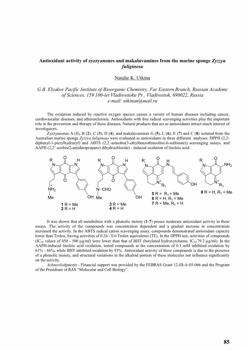
85
Antioxidant activity of zyzzyanones and makaluvamines from the marine sponge Zyzzya fuliginosa
Natalie K. Utkina
G.B. Elyakov Pacific Institute of Bioorganic Chemistry, Far Eastern Branch, Russian Academy of Sciences, 159 100-let Vladivostoku Pr., Vladivostok, 690022, Russia
e-mail: [email protected]
The oxidation induced by reactive oxygen species causes a variety of human diseases including cancer, cardiovascular diseases, and atherosclerosis. Antioxidants with free radical scavenging activities play the important role in the prevention and therapy of these diseases. Natural products that act as antioxidants attract much interest of investigators.
Zyzzyanones A (1), B (2), C (3), D (4), and makaluvamines G (5), L (6), E (7) and C (8) isolated from the Australian marine sponge Zyzzya fuliginosa were evaluated as antioxidants in three different analyses: DPPH (2,2-diphenyl-1-picrylhydrazyl) and ABTS (2,2’-azinobis(3-ethylbenzothiazoline-6-sulfonate)) scavenging assays, and AAPH (2,2’-azobis(2-amidinopropane) dihydrochloride) - induced oxidation of linoleic acid.
NN
O
O HR
NH2
OHMe
NN
O
O HR
N
OHMe
CHO
1 R = Me2 R = H
3 R = Me4 R = H
N+
NHN
O
R1
R
5 R = R1 = Me6 R = H, R1 = Me7 R = Me, R1 = H
OH N+
N NH2
O
R1
R
8 R = H, R1 = Me
It was shown that all metabolites with a phenolic moiety (1-7) posses moderate antioxidant activity in these assays. The activity of the compounds was concentration dependent and a gradual increase in concentration increased the activity. In the ABTS radical cation scavenging assay, compounds demonstrated antioxidant capacity lower than Trolox, having activities of 0.24 - 0.6 Trolox equivalents (TE). In the DPPH test, activities of compounds (IC50 values of 450 - 500 g/ml) were lower than that of BHT (butylated hydroxytoluene, IC50 79.2 g/ml). In the AAPH-induced linoleic acid oxidation, tested compounds at the concentration of 0.1 mM inhibited oxidation by 61% - 66%, while BHT inhibited oxidation by 93%. Antioxidant activity of these compounds is due to the presence of a phenolic moiety, and structural variations in the alkaloid portion of these molecules not influence significantly on the activity.
Acknowledgments - Financial support was provided by the FEBRAS Grant 12-III-A-05-066 and the Program of the Presidium of RAS “Molecular and Cell Biology”.

86
Preparation of Auricularia auricula oligosaccharides and its antioxidant activity
Yanlong Zhang, Lili Li
College of Life Science, Heilongjiang University, Harbin 150080, China e-mail: [email protected]
Auricularia auricula contains large amounts of β-1, 3–glucan. A. auricula oligosaccharides are degradated by
β-1, 3–glucanase from β-1, 3-glucan. The β-1,3-glucanase was isolated from digestive gland of scallop by precipitation with 80% saturation of (NH4)2SO4, ion-exchange on DE-52 cellulose and gel filtration on Sephadex G-150. SDS-PAGE showed that β-1,3-glucanase had a molecular mass of 157 kDa. The enzyme had optimal activity at pH 7.0 and 30oC. The enzyme had apparent Km values of 13 mg/ml by Lineweaver-Burk plot. The oligosaccharides were characterized by TLC, chromatograph of Sephadex G-100 and IR spectroscopy. Antioxidant activity of oligosaccharides from A. auricula was evaluated using 1,1-diphenyl-2-picrylhydrazyl (DPPH), hydroxyl radical and pyrogallol. The results showed that oligosaccharides from A. auricula have distinct antioxidant capacity. The oligosaccharides from A. auricula were co-cultured with bacteria, the results showed that they can stimulate the growing of Lactobacillus and inhibited Escherichia coli, Enterobacter aerogenes.

87
Proteomics of Methyl Jasmonate response in Maize leaves
Yang FengShan, Zhang YiTong ,Yang ZeZhong ,Liu ChunGuang,Zhao DanDan,Chen SiXue
Key Laboratory of Microbiology of Heilongjiang Province, College of Life Science, Heilongjiang University, Harbin 150080, China
e-mail: [email protected]
Jasmonic acid (JA) and methyl jasmonate (MJA) regulate plant development and resistance by inducing some gene expression. However, little is known about the mechanism of plants defense against herbivores attack. Here, we employed a high-throughout 2-DE to identify the MJA-responsive proteins. A total of 60 unique proteins were indentified with expression level changes. Among them, 42 proteins were unregulated and 12 proteins downregulated. There were also 8 proteins which were only expressed in response to MJA treatment. These proteins included plant defense, photosynthesis, and energy related proteins (39%),protein folding ,degradation, and regulated proteins(15%), stress and defense regulated proteins(12%) and redox-responsive proteins(8%). Further, the transcription profiles of four crucial defensive proteins were assayed by qRT-PCR. Compared with their expression levels, the changes in the two levels were not same. MJA induced plant responses which are involved in redox,stress and defense. Similarly, expression vectors of the four proteins coding genes were constructed to express in-fusion proteins. The expressed proteins were used to feed Asian corn borer and the results showed that in-fusion proteins had significant impact growth of insect. These results suggested that MJA induce defense response against insect.
Sofia A. Razgonova
Prospects to use supercritical fluid extraction method in bioorganic chemistry
, Alena I. Svetlova1, Pham Duc Thinh
Far Eastern Federal University, RussiaNhatrang Institute of Technology Research and Application, Vietnam
Extraction of biomolecules from natural sources and is the first stage of purifying this is a very important stage to further investigation. The quality of the resulting extract will influence on the number of stages of purify-ing. An Interesting method of extraction is the supercritical extraction. Supercritical fluid extraction is a new fast-paced method of processing materials. This technique has several features and advantages in compared with con-conventional methods of extraction. Very accurate extraction of the substances in accordance with the degree of polarity makes this method particularly interesting for applications in biotechnology. Alcohol extraction generally one used for these purposes, leads to the fact that excessively large amounts of extracted impurities. The analysis was conducted, and the first experimental data were obtained. This data showed that the supercritical extraction give an opportunity to obtain target-rich substance extracts. Experiments were performed on two different method of extraction of biomolecules from marine objects. The extracts were analyzed by thin layer chromatography. It has been shown that the supercritical extraction lead to extraction of compounds with small difference in polarities.
Acknowledgment:The project was supported by the Far Eastern Federal University.
88
Effects of lectins from marine invertebrates on physiological activity of microorganisms
Wei Li1, C. Tong 1, M. Qu1, Q. Jin1, P.A. Lukyanov2
1College of Food Science and Engineering, Dalian Ocean University, Dalian 116023, P. R. China
2 G.B. Elyakov Pacific Institute of Bioorganic Chemistry, Far Eastern Branch, Russian Academy of Sciences, 159 100-let Vladivostoku Pr., Vladivostok, 690022, Russia
e-mail: [email protected]
In the last decade, a large number of researchers have intensively investigated the effects of lectins from marine invertebrates on microorganisms. In these studies, several lectins from marine invertebrates showed binding activities to some bacteria and fungi. We have recently purified mucin-binding lectin from the sponge Craniella australiensis (CAL), which with a molecular mass of 54 kDa consisting of three 18 kDa subunits. Bacterial agglutination due to CAL was observed in untreated, trypsinized and acid-heat treated cells of E. coli K1. Untreated cells showed auto-agglutination partially preventing visualization of CAL-mediated agglutination. When trypsin or other proteolytic enzymes partially hydrolyse proteoglycans on the wall of E. coli K1, more carbohydrate residues become available for binding with CAL. CAL suppressed the growth of E. coli K1. A novel yeast-binding lectin was purified from hemolymph Cyclina sinensis (CSL). The carbohydrate-specificity of CSL showed that it is belong to new mannose-specific lectin. SDS-PAGE showed that the CSL protein had a molecular mass of 72 kDa, and consisted of 40 and 18 kDa subunits. CSL reveals to be a stimulator of S. cerevisiae ethanol production. CSL shows potent effect on ethanol production by S. cerevisiae, at the concentration of 59.3 μg/mL, increasing ethanol production by 13.5%。The discovery of the lectins and the demonstration that they play a crucial role in the control of physiological functions of microorganisms was a important in lectin research. Indeed, the lectins provide the best paradigm for the role of sugar-lectin interactions in biological membranes of microorganisms. These studies of effects of the lectins on microorganisms are promising for finding of new biological applications. The work was supported by grants of Natural Science Foundation of China (31071612).

89
Extraction method of fucose containing sulfated polysaccharide from the brown seaweed Fucus evanescens is important determinant for antioxidant activity development
Tatyana I. Imbs1, A.V. Skriptsova2, T.N. Zvyagintseva1, N.M. Shevchenko1 1Laboratory of Enzyme Chemistry, G.B. Elyakov Pacific Institute of Bioorganic Chemistry, Far
Eastern Branch, Russian Academy of Sciences 2A.V. Zhirmunsky Institute of Marine Biology, Far Eastern Branch, Russian Academy of
Sciences
Brown seaweeds contain fucoidans. Fucoidans designate a group of certain fucose-containing sulfated polysaccharides (FCSPs) that are represent the mixtures of structurally related polysaccharides with certain variations of carbohydrate units and non-carbohydrate substituents (mainly sulfate and acetyl groups) content. These FCSPs exhibit several potentially beneficial bioactive functions for humans. The bioactive properties may vary depending on the source of seaweed, collection time, the compositional and structural traits, the content of the sulfate groups, and the purity of the FCSP product. The presence of impurities influences the biological properties of FCSPs and therefore may currently hinder our full understanding of the biological activity of fucoidan or FCSPs. The preservation of the structural integrity of the FCSP molecules essentially depends on the extraction methodology which has a crucial significance for obtaining the relevant structural features required for specific biological activities.
FCSPs were isolated from F. evanescens using two slightly different procedures. The FCSP-1 was obtained via hot acidic extraction, followed by dialization and ethanol precipitation. Alternatively, the FCSP-2 was obtained via extraction with a 2% aqueous calcium chloride solution, followed by ultrufiltration and ethanol precipitation. Then FCSP-1 and FCSP-2 were fractionated by anion-exchange chromatography on DEAE-cellulose into four (1F1-1F4) and three (2F1-2F3) fractions, respectively.
The composition of FCSP-1 and FCSP-2 and the most highly purified fractions 1F4 and 2F3 are given in Table (fractions 1F1-1F3 and 2F1, 2F2 data not shown). The results show that the FCSP-1 obtained by hot acidic extraction method has a higher uronic acid and phenol content in comparison with FCSP-2 extracted by calcium chloride. Therefore, the extraction with 2% aqueous calcium chloride solution provides a purified preparation of the FCSP.
The antioxidant capacity values for the crude preparations and purified fractions were measured by DPPH, ABTS and Phosphomolybdenum (Mo) assays. The antioxidant capacity and the phenol content of the FCSP-2 and especially 2F3 were lower than that of FCSP-1 and fractions 1F4. This fact may indicate that phenols play a key role in the previously described antioxidant capacity of FCSPs. To investigate if the antioxidant activity of the FCSPs was due to the presence of phenolic compounds, correlations between phenolic content and antioxidant activities (DPPH, ABTS and Mo assays) were established. The results show a linear relationship between phenolic content and antioxidant activities DPPH (for FCSP-1: r2=0.987; for FCSP-2: r2=0.989), ABTS (for FCSP-1: r2=0.902). The UV-absorbance at 280 nm of FCSPs correlated well with the concentration of phenolic compounds (for FCSP-1: r2=0.981; for FCSP-2: r2=0.987). Fraction Assay Protein
(%), Lowry method
Protein (%), amino acid
Phenol,mgPhl Eq/g 3
Fuc: SO3Na
Neutral monosaccharide’s (%) Mo assaymgAc/g1
ABTS
assay,
SA2 (%)
DPPHassay, SA (%)
Fuc Gal Man Xyl Glc UAc
FCSP-1 51.0±1.0
11.0±0.27
57.6±3.04
12.5 1.2 33.29± 0.86
1:1.19 1.0 0.1 0.02 0.02 0.45 0.2
1F4 44.0±0.8
10.1±0.05
39.1±1.60
10.2 0.9 21.55± 0.67
n.d. 1.0 0.03 0 0.01 0 0.05
FCSP-2 8.31±0.57
n.d 19.43±0.48
4.23 1.2 10.23± 1.6
1:0.8 1.0 0.15 0.02 0.08 0.02 0.02
2F3 1.28±0.19
n.d 3.75±0.35
0.85 0.8 1.42± 0.16
1:1.2 1.0 0.03 0 0.01 0 0
1 mg ascorbic acid equivalents per sample dry weight; 2 SA – scavenging activity of 1 mg/mL concentration of polysaccharide; 3mg ploroglucinol equivalents per sample dry weight;

90
Cloning and expression of fucoidanases from the marine bacterium Formosa algae KMM 3553
Artem Silchenko 1,2, Mikhail Kusaykin 1, Alexander Zakharenko 1,2
1 G.B. Elyakov Pacific Institute of Bioorganic Chemistry Far Eastern Branch, Russian Academy of Sciences
2 Far Eastern Federal University [email protected]
Fucoidans belong to a family of sulphated homo- and heteropolysaccharides, including polysaccharides composed primarily of sulphated fucose. Brown algae synthesise highly branched polysaccharides with species–specific sugar compositions, and for this reason, fucoidan structures are extremely diverse. In addition, each species can form different types of fucoidans. The most studied fucoidans in terms of structure are α-L-fucans with either -13-backbones or repeating disaccharide units of -13- and -14-linked fucose residues. Depending on the structure of the main chain, fucoidans may be sulphated at the C4, C2 or both the C2 and C4 positions of the fucose units.Fucoidans from some seaweed species have recently started to be used as nutraceuticals in Australia, Japan and the United States. They have diverse biological activities, including anti-tumour, immunomodulatory, antibacterial, antiviral, anti-inflammatory, anticoagulant, and antithrombotic effects. The data reported in the literature show that fucoidans possess a wide spectrum of pharmacological activities; however, they cannot be successfully used in the construction of new drugs because of their high molecular weight and significant problems with polysaccharide standardisation. One approach to solving this problem is the use of enzymes to depolymerise the fucoidans.
The strain KMM 3553 was chosen as a producer of fucoidanase basis on screening results. The genome was sequenced by Illumina sequencing platform. Two genes encoded fucoidanases (FFA1, FFA2) were found. The products of FFA1 and FFA2 genes were a proteins with predicted molecular weight 111,1 kDa and 97,9 kDa respectively. BLASTp analysis showed 67 and 57 percent identity of FFA1 and FFA2 respectively with known fucoidanase FcnA from the marine bacterium Mariniflexile fucanivorans SW5 (NCBI DB). InterProScan searches detected three (for FFA1) and two (for FFA2) matching of repeated cadherine-like domains. The signal sequence in N-terminal region of the FFA1 fucoidanase was identified which indicate that this enzyme is extracellular as opposed to fucoidanase FFA2 in which this sequence was not found. Fucoidanases was expressed in three different forms. The first forms encoded full-length sequence of fucoidanases FFA1 and FFA2. The second constructs encoded truncated sequence without C-terminal regions (FFA1-SD, FFA2-SD). The third constructs (FFA1-KD, FFA2-KD) contain N-terminal domain only, without predicted cadherin-like domains. The soluble products were obtained for the first and second constructs for each fucoidanases. The products of third constructs were unstable and expressed both soluble (30%) and insoluble (70%) forms. Analysis of fucoidanalytic activities of soluble fractions of recombinant fucoidanases by C-PAGE indicated presence of the fucoidanase activity for all three types of the products of gene constructs (Fig. 1). Full-length fucoidanases (FFA1 and FFA2) catalyzed hydrolysis of fucoidan from Fucus evanescens containing -13- and -14-linked sulphated fucose residues but not fucoidans from Saccharina cichorioides and Undaria pinnatifida consisted of -13- linked sulphated fucose residues. This data indicate that fucoidanases (FFA1 and FFA2) able to cleavage of -14 glycosidic bounds in fucoidan molecule (Fig. 2).
Acknowledgment: We are grateful to Prof. Nils Peder Willassen and Dr. Bjerga Gro Elin Kjæreng for help in bacterial genome sequencing, annotation and design of gene constructs.
Fig. 1 – Electropherogramm of hydrolysis products of the fucoidan from F. evanescens by recombinant fucoidanases. The numbers above the lines refer to different constructs of fucoidanase: 1- FFA1, 2- FFA1-SD, 3- FFA1-KD, 4- FFA2, 5- FFA2-SD, 6- FFA2-KD; Ks – unhydrolyzed fucoidan with boiled recombinant fucoidanase.
Fig. 2 – Electropherogramm of hydrolysis products of the fucoidans from F. evanesces (F.ev), S. cichorioides (S.c), U. pinnatifida (Und.) by full-length recombinant fucoidanases FFA1 and FFA2; Ks – unhydrolyzed fucoidans.

91
Comparative Study of Adjuvant Properties of Triterpene Glycosides from Sea Cucumber
and Minor Ginseng Glycoside Rh2
Alexander M. Popov 1,2, O.N. Krivoshapko2, A.V. Tsibulsky2 1 G.B. Elyakov Pacific Institute of Bioorganic Chemistry, Far Eastern Branch, Russian Academy
of Sciences, 159 100-let Vladivostoku Pr., Vladivostok, 690022, Russia 2Far Eastern State University, Vladivostok, Russia
e-mail: [email protected]
Adjuvant activity of trepangin (sum of holotoxins A and B from Apostichopus japonica, Selenka), preparation KD (fraction of triterpene glycosides isolated from Cucumaria japonica) and minor glycoside Rh2 from Panax ginseng Meyer were investigated. The antibody responses were studied in 6-8-week-old syngenic CBA mice, immunized by corpuscular antigen, depending on the route of administration, doze and type of an adjuvant. Control and tested animals were vaccinated intraperitonially (i.p.) by 1107 cells of sheep erythrocytes. Simultaneously, adjuvants were injected per os (р.о.), i.p. or subcutaneously (s.c.). The antibody response to antigen was determined by passive hemagglutination response.
The immune response was boosted by combined administration of antigen and trepangin depending on the route of administration and the adjuvant dose. Both i.p. and s.c. injections of this agent were effective at the dose of 0,1 mg/kg. The maximal stimulation of the antibody formation (4-6 times in comparison with the control) was observed within 2 weeks with the consequent decrease by third week in all tested groups except for group, treated trepangin (i.p.) at the dose of 0,01 mg/kg. The highest antibody level was observed in the case i.p. or s.c. injections by 5th-day or 14th-day after immunization, respectively. S.c. administration of trepangin was resulted in the identical change of antibody formation depending on doses. The level of humoral response in animals, treated by KD, increased 4-8 times in comparison with control group. The dependence of the antibody level on KD doses (0,1 and 0,01 mg/kg) and on the route of administration was expressed insignificantly. The peak of the immune response was observed in animals, i.p. treated by low dose (0,01 mg/kg) of KD by 4th-day after immunization. Similarly with the immune stimulation by trepangin, KD preparation decreased the immune response by the end of the third week. Injection p.o. was the most optimal for minor ginseng glycoside Rh2 at the doses widely ranging from 0,5 up to 10 mg/kg. The specific antibody formation increased 5-8 times in comparison with the control. The comparable effect was shown at i.p. stimulation by this glycoside at dose of 2 mg/kg. The least profound effect (2-3 times less in comparison with the control) was observed at s.c. administration of Rh2. It was interesting, that dynamics of the immune response booster was identical in all groups. Maximal peak was observed during two weeks after immunization with the consequent decrease by 21th-day. It was also shown, that the investigated agents possessed the essential immunostimulating activity in relation to immunocompetent cells in vitro. The spleen B- and T-lymphocyte proliferation and phagocytosis indexes of the mice peritoneal macrophages increased. The immunostimulating mechanisms of KD and trepangin, on the one hand, and Rh2, on the other hand, are well differ. We suppose that, similarly to triterpene glycosides from Quillaja saponaria Molina, triterpene glycosides from Apostichopus japonica Selenka (trepangin) and Cucumaria japonica (KD preparation) are capable to form the complex with cholesterol, which provide the matrix of immune stimulating complexes (ISCOMs). The minor glycoside Rh2 from Panax ginseng is unable to bind cholesterol. Therefore we hypothesized that Rh2 affects the immunocompetent cells by means of the membrane mechanism preliminary signed as "soft stress" and could be used as a perspective and safe adjuvants with different mode of action for the design of various effective vaccine preparations.

92
Soluble polyelectrolyte carrageenan: chitosan complexes and their gastroprotective activity
Alexandra V. Volodko1, V.N. Davydova1, V.P. Glazunov1, E.A. Chusovitin2, I.V. Sorokina3, I.M. Yermak1
1 G.B. Elyakov Pacific Institute of Bioorganic Chemistry, Far Eastern Branch, Russian Academy of Sciences, 159 100-let Vladivostoku Pr., Vladivostok, 690022, Russia
e-mail: [email protected] 2Institute of Automation and Control Processes of Far Eastern Branch of Russian Academy of
Sciences, 5 Radio Street, Vladivostok, Russia, 690041 3 Novosibirsk Institute of Organic Chemistry, Siberian Branch of the Russian Academy of
Sciences, Lavrentjev Ave. 9, Novosibirsk, Russia, 630090
Chitosan and carrageenan, marine polysaccharides, possess versatile biological activity and various physicochemical properties. Due to their polyionic structure, chitosan and carrageenan can be used for the creation of various polyelectrolyte complexes (PEC), which give the possibility of creating medical preparation with specific properties.
The process of kappa-carrageenan:chitosan complexes formation and possibility of their use as a basis of soluble composites for biomedical application as gastroprotective and antiulcer substance have been studied. Polycationic chitosan (Ch) with acetylation degree of 6% and molecular weight (MW) 110 kDa and kappa-carrageenan (Car) from C. armatus with MW 311 kDa were used for the PECs formation.
Using the turbidimetric method we have shown that the soluble complexes of Car (0.5 mg/ml) with Ch formed in the range of the Ch:Car ratio from 0.1:1 to 1.5:1 mol/mol. For further study two different complexes Car with Ch have been prepared at Car:Ch ratios 10:1 and 1:10 w/w.
The IR spectroscopy was used for confirmation the formation of soluble Ch-Car PEC. The appearance of a new absorption band at 1540±5 cm-1 and reduction of intensity of the absorption band of sulphate groups in the spectrum of Ch:Car complex confirmed out the formation of strong PEC.
The high-speed sedimentation in a percoll gradient was another method to prove the complex formation. The formation of two types of complexes was observed by the coincidence of curves in the lower and middle parts of the gradient. In addition, part of the initial polysaccharides remained free.
The supramolecular structure of these complexes was investigated by atomic force microscopy. The macromolecular structure of complex Ch:Car was different from the initial components. That was additional evidence for complex formation. The structures similar with the structures of initial polysaccharides also were fixed. That was observed by high-speed sedimentation in a percoll gradient.
The gastroprotective and antiulcer activity of Car, Ch and their complexes (1:10 and 10:1 w/w) was investigated in vivo using indometacin ulcer model in rats. Comparative analysis of the initial polysaccharides activity showed that κ-carrageenan (4.55 mg/kg) possesses its own gastroprotective activity (reducing ulceration by 3 times compared to the control); whereas chitosan at the same dose did not have a significant protective effect (reduction factor was 1.5). The complex Car:Ch 10:1 w/w didn’t increase gastroprotective effect but the complex Car:Ch 1:10 w/w led to an increase in gastroprotective effect. The results suggest that gastroprotective activity of the complexes might be related to their role in creating a protective layer on the surface of the gastric mucosa.