
Tennis Elbow- Lateral Epicondylitis CAITLIN GARDINER.
14
Tennis Elbow- Lateral Epicondylitis CAITLIN GARDINER
-
Upload
tyrone-johns -
Category
Documents
-
view
234 -
download
3
Transcript of Tennis Elbow- Lateral Epicondylitis CAITLIN GARDINER.

Tennis Elbow- Lateral EpicondylitisCAITLIN GARDINER

Anatomy
Image From www.hss.com
-The Common Extensor Origin is a tendinous insertion onto the lateral epicondyle of the humerous of
-Extensor Digiti Minimi-Extensor Carpi Radialis
Brevis-Extensor Carpi Ulnaris -Extensor Digitorum
Communis (1)

Physiology
Repetitive traction on the osteotendinous attachment of the common extensor tendon
Results in microtrauma to the tendon causing progressive degeneration and/or partial tears or damage to the bony insertion (enthesopathy)
Angiofibroblastic infiltration occurs due to migration of vascular granulation
Crystal deposition may occur
Early abnormalities are confined to superficial fibres
Most commonly the extensor carpi radialis brevis deep fibres are involved (1,2)

Presentation
Gradual onset of symptom
Pain at the location of the lateral epicondyle
Generally a sharp pain exacerbated with gripping activities
Pain in the morning and at times when the wrist is held in flexion
Inability to grip objects.
Reduced strength with resisted grip, supination and extension of the wrist (3)
Two simple clinical tests include Chair test: Patient is asked to lift themselves out of a chair with a pronated hand.
Pain at lateral epicondyle may indicates Tennis elbow
Coffee cup test: Patient is asked to hold a cup in their hand. Pain at the lateral elbow may indicates Tennis Elbow (4)

Common Causes
Can occur at any age, but typically between the ages of 40-50
Any activity involving repetitive use of extensor muscle of the forearm
Particularly if gripping with the thumb and first two finger
Can occur in tennis players, due to extension during back stroke
Commonly occurs in occupations with highly repetitive hand tasks (4)

Symptoms
Tendinopathy: Localised pain over the CET during or after repetitive muscle activation, radiating down the lateral aspect of the elbow
Enthesopathy: Pain is confined to insertional area
Pain is exacerbated with radial deviation and pronation with the elbow extended particularly when performed with resistance (1,4)

Ultrasound Findings
Pre-insertional hypoechoic swelling
Focal or diffuse areas of decreased reflectivity
Loss of fibrillary pattern related to tendinosis
Fluid adjacent to the tendon
Ill-defined margins of the tendon
Hypervascularity in high-grade tendinosis
Spurring and cortical irregularities (1, 4, 5)

Case Presentation
Presentation: A 56 year-old male presents with severe pain of his left lateral elbow for the past three weeks. He recently began a new job where he unpacked large boxes. He finds opening the boxes, which requires significant force, increasingly painful.
Ultrasound Technique:
Use a high-frequency linear probe and/or a hockey-stick probe with the appropriate pre-set. When using colour Doppler, ensure that gain is adjusted just prior to aliasing. Be prepared to alter the patient arm to view maximum hyperaemia and apply very minimal pressure.
The patient is positioned across from the sonographer with his arms extended onto the table, with hands in a ‘praying’ position. This allows the CEO to be imaged in a lateral coronal position and has the contralateral arm in the same position for comparison (2)

Ultrasound Findings
-Thickening of the tendon-Loss of reflection of the deep fibres-Distortion of fibrillary pattern-No enthesopathy noted
-Marked hyperaemia of the deep fibres

Partial tear vs. Tendinosis
Partial tears: The CEO may appear thinned
Longitudinal splits oriented from the distal bony insertion
Anechoic cleavage planes with no fibres visible
Tendinosis Diffusely or focal (usually deep) hypoechoic
Usually has some associated hyperaemia (1)

Alternative Diagnosis
Posterior Interosseous nerve entrapment/Radial Tunnel Syndrome
Radial Humeral bursitis
Lateral Collateral Ligament injury
Dysfunction of the cervical spine at levels C5-6 or C6-7 referring as elbow pain
Osteochondral radiocapitellar lesion (1, 3)
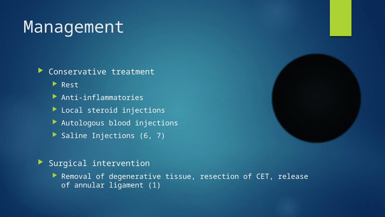
Management
Conservative treatment Rest
Anti-inflammatories
Local steroid injections
Autologous blood injections
Saline Injections (6, 7)
Surgical intervention Removal of degenerative tissue, resection of CET, release of annular
ligament (1)

Alternative Imaging Modalities
Usually clinical diagnosis is suffice
Imaging reserved for complicated cases, assisting injections, progression of damage
MRI shows sensitivity of MRI between 90-100% for Tennis Elbow. Also able to assess intra-articular processes, radial collateral ligament and to grade the tear of the extensor origin
Ultrasound shows high sensitivity but low specificity in detection of lateral epicondylitis in symptomatic cases (1) Sensitivity ranging from 72-88%
Specificity ranging from 36-48.5% (3)
References
1. Bianchi S and Martinoli C, 2007. Ultrasound of the Musculoskeletal System. Springer, New York.
2. McNally EG, 2005. Practical Musculoskeletal Ultrasound. Elsevier, London.
3. Walz DM, Newman JS, Konin GP, Ross G. Epicondylitis: Pathogenesis, imaging, and treatment. Radiographics. 2010;30(1):167-184
4. Levin D, Nazarian LN, Miller TT, et al. Lateral epicondylitis of the elbow: US findings. Radiology. 2005;237(1):230-234
5. Lin CL, Lee JS, Su WR et al, 2011. Clinical and Ultrasonographic Results of Ultrasonographically Guided Percutaneous Radiofrequency Lesioning in the Treatment of Recalcitrant Lateral Epicondylitis. Am J Sports Med; Aug 11.
6. Wolfe JM, Oxer K, Scott F et al, 2011. Comparison of Autologous Blood, corticosteroid, and Saline Injections in the Treatment of Lateral EpicondylitisL a Prospective, Radomized, Controlled Multicenter Study. J Hand Surg Am; 36(8):1269-72.
7. Altan L and Kanat E, 2008. Conservative Treatment of Lateral Epicondylitis: Comparison of Two Different Orthotic Devices. Clin Rheymatol; 27(8): 1015-1019.


![Elbow Pain - Welcome To | HealthSharehealthshare.org.uk/HS_leaflets/Elbow_Pain.pdf[ 3 ] Elbow Pain Lateral epicondylitis What is lateral epicondylitis? This is often referred to as](https://static.fdocuments.net/doc/165x107/5ac8bc037f8b9a51678c93c8/elbow-pain-welcome-to-he-3-elbow-pain-lateral-epicondylitis-what-is-lateral.jpg)

