
Subtle elbow instability associated with lateral epicondylitis
7
RESEARCH ARTICLE Open Access Subtle elbow instability associated with lateral epicondylitis Sang Ho Kwak 1 , Seung-Jun Lee 1* , Hee Seok Jeong 2 , Min Uk Do 1 and Kuen Tak Suh 1 Abstract Background: In lateral epicondylitis, even in the absence of apparent instability, subtle instability can be found under anesthesia. We wanted to ascertain the following: (1) how many elbows surgically treated with lateral epicondylitis showed subtle instability during examination under anesthesia (EUA), (2) how effective magnetic resonance imaging (MRI) was in predicting subtle instability, and (3) if any difference existed in preoperative clinical data between elbows with and without subtle instability during EUA. Methods: One hundred and twenty-two elbows (117 patients) diagnosed with intractable lateral epicondylitis underwent surgical treatment. No elbow showed apparent instability with conventional physical examination. Under general anesthesia, the elbows were examined for subtle instability via fluoroscopy and divided into unstable and stable groups. Potential prognostic factors and functional scores were assessed retrospectively. The MRIs were reviewed again by two radiologists. Results: Seventeen elbows (unstable group, 13.9%) had subtle instability in EUA, while 105 elbows (stable group, 86.1%) did not. Lateral collateral ligament (LCL) complex injury was noted in the MRIs of 28 elbows. Fifteen elbows showed subtle instability among 28 elbows with abnormal MRI (positive predictive value, 53.6%), while 81 elbows did not show subtle instability among 82 elbows with normal MRI (negative predictive value, 98.7%). The preoperative visual analog scale score was higher in the unstable group than in the stable group (p < 0.001), and a history of multiple corticosteroid injections (≥3) was related to subtle instability in EUA (p = 0.042). Other factors showed no significant differences between both groups. Conclusions: Subtle instability resulting from LCL complex injury was noted in elbows with lateral epicondylitis. This could be visualized with fluoroscopic EUA, and preoperative MRI could be used to exclude subtle instability. Surgeons should consider checking for subtle instability, especially when patients have a history of multiple corticosteroid injections (≥3) or severe pain and MRI indicates instability. Keywords: Lateral, Epicondylitis, Tendinosis, Ligament, Instability, Elbow Background Lateral epicondylitis is usually diagnosed based on clin- ical history and physical examination. Excluding condi- tions that can mimic lateral epicondylitis is very important because symptoms cannot be fully relieved if such lesions are neglected. For a differential diagnosis, multiple modalities including simple radiography, ultra- sonography, magnetic resonance imaging (MRI), and electrodiagnosis can be used. Associated ligament injuries in lateral epicondylitis were reported using MRI or via conventional physical examin- ation. MRI can be used for differentiating other pathologic conditions such as plica, elbow arthritis, and osteochon- dral defect. In addition, concomitant ligament injuries in- volving the medial collateral ligament (MCL), lateral collateral ligament (LCL), and lateral ulnar collateral liga- ment (LUCL) have also been observed in MRI-diagnosed lateral epicondylitis [1, 2]. Associated ligament injuries can induce elbow instability eventually, and posterolateral rotatory instabilities after trauma, corticosteroid injection, and iatrogenic injury during debridement for lateral epi- condylitis have been reported [3, 4]. In these reports, most * Correspondence: [email protected] 1 Department of Orthopaedic Surgery, Pusan National University Yangsan Hospital, Pusan National University School of Medicine, 20, Geumo-ro, Mulgeum-eup, Yangsan-si, Gyeongsangnam-do, Republic of Korea Full list of author information is available at the end of the article © The Author(s). 2018 Open Access This article is distributed under the terms of the Creative Commons Attribution 4.0 International License (http://creativecommons.org/licenses/by/4.0/), which permits unrestricted use, distribution, and reproduction in any medium, provided you give appropriate credit to the original author(s) and the source, provide a link to the Creative Commons license, and indicate if changes were made. The Creative Commons Public Domain Dedication waiver (http://creativecommons.org/publicdomain/zero/1.0/) applies to the data made available in this article, unless otherwise stated. Kwak et al. BMC Musculoskeletal Disorders (2018) 19:136 https://doi.org/10.1186/s12891-018-2069-8
Transcript of Subtle elbow instability associated with lateral epicondylitis

RESEARCH ARTICLE Open Access
Subtle elbow instability associated withlateral epicondylitisSang Ho Kwak1, Seung-Jun Lee1*, Hee Seok Jeong2, Min Uk Do1 and Kuen Tak Suh1
Abstract
Background: In lateral epicondylitis, even in the absence of apparent instability, subtle instability can be foundunder anesthesia. We wanted to ascertain the following: (1) how many elbows surgically treated with lateralepicondylitis showed subtle instability during examination under anesthesia (EUA), (2) how effective magneticresonance imaging (MRI) was in predicting subtle instability, and (3) if any difference existed in preoperative clinicaldata between elbows with and without subtle instability during EUA.
Methods: One hundred and twenty-two elbows (117 patients) diagnosed with intractable lateral epicondylitisunderwent surgical treatment. No elbow showed apparent instability with conventional physical examination.Under general anesthesia, the elbows were examined for subtle instability via fluoroscopy and divided into unstableand stable groups. Potential prognostic factors and functional scores were assessed retrospectively. The MRIs werereviewed again by two radiologists.
Results: Seventeen elbows (unstable group, 13.9%) had subtle instability in EUA, while 105 elbows (stable group,86.1%) did not. Lateral collateral ligament (LCL) complex injury was noted in the MRIs of 28 elbows. Fifteen elbowsshowed subtle instability among 28 elbows with abnormal MRI (positive predictive value, 53.6%), while 81 elbowsdid not show subtle instability among 82 elbows with normal MRI (negative predictive value, 98.7%). Thepreoperative visual analog scale score was higher in the unstable group than in the stable group (p < 0.001), and ahistory of multiple corticosteroid injections (≥3) was related to subtle instability in EUA (p = 0.042). Other factorsshowed no significant differences between both groups.
Conclusions: Subtle instability resulting from LCL complex injury was noted in elbows with lateral epicondylitis.This could be visualized with fluoroscopic EUA, and preoperative MRI could be used to exclude subtle instability.Surgeons should consider checking for subtle instability, especially when patients have a history of multiplecorticosteroid injections (≥3) or severe pain and MRI indicates instability.
Keywords: Lateral, Epicondylitis, Tendinosis, Ligament, Instability, Elbow
BackgroundLateral epicondylitis is usually diagnosed based on clin-ical history and physical examination. Excluding condi-tions that can mimic lateral epicondylitis is veryimportant because symptoms cannot be fully relieved ifsuch lesions are neglected. For a differential diagnosis,multiple modalities including simple radiography, ultra-sonography, magnetic resonance imaging (MRI), andelectrodiagnosis can be used.
Associated ligament injuries in lateral epicondylitis werereported using MRI or via conventional physical examin-ation. MRI can be used for differentiating other pathologicconditions such as plica, elbow arthritis, and osteochon-dral defect. In addition, concomitant ligament injuries in-volving the medial collateral ligament (MCL), lateralcollateral ligament (LCL), and lateral ulnar collateral liga-ment (LUCL) have also been observed in MRI-diagnosedlateral epicondylitis [1, 2]. Associated ligament injuriescan induce elbow instability eventually, and posterolateralrotatory instabilities after trauma, corticosteroid injection,and iatrogenic injury during debridement for lateral epi-condylitis have been reported [3, 4]. In these reports, most
* Correspondence: [email protected] of Orthopaedic Surgery, Pusan National University YangsanHospital, Pusan National University School of Medicine, 20, Geumo-ro,Mulgeum-eup, Yangsan-si, Gyeongsangnam-do, Republic of KoreaFull list of author information is available at the end of the article
© The Author(s). 2018 Open Access This article is distributed under the terms of the Creative Commons Attribution 4.0International License (http://creativecommons.org/licenses/by/4.0/), which permits unrestricted use, distribution, andreproduction in any medium, provided you give appropriate credit to the original author(s) and the source, provide a link tothe Creative Commons license, and indicate if changes were made. The Creative Commons Public Domain Dedication waiver(http://creativecommons.org/publicdomain/zero/1.0/) applies to the data made available in this article, unless otherwise stated.
Kwak et al. BMC Musculoskeletal Disorders (2018) 19:136 https://doi.org/10.1186/s12891-018-2069-8

of the patients showed apparent symptoms including in-stability via conventional physical examination, feeling of“pop,” or prominent swelling of the elbow joint [3, 4].However, Kalainov revealed that one patient showed un-specific physical examination and the posterolateral in-stability was detected only during general anesthesia [5].Morrey et al. described lax LCL and LUCL found in fluor-oscopy under local anesthesia or arthroscopic examinationas subtle instability, which the authors reported as causesof refractory lateral epicondylitis [4]. Thus, even thoughconventional physical examination may not reveal associ-ated ligament injuries, subtle instability can be foundunder specific examination with anesthesia. However, re-ports of subtle instability found in primary surgery for lat-eral epicondylitis are very rare, and except for one case[5], only one study reported about subtle instability underanesthesia and suggested a treatment algorithm [6].In this study, we examined elbow instability in fluoros-
copy during examination under anesthesia (EUA) andaimed to report the elbows that needed primary surgicalinterventions for lateral epicondylitis: (1) how many el-bows had subtle instability, (2) how closely the EUAfindings matched the MRI and operative findings in el-bows with subtle instability, and (3) whether any differ-ences existed regarding preoperative clinical databetween elbows with and without subtle instability.
MethodsThis was a retrospective case series study. After approval byour institutional board review (IRB number 05–2017-028),173 consecutive elbows (168 patients) with lateral epicondyl-itis treated surgically between March 2011 to December2016 were enrolled in this study. Definitive criteria for lateralepicondylitis included 1) pain at the elbow during the pre-ceding 30 days and 2) pain at the lateral humeral epicondyleregion and pain provoked by resisted extension of the wristwith the elbow extended [7]. All patients were treated with acombination of four conservative methods including NSAIDadministration, counterforce bracing, isometric exercise, andextensor muscle stretching. Injections including corticoster-oid, autologous blood, or botulinum toxin were not given inour protocol. The surgical indication was intractable pain(visual analog scale [VAS] ≥ 4) after conservative treatmentfor at least 6 months. Simple radiographs were taken at ini-tial diagnosis, and after deciding to treat with surgery, MRIswere performed. Elbows with previous trauma, includingelbow dislocation and fracture (n = 17); deformities, includ-ing cubitus varus and valgus (n= 11); synovial plica andosteochondral defect on MRI (n = 5); and previous surgeryfor lateral epicondylitis (n = 7) were excluded. The mediolat-eral stress, posterolateral rotatory drawer, push-up, andtabletop tests were conducted again by two orthopedic spe-cialists (KSH, LSJ) before surgery. If at least one examinerfound any instability in these examinations, the patients
(n= 8) were considered to have instability during conven-tional physical examination and excluded. None of thepatients showed generalized ligament laxity (Beighton score< 4) [8]. Patients detected with synovial plicae intraopera-tively (n= 3) were also excluded. Finally, 122 elbows (117patients) were included in this study.
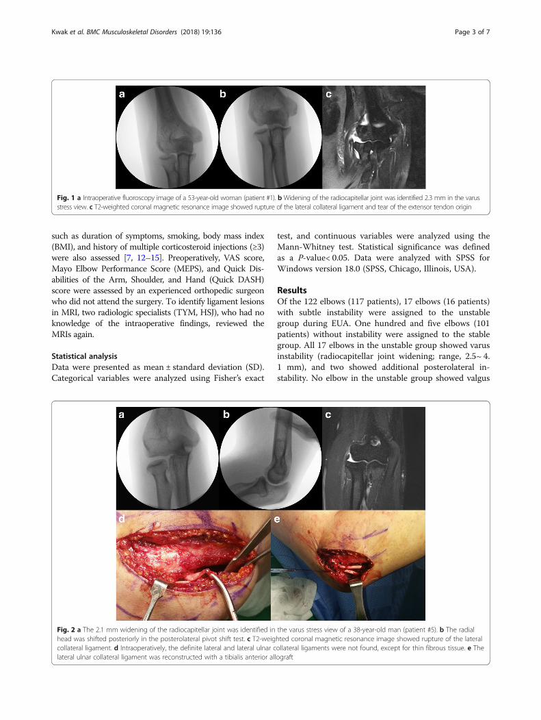
Identifying instability during EUAThe patients were placed in the supine position under gen-eral anesthesia. For valgus instability, the arm was posi-tioned with the elbow in 30° flexion, humerus in fullinternal rotation, and forearm in pronation. Then, the man-ual valgus stress test was performed, and an anteroposterior(AP) image of the elbow was acquired using a fluoroscopicimage intensifier [9]. A widening of more than 1 mm at theulnohumeral joint was considered as subtle valgus instabil-ity [9]. For varus instability, the arm was positioned withthe elbow in 15° flexion, humerus in full external rotation,and forearm in supination. Then, manual varus stress testwas performed, and an AP fluoroscopic image was ac-quired. Since a previous study reported that the radiocapi-tellar joint is 0.47 mm more redundant compared with theulnohumeral joint [10], a widening of over 1.5 mm at theradiocapitellar joint was considered as subtle varus instabil-ity (Fig. 1). In addition, a posterolateral pivot shift test wasperformed with the elbow in 90° flexion and forearm in su-pination [11]. If the longitudinal axis of the radius does notpass through the center of the capitellum, it was consideredas a posterolateral instability [9]. Thereafter, a lateral imageof the elbow was acquired to identify subtle posterolateralinstability (Fig. 2a-c) [5]. If subtle instability was found, thecontralateral asymptomatic elbow was examined to excludeunderlying normal laxity of elbow joint.
Surgical exploration of ligament statusDuring surgery, intraarticular pathology was checked viaarthrotomy, and 3 patients with synovial plicae were ex-cluded. For patients without subtle instability, we per-formed open release of lateral epicondylitis. The LCLcomplex was not explored to minimize damage to theextensor tendon. For patients with subtle instability,additional exploration for unstable ligament was per-formed. For varus or posterolateral instability, the prox-imal LCL complex was explored by elevating thecommon extensor origin, and the distal LCL complexwas explored by elevating the anconeus and extensorcarpi ulnaris. For valgus instability, the MCL was ex-plored via an additional medial longitudinal incision.
Preoperative clinical data and MRI evaluationDemographic data that were considered as potential prog-nostic factors such as patient age, gender, duration of edu-cation, and involvement of the dominant hand wereassessed retrospectively. Other potential prognostic factors
Kwak et al. BMC Musculoskeletal Disorders (2018) 19:136 Page 2 of 7
such as duration of symptoms, smoking, body mass index(BMI), and history of multiple corticosteroid injections (≥3)were also assessed [7, 12–15]. Preoperatively, VAS score,Mayo Elbow Performance Score (MEPS), and Quick Dis-abilities of the Arm, Shoulder, and Hand (Quick DASH)score were assessed by an experienced orthopedic surgeonwho did not attend the surgery. To identify ligament lesionsin MRI, two radiologic specialists (TYM, HSJ), who had noknowledge of the intraoperative findings, reviewed theMRIs again.
Statistical analysisData were presented as mean ± standard deviation (SD).Categorical variables were analyzed using Fisher’s exact
test, and continuous variables were analyzed using theMann-Whitney test. Statistical significance was definedas a P-value< 0.05. Data were analyzed with SPSS forWindows version 18.0 (SPSS, Chicago, Illinois, USA).
ResultsOf the 122 elbows (117 patients), 17 elbows (16 patients)with subtle instability were assigned to the unstablegroup during EUA. One hundred and five elbows (101patients) without instability were assigned to the stablegroup. All 17 elbows in the unstable group showed varusinstability (radiocapitellar joint widening; range, 2.5~ 4.1 mm), and two showed additional posterolateral in-stability. No elbow in the unstable group showed valgus
Fig. 1 a Intraoperative fluoroscopy image of a 53-year-old woman (patient #1). bWidening of the radiocapitellar joint was identified 2.3 mm in the varusstress view. c T2-weighted coronal magnetic resonance image showed rupture of the lateral collateral ligament and tear of the extensor tendon origin
Fig. 2 a The 2.1 mm widening of the radiocapitellar joint was identified in the varus stress view of a 38-year-old man (patient #5). b The radialhead was shifted posteriorly in the posterolateral pivot shift test. c T2-weighted coronal magnetic resonance image showed rupture of the lateralcollateral ligament. d Intraoperatively, the definite lateral and lateral ulnar collateral ligaments were not found, except for thin fibrous tissue. e Thelateral ulnar collateral ligament was reconstructed with a tibialis anterior allograft
Kwak et al. BMC Musculoskeletal Disorders (2018) 19:136 Page 3 of 7

instability under EUA (ulnohumeral joint widening;range, 0.13~ 0.66 mm). Except for 1 patient in the un-stable group (patient 2), there was no patient whoshowed subtle instability on the contralateral asymptom-atic elbow. In 105 elbows without subtle instability, theradiocapitellar and ulnohumeral joint widening wasmeasured, ranging from 0.45 to 1.32 mm and 0.15 to 0.75 mm, respectively.One elbow in the unstable group and 11 elbows in the
stable group did not undergo MRI for economic reason.Among 16 elbows which underwent MRI in the unstablegroup, 4 LCL + LUCL injuries, 6 LUCL injuries, and 5LCL injuries were found. In 1 patient (patient 8), therewas no notable ligament tear except for signal change onMRI taken preoperatively. In the stable group, 94 elbowsunderwent MRI, and 8 LCL and 5 LUCL injuries werenoted on MRI. A combined LCL + LUCL injury was notidentified. In both groups, no MCL injury was notedwith MRI. Of all 28 elbows with LCL complex injurieson MRI, 15 showed subtle instability with EUA. Intraop-eratively, in the unstable group, LCL complex abnormal-ities were confirmed at surgery in all cases; nofunctioning LCL and LUCL structure except the annularligament was considered as a deficient LCL complex(n = 8) and remained a functioning structure despite thatthe defect or attenuation was considered as a partial lossof the LCL complex (n = 9) (Fig. 3). Capsules were nottorn but attenuated in all patients (Table 1).Duration of symptoms, gender, age, dominant hand in-
volvement, occupation, educational periods, smoking,BMI, preoperative MEPS, and preoperative Quick DASHscore were not different between both groups. The mul-tiple corticosteroid injection rate (≥3) and preoperativeVAS score were higher in the unstable group than in thestable group (Table 2).
DiscussionAn elbow with significant instability can be detected viaconventional physical examinations. However, it is diffi-cult to demonstrate chronic ligament injury of subtle in-stability alone in the awake patient because of muscular
restraints. Thus, EUA with muscle relaxation should beperformed in these patients [16, 17]. On EUA, patho-logic gapping of the lateral joint in the AP stress view orposterior translation of the radial head in the lateralstress view may provide evidence of lateral elbow in-stabilities and was applied in the current study [16]. Pre-viously, EUA was not usually performed in lateralepicondylitis, and only arthroscopic EUA was reported[6]. In this study of 40 patients, 13 patients with dehis-cence between 3 and 6 mm showed slight instability,and 2 patients with dehiscence over 6 mm showed se-vere instability. Arthroscopic examination had some po-tential advantages because there was no need forradiation and it was easy to perform anytime during thesurgery. However, this procedure had a disadvantage inthat surgeons could identify dehiscence on varus or val-gus stress tests alone and obtain little information aboutposterolateral instability. Moreover, joint distension withsaline can influence the tension of the capsule and liga-ment, causing some difficulties in analyzing the stability.Fluoroscopic EUA poses a risk of radiation exposure,and it might be difficult to note minor instability. How-ever, it is possible to distinguish posterolateral instabilityfrom simple coronal instability, and it is easy to performwithout any special technique. Moreover, it can providemore intuitive images of instability. Considering theseadvantages and disadvantages, the surgeon must decidewhich method to choose in identifying instability inEUA.While few reports have been published about com-
bined instability in EUA, there are several reports aboutassociated ligament injuries with lateral epicondylitisidentified via MRI. Potter et al. reported that 4 of 20 pa-tients had associated radial collateral ligament injurieswith MRI and those lesions were confirmed during sur-gery [17]. More recent studies have reported a positivecorrelation between the severity of common extensortendon injury and that of LUCL injury [1, 2]. Moreover,in a study comparing 9 patients and 9 asymptomatic vol-unteers, MRI was reported to be effective in determiningligament abnormality in patients with subtle elbow
Fig. 3 a Example of a deficient LCL complex: no visible functioning LCL complex was found. b Example of partial defect of the LCL complex:there was a defect in the LCL complex, but a functional LCL complex partially remained
Kwak et al. BMC Musculoskeletal Disorders (2018) 19:136 Page 4 of 7

instability [18]. Thus, when suspected, it would be help-ful to perform an MRI of the elbow. However, in elbowswith lateral epicondylitis, MRI was not completely corre-lated with EUA and operative findings. In our study, 15of the 28 abnormal elbow MRIs showed subtle instability(positive predictive value, 53.6%), while 81 of the 82 nor-mal MRIs did not show subtle instability (negative
predictive value, 98.7%). Considering the relatively lowpositive and high negative predictive value, we thinkMRI alone is not recommended in determining the sur-gical procedure, and EUA should be performed whenthere are ligament abnormalities in MRI.It is clear that an unstable elbow with significant in-
stability should be stabilized because detrimental
Table 1 Patient demographics, findings in MRI and EUA, and complication in unstable group
Patient Gender Age(decade)
Handedness Duration ofsymptom
Multiple Injectionhistory
Instability underEUA
MRI Intraoperative findings of LCLcomplex
1 F 6th Rt(D) 7 + Var LCL,LUCL
Deficient
2 F 6th Rt(D) 24 + Var LUCL Deficient
Lt(ND) 24 + Var LUCL Partial defect
3 M 6th Rt(D) 36 + Var, LCL Deficient
4 M 5th Rt(D) 6 – Var, Posterolat LCL,LUCL
Partial defect
5 M 4th Rt(D) 24 + Var, Posterolat LUCL Deficient
6 F 6th Rt(D) 12 + Var LUCL Partial defect
7 F 7th Lt(ND) 12 + Var LUCL Deficient
8 M 5th Rt(D) 10 + Var intact Partial defect
9 F 5th Lt(ND) 14 + Var LCL Partial defect
10 F 5th Lt(ND) 12 + Var LUCL Deficient
11 F 6th Rt(D) 60 + Var LCL Deficient
12 M 5th Rt(D) 10 + Var LCL Partial defect
13 F 5th Rt(D) 12 + Var Nottaken
Partial defect
14 M 6th Lt(ND) 14 + Var LCL Partial defect
15 M 5th Rt(D) 12 + Var LCL,LUCL
Partial defect
16 M 4th Rt(D) 12 + Var, Posterolat LCL,LUCL
Deficient
EUA Examination under anesthesia, MRI Magnetic resonance image, LCL Lateral collateral ligament, LUCL Lateral ulnar collateral ligament
Table 2 Comparison of preoperative clinical data. Continuous variables were presented as mean (standard deviation)
Unstable Group Stable Group P-value
Gender(M/F) 8/9 45/60 0.473
Age (year) 48.9 (6.61) 50.5 (7.47) 0.392
Dominant/Nondominant 12/5 65/40 0.344
Manual worker(+/−) 9/8 46/59 0.329
Duration (month) 17.7 (13.3) 14.5 (10.3) 0.321
Eductaion (year) 12.6 (2.18) 11.9 (2.37) 0.275
Multiple corticosteroid injection(> 3)(+/−) 16/1 76/29 0.042*
Smoking(+/−) 7/10 29/75 0.195.
BMI 23.6 (2.16) 24.3 (3.21) 0.373
Pre VAS 7.29 (0.99) 5.48 (1.20) < 0.001*
Pre MEPS 59.1 (5.37) 61.8 (5.93) 0.061
Pre Quick DASH 56.4 (6.49) 54.6 (7.06) 0.333
VAS Visual analogue scale, MEPS Mayo Elbow Performance index Score, Quick DASH Quick Disabilities of the Arm, Shoulder and Hand Score*P < 0.05
Kwak et al. BMC Musculoskeletal Disorders (2018) 19:136 Page 5 of 7

articular contact in the elbow joint usually leads to irre-versible cartilage injury [19]. However, whether subtleinstability during EUA should be surgically treated iscontroversial. Aoki et al. reported that a patient withmild lateral instability had a good outcome even withconventional debridement [20]. Coleman et al. reportedthat synovial fistulae developed in 2 patients postopera-tively [21]. One patient with an irreparable defect in theelbow capsule during surgery had to undergo an anco-neus flap operation, and the other was managed conser-vatively. There was no mention about instability in these2 patients. However, Kalainov et al. reported that 3 pa-tients with large fistulae with instability had to undergoLUCL reconstruction [5]. Moreover, several authors haverecommended surgical correction for instability, whichwas only identified in EUA [16, 18]. We believe thatsuch instabilities, identified during EUA, should prefera-bly be corrected; however, evidence supporting thisproposition is still lacking. It might also be unclearwhich procedure should be selected for subtle instability.Ligament repair, including imbrication or tensioning,could be an option [22]; however, ligamentous tissueoften showed poor quality in cases of chronic posterolat-eral instability, and ligament reconstruction might pro-vide more consistent results [18, 23, 24]. In our study,13 patients underwent ligament reconstruction becauseligament repair was not feasible. Thus, if surgeons de-cide to address subtle instability, they should prepare forligament reconstruction as well as repair.We investigated preoperative factors that were known
to affect the prognosis of lateral epicondylitis, to deter-mine which of the preoperative clinical data was differ-ent between the two groups. In conservative treatments,demographic factors including age and gender were re-ported to have no significant relationship with the prog-nosis of lateral epicondylitis, while the effect of siteinvolvement was unclear. Manual work, high strain atwork, long duration of symptoms, multiple musculoskel-etal complaints, and low socioeconomic status were re-ported as poor prognostic factors at 12 months oftreatment [7, 12, 14]. After surgical treatments, highbaseline pain, sudden onset of symptoms, long duration,morbid obesity, smoking, a history of multiple cortico-steroid injections (≥3), and young age were reported aspotential risk factors for poor surgical outcome [13, 15].The effect of gender on outcome was considered contro-versial [13, 25]. Some authors even mentioned previouscorticosteroid injection as a cause of ligament tear [5,26], and a history of multiple corticosteroid injections(≥3) was reported as the most significant risk factor forsurgical treatment failure [17]. Among those factors, ourresults suggest that a high level of pain and history ofmultiple corticosteroid injections (≥3) are preoperativecharacteristics of lateral epicondylitis with subtle
instability. However, patient’s recollection of how manyinjections they received was inaccurate in our study, sowe could not perform analysis based on the exact num-ber of injections. Moreover, since the VAS score did notindicate the absolute value of pain, it was difficult toprovide cut-off values for pain level predictive of subtleinstability.The present study revealed that some patients (13.9%
of the patients that underwent surgery for chronic lateralepicondylitis) showed varus or posterolateral subtle in-stability during fluoroscopic EUA. To our knowledge,our study is the first to report the predictive value ofMRI for subtle instability. Nevertheless, our study hadseveral limitations. First, the number of patients in theunstable group was relatively small because subtle in-stability in EUA is uncommon in lateral epicondylitis.Therefore, results such as risk ratio could not be pre-sented. Second, the force applied during the stress testwas not identical. Thus, it was impossible to analyze thejoint widening quantitatively. Third, exploration of theLCL complex was not performed in the stable group.Thus, the relationship between the structural status ofligaments and MRI findings was not revealed in thisstudy. Further study should include a larger number ofpatients and the same force for stress test to overcomethese limitations.
ConclusionSubtle instability resulting from chronic LCL complexinjury was noted in elbows with lateral epicondylitis.This is difficult to detect in conventional physical exam-ination but can be easily visualized in fluoroscopic EUA.Preoperative MRIs can be used to exclude subtle in-stability with its high negative predictive value. The un-stable group tended to have higher VAS scores than thestable group, and a history of multiple corticosteroid in-jections (≥3) was an indication of subtle instability. Al-though it was unclear whether surgical treatment of theLCL complex should be performed, surgeons shouldconsider checking for subtle instability, especially whenpatients have a history of multiple corticosteroid injec-tions (≥3) or severe pain and MRI indicates instability.
AbbreviationsBMI: Body mass index; ECRB: Extensor carpi radialis brevis; EUA: Examinationunder anesthesia; LCL: Lateral collateral ligament; LUCL: Lateral ulnarcollateral ligament; MEPS: Mayo Elbow Performance Score; MRI: Magneticresonance imaging; Quick DASH: Quick Disabilities of the Arm, Shoulder, andHand; SD: Standard deviation; VAS: Visual analogue scale
AcknowledgementsWe thank Tae Yong Moon for his help with the radiographic review.
Availability of data and materialsThe datasets generated during and analyzed during the current study areavailable from the corresponding author on reasonable request.
Kwak et al. BMC Musculoskeletal Disorders (2018) 19:136 Page 6 of 7

Authors’ contributionsSK, SL, and KS have contributed to the conception of the study, acquisitionof data, or analysis of data. HJ contributed to the analysis of image data. MD,SK, and SL were involved in drafting the manuscript and revising it critically.All authors have read and approved the final version of the manuscript.
Ethics approval and consent to participateThis retrospective study was approved by the ethical review committee, PusanNational University Yangsan Hospital (05–2017-028). Written informed consentwas obtained from participants for research studies and presented data.
Competing interestsThe authors declare that they have no competing interests.
Publisher’s NoteSpringer Nature remains neutral with regard to jurisdictional claims inpublished maps and institutional affiliations.
Author details1Department of Orthopaedic Surgery, Pusan National University YangsanHospital, Pusan National University School of Medicine, 20, Geumo-ro,Mulgeum-eup, Yangsan-si, Gyeongsangnam-do, Republic of Korea.2Department of Radiology, Pusan National University Yangsan Hospital,Pusan National University School of Medicine, 20, Geumo-ro, Mulgeum-eup,Yangsan-si, Gyeongsangnam-do, Republic of Korea.
Received: 20 February 2018 Accepted: 30 April 2018
References1. Bredella M, Tirman P, Fritz R, Feller J, Wischer T, Genant H. MR imaging
findings of lateral ulnar collateral ligament abnormalities in patients withlateral epicondylitis. AJR Am J Roentgenol. 1999;173(5):1379–82.
2. Qi L, Zhu Z-F, Li F, Wang R-F. MR imaging of patients with lateralepicondylitis of the elbow: is the common extensor tendon an isolatedlesion? PLoS One. 2013;8(11):e79498.
3. Dzugan SS, Savoie FH, Field LD, O'Brien MJ, You Z. Acute radial ulno-humeral ligament injury in patients with chronic lateral epicondylitis: anobservational report. J Shoulder Elb Surg. 2012;21(12):1651–5.
4. Morrey BF. Reoperation for failed surgical treatment of refractory lateralepicondylitis. J Shoulder Elb Surg. 1992;1(1):47–55.
5. Kalainov DM, Cohen MS. Posterolateral rotatory instability of the elbow inassociation with lateral epicondylitis. J Bone Joint Surg Am. 2005;5:1120–5.
6. Kniesel B, Huth J, Bauer G, Mauch F. Systematic diagnosis and therapy oflateral elbow pain with emphasis on elbow instability. Arch Orthop TraumaSurg. 2014;134(12):1641–7.
7. Haahr J, Andersen J. Physical and psychosocial risk factors for lateralepicondylitis: a population based case-referent study. Occup Environ Med.2003;60(5):322–9.
8. Clinch J, Deere K, Sayers A, Palmer S, Riddoch C, Tobias JH, Clark EM.Epidemiology of generalized joint laxity (hypermobility) in fourteen-year-oldchildren from the UK: a population-based evaluation. Arthrits Rheum.2011;63(9):2819–27.
9. O'Driscoll S. Elbow instability. Acta Orthop Belg. 1999;65(4):404–15.10. Lee RK, Griffith JF, Yuen BT, Ng AW, Yeung DK. Elbow MR arthrography with
traction. Br J Radiol. 2016;89(1064):20160378.11. O'driscoll S, Bell D, Morrey B. Posterolateral rotatory instability of the elbow.
J Bone Joint Surg Am. 1991;73(3):440–6.12. Bot SD, van der Waal JM, Terwee C, Van der Windt D, Bouter LM, Dekker J.
Course and prognosis of elbow complaints: a cohort study in generalpractice. Ann Rheum Dis. 2005;64(9):1331–6.
13. Degen RM, Cancienne JM, Camp CL, Altchek DW, Dines JS, Werner BC.Three or more preoperative injections is the most significant risk factor forrevision surgery after operative treatment of lateral epicondylitis: an analysisof 3863 patients. J Shoulder Elb Surg. 2017;26(4):704–9.
14. Smidt N, Lewis M, Windt DAVD, Hay EM, Bouter LM, Croft P. Lateralepicondylitis in general practice: course and prognostic indicators ofoutcome. J Rheumatol. 2006;33(10):2053–9.
15. Solheim E, Hegna J, Øyen J. Extensor tendon release in tennis elbow: resultsand prognostic factors in 80 elbows. Knee Surg Sports Traumatol Arthrosc.2011;19(6):1023–7.
16. Cohen MS. Lateral collateral ligament instability of the elbow. Hand Clin.2008;24(1):69–77.
17. Potter HG, Hannafin JA, Morwessel RM, DiCarlo EF, O'Brien SJ, Altchek DW.Lateral epicondylitis: correlation of MR imaging, surgical, andhistopathologic findings. Radiology. 1995;196(1):43–6.
18. Potter HG, Weiland AJ, Schatz JA, Paletta GA, Hotchkiss RN. Posterolateralrotatory instability of the elbow: usefulness of MR imaging in diagnosis.Radiology. 1997;204(1):185–9.
19. Ring D, Jupiter JB. Reconstruction of posttraumatic elbow instability. ClinOrthop Relat Res. 2000;370:44–56.
20. Aoki M, Wada T, Isogai S, Kanaya K, Aiki H, Yamashita T. Magnetic resonanceimaging findings of refractory tennis elbows and their relationship tosurgical treatment. J Shoulder Elb Surg. 2005;14(2):172–7.
21. Coleman B, Quinlan JF, Matheson JA. Surgical treatment for lateral epicondylitis:a long-term follow-up of results. J Shoulder Elb Surg. 2010;19(3):363–7.
22. Fedorka CJ, Oh LS. Posterolateral rotatory instability of the elbow. Curr RevMusculoskelet Med. 2016;9(2):240–6.
23. Charalambous C, Stanley J. Posterolateral rotatory instability of the elbow.J Bone Joint Surg Br. 2008;90(3):272–9.
24. Sanchez-Sotelo J, Morrey B, O’driscoll S. Ligamentous repair andreconstruction for posterolateral rotatory instability of the elbow. J BoneJoint Surg Br. 2005;87(1):54–61.
25. Svernlöv B, Adolfsson L. Outcome of release of the lateral extensor muscle originfor epicondylitis. Scand J Plast Reconstr Surg Hand Surg. 2006;40(3):161–5.
26. Jones KJ, Dodson CC, Osbahr DC, Parisien RL, Weiland AJ, Altchek DW, AllenAA. The docking technique for lateral ulnar collateral ligamentreconstruction: surgical technique and clinical outcomes. J Shoulder ElbSurg. 2012;21(3):389–95.
Kwak et al. BMC Musculoskeletal Disorders (2018) 19:136 Page 7 of 7


![Arthroscopic Management of Lateral Epicondylitis · associated the symptoms with lawn tennis, and the term ‘tennis elbow’ was coined [2]. Lateral epicondylitis may occur with](https://static.fdocuments.net/doc/165x107/5ff11a9117d7151e80778efe/arthroscopic-management-of-lateral-epicondylitis-associated-the-symptoms-with-lawn.jpg)