Targeting MET in cancer: rationale and progress · MET and its physiological ligand hepatocyte...
15
MET and its physiological ligand hepatocyte growth factor/scatter factor (HGF/SF) were discovered in the mid‑1980s as a result of three independent lines of research (TIMELINE). A transforming MET fusion pro‑ tein, translocated promoter region (TPR)–MET, was first identified in a human osteogenic sarcoma cell line as an active oncogene 1 , and the proto‑oncogene was later found to encode the receptor tyrosine kinase (RTK) MET 2 . Originally named after the clastogenic carcinogen that is responsible for generating TPR‑MET (N‑methyl‑N′‑nitroso‑guanidine), the demonstration of a role in metastasis suggested renaming the RTK MET for metastasis 3 . In other studies, a liver mitogen (HGF) 4–7 and a fibroblast‑derived epithelial motility factor, scat‑ ter factor (SF) 8,9 , were independently characterized but were then found to be the same protein, referred to as HGF/SF 10,11 . In 1991, molecular biological and biochemi‑ cal experiments identified HGF/SF as the MET ligand 12 , a conclusion that was confirmed by targeted deletion of Hgf and Met alleles in mice 13–15 . Analysis of these mutant mice highlighted the essential physiological roles of the proteins encoded by these genes in survival, growth and migration of several cell types and tissues 13–15 . A striking feature of the HGF/SF–MET signalling system is the diversity of cellular responses that follow MET activation, the basis of which lies in the activation of distinct signalling pathways downstream of MET and its associated docking protein growth factor receptor‑bound protein 2 (GRB2)‑associated binding protein 1 (GAB1) 16,17 , and their cooperation with other signalling systems 18,19 . Numerous discoveries have brought into focus the role of HGF/SF and MET in cancer (TIMELINE). In addition to the large number of different cancer types in which aberrant HGF/SF–MET expression is found (see the HGF/SF ‑ MET and cancer online table (see Further information)), the large numbers of experimen‑ tal studies and clinical investigations that demonstrate activating MET kinase mutations in patients with renal carcinomas 20 have provided powerful and comprehen‑ sive evidence for a role in human cancer and a rationale for the development of MET inhibitors 21 . The roles of HGF/SF–MET in cancer and the progress in the devel‑ opment of inhibitors for therapy constitute the main focus of this Review. The regulation of HGF/SF and MET signalling Our understanding of the structure of HGF/SF and MET, as well as MET signalling, has advanced consider‑ ably in recent years. Binding of HGF/SF to MET acti‑ vates various signalling cascades that induce survival, as well as mitogenic and motogenic responses, and major advances have been made in the biochemical and genetic analysis of the crosstalk of MET with other signalling systems and their role in cancer. HGF/SF and MET activation. HGF/SF is a complex, multidomain protein (FIG. 1a) that is related to the blood proteinase precursor plasminogen, and, in addition to transcriptional regulation, a key post‑translational step in the regulation of HGF/SF–MET signalling is 1 Medical Research Council (MRC) Centre, Hills Road, Cambridge CB2 2QH, UK. 2 Division of Immunology and General Pathology, Department of Molecular Medicine, University of Pavia, 1 via A Ferrata, 27100 Pavia, Italy. 3 Max-Delbrück Center for Molecular Medicine (MDC), Berlin 13125, Germany. 4 Van Andel Research Institute, Grand Rapids, Michigan MI 49503, USA. *These authors contributed equally to this work. e-mails: egherard@mrc-lmb. cam.ac.uk; [email protected]; [email protected]; [email protected] doi:10.1038/nrc3205 Clastogenic carcinogen A chemical agent that can cause cancer as a result of its ability to induce chromosome breaks, which results in the loss or rearrangement of parts of one or more chromosomes. Targeting MET in cancer: rationale and progress Ermanno Gherardi 1,2 *, Walter Birchmeier 3 *, Carmen Birchmeier 3 and George Vande Woude 4 Abstract | Uncontrolled cell survival, growth, angiogenesis and metastasis are essential hallmarks of cancer. Genetic and biochemical data have demonstrated that the growth and motility factor hepatocyte growth factor/scatter factor (HGF/SF) and its receptor, the tyrosine kinase MET, have a causal role in all of these processes, thus providing a strong rationale for targeting these molecules in cancer. Parallel progress in understanding the structure and function of HGF/SF, MET and associated signalling components has led to the successful development of blocking antibodies and a large number of small-molecule MET kinase inhibitors. In this Review, we discuss these advances, as well as results from recent clinical studies that demonstrate that inhibiting MET signalling in several types of solid human tumours has major therapeutic value. REVIEWS NATURE REVIEWS | CANCER VOLUME 12 | FEBRUARY 2012 | 89 © 2012 Macmillan Publishers Limited. All rights reserved
Transcript of Targeting MET in cancer: rationale and progress · MET and its physiological ligand hepatocyte...
MET and its physiological ligand hepatocyte growth factor/scatter factor (HGF/SF) were discovered in the mid‑1980s as a result of three independent lines of research (TIMELINE). A transforming MET fusion pro‑tein, translocated promoter region (TPR)–MET, was first identified in a human osteogenic sarcoma cell line as an active oncogene1, and the proto‑oncogene was later found to encode the receptor tyrosine kinase (RTK) MET2. Originally named after the clastogenic carcinogen that is responsible for generating TPR‑MET (N‑methyl‑N′‑nitroso‑guanidine), the demonstration of a role in metastasis suggested renaming the RTK MET for metastasis3. In other studies, a liver mitogen (HGF)4–7 and a fibroblast‑derived epithelial motility factor, scat‑ter factor (SF)8,9, were independently characterized but were then found to be the same protein, referred to as HGF/SF10,11. In 1991, molecular biological and biochemi‑cal experiments identified HGF/SF as the MET ligand12, a conclusion that was confirmed by targeted deletion of Hgf and Met alleles in mice13–15. Analysis of these mutant mice highlighted the essential physiological roles of the proteins encoded by these genes in survival, growth and migration of several cell types and tissues13–15.
A striking feature of the HGF/SF–MET signalling system is the diversity of cellular responses that follow MET activation, the basis of which lies in the activation of distinct signalling pathways downstream of MET and its associated docking protein growth factor receptor‑bound protein 2 (GRB2)‑associated binding protein 1 (GAB1)16,17, and their cooperation with other signalling systems18,19.
Numerous discoveries have brought into focus the role of HGF/SF and MET in cancer (TIMELINE). In addition to the large number of different cancer types in which aberrant HGF/SF–MET expression is found (see the HGF/SF ‑ MET and cancer online table (see Further information)), the large numbers of experimen‑tal studies and clinical investigations that demonstrate activating MET kinase mutations in patients with renal carcinomas20 have provided powerful and comprehen‑sive evidence for a role in human cancer and a rationale for the development of MET inhibitors21. The roles of HGF/SF–MET in cancer and the progress in the devel‑opment of inhibitors for therapy constitute the main focus of this Review.
The regulation of HGF/SF and MET signallingOur understanding of the structure of HGF/SF and MET, as well as MET signalling, has advanced consider‑ably in recent years. Binding of HGF/SF to MET acti‑vates various signalling cascades that induce survival, as well as mitogenic and motogenic responses, and major advances have been made in the biochemical and genetic analysis of the crosstalk of MET with other signalling systems and their role in cancer.
HGF/SF and MET activation. HGF/SF is a complex, multidomain protein (FIG. 1a) that is related to the blood proteinase precursor plasminogen, and, in addition to transcriptional regulation, a key post‑translational step in the regulation of HGF/SF–MET signalling is
1Medical Research Council (MRC) Centre, Hills Road, Cambridge CB2 2QH, UK.2Division of Immunology and General Pathology, Department of Molecular Medicine, University of Pavia, 1 via A Ferrata, 27100 Pavia, Italy.3Max-Delbrück Center for Molecular Medicine (MDC), Berlin 13125, Germany.4Van Andel Research Institute, Grand Rapids, Michigan MI 49503, USA.*These authors contributed equally to this work.e-mails: [email protected]; [email protected]; [email protected]; [email protected]:10.1038/nrc3205
Clastogenic carcinogenA chemical agent that can cause cancer as a result of its ability to induce chromosome breaks, which results in the loss or rearrangement of parts of one or more chromosomes.
Targeting MET in cancer: rationale and progressErmanno Gherardi1,2*, Walter Birchmeier3*, Carmen Birchmeier3 and George Vande Woude4
Abstract | Uncontrolled cell survival, growth, angiogenesis and metastasis are essential hallmarks of cancer. Genetic and biochemical data have demonstrated that the growth and motility factor hepatocyte growth factor/scatter factor (HGF/SF) and its receptor, the tyrosine kinase MET, have a causal role in all of these processes, thus providing a strong rationale for targeting these molecules in cancer. Parallel progress in understanding the structure and function of HGF/SF, MET and associated signalling components has led to the successful development of blocking antibodies and a large number of small-molecule MET kinase inhibitors. In this Review, we discuss these advances, as well as results from recent clinical studies that demonstrate that inhibiting MET signalling in several types of solid human tumours has major therapeutic value.
REVIEWS
NATURE REVIEWS | CANCER VOLUME 12 | FEBRUARY 2012 | 89
© 2012 Macmillan Publishers Limited. All rights reserved
Coagulation cascadeAn ordered sequence of chemical reactions triggered by tissue components after tissue damage and catalysed by enzymes present in serum that ultimately causes the formation of a blood clot.
the proteolytic activation of pro‑HGF/SF to the active ligand. Activation of pro‑HGF/SF causes both global22 and local23 structural rearrangements and is effected by three serine proteinases: the soluble HGF activa‑tor (HGFA), as well as the type II transmembrane enzymes matriptase (also known as ST14) and hepsin24. Matriptase and hepsin are expressed on the surface of MET‑expressing target cells; whereas, HGFA is a soluble proteinase that is activated by thrombin, which provides an important link between the coagulation cascade and the tissue‑regenerative responses that are controlled by HGF/SF–MET25. Activation of HGF/SF is finely tuned
by the expression of at least two inhibitors, which are known as HGF activator inhibitor 1 (HAI1; also known as SPINT1) and HAI2 (also known as SPINT2)26,27. Elevated expression of matriptase28 and/or hepsin29 induces cancer cell invasion and metastasis, as does decreased expression of HAI1 and/or HAI2 (REF. 30). Therefore, membrane‑bound HGF/SF activators and their inhibitors are targets for therapeutic intervention (discussed below).
There are crystal structures available of several frag‑ments of HGF/SF (N‑terminal and kringle domain 1 (NK1)31,32, NK2 (REF. 33) and the serine proteinase
At a glance
•Thegrowthandmotilityfactorhepatocytegrowthfactor/scatterfactor(HGF/SF)anditsreceptortyrosinekinaseMET,theproductoftheMETproto-oncogene,provideessentialsignalsforsurvivalandlong-distancemigrationofepithelialandmyogenicprecursorcellsduringembryogenesis.CancercellshijackHGF/SF–METforinvasionandmetastasis,hencethesemoleculeshaveemergedaskeytargetsforcancer therapy.
•AberrantMETactivationoccursinmanytypesofcancer,andresultsfrommultiplemechanisms.ManycarcinomasoverexpressMETandthesurroundingstromaoverexpressesHGF/SF.Furthermore,certainpatientswithrenalpapillary,hepatocellularorgastriccarcinomascarrypointmutationsinMET.ThesemutationshaveprovedimportantindemonstratingacausalroleofaberrantMETsignallinginhuman cancer.
•TheintracellularsignallingcascadesactivatedbyMETincludethePI3K–AKT,RAC1–celldivisioncontrolprotein42(CDC42),RAP1andRAS–MAPKpathways.Anintricatenetworkofcross-signallinginvolvingtheMET–epidermalgrowthfactorreceptor(EGFR),MET–vascularendothelialgrowthfactorreceptor(VEGFR)andMET–WNTpathwayshasalsoemergedinthepastfewyears.Thissignallingnetworkhasmajorimplicationsfor therapy.
•StructuralstudiesofHGF/SF,theMETectodomainandthepathwaysinvolvedinactivationoftheprecursorformofHGF/SF(pro-HGF/SF)haveyieldedimportantresultsandnewopportunitiesfortherapeuticintervention,namelyspecificinhibitorsofthemajorHGF/SFactivators,HGF/SFfragmentswithantagonisticactivity—suchasNK4—andHGF/SFandMETantibodies.
•ParalleleffortsinthestructuralanalysisoftheMETkinasehaveledtoextensiveprogressinthedevelopmentofMETkinaseinhibitorsforcancertherapy,andthreemajorclassesofinhibitorshaveemergedfromthisworkthatdifferintheirbindingmode,activityonMETkinasemutantsandenzymespecificity.
•AnumberofrecentclinicaltrialshavedemonstratedstrongactivityofMETinhibitorsinpatientswithavarietyofadvancedormetastatictumours,includingnon-small-celllungcancer(NSCLC),andbreast,prostate,liverandrenalcancer.METinhibitorshavealsodisplayedclinicalbenefitsinpatientswithNSCLCandpatientswithbreastcancerwhohaddevelopedresistancetoEGFRtherapy.TheserecentdataclearlyindicatethatHGF/SF–METtherapeuticsmayhavepotentialinseveralgroupsofcancerpatientseitheraloneorincombinationwithinhibitorsofothersignallingpathways.
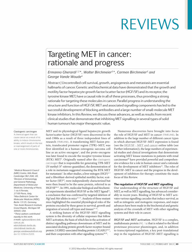
Timeline | Milestones of MET and HGF/SF discoveries
1984 1987 1989 1990 1991 1992 1994 1995 1996 1997
(1987–1989) SF, which disrupts epithelial junctions, promotes migration of epithelial cells and induces EMT, is isolated from embryo and 3T3 fibroblast cultures8,9
(1987–1989) A potent mitogen for liver parenchymal cells (HGF) is purified from rat platelets and human plasma4–7
Cloning of the full-length MET cDNA reveals that the encoded protein is an RTK2
The oncogene TPR-MET is isolated from a chemically transformed cell line1
The first sequences of HGF cDNAs reveals homology to plasminogen4,6
HGF/SF induces invasion of epithelial cells in a 3D culture assay181
HGF/SF overexpression induces growth, abnormal development and tumour formation in the liver of transgenic mice183
GAB1, a new adaptor molecule that binds to activated MET, is identified17
(1992–1994) MET is shown to be a potent oncogene182, and 3T3 cells that co-express MET and HGF/SF metastasize in an animal model3
Activating mutations of MET are found in hereditary papillary renal carcinomas and sporadic renal cancers20
HGF/SF and MET have essential roles in the development of the placenta and liver, and they control EMT of the epithelial dermomyotome and migration of myogenic precursor cells13–15
MET is identified as the receptor of HGF/SF12
(1990–1991) Protein and DNA sequencing reveal that SF and HGF are identical (HGF/SF)10,11
A bidentate docking site that binds multiple SH2 domain-containing proteins is identified in MET37
Discoveries related to cancer are outlined in red. 3D, three-dimensional; EGFR, epidermal growth factor receptor; EMT, epithelial-to-mesenchymal transition; GAB1, GRB2-associated-binding protein 1; HGF, hepatocyte growth factor; InlB, Internalin B; MACC1, metastasis-associated gene in colon cancer 1; NK1, N terminal and kringle domain 1; NSCLC, non-small-cell lung cancer; RTK, receptor tyrosine kinase; SF, scatter factor; SH2, SRC homology 2; SHP2, SRC homology 2 domain-containing phosphatase 2; SPH, serine proteinase homology; TPR, translocated promoter region.
R E V I E W S
90 | FEBRUARY 2012 | VOLUME 12 www.nature.com/reviews/cancer
© 2012 Macmillan Publishers Limited. All rights reserved
homology (SPH) domain23), as well as fragments of the MET ectodomain in complex with the SPH domain of HGF/SF34 (FIG. 1b) and in complex with Internalin B34,35 (InlB) (FIG. 1c), which is a protein expressed by the bacte‑rium Listeria monocytogenes and which is responsible for bacterial internalization in hepatocytes and macrophages through the MET receptor. The SPH domain binds the large, amino‑terminal domain of MET34 (the SEMA domain (FIG. 1b)). By contrast, InlB binds the first of four immunoglobulin‑like (Ig) domains that are present in the extracellular portion of MET35 (FIG. 1c). Low‑resolution small angle X‑ray scattering (SAXS) models of the HGF/SF–MET complex22 and analysis of InlB‑MET crys‑tal structures36 have established that minimal, signalling‑ competent MET complexes have 2:2 stoichiometry, with a ligand dimer at the centre of the structure and two MET molecules at the periphery. High‑resolution struc‑tures of the catalytic domain of MET, which is cytoplas‑mic, alone or in complex with inhibitors, are available and are discussed below.
Basic mechanisms of MET signalling. Ligand‑induced MET dimerization activates the tyrosine kinase by phosphorylation of tyrosine residues (Tyr1230, Tyr1234 and Tyr1235) in the kinase domain, which leads to autophosphorylation of the carboxy‑terminal bidentate substrate‑binding site (Tyr1349 and Tyr1356) of MET16,17,37. Various cytoplasmic effector proteins, including GAB1, GRB2, phospholipase C (PLC) and SRC are directly recruited to this site, and these proteins become frequently phosphorylated on tyrosine residues (reviewed in REFS 16,18) (FIG. 2). Phosphorylated GAB1 bound to MET at the plasma membrane can attract further docking molecules and enzymes such as SRC homology 2 domain‑containing phosphatase 2 (SHP2; also known as PTPN11), PI3K, CRK‑like protein (CRKL) and others17,38–40 that together activate various downstream signalling cascades. MET signalling, which
is mainly mediated by the RAS–MAPK and PI3K–AKT pathways, affects gene expression and cell cycle progres‑sion through the binding of transcription factors, such as the ETS family16,18 (FIG. 2). Cytoplasmic signalling cas‑cades mediated by PI3K–AKT and the GTPases RAC1 or cell division control protein 42 (CDC42) modulate cell survival and elicit cytoskeletal changes. Signals to the plasma membrane control cell migration and cell adhesion mainly through the RAP1 and RAC1–CDC42 pathways, which affect integrins and cadherins (FIG. 2).
Genetic experiments in mice have provided impor‑tant insights into the roles of the different branches of MET signalling and have shed light on the relevance of various downstream molecules in vivo. A crucial role of the ERK–MAPK branch in MET signalling has been confirmed in mice that carry specific mutations in GAB1 that prevent binding of the protein tyrosine phosphatase SHP2 or of PI3K41. Recruitment of SHP2 to GAB1 and ERK–MAPK signalling is essential for embryonic survival, myogenic precursor cell migration and liver growth; all of these processes are controlled by HGF/SF and MET during development16 (TIMELINE). In humans, activating mutations of PTPN11, the gene that encodes SHP2, are associated with a number of malig‑nancies, most prominently juvenile myelomonocytic leukaemia (JMML)42.
Several types of signal cooperation and crosstalk between MET and other receptor pathways have been unveiled in recent years, and a recent study has dem‑onstrated cooperative signalling between MET and the epidermal growth factor receptor (EGFR) during kidney development43 (TIMELINE), providing genetic evidence for the role of HGF/SF in kidney‑branching morphogenesis that was inferred from earlier experiments with cell and organ cultures44,45. The absence of MET during renal development caused reduced branching of the ureteric bud and a decreased number of nephrons, and the defect was particularly severe in mice in which both MET and
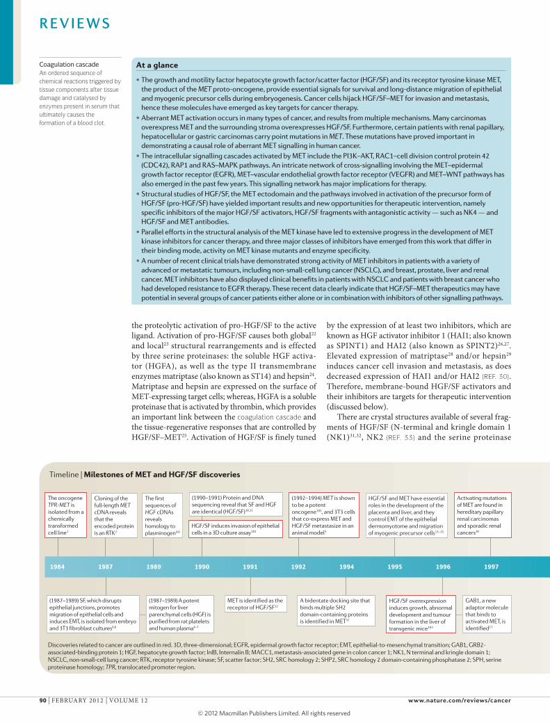
Timeline | Milestones of MET and HGF/SF discoveries
1998 2000 2001 2003 2004 2006 2007 2009 2010 2011
•The first crystal structure of the MET kinase in complex with an inhibitor is reported21
•Hypoxia controls MET expression in carcinoma and sarcoma cells98
Cryo-electron microscopy and small angle X ray scattering structures of HGF–MET complexes22
The domain structure of extracellular MET is defined178
Stromal HGF/SF is a stem cell niche factor and cooperates with epithelial MET and WNT–β-catenin signalling in the maintenance of colon cancer stem cells111
Therapeutic activity of MET inhibitors (METMab167, ARQ 197 (REF. 169), XL184 (REFS 171–174) and XL880 (REF. 175)) is shown in clinical trials
The bacterium Listeria monocytogenes uses MET in order to enter cells through the surface protein InlB186
Genetic analysis of Gab1 in mice demonstrates that this adaptor mediates MET signals184,185 and activates SHP2 (REF. 40)
(1998–1999) Crystal structures of the NK1 fragment of HGF/SF31,32 are solved
(2001–2002) Mutation of a juxtamembrane residue (Tyr1003Phe) converts MET into a transforming protein by disrupting binding to CBL and thus receptor degradation48,50
Genetic experiments in mice show that MET and EGFR cooperate during kidney development43
•Crystal structure of the InlB–MET complex35 is solved
•MET is shown to be essential for wound healing of the skin62
•The essential role of GAB1 binding to SHP2 in MET-dependent migration of muscle precursor cells is demonstrated in mice41
MET mediates resistance of NSCLC cells to EGFR kinase inhibitors75
MACC1 controls metastasis formation by upregulating MET187•MET is shown to be
essential for liver regeneration58,59
•Crystal structure of the SPH–MET complex34 is solved
R E V I E W S
NATURE REVIEWS | CANCER VOLUME 12 | FEBRUARY 2012 | 91
© 2012 Macmillan Publishers Limited. All rights reserved

Nature Reviews | Cancer
Top HGF/SF
MET
Side
βα
c
b
a
Bottom
SEMA
SEMA
SEMA
25 308 514 561 652 734 834 922 1,345 1,3901,078956
CR
CR
CR
Ig1
Ig1
Ig2 Ig3 Ig4 T K
InIB
20612132 288 383 468 728495
K1N K2 K3 K4
βα
SPH
SPH
JM
EGFR signalling was impaired43. Crosstalk between MET and other signalling systems (such as transform‑ing growth factor‑β (TGFβ), WNT and others) has emerged as a major mechanism in human cancer and is discussed below.
MET ubiquitylation, endocytosis and shedding. On ligand activation, MET — like other RTKs — is inter‑nalized through endocytosis. The internalized receptor is then either degraded or recycled to the plasma mem‑brane. Derailed receptor trafficking and degradation,
as well as unbalanced recycling, can cause sustained signalling and can contribute to cell transformation, tumorigenesis and metastasis46.
MET endocytosis and degradation are initiated by ligand‑dependent phosphorylation of the receptor, which is then internalized primarily by clathrin‑coated pits and vesicles. The internalized receptor is delivered to endosomal compartments and remains capable of signalling during vesicle trafficking. Subsequently, the receptor is either degraded or recycled. MET mutations that increase endocytosis and/or recycling activity and that decrease degradtion result in enhanced anchorage‑ independent growth, tumorigenesis and experimen‑tal metastasis in vivo47. The ubiquitin E3 ligase CBL contains a phosphotyrosine‑binding module that rec‑ognizes the phosphorylated Tyr1003 residue in the jux‑tamembrane domain of MET48 and induces degradation (TIMELINE). CBL also contains a RING finger domain that engages E2 protein ubiquitin ligases to mediate ubiquitylation of MET, which might occur at the cell membrane or in the early endocytic compartment. Ubiquitylated MET is degraded in a late endosomal or lysosomal compartment in a proteasome‑dependent manner49,50. Mutation or deletion of the CBL‑binding site converts MET into a transforming protein. Such receptor variants are still internalized on ligand activa‑tion, but they escape degradation owing to a change in endosomal sorting48. Endosomal sorting is regulated by adaptors that recognize and bind ubiquitylated proteins, among them hepatocyte growth factor‑regulated tyros‑ine kinase substrate (HRS; also known as HGS), which is a MET substrate that is involved in endosomal sort‑ing of ubiquitylated MET51. Several mechanisms that affect RTK internalization and trafficking have been reported to be altered in cancer cells (see REF. 46 for a recent review) but their role in aberrant MET activity requires further analysis.
Certain cancer cells express alternatively spliced MET mRNAs that encode a receptor without the juxtamem‑brane CBL‑binding site or with mutations in the jux‑tamembrane domain that interfere with CBL binding52. Owing to the inability to recruit the CBL E3 ubiquitin ligase, receptor downregulation is impaired48,53. Aberrant endocytosis in cancer cells and the resulting escape from degradation results in MET overexpression46.
Another mechanism that leads to downregulation of MET is regulated proteolysis and shedding of the extra‑cellular domain. Shedding is mediated by members of the a disintegrin and metalloproteinase (ADAM) family and results in the formation of a soluble MET ectodomain and a membrane‑anchored cytoplasmic tail. The surface‑associated cytoplasmic tail under‑goes proteolysis by γ‑secretase and is rapidly cleared by proteasome‑mediated degradation54.
Physiology of HGF/SF and METRecent genetic studies in mice have demonstrated that HGF/SF–MET signalling is essential for regeneration in liver and skin. These normal functions have served as a paradigm for understanding the roles of HGF/SF and MET in cancer and are discussed below.
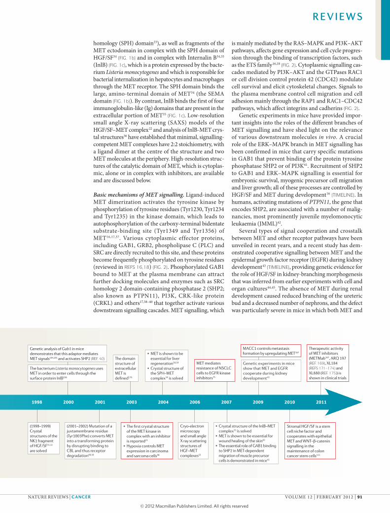
Figure 1 | The multidomain structure of MET and HGF/SF. a | MET is synthesized as a single chain precursor and cleaved by furin during transit through the endoplasmic reticulum177, thus yielding a smaller, amino-terminal α-chain and a larger β-chain. The MET ectodomain consists of a large N-terminal SEMA domain, which adopts a seven-bladed β-propeller fold and a stalk structure consisting of four immunoglobulin-like (Ig) domains178. The SEMA domain and the stalk structure are separated by a small cystine-rich (CR) domain. The transmembrane (T), the long juxtamembrane (JM) sequence, the kinase (K) domain and a carboxy-terminal sequence that contains essential motifs for downstream signalling are also shown. The SEMA domain is a structural variant of the β-propeller, a large protein domain with an irregular cylindrical shape that consists of a variable number of antiparallel β-sheets (or blades) each formed by four β-strands (named A, B, C and D). These blades are arranged radially around a central cavity, and there are seven of these in the MET β-propeller. The face of the domain that displays loops connecting β-strands B–C and D–A is called the ‘top face’; whereas, the face that displays loops connecting β-strands A–B and C–D is known as the ‘bottom face’. Hepatocyte growth factor/scatter factor (HGF/SF) is composed of six domains: an N-terminal (N) domain, four copies of the kringle domain (K1–4) and a C-terminal serine proteinase homology (SPH) domain that is structurally related to the catalytic domain of serine proteinases but that is enzymatically inactive. Mature, biologically active HGF/SF is a two-chain (α–β) protein that is produced by site-specific proteolysis in the extracellular space from single-chain, pro-HGF/SF by the serine proteinases matriptase, pepsin and HGF-activator. HGF/SF contains two MET-binding sites: one in the NK1 fragment and one in the SPH domain179. b | The crystal structure of an SPH–MET complex is shown: the SPH domain of HGF/SF binds to an area of the SEMA domain within the MET α-chain (protein databank (PDB) ID: 1SHY34). c | The crystal structure of an Internalin B (InlB)–MET compIex is shown: InlB primarily binds to the Ig1 domain of the MET stalk35. Structures were drawn using PyMOL180.
R E V I E W S
92 | FEBRUARY 2012 | VOLUME 12 www.nature.com/reviews/cancer
© 2012 Macmillan Publishers Limited. All rights reserved

SRC
GA
B1
Nature Reviews | Cancer
GAB1
GRB2GRB2
GA
B1
GRB2
SHP2PLC
PI3K
PI3K
PI3K
CRKL
DOCK
HGF/SF
HGF/SF
HG
F/SF
AKT
CDC42
RAC1
RAP1PAK
IP3PKC
ERK
RAS
RAF
FAK
MAPKK
NWASP ARP2/3
mTOR
PP
PPPPPP
PPPP
HGF/SF
HG
F/SF
PP
PPPPPP
PPPP
PIP2
NF-κB
ETS–ELK1
Integrin
NucleusCytoplasm
123012341235
13491356
1003
MET
P
PPP
PP
α-catα-catβ-cat
α-cat α-catβ-catβ-cat β-cat
E-cadherin
Actin
DAG
Myogenic progenitor cellsProgenitor cells that have the potential to differentiate into skeletal muscle.
Epithelial dermomyotomeA transient epithelial structure of the embryo that will give rise to skeletal muscle, dermis and other cell types in later development.
HGF/SF and MET in EMT and cell migration during embryogenesis. During development, MET controls the epithelial‑to‑mesenchymal transition (EMT) of myogenic progenitor cells that are released from the epithelial dermomyotome. These cells migrate in a MET‑ and GAB1‑dependent manner over long distances in the embryo15,55. MET‑dependent EMT and long‑distance migration of tumour cells also have major roles in invasion and metastasis.
MET in organ regeneration. Distinct cellular mechanisms are used for regeneration. Stem cells can provide a source, but, in certain tissues, terminally differentiated cells can re‑enter the cell cycle and sustain regeneration. It has been argued that cancer resembles a persistent regeneration
process that is unable to define its end point (tumours have been referred to as “wounds that do not heal” (REF. 56)). Loss of liver mass can be induced in rodents by admin‑istering hepatotoxic chemicals or by surgical removal of up to two‑thirds of the liver. Shortly after partial hepatec‑tomy, HGF/SF is mobilized from the extracellular matrix, resulting in MET activation in hepatocytes, which leads to DNA synthesis and cytokinesis57. Conditional ablation of Met in hepatocytes in mice interferes with hepatocyte re‑entry into S phase and cell cycle progression after par‑tial hepatectomy, resulting in impaired proliferation and incomplete liver regeneration58,59.
In the skin, stem cell populations generate differ‑ent epidermal cell types during normal turnover and wound repair60,61, and a recent study showed that MET
Figure 2 | Signalling pathways activated by HGF/SF and MET. Hepatocyte growth factor/scatter factor (HGF/SF) induces dimerization and activation of MET at the plasma membrane. The cytoplasmic tyrosine phosphorylation (P) sites of MET are indicated: Tyr1003 is in the juxtamembrane CBL-binding site (shown in green), Tyr1230, Tyr1234 and Tyr1235 are in the active site of the kinase (shown in light green), and Tyr1349 and Tyr1356 are in the bidentate docking site (shown in pink). Various cytoplasmic effector molecules such as growth factor receptor-bound protein 2 (GRB2), GRB2-associated-binding protein 1 (GAB1), phospholipase C (PLC) and SRC are recruited to the bidentate docking site. Tyrosine-phosphorylated GAB1 that is bound to MET can attract further docking proteins, including the tyrosine phosphatase SRC homology 2 domain-containing phosphatase 2 (SHP2), PI3K and others, which, together with the direct MET binders, activate various downstream signalling cascades. Signalling by the RAS–MAPK and PI3K–AKT pathways reaches the nucleus to affect gene expression and cell cycle progression. Cytoplasmic signalling cascades that are mediated by RAC1–cell division control protein 42 (CDC42) and p21-activated kinase (PAK) elicit cytoskeletal changes. Signals through the RAP1 and RAC1–CDC42 pathways reach the plasma membrane and control cadherin and integrin adhesion molecules and thereby affect cell migration. Signalling to membrane-bound lipids and protein kinase C (PKC) is also shown. These signalling pathways control cell survival and cell migration. α-cat, α-catenin; β-cat, β-catenin; ARP, actin-related protein; CRKL, CRK-like protein; DAG, diacylglycerol; DOCK, dedicator of cytokinesis; FAK, focal adhesion kinase; IP3, inositol trisphosphate; NF-κB, nuclear factor-κB; NWASP, neural Wiskott–Aldrich syndrome protein; PIP2, phosphatidylinositol 4,5-bisphosphate.
R E V I E W S
NATURE REVIEWS | CANCER VOLUME 12 | FEBRUARY 2012 | 93
© 2012 Macmillan Publishers Limited. All rights reserved

KeratinocytesEpithelial cells of the skin and its appendages, such as hair and skin glands.
Autocrine signallingA type of cell signalling in which the same cell produces both the chemical messenger (a hormone, growth factor or cytokine) and the membrane receptor that triggers the biological response to the messenger.
is essential for wound repair62. In mice with conditional ablation of Met in keratinocytes, only cells that had escaped recombination and that continued to express a functional MET62 could contribute to regeneration. This result was unexpected because growth factors of the EGF and fibroblast growth factor (FGF) families are also involved in re‑epithelialization, but cannot compen‑sate for a lack of HGF/SF–MET signalling in the skin in vivo. During the repair of skin wounds HGF/SF and MET are co‑expressed in keratinocytes, which implies that autocrine signalling occurs. Upregulated HGF/SF levels were also reported after injury to other epithelial organs, such as the kidney and lung, as well as skeletal muscle and heart63.
How HGF/SF and MET can cause cancerThe tight regulation of HGF/SF and MET signalling that is observed in development and regeneration is lost in cancer at multiple levels. These changes often involve transcriptional deregulation, but a number of other mechanisms, including inadequate degrada‑tion, receptor crosstalk or synergies in downstream signalling, have also been observed. The association of HGF/SF–MET alterations with different types of cancer can be found in a searchable, fully referenced table (see the HGF/SF ‑ MET and cancer online table; see Further information).
Genetic abnormalities that cause aberrant HGF/SF–MET signalling. Activating point mutations of MET occur in sporadic and inherited human renal carcino‑mas, hepatocellular carcinomas and several other can‑cer types20,64. Most of these mutations are located in the kinase domain and are homologous to cancer‑inducing mutations that occur in other RTKs (such as EGFR, RET and KIT). When used to replace endogenous Met in the mouse germ line, these mutations cause a vari‑ety of tumours, including sarcomas, lymphomas and carcinomas. When expressed in the mammary gland, they induce basal‑like breast carcinomas65,66. Thus, these studies constitute proof of concept that aberrant MET signalling can cause human cancer. Activating mutations of MET are clonally selected for during the metastasis of human head and neck cancers67, as their frequency increased from 2% in the primary tumours to 50% in the metastases, and this constitutes additional proof of prin‑ciple that, at least in this type of tumour, aberrant MET is associated with progression and metastasis. Finally, in certain human gastric and colorectal carcinomas, as well as in other tumours68–71, amplification of MET (on chromosome 7q31) can occur.
Crosstalk between MET and other signalling pathways. Functional crosstalk of MET with EGFR, ERBB2 or insulin‑like growth factor 1 receptor (IGF1R) has been reported in several systems72,73 and has emerged as a major mechanism for cancer progression and resist‑ance to therapy. Even in cells that express moderate levels of EGFR and MET, EGFR stimulation results in MET phosphorylation and activation74. Conversely, MET amplification can activate ERBB3–PI3K–AKT signalling
in lung cancer cells that carry EGFR mutants that are resistant to EGFR kinase inhibitors75, but resistance can be prevented by combined inhibition of EGFR and MET, as shown in human lung, pancreatic and breast tumour xenografts (see, for example, REFS 76,77). The semaphorin 4D receptor also controls cell migration by coupling with MET78,79.
Crosstalk between MET and developmental signal‑ling pathways, such as WNT–β‑catenin and TGFβ–bone morphogenetic protein (BMP), has also been demon‑strated. Mutation or overexpression of key components of the WNT–β‑catenin pathway can cause cancer80. MET is a direct transcriptional target of WNT–β‑catenin in colon cancer cell lines and other tissues81 (FIG. 3); conversely, MET and integrin α3β1 signalling regulate the tran‑scription of WNT7B in the kidney82. Moreover, HGF/SF induces the nuclear translocation of β‑catenin–TCF and the transcription of their target genes in liver and bladder cancer cells83, as well as the tyrosine phosphorylation of BCL92 (also known as BCL9L) in colon cancer cells84.
An intricate interaction between TGFβ and MET sig‑nalling has been discovered by genetic experiments in mice85. Following mutation of TGFβ receptor II (Tgfbr2) in mesenchymal cells, epithelial tumours developed in the fore‑stomach and the mammary gland as a result of the upregulation of stromal HGF/SF and MET activation in epithelial cells. Further functional links of MET have been uncovered with tetraspanins and the tumour suppres‑sors INK4A and ARF, which are encoded by the CDKN2A locus. The tetraspanins kangai 1 (KAI1; also known as CD82) and CD151 can attenuate integrin‑mediated adhesion and MET signalling in cancer cells by inhibit‑ing SRC or by preventing the binding of GRB2 and PI3K to MET86,87. Finally, genetic and biochemical experiments have defined a functional link between MET, INK4A and ARF in the origin of rhabdomyosarcoma, a tumour that occurs with high penetrance and short latency in Cdkn2a–/– mice that overexpress HGF/SF88.
HGF/SF and MET in angiogenesis. Angiogenesis and lymphangiogenesis are important processes in tumour formation and metastasis. The vascular endothelial growth factor (VEGF) and VEGF receptor (VEGFR) families have a prime role in both processes but HGF/SF–MET signalling is a potent inducer of endothe‑lial cell growth and promotes angiogenesis and lym‑phangiogenesis in vitro and in vivo89–91. Furthermore, MET signalling can induce VEGFA expression and angio genesis through common signalling intermedi‑ates such as SRC homology 2 domain‑containing pro‑teins (SHCs). Thrombospondin 1 (TSP1; also known as THBS1) is a negative regulator of angiogenesis that is sup‑pressed by HGF/SF; by ‘turning on’ VEGFA and ‘turning off ’ TSP1, HGF/SF–MET functions as a potent regulator of the angiogenic switch92. HGF/SF–MET and VEGF–VEGFR2 cooperate in inducing angiogenesis in vitro and in vivo. MET and VEGFR do not physically associate or trans‑phosphorylate each other but they synergisti‑cally activate common signalling intermediates: ERK–MAPK, AKT and focal adhesion kinase (FAK)93. In line with these results, MET kinase inhibitors and a peptide
R E V I E W S
94 | FEBRUARY 2012 | VOLUME 12 www.nature.com/reviews/cancer
© 2012 Macmillan Publishers Limited. All rights reserved

GSK
3β PI3K
SRC
GA
B1
Nature Reviews | Cancer
GAB1
GRB2GRB2
SHP2PLCPI3K
HGF/SF
HGF/SF
HGF/SFHGF/SF
HG
F/SF
AKT
SRC
PP
PPPPPP
PPPP
Nucleus Cytoplasm
β-catenin
β-catenin
β-catenin
APC
β-TrCP
Proteasomaldegradation
CBP
BCL-9PYGO
HGF/SF-producing mesenchymal (myofibrillar) cell
Epithelial stem cell
FRZ
LRP5 orLRP6
Non-activated mesenchymal cells
Minus WNT
WNT
AxinDSH DSHP
P
P
P P
GSK
3β
GSK
3β
CK1γ
CK1αUb
UbUb
TCF-LEFWNT7B
MET
β-catenin
MET
that contained the MET bidentate docking site blocked cancer growth and decreased the number of blood vessels in experimental tumours94,95.
A further interesting aspect of MET biology in tumours that has emerged in recent years is the regula‑tion of MET expression and activity by hypoxia. Hypoxia induces the expression of the transcription factor
hypoxia‑inducible factor 1α (HIF1α), and HIF (which is comprised, in this case, of HIF1α and HIF1β (also known as ARNT))‑dependent expression of MET occurs in several types of carcinoma cells96–99. As inhibitors of angiogenesis reduce tumour vascularization, thus caus‑ing tumour hypoxia, these studies raised the possibility that anti‑angiogenesis therapy alone may reduce tumour mass but may also promote MET‑dependent spread‑ing of cancer cells, and so these observations argue for combination therapies that target both angiogenesis and HGF/SF–MET. Preclinical studies with low‑molecular‑mass compounds that inhibit both VEGFR2 and MET kinases have validated this concept in mouse xenograft models100,101, and these inhibitors may prove valuable in controlling metastatic cancer102 by concurrently blocking angiogenesis and invasion.
HGF/SF and MET signalling in metastasis. The abil‑ity of HGF/SF–MET to induce metastasis in different organs has been shown experimentally with xenografts of tumour cells that are transfected with HGF/SF or MET3,103, as well as in transgenic mice that overexpress HGF/SF or MET104–106 (TIMELINE). A role of MET in meta‑static progression has also been established in patients with head and neck cancer3 (discussed above). The bidentate docking site of MET is essential for the role of MET in metastasis, because single point mutations at this site have been shown to prevent experimental metastases induced by TPR–MET in vivo in mice107. RAS–MAPK and RAC1 signalling are important in the early steps of metastasis108,109 (FIG. 2), and HGF/SF–MET controls RAS and RAC1 activity110. Last, MET and WNT signalling affect metastasis independently, but they functionally interact in colon cancer111.
HGF/SF and MET signalling in stem and cancer stem cells. Several tumours contain stem cells, known as can‑cer stem cells or cancer‑initiating cells, which constitute a variable proportion of the tumour cell population that effectively reconstitutes the whole tumour after trans‑plantation112. The presence of these cells, therefore, has implications for tumour therapy, although their actual role in progression and metastasis is debated113.
Developmental signalling pathways have crucial roles in the generation and maintenance of stem cells and cancer stem cells114–117, and a role for HGF/SF and MET in mesenchymal and haematopoietic progenitor and stem cells118,119,120 has recently been demonstrated. HGF/SF is involved in the mobilization of cardiac stem cells after myocardial infarction121 and it has been impli‑cated in the activation of satellite cells, which are the stem cells of adult muscle122. The developing liver har‑bours bipotent hepatic stem or progenitor cells, which could be enriched in the presence of HGF/SF123. Further, HGF/SF–MET signalling also has important roles in stem or progenitor cell functions in both the developing and the adult pancreas124.
A recent report has demonstrated that HGF/SF–MET signalling is essential for the maintenance of colon cancer stem cells, as these cells depend on mesenchyme (myofibroblast)‑derived HGF/SF for
Figure 3 | Cooperation between the HGF and WNT–β-catenin pathways. A recent report has shown that interaction of stroma-derived hepatocyte growth factor/scatter factor (HGF/SF) controls the maintenance of stem cell-like properties of colon cancer cells, which is a function of WNT–β-catenin signalling111. A stem cell niche (top of the figure) contains epithelial (cancer) stem cells (shown in yellow) that are surrounded by mesenchymal (myofibrillar) niche cells (shown in blue), which secrete HGF/SF. Multiple mechanisms have been reported to allow interactions between MET and WNT–β-catenin signalling in epithelial cells, of which a few are shown here. MET can contribute to the transcriptional activation of WNT ligands, such as WNT7B. MET can also induce the activation of β-catenin–TCF–LEF-target genes; for example, through direct or indirect (SRC) tyrosine phosphorylation (P) and nuclear targeting of β-catenin, or by inhibition of the β-catenin degradation complex by AKT phosphorylation of glycogen synthase kinase-3β (GSK3β). β-TrCP, β-transducin repeat-containing protein; APC, adenomatous polyposis coli; CBP, CREB-binding protein; CK1, casein kinase 1; DSH, disheveled; FRZ, frizzled; GAB1, GRB2-associated-binding protein 1; GRB2, growth factor receptor-bound protein 2; LRP, low-density lipoprotein receptor-related protein; PLC, phospholipase C; PYGO, pygopus.
R E V I E W S
NATURE REVIEWS | CANCER VOLUME 12 | FEBRUARY 2012 | 95
© 2012 Macmillan Publishers Limited. All rights reserved

WNT–β‑catenin‑dependent transcription and stemness111 (FIG. 3). These findings are important because, although the role of WNT–β‑catenin signalling in cancer stem cells is well known, that of HGF/SF–MET has only just emerged117,125. There may be additional tumour types in which the WNT and MET pathways cooperate in main‑taining a cancer stem cell compartment. For example, in human breast cancer metastases to the bone, bone‑derived or autocrine HGF/SF activates MET–SRC–WNT signalling126. Intratumoral MET and stromal HGF/SF affect growth and the prognosis of patients with non‑small‑cell lung cancer (NSCLC)127. In multiple mye‑loma, HGF/SF–MET and WNT are aberrantly activated and interference with either pathway inhibits the growth of multiple myeloma cells128,129.
HGF/SF and MET inhibitors for cancer therapyAn increased understanding of the structure–function relationship of ligand, receptor and activators has enabled considerable progress in the development of HGF/SF–MET inhibitors for cancer therapy. The first three classes of inhibitors are discussed below. Inhibitors of HGF/SF activators, HGF/SF inhibitors and MET antagonists are protein therapeutics that function out‑side target cells. The kinase inhibitors function inside the cell and have constituted, so far, the largest effort within the pharmaceutical industry towards MET‑based therapies. Inhibitors of the downstream components of MET signalling, such as inhibitors of RAS, RAF1, SRC, SHP2, MAPKs, AKT and others, are not discussed in this Review.
Inhibitors of HGF/SF activators and HGF/SF. Crystallographic and functional studies by scientists at Genentech, USA, have shown that the first Kunitz domain (KD1) of HAI1 inhibits the activity of the cata‑lytic domain of HGFA, blocking access to pro‑HGF/SF130. The specificity of KD1 mirrors that of the HAI1 ecto‑domain, and pegylated KD1 was shown to inhibit the invasion and metastasis of human prostatic cancer cells that overexpressed hepsin in severe combined immu‑nodeficient (SCID) mice131. HGFA antibodies that bind either the substrate‑binding domain (Fab58 (REF. 132)) or outside the catalytic cleft (Fab40 (REF. 133)), as well as anti‑matriptase antibodies (FabE2 (REF. 134)), have also been developed (FIG. 4a).
In a separate approach, Vande Woude and col‑leagues135 have demonstrated that antibodies directed towards HGF/SF inhibit the growth of cancer cell lines that are dependent on HGF/SF–MET signalling135. These early studies relied on mixtures of antibodies directed against two or more epitopes, but subsequent work has led to the isolation of individual monoclonal antibodies that can block HGF/SF binding to MET. One of these, AMG102, binds the SPH domain136 (FIG. 4b).
MET antagonists. Truncated splice variants of HGF/SF have formed a strong basis for developing MET antago‑nists (FIG. 4c). NK1, the shortest splice variant of HGF/SF, has agonistic activity in vivo137 and dimerizes in solu‑tion in the presence of heparin, but it can be converted
into a receptor antagonist by the mutation of residues at the dimer interface, such as Tyr124 and Asn127 (REFS 138,139). NK2, the most abundant alternative splice variant of HGF/SF, has partial agonistic and antagonistic activity in vivo140, and mutation of an unpaired cysteine (Cys214) yields a variant with receptor antagonistic activity33. Finally NK4, a fragment that corresponds to the α‑chain of HGF/SF (FIG. 1a), has been extensively characterized as a receptor antagonist by Nakamura, Matsumoto and colleagues141. In addition to MET antagonistic activity, NK4 has broad anti‑angiogenic activities against HGF/SF‑, VEGF‑ or bFGF (also known as FGF2)‑induced angiogenesis142; however, NK4 is more difficult to produce than NK1 and NK2 and may require delivery via gene therapy.
Several MET antibodies with antagonistic activity are now available (FIG. 4c). METMab (also known as onartuzumab) is a monovalent antibody developed at Genentech that binds the SEMA domain of MET143 and that displays potent antagonistic activity144. METMab may act as a classic receptor antagonist by competing for the binding of HGF/SF to MET. The MET antibody DN‑30 causes MET activation and shedding through ADAM10 (REFS 145,146). Conversion of the intact IgG into a monovalent form abolished agonistic activity and yielded a bona fide antagonist147. A MET antibody with antagonistic activity in a bivalent format (11E1) has also been described148. The mechanism for the antagonistic activity of 11E1 is unclear but is probably different from those of METMab and DN‑30.
MET kinase inhibitors. Impressive numbers of MET kinase inhibitors have been developed over the past 10 years. Compounds for which structural data have been made available (see the MET inhibitors online table; see Further information) are discussed below (see REF. 149 for a recent review). The catalytic domain of MET has the typical architecture of other protein kinases in which an N‑terminal lobe (N) with a predominant β‑structure is connected by a short linker to an α‑helical C‑terminal lobe (C). Structures of the auto‑inhibited150 and catalytically active form of the MET kinase151 are available and, in line with results from studies on other protein kinases, the switch from the inactive to the active conformation is associated with major struc‑tural rearrangements (FIG. 5a). In the inactive confor‑mation, the activation loop blocks access of ATP to the enzyme but on activation the loop is extruded (FIG. 5b). ATP binds in a pocket between the N and C lobes and makes extensive hydrogen and ion bonds with residues in the linker (Pro1158 and Met1160), the nucleotide‑binding loop (Gly1087 and His1088), the catalytic loop (Asp1204, Arg1208 and Asn1209) and the activation loop (Asp1222)152 (FIG. 5c).
Superimposed structures of the ATP–MET kinase complex and 25 crystal structures of inhibitor–MET kinase complexes show that inhibitors can be clustered in three groups that differ in their mode of binding (FIG. 5d) (see the MET inhibitors online table; see Further information). Type I compounds and the binding of one of these (PF‑02341066 (also known as crizotinib);
R E V I E W S
96 | FEBRUARY 2012 | VOLUME 12 www.nature.com/reviews/cancer
© 2012 Macmillan Publishers Limited. All rights reserved

K1
Nature Reviews | Cancer
cba
Fab58
mAbs
AMG 102,AV-299 andHuL2G7
Fab40
NK1
mAbs
METMab, DN30,CE-355621, 11E1LA480 and LMH87
NK4
KD1
pro-HGF/SF
METN
K4
K3
SPH
SEMA
NK2
FabE2
K2K1
HGF/SF
N
K4
K3
SPH
K2
Ig1
Ig2
Ig3
Ig4
K
CR
π stacking interactionsA chemical interaction between aromatic rings that is commonly seen in DNA and RNA structures, nucleoprotein complexes and between complexes of small organic compounds with proteins. The interaction is mediated by π orbitals, and the two rings are piled (stacked).
protein databank (PDB) ID: 2WGJ153) are shown in FIG. 5d,e. These compounds occupy the ATP‑binding pocket, are competitive inhibitors of ATP binding and typically form hydrogen bonds to backbone atoms of Met1160 and Asp1222, and π stacking interactions with Tyr1230 of the activation loop. Most type I compounds display preferential binding to the inactive conforma‑tion of the enzyme and have limited activity against the Tyr1230H mutation that is present in certain human tumours. However, there are type I compounds, for example MK‑2461, that have a different set of contacts and a strong binding preference for the active form of the kinase151.
Type II inhibitors (FIG. 5d) also occupy the ATP‑binding pocket but also extend into a second pocket that is formed when the side chain of Asp1222, a residue of the activation loop that coordinates a Mg2+ ion bound to ATP during catalysis, instead points away from the ATP‑binding pocket. The resulting, inactive ‘DFG out’ con‑formation that is adopted by residues Asp1222, Phe1223 and Gly1224 enables the binding of type II inhibitors (FIG. 5f) (such as compound BMS‑777607 (PDB ID: 3F82)154). A smaller number of type III compounds (FIG. 5d) occupy the ATP‑binding pocket and extend into a hydrophobic cavity that is formed by the displacement of the αC helix rather than into the pocket that is formed by the DFG out conformation (such as compound MT3 (PDB ID: 3EFJ155) (FIG. 5g). Whereas the majority of MET kinase inhibitors that are under development can be clustered into these three main types, there are exceptions. For example, ARQ 197 (also known as tivan‑tinib), like type I inhibitors, inhibits ATP binding to the MET kinase in a non‑competitive manner156, binds the ATP binding cleft and makes canonical contacts
with Met1160 but it also occupies a small hydrophobic pocket located between Phe1089 of the glycine‑rich loop and Phe1223 (REF. 157).
The different binding modes of the available MET inhibitors have implications for specificity and activ‑ity. Although the body of data available is very large, it remains incomplete. As a result, the activity of the MET inhibitors against kinases other than in vitro kinase plat‑forms or a limited set of kinase mutants has not been extensively studied and the activity profile of individual inhibitors may change as additional data are obtained. It is also clear that even small modifications can have a profound effect on both potency and/or specificity and, as a great deal of work on the MET kinase inhibitors is still in progress, the activity of several final drugs might display significantly improved profiles compared with those of the lead compounds that have been initially reported.
For example, type I inhibitors have generally been described as specific for the MET kinase (see the MET inhibitors online table; see Further information). However, PF‑02341066 has strong activity against ana‑plastic lymphoma kinase (ALK) and has recently dem‑onstrated impressive therapeutic activity in a group of patients with NSCLC carrying EML4–ALK fusions158. Among the type II inhibitors are compounds that have been described as specific (for example, L8V) but also others that inhibit multiple RTKs. For example, BMS‑777607 has strong activity against MET, RON (also known as MST1R), AXL (also known as UFO) and TYRO3, and XL880 (also known as GSK1363089 and foretinib) inhibits MET, AXL, VEGFR2, platelet‑derived growth factor receptor‑β (PDGFRβ) and TIE2 (also known as TEK) (see the MET inhibitors online
Figure 4 | Extracellular inhibitors of HGF/SF and MET. Representative examples of several classes of extracellular hepatocyte growth factor/scatter factor (HGF/SF) and MET inhibitors are shown. a | Inhibitors of pro-HGF/SF convertases include the HGF activator (HGFA)-specific Kunitz domain inhibitor KD1, the HGFA antibodies Fab40 and Fab58 and the matriptase antibody FabE2 (protein databank (PDB) IDs: 1YC0, 2R0K, 3K2U and 3BN9, respectively). b | Inhibitors of HGF/SF include several monoclonal antibodies (mAbs), including AMG102, AV299 (also known as ficlatuzumab) and HuL2G7. c | MET antagonists include several engineered fragments of HGF/SF, namely NK1 linker mutants (Tyr124Ala and Asn127Ala), the NK2 mutant Cys214Ala and NK4, as well as MET antibodies. The structures of NK2 and the NK1 monomer are from PDB IDs 3HN4 and 1NK1, respectively. Antibodies are shown as Fv models (VH is shown in yellow and VL is shown in pink) using PDB ID 1HAW as a template. NK4 is also shown as a model using the 3HN4 structure as a template. Structures and models were drawn with PyMOL180.
R E V I E W S
NATURE REVIEWS | CANCER VOLUME 12 | FEBRUARY 2012 | 97
© 2012 Macmillan Publishers Limited. All rights reserved
Nature Reviews | Cancer
c
d
b
f
g
ea
N lobe
G-richloop
C lobe
Activation loop
αC
1234 1230
1230 12351235 1234
C
ATP
ATP
Type III
Type I Type II
PP
table; see Further information). Type III inhibitors have been described as selective, but a derivative of MT4 has much more potent activity against RON than the lead compound, thus confirming that medicinal chemistry can reshape both potency and specificity.
Targeting HGF/SF–MET in cancerPatient stratification. The most notable advances in can‑cer therapy that have occurred in the past decade — for example, with tumours carrying BCR–ABL or EML4–ALK fusion genes158,159 — have resulted from three crucial factors: a genetic defect yielding a single target for therapy and the availability of an effective inhibitor and effective methods for the identification of tumours carrying the relevant genetic defect. In the case of HGF/SF–MET, the role of aberrant signalling in cancer is clear, and effective therapeutics are now available, but methods for assessing the level of HGF/SF–MET expres‑sion and activity have not been extensively validated and deployed. Thus, patient stratification according to HGF/SF–MET expression or MET phosphorylation needs further development and is not currently an important component of study design in the numer‑ous clinical trials that are in progress (FIG. 6a,b) (see the Clinical Trials Involving HGF/SF‑MET Inhibitors online table; see Further information). Here, we argue for patient stratification as an essential component for ther‑apeutic success and suggest that antibody‑based analy‑sis of HGF/SF–MET expression levels160 and/or receptor phosphorylation161 may constitute valid strategies.
MET signalling crosstalk and therapy. In recent years signalling crosstalk has evolved from a loose biochemi‑cal concept to one with a rigorous genetic founda‑tion43,85 and major clinical relevance, as demonstrated by the findings of studies with MET and EGFR in NSCLC75,76 (discussed above). This mechanism is also active in a subset of breast cancers162 and might be at work in other tumours, as revealed by preclinical stud‑ies with human tumour xenografts163. Conversely, the treatment of tumour cells with MET kinase inhibitors may lead to the selection of tumour cell populations that escape growth inhibition via the EGFR or SRC kinases164–166. The implications of these findings for therapy are clear and argue for a shift from monother‑apy to combination (multi‑target) therapies in which both the signalling pathway primarily responsible for the cancer phenotype and the ‘rescue pathways’ are targeted concurrently (FIG. 6c).
Anti-angiogenesis therapy and MET activation. Anti‑angiogenesis therapies have been shown to impair the growth of a number of experimental and human tumours and are currently used in metastatic colon cancer and NSCLC; the rationale for combining anti‑angiogenesis therapies with inhibitors of MET is dis‑cussed above. Concurrent inhibition of VEGFR and MET can be achieved either by combining specific VEGF–VEGFR and MET inhibitors or by dual or multi‑specificity kinase inhibitors that inhibit both MET and VEGFR2, as has been shown in human tumour
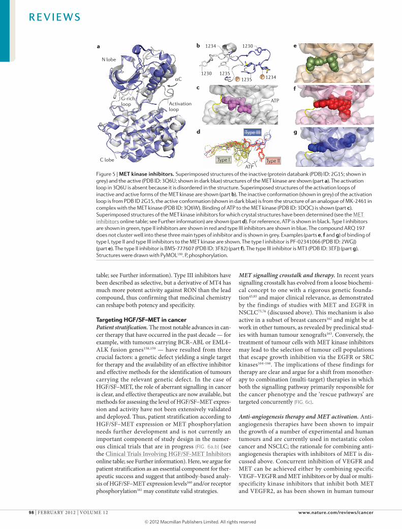
Figure 5 | MET kinase inhibitors. Superimposed structures of the inactive (protein databank (PDB) ID: 2G15; shown in grey) and the active (PDB ID: 3Q6U; shown in dark blue) structures of the MET kinase are shown (part a). The activation loop in 3Q6U is absent because it is disordered in the structure. Superimposed structures of the activation loops of inactive and active forms of the MET kinase are shown (part b). The inactive conformation (shown in grey) of the activation loop is from PDB ID 2G15, the active conformation (shown in dark blue) is from the structure of an analogue of MK-2461 in complex with the MET kinase (PDB ID: 3Q6W). Binding of ATP to the MET kinase (PDB ID: 3DQC) is shown (part c). Superimposed structures of the MET kinase inhibitors for which crystal structures have been determined (see the MET inhibitors online table; see Further information) are shown (part d). For reference, ATP is shown in black. Type I inhibitors are shown in green, type II inhibitors are shown in red and type III inhibitors are shown in blue. The compound ARQ 197 does not cluster well into these three main types of inhibitor and is shown in grey. Examples (parts e, f and g) of binding of type I, type II and type III inhibitors to the MET kinase are shown. The type I inhibitor is PF‑02341066 (PDB ID: 2WGJ) (part e). The type II inhibitor is BMS‑777607 (PDB ID: 3F82) (part f). The type III inhibitor is MT3 (PDB ID: 3EFJ) (part g). Structures were drawn with PyMOL180. P, phosphorylation.
R E V I E W S
98 | FEBRUARY 2012 | VOLUME 12 www.nature.com/reviews/cancer
© 2012 Macmillan Publishers Limited. All rights reserved

Nature Reviews | Cancer
3%
23%
20%
54%
Phase I Phase I/IIPhase II Phase III
14%3%
6%
21%
2% 6% 7%
41%
Solid BrainGI tract Kidney
Lung
LiverBreastOther
2%6%21%
27%44%
Monotherapy + EGFR i
+ chemotherapy+ VEGFR i+ other
a b c
Progression-free survival(PFS). A statistical parameter that measures the time — for example, after diagnosis and/or treatment — in which the disease remains stable (progression free). It can also be expressed as the proportion of patients whose disease has remained stable after diagnosis and/or treatment at a specified time.
Overall survivalA statistical parameter that measures the survival time of a patient or a patient group after diagnosis and/or treatment, regardless of the cause of death. It can also be expressed as the proportion of patients who remain alive at a specified time.
xenograft models163 (see the Clinical Trials Involving HGF/SF‑MET Inhibitors online table; see Further information).
Early results from clinical trials. The vast majority of the clinical trials that aim to define the efficacy of HGF/SF–MET therapeutics are currently in progress but initial results from several studies have been made avail‑able. Striking results with the MET antibody METMab in combination with an EGFR inhibitor (erlotinib) have been reported in patients with NSCLC167. As determined retro‑spectively by immunohistochemistry, METMab increased progression-free survival (PFS) in patients with high levels of MET expression compared with the group receiving erlotinib alone; however, patients with low or no MET expression experienced decreased PFS167. Improvement in overall survival has also been reported in a different patient group — specifically, patients with advanced gastric adenocarcinoma in which treatment with the HGF/SF monoclonal antibody AMG102 (also known as rilotumumab) combined with chemotherapy was com‑pared with chemotherapy alone168. Even in this study, the best response was observed in patients expressing a high level of MET in the tumour168. Thus, both the study with METMab in NSCLC and the study with AMG102 in gastric cancer highlight an essential requirement for patient stratification to ensure clinical benefit.
A large number of kinase inhibitors are now in clinical trials (FIG. 6c) (see the Clinical Trials Involving HGF/SF‑MET Inhibitors online table; see Further
information). A Phase II study with ARQ 197 in patients with NSCLC has shown a clear trend of improved PFS and overall survival in patients treated with the inhibitor plus erlotinib compared with patients who received erlo‑tinib and a placebo169; a trial with this drug combination has now advanced to a Phase III study. In other clinical trials, treatment with ARQ 197 alone also inhibited the growth of hepatocellular and pancreatic carcinomas, as well as tumours driven by microphthalmia‑associated transcription factor (MITF)170. A recent report has docu‑mented striking activity of the MET and ALK inhibitor PF‑02341066 in patients with NSCLC carrying an EML4–ALK fusion158. The EML4–ALK fusion protein occurs in 2–7% of patients with NSCLC, and PF‑02341066 induced a major therapeutic response in this patient group com‑pared with standard chemotherapy158. The bulk of the therapeutic effect of PF‑02341066 in patients with NSCLC carrying the EML4–ALK fusion protein is most probably due to inhibition of ALK158, but the drug is also a potent MET kinase inhibitor and it would be interesting, there‑fore, to further analyse the patient response on the basis of the level of MET expression in the tumour.
Studies with the multi‑target MET inhibitor XL184 (also known as cabozantinib) have shown significant activity against a number of solid tumours, including breast cancer, NSCLC, melanoma and liver cancer171. Ovarian cancer172 displayed notable responses to XL184, but the most remarkable response was seen in both soft tissue and bone metastatic lesions in patients with meta‑static castration‑resistant prostate cancer (CRPC)173. The success of XL184 against CRPC primary and metastatic tumours marks a turning point for MET kinase inhibi‑tors and their power to change terminal cancer prog‑noses. XL184 also showed activity against medullary thyroid cancer174 and the range of applications for this drug may further expand.
Finally, the multi‑target MET inhibitor XL880 has been reported to cause tumour reduction in patients with breast cancer with resistance to inhibitors of EGFR (such as erlotinib) or EGFR and ERBB2 (such as lapatinib)175, a result that mirrors those obtained with MET and EGFR inhibitors in NSCLC.
Conclusions and perspectivesThe availability of a wealth of HGF/SF–MET inhibitors with a range of potencies and specificities has provided a strong basis for assessing the therapeutic value of HGF/SF–MET inhibition in human cancer, and initial results from clinical studies have demonstrated thera‑peutic benefits in patients with a variety of advanced or metastatic tumours, including NSCLC, and breast, pros‑tate, liver and renal cancer. These results have enabled the progression of several compounds to Phase III trials, and larger studies and rigorous patient stratification procedures will further clarify the therapeutic value and long‑term safety of HGF/SF–MET inhibitors in cancer patients.
With the exception of biological agents such as METMab, and the low‑molecular‑mass compound ARQ 197, the first group of therapeutics to reach Phase II and Phase III studies predominantly included inhibi‑tors with multiple specificity that, in addition to MET,
Figure 6 | Clinical trials with HGF/SF–MET inhibitors. The figure illustrates data from 96 clinical studies involving antibodies to hepatocyte growth factor/scatter factor (HGF/SF) or MET and small-molecule inhibitors of the MET kinase listed in the US National Institutes of Health registry of Clinical Trials (see the ClinicalTrials.gov website and the Clinical Trials Involving HGF/SF-MET Inhibitors online table; see Further information). a | The distribution according to study type and stage (96 trials) is shown. 54% of these trials are Phase I studies primarily focusing on drug dosage and safety; whereas, 46% are Phase I/II, Phase II and Phase III trials addressing clinical efficacy. b | The distribution according to tumour type (84 trials) is shown. These 84 trials involve cancer patients, of which 41% had advanced stage multiple solid tumours; whereas, 59% had specific tumour types. Among the 59%, the studies on lung tumours constitute the largest group (21%), followed by brain tumours (7%) and tumours of the gastrointestinal (GI) tract (6%) and liver (6%). c | The distribution according to therapeutic strategy (monotherapy versus combined therapy) is shown. Of 44 efficacy studies (Phase I/II, Phase II and Phase III), 41% involve HGF/SF–MET monotherapies, 21% involve an HGF/SF–MET drug combined with chemotherapy, and 27% and 6% involve an HGF/SF-MET drug combined with inhibitors of epidermal growth factor receptor (EGFR i) or vascular endothelial growth factor receptor (VEGFR i), respectively. Note that monotherapy includes not only specific HGF/SF–MET inhibitors but also agents with multiple targets.
R E V I E W S
NATURE REVIEWS | CANCER VOLUME 12 | FEBRUARY 2012 | 99
© 2012 Macmillan Publishers Limited. All rights reserved

1. Cooper, C. S. et al. Molecular cloning of a new transforming gene from a chemically transformed human cell line. Nature 311, 29–33 (1984).
2. Park, M. et al. Sequence of MET protooncogene cDNA has features characteristic of the tyrosine kinase family of growth-factor receptors. Proc. Natl Acad. Sci. USA 84, 6379–6383 (1987).References 1 and 2 report a new transforming gene (MET) from a human osteogenic sarcoma cell line treated with N‑methyl‑N′‑nitronitrosoguanidine. Subsequent work established that it is the fusion of regulatory sequences from chromosome 1 (TPR) and sequences from chromosome 7 encoding a receptor tyrosine kinase (MET).
3. Rong, S., Segal, S., Anver, M., Resau, J. H. & Vande Woude, G. F. Invasiveness and metastasis of NIH 3T3 cells induced by Met-hepatocyte growth factor/scatter factor autocrine stimulation. Proc. Natl Acad. Sci. USA 91, 4731–4735 (1994).Reference 3 shows that cells made autocrine for HGF/SF–MET expression become highly metastatic in immunocompromised mice.
4. Miyazawa, K. et al. Molecular cloning and sequence analysis of cDNA for human hepatocyte growth factor. Biochem. Biophys. Res. Commun. 163, 967–973 (1989).
5. Nakamura, T., Nawa, K., Ichihara, A., Kaise, N. & Nishino, T. Purification and subunit structure of hepatocyte growth factor from rat platelets. FEBS Lett. 224, 311–316 (1987).
6. Nakamura, T. et al. Molecular cloning and expression of human hepatocyte growth factor. Nature 342, 440–443 (1989).
7. Zarnegar, R. & Michalopoulos, G. Purification and biological characterization of human hepatopoietin A, a polypeptide growth factor for hepatocytes. Cancer Res. 49, 3314–3320 (1989).References 4–7 describe the isolation, cloning and sequencing of a potent mitogen for rat hepatocyte cultures (HGF). Reference 6 further describes the sequence similarity between HGF and plasminogen.
8. Stoker, M., Gherardi, E., Perryman, M. & Gray, J. Scatter factor is a fibroblast-derived modulator of epithelial cell mobility. Nature 327, 239–242 (1987).
9. Gherardi, E., Gray, J., Stoker, M., Perryman, M. & Furlong, R. Purification of scatter factor, a fibroblast-derived basic protein that modulates epithelial interactions and movement. Proc. Natl Acad. Sci. USA 86, 5844–5848 (1989).References 8 and 9 describe the discovery and characterization of a fibroblast‑derived protein that causes dispersion of epithelial colonies (scatter factor). The reports establish a paracrine mechanism of action and describe changes in epithelial cells in culture that have now become known as EMT.
10. Gherardi, E. & Stoker, M. Hepatocytes and scatter factor. Nature 346, 228 (1990).
11. Weidner, K. M. et al. Evidence for the identity of human scatter factor and human hepatocyte growth factor. Proc. Natl Acad. Sci. USA 88, 7001–7005 (1991).
12. Bottaro, D. P. et al. Identification of the hepatocyte growth factor receptor as the c-met proto-oncogene product. Science 251, 802–804 (1991).A molecular biological and biochemical study establishes that MET is the receptor for HGF/SF.
13. Schmidt, C. et al. Scatter factor/hepatocyte growth factor is essential for liver development. Nature 373, 699–702 (1995).
14. Uehara, Y. et al. Placental defect and embryonic lethality in mice lacking hepatocyte growth factor/scatter factor. Nature 373, 702–705 (1995).
15. Bladt, F., Riethmacher, D., Isenmann, S., Aguzzi, A. & Birchmeier, C. Essential role for the c-met receptor in the migration of myogenic precursor cells into the limb bud. Nature 376, 768–771 (1995).References 13–15 define the roles of HGF/SF and MET in mouse development through genetic experiments. References 13 and 14 demonstrate roles in survival and differentiation of epithelial cells of the liver and placenta. Reference 15 reports that MET is essential for EMT of the ventral dermomyotome and migration of myogenic precursor cells into the limbs, tongue and other organs.
16. Birchmeier, C., Birchmeier, W., Gherardi, E. & Vande Woude, G. F. Met, metastasis, motility and more. Nature Rev. Mol. Cell Biol. 4, 915–925 (2003).
17. Weidner, K. M. et al. Interaction between Gab1 and the c-Met receptor tyrosine kinase is responsible for epithelial morphogenesis. Nature 384, 173–176 (1996).This report characterizes GAB1 as a universal docking protein of MET.
18. Lai, A. Z., Abella, J. V. & Park, M. Crosstalk in Met receptor oncogenesis. Trends Cell Biol. 19, 542–551 (2009).
19. Trusolino, L., Bertotti, A. & Comoglio, P. M. MET signalling: principles and functions in development, organ regeneration and cancer. Nature Rev. Mol. Cell Biol. 11, 834–848 (2010).
20. Schmidt, L. et al. Germline and somatic mutations in the tyrosine kinase domain of the MET proto-oncogene in papillary renal carcinomas. Nature Genet. 16, 68–73 (1997).This is the first report of missense mutations in MET in patients with hereditary papillary renal carcinoma and in certain non‑familial forms of renal cancer.
21. Schiering, N. et al. Crystal structure of the tyrosine kinase domain of the hepatocyte growth factor receptor c-Met and its complex with the microbial alkaloid K-252a. Proc. Natl Acad. Sci. USA 100, 12654–12659 (2003).
22. Gherardi, E. et al. Structural basis of hepatocyte growth factor/scatter factor and MET signalling. Proc. Natl Acad. Sci. USA 103, 4046–4051 (2006).Reference 21 describes the first crystal structures of the kinase domain of MET. The report describes both the apo structure, as well as the structure of the kinase domain in complex with the inhibitor K‑252A. Reference 22 describes Cryo‑EM and SAXS structures of HGF/SF–MET complexes.
23. Kirchhofer, D. et al. Structural and functional basis of the serine protease-like hepatocyte growth factor β-chain in Met binding and signaling. J. Biol. Chem. 279, 39915–39924 (2004).
24. Owen, K. A. et al. Pericellular activation of hepatocyte growth factor by the transmembrane serine proteases matriptase and hepsin, but not by the membrane-associated protease uPA. Biochem. J. 426, 219–228 (2010).
25. Shimomura, T. et al. Activation of the zymogen of hepatocyte growth factor activator by thrombin. J. Biol. Chem. 268, 22927–22932 (1993).
26. Shimomura, T. et al. Hepatocyte growth factor activator inhibitor, a novel Kunitz-type serine protease inhibitor. J. Biol. Chem. 272, 6370–6376 (1997).
27. Kawaguchi, T. et al. Purification and cloning of hepatocyte growth factor activator inhibitor type 2, a Kunitz-type serine protease inhibitor. J. Biol. Chem. 272, 27558–27564 (1997).
28. List, K. et al. Deregulated matriptase causes ras-independent multistage carcinogenesis and promotes ras-mediated malignant transformation. Genes Dev. 19, 1934–1950 (2005).
29. Klezovitch, O. et al. Hepsin promotes prostate cancer progression and metastasis. Cancer Cell 6, 185–195 (2004).
30. Morris, M. R. et al. Tumor suppressor activity and epigenetic inactivation of hepatocyte growth factor activator inhibitor type 2/SPINT2 in papillary and clear cell renal cell carcinoma. Cancer Res. 65, 4598–4606 (2005).
31. Chirgadze, D. Y. et al. Crystal structure of the NK1 fragment of HGF/SF suggests a novel mode for growth factor dimerization and receptor binding. Nature Struct. Biol. 6, 72–79 (1999).
32. Ultsch, M., Lokker, N. A., Godowski, P. J. & de Vos, A. M. Crystal structure of the NK1 fragment of human hepatocyte growth factor at 2.0 A resolution. Structure 6, 1383–1393 (1998).
33. Tolbert, W. D., Daugherty-Holtrop, J., Gherardi, E., Vande Woude, G. & Xu, H. E. Structural basis for agonism and antagonism of hepatocyte growth factor. Proc. Natl Acad. Sci. USA 107, 13264–13269 (2010).References 31 and 32 are the first reports of the crystal structure of the NK1 fragment of HGF/SF. An identical head‑to‑tail dimer is described in two different crystal forms. Reference 33 provides the first crystal structure of NK2, the product of the major HGF/SF splice variant.
34. Stamos, J., Lazarus, R. A., Yao, X., Kirchhofer, D. & Wiesmann, C. Crystal structure of the HGF β-chain in complex with the Sema domain of the Met receptor. EMBO J. 23, 2325–2335 (2004).
35. Niemann, H. H. et al. Structure of the human receptor tyrosine kinase met in complex with the listeria invasion protein InlB. Cell 130, 235–246 (2007).References 34 and 35 report on the first two crystal structures of fragments of the MET ectodomain in complex with the SPH domain of HGF/SF (reference 34) or the bacterial protein InlB (reference 35).
36. Ferraris, D. M., Gherardi, E., Di, Y., Heinz, D. W. & Niemann, H. H. Ligand-mediated dimerization of the Met receptor tyrosine kinase by the bacterial invasion protein InlB. J. Mol. Biol. 395, 522–532 (2010).
37. Ponzetto, C. et al. A multifunctional docking site mediates signaling and transformation by the hepatocyte growth factor/scatter factor receptor family. Cell 77, 261–271 (1994).This report describes the bidentate docking site of MET (Y1349 and Y1356), which is essential in MET signalling and binds various adaptor molecules.
38. Maroun, C. R., Naujokas, M. A., Holgado-Madruga, M., Wong, A. J. & Park, M. The tyrosine phosphatase SHP-2 is required for sustained activation of extracellular signal-regulated kinase and epithelial morphogenesis downstream from the met receptor tyrosine kinase. Mol. Cell. Biol. 20, 8513–8525 (2000).
39. Paliouras, G. N., Naujokas, M. A. & Park, M. Pak4, a novel Gab1 binding partner, modulates cell migration and invasion by the Met receptor. Mol. Cell. Biol. 29, 3018–3032 (2009).
40. Schaeper, U. et al. Coupling of Gab1 to c-Met, Grb2, and Shp2 mediates biological responses. J. Cell Biol. 149, 1419–1432 (2000).
target other RTKs involved in cancer, such as VEGFR and RET. This makes it currently difficult to define the contribution of MET inhibition to the overall therapeu‑tic response but comparisons of results with more spe‑cific kinase inhibitors or biological molecules is likely to define the clinical benefit derived from targeting MET in the future. Whether the emergence of resistance to MET kinase inhibitors will constitute a serious limitation for this class of therapeutics is currently difficult to assess. Resistance can rapidly develop with cancer cell lines in culture through multiple mechanisms164–166, but patient
data are clearly required to define the potential effect on therapeutic outcome. Resistance to kinase inhibitors can also be circumvented by combining different types of inhibitors, as demonstrated recently for BCR–ABL176, or by shifting to the use of MET antagonists.
Even at this early stage of clinical investigation, how‑ever, it is safe to conclude that inhibition of HGF/SF–MET signalling in cancer has evolved during the past decade from a concept built on strong experimental foundations (activity on cells and mouse models of disease) to one with considerable scope for the control of human cancer.
R E V I E W S
100 | FEBRUARY 2012 | VOLUME 12 www.nature.com/reviews/cancer
© 2012 Macmillan Publishers Limited. All rights reserved
41. Schaeper, U. et al. Distinct requirements for Gab1 in Met and EGF receptor signaling in vivo. Proc. Natl Acad. Sci. USA 104, 15376–15381 (2007).References 40 and 41 describe the involvement of the tyrosine phosphatase SHP2 in downstream signalling of MET.
42. Grossmann, K. S., Rosario, M., Birchmeier, C. & Birchmeier, W. The tyrosine phosphatase Shp2 in development and cancer. Adv. Cancer Res. 106, 53–89 (2010).
43. Ishibe, S. et al. Met and the epidermal growth factor receptor act cooperatively to regulate final nephron number and maintain collecting duct morphology. Development 136, 337–345 (2009).
44. Montesano, R., Matsumoto, K., Nakamura, T. & Orci, L. Identification of a fibroblast-derived epithelial morphogen as hepatocyte growth factor. Cell 67, 901–908 (1991).
45. Woolf, A. S. et al. Roles of hepatocyte growth factor/scatter factor and the met receptor in the early development of the metanephros. J. Cell Biol. 128, 171–184 (1995).
46. Mosesson, Y., Mills, G. B. & Yarden, Y. Derailed endocytosis: an emerging feature of cancer. Nature Rev. Cancer 8, 835–850 (2008).
47. Joffre, C. et al. A direct role for Met endocytosis in tumorigenesis. Nature Cell Biol. 13, 827–837 (2011).This report describes binding of the E3‑ubiquitin ligase CBL to the juxtamembrane region of MET leading to downregulation of the receptor.
48. Peschard, P. et al. Mutation of the c-Cbl TKB domain binding site on the Met receptor tyrosine kinase converts it into a transforming protein. Mol. Cell 8, 995–1004 (2001).
49. Hammond, D. E., Urbe, S., Vande Woude, G. F. & Clague, M. J. Down-regulation of MET, the receptor for hepatocyte growth factor. Oncogene 20, 2761–2770 (2001).
50. Petrelli, A. et al. The endophilin-CIN85-Cbl complex mediates ligand-dependent downregulation of c-Met. Nature 416, 187–190 (2002).
51. Abella, J. V. et al. Met/Hepatocyte growth factor receptor ubiquitination suppresses transformation and is required for Hrs phosphorylation. Mol. Cell. Biol. 25, 9632–9645 (2005).
52. Lee, J. H. et al. A novel germ line juxtamembrane Met mutation in human gastric cancer. Oncogene 19, 4947–4953 (2000).
53. Asaoka, Y. et al. Gastric cancer cell line Hs746T harbors a splice site mutation of c-Met causing juxtamembrane domain deletion. Biochem. Biophys. Res. Commun. 394, 1042–1046 (2010).
54. Foveau, B. et al. Down-regulation of the met receptor tyrosine kinase by presenilin-dependent regulated intramembrane proteolysis. Mol. Biol. Cell 20, 2495–2507 (2009).
55. Dietrich, S. et al. The role of SF/HGF and c-Met in the development of skeletal muscle. Development 126, 1621–1629 (1999).
56. Dvorak, H. F. Tumors: wounds that do not heal. Similarities between tumor stroma generation and wound healing. N. Engl. J. Med. 315, 1650–1659 (1986).
57. Michalopoulos, G. K. & DeFrances, M. C. Liver regeneration. Science 276, 60–66 (1997).
58. Borowiak, M. et al. Met provides essential signals for liver regeneration. Proc. Natl Acad. Sci. USA 101, 10608–10613 (2004).
59. Huh, C. G. et al. Hepatocyte growth factor/c-met signaling pathway is required for efficient liver regeneration and repair. Proc. Natl Acad. Sci. USA 101, 4477–4482 (2004).
60. Huelsken, J., Vogel, R., Erdmann, B., Cotsarelis, G. & Birchmeier, W. β-Catenin controls hair follicle morphogenesis and stem cell differentiation in the skin. Cell 105, 533–545 (2001).References 56, 57 and 60 describe an essential role of MET in liver regeneration and skin wound healing.
61. Snippert, H. J. et al. Lgr6 marks stem cells in the hair follicle that generate all cell lineages of the skin. Science 327, 1385–1389 (2010).
62. Chmielowiec, J. et al. c-Met is essential for wound healing in the skin. J. Cell Biol. 177, 151–162 (2007).
63. Nakamura, T., Mizuno, S., Matsumoto, K., Sawa, Y. & Matsuda, H. Myocardial protection from ischemia/reperfusion injury by endogenous and exogenous HGF. J. Clin. Invest. 106, 1511–1519 (2000).
64. Ma, P. C. et al. Expression and mutational analysis of MET in human solid cancers. Genes Chromosomes Cancer 47, 1025–1037 (2008).
65. Graveel, C. et al. Activating Met mutations produce unique tumor profiles in mice with selective duplication of the mutant allele. Proc. Natl Acad. Sci. USA 101, 17198–17203 (2004).
66. Ponzo, M. G. et al. Met induces mammary tumors with diverse histologies and is associated with poor outcome and human basal breast cancer. Proc. Natl Acad. Sci. USA 106, 12903–12908 (2009).
67. Di Renzo, M. F. et al. Somatic mutations of the MET oncogene are selected during metastatic spread of human HNSC carcinomas. Oncogene 19, 1547–1555 (2000).
68. Houldsworth, J., Cordon-Cardo, C., Ladanyi, M., Kelsen, D. P. & Chaganti, R. S. Gene amplification in gastric and esophageal adenocarcinomas. Cancer Res. 50, 6417–6422 (1990).
69. Kuniyasu, H. et al. Frequent amplification of the c-met gene in scirrhous type stomach cancer. Biochem. Biophys. Res. Commun. 189, 227–232 (1992).
70. Rege-Cambrin, G. et al. Karyotypic analysis of gastric carcinoma cell lines carrying an amplified c-met oncogene. Cancer Genet. Cytogenet. 64, 170–173 (1992).
71. Knudsen, B. S. & Vande Woude, G. Showering c-MET-dependent cancers with drugs. Curr. Opin. Genet. Dev. 18, 87–96 (2008).
72. Bauer, T. W. et al. Regulatory role of c-Met in insulin-like growth factor-I receptor-mediated migration and invasion of human pancreatic carcinoma cells. Mol. Cancer Ther. 5, 1676–1682 (2006).
73. Khoury, H. et al. HGF converts ErbB2/Neu epithelial morphogenesis to cell invasion. Mol. Biol. Cell 16, 550–561 (2005).
74. Yamamoto, N., Mammadova, G., Song, R. X., Fukami, Y. & Sato, K. Tyrosine phosphorylation of p145met mediated by EGFR and Src is required for serum-independent survival of human bladder carcinoma cells. J. Cell Sci. 119, 4623–4633 (2006).
75. Engelman, J. A. et al. MET amplification leads to gefitinib resistance in lung cancer by activating ERBB3 signaling. Science 316, 1039–1043 (2007).The first report that cancer cells from patients with NSCLC acquire resistance to EGFR inhibitors through MET and ERBB3 signalling, and that combinations of EGFR and MET inhibitors can restore the suppression of cell growth.
76. Turke, A. B. et al. Preexistence and clonal selection of MET amplification in EGFR mutant NSCLC. Cancer Cell 17, 77–88 (2010).
77. Zhang, Y. W. et al. MET kinase inhibitor SGX523 synergizes with epidermal growth factor receptor inhibitor erlotinib in a hepatocyte growth factor-dependent fashion to suppress carcinoma growth. Cancer Res. 70, 6880–6890 (2010).
78. Giordano, S. et al. The semaphorin 4D receptor controls invasive growth by coupling with Met. Nature Cell Biol. 4, 720–724 (2002).
79. Swiercz, J. M., Worzfeld, T. & Offermanns, S. Semaphorin 4D signaling requires the recruitment of phospholipase C γ into the plexin-B1 receptor complex. Mol. Cell. Biol. 29, 6321–6334 (2009).
80. Klaus, A. & Birchmeier, W. Wnt signalling and its impact on development and cancer. Nature Rev. Cancer 8, 387–398 (2008).
81. Boon, E. M., van der Neut, R., van de Wetering, M., Clevers, H. & Pals, S. T. Wnt signaling regulates expression of the receptor tyrosine kinase met in colorectal cancer. Cancer Res. 62, 5126–5128 (2002).
82. Liu, Y. et al. Coordinate integrin and c-Met signaling regulate Wnt gene expression during epithelial morphogenesis. Development 136, 843–853 (2009).
83. Monga, S. P. et al. Hepatocyte growth factor induces Wnt-independent nuclear translocation of β-catenin after Met-β-catenin dissociation in hepatocytes. Cancer Res. 62, 2064–2071 (2002).
84. Brembeck, F. H. et al. Essential role of BCL9–2 in the switch between β-catenin’s adhesive and transcriptional functions. Genes Dev. 18, 2225–2230 (2004).
85. Bhowmick, N. A. et al. TGF-β signaling in fibroblasts modulates the oncogenic potential of adjacent epithelia. Science 303, 848–851 (2004).
86. Sridhar, S. C. & Miranti, C. K. Tetraspanin KAI1/CD82 suppresses invasion by inhibiting integrin-dependent crosstalk with c-Met receptor and Src kinases. Oncogene 25, 2367–2378 (2006).
87. Takahashi, M., Sugiura, T., Abe, M., Ishii, K. & Shirasuna, K. Regulation of c-Met signaling by the tetraspanin KAI-1/CD82 affects cancer cell migration. Int. J. Cancer 121, 1919–1929 (2007).
88. Sharp, R. et al. Synergism between INK4a/ARF inactivation and aberrant HGF/SF signaling in rhabdomyosarcomagenesis. Nature Med. 8, 1276–1280 (2002).
89. Abounader, R. & Laterra, J. Scatter factor/hepatocyte growth factor in brain tumor growth and angiogenesis. Neuro Oncol. 7, 436–451 (2005).
90. Bussolino, F. et al. Hepatocyte growth factor is a potent angiogenic factor which stimulates endothelial cell motility and growth. J. Cell Biol. 119, 629–641 (1992).
91. Grant, D. S. et al. Scatter factor induces blood vessel formation in vivo. Proc. Natl Acad. Sci. USA 90, 1937–1941 (1993).
92. Zhang, Y. W., Su, Y., Volpert, O. V. & Vande Woude, G. F. Hepatocyte growth factor/scatter factor mediates angiogenesis through positive VEGF and negative thrombospondin 1 regulation. Proc. Natl Acad. Sci. USA 100, 12718–12723 (2003).
93. Sulpice, E. et al. Cross-talk between the VEGF-A and HGF signalling pathways in endothelial cells. Biol. Cell 101, 525–539 (2009).
94. Puri, N. et al. A selective small molecule inhibitor of c-Met, PHA665752, inhibits tumorigenicity and angiogenesis in mouse lung cancer xenografts. Cancer Res. 67, 3529–3534 (2007).
95. Cantelmo, A. R. et al. Cell delivery of Met docking site peptides inhibit angiogenesis and vascular tumor growth. Oncogene 29, 5286–5298 (2010).
96. Hara, S. et al. Hypoxia enhances c-Met/HGF receptor expression and signaling by activating HIF-1α in human salivary gland cancer cells. Oral Oncol. 42, 593–598 (2006).
97. Ide, T. et al. Tumor-stromal cell interaction under hypoxia increases the invasiveness of pancreatic cancer cells through the hepatocyte growth factor/c-Met pathway. Int. J. Cancer 119, 2750–2759 (2006).
98. Pennacchietti, S. et al. Hypoxia promotes invasive growth by transcriptional activation of the met protooncogene. Cancer Cell 3, 347–361 (2003).This report shows that hypoxia controls MET expression in carcinoma and sarcoma cells, a finding with important consequences for therapy.
99. Scarpino, S. et al. Increased expression of Met protein is associated with up-regulation of hypoxia inducible factor-1 (HIF-1) in tumour cells in papillary carcinoma of the thyroid. J. Pathol. 202, 352–358 (2004).
100. Qian, F. et al. Inhibition of tumor cell growth, invasion, and metastasis by EXEL-2880 (XL880, GSK1363089), a novel inhibitor of HGF and VEGF receptor tyrosine kinases. Cancer Res. 69, 8009–8016 (2009).
101. Nakagawa, T. et al. E7050: a dual c-Met and VEGFR-2 tyrosine kinase inhibitor promotes tumor regression and prolongs survival in mouse xenograft models. Cancer Sci. 101, 210–215 (2010).
102. You, W. K. & McDonald, D. M. The hepatocyte growth factor/c-Met signaling pathway as a therapeutic target to inhibit angiogenesis. BMB Rep. 41, 833–839 (2008).
103. Meiners, S., Brinkmann, V., Naundorf, H. & Birchmeier, W. Role of morphogenetic factors in metastasis of mammary carcinoma cells. Oncogene 16, 9–20 (1998).
104. Gallego, M. I., Bierie, B. & Hennighausen, L. Targeted expression of HGF/SF in mouse mammary epithelium leads to metastatic adenosquamous carcinomas through the activation of multiple signal transduction pathways. Oncogene 22, 8498–8508 (2003).
105. Jeffers, M. et al. The mutationally activated Met receptor mediates motility and metastasis. Proc. Natl Acad. Sci. USA 95, 14417–14422 (1998).
106. Moshitch-Moshkovitz, S. et al. In vivo direct molecular imaging of early tumorigenesis and malignant progression induced by transgenic expression of GFP-Met. Neoplasia 8, 353–363 (2006).
107. Giordano, S. et al. A point mutation in the MET oncogene abrogates metastasis without affecting transformation. Proc. Natl Acad. Sci. USA 94, 13868–13872 (1997).
108. Muschel, R. J., Williams, J. E., Lowy, D. R. & Liotta, L. A. Harvey ras induction of metastatic potential depends upon oncogene activation and the type of recipient cell. Am. J. Pathol. 121, 1–8 (1985).
109. Webb, C. P. et al. Evidence for a role of Met-HGF/SF during Ras-mediated tumorigenesis/metastasis. Oncogene 17, 2019–2025 (1998).
R E V I E W S
NATURE REVIEWS | CANCER VOLUME 12 | FEBRUARY 2012 | 101
© 2012 Macmillan Publishers Limited. All rights reserved

110. Ridley, A. J., Comoglio, P. M. & Hall, A. Regulation of scatter factor/hepatocyte growth factor responses by Ras, Rac, and Rho in MDCK cells. Mol. Cell. Biol. 15, 1110–1122 (1995).
111. Vermeulen, L. et al. Wnt activity defines colon cancer stem cells and is regulated by the microenvironment. Nature Cell Biol. 12, 468–476 (2010).A report describing stromal HGF/SF as a mesenchymal niche factor that cooperates with epithelial MET and WNT–β‑catenin signalling in the maintenance of colon cancer stem cells.
112. Bonnet, D. & Dick, J. E. Human acute myeloid leukemia is organized as a hierarchy that originates from a primitive hematopoietic cell. Nature Med. 3, 730–737 (1997).
113. Kelly, P. N., Dakic, A., Adams, J. M., Nutt, S. L. & Strasser, A. Tumor growth need not be driven by rare cancer stem cells. Science 317, 337 (2007).
114. Clevers, H. Wnt/β-catenin signaling in development and disease. Cell 127, 469–480 (2006).
115. Malanchi, I. et al. Cutaneous cancer stem cell maintenance is dependent on β-catenin signalling. Nature 452, 650–653 (2008).
116. Piccirillo, S. G. et al. Bone morphogenetic proteins inhibit the tumorigenic potential of human brain tumour-initiating cells. Nature 444, 761–765 (2006).
117. Wend, P., Holland, J. D., Ziebold, U. & Birchmeier, W. Wnt signaling in stem and cancer stem cells. Semin. Cell Dev. Biol. 21, 855–863 (2010).
118. Neuss, S., Becher, E., Woltje, M., Tietze, L. & Jahnen-Dechent, W. Functional expression of HGF and HGF receptor/c-met in adult human mesenchymal stem cells suggests a role in cell mobilization, tissue repair, and wound healing. Stem Cells 22, 405–414 (2004).
119. Son, B. R. et al. Migration of bone marrow and cord blood mesenchymal stem cells in vitro is regulated by stromal-derived factor-1-CXCR4 and hepatocyte growth factor-c-met axes and involves matrix metalloproteinases. Stem Cells 24, 1254–1264 (2006).
120. Tesio, M. et al. Enhanced c-Met activity promotes G.-CSF-induced mobilization of hematopoietic progenitor cells via ROS signaling. Blood 117, 419–428.
121. Urbanek, K. et al. Cardiac stem cells possess growth factor-receptor systems that after activation regenerate the infarcted myocardium, improving ventricular function and long-term survival. Circ. Res. 97, 663–673 (2005).
122. Tatsumi, R., Anderson, J. E., Nevoret, C. J., Halevy, O. & Allen, R. E. HGF/SF is present in normal adult skeletal muscle and is capable of activating satellite cells. Dev. Biol. 194, 114–128 (1998).
123. Kamiya, A., Gonzalez, F. J. & Nakauchi, H. Identification and differentiation of hepatic stem cells during liver development. Front. Biosci. 11, 1302–1310 (2006).
124. Suzuki, A., Nakauchi, H. & Taniguchi, H. Prospective isolation of multipotent pancreatic progenitors using flow-cytometric cell sorting. Diabetes 53, 2143–2152 (2004).
125. Barker, N. et al. Crypt stem cells as the cells-of-origin of intestinal cancer. Nature 457, 608–611 (2009).
126. Previdi, S. et al. Interaction between human-breast cancer metastasis and bone microenvironment through activated hepatocyte growth factor/Met and β-catenin/Wnt pathways. Eur. J. Cancer 46, 1679–1691 (2010).
127. Masuya, D. et al. The tumour-stromal interaction between intratumoral c-Met and stromal hepatocyte growth factor associated with tumour growth and prognosis in non-small-cell lung cancer patients. Br. J. Cancer 90, 1555–1562 (2004).
128. Mahtouk, K., Tjin, E. P., Spaargaren, M. & Pals, S. T. The HGF/MET pathway as target for the treatment of multiple myeloma and B-cell lymphomas. Biochim. Biophys. Acta 1806, 208–219 (2010).
129. Sukhdeo, K. et al. Targeting the β-catenin/TCF transcriptional complex in the treatment of multiple myeloma. Proc. Natl Acad. Sci. USA 104, 7516–7521 (2007).
130. Shia, S. et al. Conformational lability in serine protease active sites: structures of Hepatocyte Growth Factor Activator (HGFA) alone and with the inhibitory domain from HGFA inhibitor-1B. J. Mol. Biol. 346, 1335–1349 (2005).
131. Li, W. et al. Pegylated kunitz domain inhibitor suppresses hepsin-mediated invasive tumor growth and metastasis. Cancer Res. 69, 8395–8402 (2009).
132. Wu, Y. et al. Structural insight into distinct mechanisms of protease inhibition by antibodies. Proc. Natl Acad. Sci. USA 104, 19784–19789 (2007).
133. Ganesan, R. et al. Unraveling the allosteric mechanism of serine protease inhibition by an antibody. Structure 17, 1614–1624 (2009).
134. Farady, C. J., Sun, J., Darragh, M. R., Miller, S. M. & Craik, C. S. The mechanism of inhibition of antibody-based inhibitors of membrane-type serine protease 1 (MT-SP1). J. Mol. Biol. 369, 1041–1051 (2007).
135. Cao, B. et al. Neutralizing monoclonal antibodies to hepatocyte growth factor/scatter factor (HGF/SF) display antitumor activity in animal models. Proc. Natl Acad. Sci. USA 98, 7443–7448 (2001).
136. Burgess, T. L. et al. Biochemical characterization of AMG 102: a neutralizing, fully human monoclonal antibody to human and nonhuman primate hepatocyte growth factor. Mol. Cancer Ther. 9, 400–409 (2010).
137. Jakubczak, J. L., LaRochelle, W. J. & Merlino, G. NK1, a natural splice variant of hepatocyte growth factor/scatter factor, is a partial agonist in vivo. Mol. Cell. Biol. 18, 1275–1283 (1998).
138. Tolbert, W. D. et al. A mechanistic basis for converting a receptor tyrosine kinase agonist to an antagonist. Proc. Natl Acad. Sci. USA 104, 14592–14597 (2007).
139. Youles, M. et al. Engineering the NK1 fragment of hepatocyte growth factor/scatter factor as a MET receptor antagonist. J. Mol. Biol. 377, 616–622 (2008).
140. Otsuka, T. et al. Disassociation of met-mediated biological responses in vivo: the natural hepatocyte growth factor/scatter factor splice variant NK2 antagonizes growth but facilitates metastasis. Mol. Cell. Biol. 20, 2055–2065 (2000).
141. Date, K., Matsumoto, K., Shimura, H., Tanaka, M. & Nakamura, T. HGF/NK4 is a specific antagonist for pleiotrophic actions of hepatocyte growth factor. FEBS Lett. 420, 1–6 (1997).
142. Nakamura, T., Sakai, K. & Matsumoto, K. Anti-cancer approach with NK4: bivalent action and mechanisms. Anticancer Agents Med. Chem. 10, 36–46 (2010).
143. Kong-Beltran, M., Stamos, J. & Wickramasinghe, D. The Sema domain of Met is necessary for receptor dimerization and activation. Cancer Cell 6, 75–84 (2004).
144. Jin, H. et al. MetMAb, the one-armed 5D5 anti-c-Met antibody, inhibits orthotopic pancreatic tumor growth and improves survival. Cancer Res. 68, 4360–4368 (2008).
145. Petrelli, A. et al. Ab-induced ectodomain shedding mediates hepatocyte growth factor receptor down-regulation and hampers biological activity. Proc. Natl Acad. Sci. USA 103, 5090–5095 (2006).
146. Schelter, F. et al. A disintegrin and metalloproteinase-10 (ADAM-10) mediates DN30 antibody-induced shedding of the met surface receptor. J. Biol. Chem. 285, 26335–26340 (2010).
147. Pacchiana, G. et al. Monovalency unleashes the full therapeutic potential of the DN-30 anti-Met antibody. J. Biol. Chem. 285, 36149–36157 (2010).
148. Goetsch, L. Novel antibodies inhibitong c-met dimerization, and uses thereof (2007). http://ip.com/patapp/EP2188312A2.
149. Underiner, T. L., Herbertz, T. & Miknyoczki, S. J. Discovery of small molecule c-Met inhibitors: evolution and profiles of clinical candidates. Anticancer Agents Med. Chem. 10, 2188317–2188327 (2010).
150. Wang, W. et al. Structural characterization of autoinhibited c-Met kinase produced by coexpression in bacteria with phosphatase. Proc. Natl Acad. Sci. USA 103, 3563–3568 (2006).
151. Rickert, K. W. et al. Structural basis for selective small-molecule kinase inhibition of activated c-Met. J. Biol. Chem. 286, 11218– 11225 (2011).
152. Buchanan, S. G. et al. SGX523 is an exquisitely selective, ATP-competitive inhibitor of the MET receptor tyrosine kinase with antitumor activity in vivo. Mol. Cancer Ther. 8, 3181–3190 (2009).
153. Timofeevski, S. L. et al. Enzymatic characterization of c-Met receptor tyrosine kinase oncogenic mutants and kinetic studies with aminopyridine and triazolopyrazine inhibitors. Biochemistry 48, 5339–5349 (2009).
154. Schroeder, G. M. et al. Discovery of N-(4-(2-amino-3-chloropyridin-4-yloxy)-3-fluorophenyl)-4-ethoxy-1-(4-fluor ophenyl)-2-oxo-1,2-dihydropyridine-3-carboxamide (BMS-777607), a selective and
orally efficacious inhibitor of the Met kinase superfamily. J. Med. Chem. 52, 1251–1254 (2009).
155. D’Angelo, N. D. et al. Design, synthesis, and biological evaluation of potent c-Met inhibitors. J. Med. Chem. 51, 5766–5779 (2008).
156. Munshi, N. et al. ARQ 197, a novel and selective inhibitor of the human c-Met receptor tyrosine kinase with antitumor activity. Mol. Cancer Ther. 9, 1544–1553 (2010).
157. Eathiraj, S. et al. Discovery of a novel mode of protein kinase inhibition characterized by the mechanism of inhibition of human mesenchymal-epithelial transition factor (c-Met) protein autophosphorylation by ARQ 197. J. Biol. Chem. 286, 20666–20676 (2011).
158. Kwak, E. L. et al. Anaplastic lymphoma kinase inhibition in non-small-cell lung cancer. N. Engl. J. Med. 363, 1693–1703 (2010).
159. O’Brien, S. G. et al. Imatinib compared with interferon and low-dose cytarabine for newly diagnosed chronic-phase chronic myeloid leukemia. N. Engl. J. Med. 348, 994–1004 (2003).
160. Knudsen, B. S. et al. A novel multipurpose monoclonal antibody for evaluating human c-Met expression in preclinical and clinical settings. Appl. Immunohistochem. Mol. Morphol. 17, 57–67 (2009).
161. Inoue, T. et al. Activation of c-Met (hepatocyte growth factor receptor) in human gastric cancer tissue. Cancer Sci. 95, 803–808 (2004).
162. Mueller, K. L., Hunter, L. A., Ethier, S. P. & Boerner, J. L. Met and c-Src cooperate to compensate for loss of epidermal growth factor receptor kinase activity in breast cancer cells. Cancer Res. 68, 3314–3322 (2008).
163. Zhang, Y., Guessous, F., Kofman, A., Schiff, D. & Abounader, R. XL-184, a MET, VEGFR-2 and RET kinase inhibitor for the treatment of thyroid cancer, glioblastoma multiforme and NSCLC. IDrugs 13, 112–121 (2010).
164. Cepero, V. et al. MET and KRAS gene amplification mediates acquired resistance to MET tyrosine kinase inhibitors. Cancer Res. 70, 7580–7590 (2010).
165. Corso, S. et al. Activation of HER family members in gastric carcinoma cells mediates resistance to MET inhibition. Mol. Cancer 9, 121 (2010).
166. Qi, J. et al. Multiple mutations and bypass mechanisms can contribute to development of acquired resistance to MET inhibitors. Cancer Res. 71, 1081–1091 (2011).
167. Spigel DR. et al. Final efficacy results from OAM4558g, a randomized phase II study evaluating MetMAb or placebo in combination with erlotinib in advanced NSCLC. J. Clin. Oncol. Abstr. 29, 7505 (2011).References 166 and 167 are the first reports to demonstrate that combined treatment of subgroups of patients with NSCLC with EGFR and MET inhibitors increases progression‑free survival and overall survival.
168. Iveson, T. et al. Safety and efficacy of epirubicin, cisplatin, and capecitabine (ECX) plus rilotumumab (R) as first-line treatment for unresectable locally advanced (LA) or metastatic (M) gastric or esophagogastric junction (EGJ) adenocarcinoma. Proc. Eur. Multidisc. Cancer Congr. Abstr. 6.504 (Stockholm, 2011).
169. Von Pawel J. et al. Final results from Arq 197–209: a global randomized placebo- controlled phase 2 clinical trial of erlotinib plus ARQ 197 versus erlotinib plus placebo in previously treated EGFR- inhibitor Naıve patients with advanced non-small cell lung cancer (NSCLC). J. Thoracic Oncol. Abstr. 5, 1 (2010).
170. Bagai, R., Fan, W. & Ma, P. C. ARQ-197, an oral small-molecule inhibitor of c-Met for the treatment of solid tumors. IDrugs 13, 404–414 (2010).
171. Gordon MS. et al. Activity of cabozantinib (XL184) in soft tissue and bone: results of a phase II randomized discontinuation trial (RDT) in patients (pts) with advanced solid tumors. J. Clin. Oncol. Abstr. 29, 3010 (2011).
172. Buckanovich RJ. et al. Activity of cabozantinib (XL184) in advanced ovarian cancer patients (pts): results from a phase II randomized discontinuation trial (RDT). J. Clin. Oncol. Abstr. 29, 5008 (2011).
173. Hussain M. et al. Cabozantinib (XL184) in metastatic castration-resistant prostate cancer (mCRPC): results from a phase II randomized discontinuation trial. J. Clin. Oncol. 29, 4516 (2011).
R E V I E W S
102 | FEBRUARY 2012 | VOLUME 12 www.nature.com/reviews/cancer
© 2012 Macmillan Publishers Limited. All rights reserved

174. Kurzrock, R. et al. Activity of XL184 (Cabozantinib), an oral tyrosine kinase inhibitor, in patients with medullary thyroid cancer. J. Clin. Oncol. 29, 2660–2666 (2011).
175. Liu, L. et al. Synergistic effects of foretinib with HER-targeted agents in MET and HER1- or HER2-coactivated tumor cells. Mol. Cancer Ther. 10, 518–530 (2011).
176. Zhang, J. et al. Targeting Bcr-Abl by combining allosteric with ATP-binding-site inhibitors. Nature 463, 501–506 (2010).
177. Komada, M. et al. Proteolytic processing of the hepatocyte growth factor/scatter factor receptor by furin. FEBS Lett. 328, 25–29 (1993).
178. Gherardi, E. et al. Functional map and domain structure of MET, the product of the c-met protooncogene and receptor for hepatocyte growth factor/scatter factor. Proc. Natl Acad. Sci. USA 100, 12039–12044 (2003).This report defines the domain structure of extracellular MET through deletion mutagenesis and computational studies. The report establishes that MET contains a 7‑balded β‑propeller similar to the one present in the integrin α‑chain.
179. Holmes, O. et al. Insights into the structure/function of hepatocyte growth factor/scatter factor from studies with individual domains. J. Mol. Biol. 367, 395–408 (2007).
180. DeLano, W. L. The PyMOL Molecular Graphics System. (DeLano Scientific, 2002).
181. Weidner, K. M., Behrens, J., Vandekerckhove, J. & Birchmeier, W. Scatter factor: molecular characteristics and effect on the invasiveness of epithelial cells. J. Cell Biol. 111, 2097–2108 (1990).This report demonstrates the first time that HGF/SF induces invasion of human carcinoma cells into three‑dimensional matrices (that is, induces the invasive phenotype).
182. Rong, S. et al. Tumorigenicity of the met proto-oncogene and the gene for hepatocyte growth factor. Mol. Cell. Biol. 12, 5152–5158 (1992).
183. Sakata, H. et al. Hepatocyte growth factor/scatter factor overexpression induces growth, abnormal development, and tumor formation in transgenic mouse livers. Cell Growth Differ. 7, 1513–1523 (1996).
184. Itoh, M. et al. Role of Gab1 in heart, placenta, and skin development and growth factor- and cytokine-induced extracellular signal-regulated kinase mitogen-activated protein kinase activation. Mol. Cell. Biol. 20, 3695–3704 (2000).
185. Sachs, M. et al. Essential role of Gab1 for signaling by the c-Met receptor in vivo. J. Cell Biol. 150, 1375–1384 (2000).
186. Shen, Y., Naujokas, M., Park, M. & Ireton, K. InIB-dependent internalization of Listeria is mediated by the Met receptor tyrosine kinase. Cell 103, 501–510 (2000).
187. Stein, U. et al. MACC1, a newly identified key regulator of HGF-MET signaling, predicts colon cancer metastasis. Nature Med. 15, 59–67 (2009).
AcknowledgementsThe authors would like to acknowledge J. Heuberger (MDC Berlin) for initial drawing of figures 2 and 3 and H. Niemann (University of Bielefeld) for critical reading of the manuscript. E.G. and W.B. acknowledge funding under the SFMET Project of the EU FP7 Programme and the generosity of the Jay and Betty Van Andel Foundation.
Competing interests statementThe authors declare no competing financial interests.
DATABASESNational Cancer Institute Drug Dictionary: http://www.cancer.gov/drugdictionaryAMG102 | ARQ 197 | BMS-777607 | erlotinib | lapatinib | METMab | PF-02341066 | XL184 | XL880
FURTHER INFORMATIONClinicalTrials.gov: http://clinicaltrials.gov/ct2/homeClinical Trials Involving HGF/SF-MET Inhibitors:www.vai.org/metclinicaltrialsHGF/SF - MET and cancer: http://www.vai.org/met/MET inhibitors: http://www.vai.org/metinhibitors/
ALL LINKS ARE ACTIVE IN THE ONLINE PDF
R E V I E W S
NATURE REVIEWS | CANCER VOLUME 12 | FEBRUARY 2012 | 103
© 2012 Macmillan Publishers Limited. All rights reserved


![Hepatocyte GrowthFactor/Scatter Factor Effects Epithelia ... · biochemically purified rodentHGF/SFandMadin-Darbyca- ... Corp. [WestChester, PA] andFITCgoatanti-mouseIgGfromBoeh-ringer](https://static.fdocuments.net/doc/165x107/6033fd7583310d3352417cc3/hepatocyte-growthfactorscatter-factor-effects-epithelia-biochemically-purified.jpg)