Tangier Disease (Familial High Density Lipoprotein Deficiency)” · 2002-09-06 · Tangier Disease...
12
Tangier Disease (Familial High Density Lipoprotein Deficiency)” Clinical and Genetic Features in Two Adults HARRY N. HOPF~IAN, II, XD. and DONALD S. FREDRICKSON, M.D. Rochester, Afinnesota I N 1’961 Fredrickson and associates (7,2] de- scribed a new disorder of cholesterol and liiloprotcin metabolism in two young siblings. The significant clinical features were hypo- cholcsterolemia and enlarged tonsils of unusual appearance. One sibling also had hepatosple- nomegaly and lymphadenopathy. Foam cells observed in the tonsils and lymph nodes con- tained large amounts of cholesterol esters. Both siblings were found to have nearly complete absence of plasma high-density or alpha1 lipo- protein (HDL). In the absence of further knowl- edge of the basic defect, the disorder was named “Tangier disease,” after Tangier Island, the Chesapeake Bay home of the family. More re- cently, two other cases in children of a family in Missouri unrelated to the Tangier Island pa- tients have been discovered [3,4]. Studies of the kindreds of these two involved sibships revealed that many family members had HDL concentrations well below those of comparable control subjects [4]. The data ap- peared to be consistent with the hypothesis that control of plasma HDL concentrations resides in a single pair of autosomal genes. The pheno- type resulting from a single abnormal mutant is characterized by low HDL concentrations; the homozygous individual exhibits the full blown syndrome of tissue storage of cholesterol esters and has no significant plasma HDL. The present report describes the fifth and sixth cases of Tangier disease, which involve two brothers. These are the first cases observed among adults and provide new insight into the manifestations and course of this hereditary Bethesda, Maryland disease. Included also are the results of plasma lipid and lipoprotein determinations among members of this kindred. REPORT OF CASES CASE 1. A forty-two year old, white, oil-refinery worker who first registered at the Mayo Clinic in hfarch 1962 was referred by his family physician for the evaluation of splcnomcgaly. He had consulted his physician in the fall of 1361 because of fatigue, at which time mild anemia and splenomegaly were found. The anemia was said to have diminished fol- lowing iron therapy, but splcnomegaly and mild fa- tigue persisted. The patient had been well otherwise and able to work regularly; no other symptoms were elici- ted except for intermittent mild diarrhea of several years’ duration, manifested as two to four soft to loose, floating, occasionally foul-smelling stools daily. The history disclosed none but the usual childhood illnesses. He had undergone tonsillectomy at age fourteen in a physician’s of&x, but recalled little about prior indications except recurrent sore throat and the judgment that his tonsils were quite enlarged. A cyst on the thyroglossal duct had been removed in 1945 while he was in naval service. On physical examination, the patient appeared generally healthy and alert. Weight was 172 pounds and height 68 inches. Blood pressure was 135 mm. Hg systolic and 90 diastolic. No skin lesions were noted except for a few hyperpigmented areas over the anterior tibia1 regions. The corneas had a slightly cloudy appearance. Slit-lamp examination revealed the clarity and relucence of the corneas to be defi- nitely diminished, with random soft densities involv- ing the entire stromal thickness of each cornea. In the pharynx were several flat patches of yellow-orange lymphoid tissue. No lymphadenopathy was noted. The splenic tip was palpated 6 to 8 cm. below the left * From the Section of Medicine, Mayo Clinic and Mayo Foundation, Rochester, Minnesota, and the Section on Molecular Diseases, Laboratory of Metabolism, National I Ieart Institute, Bethesda, Maryland. This investigation was supported in part by Research Grant AM-06908 from the National Institutes of Health, Public Health Service, to the Gastrointestinal Research Unit, Mayo Clinic and Mayo Foundation. Manuscript received October 26, 1964. 582 AMERICAN JOVRNAL OP MEDICINE
Transcript of Tangier Disease (Familial High Density Lipoprotein Deficiency)” · 2002-09-06 · Tangier Disease...

Tangier Disease (Familial High Density Lipoprotein Deficiency)”
Clinical and Genetic Features in Two Adults
HARRY N. HOPF~IAN, II, XD. and DONALD S. FREDRICKSON, M.D.
Rochester, Afinnesota
I N 1’961 Fredrickson and associates (7,2] de-
scribed a new disorder of cholesterol and liiloprotcin metabolism in two young siblings. The significant clinical features were hypo- cholcsterolemia and enlarged tonsils of unusual appearance. One sibling also had hepatosple- nomegaly and lymphadenopathy. Foam cells observed in the tonsils and lymph nodes con- tained large amounts of cholesterol esters. Both siblings were found to have nearly complete absence of plasma high-density or alpha1 lipo- protein (HDL). In the absence of further knowl- edge of the basic defect, the disorder was named “Tangier disease,” after Tangier Island, the Chesapeake Bay home of the family. More re- cently, two other cases in children of a family in Missouri unrelated to the Tangier Island pa- tients have been discovered [3,4].
Studies of the kindreds of these two involved sibships revealed that many family members had HDL concentrations well below those of comparable control subjects [4]. The data ap- peared to be consistent with the hypothesis that control of plasma HDL concentrations resides in a single pair of autosomal genes. The pheno- type resulting from a single abnormal mutant is characterized by low HDL concentrations; the homozygous individual exhibits the full blown syndrome of tissue storage of cholesterol esters and has no significant plasma HDL.
The present report describes the fifth and sixth cases of Tangier disease, which involve two brothers. These are the first cases observed among adults and provide new insight into the manifestations and course of this hereditary
Bethesda, Maryland
disease. Included also are the results of plasma lipid and lipoprotein determinations among members of this kindred.
REPORT OF CASES
CASE 1. A forty-two year old, white, oil-refinery worker who first registered at the Mayo Clinic in hfarch 1962 was referred by his family physician for the evaluation of splcnomcgaly. He had consulted his physician in the fall of 1361 because of fatigue, at which time mild anemia and splenomegaly were found. The anemia was said to have diminished fol- lowing iron therapy, but splcnomegaly and mild fa- tigue persisted. The patient had been well otherwise and able to work regularly; no other symptoms were elici- ted except for intermittent mild diarrhea of several years’ duration, manifested as two to four soft to loose, floating, occasionally foul-smelling stools daily. The history disclosed none but the usual childhood illnesses. He had undergone tonsillectomy at age fourteen in a physician’s of&x, but recalled little about prior indications except recurrent sore throat and the judgment that his tonsils were quite enlarged. A cyst on the thyroglossal duct had been removed in 1945 while he was in naval service.
On physical examination, the patient appeared generally healthy and alert. Weight was 172 pounds and height 68 inches. Blood pressure was 135 mm. Hg systolic and 90 diastolic. No skin lesions were noted except for a few hyperpigmented areas over the anterior tibia1 regions. The corneas had a slightly cloudy appearance. Slit-lamp examination revealed the clarity and relucence of the corneas to be defi- nitely diminished, with random soft densities involv- ing the entire stromal thickness of each cornea. In the pharynx were several flat patches of yellow-orange lymphoid tissue. No lymphadenopathy was noted. The splenic tip was palpated 6 to 8 cm. below the left
* From the Section of Medicine, Mayo Clinic and Mayo Foundation, Rochester, Minnesota, and the Section on Molecular Diseases, Laboratory of Metabolism, National I Ieart Institute, Bethesda, Maryland. This investigation was supported in part by Research Grant AM-06908 from the National Institutes of Health, Public Health Service, to the Gastrointestinal Research Unit, Mayo Clinic and Mayo Foundation. Manuscript received October 26, 1964.
582 AMERICAN JOVRNAL OP MEDICINE
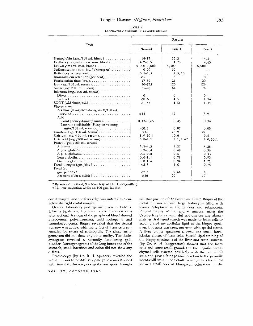
Tangier Disease-HoJman, Fredrickson 583
TABLE I LABORATORY FINDINGS IN TANGIER DISEASE
Tests -
Ifemoglobin (gm./lOO ml. blood). . Erytluocytes (million cu. mm. blood). Leukocytes (cu. mm. blood). Sedimentation (mm. hr., \\‘estergren). Rcticulocytes (per cent). Bromsulfalein retention (per cent). l’rotIuombin time (sec.). . Iron (pg./100 ml. serum). . Sugar (mg./lOO ml. blood). Bilirubin (mg. / 100 ml. serum)
Direct............................... Indirect
SCOT (rMlhour/ml.). . . Phrxphatase
Alkaline (King-Armstrong units/100 ml. serum). . . .
Acid l‘otal (nessey-LowTey units). . . Tartrate-inllil)ita~le (King-Armstrong
units/100 1111. serum). Carotene (pg. 1100 ml. serum). Calcium (mg./lOO ml. serum) Uric acid (mg./lOO ml. serum). . Protein (gm./lOO ml. serum)
All mmm.............................. Alpl~a, globulin.. _. . hlplla, globulin.. Beta globulin. Gamma globulin.. . .
Fecal nitrogen (gm./dayt). . Fecal fat
gm.perdayt.......................... Per cent of feral solids t
II
-
Normal Case 1 Case 2
14-17 13.2 14.2 4.2-5.5 4.73 4.65
5,000-9,000 3,300 6,000 O-20 10
0.5-2.5 2.5, 10 <6 8 0 17-19 21 20 50-l 75 120 126 65-90 84 76
0 0 0 <0.6 1.5 1.94 <1.43 1.61 1.34
<14 17 5.9
0.13-0.63 0.45 0.34
co.7 0.07 0.09 >G3 26.9 27
8.9-10.1 10.0 9.6 3.8-7.1 9.1,9.6* 9.0,lO.l
3.3-4.3 4.27 4.28 0.3-0.4 0.48 0.36 0.5-0.8 0.5 0.43 0.6-1.1 0.71 0.93 0.8-1.6 0.94 1.21 <2.5 1.6 0.75
<7.5 >30
9.66 30
4 17
- * By uricase method, 9.4 (courtesy of Dr. J. Seegmiller). t 72-1101~ collection while on 100 gm. Sat diet.
costal margin, and the liver edge was noted 2 to 3 cm. below the right costal margin.
General laboratory findings are given in Table I. (Plasma lipids and lipoproteins are described in a later section.) A smear of the peripheral blood showed anisocytosis, polychromasia, mild leukopcnia and thrombocytopenia. Biopsy revealed that the sternal marrow was active, with many foci of foam cells sur- rounded by excess of eosinophils. The chest roent- genogram did not show any abnormality. The chole- cystogram revealed a normally funcLioning gall- bladder. Roentgenograms of the long bones and of the stomach, small intestines and colon did not show any dcrccts.
Proctoscopy (by Dr. R. J. Spencer) revealed the rectal mucosa to be diffusely pale yellow and studded with tiny flat, discrete, orange-brown spots through-
VOL. 39, OCTOBER 1965
Results
I -
out that portion of the bowel visualized. Biopsy of the rectal mucosa showed large histiocytes filled with foamy cytoplasm in the mucosa and submucosa. Peroral biopsy of the jejunal mucosa, using the Crosby-Kugler capsule, did not disclose any abnor- malities. A diligent search was made for foam cells or accumulated intracellular lipid in the biopsy speci- men, but none was seen, not even with special stains. A liver biopsy specimen showed one small intra- lobular cluster of foam cells. Special lipid staining of the biopsy specimens of the liver and rectal mucosa (by Dr. A. H. Baggenstoss) showed that the foam cells and some small granules in the hepatic paren- chymal cells reacted positively with the oil red 0 stain and gave a faint positive reaction to the periodic acid-Schiff stain. The Schultz reaction for cholesterol showed small foci of blue-green coloration in the
584 Tangier Disease-Hogman, Fredrickson
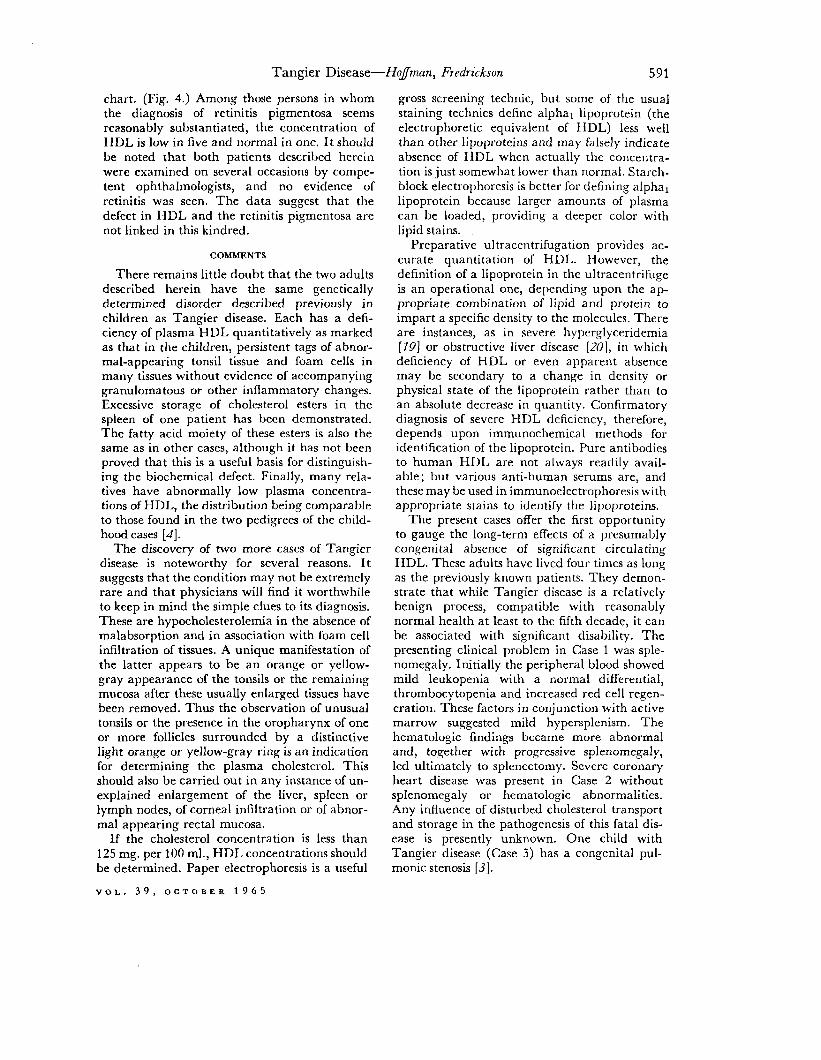
FIG. 1. Case 1. Spleen showing lipid-filled reticulo- endothelial cells of pulp. Hematoxylin and eosin stain, original magnification X 250.
parenchymal cells and brilliant blue-green staining of the foam cells in the rectal tissue.
No abnormalities were noted (by Dr. J. A. Gregg) in the relative proportions of the unconjugated bile acids and their taurine and glycine conjugates in a specimen of bile obtained by duodenal intubation [5]; the total bile acid concentration was within the lower range of normal. The free amino acids in a 24- hour urine sample were normal, and the alpha-amino nitrogen content was 210 mg. (as determined by Dr. J. D. Jones). The serum contained normal amounts of total and alpha1 and alpha2 globulin glycoprotein [G] (as established by Dr. \V. F. McCuckin).
Since then, the patient has been re-evaluated at the Mayo Clinic or National Institutes of Health every three to six months. Plasma cholesterol values have
TABLE II SPLEEN LIPIDS (MG./GM. WET TISSUE)
Data Case 1 7
Total lipid.. Phospholipid.
Phosphatidyl choline, Sphingomyelin.
Cholesterol Free............... Esterified
Cholesterol-ester fatty acids (estimated).
Triglyceride. Lipid hexose.
-(-
A
50.7 20.6 15.4 10.4
4.6 2.1 2.3 2.0
4.8 7.8
5.5 2.0 0.2
4.0 0.7
0.5 0.4 0.2
I Controls *
B
- * Control tissues were obtained from (A) a child
and from (B) an adult who had died following cardiac surgery. Both tissues showed histologic evidence of chronic passive congestion.
remained low. The spleen has increased in size, and on each visit the patient has complained of increased upper abdominal fullness and discomfort. Thrombo- cytopenia and leukopenia have become more marked, along with reticulocytosis and other evidence of in- creased regeneration of erythrocytes, although hemo- globin values have not decreased signilicantly.
In September 1963 the patient complained of dis- abling physical discomfort produced by the spleen, the inferior margin of which was then below the level ol the umbilicus. The liver had not increased in size since the initial examination. Splenectomy was per- formed on September 7. At operation, the spleen was found to be greatly enlarged and much firmer than normal. It weighed 1,160 gm. and was yellowish red. The liver was only slightly enlarged and appeared normal grossly. There were many enlarged lymph nodes along the entire root of the mescntery. The other findings at intra-abdominal exploration were within normal limits. Microscopic sections of the spleen showed the reticuloendothclial cells of the pulp to be filled with intracytoplasmic lipid droplets. There were also scattered clusters of cholesterol crys- tals in the pulp. (Fig. 1.) Biopsy of a lymph node showed marked fatty change. The results from anal- ysis oC spleen lipids are shown in Tables u and III. The patient’s convalescence was uneventful, and he re- sumed a normally active life.
In February 1364 he noted enlargement of the right breast. Other findings on physical examination at this time were within normal limits. Excisional biopsy of the right breast disclosed benign hyperplasia of the ductile tissue and stroma. Staining for fat did not reveal any foam cells in the resected tissue. Labo- ratory studies on two occasions showed the alkaline phosphatase to be 28 and 34 King-Armstrong units. The bromsulfalein test showed 20 per cent dye reten- tion. The plasma total cholesterol was 125 mg. per
TABLE III FATTY ACIDS IN CHOLESTEROL ESTERS FROM SPLEENS
Fatty Case 1 Acid ‘*
12:o 14:o 15:o 16:0 16:l 17:o 18:O 18:l 18:2 20:4 24:l I -
(%)
1.5 5.7 4.9
21.8 13.7
0.6 4.1
40.0 5.1 3.6 1.9
-T
-
Control Bf
(%)
0.8 0.3
31 .l 4.2
13.b 21.9 19.6
9.0 . . .
* Number carbons in chain: number double bonds, t Source as in Table U.
AMERICAN JOURNAL OF MEDICINE

Tangier Disease-Hoj’man, Fredrickson
100 ml., and on paper electrophoresis an increase in low-density lipoprotein (LDL) concentration was noted.
CASE 2. A farmer and distillery worker, the forty- eight year old brother of the first patient, was seen at the Mayo Clinic in March 1963 after a preliminary survey of the family led to the suspicion that he also might have Tangier disease. His medical history in- cluded tonsillectomy in 1950 because of recurrent sore throat and “enlarged tonsils.” (The surgical transcripts were obtained from his local hospital, but there was no comment about the unusual features of the tonsils and no histologic studies were reported.) Typical angina pectoris of moderate severity had been noted daily for the past five years. A prolonged epi- sode of chest pain had led to the patient’s hospitaliza- tion near his home in 1961, but “no permanent dam- age” had been noted. Dull, nonspecific, right abdomi- nal discomfort of several years’ duration was the pa- tient’s only other current complaint. No history of diarrhea or steatorrhea could be elicited.
On physical examination, the weight was 208 pounds and height 67 inches. The blood pressure was 142/100 mm. Hg. The only skin lesions were two 3 mm. yellow-brown spots on the lower calf and tibia1 areas. There was no cornea1 clouding on gross exami- nation, but slit-lamp examination showed a hazily flocculent infiltration throughout the cornea1 stroma. This was described by an ophthalmologist observer (Dr. Ludwig Von Sallmann) as “decreased trans- parency of the cornea, more marked centrally than peripherally, and more prominent in the posterior than the anterior stroma. This slight diffused opacity can be resclved into fine equidistant dots.” Fundu- scopic examination revealed minimal sclerosis of the retinal arterioles in both eyes and a fluffy retinitic spot in the inferior temporal region of the left. Cystic limbal lesions thought to be pingueculae were present bilaterally. In the pharynx a few follicles remained, surrounded by a pale yellow-orange ring in several instances. The liver edge was palpable at the right costal margin ; the spleen was not palpable. The other physical findings were not remarkable.
General laboratory findings are listed in Table I. The standard thoracic roentgenogram, roentgeno- grams of the gallbladder, colon and lumbar vertebrae and the excretory urograms did not reveal any ab- normality. An electrocardiogram showed a non- specific repolarization abnormality in the mid-pre- cordial leads. Marrow aspirated from the iliac crest contained a few macrophages (“foam cells”), their cytoplasm loaded with doubly refractile droplets. Many other scattered droplets appeared to be out- side any cytoplasmic membrane. The remaining marrow elements were normal. Proctoscopic examina- tion revealed a smooth yellowish mucosa with tiny, flat, dark red-brown 1 mm. spots scattered through- out. A rectal biopsy specimen showed mucosal and
VOL. 39, OCTOBER 1965
FIG. 2. Case 2. Rectal biopsy specimen showing large lipid-laden cells in lamina propria and beneath mus- cularis mucosa. Hernatosylin and eosin stain, original magniIicalion X 150.
submucosal collections of foam cells. Biopsy of tonsil and adenoid remnants revealed clusters of pale-stain- ing foamy macrophages. A jejunal biopsy specimen did not show any histologic abnormalities, but a needle biopsy specimen of the liver revealed one small intra- lobular focus of foam cells, as in the previous case. The foam cells from pharyngeal, rectal and liver biopsy specimens exhibited histochemical character- istics identical with those in Case 1. (Fig. 2.)
The patient’s symptomatic coronary heart disease was discussed with him, and he was also advised to reduce his weight. On July 22, 1963, he died suddenly while driving his farm truck. Necropsy was not performed.
METHODS
Measurements of cholesterol [7], phospholipid (S] and triglycerides [9] were performed on chloroform- methanol extracts [7U] of tissue, plasma or lipoprotein fractions isolated by preparative ultracentrifugation [77]. Tissue was homogenized in chloroform: meth- anol (2: l), using two 10 to 20 ml. portions of solvent per gram of wet tissue. After the combined extracts were filtered and the chloroform phase washed ac- cording to the method of Folch and associates [72], they were kept at 4”~. until analyzed. Total lipid was determined gravimetrically and lipid hexose was measured by the anthrone method [73] following

586 Tangier Disease-Hofman, Fredrickson
TABLE IV PLASMA LIPlDS (MC./100 ML.)
Cholesterol ,
source I Phospholipids Triglycerides Total / Free
-__ I I Cax 1 38 (30-45) 14 86 (68-104) 142(136-152) Case 2 69 (50-88) 21 114(104-124) 213(195-238) Controls ; 250 (176-324)
: I . 280 (205-355) 70 (20-120)
NOTE: Values for phosphohpids and triglycerides of patients represent two to five postabsorptive samples obtained on different days. Control cholesterol and phospholipid values represent mean (and 90 per cent confidence limits) obtained from forty-three normal men forty to forty-nine years old, averaging 44.5 years of age. Control triglyceride values were derived from major peak of distribution of values obtained from 390 fasting and apparently healthy subjects [23].
TABLE v ULTRACENTRIFUGAL ANALYSIS OF LIPOPROTEINS
(MC./100 ML. PLASMA)
Lipoprotein Fraction
Case 1 Case 2 Controls *
ct =t C PL C PL
Plasma <1.019 1.021-1.063 1,063-l .21 >1.21
30 68 50 104 171 215 19 44 33 70 26 31 11 15 13 23 97 69
1 1 1 :: / :
43 111
i O 0 21
* Mean of samples from ten men, aged twenty-one to twenty-eight years. From Have1 et al. [ 7 I] t C = cholesterol. PL = phospholipid.
hydrolysis of the sample at 100’~. for 15 hours in 6 N hydrochloride. Phospholipids were separated and quantified by the method of Skidmore and Entenman [ 74) using two-dimensional thin layer chromatog- raphy. Cholesterol esters were isolated by chroma- tography on alumina. Methyl esters of fatty acids were chromatographed on 15 per cent ethylene glycol succinate at 195’~. The ionization detector was calibrated for response with NIH Metabolism Study Section standards. Paper electrophoresis for lipopro- teins was performed by the technics of Swahn [75] and of Lees and Ilatch [/G].
RESULTS
Plasma Lipids and Lipoproteins. In the tlvo cases, the plasma lipids (Table IV) followed the abnormal pattern which is characteristic of Tangier disease, although not necessarilyunique. This is the association of low levels of cholesterol and phospholipids with normal or elevated con- centrations of glycerides. On the basis of the known composition of the major plasma lipo- protein systems, it may be predicted from this
lipid pattern that there \vill be a deficiency in HDL or in that portion of LDL of density be- tween 1.019 and 1.063, with an associated fmd- ing of normal or elevated concentrations of tri- glyceride-rich LDL of density less than 1.019.
In these cases all three features proved to be present. On paper electrophoresis, no alphai lipoprotein could be seen. On preparative ultra- centrifugation (Table v), there was no significant HDL in the appropriate density band, 1.063 to 1.21. Also, there was a definite decrease in LDL of density 1.019 to 1.063 (which corresponds to beta lipoproteins on paper electrophoresis or Sf 0 to 12 in the analytical ultracentrifuge) and probably a significant increase in LDL of den- sity less than 1.019 in Case 2.
Immunoelectrophoresis on agar, using either rabbit antibodies to human HDL or horse anti- human serum, revealed no precipitation line attributable to HDL. The results of agar diffu- sion studies are shown in Figure 3A. The rabbit anti-HDL serum used in these experiments has
AMERICAN JOURNAL OF MEDICINE

Tangier Disease--lZ~J~nan, Fredrickson 587
FIG. 3. A, precipitation lines formed by Ouchterlony agar dillusion technic with serum from normal subject (I ), Cav 1 (2), Case 2 (3) and patient with acanthocytosis and abetalipoproteinemia (4) against rabbit antibodies pre- pared against human lipoproteins of density 1.063 to 1.21 (“anti-l IDL”). Both precipitation lines at (1) are attributed to HDL (see text). H, arrangement same as in A, except rabbit antibodies were prepared against human lipoprotein of density 1.063 (“anti-LDL”). Here both patients with Tangier disease demonstrated LDL, which was further shown by immunoelectrophoresis. ThFse plates were prepared by Dr. Robert I. Levy.
been shown to detect two different HDL anti- gens and to contain no antibodies to either albumin or LDL [ 171. A very faint precipitation line was formed by serums from both patients, suggesting the presence of a very small amount of HDL. (Fig. 3A.) This is very similar to results obtained in the four other known cases of Tan- gier disease, and precise identification of the faintly detectable antigen is under way. In contrast to serum from a patient with abetalipo- proteinemia (kindly supplied by Dr. Leonard Laster), serums from both of the present pa- tients had easily detectable LDL, antigenically similar to that in normal subjects although some- what decreased in amount. (Fig. 3B.)
The abnormal concentrations in the LDL fractions were further associated with lower than normal cholesterol : phospholipid ratios in these lipoproteins. Both of these abnormalities have also been observed in lesser degree in the four children with Tangier disease [IS].
Tissue Lipids. The significant splenic abnor- mality in Case 1 (Table II) was the presence of cholesterol esters in amounts which far exceeded that in the two control tissues analyzed concur- rently. The fatty acid pattern of the cholesterol esters in the spleen in this case (Table III) was
VOL.. 39, OCTOBER 1965
similar to that seen earlier in tonsils and lymph nodes from the children with Tangier disease [Z]. Oleic acid predominated and the relative amounts of linoleic and palmitic acids were less than those seen in the control spleen. (Table III.)
A considerable amount-about 30 per cent- of the total lipid of the spleen in Case 1, as esti- mated gravimetrically, could not be accounted for in the summation of the cholesterol, phos- pholipid and glyceride fractions. (Table II.) Such measurements were made a total of nine times on three different extracts of the abnormal spleen. Since 13 to 22 per cent of lipid was un- accounted for in the controls also, it is likely that the gravimetric measurements of total lipid were uniformly and inexplicably too high; but the possibility of an abnormal and undetected lipid in the tissue in Case 1 has not been excluded completely.
The lipid hexose content was normal, how- ever; and the total fatty acid content, deter- mined by quantitative gas chromatography after saponification and methylation, was equivalent to that predicted from the quantities of estcrified lipids specifically measured and pre- sented in Table II. The gas chromatographic pattern of the total fatty acids in the spleen
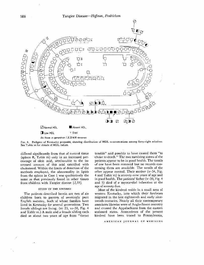
588 Tangier Disease-I-logman, Fredrickson
q Normal HDL q Absenl HDL
GLOW HDL t Died
All those ~n generotion I.lI.IUW!Z-deceased
FIG. 4. Pedigree of Kentucky propositi, showing distribution of HDL ccncentratiom among forty-eight relatives. See Table VI for details of HDL values.
differed significantly from that of control tissue (spleen B, Table III) only in an increased per- centage of oleic acid, attributable to the in- creased amount of this acid esterified with cholesterol. Within the limits of detection of the methods employed, the abnormality in lipids from the spleen in Case 1 was qualitatively the same as that previously found in other tissues from children with Tangier disease [2,78].
STUDY OF THE KINDRED
The patients described herein are two of six children born to parents of seemingly pure English ancestry, both of whose families have lived in Kentucky for several generations. Two female siblings are living. (~1-23, ~1-30, Fig. 4 and Table VI.) A male and a female sibling each died at about two years of age from “throat
trouble” said possibly to have caused them “to choke to death.” The two surviving sisters of the patients appear to be in good health. The tonsils of one have been removed but no records con- cerning them are available. The tonsils of the other appear normal. Their mother (v-34, Fig. 4 and Table VI) is seventy-nine years of age and in good health. The patients’ father (v-30, Fig. 4 and 5) died of a myocardial infarction at the age of seventy-five.
Most of the kindred reside in a small area of western Kentucky, into which their forebears migrated in the late eighteenth and early nine- teenth centuries. Nearly all their contemporary associates likewise were of Anglo-Saxon ancestry and crossed the Appalachians from the eastern seaboard states. Antecedents of the present kindred have been traced to Pennsylvania,
AMERICAN JOURNAL OF MEDICINE

v-2 v-3 v-4 v-9 v-17 v-21 v-24 v-26 v-27 v-28 v-29 v-32 v-34 v-37 V-41
VI-4 VI-9 VI-1 0 VI-1 I w-1 2 m-1 3 VI-1 6 VI-17 VI-1 9 VI-20 VI-2 1 VI-22 w-23 ~1-25 ~1-26 w-27 ~1-28 VI-30 VI-31 m-35 ~1-36 ~1-38 VI-39 VI-40 VII-5 VII-7 VII-1 2 WI-1 3 w-1 4 VII-1 5 VII-1 6 w-1 7 WI-1 8
Tangier Disease--Ho$man, Fredrickson 589
DISTRIBUTION OF LIPID FRACTIONS AMONG KENTUCKY KINDRED
Age (y-r.1 and Sex
79, F 77, F 75, F 64, M 71, M 85, F 79, M 75, M 56, F 69, M 68, M 68, M 79, F 87, F 73, M 36, M 56, F 57, F 55, F 48, F 42, F 49, M 43, M 35, M 46, F 45, F 36, M 58, F 45, F 48, M 44, M 43, F 36, F 34, M 36, M 37, M 60, M 69, M 65, F 19, M 10, F 19, F 10, M 19, M 15, M 13, M 35, F
25, F
TC*
280 297 299 347 344 376 294 331 174 250 167 218 261 273 181 242 221 313
Ii;; 2% 373 353 160 190 118 166 205 248 229 301 213 305 418 389 245 315 250 239 284 304 232 281 174 274 234 .316 215 266 132 176 157 222 278 347 241 298
88 115 36 104
199 220 115 159 228 233 252 317 306 258 226 243 236 287 241 331 136 187 168 200 138 151 102 170 105 151 108 203 107 196 154 194 174 228
-
--
-
Plasma Density >l ,063
PL’ TG*
iii is5 148 209 215
ii;
iii 116
&ii 178
‘Ii, 174 213 104
ii;
ii6 138
49 54
iii
‘ii t31 41 79 76
-
-
TC
66 50 72 64 33 22 34 24 49 15 19 53 30 20 52 30 53 79 59 81 58
117 34 37 29 33 29 44 30
1 1
52 35 35 44 53 39 31 58 36 45 29 29 35 36 32 59 61
PL
HDL Pheno-
typic Classifica-
tion?
144 N 128 N 170 N 158 N
97 N 74 L 94 N 82 L
144 N 72 -L 75 L
122 N 67 L 67 L
136 N 91 L
138 N 184 N 161 N 171 N 131 N 160 N 115 N 107 N
93 L 93 L 87 L
121 N 101 L
14 A 8 A
111 N 89 N 85 N
134 N 104 N 107 N 100 L 148 N 102 N 131 N
70 L 96 L 88 N
108 N 105 L 136 N 150 N
* TC = total cholesterol. PL = phospholipids. TG = triglycerides. t N = normal HDL. L = abnormally low. HDL (heterozygous). A = absent HDL (homozygous).
VOL. 39, OCTOBER 1965
Tangier Disease---Hoffman, Fredrickson
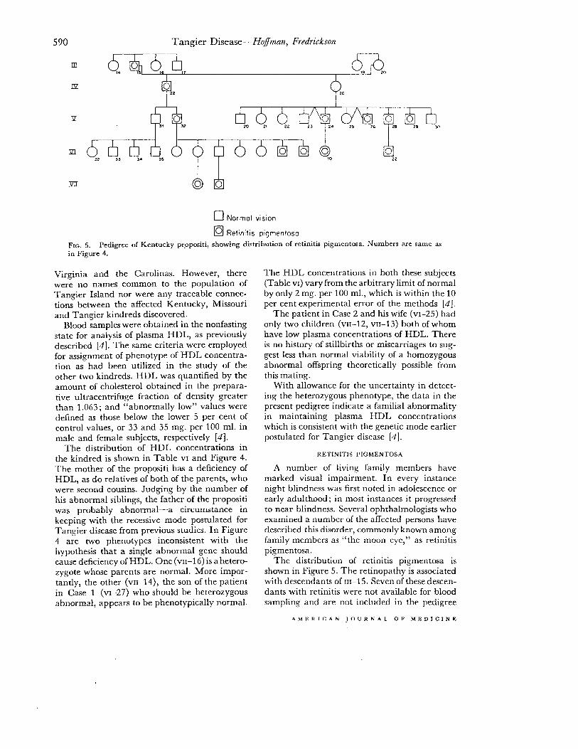
cl Normol vision
q Retinitis pigmentoso Fro. 5. Pedigree of Kentucky prppositi, showing distribution of retinitis pigmentosa. Numbers are same as in Figure 4. -
Virginia and the Carolinas. However, there were no names common to the population of Tangier Island nor were any traceable connec- tions between the affected Kentucky, Missouri and Tangier kindreds discovered.
Blood samples were obtained in the nonfasting state for analysis of plasma HDL, as previously described [#I. The same criteria were employed for assignment of phenotype of HDL concentra- tion as had been utilized in the study of the other two kindreds. HDL was quantified by the amount of cholesterol obtained in the prepara- tive ultracentrifuge fraction of density greater than 1.063; and “abnormally low” values were defined as those below the lower 5 per cent of control values, or 33 and 35 mg. per 100 ml. in male and female subjects, respectively [4].
The distribution of HDL concentrations in the kindred is shown in Table VI and Figure 4. The mother of the propositi has a deficiency of HDL, as do relatives of both of the parents, who were second cousins. Judging by the number of his abnormal siblings, the father of the propositi was probably abnormal-a circumstance in keeping with the recessive mode postulated for Tangier disease from previous studies. In Figure 4 are two phenotypes inconsistent with the hypothesis that a single abnormal gene should cause deficiency of HDL. One (VII-~ 6) is a hetero- zygote whose parents are normal. More impor- tantly, the other (VII-14), the son of the patient in Case 1 (~1-27) who should be heterozygous abnormal, appears to be phenotypically normal.
The HDL concentrations in both these subjects (Table VI) vary from the arbitrary limit of normal by only 2 mg. per 100 ml., which is within the 10 per cent experimental error of the methods [4].
The patient in Case 2 and his wife (~1-25) had only two children (~11-12, ~11-13) both of whom have low plasma concentrations of HDL. There is no history of stillbirths or miscarriages to sug- gest less than normal viability of a homozygous abnormal offspring theoretically possible from this mating.
With allowance for the uncertainty in detect- ing the heterozygous phenotype, the data in the present pedigree indicate a familial abnormality in maintaining plasma HDL concentrations which is consistent with the genetic mode earlier postulated for Tangier disease [4].
RETINITIS PICMENTOSA
A number of living family members have marked visual impairment. In every instance night blindness was first noted in adolescence or early adulthood; in most instances it progressed to near blindness. Several ophthalmologists who examined a number of the affected persons have described this disorder, commonly known among family members as “the moon eye,” as retinitis pigmentosa.
The distribution of retinitis pigmentosa is shown in Figure 5. The retinopathy is associated with descendants of 111-15. Seven of these descen- dants with retinitis were not available for blood sampling and are not included in the pedigree
AMERICAN JOURNAL OF MEDICINE
Tangier Disease--NoJman, Fredrickson 591
chart. (Fig. 4.) Among those persons in whom gross screening technic, but some of the usual the diagnosis of retinitis pigmentosa seems staining technics define alphai lipoprotein (the reasonably substantiated, the concentration of electrophoretic equivalent of HDL) less well HDL is low in five and normal in one. It should than other lipoproteins and may falsely indicate be noted that both patients described herein absence of HDL when actually the concentra- were examined on several occasions by compe- tion is just somewhat lower than normal. Starch- tent ophthalmologists, and no evidence of block electrophoresis is better for defining alphai retinitis was seen. The data suggest that the lipoprotein because larger amounts of plasma defect in HDL and the retinitis pigmentosa are can be loaded, providing a deeper color with not linked in this kindred. lipid stains.
COMMENTS
There remains little doubt that the two adults described herein have the same genetically determined disorder described previously in children as Tangier disease. Each has a defi- ciency of plasma HDL quantitatively as marked as that in the children, persistent tags of abnor- mal-appearing tonsil tissue and foam cells in many tissues without evidence of accompanying granulomatous or other inflammatory changes. Excessive storage of cholesterol esters in the spleen of one patient has been demonstrated. The fatty acid moiety of these esters is also the same as in other cases, although it has not been proved that this is a useful basis for distinguish- ing the biochemical defect. Finally, many rela- tives have abnormally low plasma concentra- tions of HDL, the distribution being comparable to those found in the two pedigrees of the child- hood cases [4].
The discovery of two more cases of Tangier disease is noteworthy for several reasons. It suggests that the condition may not be extremely rare and that physicians will find it worthwhile to keep in mind the simple clues to its diagnosis. These are hypocholesterolemia in the absence of malabsorption and in association with foam cell infiltration of tissues. A unique manifestation of the latter appears to be an orange or yellow- gray appearance of the tonsils or the remaining mucosa after these usually enlarged tissues have been removed. Thus the observation of unusual tonsils or the presence in the oropharynx of one or more follicles surrounded by a distinctive light orange or yellow-gray ring is an indication for determining the plasma cholesterol. This should also be carried out in any instance of un- explained enlargement of the liver, spleen or lymph nodes, of cornea1 infiltration or of abnor- mal appearing rectal mucosa.
If the cholesterol concentration is less than 125 mg. per 100 ml., HDL concentrations should be determined. Paper electrophoresis is a useful
Preparative ultracentrifugation provides ac- curate quantitation of HDL. However, the definition of a lipoprotein in the ultracentrifuge is an operational one, depending upon the ap- propriate combination of lipid and protein to impart a specific density to the molecules. There are instances, as in severe hyperglyceridemia [19] or obstructive liver disease [ZO], in which deficiency of HDL or even apparent absence may be secondary to a change in density or physical state of the lipoprotein rather than to an absolute decrease in quantity. Confirmatory diagnosis of severe HDL deficiency, therefore, depends upon immunochemical methods for identification of the lipoprotein. Pure antibodies to human HDL are not always readily avail- able; but various anti-human serums are, and these may be used in immunoelectrophoresis with appropriate stains to identify the lipoproteins.
The present cases offer the first opportunity to gauge the long-term effects of a presumably congenital absence of significant circulating HDL. These adults have lived four times as long as the previously known patients. They demon- strate that while Tangier disease is a relatively benign process, compatible with reasonably normal health at least to the fifth decade, it can be associated with significant disability. The presenting clinical problem in Case 1 was sple- nomegaly. Initially the peripheral blood showed mild leukopenia with a normal differential, thrombocytopenia and increased red cell regen- eration. These factors in conjunction with active marrow suggested mild hypersplenism. The hematologic findings became more abnormal and, together with progressive splenomegaly, led ultimately to splenectomy. Severe coronary heart disease was present in Case 2 without splenomegaly or hematologic abnormalities. Any influence of disturbed cholesterol transport and storage in the pathogenesis of this fatal dis- ease is presently unknown. One child with Tangier disease (Case 3) has a congenital pul- manic stenosis [3].
VOL. 39, OCTOBER 1965
592 Tangier Disease--H@man, Fredrickson
Hitherto unappreciated sites of lipid storage in Tangier disease were demonstrated in the gut and eyes of the two adult patients. Gastrointes- tinal symptoms (mild intermittent diarrhea) in Case 1 led to proctoscopic examination, which revealed a unique gross appearance of the rectal mucosa in association with mucosal and sub- mucosal foam cell infiltration. Identical findings were present in Case 2. The suggestively steator- rhea1 nature of the stools in Case 1 was investi- gated by an intake-excretion study of fecal fat and nitrogen. The value for fecal fat was slightly in excess of normal. (Table I.) Both of the brothers also had low serum concentrations of carotene. However, the absence of other evi- dence of malabsorption, despite roentgenog- raphy of the small intestine and biopsy of the jejunal mucosa in Case 1, militates against a primary or secondary malabsorption syndrome which might be responsible for the abnormal serum lipid and lipoprotein findings in these pa- tients. Moreover, in Case 2 the patient had no gastrointestinal symptoms and was obese, and his fecal fat and nitrogen excretions were normal. No foam cells or other abnormalities were seen in the biopsy specimens of jejunal mucosa from either patient. Gut biopsies have not been per- formed in the four children with Tangier disease; but they, too, have had no evidence of gastro- intestinal dysfunction.
The cornea1 clouding and stromal deposits observed on slit-lamp examination presumably are due to deposition of cholesterol esters in a manner similar to that occurring in other tissues. Moderate hyperuricemia without evidence of ab- normal renal function was present in both cases, but its significance here cannot be determined.
The basic inheritable defect in Tangier dis- ease remains to be established with certainty. HDL is one of the major mechanisms for trans- porting lipid in extracellular fluid, others being the albumin-free fatty acid complex and LDL [21]. From a physiologic standpoint, particulate emulsions such as chylomicrons may be con- sidered a fourth mechanism, although their chemical and functional relationships to the other lipoproteins are not fully understood. One of the major contributions of studies of Tangier disease has been to point out an apparent lack of absolute requirement for HDL in the absorption of fat and formation of chylomicrons [78].
The polypeptide in HDL is specific and dis- tinct from that in LDL. From the data available it seems most likely that inability to maintain
significant concentration of plasma HDL in Tangier disease is due to a genetically deter- mined defect in elaboration of this polypeptide. If this be true, the widespread accumulation of cholesterol esters in tissues suggests that HDL may have some specific function in maintaining the equilibrium of cholesterol between plasma and tissues.
It is noteworthy that there are also abnor- malities in LDL in Tangier disease. In the adults they have been especially marked (Table v), but qualitatively similar findings have been present in the affected children [18]. The hypocholes- terolemia in Tangier disease thus is not solely due to an absence of significant HDL, although the latter lipoprotein class is by far the most profoundly affected. In the ultimate explanation for Tangier disease as based on alteration at a single gene locus, it will be necessary to account for changes in all classes of lipoproteins as, at the least, secondary phenomena.
One of the few conditions in which cholesterol esters are stored selectively in reticuloendothelial tissues is eosinophilic granuloma [22]. Neither the diagnostic granulomatous changes nor other typical clinical features are found in Tangier disease. One of us (D. S. F.) has had oppor- tunity to measure plasma HDL in two patients with an active form of the disseminated variety of eosinophilic granuloma. In one, HDL was normal; in the other, it was abnormally low (HDL cholesterol measured 24 mg. per 100 ml., the mean for her age and sex being 54). How- ever, the decrease was not comparable to the near absence seen in Tangier disease [23]. Eosinophilic granuloma is not a familial disease, and tonsillar abnormalities have not been re- ported in association with it.
While the frequency of the gene or genes asso- ciated with Tangier disease must undoubtedly be quite low in the general population, the finding of six cases within a three year period of awareness of the disease suggests that more are to be found. It still is less common than its counterpart abnormality, abetalipoproteinemia [24,23’3. The latter is characterized by the absence of both the LDL and particulate forms of lipoproteins and is associated with acantho- cytosis, steatorrhea, neurologic changes and retinopathy. While LDL was reported as being deficient in the parents of the patient first de- scribed [2J], no definite mode of genetic in- fluence on the development of this latter disease has yet been established. The extension of clini-

Tangier Disease-HoJman, Fredrickson
cal observations in Tangier disease to adults suggests that severe deficiency of HDL is asso- ciated with a more benign long-term course than abetalipoproteinemia. Both of these diseases offer unusual opportunities for further exploring the functions and control of the major plasma lipoprotein systems, and all new patients deserve careful study and extensive documentation.
SUMMARY
Two brothers, aged forty-five and forty-eight, with familial deficiency of high-density lipo- protein (HDL) (Tangier disease) are described. These are the fifth and sixth known patients and the first adults with the disease. They demon- strated the hypocholesterolemia, abnormal ton- sils and tissue storage of cholesterol esters char- acteristic of this condition. One brother had hypersplenism and both had unusual infiltration of the corneas and rectal mucosa, new findings presumably related to the chronic course of Tangier disease. One brother had coronary artery disease which apparently caused his death. Low plasma concentrations of high- density lipoprotein were distributed among family members in a manner similar to that reported in the two other involved kindreds.
Acknowledgment: We thank Dr. Charles F. Stroebel and Dr. Haddon M. Carryer of the Mayo Clinic for their clinical contributions to the management of these patients.
REFERENCES
1. FREDRICKSON, D. S., ALTROCCHI, P. H., AVIOLI, L. V., GOODMAN, D. S. and GOODMAN, H. C. Tangier disease. Ann. Int. Med., 55: 1016, 1961.
2. FREDRICKSON, D. S. and ALTROCCHI, P. H. Tangier disease (familial cholesterolosis with high-density lipoprotein deficiency). In: Cerebral Sphingo- lipidoses: A Symposium on Tay-Sachs Disease and Allied Disorders, pp. 343-357. Edited by Aronson, S. U. and Volk, B. W. New York, 1962. Academic Press, Inc.
3. FREDRICKSON, D. S., SHIRATORI, T. and YOUNG, 0. M. Genetic control of high-density lipoprotein concentrations in man. (Abstract.) Crrculation, 26: 653,1962.
4. FREDRICKSON, D. S.,You~o, O., SHIRATORI, T. and BRIGGS, N. The inheritance of high density lipo- protein deficiency (Tangier disease). J. Clin. Inmt., 43: 228, 1964.
5. SJGVALL, J. The determination of bile acids in bile and duodenal contents by quantitative paper chromatography. Clin. chim. a&z, 4: 652, 1959.
6. MCGUCKIN, W. F. and MCKENZIE, B. F. An im- proved periodic acid fuchsin sulfite staining method for evaluation of glycoproteins. Clin. Chcm., 4: 476, 1958.
7. SPERRY, W. M. and WEBB, MERRILL. A revision of
Reprinted from the October issue of The
the Schoenheimer-Sperry method for cholesterol determination. J. Biol. Chcm., 187: 97, 1950.
8. STEWART, C. P. and HENDRY, E. B. The phospho- lipins of blood. Biochem. J., 29: 1683, 1935.
9. VAN HANDEL, E. and ZILVERSNT, D. B. Micro- method for the direct determination of serum tri- glycerides. J. Lob. H Clin. Med., 50: 152, 1957.
10. BRAGDON, J. H. Extraction of lipids from serum. In: Lipids and the Steroid Hormones in Clinical Medicine, p. 6. Edited by Sunderman, F. W. and Sunderman, F. W., Jr. Philadelphia, 1960. J. B. Lippincott Co.
11. HAVEL, R. J., EDER, H. A. and BRAGDON, J. H. The distribution and chemical composition of ultracentrifugally separated lipoproteins in human serum. J. Clin. Znuest., 34: 1345, 1955.
12. FOLCH, J., LEES, M. and STANLEY, G. H. S. A simple method for the isolation and purification of total lipides from animal tissues. J. Biol. Chcm., 226: 497, 1957.
13. HASSID, W. Z. and ABRAHAM, S. Chemical pro- cedures for analysis of polysaccharides. In: Meth- ods in Enzymology, vol. 3, pp. 34-50. Edited by Colurick, S. P. and Kaplan, N. 0. New York, 1957. Academic Press, Inc.
14. SKIDMORE, W. D. and ENTENMAN, C. Two-dimen- sional thin-layer chromatography of rat liver phosphatides. J. Lipid Rcs., 3: 471, 1962.
15. SWAHN, B. A method for localization and deter- mination of serum lipids after electrophoretical separation on filter paper. &and&m. J. Clin. G? Lab. Znucrt., 4: 98, 1952.
16. LEES, R. S. and HATCH, F. T. Sharper separation of lipoprotein species by paper electrophorcsis in albumin-containing buffer. J. Lab. E? Clin. Med., 61: 518, 1963.
17. LEVY, R. I. and FREDRICKSON, D. S. Antigenic heterogeneitv of human plasma high density lipo- protein. J. Clil. Invest., 43: 1286, 1964.
18. FREDRICKSON, D. S. Tangier disease. In: Metabolic Basis of Inherited Disease, 2nd ed. Fdited by Stanbury, J. B., Wyngaarden, J. B. and Fredrick- son, D. S. New York, 1965. McGraw-Hill Book Co., Inc.
19. HAVEL, R. J. and GORDON, R. S., JR. Idiopathic hyperlipemia: metabolic studies in an ahected family. J. Clin. Inuest., 39: 1777, 1960.
20. Russ, ELLA M., RAYMUNT, JULIE and BARR, D. P. Lipoproteins in primary biliary cirrhosis. J. C/in. Invest., 35: 133, 1956.
21. FREDRICKSON, D. S. and GORDON, R. S., JR. Trans- port of fatty acids. Physiol. Rev., 38: 585, 1958.
22. THANNHAUSER, S. J. Lipidosis: Diseases of the Intra- cellular Lipid Metabolism. New York, 1958. Grune & Stratton, Inc.
23. FREDRICKSON, D. S. Unpublished data. 24. MIER, M., SCHWARTZ, S. 0. and BOSHES, B.
Acanthocytosis, pigmentary degeneration of the retina and ataxic neuropathy. A genetically determined syndrome with associated metabolic disorder. Blood, 16: 1586, 1960.
25. SCHWARTZ, J. F., ROWLAND, L. P., EDER, I-I., MARKS, P. A., OSSERMAN, E. F., HIRSCRBERG, E. and ANDERSON, H. Bassen-Kornzweig syndrome. Deficiency of serum p-lipoprotein. Arch. Neural., 8: 438, 1963.
American Journal of Medicine volume 39. number 4, pages 582-593. copyright 1965 and printed in the U.S.A. Published by Thr Reuben H. Dnnnelley Corpnrnlion, 466 Lexington Ave., New York 10017.
![Tangier City Apartments[1]](https://static.fdocuments.net/doc/165x107/54e8685a4a7959704f8b4c0d/tangier-city-apartments1.jpg)