Syndrome of absent two with microcephaly, polymicrogyria,...
7
J. Neurol. Neurosurg. Psychiat., 1970, 33, 844-850 Syndrome of absent abdominal muscles: two cases with microcephaly, polymicrogyria, and cerebellar malformations REID R. HEFFNER1 From the Department of Pathology, Yale University School of Medicine, New Haven, Connecticut, U.S.A. SUMMARY Two unique cases of the syndrome of absent abdominal muscles with central nervous system involvement are presented. Microcephaly, polymicrogyria, and cerebellar heterotopiae were present in both. In case 1 there was also absence of the corpus callosum and agenesis of the cerebellar vermis. In case 2 a count of anterior horn cells in the spinal cord showed a reduction of approxi- mately 50% in the lower thoracic region. The pertinent literature is briefly discussed. The findings in the nervous system suggest that the syndrome is the result of defective embryogenesis during the first trimester. Congenital absence of the abdominal muscles was first described by Frohlich in 1839 (Bourne and Cerny, 1967), but not until the turn of the century (Parker, 1895) was this finding known to be associ- ated with genitourinary abnormalities. Eagle and Barrett (1950), who reviewed 42 cases in the world literature and added nine of their own, were among the first to indicate that the deficiency of abdominal muscles is actually part of a syndrome, usually occurring in males, which consists of a triad of abdominal muscle aplasia, massive bladder and ureteral dilatation, and cryptorchism. The kidneys often are affected by secondary hydronephrosis (Silverman and Huang, 1950) or by polycystic dysplasia (Nunn and Stephens, 1961). Commonly associated anomalies include lower urinary tract obstruction, patent urachus, pigeon breast, and talipes. Dislocation of the hip (Metrick, Brown, and Rosenblum, 1957), pulmonary atresia (Brierre, 1963), choanal atresia (Brierre, 1963), and abnor- malities of the gastrointestinal and cardiovascular systems (Metrick, Brown, and Rosenblum, 1957) have been reported. Since the original observations by Parker (1895), over 120 cases have been described (Williams and Burkholder, 1967), yet investigations 'Present address (including reprint requests): Section of Neuropath- ology, Department of Pathology, New York Hospital-Cornell Medical Center, 1300 York Avenue, New York City, New York, 10021, U.S.A. of neurological interest have been few. This report concerns two cases of the syndrome of absent abdominal muscles with involvement of the central nervous system. CASE 1 This 1 day old male was transferred from a neighbouring hospital to the Yale-New Haven Medical Center for evaluation of absent abdominal muscles. The baby's mother, aged 26, had produced two normal children previously. The present pregnancy and delivery had been uneventful. The gestational age was 37 weeks. On admission the infant weighed 3,460 g and measured 50 cm in crown-heel length. The head was quite small, measuring only 30 cm in circumference, as compared with a chest circumference of 34 cm. The ears were low- set and the vertex of the skull was flat, giving the forehead a receding appearance. The neck was extremely short. A high-arched palate was noted. The abdomen was dis- tended due to a lack of abdominal muscles. A patent urachus was present. The testes were undescended. Bilateral talipes equinovarus and a dislocated left hip were evident. Neurological examination disclosed generalized hypotonia and diminished suck and grasp reflexes. Intravenous pyelography demonstrated poor visualization bilaterally. Markedly dilated and tortuous ureters were seen on retrograde pyelography. A right nephrostomy was performed. Post-operatively the patient had several generalized grand mal convulsions for which he was given phenobarbitone. At the age of 7 days the child suffered a fatal cardiorespiratory arrest. 844 guest. Protected by copyright. on 26 October 2018 by http://jnnp.bmj.com/ J Neurol Neurosurg Psychiatry: first published as 10.1136/jnnp.33.6.844 on 1 December 1970. Downloaded from
Transcript of Syndrome of absent two with microcephaly, polymicrogyria,...

J. Neurol. Neurosurg. Psychiat., 1970, 33, 844-850
Syndrome of absent abdominal muscles:two cases with microcephaly, polymicrogyria, and
cerebellar malformationsREID R. HEFFNER1
From the Department ofPathology, Yale University School of Medicine, New Haven,Connecticut, U.S.A.
SUMMARY Two unique cases of the syndrome of absent abdominal muscles with central nervoussystem involvement are presented. Microcephaly, polymicrogyria, and cerebellar heterotopiae werepresent in both. In case 1 there was also absence of the corpus callosum and agenesis of the cerebellarvermis. In case 2 a count of anterior horn cells in the spinal cord showed a reduction of approxi-mately 50% in the lower thoracic region. The pertinent literature is briefly discussed. The findings inthe nervous system suggest that the syndrome is the result of defective embryogenesis during thefirst trimester.
Congenital absence of the abdominal muscles wasfirst described by Frohlich in 1839 (Bourne andCerny, 1967), but not until the turn of the century(Parker, 1895) was this finding known to be associ-ated with genitourinary abnormalities. Eagle andBarrett (1950), who reviewed 42 cases in the worldliterature and added nine of their own, were amongthe first to indicate that the deficiency of abdominalmuscles is actually part of a syndrome, usuallyoccurring in males, which consists of a triad ofabdominal muscle aplasia, massive bladder andureteral dilatation, and cryptorchism. The kidneysoften are affected by secondary hydronephrosis(Silverman and Huang, 1950) or by polycysticdysplasia (Nunn and Stephens, 1961). Commonlyassociated anomalies include lower urinary tractobstruction, patent urachus, pigeon breast, andtalipes. Dislocation of the hip (Metrick, Brown, andRosenblum, 1957), pulmonary atresia (Brierre,1963), choanal atresia (Brierre, 1963), and abnor-malities of the gastrointestinal and cardiovascularsystems (Metrick, Brown, and Rosenblum, 1957)have been reported. Since the original observationsby Parker (1895), over 120 cases have been described(Williams and Burkholder, 1967), yet investigations
'Present address (including reprint requests): Section of Neuropath-ology, Department of Pathology, New York Hospital-CornellMedical Center, 1300 York Avenue, New York City, New York,10021, U.S.A.
of neurological interest have been few. This reportconcerns two cases of the syndrome of absentabdominal muscles with involvement of the centralnervous system.
CASE 1
This 1 day old male was transferred from a neighbouringhospital to the Yale-New Haven Medical Center forevaluation of absent abdominal muscles. The baby'smother, aged 26, had produced two normal childrenpreviously. The present pregnancy and delivery had beenuneventful. The gestational age was 37 weeks. Onadmission the infant weighed 3,460 g and measured 50cm in crown-heel length. The head was quite small,measuring only 30 cm in circumference, as comparedwith a chest circumference of 34 cm. The ears were low-set and the vertex of the skull was flat, giving the foreheada receding appearance. The neck was extremely short. Ahigh-arched palate was noted. The abdomen was dis-tended due to a lack of abdominal muscles. A patenturachus was present. The testes were undescended.Bilateral talipes equinovarus and a dislocated left hipwere evident. Neurological examination disclosedgeneralized hypotonia and diminished suck and graspreflexes. Intravenous pyelography demonstrated poorvisualization bilaterally. Markedly dilated and tortuousureters were seen on retrograde pyelography. A rightnephrostomy was performed. Post-operatively the patienthad several generalized grand mal convulsions for whichhe was given phenobarbitone. At the age of 7 days thechild suffered a fatal cardiorespiratory arrest.
844
guest. Protected by copyright.
on 26 October 2018 by
http://jnnp.bmj.com
/J N
eurol Neurosurg P
sychiatry: first published as 10.1136/jnnp.33.6.844 on 1 Decem
ber 1970. Dow
nloaded from

Sylidrome ofabsent abdominal muscles: microcephaly, polymicrogyria, and cerebellar malformations 845
Necropsy revealed bilateral pleural effusions and focalpulmonary atelectasis. Multiple sections through theanterior abdominal wall failed to yield any gross orhistological evidence of the rectus abdominis, external orinternal oblique, or transversus abdominis muscles. Themuscles of the back and thorax including the intercostalswere normal. The kidneys were not enlarged but showedmulticystic dysplasia microscopically. The dilated uretersentered the bladder normally. Ganglion cells werenumerous in the ureters and bladder. No evidence oflower urinary tract obstruction was found. The testeswere located in the abdominal cavity lying superior tothe ureters at the ureterovesical junction.
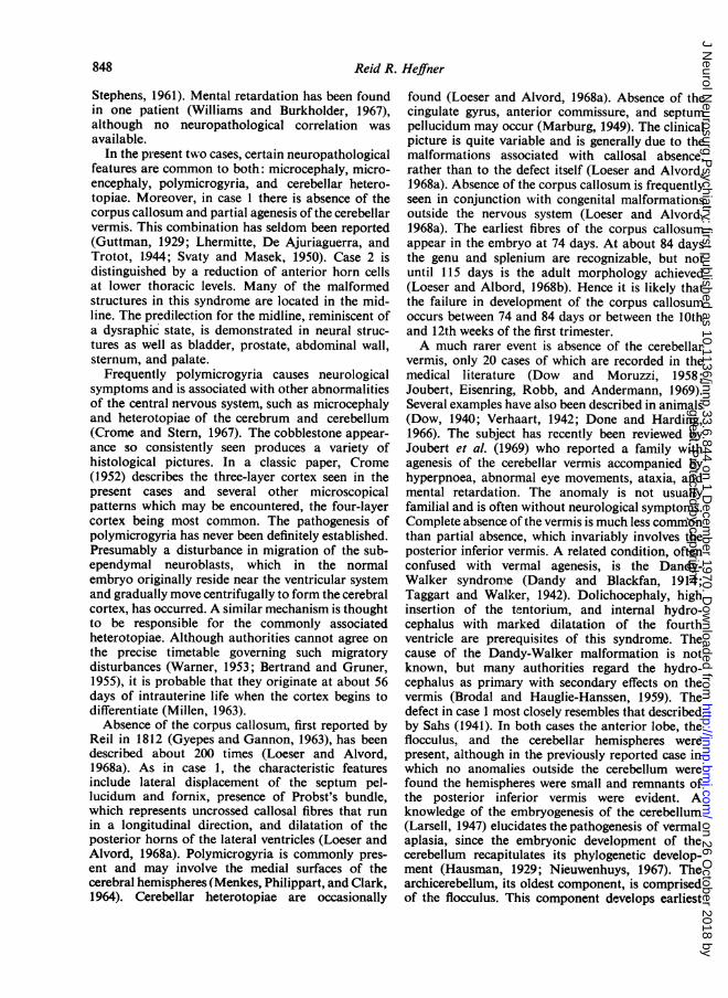
After formalin fixation for 10 days the brain wasexamined. For the size of the infant, it was small, weigh-ing 220 g. There was polymicrogyria of the medialportions of the frontal and parietal lobes, imparting aMorocco-leather appearance to the superior frontalgyrus and the paracentral lobule. Coronal sections of thecerebral hemispheres revealed absence of the corpuscallosum (Fig. 1). The septum pellucidum and fornixwere displaced laterally. The anterior commissure andcingulate gyrus appeared normal. The bundle of Probstwas prominent. There was apparent dilatation of thethird ventricle due to the absent corpus callosum anddisplaced related structures and actual dilatation of theoccipital horns of the lateral ventricles. The posteriorinferior cerebellar vermis was absent. From the posteriorsurface of the cerebellum a direct view could be obtainedof the floor of the deep, trough-shaped caudal fourthventricle, which was covered only by an opaque, thickenedpia-arachnoid membrane. Serial horizontal sectionsdemonstrated partial agenesis of the vermis, sparing theflocculus, anterior lobe (Fig. 2), and cerebellar hemi-spheres. Absence of the declive, folium, tuber, pyramis,uvula, and nodulus resulted in striking anterior-posteriorelongation of the caudal fourth ventricle (Fig. 3). Thebrain-stem was grossly normal. Sections for microscopy
FIG. 2. Horizontal section of cerebellum and rostralpons. Anterior lobe of cerebellum is normal.
were obtained from the frontal, temporal, parietal, andoccipital lobes, Ammon's horn, basal ganglia, thalamus,midbrain, pons, medulla, and cerebellum. They wereembedded in paraffin, cut at 6 ,u and stained withhaematoxylin and eosin, Nissl (thionin), and for myelin(Weil). The glandular gyral pattern of polymicrogyriawas observed in specimens from the mesial frontal andparietal lobes (Fig. 4). A simplified, three-layer micro-
FIG. 3. Horizontal section of cerebellum and medulla.FIG. 1. Coronal section of cerebrum showing absence of Reconstructed specimen shows absence of cerebellarcorpus callosum. Note bundle ofProbst at superior lateral vermis, producing trough-shaped caudal fourth ventriclelip offrontal horn. which is elongated in anterior-posterior plane.
guest. Protected by copyright.
on 26 October 2018 by
http://jnnp.bmj.com
/J N
eurol Neurosurg P
sychiatry: first published as 10.1136/jnnp.33.6.844 on 1 Decem
ber 1970. Dow
nloaded from

Reid R. Heffner
,. ~~~~~*a
gyric pattern replaced the normal pallium which was layer, whiciabout twice the thickness of the abnormal cortex. The underlyingsuperficial layer was relatively acellular and resembled myelin. Ncthe molecular layer of normal cerebrum. Mainly tiny cerebellumspindle-shaped cells were seen at this level. There was heterotopicapparent fusion of adjacent 'molecular layers', so that the near the rrpia-arachnoid membrane, normally interposed between arrangemerindividual gyri, was not seen. The second layer, only three cortex. Sevto four cells thick, was comprised of small dark granular the ependlyineurones which were closely packed together and often hillocks thagrouped in clusters. Similar granular cells, more widely No abnorrseparated from each other, and occasional larger nuclei, spiipyramidal cells were the components of the innermost other parts
14s
FIG. 4. Low power micrograph ofpolymicrogyria. Glandular pattern ofthe gyri and apparent fiusion of the'molecular layers' is illustrated.H and E, x 30.
-h by comparison was relatively thick. Thewhite matter was pale and stained poorly foro cerebral heterotopiae were present. In thethe deep nuclei were normal. Innumerablefoci of neurones were seen in the white matternidline. These foci consisted of a haphazardnt of cells from all three layers of the cerebellar,eral of them were located immediately beneathmal lining of the fourth ventricle, forming smallit protruded into the ventricular cavity (Fig. 5).malities of the red nuclei, inferior olivarynocerebellar tracts, cerebellar peduncles, orof the brain-stem were seen.
FIG. 5. Microscopic view of cerebellarheterotopia which forms a hillock pro-truding into fourth ventricle.Hand E, x 90.
846
guest. Protected by copyright.
on 26 October 2018 by
http://jnnp.bmj.com
/J N
eurol Neurosurg P
sychiatry: first published as 10.1136/jnnp.33.6.844 on 1 Decem
ber 1970. Dow
nloaded from

Syndrome ofabsent abdominal muscles: microcephaly, polymicrogyria, and cerebellar malformations 847
CASE 2
The patient was the second born of fraternal twins whosegestational age was 38 weeks and whose intrauterinedevelopment had been uncomplicated. Their 25 year oldmother had three normal children at home. The olderinfant who weighed 2,875 g and measured 49 cm incrown-heel length was entirely normal. After a prolongedsecond stage of labour lasting 40 minutes, the youngerchild was delivered. His birth weight was 2,749 g and hiscrown-heel length was 49 cm. Absence of the abdominalmuscles which produced massive enlargement of theabdomen was noteworthy. Both testes were undescended.In the premature nursery cystourethrography was carriedout to show dilatation of the bladder and ureters.Bilateral nephrostomies were performed. On the tenthday of life the child developed an E. coli urinary tractinfection. Although antibiotic therapy was instituted, thebaby began to lose weight, his urinary output diminished,and his blood urea nitrogen began to rise. At age 23 daysProteus and Pseudomonas were cultured from the urine.The child's respirations became more feeble and cyanosissupervened. He died on the 28th day of life.At necropsy the baby weighed 2,480 g. The head
circumference measured only 28-5 cm, while the chestmeasured 33 cm. The vertex of the skull was flat and theears were low set. The arch of the palate was normal. Apigeon breast deformity was present. The intercostal,serratus posterior, and deep muscles of the back werenormal. However, the rectus abdominis, external andinternal oblique, and transversalis muscles were totallyabsent on gross and histological inspection. The lungswere firm, reflecting a bilateral confluent E. coli pneu-monia. There was multicystic dysplasia of the kidneys.The ureters were massively dilated. They joined theseminal vesicles which were dilated and which entereda distended bladder at its trigone. The utriculus of theprostate was greatly enlarged, causing bladder neckobstruction. There was a patent urachus which com-municated with the fundus of the bladder. The testes hadremained in the abdomen overlying the ureters.The brain and spinal cord were fixed in 10% formalin
for 10 days. The brain weighed only 260 g. There waspolymicrogyria of the superior frontal gyrus, cingulategyrus, paracentral lobule, and superior parietal lobule.Multiple coronal sections made 5 mm apart throughthe cerebral hemispheres revealed a normal ventricularsystem. No abnormalities of the corpus callosum or itsneighbouring structures were noted. No gross lesionswere apparent in serial sections of the brain-stem.cerebellum, and spinal cord. Specimens from the frontal,temporal, parietal, and occipital lobes, basal ganglia,thalamus, brain-stem, cerebellum, and spinal cord atcervical, thoracic, lumbar, and sacral levels were processedfor microscopy and subjected to haematoxylin andeosin, Nissl (thionin), myelin (Weil), and Holzer stains.Microscopically a pattern of polymicrogyria similar tothat found in case 1 was seen. Numerous similar foci ofheterotopic grey matter were clustered near the midlineof the corpus medullaris in the cerebellum. The brain-stem was histologically normal. The spinal anterior horncells were reduced in number at lower thoracic levels
-. r4 .
HadEx 0.
4. I -i
between T7 and Tl12 segments (Fig. 6). Involved anteriorhorn regions contained an average of 10 to 12 motorneurones, compared with ihe normal complement of 20to 25 at similar levels of spinal cords from comparablyaged infants in this laboratory. The remaining anteriorhorn cells in the affected regions were normal. No glialreaction was apparent. The anterior roots of segmentsT7 through T12 stained poorly for myelin.
DISCUSSION
In the earliest published case of this syndrome withneurological implications, there was presumed spinalbifida, but no radiological or neuropathologicalstudies were done (Housden, 1934). Several refer-ences to associated meningomyelocele and hydro-cephalus in the literature (Roberts, 1956; Andren,Bjersing, and Lagergren, 1964) seem to be based onthe same case described by Mathieu, Goldowsky,Chaset, and Mathieu (1953), in which there wasabsence of the abdominal muscles and an un-descended left testis. Aplasia of ganglion cells in thebladders of two patients, linking this syndrome per-haps to Hirschsprung's disease, has been reported(Henley and Hyman, 1953). This work has not beensubstantiated by other investigators (Nunn and
guest. Protected by copyright.
on 26 October 2018 by
http://jnnp.bmj.com
/J N
eurol Neurosurg P
sychiatry: first published as 10.1136/jnnp.33.6.844 on 1 Decem
ber 1970. Dow
nloaded from
Reid R. Heffner
Stephens, 1961). Mental retardation has been foundin one patient (Williams and Burkholder, 1967),although no neuropathological correlation wasavailable.
In the present two cases, certain neuropathologicalfeatures are common to both: microcephaly, micro-encephaly, polymicrogyria, and cerebellar hetero-topiae. Moreover, in case 1 there is absence of thecorpus callosum and partial agenesis of the cerebellarvermis. This combination has seldom been reported(Guttman, 1929; Lhermitte, De Ajuriaguerra, andTrotot, 1944; Svaty and Masek, 1950). Case 2 isdistinguished by a reduction of anterior horn cellsat lower thoracic levels. Many of the malformedstructures in this syndrome are located in the mid-line. The predilection for the midline, reminiscent ofa dysraphic state, is demonstrated in neural struc-tures as well as bladder, prostate, abdominal wall,sternum, and palate.
Frequently polymicrogyria causes neurologicalsymptoms and is associated with other abnormalitiesof the central nervous system, such as microcephalyand heterotopiae of the cerebrum and cerebellum(Crome and Stern, 1967). The cobblestone appear-ance so consistently seen produces a variety ofhistological pictures. In a classic paper, Crome(1952) describes the three-layer cortex seen in thepresent cases and several other microscopicalpatterns which may be encountered, the four-layercortex being most common. The pathogenesis ofpolymicrogyria has never been definitely established.Presumably a disturbance in migration of the sub-ependymal neuroblasts, which in the normalembryo originally reside near the ventricular systemand gradually move centrifugally to form the cerebralcortex, has occurred. A similar mechanism is thoughtto be responsible for the commonly associatedheterotopiae. Although authorities cannot agree onthe precise timetable governing such migratorydisturbances (Warner, 1953; Bertrand and Gruner,1955), it is probable that they originate at about 56days of intrauterine life when the cortex begins todifferentiate (Millen, 1963).Absence of the corpus callosum, first reported by
Reil in 1812 (Gyepes and Gannon, 1963), has beendescribed about 200 times (Loeser and Alvord,1968a). As in case 1, the characteristic featuresinclude lateral displacement of the septum pel-lucidum and fornix, presence of Probst's bundle,which represents uncrossed callosal fibres that runin a longitudinal direction, and dilatation of theposterior horns of the lateral ventricles (Loeser andAlvord, 1968a). Polymicrogyria is commonly pres-ent and may involve the medial surfaces of thecerebral hemispheres (Menkes, Philippart, and Clark,1964). Cerebellar heterotopiae are occasionally
found (Loeser and Alvord, 1968a). Absence of thecingulate gyrus, anterior commissure, and septumpellucidum may occur (Marburg, 1949). The clinicalpicture is quite variable and is generally due to themalformations associated with callosal absence,rather than to the defect itself (Loeser and Alvord,1968a). Absence of the corpus callosum is frequentlyseen in conjunction with congenital malformationsoutside the nervous system (Loeser and Alvord,1968a). The earliest fibres of the corpus callosumappear in the embryo at 74 days. At about 84 daysthe genu and splenium are recognizable, but notuntil 115 days is the adult morphology achieved(Loeser and Albord, 1968b). Hence it is likely thatthe failure in development of the corpus callosumoccurs between 74 and 84 days or between the 10thand 12th weeks of the first trimester.A much rarer event is absence of the cerebellar
vermis, only 20 cases of which are recorded in themedical literature (Dow and Moruzzi, 1958;Joubert, Eisenring, Robb, and Andermann, 1969).Several examples have also been described in animals(Dow, 1940; Verhaart, 1942; Done and Harding,1966). The subject has recently been reviewed byJoubert et al. (1969) who reported a family withagenesis of the cerebellar vermis accompanied byhyperpnoea, abnormal eye movements, ataxia, andmental retardation. The anomaly is not usuallyfamilial and is often without neurological symptoms.Complete absence of the vermis is much less commonthan partial absence, which invariably involves theposterior inferior vermis. A related condition, oftenconfused with vermal agenesis, is the Dandy-Walker syndrome (Dandy and Blackfan, 1914;Taggart and Walker, 1942). Dolichocephaly, highinsertion of the tentorium, and internal hydro-cephalus with marked dilatation of the fourthventricle are prerequisites of this syndrome. Thecause of the Dandy-Walker malformation is notknown, but many authorities regard the hydro-cephalus as primary with secondary effects on thevermis (Brodal and Hauglie-Hanssen, 1959). Thedefect in case 1 most closely resembles that describedby Sahs (1941). In both cases the anterior lobe, theflocculus, and the cerebellar hemispheres werepresent, although in the previously reported case inwhich no anomalies outside the cerebellum werefound the hemispheres were small and remnants ofthe posterior inferior vermis were evident. Aknowledge of the embryogenesis of the cerebellum(Larsell, 1947) elucidates the pathogenesis of vermalaplasia, since the embryonic development of thecerebellum recapitulates its phylogenetic develop-ment (Hausman, 1929; Nieuwenhuys, 1967). Thearchicerebellum, its oldest component, is comprisedof the flocculus. This component develops earliest
848
guest. Protected by copyright.
on 26 October 2018 by
http://jnnp.bmj.com
/J N
eurol Neurosurg P
sychiatry: first published as 10.1136/jnnp.33.6.844 on 1 Decem
ber 1970. Dow
nloaded from

Syndrome ofabsent abdominal muscles: microcephaly, polymicrogyria, and cerebellar malformations 849
on the phylogenetic scale, being the major or soleportion of the cerebellum in lower vertebrates suchas the cyclostomes. It appears in the human embryoat 7 to 8 weeks. The paleocerebellum or anteriorlobe, made up of the lingula, central lobule, culmen,and anterior quadrangular lobule, represents thatpart of the cerebellum receiving the spinocerebellartracts and is most prominent in fish and reptiles.In the embryo of man the anterior lobe develops bythe 1 1th week. The posterior inferior vermis and thehemispheres, the neocerebellum, reach their greatestdevelopment in primates. These structures are notfully formed until nearly 15 weeks in man. Amalformation of the posterior inferior vermis as seenin case 1 probably occurred during the latter firsttrimester.The spinal cord from a case of absent abdominal
muscles was examined at necropsy by Lichtenstein(1939) and, as in case 2, was found to containa decreased population of anterior horn cells in thethoracic area. Since this change was confined to thesame level of the cord that innervated the muscles ofthe abdomen, Lichtenstein considered it retrogradeneuronal loss reflecting the absence of muscles. Thisinterpretation may be valid for the present case aswell. Alternatively, one cannot exclude the possibilitythat certain anterior horn neurones were congenitallyabsent, causing instead failure of muscular develop-ment in the abdominal region. Such a conditionwould occur during the time of embryonic anteriorhorn cell formation at approximately 27 days offoetal life (Millen, 1963). The concept of motorneurone agenesis is well established. Mobius's syn-drome, or congenital facial diplegia, is commonlyconsidered the product of nuclear aplasia (Hender-son, 1939). Some cases of arthrogryposis multiplexcongenita may be due to hypoplasia of the motorneurone system of the spinal cord (Drachman,1968). In both Mobius's syndrome (Henderson,1939; Evans, 1955) and arthrogryposis multiplexcongenita (Drachman, 1968) club foot is frequentlyfound. Although pathological evaluation is as yetincomplete, some authors attribute the deformity inmany cases to anterior horn cell disease (Drachman,1968). The finding of talipes equinovarus in case 1is thus intriguing, but unfortunately permission tostudy the spinal cord and peripheral structures wasnot obtained.
Since the syndrome of absent abdominal muscleswas first described, investigators have argued aboutits pathogenesis. Because distal urinary tractobstruction has been present in nearly 50% of casesin some series (Bourne and Cerny, 1967), someworkers have theorized that obstruction early infoetal development, producing massive dilatation ofthe bladder, interfered with the formation of the
abdominal muscles (Greene, Emmett, Culp, andKennedy, 1952; Lattimer, 1958). However, thisproposal does not explain the urogenital and ab-dominal defects in the cases without urinary tractobstruction, nor does it explain the frequentlyobserved congenital anomalies unassociated withthe urinary tract. Furthermore, it ignores thosenumerous cases of congenital urinary tract obstruc-tion with megalobladder and megaloureter which arenot accompanied by absence of abdominal muscles(Eagle and Barrett, 1950). Nunn and Stephens (1961)believe that this syndrome is the result of faultyembryogenesis rather than mechanical obstruction.They contend that the basic problem arises fromderanged muscularization of the bladder, ureteral,and abdominal walls occurring between 6 and 10weeks of foetal life. Those cases in which polycysticdysplasia of the kidneys is found support such atheory, since the crucial development of the meta-nephros occurs from 4 to 8 weeks (Arey, 1965). Theneuropathological findings in the two cases presentedhere suggests that this syndrome is, indeed, a develop-mental defect involving many organ systems and thatwhatever influence produced the defects was presentduring the first trimester.
REFERENCES
Andren, L., Bjersing, L., and Lagergren, J. (1964). Congenitalaplasia of the abdominal muscles with urogenital mal-formations. Acta radiol. Diagn. (Stockh.), 2, 298-304.
Arey, L. B. (1965). Developmental Anatomy, 7th edition,pp. 301-308. W. B. Saunders: Philadelphia.
Bertrand, I., and Gruner, J. (1955). The status verrucosus ofthe cerebral cortex. J. Neuropath. exp. Neurol., 14, 331-347.
Bourne, C. W., and Cerny, J. C. (1967). Congenital absenceof abdominal muscles: report of 6 cases. J. Urol.(Baltimore), 98, 252-259.
Brierre, J. T. jun. (1963). Congenital abnormalities of thegenitourinary tract: abdominal muscle dysplasia andchoanal atresia. Pediatrics, 31, 290-296.
Brodal, A., and Hauglie-Hanssen, E. (1959). Congenitalhydrocephalus with defective development of the cerebellarvermis (Dandy-Walker syndrome). Clinical and anatomicalfindings in two cases with particular reference to the so-called atresia of the foramina of Magendie and Luschka.J. Neurol. Neurosurg. Psychiat., 22, 99-108.
Crome, L. (1952). Microgyria. J. Path. Bact., 64, 479-495.Crome, L., and Stern, J. (1967). The Pathology of Mental
Retardation, pp. 109- 110. J. and A. Churchill: London.Dandy, W. E., and Blackfan, K. D. (1914). Internal hydro-
cephalus, an experimental, clinical, and pathologicalstudy. Amer. J. Dis. Child., 8, 406-482.
Done, J. T., and Harding, J. D. J. (1966). The relationship ofmaternal swine fever infection to cerebellar hypoplasia inpiglets. Proc. roy. Soc. Med., 59, 1083-1084.
Dow, R. S. (1940). Partial agenesis of the cerebellum in dogs.J. comp. Neurol., 72, 569-586.
Dow, R. S., and Moruzzi, G. (1958). The Physiology andPathology of the Cerebellum, pp. 416-444. University ofMinnesota Press: Minneapolis.
Drachman, D. B. (1968). Congenital deformities produced byneuromuscular disorders of the developing embryo, pp.
guest. Protected by copyright.
on 26 October 2018 by
http://jnnp.bmj.com
/J N
eurol Neurosurg P
sychiatry: first published as 10.1136/jnnp.33.6.844 on 1 Decem
ber 1970. Dow
nloaded from

Reid R. Heffner
112-121 in: Motor Neuron Diseases. ContemporaryNeurology Symposia, Vol. II. Edited by F. H. Norris andL. T. Kurland. Grune and Stratton: New York.
Eagle, J. F., jun. and Barrett, G. S. (1950). Congenitaldeficiency of abdominal musculature with associatedgenitourinary abnormalities: a syndrome. Report of 9cases. Pediatrics, 6, 721-736.
Evans, P. R. (1955). Nuclear agenesis. Mobius' syndrome:The congenital facial diplegia syndrome. Arch. Dis.Childh., 30, 237-243.
Greene, L. F., Emmett, J. L., Culp, O. S., and Kennedy,R. L. J. (1952). Urologic abnormalities associated withcongenital absence or deficiency ofabdominal musculature.J. Urol. (Baltimore), 68, 217-229.
Guttman, L. (1929). Uber einen Fall von Entwicklungs-storung des Gross- und Kleinhirns mit Balkenmangel.Psychiat.-neurol. Wschr., 31, 453-455.
Gyepes, M. T., and Gannon, W. E. (1963). Agenesis ofcorpuscallosum. N. Y. St. J. Med., 63, 1385-1387.
Hausman, L. (1929). The comparative morphology of thecerebellar vermis, the cerebellar nuclei, and the vestibularmass. Experimental investigations. Ass. Res. nerv. Dis.Proc., 6, 193-237.
Henderson, J. L. (1939). The congenital facial diplegiasyndrome: Clinical features, pathology, and aetiology;A review of sixty-one cases. Brain, 62, 381-403.
Henley, W. L., and Hyman, A. (1953). Absent abdominalmusculature, genitourinary anomalies and deficiency inpelvic autonomic nervous system. Amer. J. Dis. Child.,86, 795-798.
Housden, L. G. (1934). Congenital absence of the abdominalmuscles. Arch. Dis. Childh., 9, 219-232.
Joubert, M., Eisenring, J.-J., Robb, J. P., and Andermann,F. (1969). Familial agenesis of the cerebellar vermis: Asyndrome ofepisodic hyperpnea, abnormal eye movements,ataxia, and retardation. Neurology (Minneap.), 19, 813-825.
Larsell, 0. (1947). The development of the cerebellum in manin relation to its comparative anatomy. J. comp. Neurol.,87, 85-129.
Lattimer, J. K. (1958). Congenital deficiency of the abdominalmusculature and associated genitourinary anomalies: Areport of 22 cases. J. Urol. (Baltimore), 79, 343-352.
Lhermitte, J., de Ajuriaguerra, J., and Trotot, R. P. (1944).Oxycephalie avec agenesie de la commissure calleuse et duvermis inferieur. Rev. neurol., 76, 146-147.
Lichtenstein, B. W. (1939). Congenital absence of the ab-dominal musculature. Associated changes in the genitourin-ary tract and in the spinal cord. Amiier. J. Dis. Child., 58,339-348.
Loeser, J. D., and Alvord, E. C. (1968a). Clinicopathologiccorrelations in agenesis of the corpus callosum. Neurology(Minneap.), 18, 745-756.
Loeser, J. D., and Alvord, E. C., jun. (1968b). Agenesis ofthe corpus callosum. Brain, 91, 553-570.
Marburg, 0. (1949). So-called agenesia of the corpus callosum(callosal defect) anterior cerebral dysraphism. Arch.Neurol. Psychiat. (Chic.), 61, 297-312.
Mathieu, B. J., Goldowsky, S., Chaset, N., and Mathieu,P. L., jun. (1953). Congenital deficiency of the abdominalmuscles (with associated multiple anomalies). J. Pediat.,42, 92-98.
Menkes, J. H., Philippart, H., and Clark, D. B. (1964).Hereditary partial agenesis of corpus callosum. Bio-chemical and pathological studies. Arch. Neurol. (Chic.),11, 198-208.
Metrick, S., Brown, R. H., and Rosenblum, A. (1957).Congenital absence of the abdominal musculature andassociated anomalies. Review of recent literature and fournew cases. Pediatrics, 19, 1043-1052.
Millen, J. W. (1963). Timing of human congenital mal-formations with a time-table of human development.Develop. Med. Child Neurol., 5, 343-350.
Nieuwenhuys, R. (1967). Comparative anatomy of thecerebellum. Progr. Brain Res., 25, 1-93.
Nunn, l. N., and Stephens, F. D. (1961). The triad syndrome:A composite anomaly of the abdominal wall, urinarysystem, and testes. J. Urol. (Baltimore), 86, 782-794.
Parker, R. W. (1895). Case of an infant in whom some of theabdominal muscles were absent. Trans. clin. Soc. Lond.,28, 201-203.
Roberts, P. (1956). Congenital absence of the abdominalmuscles with associated abnormalities of the genito-urinary tract. Arch. Dis. Childh., 31, 236-239.
Sahs, A. L. (1941). Congenital anomaly of the cerebellarvermis. Arch. Path., 32, 52-63.
Silverman, F. N., and Huang, N. (1950). Congenital absenceof the abdominal muscles associated with malformationof the genitourinary and alimentary tracts; report of casesand review of literature. Amer. J. Dis. Child., 80, 91-124.
Svaty, J., and Masek, R. (1950). Agenesia of corpus callosumand septum pellucidum with porencephaly of cerebellum.Cas. Lc;k. Ce&s., 89, 1171-1177
Taggart, J. K., Jr., and Walker, A. E. (1942). Congenitalatresia of the foramens of Luschka and Magendie. Arch.Neurol. Psychiat. (Chic.), 48, 583-612.
Verhaart, W. J. C. (1942). Partial agenesis of the cerebellumand medulla and total agenesis of the corpus callosum ina goat. J. comp. Neurol., 77, 49-60.
Warner, F. J. (1953). The histogenic principle of microgyriaand related cerebral malformations. J. nerv. ment. Dis.,118, 1-18.
Williams, D. I., and Burkholder, G. V. (1967). The prunebelly syndrome. J. Urol. (Baltimore), 98, 244-251.
850
guest. Protected by copyright.
on 26 October 2018 by
http://jnnp.bmj.com
/J N
eurol Neurosurg P
sychiatry: first published as 10.1136/jnnp.33.6.844 on 1 Decem
ber 1970. Dow
nloaded from


