Symbiosis Teaching LAB ACTIVITY - ULisboaazolla.fc.ul.pt/aulas/documents/Azolla_Lab2006.pdf ·...
11
Symbiosis Teaching Workshop - 5th International Symbiosis Society Congress, Vienna, August 4-10, 2006 1 L L AB AB A A CTIVITY CTIVITY F F OR OR S S YMBIOSIS YMBIOSIS T T EACHING EACHING D ISCOVERING ISCOVERING A ZOLLA ZOLLA Isa Pereira, Olga Rita, Ana C. Paz & Francisco Carrapiço University of Lisbon, Faculty of Sciences, Department of Plant Biology, Centre for Environmental Biology, 1749-016 Lisbon, Portugal INTRODUCTION What is Azolla? Figure 1. Azolla filiculoides Taxonomy (Saunders and Fowler, 1993): Division: Pteridophyta Class: Filicopsida Order: Salviniales Family: Azollaceae Genus: Azolla Subgenus: Azolla Section: Azolla Species: Azolla filiculoides
Transcript of Symbiosis Teaching LAB ACTIVITY - ULisboaazolla.fc.ul.pt/aulas/documents/Azolla_Lab2006.pdf ·...
Symbiosis Teaching Workshop - 5th International Symbiosis Society Congress, Vienna, August 4-10, 2006
1
LL AB AB AA CTIVITYCTIVITY FF OR OR SS YMBIOSIS YMBIOSIS TT EACHINGEACHING
DDISCOVERING ISCOVERING AAZOLLAZOLLA
Isa Pereira, Olga Rita, Ana C. Paz & Francisco Carrapiço
University of Lisbon, Faculty of Sciences, Department of Plant Biology, Centre for Environmental Biology, 1749-016 Lisbon, Portugal
INTRODUCTION
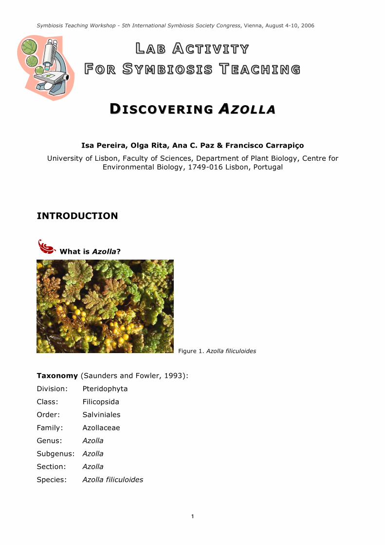
What is Azolla?
Figure 1. Azolla filiculoides
Taxonomy (Saunders and Fowler, 1993):
Division: Pteridophyta
Class: Filicopsida
Order: Salviniales
Family: Azollaceae
Genus: Azolla
Subgenus: Azolla
Section: Azolla
Species: Azolla filiculoides
Symbiosis Teaching Workshop - 5th International Symbiosis Society Congress, Vienna, August 4-10, 2006
2
Azolla is a small aquatic floating fern, whose genus was established by Lamarck in
1783 with a fossil record dating back to the mid-Cretaceous, that presents a very
peculiar structure:
• The stems (rhizomes) are thin, ramified, horizontaly distributed and covered
with leaves.
• The fern has numerous and simple roots that emerge in the ramification points
of the ventral side of the stem.
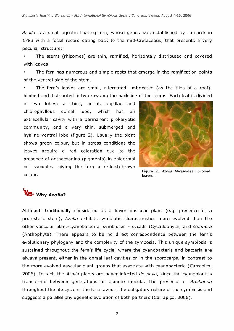
• The fern’s leaves are small, alternated, imbricated (as the tiles of a roof),
bilobed and distributed in two rows on the backside of the stems. Each leaf is divided
in two lobes: a thick, aerial, papillae and
chlorophyllous dorsal lobe, which has an
extracellular cavity with a permanent prokaryotic
community, and a very thin, submerged and
hyaline ventral lobe (figure 2). Usually the plant
shows green colour, but in stress conditions the
leaves acquire a red coloration due to the
presence of anthocyanins (pigments) in epidermal
cell vacuoles, giving the fern a reddish-brown
colour.
Why Azolla?
Although traditionally considered as a lower vascular plant (e.g. presence of a
protostelic stem), Azolla exhibits symbiotic characteristics more evolved than the
other vascular plant-cyanobacterial symbioses - cycads (Cycadophyta) and Gunnera
(Anthophyta). There appears to be no direct correspondence between the fern’s
evolutionary phylogeny and the complexity of the symbosis. This unique symbiosis is
sustained throughout the fern’s life cycle, where the cyanobacteria and bacteria are
always present, either in the dorsal leaf cavities or in the sporocarps, in contrast to
the more evolved vascular plant groups that associate with cyanobacteria (Carrapiço,
2006). In fact, the Azolla plants are never infected de novo, since the cyanobiont is
transferred between generations as akinete inocula. The presence of Anabaena
throughout the life cycle of the fern favours the obligatory nature of the symbiosis and
suggests a parallel phylogenetic evolution of both partners (Carrapiço, 2006).
Figure 2. Azolla filiculoides: bilobed leaves.
Symbiosis Teaching Workshop - 5th International Symbiosis Society Congress, Vienna, August 4-10, 2006
3
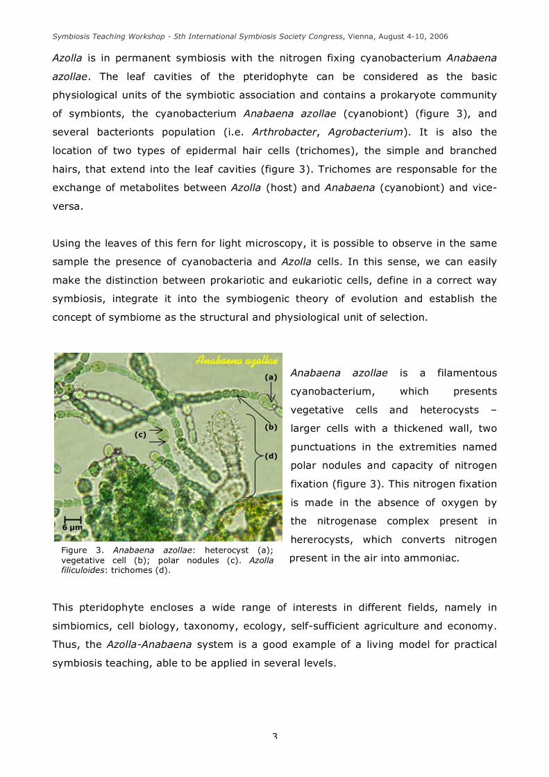
Figure 3. Anabaena azollae: heterocyst (a); vegetative cell (b); polar nodules (c). Azolla filiculoides: trichomes (d).
Azolla is in permanent symbiosis with the nitrogen fixing cyanobacterium Anabaena
azollae. The leaf cavities of the pteridophyte can be considered as the basic
physiological units of the symbiotic association and contains a prokaryote community
of symbionts, the cyanobacterium Anabaena azollae (cyanobiont) (figure 3), and
several bacterionts population (i.e. Arthrobacter, Agrobacterium). It is also the
location of two types of epidermal hair cells (trichomes), the simple and branched
hairs, that extend into the leaf cavities (figure 3). Trichomes are responsable for the
exchange of metabolites between Azolla (host) and Anabaena (cyanobiont) and vice-
versa.
Using the leaves of this fern for light microscopy, it is possible to observe in the same
sample the presence of cyanobacteria and Azolla cells. In this sense, we can easily
make the distinction between prokariotic and eukariotic cells, define in a correct way
symbiosis, integrate it into the symbiogenic theory of evolution and establish the
concept of symbiome as the structural and physiological unit of selection.
Anabaena azollae is a filamentous
cyanobacterium, which presents
vegetative cells and heterocysts –
larger cells with a thickened wall, two
punctuations in the extremities named
polar nodules and capacity of nitrogen
fixation (figure 3). This nitrogen fixation
is made in the absence of oxygen by
the nitrogenase complex present in
hererocysts, which converts nitrogen
present in the air into ammoniac.
This pteridophyte encloses a wide range of interests in different fields, namely in
simbiomics, cell biology, taxonomy, ecology, self-sufficient agriculture and economy.
Thus, the Azolla-Anabaena system is a good example of a living model for practical
symbiosis teaching, able to be applied in several levels.
6 µm
(a)
(b)
(d)
(c)
Symbiosis Teaching Workshop - 5th International Symbiosis Society Congress, Vienna, August 4-10, 2006
4
MATERIAL
• Azolla filiculoides
• coverslips
• dissection needle
• distilled water
• Pasteur pipettes
• immersion oil
• light microscope
• Petri dishes
• slides
• stereomicroscope
• tweezers
• filter paper
METHODOLOGY
Observation of Azolla with stereomicroscope
1. Place one or two specimens of Azolla filiculoides in a Petri dish.
2. Observe the sample with a stereomicroscope.
3. Make a diagram of the roots, rhizome and leaves (identify the dorsal and ventral
lobe of the Azolla leaf and observe the characteristics of each lobe). Label them.
Microscopic observation of Azolla
1. Remove some Azolla rhizomes, with the respective leaves.
2. Put a drop of distilled water on a slide, followed by the sample.
3. Gently, squeeze the leaves with a dissection needle and cover it with a coverslip.
4. Put the slide in the stage of the light microscope and observe.
5. Make a diagram and label the main aspects of your observation:
Symbiosis Teaching Workshop - 5th International Symbiosis Society Congress, Vienna, August 4-10, 2006
5
• the epidermal cells of Azolla leaf: observe the presence of anthocyanins,
a red pigment present in vacuoles (figure 4);
• the mesophyll cells of Azolla leaf: identify the dorsal lobe cells, which
contain chloroplasts (figure 4);
• the transfer hairs (trichomes) (figure 3);
• the cyanobacteria: identify the typical filaments of this prokaryotic
organism; observe the cell wall, the vegetative cells and the heterocysts (figure
3).
Figure 4. Azolla filiculoides: epidermal cells with anthocyanins and mesophyll cells with chloroplasts.
REGISTRY OF OBSERVATIONS
Symbiosis Teaching Workshop - 5th International Symbiosis Society Congress, Vienna, August 4-10, 2006
6
THINK ABOUT... / REFLECT...
Prokaryotic and Eukaryotic Cells - the Differences
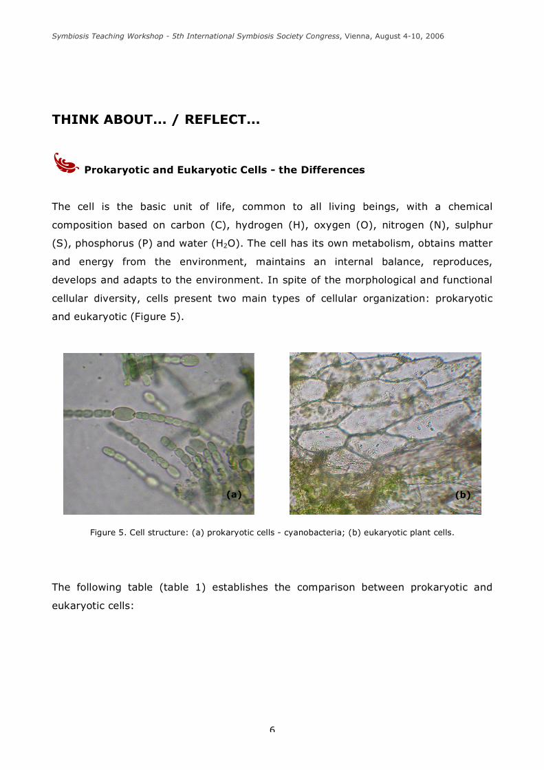
The cell is the basic unit of life, common to all living beings, with a chemical
composition based on carbon (C), hydrogen (H), oxygen (O), nitrogen (N), sulphur
(S), phosphorus (P) and water (H2O). The cell has its own metabolism, obtains matter
and energy from the environment, maintains an internal balance, reproduces,
develops and adapts to the environment. In spite of the morphological and functional
cellular diversity, cells present two main types of cellular organization: prokaryotic
and eukaryotic (Figure 5).
Figure 5. Cell structure: (a) prokaryotic cells - cyanobacteria; (b) eukaryotic plant cells.
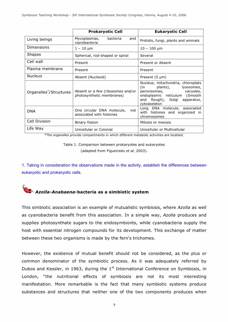
The following table (table 1) establishes the comparison between prokaryotic and
eukaryotic cells:
(a) (b)
Symbiosis Teaching Workshop - 5th International Symbiosis Society Congress, Vienna, August 4-10, 2006
7
Prokaryotic Cell Eukaryotic Cell
Living beings Mycoplasmas, bacteria and cianobacteria
Protists, fungi, plants and animals
Dimensions 1 – 10 µm 10 – 100 µm
Shapes Spherical, rod-shaped or spiral Several
Cell wall Present Present or Absent
Plasma membrane Present Present
Nucleus Absent (Nucleoid) Present (5 µm)
Organelles*/Structures Absent or a few (ribosomes and/or photosynthetic membranes)
Nucleus, mitochondria, chloroplats (in plants), lysosomes, peroxisomes, vacuoles, endoplasmic reticulum (Smooth and Rough), Golgi apparatus, cytoskeleton
DNA One circular DNA molecule, not associated with histones
Long DNA molecule, associated with histones and organized in chromosomes
Cell Division Binary fission Mitosis or meiosis
Life Way Unicellular or Colonial Unicellular or Multicellular
*The organelles provide compartments in which different metabolic activities are locatted.
Table 1. Comparison between prokaryotes and eukaryotes
(adapted from Figueiredo et al, 2003).
1. Taking in consideration the observations made in the activity, establish the differences between
eukaryotic and prokaryotic cells.
Azolla-Anabaena-bacteria as a simbiotic system
This simbiotic association is an example of mutualistic symbiosis, where Azolla as well
as cyanobacteria benefit from this association. In a simple way, Azolla produces and
supplies photosynthate sugars to the endosymbionts, while cyanobacteria supply the
host with essential nitrogen compounds for its development. This exchange of matter
between these two organisms is made by the fern’s trichomes.
However, the existence of mutual benefit should not be considered, as the plus or
common denominator of the symbiotic process. As it was adequately referred by
Dubos and Kessler, in 1963, during the 1st International Conference on Symbiosis, in
London, “the nutritional effects of symbiosis are not its most interesting
manifestation. More remarkable is the fact that many symbiotic systems produce
substances and structures that neither one of the two components produces when
Symbiosis Teaching Workshop - 5th International Symbiosis Society Congress, Vienna, August 4-10, 2006
8
growing alone” (Carrapiço & Rodrigues, 2005). We belive that this concept can also be
applied to the Azolla-Anabaena symbiotic system.
1. How can the Azolla-Anabaena simbiotic system be an example of exchange of matter and
energy?
2. Discuss the following statement: “The Azolla-Anabaena simbiotic system can be compared to an
ecosystem.”
Azolla as a symbiome
In 2003, Jan Sapp (Sapp, 2003) introduces the concept of symbiome, referring that
“every eukaryote is a superorganism, a symbiome composed of chromosomal genes,
organellar genes, and often other bacterial symbionts as well as viruses. The
symbiome, the limit of the multicellular organism, extends beyond the activities of its
own cells. All plants and animals involve complex ecological communities of microbes,
some of which function as commensals, some as mutualists, and others as parasites,
depending on their nature and context”. In the same sense, we believe that this idea
can be applied to the Azolla-Anabaena-bacteria symbiosis. The Azolla leaf cavity can
be considered as the basic physiological unit of the symbiotic association (Grilli Caiola
and Forni, 1999), where complex ecological communities of permanent
microorganisms co-exist with the fern to maintain the whole. New novel metabolic and
organic capabilities are acquired and developed by the partners to establish a new
level of organization, extending beyond the capability of each individual forming the
association (Carrapiço, 2006).
1. Why can we consider Azolla as a superorganism?
Symbiosis and evolution
Symbiomics is a new term created by Jan Sapp (2003) and defined, through personal
communication between him and Francisco Carrapiço (2004), as a field that studies
the biochemistry, physiology, genetics, ecology and evolution of the simbiotic
systems, as well as their dynamic interfaces. This science considers every plant and
animal as a symbiome - a poligenomic entity constituted by chromosomal genes,
organellar genes, viral genes and other microbial symbionts – in which multiple
Symbiosis Teaching Workshop - 5th International Symbiosis Society Congress, Vienna, August 4-10, 2006
9
species function to maintain the whole system. Many protists, plants and animals
harbor symbiotic bacteria, that are transmitted hereditarily from one generation to
another. In this case, the symbiosis present in Azolla-Anabaena system is sustained
throughout the whole fern´s life cycle, where the cyanobacteria and bacteria are
always present, showing a syncronic cycle with the host (Selosse, 2005). This fact
favours the obligatory nature of the symbiosis and suggests a parallel phylogenetic
evolution of both partners – a successful co-evolved system (Carrapiço, 2006). Thus,
symbiosis can be recognized as an evolutionary process which has an important role
in evolution.
Even the eukaryotic cell can also be seen as a symbiome. According to the
contemporary conceptual consensus, all eukaryotes emerged from mergers between
different kinds of bacteria. The mitochondria of eukaryotic cells and the chloroplasts of
plants and protists were once free-living bacteria that became incorporated in a
primitive host cell (Sagan, 1967) and, in the course of evolution, some of their
bacterial genes were lost and others transfered to the cell nucleus. In this sense, the
horizontal gene transfer is a powerful mechanism of novelty allowing symbiosis can be
recognized as an evolutionary mechanism with an important role in evolution. It is at
the symbiome level, composed by an integrate multigenomic genetic pool, in which
natural selection acts, constituting the unit of selection.
1. Explain how symbiosis contributes for the evolutionary process.
2. Why is the symbiome a poligenomic entity?
3. Which are the main reasons to consider the symbiome as the unit of selection?

Symbiosis Teaching Workshop - 5th International Symbiosis Society Congress, Vienna, August 4-10, 2006
10
REFERENCES
• CARRAPIÇO, F., 1988 - "A evolução celular". Boletim da Sociedade Portuguesa de
Ciências Naturais, 2ª Série, 24: 35-70.
• CARRAPIÇO, F., 2000 - Apontamentos de Biologia Celular – Divisão Cyanophyta.
• CARRAPIÇO F., ANTUNES T., SEVINATE-PINTO I., TEIXEIRA G., SERRANO R., BAIOA
V., PEREIRA A.L., ELIAS F., BASTOS M., CAIXINHAS R. FALCÃO M. & RAFAEL T., 2001
- “Azolla em Portugal”. Brochura INAG-CBA, Lisboa, 15 pp. (ISBN: 972-9412-58-8).
• CARRAPIÇO, F., 2002 - “The Azolla-Anabaena-Bacteria System as a Natural
Microcosm”. Proceedings of SPIE, 4495: 261-265.
• CARRAPIÇO, F. & RODRIGUES, T., 2005- “Symbiogenesis and the Early Evolution of
Life”. Proceedings of SPIE, 5906: 59060R-1 - 59060R-4.
• CARRAPIÇO, F., 2006 - “Is the Azolla-Anabaena symbiosis a co-evolution case?”.
Proceedings of the International Conference “General Botany: Traditions and
Perspectives”, Department of Botany of the Kazan University, Jan 23-27, 2006.
• FIGUEIREDO, A., BARROSO, J., PEDRO, L., & OLIVEIRA, M., 2003 – “Guia Prático de
Biologia Celular”. Associação dos Estudantes da Faculdade de Ciências de Lisboa.
Lisboa.
• GRILLI CAIOLA, M. & FORNI, C. (1999). “The hard life of prokaryotes in the leaf
cavities of Azolla”. J. Seckbach (ed.), Enigmatic microorganisms and life in extreme
environments, pp. 629-639.
• LÓPEZ-GARCIA, P. & MOREIRA, D., 1999 - Metabolic symbiosis at the origins of
eukaryotes. in TIBS, 24: 88-93.
• KUTSCHERA, U. & NIKLAS, K. J., 2005 – “Endosymbiosis, cell evolution and
speciation”. Theory in Biosciences, 124: 1-24. (Available online at
www.sciencedirect.com).
Symbiosis Teaching Workshop - 5th International Symbiosis Society Congress, Vienna, August 4-10, 2006
11
• SAGAN, L., 1967 – “On the origin of mitosing cells”. J. Theor.Biol., 14: 225-274.
• SAPP, J., 2003 - Genesis – The Evolution of Biology. Oxford University Press, Inc.
New York.
• SAPP, J., 2004 – “The dynamics of symbiosis: an historical overview”. Can. J. Bot,
82: 1046-1056.
• SAUNDERS, R. M. K. & FOWLER, K., 1993 - "The supraspecific taxonomy and
evolution of the fern genus Azolla (Azollaceae)". Plant Systematics & Evolution, 184:
175-193.
• SELOSSE, M-A., 2005 - “La Symbiose. Structures et fonctions, rôle écologique et
évolutif”. Vuibert, Paris.