Sutter Standard Operating Procedures · Sutter Standard Operating Procedures ... For breast imaging...
42
1 Sutter Standard Operating Procedures
Transcript of Sutter Standard Operating Procedures · Sutter Standard Operating Procedures ... For breast imaging...

1
Sutter Standard Operating Procedures

{00241664 v.1} Policies Page X of X Department Policy: XX-XXX
Sutter Health and Affiliates Administrative Policies and Procedures
Modifying Imaging Procedure Orders to Meet the Needs of the Patient
Department: Medical Foundation Radiology / Diagnostic Imaging Policy: 00-000
Origination Date: 07/01/2012 Revised Date: Next Review Date:
Approved by:
Diagnostic Imaging Oversight Committee -
June 1st, 2012
System Management Team
POLICY
Radiologists practicing in Sutter Health affiliated Medical Foundation imaging departments are permitted to modify the test design and/or order alternative procedures for ambulatory patients within specified limits detailed in relevant Standard Operating Procedures (SOPs) or Radiology Protocols without specific notification to or additional orders from the referring/ordering provider. The patient’s medical and physical condition, the imaging department’s equipment and the training and expertise of the imagers will be used in determining the optimal study(ies) to be performed.
PURPOSE Sutter Health Medical Foundation imaging departments and breast centers require the ability to efficiently generate and fulfill orders and provide optimal patient care while remaining in compliance with payor guidelines. SCOPE This policy applies to Sutter Health affiliated Medical Foundations. Note: In the hospital setting, radiologists may modify or give orders according to their privileges and the hospital’s general policies on documenting physician orders.
PROCEDURES Ordering physicians who use Epic are presented with the following question which authorizes the Medical Foundation imaging department to modify the order if needed per imaging protocol: “Radiologist may modify the order per protocol to meet the clinical needs of the patient?” The default answer is “Yes,” to authorize modifications to test orders. Ordering providers who do not want the radiologist to make any changes can change the response to “No.” External ordering physicians are informed by affiliate imaging departments of Sutter Health radiologists’ ability to use SOPs and protocols to modify the test design of radiology procedures and their ability to limit their orders to only particular studies. Unless otherwise specified by the external ordering physician, Sutter Health users transcribing radiology procedure orders for

{00241664 v.1} Policies Page X of X Department Policy: XX-XXX
external ordering providers will respond “Yes” to the ordering question of “Radiologist may modify the order per protocol to meet the clinical needs of the patient?” For breast imaging studies, follow the Standard Operating Procedures for breast imaging results and recommended actions outlined in the attached Breast Imaging Standard Operating Procedure. For all imaging studies, radiologists may modify the following aspects of test design to optimally meet the clinical needs of the patient based on presenting clinical history, signs and symptoms:
• Use or non-use of contrast • Laterality of exam • Number and type of views • CT slice thickness • Imaging of adjacent or alternate body part
The radiologist should include the reason for the modification in the report to the ordering provider.
The radiologist may modify an order with clear and obvious errors that would be apparent to a reasonable layperson, such as the patient receiving the test (e.g., x-ray of wrong foot ordered), without notifying the treating physician/practitioner, Test design does not include adding tests using a different modality, except in the case of breast imaging, which is covered by a specific SOP. If the radiologist determines than another imaging procedure using a different modality than the one ordered is needed, the imaging department staff will contact the ordering physician for new orders. References: Medicare Benefit Policy Manual, Ch. 15, sec. 80.6.4 Palmetto GBA article Standard Operating Procedures (SOPs) for Diagnostic Testing http://www.palmettogba.com/palmetto/providers.nsf/DocsCat/Providers~Jurisdiction%201%20Part%20B~Browse%20by%20Topic~General~7X9NRW2052?open&navmenu=%7C%7C Attachment: Breast Imaging Standard Operating Procedure

Page #1 of 6
CT Standard Operating Procedures
Body Part Reason For Exam IV Contrast Procedure to Pre-Cert CPT Code Head/Brain
Alzheimer’s Bleed, Hemorrhage CVA, Stroke Headaches Hydrocephalus Memory Loss, Confusion Shunt Check Trauma Vertigo, Dizziness* Headache w/Associated Neurologic Signs* Infection* Mass/Tumor* Metastatic Staging* *MRI PREFERRED HIV Melanoma Toxoplasmosis
No
Yes
Yes
CT Head, Brain Without Contrast CT Head, Brain With Contrast CT Head, Brain Without and With Contrast
70450 70460 70470
CTA Brain (Head)
Aneurysm AVM (Arterio/Venous Malformation CVA TIA Vascular Malformation
Yes
CTA Brain
75671
Orbit
Foreign Body Fracture Graves Disease Trauma Abscess Exopthalmus Mass Pain Pseudo Tumor Retinoblastoma
No
Yes
Yes
CT Orbit Without Contrast CT Orbit With Contrast CT Orbit without & with Contrast
70480 70481 70482
Sinus Limited
Sinusitis (billing will apply modifier 52) *This is for limited exam ONLY*
No
CT Sinus Screen (same CPT code as sinus complete)
70486
Sinus Full Osteomeatal Complex
Osteomeatal Complex Functional Endoscopic Sinus Surgery
No
CT Sinus Complete
70486

Page #2 of 6
Body Part Reason For Exam IV Contrast Procedure to Pre-Cert CPT Code Temporal Bone
Cholesteotoma Hearing Loss, Conductive* Trauma *Sensory neuro hearing loss, order MRI with and without contrast
No
CT Inner Ears, Temporal Bones
70480
CTA Carotid
Bruit Carotid Stenosis CVA TIA
Yes
CTA Carotid
75662
Spine Cervical/Thoracic
Trauma, Fracture, Fusion Assess Bony Degenerative Changes *MRI recommended for disc herniation, Mets or infection
No
CT Cervical Spine Without Contrast CT Thoracic Spine Without Contrast
72125
72128
Spine Lumbar/Sacral
Trauma, Fracture, Fusion, Pars Defect - *MRI recommended for disc herniation, mets, infection.
No
CT Lumbar Spine Without Contrast
72131
Neck/Parotid
All necks should be ordered with contrast unless there is renal failure Parotid Mass Gland Infection of Parotid Parotid Stone
Yes
Yes
CT Neck, Parotid With Contrast CT Neck, Parotid Without and With Contrast
70491
70492
Neck/Parotid/Nasopharynx
If elevated creatinine, order without contrast.
No
CT Neck Without Contrast
70490
Axilla
Mass, Chest Wall Mass
Yes
CT Chest with Contrast
71260

Page #3 of 6
Body Part Reason For Exam IV Contrast Procedure to Pre-Cert CPT Code Chest
All chest are ordered with contrast except for these indications Renal Failure F/U Nodules (Contrast needed for 1st exam, then all F/U nodules without contrast.) Tracheal Stenosis Asbestosis COPD Cough Esophageal CA Hemoptysis Lymphoma Lung CA Lung Nodule Mass Pneumonia Sarcoidosis
No
Yes
CT Chest Without Contrast CT Chest With Contrast
71250
71260
Chest, High Resolution
Bronchiectasis Fibrosis Interstitial Disease Pleural Plaques
No
CT Chest Without High Resolution
71250
CTA Chest & Abd Thoracic Aortic Dissection
Aneurysm Aortic Dissection Thoracic Aortic
Yes
CTA Chest and Abdomen (Please authorize BOTH codes)
71275 75635
CTA Chest ( PE Study)
AAA / Aortic Dissection Chest Pain / Dyspnea + D Dimers DVT Hemoptysis Pulmonary Hypertension Shortness of Breath Tachypnea
Yes
CTA Chest
71275

Page #4 of 6
Body Part Reason For Exam IV Contrast Procedure to Pre-Cert CPT Code Abdomen
F/U for patients with renal cell carcinoma in renal failure - *Recommend MRI Abdominal Pain (generalized) Epigastric pain LUQ Pain Mass Pancreatitis Pseudocyst RUQ Pain
No
Yes
CT Abdomen Without Contrast CT Abdomen With Contrast
74150
74160
Abdomen and/or Kidney
Adrenal Mass- (MR preferred) Cirrhosis— (MR preferred) Embolization Hepatoma, Hepatitis Liver Hemangioma- (MR preferred) Radiofrequency Ablation
Yes
CT Abdomen Without and With Contrast
74170
Abdomen and/or Kidney and Pelvis (if ordering pelvis, both codes must be authorized)
Carcinoid Kidney Cyst vs. Mass - (MRI Preferred) Painless Hematuria Melanoma
Yes
CT Abdomen Without and With Contrast CT Pelvis With Contrast
74170
72193
Abdomen/Pelvis
Hematuria Stone (Stone Study) Abdominal Pain (upper & lower quardrants) Abscess Appendicitis Cancer Staging (except melanoma & carcinoid)* Colitis/ IBD Crohns / Ulcerative Diverticulitis Hematuria Hernia (i.e. ventral, umbilical, inguinal)** Mass *For certain CA’s, some insurance companies will not cover CT pelvis (i.e. breast & lung CA) ** Non covered indication for upper quadrant.
No
Yes
CT Abdomen Without Contrast AND CT Pelvis Without Contrast CT Abdomen With Contrast AND CT Pelvis With Contrast
74176
74177

Page #5 of 6
Body Part Reason For Exam IV Contrast Procedure to Pre-Cert CPT Code Pelvis (Routine)
Cancer Staging Cysts Infection Mass Pain
Yes
CT Pelvis With Contrast Note: Symptoms must be in pelvis or lower abdomen to be covered by Medicare.
72193
Pelvis
Bone Infection Infection* Tumor/Mass/Cancer/Mets *Recommend MRI
Yes
CT Pelvis Without and With Contrast Note: Must be in pelvic region (lower abdomen) to be a covered service of Medicare.
72194
CT Urogram
Carcinoma Kidney and/or Bladder Transitional Cell
Yes
CT Abdomen and Pelvis With and Without Contrast
74170
CTA Abdomen & Run Off
Peripheral Artery Disease (PAD)
Yes
CT Abdomen and Run Off (Please authorize BOTH codes.)
75635 73706
Pubic Arch Study Protocol
Prostate Treatment Planning
No
CT Pelvis Without Contrast
72192
Pelvis Hips
Fracture Non Union Arthritis
No
CT Pelvis Without Contrast
72192
Extremity Foot Toe Ankle Calf Knee Thigh Femur Lower Leg Finger Hand Wrist Forearm Elbow Humerus
Arthritis Femoral Anteversion Fracture Fusion Loose body Malunion Malignment Knee (indicate Lt, Rt, or Bil) Non Union Osteochondral lesion
No
CT Without Contrast Lower Extremity Upper Extremity
73700
73200
Extremities
Infection* Tumor/Mass/Cancer/Mets *Recommend MRI
Yes
CT Without and With Contrast –Lower CT Without and With Contrast –Upper
73702
73202

Page #6 of 6
Body Part Reason For Exam IV Contrast Procedure to Pre-Cert CPT Code CT Angiography - Extremities
Arterial Stenosis – Lower Claudication -extremity Ischemia - extremity Peripheral Artery Disease
Yes
CT Angiography Lower Extremity Upper Extremity
73706
73206
CT Arthrography
Cartilage Abnormally Labrum Abnormality Loose Bodies Meniscus Abnormally
Yes
CT With Contrast Lower Extremity with Contrast Upper Extremity with Contrast Fluoro Guided Joint Injection *Please state which joint in comments
73701
73201
76000

Diagnostic Imaging Services Policy and Procedure
{00241664 v.1}Page 1 of 5
TITLE: Breast Imaging Standard Operating Procedure
APPROVED BY: Diagnostic Imaging Oversight Committee
CHAPTER: Sutter Epic Weblinks SECTION: Specialty/Clinical Topic>Radiology
ISSUED: 7/1/2011 Updated: 01/06/2012
REVIEWED: DIOC Chair
REPLACES: N/A PAGE 1/4
Purpose - These operating procedures allow Sutter Health imaging departments and breast centers to efficiently generate and fulfill compliant orders for indicated breast imaging and diagnostic studies as needed based upon radiologist recommendations.
Sutter Health imaging departments and breast centers will recommend additional breast imaging and diagnostic studies, including mammography, breast ultrasound, Breast MRI and image guided breast aspiration and/or biopsy, based upon the clinical history, physical findings and/or Breast Imaging Reporting and Data System (BI-RADS) classification as developed by the American College of Radiology.
What is the Breast Imaging Reporting and Database System (BI-RADS)?
The American College of Radiology (ACR) has established a uniform system for radiologists to describe and manage mammogram findings. The system includes seven standardized Categories, or Assessments. Each BI-RADS Category has an associated recommended Follow-up plan to assist radiologists and other physicians in appropriately managing a patient’s care.
Breast Imaging Reporting and Database System (BI-RADS)
Category Assessment Follow-up
0 Need additional imaging evaluation Additional imaging needed before a category can be assigned
1 Negative Continue routine screening mammograms (for women over age 40)
2 Benign (noncancerous) findingj Continue routine screening mammograms (for women over age 40)
3 Probably benign Receive a 6-month follow-up mammogram or ultrasound, then every 6-12 months for 1-2 years.
4 Suspicious abnormality Requires biopsy 5 Highly suggestive of malignancy
(cancer) Requires biopsy
6 Known biopsy-proven malignancy (cancer)
Assure that treatment for known cancer is completed.

Diagnostic Imaging Services Policy and Procedure
{00241664 v.1}Page 2 of 5
Additional information about BI-RADS is available on the ACR Web site at http://www.acr.org/Quality-Safety/Resources/BIRADS/Mammography or by calling the ACR at 1–800–ACR–LINE (1–800–227–5463).
There are certain clinical situations that BI-RADS do not address, where it is clinically appropriate for the radiologist to modify the referring physician's order based on prior history or patient current conditions, the radiologist may change the patient's referral order to the appropriate study under the general SOP permission given by the referring physician. Examples include:
1. Patients that present with orders for screening mammograms who report a palpable abnormality or history of prior abnormal breast imaging study
2. Patients who present with a diagnostic mammogram order with no signs or symptoms or history of breast cancer that should undergo screening mammography
3. Patient with a palpable abnormality whose diagnostic mammogram order does not include breast ultrasound and for which ultrasound is indicated

Diagnostic Imaging Services Policy and Procedure
{00241664 v.1}Page 3 of 5
PALPABLE BREAST LUMPS and FOCAL BREAST PAIN WORKFLOW
Patient < 30 Patient > 30
Diagnostic US first. Add diagnostic mammogram based on radiologist recommendations
Diagnostic mammogram and likely US
Specific imaging findings
BI-RADS 0 Assessment incomplete
Need to complete additional imaging or obtain and review prior studies
BI-RADS 1 & 2 Negative / benign
BI-RADS 3 Probably benign
BI-RADS 4 & 5 Suspicious
Follow-up by PCP, continued routine screening
Follow up as specified by radiologist
Image guided core needle biopsy
If not available refer to surgeon for excisional biopsy
Biopsy results reviewed by radiologist & communicated to PCP

Diagnostic Imaging Services Policy and Procedure
{00241664 v.1}Page 4 of 5
Discordant Malignant
Atypia Benign
6 month possible follow-up mammo or U/S
Refer to surgeon Re-biopsy Refer to surgeon

{00241664 v.1} Policies Page X of X Department Policy: XX-XXX
SCREENING MAMOGRAM – WORKFLOW
BI-RADS Category 1 & 2
Image guided core needle biopsy
Follow radiology advice for follow up imaging
Follow up by PCP continue routine screening
If not available or amenable, refer to surgeon for excisional biopsy
Biopsy results reviewed by radiologist and communicated to PCP
Malignant Atypia Benign
Refer to surgeon Refer to surgeon 6 month follow-up mammo/ultrasound possible
BI-RADS Category 0 & 3
BI-RADS Category 4 & 5
Page #1 of 7
MRI Standard Operating Procedures
Body Part Reason For Exam IV Contrast Procedure to Pre-Cert CPT Code Brain
Alzheimer’s Confusion CVA Dementia Headache w/o Focal Symptoms Memory Loss Mental Status Changes Seizures Stroke TIA Trauma Adenoma Cranial Nerve Lesions Dizziness Elevated Prolactin HIV IAC/Hearing Loss Infection Multiple Sclerosis Neurofibromatosis Pituitary Lesion Tumor/Mass/Cancer/Mets Vascular Lesions Vertigo Vision Changes To guide surgery planning, radiation therapy, or other surgical treatments for the brain, such as laser ablation; for preoperative planning to localize language dominance and for functional localization of memory; to localize abnormal brain functionfor epilepsy surgery
No
Yes
MRI Without Contrast MRI With & Without Contrast fMRI
70551
70553
70555 96020
MRA Brain
Stroke CVA TIA Aneurysm Arterial Venous
No
MRA Brain Without Contrast
70544
Brain Spectroscopy
Alzheimer’s Dementia Seizures Encephalopathy Ischemia Hypoxia Brain Injury Tumor/Mass/Cancer/Mets Infection Multiple Sclerosis
No
Yes
MRI Spectroscopy Without Contrast MRI Spectroscopy With and/or Without Contrast
76390
76390

Page #2 of 7
Body Part Reason For Exam IV Contrast Procedure to Pre-Cert CPT Code MRV Brain
Venous Thrombosis
No
MRA Without Contrast
70544
Orbits
Demyelination/Multiple Diplopia Dysthyroid Eye Disease Exopthalmos Grave’s Disease Proptosis Pseudotumor Sclerosis Trauma Tumor/Mass/Cancer/Mets Vascular Lesions
Yes
MRI Orbits/Face/Neck With & Without Contrast
70543
Neck
Infection Pain Tumor/Mass/Cancer/Mets Vocal Cord Paralysis
Yes
MRI Orbits/Face/Neck With & Without Contrast
70543
MRA Neck
Aneurysm Arterial Venous Malformation CVA Stroke Subclavian Steal TIA
Yes
MRA Neck With & Without Contrast
70549
MRA Arch & Great Vessels
Aneurysm Arterial Venous Malformation CVA Stroke Subclavian Steal TIA
Yes
MRA Neck With & Without Contrast
70549
Spine: Cervical
Arm/Shoulder Pain and/or Weakness Degenerative Disease Disc Herniation Neck Pain Radiculopathy Post-op (any hx cervical surgery) Abscess/Infection Discitis Multiple Sclerosis Myelopathy Osteomyelitis Syrinx Tumor/Mass/Cancer/Mets Vascular Lesions/AVM
No
Yes
MRI Cervical Spine Without Contrast MRI Cervical Spine With & Without Contrast
2141
72156

Page #3 of 7
Body Part Reason For Exam IV Contrast Procedure to Pre-Cert CPT Code Spine: Thoracic
Back Pain Compression Fracture (no hx cancer/mets) Degenerative Disease Disc Herniation Radiculopathy Trauma Vertebroplasty Planning (no hx of cancer/mets) Compression Fracture (with hx of cancer/mets) Post-op (any hx thoracic surgery) Abscess/Infection AVM Discitis Multiple Sclerosis Myelopathy Osteomyelitis Syrinx Tumor/Mass/Cancer/Mets Vascular Lesions Vertebroplasty Planning (with hx of cancer/mets)
No Yes
MRI Thoracic Without Contrast MRI Thoracic With & Without Contrast
72146
72157
Spine: Lumber
Back Pain Compression Fracture (no hx cancer/mets) Degenerative Disease Disc Herniation Radiculopathy Sciatica Spondylolithesis Stenosis Trauma Vertebroplasty (no hx of cancer/mets) Postop (any hx lumbar surgery) Abscess/Infection Compression Fracture (with hx of cancer/mets) Discitis Osteomyelitis Post-op (any hx lumbar surgery) Tumor/Mass/Cancer/Mets Vertebroplasty Planning (with hx of cancer/mets)
No
Yes
MRI Lumbar Without Contrast MRI Lumbar With & Without Contrast
72148
72158
Page #4 of 7
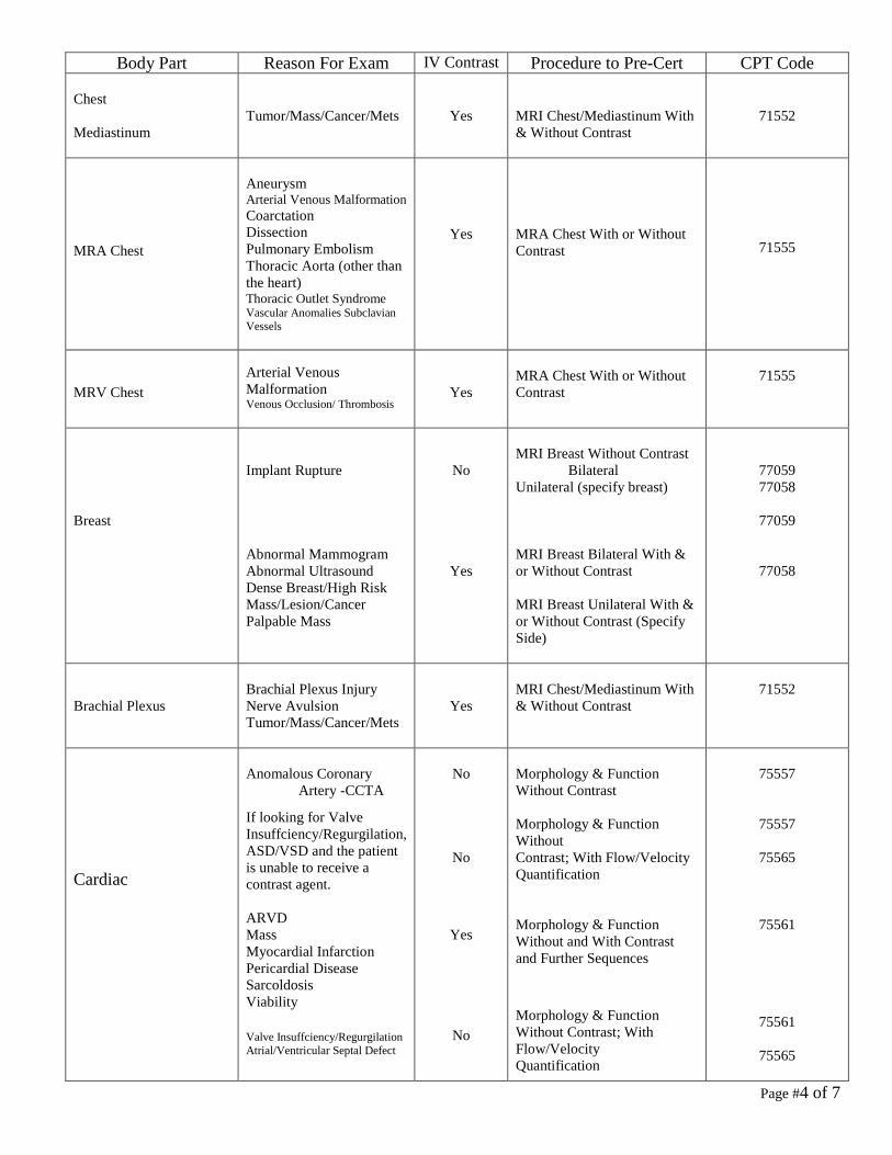
Body Part Reason For Exam IV Contrast Procedure to Pre-Cert CPT Code Chest Mediastinum
Tumor/Mass/Cancer/Mets
Yes
MRI Chest/Mediastinum With & Without Contrast
71552
MRA Chest
Aneurysm Arterial Venous Malformation Coarctation Dissection Pulmonary Embolism Thoracic Aorta (other than the heart) Thoracic Outlet Syndrome Vascular Anomalies Subclavian Vessels
Yes
MRA Chest With or Without Contrast
71555
MRV Chest
Arterial Venous Malformation Venous Occlusion/ Thrombosis
Yes
MRA Chest With or Without Contrast
71555
Breast
Implant Rupture Abnormal Mammogram Abnormal Ultrasound Dense Breast/High Risk Mass/Lesion/Cancer Palpable Mass
No
Yes
MRI Breast Without Contrast Bilateral Unilateral (specify breast) MRI Breast Bilateral With & or Without Contrast MRI Breast Unilateral With & or Without Contrast (Specify Side)
77059 77058
77059
77058
Brachial Plexus
Brachial Plexus Injury Nerve Avulsion Tumor/Mass/Cancer/Mets
Yes
MRI Chest/Mediastinum With & Without Contrast
71552
Cardiac
Anomalous Coronary
Artery -CCTA
If looking for Valve Insuffciency/Regurgilation, ASD/VSD and the patient is unable to receive a contrast agent. ARVD Mass Myocardial Infarction Pericardial Disease Sarcoldosis Viability Valve Insuffciency/Regurgilation Atrial/Ventricular Septal Defect
No
No
Yes
No
Morphology & Function Without Contrast Morphology & Function Without Contrast; With Flow/Velocity Quantification Morphology & Function Without and With Contrast and Further Sequences Morphology & Function Without Contrast; With Flow/Velocity Quantification
75557
75557
75565
75561
75561
75565

Page #5 of 7
Body Part Reason For Exam IV Contrast Procedure to Pre-Cert CPT Code Abdomen
Abnormal Enzymes Fetal MRI MRCP (Biliary/Pancreatic Ducts, Stones, Jaundice) Abdominal Pain Abscess/Ascites Adrenal Mass Liver Pancreatic Mass/Lesion Renal Lesion Tumor/Mass/Cancer/Mets
No
Yes
MRI Abdomen Without Contrast MRI Abdomen With & Without Contrast
74181 74183
MRA Abdomen
AAA(abdominal aortic aneurysm) Dissection Mesenteric Ischemia Renal Artery Stenosis Pre Liver Transplant Pre Kidney Transplant Renal Mass
Yes
Yes
MRA Abdomen With or Without contrast Order 2 Studies MRA Abdomend With or Without Contrast MRI Abdomen With Or Without Contrast
74185 74185 74183
MRV Abdomen
Venous Anomaly Venous Occlusion Venous Thrombosis
Yes
MRA Abdomen With or Without Contrast
74185
Pelvis
Fracture MRI Defecogram Muscle/Tendon Tear Urethral Diveticulum Abscess Adenomyosis Embolization Endometrioma Fibroid Osteomyetits Plexopathy Pre/Post Fibroid Prostate Cancer Septic Arthritis Tumor/Mass/Cancer/Mets Ulcer
No
Yes
MRI Pelvis Without Contrast MRI Pelvis With & Without Contrast
72195
72197
MRA Pelvis Or MRV Pelvis
Aneurysm AVM (artriovenous malformation) May Thurner Syndrome Pelvic Congestion Venous Occlusion Aneurysm Pelvic Congestion
Yes
Yes
MRA Pelvis With or Without Contrast Order 2 Exams: MRA Pelvis With or Without Contrast MRI Pelvis With and Without Contrast
72198
72198
72197

Page #6 of 7
Body Part Reason For Exam IV Contrast Procedure to Pre-Cert CPT Code MRA Peripheral (Run-off)
Claudication Cold foot Gangrene Pain Ulcer
Yes
Order 3 Exams: MRA Abdomen with/without contrast & MRA Lower Extremity with/without contrast LEFT & MRA Lower Extremity with/without contrast RIGHT
74185
73725
73725
MRA Extremity or MRV Extremity
Aneurysm Arterial Occlusion/Stenosis Venous Occlusion
Yes
MRA Extremity with & without contrast Upper Extremity
Lower Extremity
73225
73725
Extremity, Non-Joint Arm Hand Finger Femur Lower Leg Foot Toe
Fracture Muscle/Tendon tear Stress Fracture Abscess Cellultis Fasciitis Myositis Osteomyelitis Soft Tissue Tumor/Mass/ Cancer/Mets Ulcer
No
Yes
MRI Non-Joint Without Contrast Lower-Extremity Upper-Extremity MRI Non-Joint Without & With Contrast Lower-Extremity Upper-Extremity
73718
73218
73720
73220
Extremity, Joint Shoulder Elbow Wrist Hip Knee Ankle Foot
Arthritis AVN (Avascular Necrosis) Cartilage Tear Fracture Internal Derangement Joint Pain (specify joint) Labral Tear Ligament Tear Meniscus Tear Muscle Tear Osteochondritis Dessicans (OCD) Stress Fracture Sprain/Strain Tendon Tear
No
MRI Joint Without Contrast Lower-Extremity Upper-Extremity
73721
73221
Extremity, Joint Shoulder Elbow Wrist Hip Knee Ankle
Abscess Cellulitis Fasciitis Inflamatory Arthritis Myositis Osteomyelitis Septic Arthritis Tumor/Mass/Cancer/Mets Ulcer
Yes
MRI Joint Without & With Contrast Lower-Extremity Upper-Extremity
73723
73223

Page #7 of 7
Body Part Reason For Exam IV Contrast Procedure to Pre-Cert CPT Code MR Arthrography
Labral Tear Loose Bodies OCD Stability Post-op shoulder Post-op meniscus repair
Yes
MRI Joint with Contrast-Order 2 Exams Lower Extremity With Contrast Upper Extremity With Contrast Fluoroscopy Guided Joint
Injection Please state which joint in
comments
73722 73222 76000

NUCLEAR MEDICINE STUDIES STANDARD OPERATING PROCEDURES 1. Bone Scan: ( Order the following as: 78315 – 3 Phase Bone scan with option to do 78320 - Tomographic SPECT) RADO255 with RAD025 A. Procedure for evaluating bone disorders including:
• Skeletal Pain (bone or joint pain) / Fracture (For skeletal injuries/fractures it is preferable to image 48 hours or more after the injury) • Inflammatory / Septic Arthritis • Cellulitis / Osteomyelitis • Renal Osteodystrophy • Avascular Necrosis / Aseptic Necrosis • Skeletal Lesions / Primary Bone Tumors • Prosthetic Infection or Loosening or Painful Prosthesis • Arthritis / Heterotopic Ossification • Reflex Sympathetic Dystrophy
-- 3 Phase Bone scan includes imaging Single or Multiple regions or as a Whole Body exam. -- SPECT may be used by the Nuclear Medicine physician after reviewing initial set of images -- Three Phase Bone Scans are performed on pediatric patients, unless contraindicated. B. Skeletal Metasases: (78306 -Whole Body Bone) RAD0259 -- SPECT may be performed if indicated by radiologist after reviewing initial set of images. 2. Bone Marrow Scan: (78104 – Whole Body) RADO252 Procedure for evaluation of regional bone marrow abnormalities.
• Diagnosis of osteomyelitis in conjunction with an 111In WBC scan. • Diagnosis of prosthetic infection versus normal marrow accumulation at site of prosthetic. • Avascular Necrosis / Bone Infarction
3. Brain Tumor Imaging: (78607 – Tomographic SPECT) RAD0267
• Screening of patients suspected of having primary and/or metastatic brain tumors • To determine the viable tumor burden in patients with known gliomas.
-- Baseline tumor burden prior to therapy -- Follow residual tumor burden following treatment
-- Evaluation of patients suspected of having tumor versus infection (gallium utilized) 4. Cardiac Shunt Study: Right to Left (78428 – Cardiac Shunt Detection) RAD0271
• Detection, evaluation and quantitation of intra-cardiac shunts • Follow-up from surgery for correction of intracardiac shunts.

5. Gallium Inflamation and Infection Scan: (Order the following as: 78806 – Whole Body) RAD0243
• To localize source of fever in patients with fever of unknown origin (FUO) or elevated WBC count. • Diagnosis and follow-up of retroperitoneal fibrosis
(Order the following as: 78807—Tomographic SPECT RAD0338
• Detection of pulmonary and mediastinal inflammation/infection, especially in the immunocompromised patient. • Evaluation and follow-up of active lymphocytic or granulomatous inflammatory processes, such as sarcoidosis or tuberculosis. • Diagnosing osteomyelitis and/or disk space infection. (67 Ga is preferred over labeled leukocytes for disk space infection and vertebral osteomyelitis, and orthopedic
hardware). • Evaluation and follow-up of drug-induced pulmonary toxicity (e.g., bleomycin, amiodarone). • Follow-up post surgery / transplant for infection / osteomyelitis.
6. Gallium Tumor Scan: (78804 – Whole Body Mult. Days with 78803-Tomographic SPECT) RAD0395 with RAD0445
• Lymphoma -- Hodgin's Disease (HDL) Sarcoma -- Non Hodgkin's Lymphoma (NHL) Testicular Tumors -- Lung cancer Head and Neck Tumors -- Melanoma Multiple Myeloma -- Hepatocellular carcinoma Neuroblastoma
• Recurrence, Restaging, Management, and outcome of both HD and NHL • Differentiation of brain tumor from infection
7. Gastric Empty Study: (78264 – Gastric Emptying Study) RAD0263 • Determination of possible delayed gastric emptying and quantitation of gastric emptying rate in patients with suspected gastroparesis of other motility problems. • Evaluation of response to therapy in those patients who have proven delayed gastric
emptying. • Evaluation of gastric motility in post-operative or post-radiotherapy patients. • Evaluation of gastric outlet obstruction
8. Hepatobiliary (HIDA) Scan: (78223 – Hepatobiliary Ductal System Imaging w or w/o Pharm. Intervnt.) RAD0299
• Functional assessment of the hepatobiliary system / Or asses the integrity of the hepatobiliary tree. These broad categories include:
-- Suspected acute cholecystitis -- Evaluate for choledochal cyst -- Suspected chronic biliary tract disorders -- Jaundice -- Common bile duct obstruction / Extravasation / Leak -- Gallstone/lithotripsy patients -- Evaluation of congenital abnormalities of the biliary tree -- Evaluation of right upper quadrant pain -- Evaluation of route of biliary drainage in post-op biliary diversion -- Evaluation of biliary atresia vs. neonatal hepatitis
9. Liver and Spleen Scan: (78205 – Liver Imaging SPECT) RAD0324 • This study can be used for determining the size and shape of the liver and spleen. • For suspected focal nodular hyperplasia of the liver. These lesions often have normal or
increased uptake on sulfur colloid imaging.
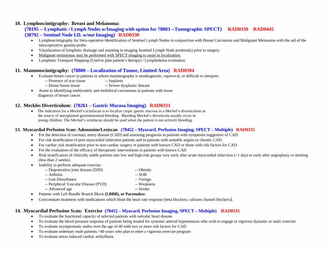
10. Lymphoscintigraphy: Breast and Melanoma: (78195 -- Lymphatic / Lymph Nodes w/Imaging with option for 78803 --Tomographic SPECT) RAD0330 RAD0445 (38792 – Sentinel Node I.D. w/out Imaging) RAD0330
• Lymphoscintigraphy for Intra-operative Identification of Sentinel Lymph Nodes in conjunction with Breast Carcinoma and Malignant Melanoma with the aid of the intra-operative gamma probe.
• Visualization of lymphatic drainage and assisting in imaging Sentinel Lymph Node position(s) prior to surgery. • Malignant melanomas may be performed with SPECT imaging to assist in localization. • Lymphatic Transport Mapping (Used to plan patient’s therapy) / Lymphedema evaluation
11. Mammoscintigraphy: (78800 – Localization of Tumor, Limited Area) RAD0394
• Evaluate breast cancer in patients in whom mammography is nondiagnostic, equivocal, or difficult to interpret: -- Presence of scar tissue -- Implants -- Dense breast tissue -- Severe dysplastic disease • Assist in identifying multicentric and multifocal carcinomas in patients with tissue
diagnosis of breast cancer.
12. Meckles Diverticulum: (78261 – Gastric Mucosa Imaging) RAD0331 • The indication for a Meckel’s scintiscan is to localize ctopic gastric mucosa in a Meckel’s diverticulum as
the source of unexplained gastrointestinal bleeding. Bleeding Meckel’s diverticula usually occur in young children. The Meckel’s scintiscan should be used when the patient is not actively bleeding. 13. Myocardial Perfusion Scan: Adensosine/Lexiscan (78452 – Myocard. Perfusion Imaging, SPECT – Multiple) RAD0335
• For the detection of coronary artery disease (CAD) and assessing prognosis in patients with symptoms suggestive of CAD. • For risk stratification of post myocardial infarction patients and in patients with unstable angina or chronic CAD. • For cardiac risk stratification prior to non-cardiac surgery in patients with known CAD or those with risk factors for CAD. • For the evaluation of the efficacy of therapeutic interventions in patients with known CAD • Risk stratification of clinically stable patients into low and high-risk groups very early after acute myocardial infarction (>1 day) or early after angioplasty or stenting
(less than 2 weeks). • Inability to perform adequate exercise.
-- Degenerative joint disease (DJD) -- Obesity -- Arthritis -- SOB -- Gait Disturbance -- Vertigo -- Peripheral Vascular Disease (PVD) -- Weakness -- Advanced age -- Stroke
• Patients with Left Bundle Branch Block (LBBB), or Pacemaker. • Concomitant treatment with medications which blunt the heart rate response (beta blockers, calcium channel blockers).
14. Myocardial Perfusion Scan: Exercise (78452 – Myocard. Perfusion Imaging, SPECT – Multiple) RAD0335
• To evaluate the functional capacity of selected patients with valvular heart disease • To evaluate the blood pressure response of patients being treated for systemic arterial hypertension who wish to engage in vigorous dynamic or static exercise • To evaluate asymptomatic males over the age of 40 with two or more risk factors for CAD • To evaluate sedentary male patients >40 years who plan to enter a vigorous exercise program • To evaluate stress induced cardiac arrhythmia

15. Myocardial Infarct Study w/PYP: (78469 – Myoc. Imaging, Infact Tomographic SPECT) RAD0295
• Detection of Acute Myocardial Infarction • Patients with pre-existing ECG abnormalities • Patients with atypical symptoms • Patients who are post-op open heart surgery
Note: Myocardial scanning is not indicated in evaluating a patient with a typical MI. 16. Neuroendocrine Tumor Imaging: (78804 – Whole Body Mult. Days with 78803-Tomographic SPECT) RAD0395 with RAD0445
• Detecting primary and metastic pheochromocytoma in adults. • Improving the diagnosis of neuroblastomas in pediatric patients.
17. Octreoscan: (78804 – Whole Body Mult. Days with 78803-Tomographic SPECT) RAD0395 with RAD0445 A. Detection and localization of a variety of suspected neuroendocrine and some non-neuroendocrine tumors and their metastases B. Staging patients with neuroendocrine tumors. C. Determination of somatostatin-receptor status (patients with somatostatin receptor-positive tumors may be more likely to respond to octreotide therapy). D. Follow-up of patients with known disease to evaluate potential recurrence. E. Staging patients with Neuroendocrine tumors.
• Adrenal medullary tumors ( pheochromocytoma, neuroblastoma, ganglioneuroma) • GEP (gastroenteropancreatic) tumors, e.g., gastrinoma, insulinoma, glucagonoma, VIPoma (vasoactive intestinal polypeptide secreting tumor) and non-functioning GEP tumors • Carcinoid tumors (The following is a partial list as other entities may also demonstrate somatostatin positive receptor uptake.) -- Merkel Cell tumor of the skin -- Paraganglioma -- Small-Cell Lung Carcinoma -- Benign and malignant bone tumors -- Lymphoma (Hodgkin’s and non-Hodgkin’s -- Differentiated thyroid carcinoma (Papillary, follicular, Hürthle cell)
-- Medullary Thyroid Carcinoma -- Melanoma -- Pituitary Adenomas -- Astrocytomas / Meningioma -- Breast Carcinoma -- Non-small cell lung carcinoma 18. Parathyroid Imaging: (78070 – Parathyroid Imaging) RAD0342
• To localize hyperfunctioning parathyroid tissue in primary hyperparathyroidism in patients with newly diagnosed hypercalcemia and elevated PTH levels. • To localize hyperfunctioning parathyroid tissue (usually adenomas) in patients
with persistent or recurrent disease. • To provide localization information prior to parathyroid surgery.
19. Pulmonary Aspiration: Pediatric and Adult (78262 – Gastroesophageal Reflux Study) RAD0285
• Suspected aspiration into the lungs due to gastroesophageal reflux. Pulmonary Aspiration with Gastric Empty Study (78264 – G. E. Study & 78262 – Reflux Study) RAD0263 and RAD0285
• Suspected aspiration into the lungs due to gastroesophageal reflux. • Determination of possible delayed gastric emptying and quantitation of gastric emptying rate in patients with suspected gastroparesis of other motility problems.
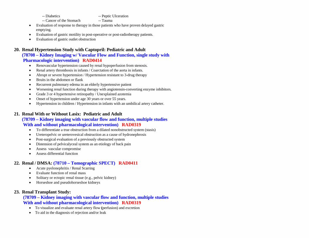
-- Diabetics -- Peptic Ulceration -- Cancer of the Stomach -- Tauma
• Evaluation of response to therapy in those patients who have proven delayed gastric emptying.
• Evaluation of gastric motility in post-operative or post-radiotherapy patients. • Evaluation of gastric outlet obstruction
20. Renal Hypertension Study with Captopril: Pediatric and Adult (78708 – Kidney Imaging w/ Vascular Flow and Function, single study with Pharmacologic intervention) RAD0414
• Renovascular hypertension caused by renal hypoperfusion from stenosis. • Renal artery thrombosis in infants / Coarctation of the aorta in infants. • Abrupt or severe hypertension / Hypertension resistant to 3-drug therapy • Bruits in the abdomen or flank • Recurrent pulmonary edema in an elderly hypertensive patient • Worsening renal function during therapy with angiotensin-converting enzyme inhibitors. • Grade 3 or 4 hypertensive retinopathy / Unexplained azotemia • Onset of hypertension under age 30 years or over 55 years. • Hypertension in children / Hypertension in infants with an umbilical artery catheter.
21. Renal With or Without Lasix: Pediatric and Adult (78709 – Kidney imaging with vascular flow and function, multiple studies With and without pharmacological intervention) RAD0319
• To differentiate a true obstruction from a dilated nonobstructed system (stasis) • Ureteropelvic or ureterovesical obstruction as a cause of hydronephrosis • Post-surgical evaluation of a previously obstructed system • Distension of pelvicalyceal system as an etiology of back pain • Assess vascular compromise • Assess differential function
22. Renal / DMSA: (78710 – Tomographic SPECT) RAD0411
• Acute pyelonephritis / Renal Scarring • Evaluate function of renal mass • Solitary or ectopic renal tissue (e.g., pelvic kidney) • Horseshoe and pseudohorseshoe kidneys
23. Renal Transplant Study: (78709 – Kidney imaging with vascular flow and function, multiple studies With and without pharmacological intervention) RAD0319
• To visualize and evaluate renal artery flow (perfusion) and excretion • To aid in the diagnosis of rejection and/or leak
24. Thyroid Scan and Uptake: (78007 – Thyroid imaging, w/ uptake; multiple determinations) RAD0435
• Evaluate for Hyperthyroidism or Hypothyroidism • Evaluate function of a thyroid nodule • Document the existence , the size and location of the thyroid gland or location of ectopic thyroid tissue. • Demonstrate heterogeneity of function within a hyperthyroid gland (Toxic Nodular Goiter) • Post-operative evaluation of the thyroid gland
25. Voiding Cysternogram: (78470 & 78730 – Ureteral reflux study, with bladder residual study) RAD0457 and RAD0448
• Initial evaluation of females with urinary tract infection for reflux • Diagnosis of familial reflux • Evaluation of vesicoureteral reflux after medical management or assessment of the results of antireflux surgery • Serial evaluation of bladder dysfunction (e.g., neurogenic bladder) for reflux
26. White Blood Cell Study: (78806 – Whole Body, with option to do 78807 – Tomographic SPECT) RAD0243 with RAD0338
• To evaluate febrile postoperative patient without localizing signs or symptoms. Fluid collections, ileus, bowel gas, fluid, and/or healing wounds may reduce the specificity of CT and ultrasound to detect site(s) and extent of inflammatory bowel disease. 99mTc-labeled leukocytes may be preferable for this indication.
• To detect acute osteomyelitis (less than one week duration) in conjunction with bone imaging. Gallium imaging is preferred performed in conjunction with bone imaging for chronic infection greater than one week, in the presence of prior surgery or hardware, spine or disc infections and in diabetic patients when degenerative or traumatic changes, neuropathic osteoarthropathy, or prior osteomyelitis have caused increased bone remodeling.
• To detect mycotic aneurysms, vascular graft infections, and shunt infections.
Note: The 99mTc Ceretec WBC study is performed instead of the 111In WBC study when acute appendicitis is suspected, a STAT study is needed, in pediatric population or if 111Indium is not available.

Page #1 of 5
Ultrasound Standard Operating Procedures EPIC Exam to Order Reason for Exam Exam Description CPT Code RAD1199 US AAA Exam Screen Only (Medicare)
Abdominal Aorta AAA Screening
G0389
RAD0459 US Abdomen Complete
Abdominal Pain Cirrhosis r/o Gallstones Hepatitis Nausea Pancreatitis Vomiting Elevated LFT’s
Includes Liver, GB, CBD, Pancreas, Spleen, Kidneys, Upper Abdominal Aorta and IVC
76700
RAD0460 US Abdomen Limited
Abdominal Mass Hernia R/O Ascitis Pyloric Stenosis R/O Gallstones R/O Appendicitis
Single abdominal organ or single quadrant only i.e. Gallbladder
76705
RAD0461 US Abdomen Retroperitoneal
AAA Bladder Mass/CA Chronic Kidney Disease Hematuria Hydronephrosis Renal Insufficiency Renal/kidney Stones UTI
Kidneys Bladder Aorta Iliacs IVC
76770
RAD0462 US Abdomen Retroperitoneal Limited
Aorta/IVC Bladder Only
76775
RAD0465 US Chest
R/O Pleural Fluid Superficial Mass Chest area
76604
RAD1056 or 0770 US Breast
Finding in axilla
Perform 1799 / 1800 US Axilla left or right
76882
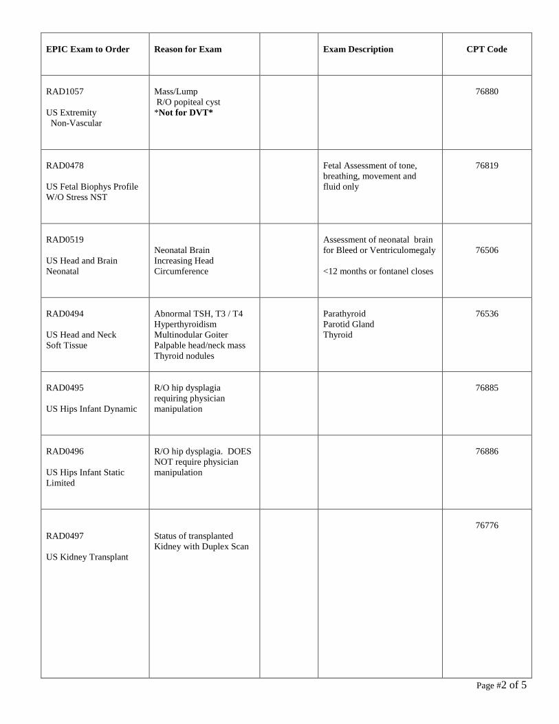
Page #2 of 5
EPIC Exam to Order
Reason for Exam
Exam Description
CPT Code
RAD1057 US Extremity Non-Vascular
Mass/Lump R/O popiteal cyst *Not for DVT*
76880
RAD0478 US Fetal Biophys Profile W/O Stress NST
Fetal Assessment of tone, breathing, movement and fluid only
76819
RAD0519 US Head and Brain Neonatal
Neonatal Brain Increasing Head Circumference
Assessment of neonatal brain for Bleed or Ventriculomegaly <12 months or fontanel closes
76506
RAD0494 US Head and Neck Soft Tissue
Abnormal TSH, T3 / T4 Hyperthyroidism Multinodular Goiter Palpable head/neck mass Thyroid nodules
Parathyroid Parotid Gland Thyroid
76536
RAD0495 US Hips Infant Dynamic
R/O hip dysplagia requiring physician manipulation
76885
RAD0496 US Hips Infant Static Limited
R/O hip dysplagia. DOES NOT require physician manipulation
76886
RAD0497 US Kidney Transplant
Status of transplanted Kidney with Duplex Scan
76776

Page #3 of 5
EPIC Exam to Order Reason for Exam Exam Description CPT Code RAD0500 US OB Complete Greater than 14 Weeks
If the pt is >or = 14 weeks and this is her 1st US and will not be the detailed scan
Fetal and Maternal Evaluation after 1st trimester
76805
RAD0502 US OB Detailed Single Fetus
Fetal and Maternal evaluation plus detailed fetal anatomic examination. Only used for high-risk pregnancy evaluation by OB specialist – otherwise, use 76805
Detailed anatomy scan usually done between 18 – 22 weeks * Preferably at least 20 weeks
76811
RAD0504 US OB Exam Limited
AFI Cervical Length Fetal Hearbeat Fetal Position Placenta location
Any or all of the reasons for exam
76815
RAD0469 US OB Exam Repeat
Follow-up Fetal Size & AFI Re-evaluation of organ systems suspected to be abnormal
76816
RAD0499 US OB Less than 14 weeks Single Fetus
Fetal and Maternal Evaluation 1st trimester
1st trimester scan – May need OB Transvaginal Obstetric order if really early IUP RAD0518
76801
RAD0503 US OB Multiple Gestation Complete
Fetal and Maternal evaluation plus detailed fetal anatomic examination, transabdominal approach
If the pt is >or = 14 weeks and this is her 1st US and it will not be the detailed scan
76812
RAD0518 US Transvaginal Obstetric
Typically only done during 1st trimester.if not well seen on transabdominal
76817
RAD1175 US OB Nuchal Translucency
First trimester fetal nuchal translucency measurement transabdominal and transvaginal approach
Performed during weeks 11wk2d – 13wk2d
76813
RAD1176 US OB Nuchal Translucency add’l fetus
First trimester fetal nuchal translucency measurement transabdominal and transvaginal approach each add’l fetus
Performed during weeks 11wk2d – 13wk2d
76814
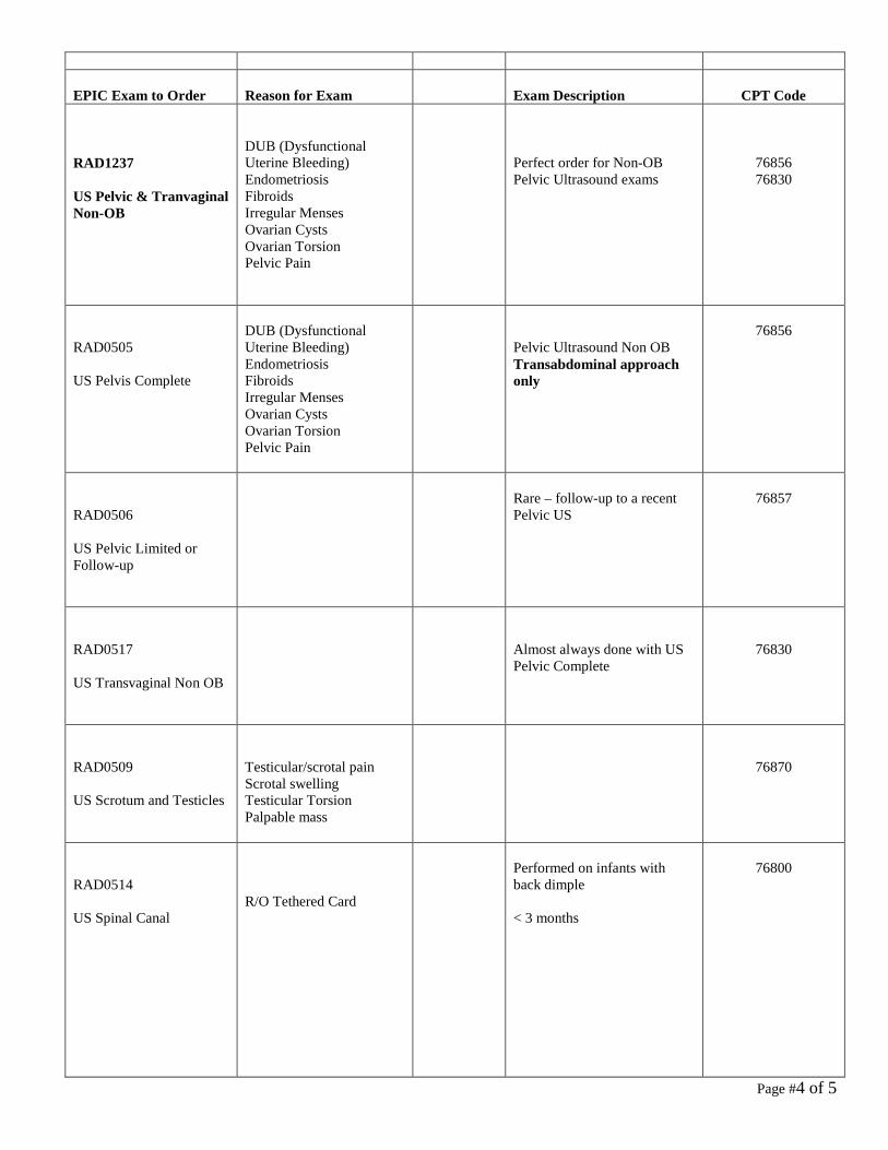
Page #4 of 5
EPIC Exam to Order
Reason for Exam
Exam Description
CPT Code
RAD1237 US Pelvic & Tranvaginal Non-OB
DUB (Dysfunctional Uterine Bleeding) Endometriosis Fibroids Irregular Menses Ovarian Cysts Ovarian Torsion Pelvic Pain
Perfect order for Non-OB Pelvic Ultrasound exams
76856 76830
RAD0505 US Pelvis Complete
DUB (Dysfunctional Uterine Bleeding) Endometriosis Fibroids Irregular Menses Ovarian Cysts Ovarian Torsion Pelvic Pain
Pelvic Ultrasound Non OB Transabdominal approach only
76856
RAD0506 US Pelvic Limited or Follow-up
Rare – follow-up to a recent Pelvic US
76857
RAD0517 US Transvaginal Non OB
Almost always done with US Pelvic Complete
76830
RAD0509 US Scrotum and Testicles
Testicular/scrotal pain Scrotal swelling Testicular Torsion Palpable mass
76870
RAD0514 US Spinal Canal
R/O Tethered Card
Performed on infants with back dimple < 3 months
76800

Page #5 of 5
EPIC Exam to Order Reason for Exam Exam Description CPT Code RAD0236 NIVL Duplex Abd Pelvis Study
RAS Hypertension
Renal Artery Stenosis SMA Study Liver Colorflow
93975
RAD0810 NIVL Duplex Scan Extracranial Arteries
Carotid Bruit Carotid Stenosis CVA/TIA symptoms S/P CEA
Carotid Doppler
93880
RAD 0235 NIVL Venous Duplex Extremity Bilateral
DVT Bilateral Extremities swelling/pain/redness
Venous Doppler Bilateral
93970
RAD1053 NIVL Venous Duplex Extremity Left
DVT Left leg or arm swelling/pain/redness
Venous Doppler Left leg or arm
93971
RAD0774 NIVL Venous Duplex Extremity Right
DVT Right leg or arm swelling/pain/redness
Venous Doppler Right leg or arm
93971

Last rev. 5/7/2012
SMF (Central) RADIOLOGY Protocols for STANDARD OPERATING PROCEDURES
HEAD SKULL < 4 views (limited)
o PA o LAT 70250
4 views or more (complete) o PA o TOWNES o BOTH LATERALS 70260
FACIAL BONES < 3 views (limited)
o UPRIGHT WATERS 70140 3 views or more (complete)
o UPRIGHT WATERS o CALDWELL o LATERAL SMV for zygomas as indicated by pain in that area 70150
NASAL BONES At least 3 views
o UPRIGHT WATERS o BOTH LATERALS 70160
SINUS Less than 3 views
o UPRIGHT WATERS o LATERAL 70210
EYE FOR FB (screening orbits for MRI) 1 view
o WATERS VIEW 70030 TMJ X-RAY indicated for trauma only
o TOWNES o BILATERAL o OPEN SCHULLERS (modified lateral) o CLOSED SCHULLERS 70330

Last rev. 5/7/2012
(CT RECOMMENDED) FOR ALL OTHER COMPLAINTS – Call ordering physician ORBITS At least 4 views
o WATERS o CALDWELL W/ 25-30 degree caudad o LATERAL of affected side o BOTH 3 POINT LANDINGS 70200
MANDIBLE < 4 views (limited)
o LATERAL (only for pediatric teeth) 70100 4 views or more (complete)
o PA o TOWNES o LATERAL o BOTH OBLIQUES 70110
MASTOIDS CT Highly recommended – Call ordering physician NECK SOFT TISSUE LATERAL NECK to include adenoids 70360
SPINE & PELVIS CERVICAL For TRAUMA
o AP o ODONTOID o LATERAL (Incl C-7); SWIMMERS as needed 72040
For PAIN
o AP o ODONTOID o BOTH OBLIQUES, LAT (Incl C-7) SWIMMERS as needed 72050
Complete w/Flexion & Extension
o AP o ODONTOID o BOTH OBLIQUES o LATERAL (Incl C-7); SWIMMERS as needed; o LAT FLEXION

Last rev. 5/7/2012
o LAT EXTENSION 72052 THORACIC AP LATERAL SWIMMERS 72070
LUMBAR For TRAUMA
o AP o LATERAL o CONE OF L4-5 if not open on lateral 72100
For PAIN
o AP o BOTH OBLIQUES o LATERAL o CONE OF L5-S1 if not open on lateral 72110
Complete, including bending views
o AP o LATERAL o BOTH OBLIQUES o CONE OF L5-S1 if not open on latearl o LATERAL FLEXION o LATERAL EXTENSION 72114
LUMBOSACRAL, bending views only
o STANDING AP o NEUTRAL LATERAL o FLEXION LATERAL o EXTENSION LATERAL 72120
THORACOLUMBAR AP LATERAL centered at T-12 / L-1, to includeT-10 TO L-3 72080
SCOLIOSIS UPRIGHT PA T/L to include entire pelvic crests for pelvic tilt & Risser grade
assessment; include separate AP pelvis, only if necessary (72170) UPRIGHT LATERAL T & L SPINE on one projection if possible 72069
*Please note: AP should be standing, with both legs fully extended and patellae facing forward.
No shoes 72-inch distance with radiographic ruler down the middle (from top to bottom) Include top of the acetabulum to the talus (this may require > than 72 inches from

Last rev. 5/7/2012
the tube to the imaging plate, depending on patient’s height. Patient may stand on step stool, with handle at side, in order to get the film low enough.
Digital image may require “stitching” to merge it into one film SACRUM & COCCYX 15 DEGREE CEPHALAD AP VIEW OF SACRUM 10 DEGREE CAUDAD VIEW OF COCCYX LATERAL VIEW TO INCLUDE BOTH 72220
SI JOINTS Always Bilateral 20 DEGREE CEPHALAD VIEW OF SACRUM to include both SI joints 45 DEGREE UPSIDE OBLIQUE VIEWS of each SI joint 72202
PELVIS 1-2 views (limited)
o AP PELVIS 72170 At least 3 views (complete)
o AP o LPO o RPO 72190
UPPER EXTREMITIES AC JOINTS *Always Bilateral AP VIEWS bilateral AC JOINTS, with and without weights 73050
STENOCLAVICULAR JOINTS RAO LAO PA CENTERED AT SC JOINTS 71130 CT RECOMMENDED – Call ordering physician
SCAPULA AP “Y” VIEW 73010
SHOULDER Minimum 2 views (complete) for Trauma
o INTERNAL ROTATION o EXTERNAL ROTATION

Last rev. 5/7/2012
o AXILLARY VIEW o TRANSCAPULAR VIEW 73030
Minimum 2 views (complete) for Non-Trauma
o INTERNAL ROTATION o GRASHEY VIEW o AXILLARY VIEW o SUPRASPINATUS OUTLET 73030
CLAVICLE 2 views
o AP o AP WITH 20 – 25 CEPHAL ANGLE 73000
HUMERUS 2 views
o AP o LATERAL 73060
ELBOW AP LATERAL OBLIQUE LATERAL 73080
FOREARM AP LATERAL 73090
WRIST PA OBLIQUE w/45 degree pronation LATERAL SCAPHOID as indicated by pain over snuffbox 73110
HAND PA OBLIQUE w/45 degree pronation LATERAL 73130
FINGER PA OBLIQUE LATERAL 73140

Last rev. 5/7/2012
UPPER EXTREMITY INFANT < 24 months AP LATERAL (Include shoulder to fingers) 73092
LOWER EXTREMITIES
All lower extremity images ordered by podiatry and orthopaedics will be performed as “weight-bearing” exams unless there is recent injury or concern for fracture.
HIP (S) Unilateral
o AP PELVIS 72170 o FROG LATERAL of affected hip (Cross table as indicated) 73500
BILATERAL o AP (may substitute AP Pelvis) o FROG LATERAL both hips 73520
PELVIS & HIPS INFANT (< 24 months) AP Frog leg PELVIS and HIPS 73540
FEMUR AP LATERAL (INCLUDE HIP & KNEE) 73550 (* If 2 films needed for AP & Lat, ensure overlap at midshaft)
KNEE 3 view
o AP (upright weight-bearing, as directed) o LATERAL o SUNRISE 73562
Min 4 views (complete) o AP (upright weight-bearing, as directed) 73564 o LATERAL o TUNNEL o SUNRISE or MERCHANT
AP STANDING *BILATERAL KNEES* 73565 TIB-FIB

Last rev. 5/7/2012
AP LATERAL (Include knee and ankle) 73590
ANKLE AP OBLIQUE LATERAL (Weight Bearing as directed) 73610
FOOT AP OBLIQUE LATERAL (Weight Bearing as directed) 73630
CALCANEUS (HEEL) AP LATERAL 73650
TOES AP OBLIQUE LATERAL 73660
LOWER EXTREMITY INFANT < 24 months AP LATERAL ( Include hips to feet) 73592
CHEST CHEST Single view (for positive PPD test or as ordered)
o PA 71010 o
2 view o PA o LATERAL 71020
PA w/APICAL LORDOTIC 71021 PA w/ BOTH OBLIQUES 71022 CHEST COMPLETE 4 views
o PA o LATERAL o BOTH OBLIQUES 71030
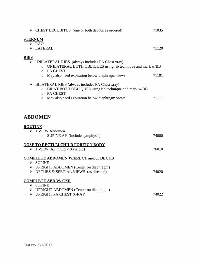
Last rev. 5/7/2012
CHEST DECUBITUS (one or both decubs as ordered) 71035
STERNUM RAO LATERAL 71120
RIBS UNILATERAL RIBS (always includes PA Chest xray)
o UNILATERAL BOTH OBLIQUES using rib technique and mark w/BB o PA CHEST o May also need expiration below diaphragm views 71101
BILATERAL RIBS (always includes PA Chest xray)
o BILAT BOTH OBLIQUES using rib technique and mark w/BB o PA CHEST o May also need expiration below diaphragm views 71111
ABDOMEN ROUTINE 1 VIEW Abdomen
o SUPINE AP (include symphysis) 74000 NOSE TO RECTUM CHILD FOREIGN BODY 1 VIEW AP (child < 8 yrs old) 76010
COMPLETE ABDOMEN W/ERECT and/or DECUB SUPINE UPRIGHT ABDOMEN (Center on diaphragm) DECUBS & SPECIAL VIEWS (as directed) 74020
COMPLETE ABD W/ CXR SUPINE UPRIGHT ABDOMEN (Center on diaphragm) UPRIGHT PA CHEST X-RAY 74022

Last rev. 5/7/2012
MISCELLANEOUS BONE AGE PA ONLY LEFT HAND TO INCLUDE WRIST 77072
INFANT OSSEOUS SURVEY / Battered child series (for suspected child abuse) 77076
Appendicular Skeleton: AP Humeri AP Forearms PA Hands AP Femurs AP Lower Legs (Tib/Fib) AP Feet
*Each must be obtained as a separate exposure to ensure uniform image density and maximize image sharpness; it is NOT acceptable to do single views of an entire upper or lower extremity.*
Axial Skeleton: Chest: AP, Lateral, and Bilateral Obliques, to include all 12 ribs and entire thoracic spine through thoracolumbar junction
Pelvis: AP to iclude the lumbar spine Lumbosacral Spine: Lateral Cervical Spine: Lateral Skull: Frontal (AP) and Lateral
If possible, the exam should be reviewed for completeness and possible inclusion of additional views by the covering Radiologist.
Evaluation of suspected metaphyseal irregularities may require additional coned views of a joint/extremity. Additional Townes and/or oblique views of skull may be necessary in cases of suspected occipital trauma to skull.
SKELETAL / OSSEOUS SURVEY PA & LAT CHEST, LAT SKULL, PELVIS, LATERAL C-T-L SPINES, AP ALL LONG BONES BILATERALLY ( NO LIMITED SURVEYS = ORDER SPECIFIC EXAMS ) 77075
METASTATIC SURVEY PA & LAT CHEST, LAT SKULL, PELVIS, LATERAL C-T-L SPINES, AP LONG BONES 77074
METABOLIC SURVEY (JOINT SURVEY) KNEES & WRISTS AP/LAT 77077

Last rev. 5/7/2012
ARTHRITIS SERIES (Bilat Hands) PA LATERAL NORGAARD VIEW (Allstate view – both hands semi-cupped)


![Operating Procedures 1 G2 - OPERATING PROCEDURES [6 Exam Questions - 6 Groups] G2APhone operating procedures; USB/LSB utilization conventions; procedural.](https://static.fdocuments.net/doc/165x107/56649e4d5503460f94b4351a/operating-procedures-1-g2-operating-procedures-6-exam-questions-6-groups.jpg)