Surgical Bleeding and Blood Replacement education/Didactic Support Mate… · Surgical Bleeding and...
55
Surgical Bleeding and Blood Replacement Stepheny Berry, MD Department of Surgery University of Kansas
-
Upload
truongthuy -
Category
Documents
-
view
219 -
download
0
Transcript of Surgical Bleeding and Blood Replacement education/Didactic Support Mate… · Surgical Bleeding and...

Surgical Bleeding and Blood Replacement
Stepheny Berry, MD
Department of Surgery
University of Kansas

Introduction
► Hemostasis defined by Virchow as the balance among blood flow, humoral factors, and cellular elements of the vascular system.
► Two coagulation pathways Intrinsic
Extrinsic
► Platelets play a vital role early in hemostasis with the formation of the platelet plug
► Platelets release factors that promote hemostasis at the site of injury
► The intrinsic and extrinsic pathways lead to formation of Xa which starts the common pathway to coagulation

Introduction
► Negatively charged phospholipid phosphatidylserine is found on the inner leaflet of mammalian cells.
► Collagen or thrombin exposure changes the distribution of phospholipids to the external leaf
► This provides a pro-coagulant surface for the various steps to take place
► This also selects the activation to the site of injury


Intrinsic Pathway
► Factor XII becomes activated in the contact phase of coagulation Combines with XI, prekallikrein, and high molecular weight
kininogen Come together on the highly negatively charged surfaces
experimentally
► Factor XII is then activated by an unknown mechanism ► Factor XIIa converts prekallikrein to kallikrein ► Kallikrein converts factor XII to XIIa ► XIIa converts XI to XIa ► XIa converts IX to IXa

Intrinsic Pathway
► IXa with its cofactor VIII plus calcium and phospholipid membranes form the “tenase” complex
This complex converts X -> Xa
Xa activates the common pathway
► This complex is enhanced by two mechanisms
The phospholipid membrane allows the enzymes to become more easily saturated
Helps localize coagulation response to where it’s needed

Extrinsic Pathway
►Circulating factor VII encounters tissue factor and activates
► Tissue factor Transmembrane glycoprotein normally expressed by
fibroblast like cells that surround the blood vessel
Endothelium shields circulating blood from exposure to tissue factor
Activated monocytes, atherosclerotic plaques, and activated endothelial cells express tissue factor
► Factor VII Weak procoagulant

Extrinsic Pathway
► Factor VII 10,000,000 fold increase in activity when bound to tissue factor
How VII activated unknown (? activation by Xa)
Both VII and VIIa bind to tissue factor
VIIa activates Xa
► IX activated by VII showing a cross activation of the two pathways
► Activation of X by the IXa/VIII complex is 50 times greater than the activation by VII/TF

Common Pathway
► Factor Xa Combines with Va, calcium, and the phospholipid membrane to
form prothrombinase complex Converts prothrombin to thrombin
► Factor Va Factor Xa and Va are present in stoichiometric amounts and cause
an alteration in the binding site of Xa to increase the catalytic efficiency
Binds to prothrombin and sequesters it to the site of the prothrombinase complex
► Produces a 300,000 fold increase in rate of prothrombin conversion
► Factors V and VIII are activated by proteases but are not active proteases themselves

Common pathway
► Thrombin and Fibrin Cleaves the soluble protein fibrinogen to produce the
insoluble fibrin monomer Factor XIIIa cross links these monomers and allows
formation of the meshwork of the thrombus Thrombin activates
►Factors XII, XI, VII, and V ►Activates platelets ►Activates Protein C ►Stimulates endothelial cells to produce plasminogen inhibitor

Role of Platelets
►Disc shaped, anuclear particles that circulate in a nonadhesive state in the undamaged circulation
►Changes in the platelet surface in the activated vs inactive state Inactive- mostly phosphatidylcholine
Activated- mostly phosphatidylserine
►Contain a contractile system and storage granules α granules contain platelet factor 4, thromboglobulin,
PDGF, P-selectin, fibrinogen, factor V, vWF
β granules contain ATP, ADP, and serotonin

Role of Platelets
► First step toward platelet aggregation is adhesion ► Aggregation prevented by
Heparan sulfate- activates antithrombin Thrombomodulin- activates protein C PAI- induces fibrin degradation TFPI- inhibits TF Prostacyclin I2- raises CAMP levels and NO levels
► Injured endothelium promotes adhesion of platelets ► Platelet adhesion promotes activation ► Thrombin is the most potent aggregation factor for
platelets

Other Factors
► Platelet integrins GP Ib- vWF GP Ia/IIa- collagen GP IIb/IIIa- fibrinogen and fibronectin (most abundant)
► Leukocytes Express minimal amounts of procoagulant activity normally Monocytes express TF Contain XI-VIII receptors which allows intrinsic pathway activation Linked to thrombosis in sepsis
► Endothelium Important in the regulation of coagulation Undamaged
► Thrombomodulin, fibrinolytic mediators, prostaglandins, NO, TFPI
Damaged ► TF, PAI, vWF, procoagulant proteins

Endogenous Inhibitors
► Antithrombin Serine protease inhibitor (SERPIN)
Primary inhibitor of coagulation
Targets most coagulation proteases, plasmin, and kallikrein
► Heparin cofactor II Resembles antithrombin
Only has activity against thrombin
► Protein C Keeps blood in fluid state
Activated when thrombin binds to thrombomodulin
Cleaves membrane bound Va and VIIIa
Needs Protein S and factor V as cofactors

History and Physical Exam
►Detailed bleeding history
? Bleeding after dental procedures, minor cuts, previous OR, prolonged menses, easy bruising, nose bleeds
Family history
►Physical Exam
Few true physical signs
Splenomegaly, hepatomegaly, hemarthroses, petechia (plt) or ecchymosis (coag)

Diagnostic Testing
► Bleeding is common ► Diagnosis of the underlying reason is vital ► Test of coagulation
PT ► Extrinsic pathway ►Measured by subjecting citrated plasma to TF, phospholipids, and
calcium ► Vitamin K dependent factors- II, VII, XI, X, V ► INR ► Corrected with FFP and/or Vitamin K ► Can be elevated with high doses of heparin
aPTT ► Intrinsic pathway ► Unfractionated Heparin ► Not used for low molecular weight Heparin
Diagnostic Testing
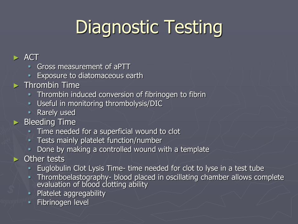
► ACT Gross measurement of aPTT Exposure to diatomaceous earth
► Thrombin Time Thrombin induced conversion of fibrinogen to fibrin Useful in monitoring thrombolysis/DIC Rarely used
► Bleeding Time Time needed for a superficial wound to clot Tests mainly platelet function/number Done by making a controlled wound with a template
► Other tests Euglobulin Clot Lysis Time- time needed for clot to lyse in a test tube Thromboelastography- blood placed in oscillating chamber allows complete
evaluation of blood clotting ability Platelet aggregability Fibrinogen level

Causes of Bleeding ► Coagulopathic bleeding Congenital
Platelet Disorders ► Rare ► Divided into problems of adhesion, aggregation, secretion, and procoagulant activity ► Treatment is platelets or DDAVP
Von Willebrand Disease ► Quantitative or qualitative defect of vWF ► Carrier for factor VIII ► Most commonly inherited bleeding disorder (incidence 1-2%) ► Easy bruising, mucosal bleeding, menorrhagia, epistaxis, etc… ► Treatment rarely required but if bleeding then DDAVP or factor VIII/vWF concentrate


Causes of Bleeding
► Coagulopathic bleeding congenital cont’d Hemophilia
► A or B ► Hallmark is repeat bleeding into joints and muscles ► Levels
<1% severe 1-5% moderately severe 6-25% mild
► Treat with factor replacement ► If immunity develops to exogenous factors activated factor VII ► A
Factor VIII X-linked recessive 1 in 5000 men affected 3% prevents spontaneous hemorrhage 30% for mild bleeding, 50% for major bleeding 80-100% during OR and 30% post op for 2 weeks

Causes of Bleeding
► Hemophilia cont’d B
► Accounts for 20% of hemophilia ► X-linked recessive ► Indistinguishable from hemophilia A ► 20-30% levels for minor bleeding ► 50-100% for 2 weeks post op
► Acquired disorders of hemostasis Liver disease (decreased prothrombin, V, VII, X) EtOH (thrombocytopenia Hypersplenism (thrombocytopenia)

Treatments
► Whole Blood Occasionally used in the military, not readily available here
► PRBC Stored @ 4 degrees Celsius up to 5 weeks Restore oxygen carrying capacity Transfuse to 7 mg/dL minimum
► FFP Replaces all coagulation factors, but not as rich in factor VIII Can be stored frozen for up to 12 months at -30 degrees Celsius Useful in elevated PT Useful when specific factor not available

Treatments
► Platelets Prophylactic in massive hemorrhage Contain a substantial amount of FFP and V Need 20/mcL minimum for normal hemostasis 50-70/mcL for active
bleeding
► Cryoprecipitate Rich in VIII, vWF, fibrinogen, and fibronectin Most commonly used to increase fibrinogen Can be stored at -30 degrees Celsius for 12 months
► Desmopressin Synthetic vasopression Increases release of factor VIII and vWF Improves platelet adhesion

Treatments
► Vitamin K Carboxylates already synthesized factors stored in hepatocytes Slower more durable correction
► Protamine Sulfate Positively charged protein that reverses the effect of negatively
charged heparin 1mg/100u heparin Can cause hypotension, pulmonary HTN, anaphylaxis, death Derived from Salmon Semen
► Antifibrinolytic agents i.e. Amicar Block plasminogen primarily or the effect of plasmin on fibrinogen
and fibrin
► Specific factors

Transfusion Reactions
► Febrile Transfusion Reactions
Most common
Treated with antipyretics and antihistamines
Removal of white cell debris from PRBC, plt and FFP reduces risk
Can be pre-treated if pt has history
► Hemolytic Transfusion Reaction
STOP administration of blood
Return to lab for repeat crossmatch
May require pressors to support BP, maintenance of renal perfusion, management of DIC
Treat with volume support first, pressors if needed, diuretics to maintain UO, and HD if renal faiure

Transfusion Reactios
► Infection
Hep C – 1 in 1,390,000
Hep B – 1 in 200,000-500,000
HIV – 1 in 2,000,000
HTLV - <1 in 2,000,000
West Nile Virus (11 documented cases)
Syphilis – none in 30 years
Chagas Disease – extremely low, red cross qualifies each donor rather than each donation for negativity
Bacterial infection most common with plt transfusion
► Volume Overload

Transfusion Reactions
► Massive Transfusion
Coagulopathy, hypothremia, citrate toxicity (liver dysfxn), electrolyte abnormalities (hyperkalemia, acidemia, hypocalcemia)
► TRALI
Acute lung injury developing within 6 hours of transfusion
Rapid onset of tachypnea, cyanosis, dyspnea, fever
Acute hypoxemia (paO2/FiO2 <300)
Wedge pressure < 18mmHg
Treatmens: aggressive resp support, may need mechanical ventilation
Leading reported cause of fatal transfusion reactions in the US in 2003/4

Hypercoagulable States
► Congential disorders Activated protein C resistance most common
►Most common cause of APCR is Factor V leiden deficiency
ATIII deficiency
Protein C & S deficiency
Hyperhomocytstinemia
► Acquired disorders Decreased production (liver failure)
Ineffective fibrinolysis
High levels of clotting factors (upregulated during stress)
Thrombocytosis
Antiphospholipid syndromes
Chronic cases of DIC
Hyperhomocystenemia (in pts with renal faliure)

Hypercoagulable States
► Diagnostic Evaluation
Activated protein C resistance test
Antithrombin III activity assay
Proteins C & S activity
Antiphospholipid antibody
Prothrombin activity (screening for prothombin 20210)
Serum homocystine level
► Management
Theraputic anticoagulation for VTE (heparin/coumadin/antiplatelet)
Treat hyperhomocystenemia
DVT prophylaxis for high risk patients

Spleen
Stepheny Berry, MD
Department of Surgery
University of Kansas

Objectives
►Anatomy & Physiology
►Surgical disorders
►Consequenses of Splenectomy
►Complications

Anatomy
►Embrology – develops from the dorsal mesogastrium by the 6th gestational week
►Receives 5% cardiac output
►Dual arterial/venous supply (splenic vessels and short gastric vessels)
Splenic artery – branch of the celiac
Short gastrics – from left gastroepiploic artery

Anatomy
► LUQ bound by the diaphragm and rib cage
► Intimately assoc w/ pancreas, stomach, left kidney, colon and diaphragm
► Multiple ligaments: splenorenal, gastrosplenic, splenocolic and splenophrenic ligaments

Anatomy
► Accessory spleens: most commonly found in the splenic hilum, followed by the splenocolic ligament, gastrocolic ligament, splenorenal ligament and omentum.
Important to know when performing splenectomy for hematologic disorders
► Polysplenia: multiple small spleens, no normal spleen
Assoc w/ cardiac defects, situs inversus, biliary atresia
► Asplenia: absence of spleen
Lethal condition assoc w/ cardiac defects and situs inversus
► Splenogonadal Fusion
Rare disorder. Splenic tissue found in scrotum, attached to testicle.

Physiology
► Functions
Hematopoiesis
Blood filtering
Immune modulation
► Structure
Blood enters spleen through central arteries
Branch to trabecular arteries – white pulp
Then goes to the marginal zone (sinuses) and directed either to the red pulp or back to white pulp

Physiology
► White Pulp Surrounded by lymphatic
sheaths (T-lymphocytes and macrophages) that process soluble antigens
Some goes into lymphatic follicles, where B-lymphocytes can proliferate. Plasma cells are also found here.
► Red Pulp Reticular network, no
endothelial cells, moves slowly through numerous macrophages, then enters sinuses
Antibody-sensitized and particulate material removed

Physiology
► Filters and sequesters abnormal and aged erythrocytes, granulcytes and platelets
►Nearly 350 L/day filtered through spleen
► Immune function – reticuloendotheial system
Specific
►Antigen processing and antibody production.
►Largest producer of IgM
Non-specific
►Clearance of opsonized particles and bacteria by splenic macrophages
►Production of opsonin (properdin, tuftsin, fibronectin)

Surgical Disorders of the Spleen
► Splenic Rupture
Trauma
Spontaneus
Iatrogenic injury
► Hematologic Disorders
Hematolymic anemias
Hereditary spherocytosis
Thalassemias
ITP

Surgical Disorders of the Spleen
► Hypersplenism from other Diseases
Inflammation
Infiltrative diseases
congestion
► Leukemia and Lymphoma
► Other Diseases
Splenic abscess
Primary and metastatic tumors
Cysts
Splenic artery aneurysm
Isolated gastric varicies

Surgical Disorders of the Spleen
► Splenic Trauma
Most commonly injured organ after blunt trauma
► Usually has associated injuries (rib fracturs, TBI, ortho injuries, liver
injuries)
Also common in penetrating trauma
Management based on hemodynamic stability of the patient.

Splenic Trauma
► ATLS protocol for all patients
► Physical Exam
Unreliable in trauma patients
20% of patients with splenic injuries have rib fx
LUQ pain/tenderness
Kehr’s sign – referred pain in left shoulder
Balance’s sign – percussion dullness to left flank

Splenic Trauma
► FAST
Quick, good in “unstable” patients
Non-specific – tells you fluid or no fluid
Unstable pt with positive fast = OR
► CT
Better for stable patients, more specific, diagnostic test of choice
Allows for grading of injury and measurement of hemoperitoneum
Able to diagnose other injuries as well

Splenic Trauma
► Grading scale to determine severity and uniformity of diagnosis
► No rule about certain grade needing splenectomy, but some generalizations
Elderly more likely to fail non-op
Higher grades more likely to fail non-op
Large amt of hemoperitoneum more likely to fail non-op

Hematologic Disorders
► Hypersplenism vs splenomegaly
Hypersplenism – excess fxn of spleen and causes cytopenia (anemia, leukopenia, thrombocytopenia)
Splenomegaly (anatomic enlargement of the spleen)
► Hereditary Spherocytosis
Autosomal dominant
Deficiency in spectrin and makes rigid RBC which become sequestered in the red pulp
Splenectomy to prevent anemia (wait until age 5, if possible)
► Metabolic hemolytic anemia
Pyruvate kinase deficiency, G6PD deficiency, etc
Not responsive to splenectomy

Hematologic Disorders
► Sickle Cell
Autosomal recessive
Rigid cells at low O2 sats
Also lead to increased viscosity, stasis and thrombocytosis
Usually infarct spleen and become functionally asplenic
Splenectomy may be beneficial during hemolytic crisis and splenomegaly
► Thalassemias
Major (homozygous beta thalassemia) – reduces transfusion requirements, splenomegaly and rupture
Minor (heterozygous beta thalassemia) – decreases transfusion requirements and issues with iron overload

Hematologic Disorders
► Thrombocytopenia
Splenectomy only appropriate for idiopathic immune mediated thrombocytopenia (cause can’t be found)
► ITP
Usually after an acute viral infection
Women > men
Steroids first
If no response, may benefit from splenectomy
Better response to splenectomy if good response w/ steroids, but recurrence once steroids are tapered.

Hematologic Disorders
► TTP
Disease of arteries or capillaries
Fevers, purpura, hemolytic anemia, neurologic manifestations, renal disease
Plasmapheresis is treatment
► HIV assoc Thrombocytopenia
Splenectomy if AIDS and symptomatic thrombocytopenia resistant to medical management

Hypersplenism from other diseases
► Congestive splenomegaly
Usually as result of liver failure
Treatment of portal hypertension
Splenectomy contra-indicated as one of the treatments for portal hypertension is splenorenal shunt
► Infiltrative Splenomegaly
e.g Gaucher’s disease ► Partial splenectomy or embolization used to treat symptoms (hypersplenism an
pain from splenomegaly)
► Felty’s Syndrome
RA pts with leg ulcers and assoc splenomegaly ane neutropenia
Splenectomy controversial as results are unpredictable

Hematologic Malignancies
► Acute Leukemia
Not indicated
► Chronic Leukemia
Rarely for hypersplenism or sx of splenomegaly
► Leukemic reticuloendotheliosis
Hairy cell leukemia
For palliation of cytopenia and sx of splenomegaly with advent of medications
► Hodgkin’s disease
Not routinely part of staging laparotomy anymore because of newer imaging techniques
► Non-hodgkin’s lymphoma
Rarely indicated for hypersplenism or sx of splenomegaly

Splenectomy
► Midline incision, left subcostal incision (Kehr’s incision), or laparoscopic
► Mobilized from its retroperitoneal attachments bluntly
► Splenocolic, splenophrenic and splenorenal ligaments divided with electrocautery
► Short gastric arteries individually ligated in the gastrosplenic ligament near the spleen
► Splenic artery and vein are then individually ligated close to the spleen

Consequences and Complications
► Transient leukocytosis and thrombocytosis
WBC increases by avg of 50% from baseline
Usually normalizes within 5-7 days
Plt increases by avg 30%
Usually normalizes within 2 wks
► Postsplenectomy sepsis
Higher incidence in children (2-4% vs 1-2% in adults)
Hematologic disorders at highest risk
Strep pneumo most common organism (H.flu, N.meningitidis, beta-hemolytic strep, S.aureus, E.coli, Pseudomonas)
Pts progress rapidly (within hours) to sepsis and even death despite appropriate abx
Waterhouse-Friderichsen’s Syndrome not uncommon

Consequences and Complications
► Encapsulated organisms
Vaccinate against S.pneumo, H.flu, N. meningiditidis
Before surgery appx 1 week, if possible
► Atelectasis
Most common complication, from discomfort related to upper abd incision
Pulmonary toilet
► Subphrenic abscess
Can develop assoc plerual effusion
Fluid collects in splenic fossa. Can become infected
No role for routine drain

Consequences and Complications
► Pancreatic injury
1-3% of patients
Increased risk of abscess
Can have pancreatic fistula, local pancreatitis, pancreatic pseudocyst
Sx similar to subphrenic abscess
► Stomach injury
Usually related to where short gastric arteries were ligated
Subphrenic abscess or gastrocutaneous fistula
Some surgeons advocate keeping NG for a couple days, but no data to support

Questions?