1-4 Hemostasis, Surgical Bleeding and Transfusion
17
1 of 17 NAMES NG TRANS PEOPLE Surgery Hemostasis, Surgical Bleeding, and Transfusion DR. Bibera July 5, 2012 1-4 a complex process whose function is to limit blood loss from an injured vessel 4 major physiologic events o vascular constriction o platelet plug formation o fibrin formation o fibrinolysis Vascular Constriction is the initial response to vessel injury, more pronounced in vessels with medial smooth muscles dependent on local contraction of smooth muscle subsequently linked to platelet plug formation potent vasoconstrictors: o Thromboxane A2 (TXA2) is produced locally at the site of injury via the release of arachidonic acid from platelet membranes o Endothelinsynthesized by injured endothelium and serotonin (5-hydroxytryptamine) released during platelet aggregation o Bradykinin and Fibrinopeptides the extent of vasoconstriction varies with the degree of vessel injury Platelet Function platelets are anucleate fragments of megakaryocytes, normal circulating number of platelets ranges between 150,000 and 400,000/ L up to 30% may be sequestered in the spleen if not consumed in a clotting reaction, platelets are normally removed by the spleen and have an average life span of 7 to 10 days platelets play an integral role in hemostasis by forming a hemostatic plug and by contributing to thrombin formation injury to the intimal layer in the vascular wall exposes subendothelial collagen to which platelets adhere, which requires von Willebrand's factor (vWF) binds to glycoprotein I/IX/V on the platelet membrane after adhesion, platelets initiate a release reaction that recruits other platelets from the circulating blood to seal the disrupted vessel. Up to this point, this process is known as primary hemostasis platelet aggregation is reversible and is not associated with secretion o heparin does not interfere with this reaction o adenosine diphosphate (ADP) and serotonin are the principal mediators in platelet aggregation arachidonic acid released converted by COX to prostaglandin G2 (PGG2) prostaglandin H2 (PGH2) converted to TXA2 effects:Arachidonic acid shuttled to adjacent endothelial cells converted to prostacyclin (PGI2 ) vasodilation and acts to inhibit platelet aggregation platelet COX is o irreversibly inhibited by aspirin o reversibly blocked by NSAIDs o but is not affected by COX-2 inhibitor in the second wave of platelet aggregation, a release reaction occurs in which several substances, including ADP, Ca2+,serotonin, TXA2, and -granule proteins are discharged fibrinogen is a required cofactor, acting as a bridge for the glycoprotein IIb/IIIa receptor on the activated platelets its release causes compaction of the plateletsinto a plug, a process that is irreversible thrombospondin, secreted by the granule, stabilizes fibrinogen binding to the activated platelet surface and strengthens the platelet-platelet interactions. platelet factor 4 (PF4), potent heparin antagonist, and thromboglobulin also are secreted during the release reaction the second wave of platelet aggregation is inhibited by aspirin and NSAIDs, by (cAMP), and by nitric oxide alterations occur in the phospholipids of the platelet membrane that allow Ca2+ and clotting factors bind to the platelet surface enzymatically active complexes BIOLOGY OF HOMEOSTASIS
-
Upload
robin-hesita-tolentino -
Category
Documents
-
view
57 -
download
3
description
Filename: 1-4 Hemostasis, Surgical Bleeding and Transfusion.pdf
Transcript of 1-4 Hemostasis, Surgical Bleeding and Transfusion

1 of 17
NAMES NG TRANS PEOPLE
Surgery
Hemostasis, Surgical Bleeding, and
Transfusion
DR. Bibera
July 5, 2012 1-4
a complex process whose function is to limit blood loss
from an injured vessel 4 major physiologic events
o vascular constriction o platelet plug formation o fibrin formation o fibrinolysis
Vascular Constriction
is the initial response to vessel injury, more pronounced in vessels with medial smooth muscles
dependent on local contraction of smooth muscle subsequently linked to platelet plug formation potent vasoconstrictors:
o Thromboxane A2 (TXA2) is produced locally at the site of injury via the release of arachidonic acid
from platelet membranes
o Endothelinsynthesized by injured endothelium and serotonin (5-hydroxytryptamine) released during platelet aggregation
o Bradykinin and Fibrinopeptides the extent of vasoconstriction varies with the degree of
vessel injury
Platelet Function
platelets are anucleate fragments of megakaryocytes, normal circulating number of platelets ranges between 150,000 and 400,000/ L
up to 30% may be sequestered in the spleen if not consumed in a clotting reaction, platelets are
normally removed by the spleen and have an average life span of 7 to 10 days
platelets play an integral role in hemostasis by forming a hemostatic plug and by contributing to thrombin formation
injury to the intimal layer in the vascular wall exposes subendothelial collagen to which platelets adhere, which requires von Willebrand's factor (vWF) binds to glycoprotein I/IX/V on the platelet
membrane
after adhesion, platelets initiate a release reaction that recruits other platelets from the circulating blood to seal the disrupted vessel. Up to this point, this process is known as primary hemostasis
platelet aggregation is reversible and is not associated with secretion
o heparin does not interfere with this reaction o adenosine diphosphate (ADP) and serotonin are
the principal mediators in platelet aggregation
arachidonic acid released converted by COX to
prostaglandin G2 (PGG2) prostaglandin H2 (PGH2) converted to TXA2
effects:Arachidonic acid shuttled to adjacent endothelial cells converted to prostacyclin (PGI2 )
vasodilation and acts to inhibit platelet aggregation
platelet COX is
o irreversibly inhibited by aspirin o reversibly blocked by NSAIDs
o but is not affected by COX-2 inhibitor
in the second wave of platelet aggregation, a release
reaction occurs in which several substances, including ADP, Ca2+,serotonin, TXA2, and -granule proteins are discharged
fibrinogen is a required cofactor, acting as a bridge for the glycoprotein IIb/IIIa receptor on the activated platelets its release causes compaction of the plateletsinto a
plug, a process that is irreversible thrombospondin, secreted by the granule, stabilizes
fibrinogen binding to the activated platelet surface and strengthens the platelet-platelet interactions.
platelet factor 4 (PF4), potent heparin antagonist, and thromboglobulin also are secreted during the release reaction
the second wave of platelet aggregation is inhibited by aspirin and NSAIDs, by (cAMP), and by nitric oxide
alterations occur in the phospholipids of the platelet membrane that allow Ca2+ and clotting factors bind
to the platelet surface enzymatically active
complexes
BIOLOGY OF HOMEOSTASIS

2 of 17
Dave.Linelle.Deane.Mhe.Te.Paul
o the altered lipoprotein surface (sometimes referred to as platelet factor 3) catalyzes reactions that are involved in:
- conversion of prothrombin to thrombin by
activated factor X (Xa) in the presence of factor V and Ca2+
- is also involved in the reaction by which activated factor IX (IXa), factor VIII, and Ca2+ activate factor X
Coagulation
the coagulation cascade has 2 intersecting pathways: o Intrinsic pathway
- begins with factor XII and through a cascade of reactions activates factors XI, IX, and VII in sequence fibrin clot formation, intrinsic to the
circulating plasma and no surface is required to
initiate the process o Extrinsic pathway
- requires exposure of tissue factor on the surface of the injured vessel wall to initiate the cascade beginning with factor VII
the two arms of the coagulation cascade merge to a common pathway at factor X activation sequence of factors II (prothrombin) and I
(fibrinogen) clot formation occurs after proteolytic conversion of
fibrinogen to fibrin an elevated activated partial thromboplastin
time(aPTT) abnormal function Intrinsic pathway an elevated prothrombin time (PT) abnormal
extrinsic pathway vitamin K deficiency and warfarin use affect factors II,
VII, IX, and X fibrinogen levels of <50 mg/dL causes prolongation of
the PT and aPTT primary pathway for coagulation is initiated by
theexposure of subendothelial tissue factor when vessel surface is injured
propagation of the clotting reaction then ensues with a
sequence of enzymatic reactions, which involves a proteolytic enzyme
cleavage of a proenzyme and a phospholipid surface
generates the next enzyme in a cascade manner
o each reaction requires a helper protein (i.e. Factor VIIa binds to tissue factor, and tissue factor-VIIa complex catalyzes the activation of factor X to factor Xa)
o the reaction takes place on the phospholipid
surface of activated platelets
o this complex is four orders of magnitude more active at converting factor X than is factor VIIa alone and also activates factor IX to factor IXa
o factor Xa, together with factor Va and Ca2+ and phospholipid, comprises the prothrombinase complex that converts prothrombin to thrombin
o thrombin has multiple functions in the clotting process, including conversion of fibrinogen to fibrin and activation of factors V, VII, VIII, XI,and
XIII, as well as activation of platelets
o factor VIIIa combines with factor IXa to form the intrinsic factor complex (VIIIa-IXa), which is responsible for the bulk of the conversion of factor X to Xa50x more effective at catalyzing factor X
activation than is extrinsic (tissue factor-VIIa) complex, five to six orders of magnitude more effective than is factor IXa alone
o the prothrombinase is significantly more effective
at catalyzing its substrate than is factor Xa alone
o once formed, thrombin leaves the membrane surface converts fibrinogen by two cleavage
steps into fibrin and 2 small peptides termed fibrinopeptides A and B
o removal of fibrinopeptide A permits end-to-end
polymerization of the fibrin
o cleavage of fibrinopeptide B allows side-to-side polymerization of the fibrin clot, facilitated by thrombin-activatable fibrinolysis inhibitor(TAFI)
the coagulation system is exquisitely regulated. Feedback inhibition on the coagulation cascade
deactivates the enzyme complexes leading to thrombin formation
exists at upstream, intermediate, and downstream
portions of the coagulation cascade to "turn off" thrombin formation once the procoagulantsequence is initially activated
Coagulation Factors Tested
by the PT and the aPTT
PT aPTT
VII XII
X High molecular weight
kininogen
V Prekallikrein
II (prothrombin) XI
Fibrinogen
IX
VIII
X
V
II
Fibrinogen
Based on 3 mechanisms:
o mechanisms of fibrinolysis allow for breakdown of the fibrin clot and subsequent repair of vessel with deposition of connective tissue
o tissue factor pathway inhibitor (TFPI) blocks the extrinsic tissue factor–VIIa complex eliminating this production of factors Xa and IXa
- Antithrombin III effectively neutralizes all of the procoagulant serine proteases and weakly inhibits the tissue factor–VIIa complex
o mechanism of inhibition of thrombin formation is the protein C system
- thrombin binds to thrombomodulin and activates protein C to activated protein C (APC), which
then forms a complex with its cofactor, protein S, on a phospholipid surface cleaves factors Va and VIIIa no longer able to participate in the formation of
tissue factor–VIIa or prothrombinasecomplexe
- also activates TAFI, which removes the terminal lysine on the fibrin molecule clot more
susceptible to lysis by plasmin

3 of 17
Dave.Linelle.Deane.Mhe.Te.Paul
- factor V Leide, gene mutation, that is resistant to
cleavage by APC predisposed to venous
thromboembolic events
degradation of fibrin clot is accomplished by plasmin, a serine protease derived from the proenzyme plasminogen
tissue plasminogen activator (tPA) is made by the endothelium and is the main circulating form of this
family of enzymes o is selective for fibrin-bound plasminogen so that
endogenous fibrinolytic activity occurs predominately at the site of clot
o urokinase plasminogen activator (uPA), also
produced by endothelial cells, as well as by urothelium, is not selective for fibrin-bound plasminogen
Fibrinolysis
fibrin clot undergoes clot lysis, which permits restoration of blood flow
fibrinolysis is initiated at the same time as the clotting mechanismunder the influence of circulating kinases,
tissue activators, and kallikrein
plasmin- main enzyme degrades the fibrin mesh at various places
plasminogen may be converted by one of several plasminogen activators, including tPA and uPA
bradykinin, a potent endothelium-dependent vasodilator cleaved from high molecular weight
kininogen by kallikrein, causes contraction of nonvascular smooth muscle, increases vascular permeability, and enhances release of tPA
plasminogen activation may be initiated by activation of factor XII
the tPA activates plasminogen more efficiently when it is bound to fibrin, so that plasmin is formed selectively
on the clot o plasmin is inhibited by 2-antiplasmin, a protein
that is cross-linked to fibrin by factor XIII, which helps to ensure that clot lysis does not occur too quickly
o any circulating plasmin also is inhibited by 2-
antiplasmin and circulating tPA or urokinase
clot lysis yields fibrin degradation products, including E-nodules and D-dimers o the smaller fragments interfere with normal
platelet aggregation and the larger fragments may
be incorporated into the clot in lieu of normal fibrin monomers unstable clot.
o D-dimers in the circulationmarker of thrombosis
or other conditions in which a significant activation of the fibrinolytic system is present
Most frequent inherited factor deficiencies
factor VIII deficiency (hemophilia A and von Willebrand's disease)
factor IX deficiency (hemophilia B or Christmas disease)
factor XI deficiency
Factor VIII And Factor IX Hemophilia
sex-linked recessive disorders
Severity of both hemophilia A and hemophilia B depends on the level of factor VIII or factor IX in the patient's plasma
Disease factor levels:
<1% normal: Severe Disease 1 - 5%: moderately severe disease 5 - 30%: mild disease
MANIFESTATIONS:
Intracranial bleeding, retropharyngeal bleeding, and
bleeding from the tongue or lingual frenulum may be life-threatening
Moderately severe hemophilia: less spontaneous bleeding but are likely to bleed severely after trauma
or surgery Retroperitoneal hematomas Mild hemophiliacs: do not bleed spontaneously and
have mild bleeding after major trauma or surgery May not bleed immediately after an injury or minor
surgery but will begin to bleed several hours later because of normal platelet function
TREATMENT:
Factor VIII (hemo A) or factor IX (hemo B) concentrate
Recombinant factor VIII recommended for HIV and hepa C virus (HCV)-seronegative
For factor IX replacement :recommended tx are
recombinant or high purity factor IX Intermediate purity factor IX (prothrombin complex)
concentrates (not use: risk of thrombosis) 1-deamino-D-argininevasopressin (DDAVP,
desmopressin): induces the release of vWF from endothelial cells, raising the levels of vWF and
associated factor VIII
CONGENITAL FACTOR DEFICIENCIES

4 of 17
Dave.Linelle.Deane.Mhe.Te.Paul
Aminocaproic acid (Amicar): inhibitor of fibrinolysis, useful adjunct to factor VIII or IX or DDAVP especially for oral and urinary tract bleeding
Patients with high titer inhibitors is not possible to achieve adequate factor VIII levels with factor VIII preparations
Alternatives:
Porcine factor VIII Prothrombin complex concentrates Recombinant factor VIIa (most effective, given every 2
hrs, expensive)
Von Willebrand’s Disease
Disorder with low factor VIII Autosomal dominant disorder
Primary defect: low level of the vWF, a large glycoprotein with two functions
1. Serve as a carrier for factor VIII 2. Necessary for normal platelet adhesion and
normal aggregation under high shear conditions
Three types:
a) Type I (partial quantitative deficiency) b) Type II (qualitative defect) c) Type III (total deficiency)
MANIFESTATIONS:
Menorrhagia is common in women with vWD Easy bruising and mucosal bleeding (platelet disorder)
TREATMENT:
Intermediate purity factor VIII concentrates (Humate-
P: contains vWF and factor VIII) DDAVP: raises endogenous vWF levels by release of
the factor from endothelial cells - EACA (Amicar) is a useful adjunct
In general, type I patients respond well to DDAVP, type II patients may respond, depending on the
particular defect and type III patients usually do not respond.
Factor XI Deficiency
Hemophilia C
Prevalent in the Ashkenazi Jewish population (heterozygote frequency about 1:8)
Mild bleeding disorder, autosomal recessive trait
MANIFESTATIONS:
Spontaneous bleeding is rare, but may occur after
surgery or trauma
TREATMENT:
Fresh-frozen plasma (FFP) infusion Factor XI concentrates
DDAVP: useful in prevention of surgical bleeding
Deficiencies Of Factors II (Prothrombin), V & X
Rare inherited deficiencies autosomal recessive traits Significant bleeding in homozygotes with <1% of
normal activity Half-life of prothrombin (factor II) is approximately 72
hours
Half-life of factor X is approximately 48 hours Factor V deficiency may be coinherited with factor VIII
deficiency
TREATMENT:
FFP. Contains 1 unit of activity of each (factors X and
II) per milliliter. However, factor V activity is decreased because of its inherent instability.
Half-life of factor II is long (approximately 72 hours)
and only 25% of the normal level is needed for hemostasis, single infusion of FFP is sufficient.
Prothrombin complex concentrates can be used to
treat deficiencies of prothrombin or factor X. Treatment of bleeding in combined deficiency (factor V
and factor VIII deficiency) requires factor VIII
concentrate and FFP.
Some factor V deficient pt also lacks factor V normally
present in platelets and may need platelet transfusions as well as FFP
Factor VII Deficiency
Rare disorder Bleeding is uncommon unless the level is less than 3%
TREATMENT:
FFP or with recombinant factor VIIa
Half-life of recombinant factor VIIa is approximately 2 hours
Half-life of factor VII in FFP is approximately 4 hours
Factor XIII Deficiency
Rare, autosomal recessive trait
MANIFESTATIONS:
Bleeding is delayed because clots form normally but are susceptible to fibrinolysis
Umbilical stump bleeding
high risk of intracranial bleeding Spontaneous abortion is usual in women unless they
receive replacement therapy Half-life of factor XIII is approximately 9 to 14 days
TREATMENT:
Replacement with FFP, cryoprecipitate, or a factor XIII concentrate
Levels of 1 - 2% : adequate for hemostasis
Inherited Defects
Rare defects Abnormalities of platelet surface proteins, platelet
granules, and enzyme defects
Major surface protein abnormalities are thrombasthenia and Bernard-Soulier syndrome
THROMBASTHENIA (GLANZMANN'S DISEASE)
Caused by an absence of functional glycoprotein IIb IIIa, the receptor for fibrinogen and also a receptor for vWF
Because platelets must bind fibrinogen or vWF to expose the ADP receptor so they can bind ADP and
PLATELET FUNCTIONAL DEFECTS
5 of 17
Dave.Linelle.Deane.Mhe.Te.Paul
aggregate, platelets of thrombasthenic patients do not aggregate
Treatment: platelet transfusions
BERNARD-SOULIER SYNDROME
Caused by a defect in the GP Ib/IX/V receptor for vWF
-necessary for platelet adhesion Treatment: Platelet transfusion
STORAGE POOL DISEASE
Most common intrinsic platelet defect
May involve loss of dense granules (storage sites for ADP, ATP, Ca2+, and inorganic phosphate) and α granules
DENSE GRANULE DEFICIENCY
Most prevalent
May be an isolated defect or occur with partial albinism in the Hermansky-Pudlak syndrome
Bleeding is variable depending on how severe the granule defect is
Bleeding is primarily caused by the decreased release of ADP from these platelets
GRAY PLATELET SYNDROME
Isolated defect of the α-granules Bleeding is usually mild dense and α-granules: more severe bleeding disorder o Treatment:
DDAVP
platelet transfusion: severe bleeding
Other intrinsic platelet defects:
Deficiency of cyclooxygenase Abnormalities in platelet actin, myosin, cytoskeletal
proteins, and enzymes involved in various aspects of platelet metabolism
Treatment:
DDAVP- mild bleeding Platelet transfusion
Quantitative Platelet Defects
Inherited Thrombocytopenia
Rare Treatment: platelet transfusion, if significant
Platelet Abnormalities
a. Quantitative
Due to failure of production o as in bone marrow disorders (cuased by leukemia,
myelodysplastic syndrome, severe vitaminB12 or folate deficiency, chemotherapeutic drug use, radiation
therapy, acute ethanol intoxication, or viral infection) Shortened survival Sequestration
b. Qualitative
With indicated treatment, due to symptoms or the
need for an invasive procedure platelet transfusion is used
Etiology of Platelet Disorders
A. Quantitative disorders
1. Failure of production: related to impairment of
bone marrow function
a. Leukemia
b. Myeloproliferative disorders
c. Vitamin B12 or Folate deficiency
d. Chemotherapy or radiation therapy
e. Acute alcohol intoxication
f. Viral infections
2. Decreased survival
a. Immune-mediated disorders
o Idiopathic thrombocytopenia
o Heparin-induced thrombocytopenia
o Autoimmune disorders or B-cell
malignancies
o Secondary thrombocytopenia
b. Disseminated intravascular coagulation
c. Disorders related to platelet thrombi
o Thrombocytopenic purpura
o Hemolytic uremic syndrome
3. Sequestration
a. Portal hypertension
b. Sarcoid
c. Lymphoma
d. Gaucher's disease
A. Qualitative disorders
1. Massive transfusion
2. Therapeutic administration of platelet inhibitors
3. Disease states
a. Myeloproliferative disorders
b. Monoclonal gammopathies
c. Liver disease
QUANTITATIVE DEFECTS
marrow related diseases (leukemia or myelodysplasia, vitamin B12 or folate deficiencies, chemotherapy or radiation therapy, acute alcohol intoxication, or viral illnesses ) affects bone marrow production
Shortened platelet survival in immune
thrombocytopenia
o may be idiopathic
o associated with other autoimmune disorders or low-grade B-cell malignancies disseminated intravascular coagulation
o secondary to viral infections (HIV infection) or use of drugs and disorders (thrombotic thrombocytopenic purpura and hemolytic uremic syndrome)
Secondary immune thrombocytopenia
o very low platelet count
o with petechiae and purpura
o with epistaxis
o initial treatment
corticosteroids
IV gamma globulin
anti-D immunoglobulin in patients who are Rh-positive
Gamma globulin and anti-D immunoglobulin
rapid onset
Survival of transfused platelets
o Short
ACQUIRED HEMOSTATIC DEFECTS

6 of 17
Dave.Linelle.Deane.Mhe.Te.Paul
Primary Immune Thrombocytopenia
known as idiopathic thrombocytopenic purpura(ITP)
o In children
o acute and short lived
o typically follows a viral illness
In adults
o gradual in onset
o chronic
o no identifiable cause
Circulating platelets: young functional
Bleeding
o less for a given platelet count than when there is failure of platelet production
Pathophysiology
o involve both impaired platelet production and T cell–mediated platelet destruction
Drug-Induced Immune Thrombocytopenia
Treatment
o Withdrawal of the offending drug
Hastens recovery
o Corticosteroids
o Gamma globulin
o Anti-D immunoglobulin
Management of Idiopathic Thrombocytopenic Purpura (ITP) in Adults
First Line:
a. Corticosteroids: The majority of patients respond, but only a few long term.
b. IV immunoglobulin: Indicated with clinical bleeding, along with platelet transfusion, and when condition is steroid unresponsive. Response is rapid but transient.
c. Anti-D immunoglobulin: Active only in Rh-positive patients before splenectomy. Response is transient
Second Line:
a. SPLENECTOMY: Open or laparoscopic. Criteria
include severe thrombocytopenia, high risk of bleeding, and continued need for steroids. Treatment failure may be due to retained accessory splenic tissue.
Third Line:
a. Patients for whom firstand second-line therapies fail are considered to have chronic ITP. The objective in this subset of patients is to maintain
the platelet count >20–30 x 109/L and to minimize side effects of medications.
b. Rituximab, an anti-CD20 monoclonal antibody: Acts by eliminating B cells.
c. Alternative medications producing mixed results and a limited response: Danazol, cyclosporine A, dapsone, azathioprine, and vinca alkaloids.
Thrombopoietic agents: A new class of drugs for
patients with impaired production of platelets rather than accelerated destruction of platelets. Second-generation drugs still in clinical trials include AMG531 and
eltrombopag.
Heparin-Induced Thrombocytopenia (HIT)
form of drug-induced immune thrombocytopenia immunologic disorder o antibodies against PF4 affect platelet activation and
endothelial function with resultant thrombocytopenia and intravascular thrombosis
platelet count
o fall 5 to 7 days after heparin has been started re-exposure o decrease in count may occur within 1 to 2 days
should be suspected if the platelet count falls to <100,000/L or drops by 50% from baseline in a patient receiving heparinmore common with full-dose unfractionated heparin (1 to 3%)
occur with prophylactic doses or with low molecular weight heparins
approximately17% of patients receiving unfractionated heparin and 8% of those receiving low molecular weight heparin o develop antibodies against PF4
with high incidence of thrombosis may be arterial or venous absence of thrombocytopenia in these patients o does not preclude the diagnosis of HIT
Diagnosis of HIT
uses either a serotonin release assay or enzyme-linked immunosorbent assay (ELISA)
o serotonin release assay o highly specific but not sensitive o ELISA has a low specificity o negative ELISA result essentially rules out HIT
Initial treatment of HIT
o Goal
to stop heparin start with alternative anticoagulant Alternative anticoagulants are primarily thrombin
inhibitors
o Lepirudin
o Argatroban
o Bivalirudin
o In Canada and Europe, danaparoid also is available
Danaparoid
heparinoid that has approximately 20% cross reactivity with HIT antibodies (vivo < vitro)
Thrombotic Thrombocytopenic Purpura (TTP)
large vWF molecules interact with plateletsactivation
inhibition of metalloproteinase enzyme (ADAMTS13) characterized by
thrombocytopenia
microangiopathic hemolytic anemia fever renal and neurologic signs or symptoms finding of schistocytes on a peripheral blood smear
aids in the diagnosis
most effective treatment for TTP
o plasmapheresis
RITUXIMAB
Monoclonal antibody against the CD20 protein on B lymphocytes
Immunomodulatory therapy against acquired TTP (majority:autoimmune mediated)
Hemolytic Uremic Syndrome (HUS)
often occurs secondary to infection
o Escherichia coli 0157:H7
o other Shiga toxin– producing bacteria
metalloproteinase
o normal
usually is associated with some degree of renal failure

7 of 17
Dave.Linelle.Deane.Mhe.Te.Paul
many patients requiring renal replacement therapy
less frequent neurologic symptoms
TTP and HUS developed by patients w/
Autoimmune diseases (SLE)
HIV infection
in association with certain drugs (such as ticlopidine, mitomycin C, gemcitabin
Associated with immunosuppressive agents (such as cyclosporine and tacrolimus)
Sequestration
important cause of thrombocytopenia
sequestration of platelets in an enlarged spleen (related to portal hypertension, sarcoid, lymphoma, or Gaucher's disease)
In patients with hypersplenism:
they have normal total body platelet mass
larger fraction of the platelets are in the enlarged spleen
Platelet survival: mildly decreased
Bleeding is less than anticipated from the count
Platelet transfusion does not increase the platelet count as much as it would in a normal person
o because transfused platelets are similarly sequestered in the spleen
Splenectomy
do not correct the thrombocytopenia of hypersplenism caused by portal hypertension
QUALITATIVE PLATELET DEFECTS
Thrombocytopenia
most common abnormality of hemostasis bleeding in
the surgical patient
reduced platelet count due to a variety of disease processes
marrow usually demonstrates a normal or increased
number of megakaryocytes also occurs in surgical patients as a result of massive blood loss and replacement with product deficient in platelets
induced by
o heparin administration (in cardiac and vascular disorders)
In patients with leukemia or uremia and receiving cytotoxic therapy
o reduced number of megakaryocytes in the
marrow
In patient for whom an elective operation is being considered
o management is contingent on the extent and cause of platelet reduction
o count of >50,000/ L generally requires no specific therapy
In patients whose thrombocytopenia is refractory to standard platelet transfusion
o use of human leukocyte antigen (HLA)–
compatible platelets coupled with special processors has proved effective
Platelets
administered preoperatively
o to increase the platelet count in surgical patients with underlying thrombocytopenia
One unit of platelet concentrate
o with approx. 5.5 x 10 platelets
o increase the circulating platelet count by approximately 10,000/ L in the average 70-
kg person
Impaired function
o accompanies thrombocytopenia
Decreased effectiveness of platelet transfusion
Fever
Infection
Hepatosplenomegaly
Presence of antiplatelet alloantibodies
o decrease the effectiveness of platelet transfusions
Impaired ADP-stimulated aggregation
occurs with massive transfusion (>10 units of packed red blood cells)
Uremia
may be associated with increased bleeding time and impaired aggregation
can be corrected by hemodialysis or peritoneal dialysis
Defective aggregation and platelet secretion
In patients with
o thrombocythemia
o polycythemia vera
o myelofibrosis
Drugs that interfere with platelet function by design
Aspirin
o through irreversible acetylation of platelet prostaglandin synthase
Clopidogrel
o Both aspirin and clopidogrel irreversibly inhibit platelet function, clopidogrel through selective irreversible
inhibition of ADP-induced platelet aggregation
Dipyridamole
Glycoprotein IIB/IIIA Inhibitors
For each drugs (mentioned above)
o a period of approximately 7 days is required from the time the drug is stopped until an
elective procedure can be performed
Other disorders associated with abnormal platelet function
Uremia
Myeloproliferative Disorders
o intrinsic to the platelets
o usually improves if the platelet count can be reduced to normal with chemotherapy
surgery should be delayed until the count has been decreased
These patients are at risk for both bleeding and thrombosis
Monoclonal Gammopathies
o result of interaction of the monoclonal protein with
platelets
o treatment with chemotherapy, or occasionally plasmapheresis
to lower the amount of monoclonal protein
Liver Disease

8 of 17
Dave.Linelle.Deane.Mhe.Te.Paul
Administration of desmopressin acetate/ dialysis
o corrects platelet dysfunction in surgical patients
Acquired Hypofibrinogenemia
Disseminated Intravascular Coagulation (DIC)
Characterized by the intravascular activation of coagulation with the loss of localization arising from different causes. It can originate from and cause damage to the microvasculature, , can produce organ
dysfunction
o Additional causes
o Malignancy o Organ injury (such as severe pancreatitis) o Liver failure o Certain vascular abnormalities (such as large
aneurysms) o Snakebites
o Illicit drugs o Transfusion reactions o Transplant rejection o Sepsis
o Accompanies sepsis and may be associated with multiple organ failure
o Diagnosis
o inciting cause with associated thrombocytopenia
o prolongation of the PT
o low fibrinogen level
o elevated levels of fibrin markers (fibrin degradation products, D-dimer, soluble fibrin monomers)
facets of treatment
o relieving the patient's causative primary medical or
surgical problem
o maintaining adequate perfusion
heparin therapy has been proposed
Specific injuries (ofDIC)
o central nervous system injuries with embolization of brain matter
o fractures with embolization of bone marrow
o amniotic fluid embolization
Excessive Thrombin Generation
leads to microthrombus formationconsumption and depletion of coagulation factors and platelets classic
picture of diffuse bleeding
Primary Fibrinolysis
caused by acquired hypofibrinogenic state in the surgical patient
occur in patients after prostate resection when urokinase is released during surgical manipulation of the prostate or in patients undergoing extracorporeal bypass
Fibrinolytic bleeding
o dependent on the concentration of breakdown products in the circulation
Synthetic amino acid-aminocaproic acid
o interferes with fibrinolysis by inhibiting plasminogen
activation
Myeloproliferative Diseases
Polycythemia - particularly with marked thrombocytosis
o presents a major surgical risk
Operations are considered only for the most grave surgical emergencies
Defer operation until medical management has restored normal blood volume, hematocrit level, and platelet count
Spontaneous thrombosis
o complication of polycythemia vera
o explained in part by increased blood viscosity
o increased platelet count
o increased tendency toward stasis
Myeloid metaplasia
o frequently represents part of the natural history of polycythemia vera
o Approximately 50% of patients with myeloid metaplasia are postpolycythemic
Thrombocytosis
o reduced by the administration of hydroxyurea or anagrelide
o Elective surgical procedures should be delayed until the institution of appropriate treatment
o hematocrit level is kept below 48% and platelet count under 400,000/ L
o In emergency procedure
phlebotomy and blood replacement with lactated Ringer's solution may be beneficial
Coagulopathy of Liver Disease
Liver
Plays a key role in hemostasis
o responsible for the synthesis of many of the coagulation factors
Most common coagulation abnormalities associated with liver dysfunction:
o thrombocytopenia
o impaired humoral coagulation function manifested as prolongation of the PT
o increase in the International Normalized Ratio (INR)
Thrombocytopenia in Patients with Liver Disease
typically related to hypersplenism
reduced production of thrombopoietin
immune-mediated destruction of platelets
Immune-mediated thrombocytopenia
o may also occur in cirrhotic patients (w/ hepatitis C and primary biliary cirrhosis)
Ameliorate thrombocytopenia
o before therapy, the actual need for correction should be strongly considered
o In general, correction based solely on a low platelet count should be discouraged
Patients with Hypersplenism
the total body platelet mass is basically normal
abnormally high proportion of the platelets
Less bleeding is seen than would be anticipated from the platelet count because some of the sequestered platelets can be released into the circulation
Splenectomy
o less well accepted option is splenectomy or splenic embolization
reduce hypersplenism
reduced splenic blood flow
reduce portal vein flow with subsequent portal vein thrombosis
Results are mixed after transjugular intrahepatic portosystemic shunt (TIPS)

9 of 17
Dave.Linelle.Deane.Mhe.Te.Paul
o treatment of thrombocytopenia should not be the primary indication for a TIPS procedure
Thrombopoietin
primary stimulus for thrombopoiesis
responsible for some cases of thrombocytopenia in cirrhotic patients
o should be withheld for invasive procedures and surgery
Platelet transfusions
mainstay of therapy
effect typically lasts only several hours
Administration of Il-11
potential alternative
stimulates proliferation of hematopoietic stem cells and megakaryocyte progenitors
Most studies using interleukin-11 have been in patients with cancer
Significant side effects limit its usefulness
Decreased production or increased destruction of coagulation factors and vitamin K deficiency
contribute to a prolonged PT
increased INR in patients with liver disease
Correction of Coagulopathy
reserved for treatment of active bleeding and prophylaxis for invasive procedures and surgery
w/ liver disease, treated with FFP
Complete correction is not possible
Fibrinogen level is <100 mg/dL
Administration of cryoprecipitate may be helpful
Cryoprecipitate
o source of factor VIII for the rare patient with a low factor VIII level
Coagulopathy of Trauma
Causes
o Acidosis
o Hypothermia
o Dilution of coagulation factors
Only patients in shock arrive coagulopathic and that it is the shock that induces coagulopathy through systemic activation of anticoagulant and fibrinolytic pathways
hypoperfusion
o causes activation of thrombomodulin on the surface of endothelial cells
Circulating thrombin
complexes with thrombomodulin.
induces an anticoagulant state through activation of protein C
enhances fibrinolysis by deinhibition of tPA through the consumption of plasminogen activator inhibitor 1
Acquired Coagulation Inhibition
Antiphospholipid Syndrome (APLS)
most common acquired disorder of coagulation inhibition
lupus anticoagulant and anticardiolipin antibodies are present
these antibodies are associated with either arterial or venous thrombosis
APLS is very common in patients with systemic lupus erythematosus (SLE), and associated with rheumatoid arthritis and Sjörgen’s Syndrome
Hallmark is prolonged aPTT in vitro but an increased risk of thrombosis in vivo
Other Diseases
Paraprotein Disorders
production of abnormal globulin or fibrinogen that interferes with clotting or platelet function
IgM Waldenström's
macroglobulinemia
IgG or IgA multiple myeloma
Cryoglobulin or cryofibrinogen
liver disease (especially hepatitis C) or autoimmune diseases
Treatment:
Chemotherapy -effective in lowering the paraproteins of macroglobulinemia and myeloma,
Plasmapheresis- usually removes Cryoglobulins and cryofibrinogens
Hypersplenism
associated with platelet sequestration and platelet survival is mildly decreased
total body platelet mass essentially normal, but a much larger fraction of the platelets than normal are in the enlarged spleen
Bleeding is less anticipated because sequestered platelets can be mobilized and enter
the circulation Platelet transfusion not helpful will end up in
spleen ACQUIRED HEMOSTATIC DEFECTS
Spontaneous bleeding - complication of anticoagulant therapy with:
o low molecular weight heparins o factor Xa inhibitors
To reduce bleeding with continuous infusion of heparin:
o aPTT must be regulated between 1.5
and 2.5 times the upper limit of normal Therapeutic anticoagulation is more reliably
achieved with low molecular weight heparin o laboratory testing is not routinely used to
monitor dosing of these agents An exaggerated response to oral anticoagulants
may occur if dietary vitamin K is inadequate. Anticoagulant effect of the warfarin is reduced in
patients receiving barbiturates, contraceptives, other estrogen-containing compounds, corticosteroids, ACTH
o Reduced anticoagulant dosage should be instituted after discontinuance of any of
these drugs. Medications known to increase the effect of oral
anticoagulants o Phenylbutazone o Clofibrate (cholesterol-lowering agent) o a variety of antibiotics (particularly the
Cephalosporins)
o Anabolic steroids (norethandrolone) o Amiodarone o Glucagons
o L-thyroxine o Quinidine
Onset of hematuria or melena in the patient receiving anticoagulants should be investigated may unmask underlying tumors.
o PE reveals other signs of bleeding, such as ecchymoses, petechiae, or hematoma
ANTICOAGULATION AND BLEEDING
10 of 17
Dave.Linelle.Deane.Mhe.Te.Paul
CNS and eye surgeries minor bleeding poses a
great problem: anticoagulants should be discontinued, and, if necessary, reversed
Rebound phenomenon – risk of thrombotic
complications is increased when anticoagulation
therapy is discontinued suddenly
When the aPTT is <1.3 times control in a heparinized patient, or when the INR is<1.5 in a patient on warfarin, meticulous surgical technique is mandatory
Certain surgical procedures should not be performed in the face of anticoagulation; Procedures requiring blind needle introduction should be avoided
Management:
Discontinuation of heparin may be
sufficient if the operation can be delayed for several hours
For more rapid reversal 1 mg of protamine
sulfate for every 100 units of heparin most recently administered
The reversal of warfarin may take several hours; more rapid reversal can be accomplished with fresh-frozen plasma or prothrombin complex concentrate (Konyne or Proplex)
Parenteral administration of vitamin K
indicated in elective surgical treatment of patients with biliary obstruction or malabsorption
Excessive Bleeding Associated With
Cardiopulmonary Bypass
Triggering factors: o excessive fibrinolysis o abnormal platelet functions
Laboratory evaluation tests may include: o INR, aPTT, CBC, platelet count,
peripheral blood smear examination and measurement of fibrin degradation products
Treatment may include: o Administration of platelets
o Protamine o ε- aminocaproic acid o aprotinin
o desmopressin acetate
Local Hemostasis
The goal is to prevent or interrupt the flow of blood from a disrupted vessel that has been incised or transected.
May be accomplished by: o interrupting the flow of blood to the
involved area o direct closure of the blood vessel wall
defect The techniques are classified as:
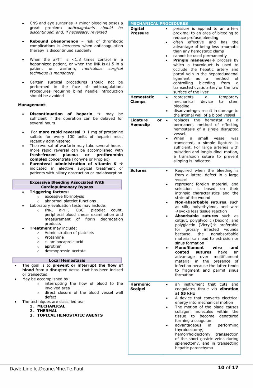
1. MECHANICAL 2. THERMAL 3. TOPICAL HEMOSTATIC AGENTS
MECHANICAL PROCEDURES
Digital
Pressure
pressure is applied to an artery
proximal to an area of bleeding to
reduce profuse bleeding often effective and has the
advantage of being less traumatic than any hemostatic clamp
cannot be used permanently Pringle maneuver process by
which a tourniquet is used to occlude the hepatic artery and portal vein in the hepatoduodenal ligament as a method of controlling bleeding from a transected cystic artery or the raw
surface of the liver
Hemostatic Clamps
represents a temporary mechanical device to stem
bleeding
disadvantage: result in damage to the intimal wall of a blood vessel
Ligature or Hemoclip
replaces the hemostat as a permanent method of effecting hemostasis of a single disrupted
vessel. When a small vessel was
transected, a simple ligature is sufficient. For large arteries with pulsation and longitudinal motion, a transfixion suture to prevent slipping is indicated.
Sutures Required when the bleeding is from a lateral defect in a large
vessel
represent foreign material, and selection is based on their intrinsic characteristics and the state of the wound
Non-absorbable sutures, such as silk, polyethylene, and wire evoke less tissue reaction
Absorbable sutures such as catgut, polyglycolic (Dexon), and polyglactin (Vicryl) preferable
for grossly infected wounds because the nonabsorbable
material can lead to extrusion or sinus formation
Monofilament wire and
coated sutures have an advantage over multifilament material in the presence of infection because the latter tends
to fragment and permit sinus formation
Harmonic Scalpel
an instrument that cuts and coagulates tissue via vibration
at 55 kHz A device that converts electrical
energy into mechanical motion The motion of the blade causes
collagen molecules within the tissue to become denatured
forming a coagulum advantageous in performing
thyroidectomy, hemorrhoidectomy, transsection of the short gastric veins during splenectomy, and in transecting hepatic parenchyma

11 of 17
Dave.Linelle.Deane.Mhe.Te.Paul
THERMAL AGENTS
Heat achieves hemostasis by
denaturation of protein that
results in coagulation of large areas of tissue
ACTUAL CAUTERYheat is
transmitted from the instrument by conduction directly to the
tissue ELECTROCAUTERYheating
occurs by induction from an alternating current source
DIRECT CURRENT (20- to 100-mA range) have successfully controlled diffuse bleeding from a
raw surface; because the protein moieties and cellular elements of blood have a negative surface
charge, they are attracted to a positive pole where a thrombus is formed
Local Cooling (decreased temp)
has been applied to control bleeding from the eroded mucosa of the esophagus and stomach.
Direct cooling with iced saline is effective and acts by increasing
the local intravascular hematocrit and by causing vasoconstriction of the arterioles
EXTREME COOLING, i.e., cryogenic procedures, have been applicable in gynecology and as a
method of destroying hepatic metastases
TOPICAL HEMOSTATIC AGENTS
can be classified based on their mechanism of action and include physical or mechanical, caustic, biologic,
and physiologic agents Some agents induce protein coagulation and
precipitation occlusion of small cutaneous vessels
Others take advantage of later stages in the coagulation cascade activating biologic responses
to bleeding
The ideal topical hemostatic agent: o With significant hemostatic action
o Has minimal tissue reactivity
o Is nonantigenic o Biodegrades in vivo
o Provides ease of sterilization
o Low in cost o Can be tailored to specific needs
Thrombin- derivative
products
direct the conversion of fibrinogen to fibrin, aiding in clot formation
takes advantage of natural physiologic processes avoiding foreign body or inflammatory reactions wound bed is not disturbed
thrombin entry into larger-caliber vessels can result in systemic exposure to thrombin with a risk of disseminated intravascular clotting or death
Fibrin sealants prepared from cryoprecipitate (homologous or synthetic)
have the advantage of not
promoting inflammation or tissue necrosis
particularly helpful in patients who have received heparin or who have deficiencies in coagulation (e.g., hemophilia or von Willebrand's disease)
Purified gelatin solution
can be prepared into several vehicles, including powders, sponges or foams, and sheets or films
Hygroscopic - absorbing many times its weight in water or liquid
effectively metabolized and degraded by proteinases in the wound bed over a period of 4 to 6 weeks
provides effective hemostasis for operative fields with diffuse small-vessel oozing
Thrombin may be applied to boost hemostasis
Advantages: relatively inexpensive, readily available, pliable, and easy to handle
Disadvantages: implanted gelatin can serve as a nidus for infection
BACKGROUND
Late 19th century social acceptance of human blood replacement
therapy 1900 Introduction of ABO blood
grouping (Dr. Karl
Landsteiner) 1939 Rh grouping (Dr. Levine &
Dr. Stetson)
Late 1970s Whole blood was
considered the standard
in transfusion
Typing and Cross Matching
serologic compatibility is established routinely for the recipients' and donors' A, B, O, and Rh groups in selecting blood for transfusion
cross-matching between the donors' red blood cells and the recipients' sera (the major cross-match) is performed
as a rule, Rh-negative recipients should be transfused
only with Rh-negative blood (If the recipient is an elderly male who has not been transfused previously, the administration of Rh-positive blood is acceptable if
Rh-negative blood is not available anti-Rh antibodies form within several weeks of
transfusion anti-Rh antiserum (RhoGAM) should be given if Rh-
positive products have been given to an Rh-negative patient
REPLACEMENT THERAPY
TRANSFUSION
12 of 17
Dave.Linelle.Deane.Mhe.Te.Paul
Rh-positive blood should not be transfused to Rh-negative females who are capable of child-bearing
administration of hyperimmune anti-Rh globulin to Rh-
negative women shortly before or after childbirth largely eliminates Rh disease in subsequent offspring
for patients receiving repeated transfusions, serum drawn not more than 72 hours before cross-matching
should be used for matching with cells of the donor (Emergency transfusion can be accomplished with type O blood)
O-negative and type-specific red blood cells are equally safe for emergency transfusion
problems are associated with the administration of four
or more units of O-negative blood because there is a significant increase in the risk of hemolysis
for patients with clinically significant cold agglutinins, blood should be administered through a blood warmer (If these antibodies are present in high titer, hypothermia is contraindicated)
for patients with multiple transfusion and who have
developed alloantibodies, or who have autoimmune hemolytic anemia with pan-red blood cell antibodies, typing and cross-matching is often difficult, and sufficient time should be allotted preoperatively to accumulate blood that might be required during the operation
cross-matching should always be performed before the
administration of dextran because it interferes with the typing procedure
for autologous transfusion, up to 5 units can be collected for subsequent use during elective procedures
patients can donate blood if their hemoglobin
concentration exceeds 11 g/dL or if the hematocrit is
greater than 34% first procurement is performed 40 days before the
planned operation and the last one is performed 3 days before the operation
donations can be scheduled at intervals of 3 to 4 days recombinant human erythropoietin (rHuEPO)
accelerates generation of red blood cells and allows for more frequent harvesting of blood
Banked Whole Blood
shelf life extended to 40 ± 5 days
at least 70% of the transfused erythrocytes remain in the circulation for 24 hours after transfusion and are viable
rarely indicated and rarely available changes in the red blood cells that occur during
storage include reduction of intracellular ADP and 2,3-diphosphoglycerate (2,3-DPG), which alters the curve
of oxygen dissociation from hemoglobin, decreasing the function of oxygen transport
along with the clotting factors, only factor V and VIII are stable in banked blood
pH decreases from 7.00 to 6.68, and the lactic acid level increases from 20 to 150 mg/dL within 21 days of storage
potassium concentration rises steadily to 32 mEq/dL, and the ammonia concentration rises from 50 to 680 mg/dL at the end of 21 days
Fresh Whole Blood
blood administered within 24 hours of its donation
rarely indicated must be administered untested because of the time
required for testing for infectious disease 1 unit of platelet concentrate has more viable platelets
than 1 unit of fresh blood poor source of platelets and factor VIII
Packed Red Blood Cells and Frozen Red Blood Cells
product of choice for most clinical situations
concentrated suspensions of red blood cells can be prepared by removing most of the supernatant plasma after centrifugation
preparation reduces but does not eliminate reaction caused by plasma components (also reduces the amount of sodium, potassium, lactic acid, and citrate
administered) provides oxygen-carrying capacity frozen red blood cells are not available for use in
emergencies (used for patients who are known to have been previously sensitized)
improved red blood cell viability and the ATP and 2,3-DPG concentrations are maintained
Leukocyte- Reduced and Leukocyte-
Reduced/Washed Red Blood Cells
prepared by filtration eliminate 99.9% of the WBCs and most of the
platelets (leukocyte-reduced red blood cells), and, if needed, by additional saline washing (leukocyte-reduced/washed red blood cells)
leukocyte-reduction prevents almost all febrile, nonhemolytic transfusion reactions (fever and/or rigors), alloimmunization to HLA class I antigens, and platelet transfusion refractoriness and cytomegalovirus
transmission washed, leukocyte-reduced red blood cells are usually
given only to patients who have had reactions (rash, urticaria, anaphylaxis) to unwashed red blood cells
Platelet Concentrates
indicated for thrombocytopenia caused by massive blood loss and replacement with platelet-poor products; and by inadequate production
also given to patients with qualitative platelet disorders preparations should be used within 120 hours of
donation
1 unit of platelet concentrate = 50 mL can transmit infectious diseases and account for
allergic reactions similar to those caused by blood transfusion
elevate the platelet count to the range of 50,000 to 100,000/microL when treating bleeding caused by
thrombocytopenia or preparing some thrombocytopenic patients for an operation
leukocyte reduction through filtration prevents HLA
alloimmunization patients who become alloimmunized through previous
transfusion, or those patients who are refractory from sensitization through prior pregnancies, HLA-matched
platelets can be used platelet transfusion thresholds can safely be lowered in
patients without signs of hemostatic deficiency and who have no history of poor tolerance to low platelet counts
multiple platelet transfusions predispose to multiorgan failure and mortality is dose-dependent
shelf life of platelets: 120 hrs from time of donation
Frozen Plasma and Volume Expanders
frozen plasma prepared from freshly donated blood is
the standard source of the vitamin K–dependent factors (and is the only source of factor V) factor V is less stable than the vitamin K–dependent factors)
risk of infectious disease is the same whether FFP, whole blood, or red blood cells is administered
Lactated Ringer's solution or buffered saline solution administered in amounts 2 to 3 times the estimated
blood loss is effective and associated with fewer complications

13 of 17
Dave.Linelle.Deane.Mhe.Te.Paul
Dextran or a combination of lactated Ringer's solution and normal serum albumin are preferred for rapid volume expansion
commercially available dextran should not be administered more than 1 L/d because of prolonged bleeding time and consequent hemorrhage
low molecular weight dextran (30–40,000 Da)
possesses a higher colloidal pressure than plasma and effects some reversal of erythrocyte agglutination
Concentrates and Recombinant DNA Technology
antihemophilic concentrates are prepared from plasma
and are available for treatment of factor VIII or factor IX deficiency
various concentrates are 20 to 30 times as potent as an equal volume of FFP
concentrated albumin of 25 g can be administered to provide the equivalent of 500 mL of plasma and has the advantage of being hepatitis-free
Human Polymerized Hemoglobin (Polyheme)
universally compatible immediately available disease-free
oxygen-carrying resuscitative fluid that has been successfully used in massively bleeding patients when red blood cells were not transfused
absence of blood-type antigens (no cross-match needed) and viral infections
long-term stability disadvantages though are shorter half-life in the
bloodstream and cardiovascular complications
General Indications
Improvement in Oxygen-Carrying Capacity
oxygen-carrying capacity is chiefly a function of RBCs transfusion should be withheld when anemia can be
treated by specific therapy, such as erythropoietin acute anemias are more disabling than chronic anemia
because patients with chronic anemia have undergone an adjustment to the deficiency
moderate drop in the hematocrit level and transfusions
are not indicated to correct the physiologic anemia in pregnancy if an operation is required
correction of chronic anemia before surgical intervention is often not necessary
hemoglobin value of less than 10 g/dL or a hematocrit level less than 30% indicates a need for preoperative
red blood cell transfusion cardiac output does not increase significantly in
healthy individuals until the hemoglobin value decreases to approximately 7 g/dL
patients with chronic anemia and a hemoglobin value of less than 7 g/dL in whom intraoperative bleeding is not anticipated do not require a transfusion
preoperatively blood volume can be replaced with dextran solution or
lactated Ringer's solution with a reduction of the hemoglobin value to levels below 10 g/dL
human polymerized hemoglobin can be used to increase oxygen-carrying capacity
whole blood substitute, Fluosol-DA, has been proposed as a solution with increased oxygen-handling capability
Replacement of Clotting Factors transfusion of platelets and/or proteins contributing to
coagulation may be indicated in specific patients before or during an operative procedure
Table for replacement of clotting factors (refer to the last page)
Specific Indications
Massive Transfusion
entails a single transfusion greater than 2500 mL or 5000 mL transfused over a period of 24 hours
circulatory overload or DIC might occur
dilutional thrombocytopenia, impaired platelet function, and deficiencies of factors V, VIII, and XI can
also be encountered routine alkalization is not advisable because this could
have an adverse effect on the hemoglobin dissociation curve and also is accompanied by an increased sodium load
Percentage of Original Blood Volume Remaining
in a Patient with a 5-L Blood Volume Transfused
with 500-mL Units
Magnitude of Hemorrhage and Transfusion
Situation 1 Blood
Volume
(10 Units)
2 Blood
Volume
(20 Units)
3 Blood
Volume
(30 Units)
Best 37 14 5
Usual 25–30 10 2–4
Worst 18 3 0.4
"best" situation requires simultaneous and equal replacement during hemorrhage;
"worst" situation means initial loss of one-half blood volume not replaced until the hemorrhage has stopped
citrate toxicity from the use of stored blood may result in young children, in patients with severe hypotension, or in patients with liver disease (toxicity is related to
an excessive binding of ionized calcium) use of stored blood also provides a potassium load, but
there are no effects in the face of normal renal
function when large volume transfusions are administered, a
heat exchanger should be used because hypothermia can cause a decrease in cardiac rate and output and blood pH
use of blood from multiple donors increases the risk of hemolytic reaction as a consequence of incompatibility
when massive transfusions are administered, the pH, blood gases, and potassium should be measured regularly and abnormalities corrected immediately
if diffuse bleeding is noted, coagulation tests and platelet counts should be measured and deficiencies corrected
INDICATIONS FOR REPLACEMENT OF
BLOOD AND ITS ELEMENTS

14 of 17
Dave.Linelle.Deane.Mhe.Te.Paul
Routine Administration
rate of transfusion depends upon the patient's status Usually 5 mL/min is administered for the first minute,
after which 10 to 20 mL/min is given when there is marked oligemia, 500 mL can be given
within 10 minutes and a second 500 mL also can be given within 10 minutes
approximately 1500 mL/min can be administered through two 7.5-F catheters
Other Methods
blood can be instilled intraperitoneally or into the medullary cavity of long bones and the sternum
approximately 90% of red blood cells injected intraperitoneally enter the circulation, but uptake is not complete for at least a week
intraoperative autotransfusion is a potentially life-saving adjunct
roughly 250 mL of blood can be retrieved, washed or filtered, and returned to the patient over a 5-minute period
another approach to anticipated intraoperative large
blood losses is hemodilution (at the onset of the procedure, RBCs are removed while the intravascular volume is maintained with crystalloid or colloid)
reduced blood viscosity improves the microcirculatory perfusion
removed blood can then be retransfused during the operation to replace lost blood
Nonhemolytic Reactions/Febrile Reactions
increase in temperature [>1°C (1.8°F)] associated with a transfusion
CAUSE: Preformed cytokines in donated blood
and recipient antibodies reacting with donated antibodies
PREVENTION:use of leukocyte-reduced and/or out of date blood products
Acetaminophen/Paracetamol- reduces the severity of the reaction.
Bacterial contamination of infused blood is rare. CLINICAL MANIFESTATIONS
o Systemic Signs - fever and chills - tachycardia - hypotension
o GI Symptoms
- abdominal cramps - vomiting - diarrhea
o Hemorrhagic Manifestations - hemoglobinemia - hemoglobinuria - disseminated intravascular coagulation
UPON SUSPECTED DIAGNOSIS: o STOP the transfusion o Have the donated blood cultured
Emergency treatment
o administration of oxygen o adrenergic blocking agents
o antibiotics
Allergic Reactions
relatively frequent (~1% of all transfusions)
can occur after the administration of any blood
product
CLINICAL MANIFESTATIONS
o usually mild
o rash
o urticaria
o fever
(alloccurring within 60 to 90 minutes of
transfusion)
o anaphylactic shock - rare
CAUSE
o transfusion of antibodies from hypersensitive
donors
o transfusion of antigens to which the recipient is
hypersensitive
TREATMENT AND PROPHYLAXIS
o antihistamines
o epinephrine or steroids in more serious cases
Respiratory Complications
associated with transfusion-associated
circulatory overloadavoidable complication
occurs with rapid infusion of blood, plasma
expanders, and crystalloids esp. in older patients
with underlying heart disease
Central venous pressure monitoring should be
considered whenever large amounts of fluid are
administered
CLINICAL MANIFESTATION
o rise in venous pressure
o dyspnea
o cough
o rales at the lung bases
TREATMENT
o initiating diuresis
o slowing the rate of blood administration
o minimizing delivery of fluids while blood products
are being transfused
Syndrome of Transfusion-related Acute Lung Injury (TRALI)
noncardiogenic pulmonary edema related to
transfusion
can occur with the administration of any plasma-
containing blood product
CLINICAL MANIFESTATIONS
o similar to those of circulatory overload
o dyspnea
o hypoxemia
o fever
o rigors
o bilateral pulmonary infiltrates on CXR
o occurs within 1 to 2 hours of transfusion, but
virtually always before 6 hours
CAUSE
o not well established
o thought to be related to anti-HLA or anti–human
neutrophil antigen antibodies in transfused
blood that primes neutrophils in the pulmonary
circulation
RISK FACTORS
o Multiparity of the donor - major risk factor
o Female donors
TREATMENT
o discontinuation of any transfusion
METHODS OF ADMINISTERING BLOOD
COMPLICATIONS OF TRANSFUSION
15 of 17
Dave.Linelle.Deane.Mhe.Te.Paul
o notification of the transfusion service
o provision of pulmonary support
- supplemental oxygen
- mechanical ventilation
Hemolytic Reactions
high index of suspicion is needed to make the diagnosis
LABORATORY CRITERIA o hemoglobinuria o serologic findings that show incompatibility of
the donor and recipient blood o (+) Coombs' test presence of transfused cells
coated with patient antibody (diagnostic) GENERAL TREATMENT o STOP the transfusion immediately o sample of the recipient's blood drawn and sent
along with the suspect unit to the blood bank
o Monitor Urine output
o Maintain adequate hydration to prevent precipitation of hemoglobin within the tubules
Acute Hemolytic Reactions
occur with the administration of ABO-incompatible blood
fatal in up to 6% of cases CONTRIBUTING FACTORS o technical or clerical errors in the laboratory
o administration of blood of the wrong blood type CLINICAL MANIFESTATIONS o intravascular destruction of red blood cells
o hemoglobinemia o hemoglobinuria o pain at the site of transfusion o facial flushing
o back (flank) o chest pain o fever o respiratory distress o hypotension o tachycardia o for anesthetized patients:
- diffuse bleeding - hypotension
o DIC - can be initiated by activation of factor XII
and complement by antibody-antigen complexescoagulation cascade
o Acute Renal Insufficiency - results from the toxicity associated with free
hemoglobin in the plasma - tubular necrosis - precipitation of hemoglobin within the
tubules
Delayed Hemolytic Reactions
occur 2 to 10 days after transfusion occur when an individual has a low antibody titer
at the time of transfusion but the titer increases after transfusion as a result of an anamnestic response
Reactions to non-ABO antigens involve
immunoglobulin G–mediated clearance by the reticuloendothelial system.
do not usually require specific intervention
CLINICAL MANIFESTATIONS o extravascular hemolysis o mild anemia o indirect (unconjugated) hyperbilirubinemia o fever
o recurrent anemia o Jaundice o decreased haptoglobin levels
o low-grade hemoglobinemia and hemoglobinuria o (+) Coombs' test
Transmission of Disease
Diseases that can be transmitted by transfusion:
Malaria
Chagas' disease
Brucellosis
Syphilis (very rarely)
Malaria (most common: Plasmodium malariae)
incubation period: 8 to 100 days
initial manifestations
shaking chills
spiking fever
hepatitis C
HIV-1
Hepatitis B
Prion disorders (e.g., Creutzfeldt-Jakob disease)
Carefully review the patient's clinical history and drug use, and basic laboratory testing.
Common screening laboratory testing platelet count
PT or INR
Aptt
normal platelet count : 150,000 to 400,000/ µL.
bleeding or thrombotic complications Platelet counts >1,000,000/µL
Increased bleeding complications may be seen with major surgical procedures when
the platelet count is<100,000/ L minor surgical procedures when
counts are <50,000/ L Spontaneous hemorrhage:
o when count falls below 20,000/ L. PT and aPTT - variations of plasma recalcification
times initiated by the addition of a thromboplastic agent
PT reagent o contains thromboplastin and Ca
o when added to plasma, leads to the formation of a
fibrin clot PT test o measures the function of factors I, II, V, VII, and X o Factor VII -shortest half-life of the coagulation
factors, and its synthesis is vitamin K dependent. o best suited to detection of abnormal coagulation
caused by vitamin K deficiencies and warfarin
therapy Due to variations in thromboplastin activity, it can be
difficult to accurately assess the degree of anticoagulation on the basis of PT alone o determination of the INR is now the method of
choice for reporting PT values.
o International Sensitivity Index (ISI) is unique to each batch of thromboplastin and is furnished by the manufacturer tothe hematology laboratory
o Human brain thromboplastin has an ISI of 1, and the optimal reagent has an ISI between 1.3 and 1.5.
aPTT reagent
o phospholipid substitute, activator, and calcium
TEST OF HEMOSTASIS AND BLOOD
COAGULATION

16 of 17
Dave.Linelle.Deane.Mhe.Te.Paul
o in the presence of plasma leads to fibrin clot formation
o measures function of factors I, II, and V of the
common pathway and factors VIII, IX, X, and XII Heparin therapy-monitored by following aPTT values,
with a therapeutic target range of 1.5 to 2.5 times the control value (approx 50 to 80 seconds)
Low molecular weight heparins are selective factor Xa inhibitors and may mildly elevate the aPTT, but therapeutic monitoring is not routinely recommended.
bleeding time -used to evaluate platelet and vascular dysfunction
o Ivy bleeding time is most commonly
used determined by placing a
sphygmomanometer on the upper arm and inflating it to 40 mmHg and then making a 5-mm stab incision on the flexor surface
of the forearm
Time is measured to cessation of bleeding
upper limit of normal bleeding time – 7 min
o abnormal bleeding time suggests either platelet dysfunction (intrinsic or drug induced), vWD, or certain vascular
defects replacing the template bleeding time with an in vitro
test o which blood is sucked through a capillary and the
platelets adhere to the walls of the capillary and aggregate
o closure time in this system appears to be more
reproducible than the bleeding time o correlates with bleeding in patients with vWD,
primary platelet function disorders, or other platelet dysfunction disorders and patients who are taking aspirin
Additional medications may significantly impair
hemostatic function such as o antiplatelet agents (clopidogrel and glycoprotein
IIb/IIIa inhibitors) o anticoagulant agents (hirudin, chondroitin sulfate,
dermatan sulfate) o thrombolytic agents (streptokinase, tPA)
If abnormal results on any of the coagulation studies
cannot be explained by known medications->congenital abnormalities of coagulation or comorbid disease should be considered
Thromboelastography (TEG) o monitors hemostasis as a dynamic process rather
than revealing isolated information as in conventional coagulation screens.
o measures the viscoelastic properties of blood as it is induced to clot in a lowshear environment (resembling sluggish venous flow)
o patterns of change in shear elasticity allow the kinetics of clot formation and growth as well as the
strength and stability of the formed clot to be determined
o strength and stability data- provide information about the ability of the clot to perform the work of hemostasis
o kinetic data- determine the adequacy of quantitative
factors available for clot formation
o sample of celite-activated whole blood is placed into a prewarmed cuvette ->suspended piston is then lowered into the cuvette -> rotated through a 4.5-degree arc backwards and forwards
o Normal clot goes through an acceleration and strengthening phase
o fiber strands that interact with activated platelets attach to the surface of the cuvette and the suspended piston
o clot forming in the cuvette transmits its movement onto the suspended piston
o Weak clot stretches and therefore delays the arc movement of the piston, which is graphically
expressed as a narrow thromboelastogram o Strong clot will move the piston simultaneously and
proportionally to the cuvette's movements, creating a thick thromboelastogram
o strength of a clot is graphically represented over time as a characteristic cigar-shaped figure
o k- measure of the time from the beginning of clot
formation until the amplitude of the TEG tracing reaches 20 mm and represents the dynamics of clot formation
o alpha angle - angle between the line in the middle of the TEG(r) tracing and the line tangential to the developing body of the TEG(r) tracing. The alpha angle
represents the acceleration (kinetics) of fibrin buildup and cross-linking
o MA -maximum amplitude and reflects the strength of the clot, which is dependent on the number and function of platelets and the clot's interaction with
fibrin o MA60 - rate of amplitude reduction 60 minutes after
MA and represents the stability of the clot
Preoperative Evaluation of Hemostasis
Several hematologic disorders may have an impact on
the outcome of surgery.
pre-existing anemia and oral anticoagulation therapy-common clinical situations faced by the
surgeon Assessment of bleeding risk should also be considered
in patients with liver or renal dysfunction When feasible, diagnostic evaluation of the patient
with previously unrecognized anemia should be carried out before surgery, because certain types of anemia (particularly sickle cell disease and immune hemolytic anemias) may have implications for perioperative management
Hemoglobin levels below 7 or 8 g/dL appear to be associated with significantly more perioperative complications than higher levels
Determination of the need for preoperative transfusion in an individual patient must consider factors other than the absolute hemoglobin level, including
o presence of cardiopulmonary disease
o type of surgery o likelihood of surgical blood loss
Many patients have anemia postoperatively secondary to blood loss and hemodilution and do not necessarily require transfusion
directed bleeding history- most important component
of the bleeding risk assessment o provide meaningful clues to the presence
of a bleeding tendency
EVALUATION OF HEMOSTATIC RISK IN THE
SURGICAL PATIENT

17 of 17
Dave.Linelle.Deane.Mhe.Te.Paul
Patients who are reliable historians and who reveal no suggestion of abnormal bleeding on directed bleeding history and physical examination are at very low risk
for having an occult bleeding disorder o Laboratory tests of hemostatic
parameters is not required When the directed bleeding history is unreliable or
incomplete or when abnormal bleeding is suggested o formal evaluation of hemostasis should
be performed before surgery PT, the aPTT, and the platelet
count
Excessive bleeding during or after a surgical procedure may be the result of o ineffective hemostasis o blood transfusion o undetected hemostatic defect o consumptive coagulopathy
o fibrinolysis Excessive bleeding from the operative field
unassociated with bleeding from other sites usually
suggests inadequate mechanical hemostasis
Massive blood transfusion-well-known cause of
thrombocytopenia Bleeding after massive transfusion can occur due to o Hypothermia o dilutional coagulopathy
o platelet dysfunction, o fibrinolysis
o hypofibrinogenemia Another cause of hemostatic failure related to the
administration of blood is hemolytic transfusion reaction
diffuse bleeding -first sign of a transfusion reaction o release of ADP from hemolyzed red blood cells->
diffuse platelet aggregation->after which the platelet
clumps are removed out of the circulation->bleeding Transfusion purpura occurs when the donor platelets
are of the uncommon Pl(A1) group o an uncommon cause of thrombocytopenia and
associated bleeding after transfusion The platelets sensitize the recipient, who makes
antibody -> antibody then destroys the recipient's own
platelets-> thrombocytopenia and bleeding may continue for several weeks.
This uncommon cause of thrombocytopenia should be considered if bleeding follows transfusion by 5 or 6 days.
Platelet transfusions are of little help in the
management of this syndrome, because the new donor platelets usually are subject to the binding of antigen and damage from the antibody
Corticosteroids may be of some help in reducing the bleeding tendency.
Posttransfusion purpura is self-limited, and the passage of several weeks inevitably leads to
subsidence of the problem. DIC is characterized by systemic activation of the
blood coagulation system-> results in the generation
and deposition of fibrin ->leading to microvascular thrombi in various organs -> contributing to the development of multiorgan failure
Consumption and subsequent exhaustion of
coagulation proteins and platelets due to the ongoing activation of the
coagulation system may induce severe bleeding
complications
Severe hemorrhagic disorders due to thrombocytopenia have occurred as a result of gram-negative sepsis
o pathogenesis of endotoxin-induced thrombocytopemia- related to lability of factor V
Defibrination and hemostatic failure also may occur with meningococcemia, Clostridium perfringens sepsis,
and staphylococcal sepsis o Hemolysis appears to be one mechanism in sepsis leading to defibrination
From omgfacts.com:
Anatidaephobia is the fear that somewhere in the
world, there is a duck watching you.
If you touch your tongue while yawning, it can stop the yawn.
You're more likely to die on your way to buy a lottery ticket than you are to actually win the lottery.
Women speak about 7000 words a day. The average man averages just over 2000.
'Hippopotomonstrosesquippedaliophobia' is the fear of long words.
When a person is tickled, the response is actually a form of panic, as the brain interprets the tickling sensation as produced by spiders or
other creepy crawlies on your skin - the uncontrollable laughter is a response to panic.
Those stars and colours you see when you rub your eyes are called phosphenes.
Yawning is contagious - even thinking about yawning is enough. After reading this fact, there is a 50% chance you will yawn.
The total weight of all the ants on Earth is about the same as the weight of all the humans on earth.
When you kiss someone for a minute, you both burn about 2.6 calories.
An elephant can die from a broken heart.
'Dreamt' is the ONLY WORD in the entire English language that ends in the letters 'mt'.
'Dysania' is the state of finding it hard to get out of bed in the morning.
One tablespoon of semen has approximately 20 calories. Semen also contains zinc and calcium which helps prevent tooth decay.
Breathing the air in Mumbai, India for just ONE DAY, is equivalent to smoking 2.5 packs of cigarettes.
According to suicide statistics, Monday is the favored day for self-destruction.
If you walk and talk with someone, eventually you will synchronize your steps with each other.
Albert Einstein left his first wife for his COUSIN. A strawberry is not an actual berry, but a banana
is. Your earlobes line up with your nipples. (Go
try!) As a punishment for misbehavior, Thai cops have
to wear pink Hello Kitty armbands. Colgate faced big obstacle marketing toothpaste
in Spanish-speaking countries because Colgate
translates into the command 'go hang yourself.' In Topeka, Kansas it is illegal to sing the
alphabet on the streets at night. It is also illegal to install a bathtub, and you may not scream in a
haunted house. Justin Bieber can solve a Rubik's Cube in less
than 2 minutes. Humans don't just shrink with age. They shrink
EVERY DAY. (“Uh-oh.” – Deane)
EVALUATION OF EXCESSIVE
INTRAOPERATIVE OR POSTOPERATIVE
BLEEDING