Hemostasis. Mechanisms of Hemostasis Hypercoagulability States Bleeding Disorders.
60
Hemostasis
-
Upload
dorthy-sharp -
Category
Documents
-
view
241 -
download
5
Transcript of Hemostasis. Mechanisms of Hemostasis Hypercoagulability States Bleeding Disorders.
Hemostasis
Mechanisms of Hemostasis
Hypercoagulability States
Bleeding Disorders
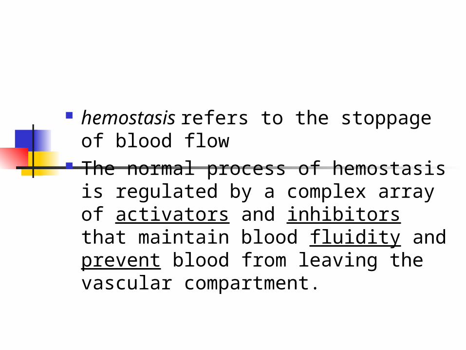
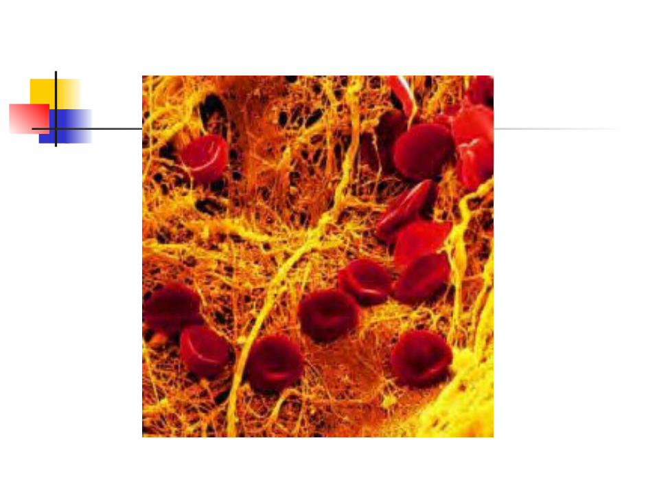
hemostasis refers to the stoppage of blood flow
The normal process of hemostasis is regulated by a complex array of activators and inhibitors that maintain blood fluidity and prevent blood from leaving the vascular compartment.
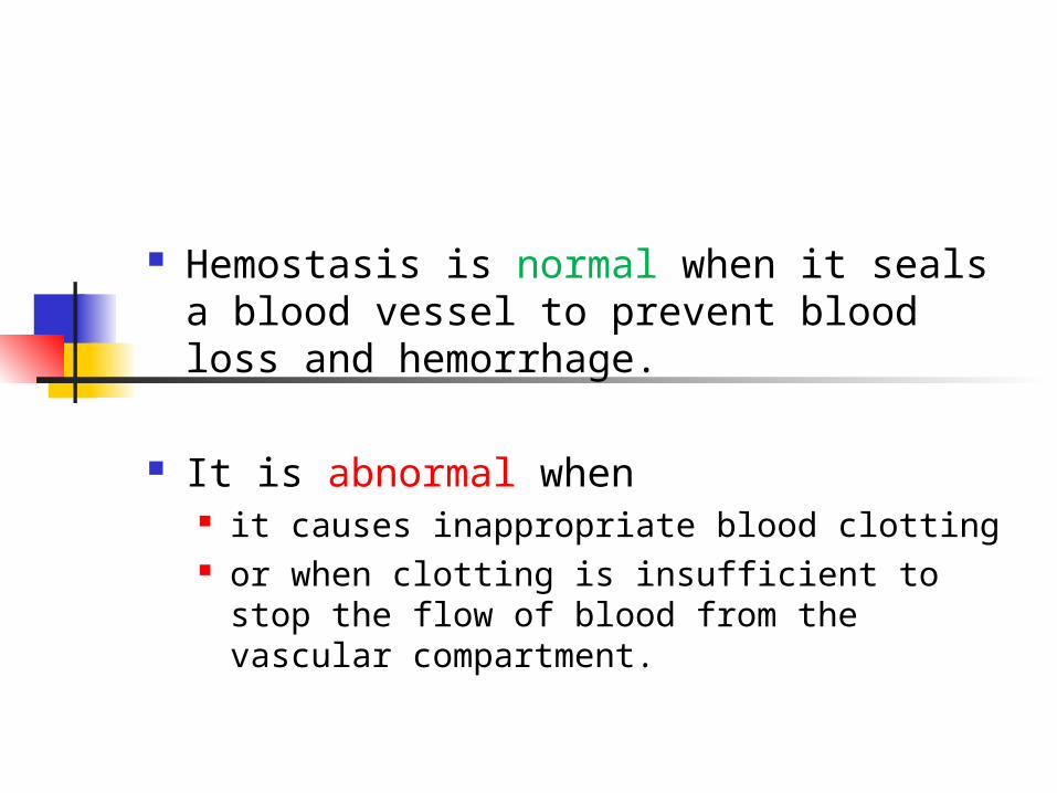
Hemostasis is normal when it seals a blood vessel to prevent blood loss and hemorrhage.
It is abnormal when it causes inappropriate blood clotting or when clotting is insufficient to stop the
flow of blood from the vascular compartment.
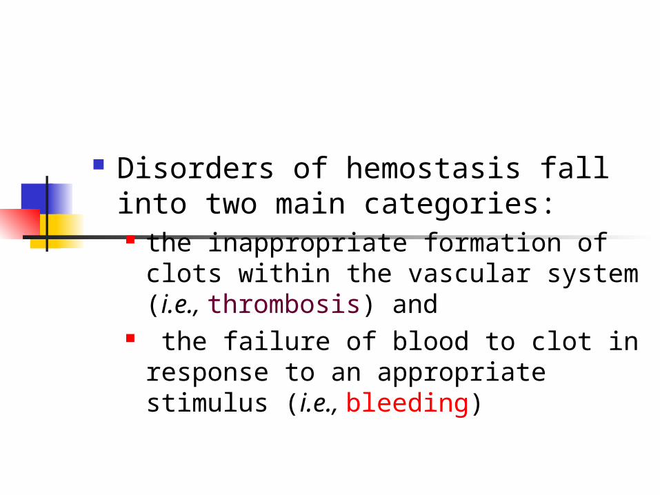
Disorders of hemostasis fall into two main categories: the inappropriate formation of clots
within the vascular system (i.e., thrombosis) and
the failure of blood to clot in response to an appropriate stimulus (i.e., bleeding)
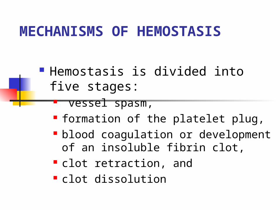
MECHANISMS OF HEMOSTASIS
Hemostasis is divided into five stages: vessel spasm, formation of the platelet plug, blood coagulation or development of
an insoluble fibrin clot, clot retraction, and clot dissolution
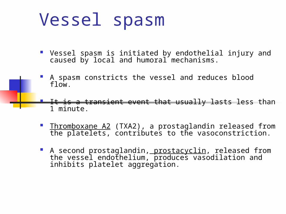
Vessel spasm
Vessel spasm is initiated by endothelial injury and caused by local and humoral mechanisms.
A spasm constricts the vessel and reduces blood flow.
It is a transient event that usually lasts less than 1 minute.
Thromboxane A2 (TXA2), a prostaglandin released from the platelets, contributes to the vasoconstriction.
A second prostaglandin, prostacyclin, released from the vessel endothelium, produces vasodilation and inhibits platelet aggregation.
Formation of the Platelet Plug
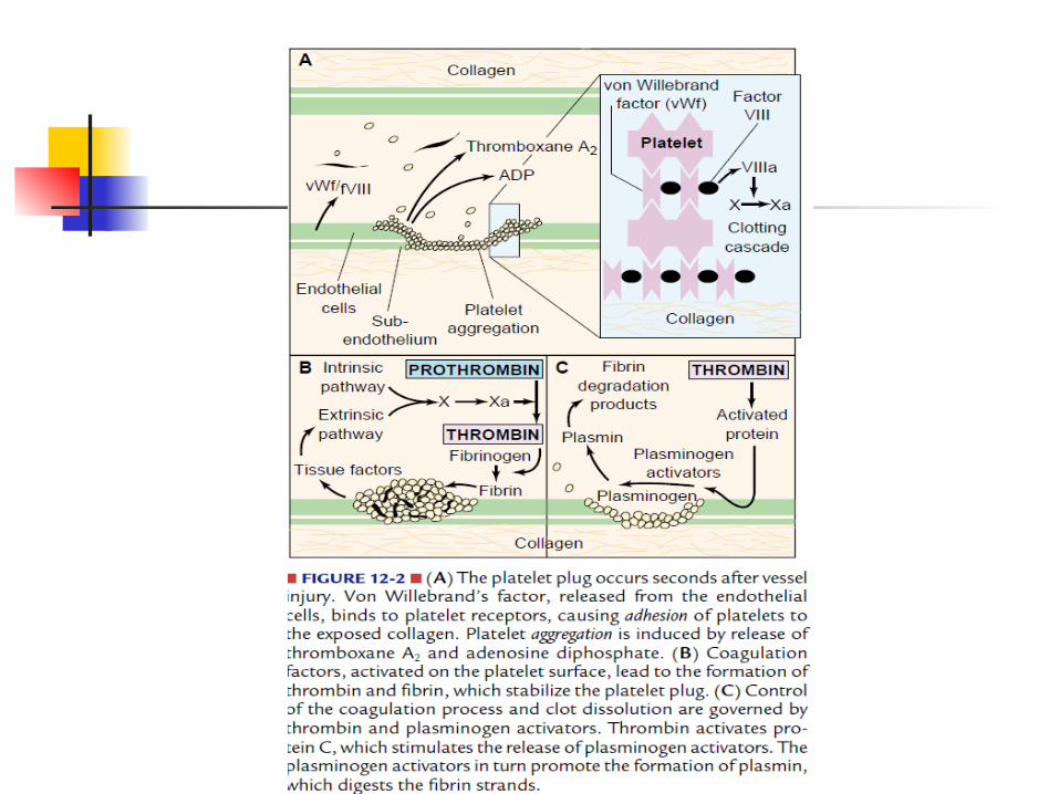
The platelet plug, the second line of defense, is initiated as platelets come in contact with the vessel wall.
The outside of the platelet membrane is coated with glycoproteins that repulse adherence to the normal vessel
endothelium, while causing adherence to injured areas of the vessel
wall, particularly the subendothelial layer.
The platelet membrane also has glycoprotein receptors that bind fibrinogen and link platelets together.
Glycoprotein receptor antagonists have been developed and are selectively used in the treatment of acute coronary myocardial infarction
Platelet plug formation involves adhesion and aggregation of platelets.
Platelet adhesion also requires a protein molecule called von Willebrand factor (vWF). vWF, which is produced by the endothelial cells of blood vessels, performs two important functions: it aids in platelet adhesion, and it circulates in the blood as a carrier protein
for coagulation factor VIII.
Platelets are attracted to a damaged vessel wall, become activated, and change from smooth disks to spiny spheres, exposing receptors on their surfaces.
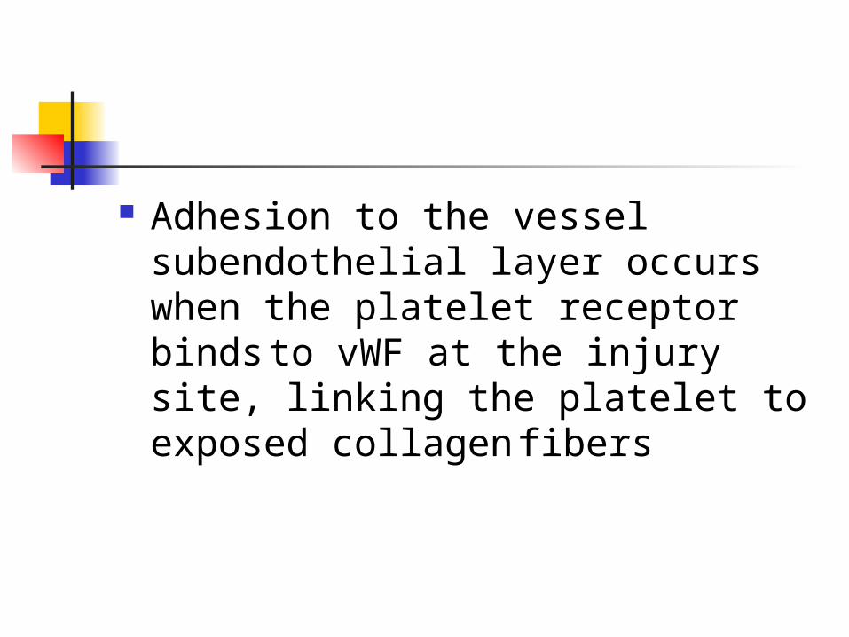
Adhesion to the vessel subendothelial layer occurs when the platelet receptor binds to vWF at the injury site, linking the platelet to exposed collagen fibers
Blood Coagulation Blood coagulation is controlled by many substances that
promote clotting (i.e., procoagulation factors) or inhibit it (i.e., anticoagulation factors).
The action of one coagulation factor or proenzyme is designed to activate the next factor in the sequence (i.e., cascade effect).
Abnormalities of the clotting process occur when one or more of the factors are deficient or when conditions lead to inappropriate activation of any of the steps.
The chemical events in the blood coagulation process involve a number of essential steps that result in the conversion of fibrinogen, a circulating plasma protein, to the fibrin strands that enmesh platelets, blood cells, and plasma to form the clot
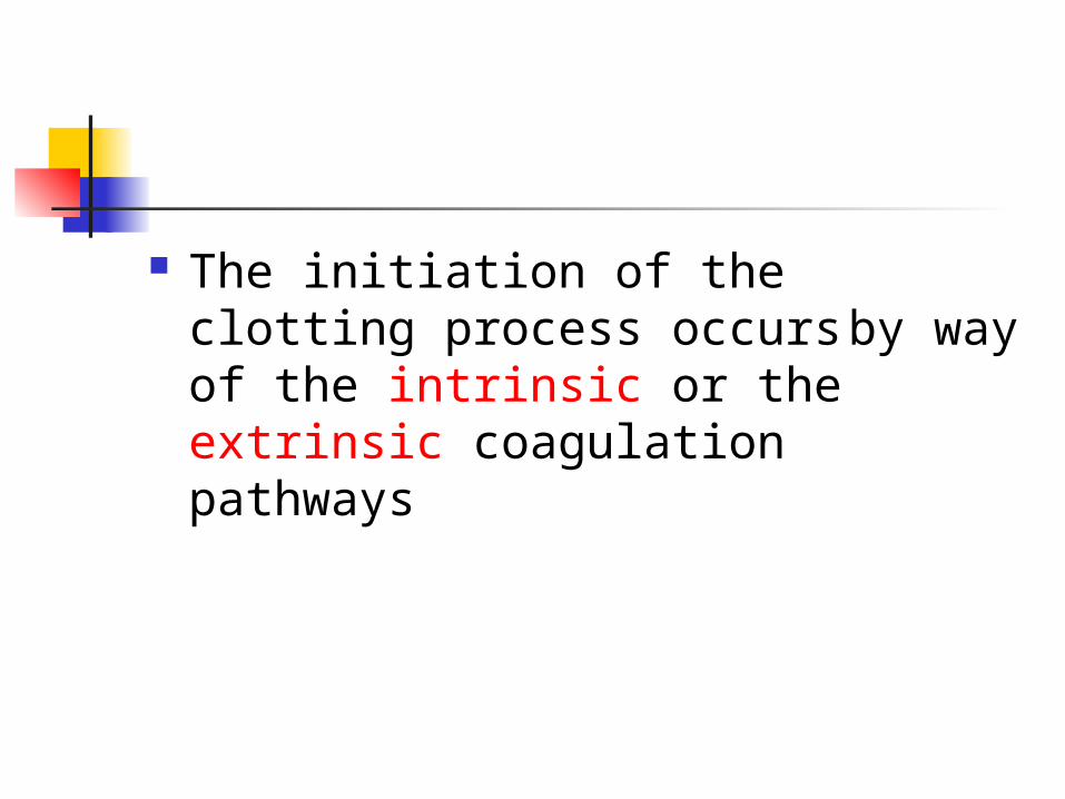
The initiation of the clotting process occurs by way of the intrinsic or the extrinsic coagulation pathways
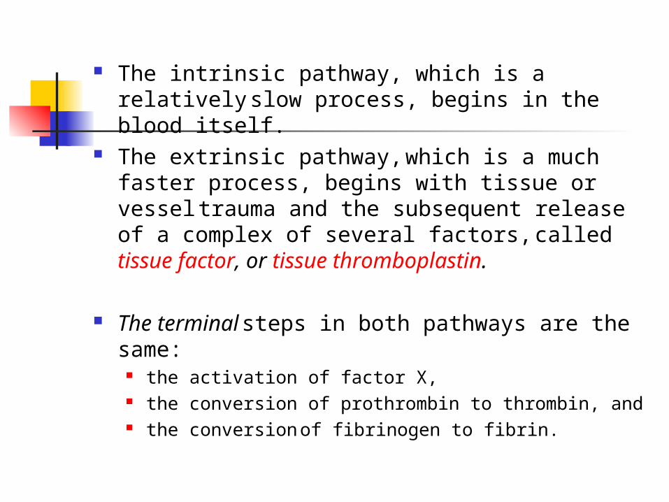
The intrinsic pathway, which is a relatively slow process, begins in the blood itself.
The extrinsic pathway, which is a much faster process, begins with tissue or vessel trauma and the subsequent release of a complex of several factors, called tissue factor, or tissue thromboplastin.
The terminal steps in both pathways are the same:
the activation of factor X, the conversion of prothrombin to thrombin, and the conversion of fibrinogen to fibrin.
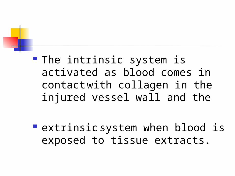
The intrinsic system is activated as blood comes in contact with collagen in the injured vessel wall and the
extrinsic system when blood is exposed to tissue extracts.
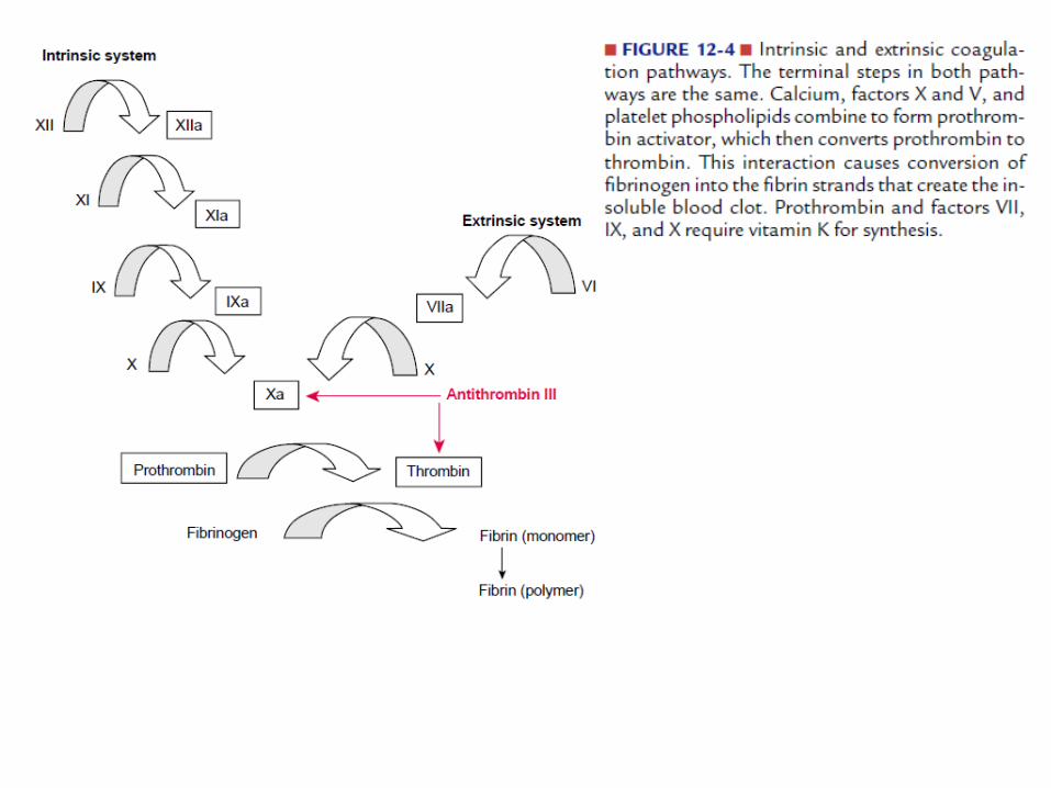
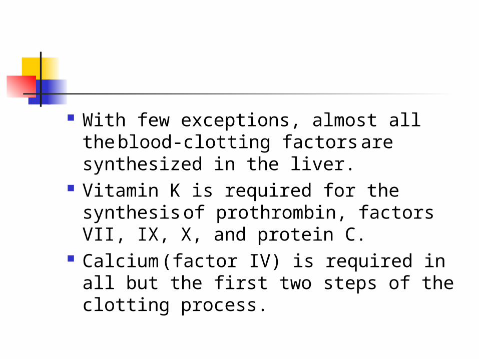
With few exceptions, almost all the blood-clotting factors are synthesized in the liver.
Vitamin K is required for the synthesis of prothrombin, factors VII, IX, X, and protein C.
Calcium (factor IV) is required in all but the first two steps of the clotting process.
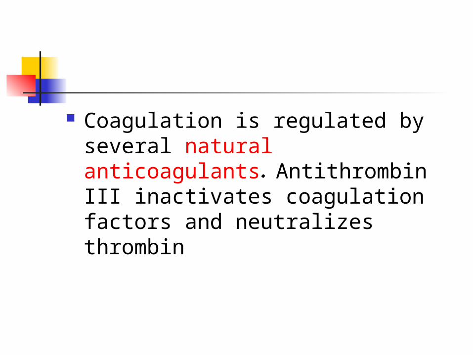
Coagulation is regulated by several natural anticoagulants. Antithrombin III inactivates coagulation factors and neutralizes thrombin
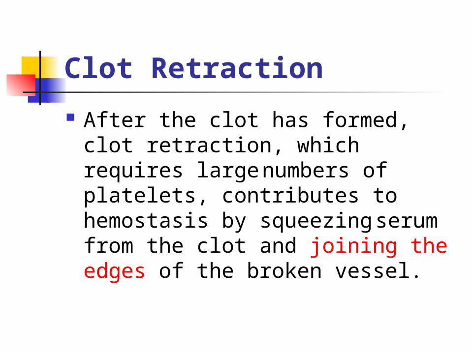
Clot Retraction
After the clot has formed, clot retraction, which requires large numbers of platelets, contributes to hemostasis by squeezing serum from the clot and joining the edges of the broken vessel.

Clot Dissolution
The dissolution of a blood clot begins shortly after its formation; this allows blood flow to be re-established and permanent tissue repair to take place
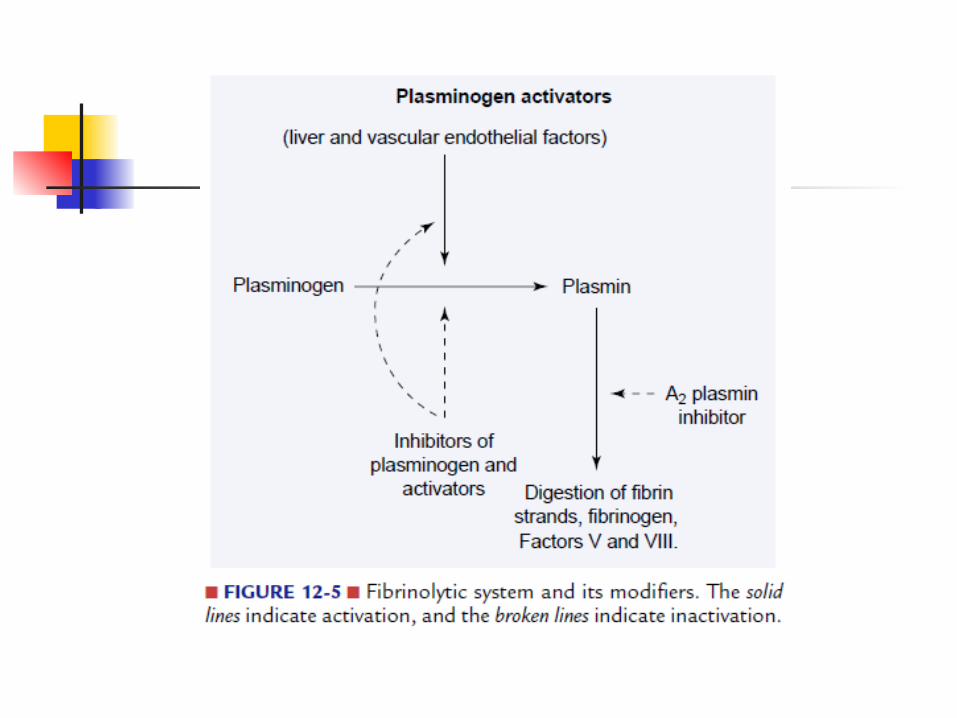
The process by which a blood clot dissolves is called fibrinolysis.

Plasminogen, the proenzyme for the fibrinolytic process, normally is present in the blood in its inactive form.
It is converted to its active form, plasmin, by plasminogen activators formed in the vascular endothelium, liver, and kidneys.
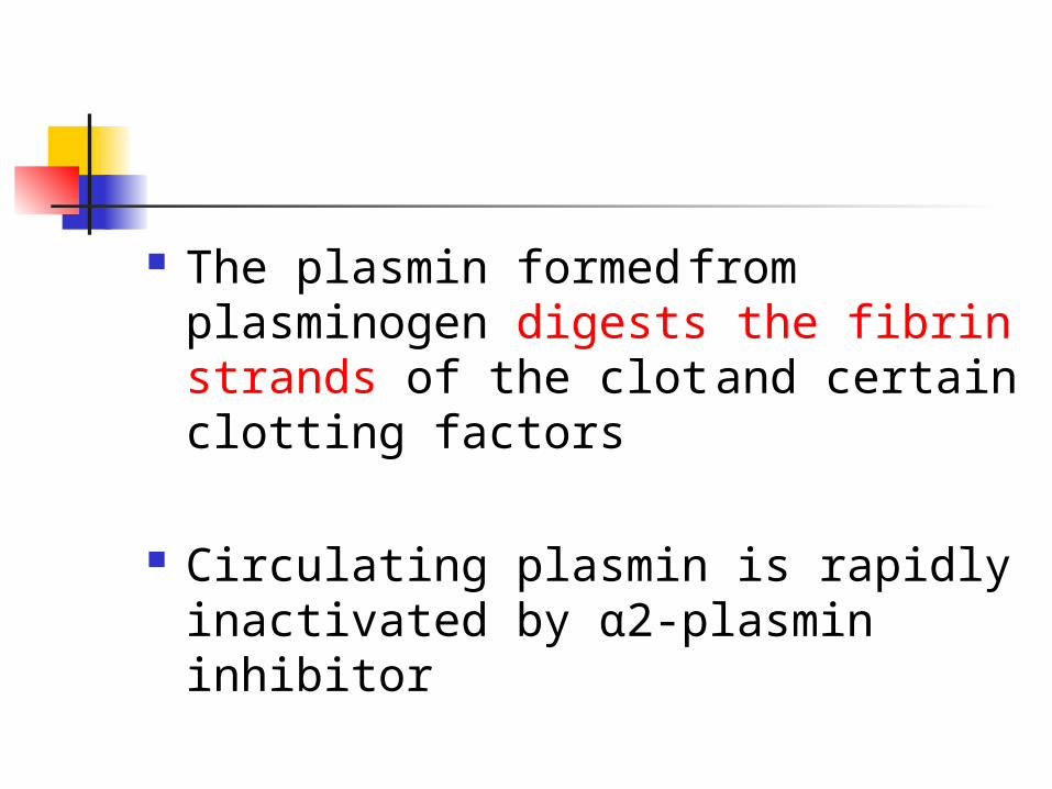
The plasmin formed from plasminogen digests the fibrin strands of the clot and certain clotting factors
Circulating plasmin is rapidly inactivated by α2-plasmin inhibitor
Hints on normal vascular hemostasisHints on normal vascular hemostasis
Normal hemostasis: Normal hemostasis: results from well-regulated results from well-regulated processes that maintain blood in a fluid, clot-free state processes that maintain blood in a fluid, clot-free state in normal vessels while inducing the rapid formation of a in normal vessels while inducing the rapid formation of a localized hemostatic plug at the site of vascular injury.localized hemostatic plug at the site of vascular injury.
Thrombosis:Thrombosis:is the pathologic converse to hemostasis it is the pathologic converse to hemostasis it can be thought of as the formation of a blood clot can be thought of as the formation of a blood clot (thrombus) in uninjured vessels, or thrombotic occlusion (thrombus) in uninjured vessels, or thrombotic occlusion of a vessel after relatively minor injury.of a vessel after relatively minor injury.
Both hemostasis and thrombosis are dependent on three Both hemostasis and thrombosis are dependent on three general components:general components:
the vascular wall, platelets, and the coagulation the vascular wall, platelets, and the coagulation cascadecascade
Hints on normal vascular hemostasis
Normal hemostasis: results from well-regulated processes that maintain blood in a fluid, clot-free state in normal vessels while inducing the rapid formation of a localized hemostatic plug at the site of vascular injury.
Thrombosis:is the pathologic converse to hemostasis it can be thought of as the formation of a blood clot (thrombus) in uninjured vessels, or thrombotic occlusion of a vessel after relatively minor injury.
Both hemostasis and thrombosis are dependent on three general components:
the vascular wall, platelets, and the coagulation cascade
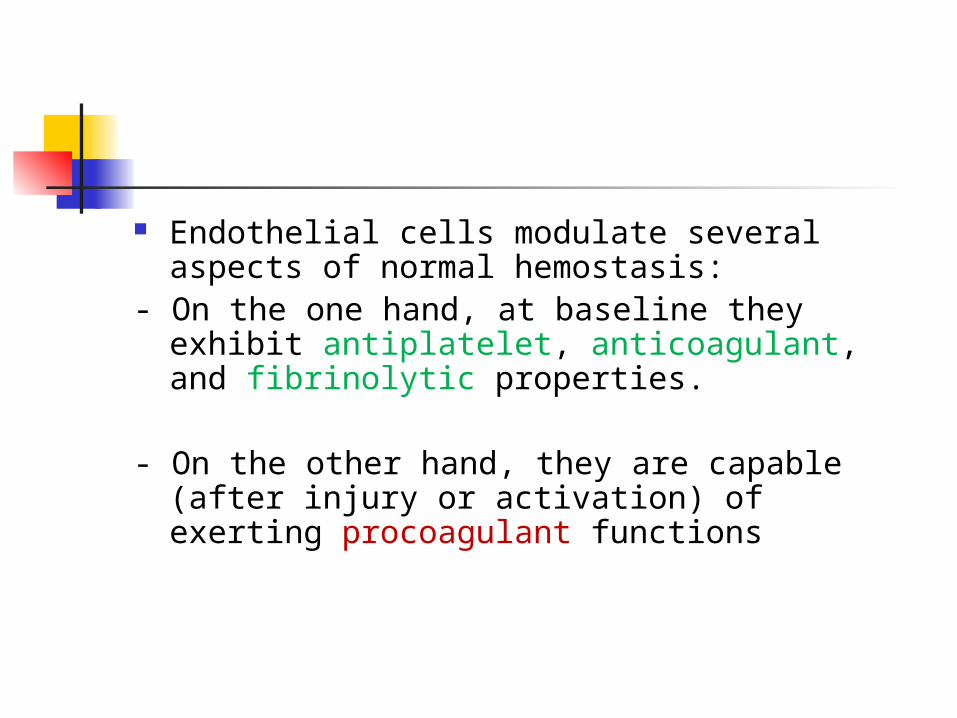
Endothelial cells modulate several aspects of normal hemostasis:
- On the one hand, at baseline they exhibit antiplatelet, anticoagulant, and fibrinolytic properties.
- On the other hand, they are capable (after injury or activation) of exerting procoagulant functions
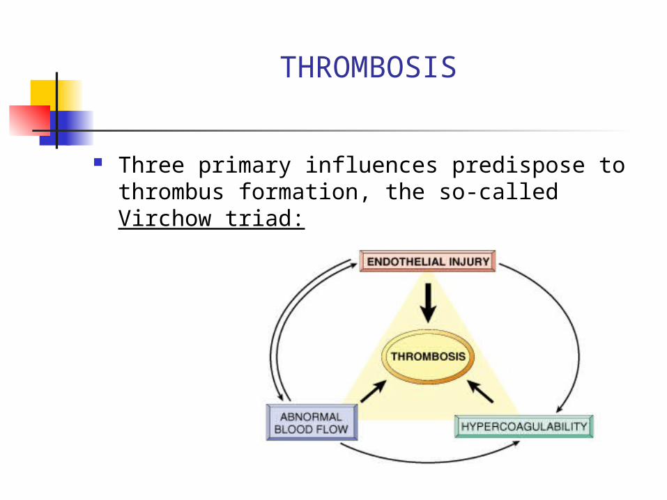
THROMBOSIS
Three primary influences predispose to thrombus formation, the so-called Virchow triad:
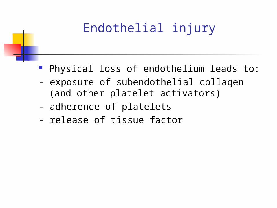
Endothelial injury
Physical loss of endothelium leads to: - exposure of subendothelial collagen (and other
platelet activators) - adherence of platelets- release of tissue factor
Dysfunctional endothelium may elaborate greater amounts of procoagulant factors(e.g., adhesion molecules to bind platelets, tissue factor, PAI, etc.) and smaller amounts of anticoagulant effectors(e.g., thrombomodulin, PGI2, t-PA).

Abnormal blood flow
Turbulence contributes to arterial and cardiac thrombosis by causing endothelial injury or dysfunction
Stasis is a major factor in the development of
venous thrombi
Normal blood flow is laminar such that the platelet elements flow centrally in the vessel lumen, separated from the endothelium by a slower-moving clear zone of plasma.
Stasis and turbulence therefore: a) Disrupt laminar flow & bring platelets into
contact with the endothelium
b) Prevent dilution of activated clotting factors by fresh-flowing blood
c) Retard the inflow of clotting factor inhibitors and permit the build-up of thrombi
d) Promote endothelial cell activation, predisposing to local thrombosis, leukocyte adhesion, and a variety of other endothelial cell effects
Examples: Myocardial infarction

HYPERCOAGULABILITY STATES
There are two general forms of hypercoagulability states: conditions that create increased
platelet function and conditions that cause accelerated
activity of the coagulation system.
Increased Platelet Function
The causes of increased platelet function are disturbances in flow, endothelial damage, and increased sensitivity of platelets to
factors that cause adhesiveness and aggregation
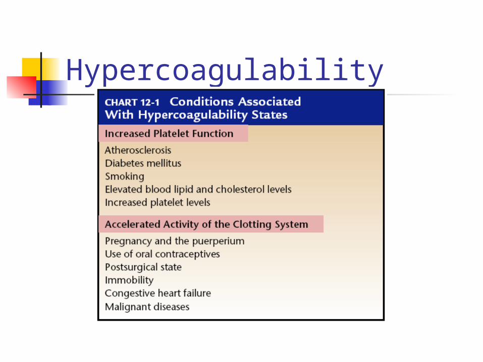
Hypercoagulability

Increased Clotting Activity
results from factors that increase the activation of the coagulation system, including stasis of blood flow and alterations in the coagulation components of the blood (i.e., an increase in procoagulation factors or a decrease in anticoagulation factors)
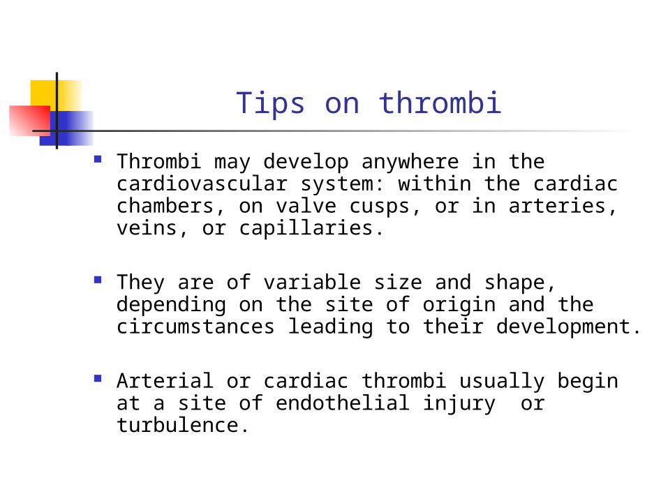
Tips on thrombi
Thrombi may develop anywhere in the cardiovascular system: within the cardiac chambers, on valve cusps, or in arteries, veins, or capillaries.
They are of variable size and shape, depending on the site of origin and the circumstances leading to their development.
Arterial or cardiac thrombi usually begin at a
site of endothelial injury or turbulence.
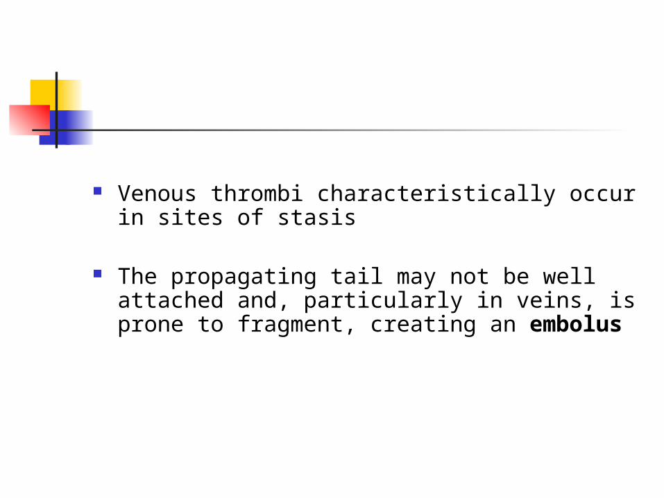
Venous thrombi characteristically occur in sites of stasis
The propagating tail may not be well attached and, particularly in veins, is prone to fragment, creating an embolus
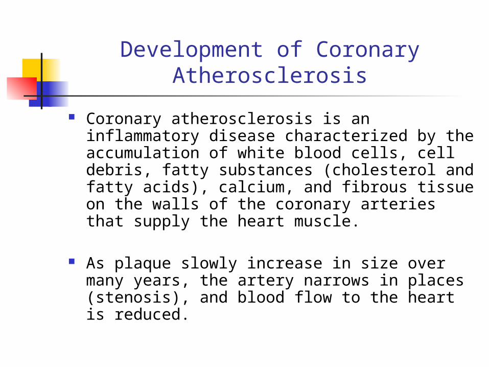
Development of Coronary Atherosclerosis
Coronary atherosclerosis is an inflammatory disease characterized by the accumulation of white blood cells, cell debris, fatty substances (cholesterol and fatty acids), calcium, and fibrous tissue on the walls of the coronary arteries that supply the heart muscle.
As plaque slowly increase in size over many years, the artery narrows in places (stenosis), and blood flow to the heart is reduced.

BLEEDING DISORDERS
Bleeding disorders or impairment of blood coagulation can result from defects in any of the factors that contribute to hemostasis.
Defects are associated with platelets, coagulation factors, and vascular integrity.
Platelet Defects
Disorders of platelet plug formation include a decrease in platelet numbers due to
inadequate platelet production (bone marrow dysfunction),
excess platelet destruction (thrombocytopenia),
abnormal platelet function (thrombocytopathia), or
defects in von Willebrand factor.
Coagulation Defects Impairment of blood coagulation can
result from deficiencies of one or more of the known clotting factors.
Deficiencies can arise because of defective synthesis, inherited defects, or increased consumption of the clotting
factors.
Bleeding that results from clotting factor deficiency typically occurs after injury or trauma.
Large bruises, hematomas, or prolonged bleeding into the gastrointestinal or urinary tracts or joints are common.

Coagulation Defects Impaired Synthesis of Coagulation
Factors factors V, VII, IX, X, XI, and XII; prothrombin; and
fibrinogen are synthesized in the liver In liver disease, synthesis of these clotting factors is reduced
Hemophilia A Deficiency or defect in factor VIII
Von Willebrand Disease Deficiency of or defect in vWF results in reduced platelet adhesion
Disseminated Intravascular Coagulation (DIC)
A variety of disorders ranging from obstetric complications to advanced malignancy may be complicated by disseminated intravascular coagulation:
It is the sudden or insidious onset of widespread fibrin thrombi in the microcirculation
While these thrombi are not usually visible on gross inspection, they are readily apparent microscopically and can cause diffuse circulatory insufficiency, particularly in the brain, lungs, heart, and kidneys
With the development of the multiple thrombi, there is a rapid concurrent consumption of platelets and coagulation proteins (hence the synonym consumption coagulopathy); at the same time, fibrinolytic mechanisms are activated, and as a result an initially thrombotic disorder can evolve into a serious bleeding disorder
It should be emphasized that DIC is not a primary
disease but rather is a potential complication of any condition

DIC

EMBOLISM
An embolus is a detached intravascular solid, liquid, or gaseous mass that is carried by the blood to a site distant from its point of origin.
Virtually 99% of all emboli represent some part of a dislodged thrombus, hence the commonly used term thromboembolism
Rare forms of emboli include droplets of fat, bubbles of air or nitrogen, atherosclerotic debris (cholesterol emboli), tumor fragments, bits of bone marrow, or foreign bodies such as bullets.
Emboli lodge in too small vessels, resulting in partial or complete vascular occlusion. The potential consequence of such thromboembolic events is the ischemic necrosis of down-stream tissue, known as infarction
INFARCTION
An infarct is an area of ischemic necrosis caused by occlusion of either the arterial supply or the venous drainage in a particular tissue
Nearly 99% of all infarcts result from thrombotic or embolic events, and almost all result from arterial occlusion
Occasionally, infarction may also be caused by other mechanisms, such as local vasospasm, extrinsic compression of a vessel, for example, by tumor
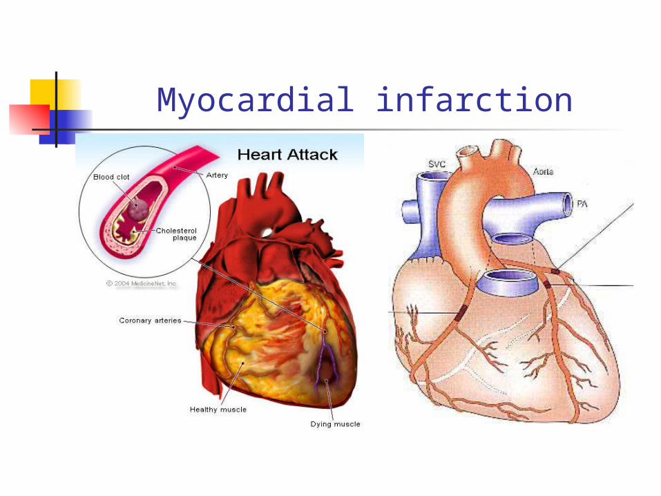
Myocardial infarction
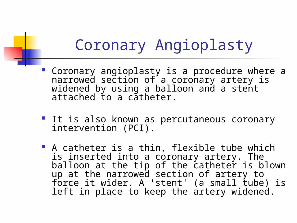
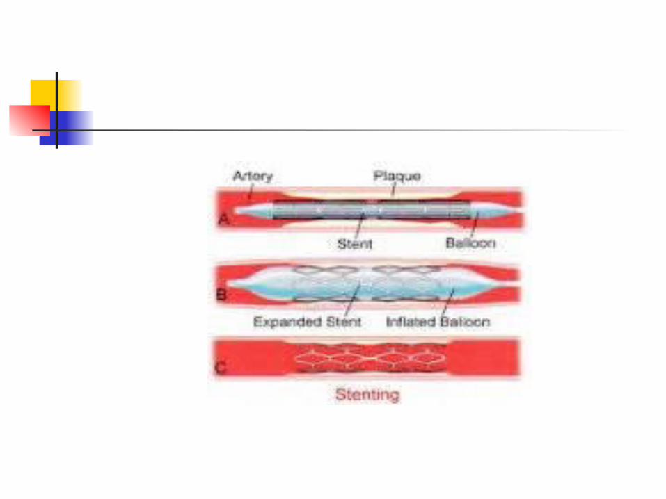
Coronary Angioplasty
Coronary angioplasty is a procedure where a narrowed section of a coronary artery is widened by using a balloon and a stent attached to a catheter.
It is also known as percutaneous coronary intervention (PCI).
A catheter is a thin, flexible tube which is inserted into a coronary artery. The balloon at the tip of the catheter is blown up at the narrowed section of artery to force it wider. A 'stent' (a small tube) is left in place to keep the artery widened.
What is the mechanism of hemostasis? the five step mechanism.
Explain in a diagram the intrinsic and extrinsic pathway.
Explain the pathogenesis of Hemophillia A Vonwillibrand disease
Conditions associated with Hypercoagulability