Superoxide Dismutase 1 Nanoparticles (Nano-SOD1) as a ...
15
biomedicines Article Superoxide Dismutase 1 Nanoparticles (Nano-SOD1) as a Potential Drug for the Treatment of Inflammatory Eye Diseases Alexander N. Vaneev 1,2 , Olga A. Kost 1 , Nikolay L. Eremeev 1 , Olga V. Beznos 3 , Anna V. Alova 4 , Peter V. Gorelkin 2 , Alexander S. Erofeev 1,2 , Natalia B. Chesnokova 3 , Alexander V. Kabanov 1,5 and Natalia L. Klyachko 1,5,6, * Citation: Vaneev, A.N.; Kost, O.A.; Eremeev, N.L.; Beznos, O.V.; Alova, A.V.; Gorelkin, P.V.; Erofeev, A.S.; Chesnokova, N.B.; Kabanov, A.V.; Klyachko, N.L. Superoxide Dismutase 1 Nanoparticles (Nano-SOD1) as a Potential Drug for the Treatment of Inflammatory Eye Diseases. Biomedicines 2021, 9, 396. https://doi.org/10.3390/ biomedicines9040396 Academic Editor: Thiruganesh Ramasamy Received: 3 March 2021 Accepted: 2 April 2021 Published: 7 April 2021 Publisher’s Note: MDPI stays neutral with regard to jurisdictional claims in published maps and institutional affil- iations. Copyright: © 2021 by the authors. Licensee MDPI, Basel, Switzerland. This article is an open access article distributed under the terms and conditions of the Creative Commons Attribution (CC BY) license (https:// creativecommons.org/licenses/by/ 4.0/). 1 School of Chemistry, Lomonosov Moscow State University, 119991 Moscow, Russia; [email protected] (A.N.V.); [email protected] (O.A.K.); [email protected] (N.L.E.); [email protected] (A.S.E.); [email protected] (A.V.K.) 2 Research Laboratory of Biophysics, National University of Science and Technology “MISIS”, 119991 Moscow, Russia; [email protected] 3 Helmholtz National Medical Research Center of Eye Diseases, 105062 Moscow, Russia; [email protected] (O.V.B.); [email protected] (N.B.C.) 4 School of Biology, Lomonosov Moscow State University, 119991 Moscow, Russia; [email protected] 5 Eshelman School of Pharmacy, University of North Carolina at Chapel Hill, Chapel Hill, NC 27599, USA 6 Research Institute “Nanotechnology and Nanomaterials”, G.R. Derzhavin Tambov State University, 392000 Tambov, Russia * Correspondence: [email protected] Abstract: Inflammatory eye diseases remain the most common clinical problem in ophthalmology. The secondary processes associated with inflammation, such as overproduction of reactive oxygen species (ROS) and exhaustion of the endogenous antioxidant system, frequently lead to tissue degeneration, vision blurring, and even blindness. Antioxidant enzymes, such as copper–zinc superoxide dismutase (SOD1), could serve as potent scavengers of ROS. However, their delivery into the eye compartments represents a major challenge due to the limited ocular penetration. This work presents a new therapeutic modality specifically formulated for the eye on the basis of multilayer polyion complex nanoparticles of SOD1 (Nano-SOD1), which is characterized by appropriate storage stability and pronounced therapeutic effect without side reactions such as eye irritation; acute, chronic, and reproductive toxicity; allergenicity; immunogenicity; mutagenicity even at high doses. The ability of Nano-SOD1 to reduce inflammatory processes in the eye was examined in vivo in rabbits with a model immunogenic uveitis—the inflammation of the inner vascular tract of the eye. It was shown during preclinical studies that topical instillations of Nano-SOD1 were much more effective compared to the free enzyme in decreasing uveitis manifestations. In particular, we noted statistically significant differences in such inflammatory signs in the eye as corneal and conjunctival edema, iris hyperemia, and fibrin clots. Moreover, Nano-SOD1 penetrates into interior eye structures more effectively than SOD itself and retains enzyme activity in the eye for a much longer period of time, decreasing inflammation and restoring antioxidant activity in the eye. Thus, the presented Nano-SOD1 can be considered as a potentially useful therapeutic agent for the treatment of ocular inflammatory disorders. Keywords: nanoparticles; Nano-SOD1; inflammation; uveitis; eye diseases; double-layered polyelec- trolyte complex; drug delivery 1. Introduction Reactive oxygen species (ROS) are excessively produced in many disease states and contribute to cell death and tissue degeneration due to the damage of multiple components of cells including cell membranes, DNA, proteins (including various enzymes), carbohy- drates, and proteinase inhibitors. Under physiological conditions, a balance exists between the level of ROS produced during normal cellular metabolism and the level of endogenous Biomedicines 2021, 9, 396. https://doi.org/10.3390/biomedicines9040396 https://www.mdpi.com/journal/biomedicines
Transcript of Superoxide Dismutase 1 Nanoparticles (Nano-SOD1) as a ...

biomedicines
Article
Superoxide Dismutase 1 Nanoparticles (Nano-SOD1) as aPotential Drug for the Treatment of Inflammatory Eye Diseases
Alexander N. Vaneev 1,2, Olga A. Kost 1, Nikolay L. Eremeev 1, Olga V. Beznos 3, Anna V. Alova 4,Peter V. Gorelkin 2 , Alexander S. Erofeev 1,2, Natalia B. Chesnokova 3 , Alexander V. Kabanov 1,5 andNatalia L. Klyachko 1,5,6,*
�����������������
Citation: Vaneev, A.N.; Kost, O.A.;
Eremeev, N.L.; Beznos, O.V.; Alova,
A.V.; Gorelkin, P.V.; Erofeev, A.S.;
Chesnokova, N.B.; Kabanov, A.V.;
Klyachko, N.L. Superoxide
Dismutase 1 Nanoparticles
(Nano-SOD1) as a Potential Drug for
the Treatment of Inflammatory Eye
Diseases. Biomedicines 2021, 9, 396.
https://doi.org/10.3390/
biomedicines9040396
Academic Editor: Thiruganesh
Ramasamy
Received: 3 March 2021
Accepted: 2 April 2021
Published: 7 April 2021
Publisher’s Note: MDPI stays neutral
with regard to jurisdictional claims in
published maps and institutional affil-
iations.
Copyright: © 2021 by the authors.
Licensee MDPI, Basel, Switzerland.
This article is an open access article
distributed under the terms and
conditions of the Creative Commons
Attribution (CC BY) license (https://
creativecommons.org/licenses/by/
4.0/).
1 School of Chemistry, Lomonosov Moscow State University, 119991 Moscow, Russia;[email protected] (A.N.V.); [email protected] (O.A.K.); [email protected] (N.L.E.);[email protected] (A.S.E.); [email protected] (A.V.K.)
2 Research Laboratory of Biophysics, National University of Science and Technology “MISIS”,119991 Moscow, Russia; [email protected]
3 Helmholtz National Medical Research Center of Eye Diseases, 105062 Moscow, Russia;[email protected] (O.V.B.); [email protected] (N.B.C.)
4 School of Biology, Lomonosov Moscow State University, 119991 Moscow, Russia; [email protected] Eshelman School of Pharmacy, University of North Carolina at Chapel Hill, Chapel Hill, NC 27599, USA6 Research Institute “Nanotechnology and Nanomaterials”, G.R. Derzhavin Tambov State University,
392000 Tambov, Russia* Correspondence: [email protected]
Abstract: Inflammatory eye diseases remain the most common clinical problem in ophthalmology.The secondary processes associated with inflammation, such as overproduction of reactive oxygenspecies (ROS) and exhaustion of the endogenous antioxidant system, frequently lead to tissuedegeneration, vision blurring, and even blindness. Antioxidant enzymes, such as copper–zincsuperoxide dismutase (SOD1), could serve as potent scavengers of ROS. However, their delivery intothe eye compartments represents a major challenge due to the limited ocular penetration. This workpresents a new therapeutic modality specifically formulated for the eye on the basis of multilayerpolyion complex nanoparticles of SOD1 (Nano-SOD1), which is characterized by appropriate storagestability and pronounced therapeutic effect without side reactions such as eye irritation; acute,chronic, and reproductive toxicity; allergenicity; immunogenicity; mutagenicity even at high doses.The ability of Nano-SOD1 to reduce inflammatory processes in the eye was examined in vivo inrabbits with a model immunogenic uveitis—the inflammation of the inner vascular tract of the eye.It was shown during preclinical studies that topical instillations of Nano-SOD1 were much moreeffective compared to the free enzyme in decreasing uveitis manifestations. In particular, we notedstatistically significant differences in such inflammatory signs in the eye as corneal and conjunctivaledema, iris hyperemia, and fibrin clots. Moreover, Nano-SOD1 penetrates into interior eye structuresmore effectively than SOD itself and retains enzyme activity in the eye for a much longer period oftime, decreasing inflammation and restoring antioxidant activity in the eye. Thus, the presentedNano-SOD1 can be considered as a potentially useful therapeutic agent for the treatment of ocularinflammatory disorders.
Keywords: nanoparticles; Nano-SOD1; inflammation; uveitis; eye diseases; double-layered polyelec-trolyte complex; drug delivery
1. Introduction
Reactive oxygen species (ROS) are excessively produced in many disease states andcontribute to cell death and tissue degeneration due to the damage of multiple componentsof cells including cell membranes, DNA, proteins (including various enzymes), carbohy-drates, and proteinase inhibitors. Under physiological conditions, a balance exists betweenthe level of ROS produced during normal cellular metabolism and the level of endogenous
Biomedicines 2021, 9, 396. https://doi.org/10.3390/biomedicines9040396 https://www.mdpi.com/journal/biomedicines
Biomedicines 2021, 9, 396 2 of 15
antioxidants that offers protection against damage caused by ROS. Disruption of this bal-ance, through increased ROS production or decreased antioxidant levels, causes a conditionreferred to as oxidative stress that is associated with an increased risk of cardiovasculardisease, atherosclerosis, cancer, and many other diseases. In particular, ROS are importantfactors in the early tissue damage that develops from inflammation [1,2]. Because of thenonspecific nature of ROS-induced tissue injury, excessive release of these agents can causesubstantial damage not only to the tissue in an inflamed state but also to the surroundingnormal tissue. This is very important for the eye, as the transparency of the cornea andlens, as well as the functioning of photoreceptor apparatus, relies on their highly orderedstructures, and excessive tissue damage will compromise visual function [3].
Antioxidants, superoxide dismutase 1 (SOD1) in particular, are known to play abeneficial role in protecting tissues against oxidative stress in many pathological states,including inflammation [4–12]. Thus, SOD1 was reported to accelerate the healing of skinlesions caused by burns, systemic lupus erythematosus, and herpes [13,14]; protect culturedhuman neurons under oxidative stress [15]; reduce ischemia-reperfusion injury [16]; beeffective in the treatment of rat adjuvant arthritis, [17] etc.
Most relevant to this study, antioxidants, including SOD1, are also thought to bebeneficial in the treatment of eye diseases. Superoxide dismutase 1 was found in differenteye tissues and fluids [18–20], where this enzyme plays an important role in confrontingoxidative stress and maintaining eye homeostasis. The eye is a rather isolated organ, andthe pathological processes within it are preferably treated not via systemic but by localdrug intake. The use of topical applications, subconjunctival, parabulbar or intraocularinjections of SOD1 could, therefore, provide a supplement for intrinsic antioxidants in eyetissues, which may be depleted at oxidative stress. According to statistics, inflammatoryeye diseases, accompanied by oxidative stress, are the most common eye pathologies thatlead to partial disability and, sometimes, to the complete loss of vision [21–23]. It wasshown that both subconjunctival injections and topical applications of SOD1 solutionswere effective in preventing corneal perforations under acute inflammation of the eye afteralkali burns [24,25]. Other beneficial effects of SOD1 were obtained in the treatment ofuveitis inflammation of the uveal tract involving both outer and inner structures of the eye.Thus, SOD1 treatment of animals with phacoanaphylactic endophthalmitis (lens-induceduveitis) resulted in the strong reduction of choroid inflammation, retinal edema and,vasculitis [26]. In S-antigen-induced and bovine serum albumin-induced passive Arthus-type uveitis, SOD1 treatment resulted in the remarkable reduction in aqueous humor cellquantity and infiltration of the inflammatory cells in the anterior retina [25,27]. Topicalinstillations of SOD1 at immunogenic uveitis induced by foreign animal serum resulted inthe enhancement of general antioxidant activity in the eye and in the substantial decreaseof leukocyte count in aqueous humor, even in comparison with dexamethasone [28,29].
However, topical application of the drug, although sparing for the patient, is lesseffective for the treatment of the diseases, which include inner structures of the eye, dueto the poor transport of the drug into the eye. In recent years, there has been significantinterest in the development of nanosize drug delivery systems to the eye. Such nanosys-tems can potentially improve the therapeutic efficacy of the drugs by overcoming thediffusion barrier, by increasing their stability in biological tissues and fluids, and enhancingcellular/tissue uptake [30–35].
Several years ago, a cross-linked polyion complex of SOD1 with a cationic blockcopolymer methoxy-poly(ethylene glycol)-block(L-lysine hydrochloride) (PEG-pLL50) wasdeveloped [36]. This SOD1 nanoformat decreased ischemia/reperfusion-induced tissueinjury and improved sensorimotor functions in a rat middle cerebral artery occlusion modelafter a single intravenous injection, [36] attenuated hypertension established in mice bychronic subcutaneous infusion of angiotensin II after intracerebroventricular injection [37],demonstrated a significant decrease in markers in endothelial/vascular cell activationand/or inflammation in visceral adipose tissue, thoracic aorta and heart of obese mice [38],and attenuated ethanol-induced steatohepatitis [39] after intraperitoneal injections. It is

Biomedicines 2021, 9, 396 3 of 15
noteworthy that this nanoformulated SOD1 also appeared to be more effective than nativeSOD1 in the treatment of inflammatory eye disease, uveitis, just at topical instillations [40].Despite all the advantages of this SOD1 formulation in treatment, it should be noted thatduring the synthesis of the drug, the loss of enzyme activity exceeds 90%.
Recently, a new SOD1 nanoformulation was introduced, based on a chemically cross-linked multilayer polyion complex in which SOD1 is initially incorporated into a complexwith polycation, then coated with a polyanion block copolymer. The loss of the enzymeactivity during the synthesis by this technique was significantly lower (60–70%), and thisdouble-coated SOD1 nanoparticles exhibited prolonged circulation of active enzymes inthe blood stream, while single intravenous injection of such a nanoformulation improvedthe recovery of locomotor functions in rats with moderate spinal cord injury [41].
In the current study, we adjusted the synthesis of double-coated SOD1 nanoparti-cles (Nano-SOD1) specified for local application in ophthalmology and evaluated theireffectiveness in reducing inflammation in the eye.
2. Materials and Methods2.1. Materials
Recombinant human SOD1 was purchased from Life Science Advanced Technologies(St. Petersburg, Russia). Methoxy-poly(ethylene glycol)113-block-poly(L-glutamic acidsodium salt)50 (PGlu-PEG) were purchased from Alamanda Polymers Inc. (USA). Pro-tamine from salmon, glutaraldehyde (GA), 0.01 Мphosphate-buffered saline (PBS, pH 7.4,0.137 МNaCl), quercetin, pyrogallol, Amicon Ultra Centrifugal Filter Units (membranewith a molecular weight cut off of 100 kDa), hemoglobin from bovine blood, luminol,6-hydroxy-2,5,7,8-tetramethylchromane (Trolox), and a sterile syringe filter with a 220 nmpore size were purchased from Merck Milipore (Carrigtwohill, Ireland). Dialysis capsules,Float-A-Lyzer® G2 with a membrane with a molecular weight cut off of 100 kDa, werepurchased from Spectrum Laboratories (CA, USA). A protein concentration assay kit andfluorescent dye, Alexa 488 Fluor, were from Thermo Fisher Scientific (CA, USA).
2.2. Synthesis of Double-Coated Cross-Linked Nano-SOD1
The Nano-SOD1 was synthesized using a modified procedure of Nukolova et al. [41].Briefly, stock solutions of SOD1, protamine, and PGlu-PEG (5 mg/mL each) were preparedin PBS or 10 mM 2-[4-(2-hydroxyethyl)piperazin-1-yl]ethanesulfonic acid (HEPES), pH 7.4,containing 0.137 M NaCl. Stock solutions of GA (0.5%, v/v) and NaBH4 (1 mg/mL) wereprepared in water immediately before the experiments. All solutions were added to eachother dropwise with stirring. First, protamine solution was added to the SOD1 solution toachieve a final SOD1/polycation charge ratio 1:2 of ionogenic groups, and the resultingsolution was incubated at room temperature (r.t.) for 30 min upon stirring. The charge ratiowas calculated as a ratio of the concentration of NH2 groups of the protamine protonated atpH 7.4 to the concentration of COOH groups of glutamic acid and aspartic acid residues ofthe enzyme (estimated using Protein Calculator v.3.3 software, Scripps Research Institute,CA, USA). Subsequently, PGlu-PEG solution was added to the SOD1/protamine complexsolution at a final charge ratio polycation/polyanion 1:1, and the reaction mixture wasincubated for 30 min at 4 ◦C without stirring. Then, GA solution was added to achieve afinal molar ratio of cross-linker to amino groups of protamine 4.4:1. The resulting mixturewas then incubated overnight at 4 ◦C, formed Schiff base bonds were reduced by thetreatment with NaBH4 solution. To remove the side products and excess of unreactedreagents, the reaction mixture was diluted 4 times by PBS or 10 mM HEPES and centrifugedon 100 kDa Amicon filters at 800× g, 4 ◦C. The procedure was repeated twice. Purifiedsamples of cross-linked Nano-SOD1 were adjusted to specific SOD1 activity 13,500 Uby pyrogallol (185,000 U by quercetin) per mL, stored at 4 ◦C, and used in experimentson animals.

Biomedicines 2021, 9, 396 4 of 15
2.3. Protein Concentration and Enzyme Activity
The protein concentration was measured using the Micro BCA Protein Assay Kit inaccordance with the manufacturer’s instructions (Thermo Fisher Scientific, CA, USA).
The catalytic activity of SOD1 and Nano-SOD1 was determined using pyrogallol orquercetin assay [41]. For the pharmacokinetics studies, the quercetin assay was applieddue to its greater sensitivity.
2.4. Dynamic Light Scattering (DLS)
Purified Nano-SOD1 was characterized by DLS using Zetasizer Nano ZS (MalvernInstruments Ltd., Malvern, UK). The hydrodynamic diameter and polydispersity index(PDI) were measured in PBS buffer; zeta (ζ)-potential was analyzed in water (the change ofsolvent was performed by gel filtration on NAP-10 column, GE Healthcare, CA, USA). Allmeasurements were performed in automatic mode at 25 ◦C at least in triplicate.
2.5. Retention of SOD1 in Nano-SOD1
The SOD1 retention in the Nano-SOD1 formulation was determined using 100 kDadialysis capsules. A capsule was filled with a solution of Nano-SOD1 (1 mL), then thecapsule was placed into PBS buffer (30 mL), and the enzyme activity and protein concentra-tion were measured in aliquots (100 µL) taken from the capsule at predetermined intervalsusing the techniques described above.
2.6. Stability of Nano-SOD1
To study the stability of the obtained Nano-SOD1, solutions of SOD1 nanoparticles(13,500 U/mL by pyrogallol) were filtered under sterile conditions through a syringemembrane with 220 nm pores, sealed in glass ampoules, and stored at 4 and 25 ◦C. Sampleswere taken at regular intervals (7, 30, 60, and 180 days), and the enzymatic activity andhydrodynamic diameter of Nano-SOD1 were measured.
2.7. Animals
A study was conducted using 45 adult male Chinchilla rabbits weighing 2.0–2.5 kg.All experiments with live rabbits were carried out in strict accordance with the Associationfor Research in Vision and Ophthalmology (ARVO) statement for the Use of Animals inOphthalmic and Vision Research. The animals were kept in individual cages with freeaccess to food and water.
2.8. Dynamics of Labeled SOD1 and Nano-SOD1 in Tear and Intraocular Fluid
For pharmacokinetics study, Nano-SOD1 was synthesized with SOD1 covalently con-jugated to Alexa 488 fluorescent dye (in accordance with the manufacturer’s instructions).
Rabbits were randomly divided into 3 groups (n = 2 per each group, i.e., 4 eyes): (1)placebo group that received PBS; (2) SOD1 group that received a solution of native SOD1labeled by Alexa; (3) experimental Nano-SOD1 group that received a solution of SOD1nanoparticles prepared with labeled SOD1. Specific activities of SOD1 and Nano-SOD1solutions were equal. Rabbits received 40 µL of drug solutions as eye drops bilaterallytwice with an interval of 5 min (total of 80 µL per eye).
Tear fluid was taken 5, 30, and 60 min after the last instillation using round pieces offilter paper with a diameter of 5 mm. Paper pieces were placed into the lower conjunctivalsack for 5 min, 6 circles to each eye. Pieces soaked with tear fluid were removed, placedinto 300 µL of PBS buffer for 20 min for the elution of tear components, and centrifuged for10 min at 940× g.
The intraocular fluid was collected by paracentesis of the cornea with an insulinsyringe under topical anesthesia (Alcain 0.5%, Alcon, Puurs, Belgium) 30 and 60 min afterthe last instillation.
The intensity of Alexa-labelled SOD1 and NanoSOD1 fluorescence and enzymaticactivity was measured in the samples of tear and intraocular fluid.

Biomedicines 2021, 9, 396 5 of 15
In addition, we applied the amperometric method to determine the H2O2 level as anindicator of Nano-SOD1 functioning in tear and intraocular fluid. For this purpose, weused platinum-coated carbon nanoelectrodes in the fluids and a silver chloride referenceelectrode. Patch-clamp amplifier Model 2400 (AM Systems; WA, USA) allowed registrationof the potential difference between platinum nanoelectrode and reference electrode. TheUSB-6211 ADC/DAC converter (National instruments; USA) and WinWCP softwareallowed data transfer and recording. The H2O2 was evaluated at the potential of +800 mVto the silver chloride electrode [42–44].
Rabbits were randomly divided into 5 groups (n = 5 per group), one group per timepoint. The Nano-SOD1 solution was instilled into the left eye of each animal, while PBSbuffer (control) was instilled into the right eye. The samples of tear and intraocular fluidwere taken (as described in previously) before instillation and 10, 30, 60, and 120 min afterthe instillation, and amperage in the sample (proportional to Н2О2 level) was measured byplatinized electrode. Due to the variability of the background levels of hydrogen peroxidein the tear and intraocular fluid of each animal, the H2O2 dynamics were calculated as thedifference between the left (experimental) and right (control) eyes of each animal.
2.9. In Vivo Studies/Treatment of Immunogenic Uveitis
Experiments were performed in three independent series with 15 rabbits each.Acute uveitis was induced in rabbits by double injection of normal equine serum:
initial subcutaneous injection of 5 mL serum for sensitization was followed on the 10thday with intravitreal injection of 70 µL serum after anesthetic Alcain 0.5% (Alcon, Puurs,Belgium) instillation [45].
Animals were randomly divided into 3 groups (n = 5 per group, i.e., 10 eyes) andtreated as follows: (1) placebo group with uveitis that received PBS buffer (placebo); (2)SOD1 group with uveitis that received native SOD1 solution (1 mg/mL, 13,500 U/mLwith pyrogallol assay) in the same buffer; (3) Nano-SOD1 group with uveitis that receivedNano-SOD1 solution in the same buffer. During the treatment, solutions were instilledthree times a day (30 µL in each eye) for 8 days. The SOD1 dose corresponded to that usedin Reference [40]. The dose of Nano-SOD1 solution was calculated based on the specificactivity of SOD1 (units per mL) so that the activities of SOD1 and Nano-SOD1 solutionswere equal.
Clinical manifestation of uveitis was followed on 1, 2, 3, 4, 7, and 8 days after theintravitreal injection of equine serum. Rabbit eyes were observed under the slit lamp andscored for clinical signs of ocular inflammation. Clinical symptoms of uveitis includedeyelid and conjunctival edema and hyperemia, corneal and iris edema, presence of fibrinclots in the anterior chamber, and presence of cataract and posterior synechiae (cohesionsbetween the papillary margin of the iris and anterior part of the lens). We compared theefficacy using the scores for the manifestations of inflammation as a common approachin ophthalmology [46]. Evaluation of the inflammation score was performed using aconventional scale: (0) no symptom, (1) low degree of manifestation, (2) medium, and(3) strong.
2.10. Leukocyte Counting and Antioxidant Activity in Intraocular Fluid
Intraocular fluid from all eyes was collected by paracentesis of the cornea with theinsulin syringe under local anesthesia (Alcain 0.5%, eye drops) on the 8th day (i.e., 16 hafter the last instillation) for leukocyte counting and total protein and antioxidant activity(AOA) measurement. A healthy group of rabbits without uveitis (n = 5) was used as acontrol for a comparative analysis of biochemical parameters during the treatment.
Leukocytes in intraocular fluid were counted in unstained preparation (20 µL) offreshly collected sample using a light microscope. Average amount of leukocytes in themicroscopic field was counted.
Antioxidant activity (AOA) was assayed via estimation of chemiluminescence kineticsparameters in the hemoglobin–H2O2–luminol model system [47] using Trolox as the stan-

Biomedicines 2021, 9, 396 6 of 15
dard. The AOA value of the sample was expressed as the Trolox equivalent concentrationcalculated on the basis of a standard curve. Protein concentration in the intraocular fluidwas determined by the Lowry method [48].
2.11. Toxicology Studies
All toxicity studies were performed according to Reference [49].Acute toxicity of Nano-SOD1 formulations was determined on two types of animals,
rats and rabbits. In series 1, Nano-SOD1 was injected in the lateral vein of rat tails (averagerat weight 280 g) in average doses 91,000 U/kg (here and after, units of activity by pyrogallolare given) that exceeded the recommended dose for humans (89 U/kg per day [40] bymore than 1000 times. In series 2, Nano-SOD1 was instilled in each eye of the rats in a totaldose of 240,000 U/kg that exceeded the recommended dose for humans by 2700 times.In series 3, Nano-SOD1 was instilled in each eye of the rabbits in a dose of 26,000 U/kg(average rabbit body weight of 2.5 kg) that exceeded the recommended dose for humansnearly 300 times.
Chronic toxicity of Nano-SOD1 was investigated on rats and rabbits who receivedinstillations of the formulation with a daily dose exceeding the recommended dose forhumans 30 times for rats and 17 times for rabbits during a month.
Assessment of the mutagenic properties of Nano-SOD1 was carried out in tests in vitrofor the induction of gene mutations in Salmonella typhimurium (Ames test) and in thetest in vivo for the induction of chromosomal damage (test accounting micronuclei inpolychromatophilic red blood cells of mice). In the latter case, the excess of the dose overthe recommended one for humans was 570 times with intraperitoneal administration and20 times with instillations, accordingly.
To determine the allergenic properties of Nano-SOD1, anaphylactogenic activityassessment, “delayed”-type hypersensitivity reactions, conjunctival tests, and mast celldegranulation were examined in guinea pigs with a 10 fold excess of the dose recommendedfor humans.
To determine the immunotoxic effect of Nano-SOD1 formulations, an assessment ofthe humoral immune response, cellular immunity, and phagocytic activity of peritonealmacrophages were examined in mice in excess of the recommended dose for humans up to20 times.
The influence of Nano-SOD1 on the reproduction was investigated in mice. Formula-tion was instilled daily—males for 48 days (spermatogenesis period), females for 15 days(3 estrous cycles)—with a 10 fold excess of the dose recommended for humans.
Detailed methods of toxicological studies are presented in the Supplementary Materials.
2.12. Data Analysis
All experiments were conducted independently at least in triplicate, the results wereexpressed as mean value ± standard deviation (SD). Origin 2016 (OriginLab Corpora-tion, MA, USA) and STATISTICA 6 (StatSoft, Inc., OK, USA) were used for statisticalanalysis. Significance was analyzed using the Mann–Whitney U test and ANOVA test. Ap-value < 0.05 was considered statistically significant; p < 0.01 and p < 0.001 were consid-ered highly statistically significant.
3. Results and Discussion3.1. Synthesis of SOD1 Nanoparticles and Their Characterization
Here, we evaluated a double-coated cross-linked polyion complex partly based on theapproach described in Reference [41]. The principal scheme of the synthesis is presentedin Figure 1A. Firstly, block–ionomer complexes were formed spontaneously as a resultof electrostatic coupling of the enzyme and cationic polymer (protamine) and anionicblock copolymer (PolyGlu-PLE). The Block–ionomer complex consists of several SOD1globules based on the size of the resulting particles (Figure 1B), assuming the size of SOD1

Biomedicines 2021, 9, 396 7 of 15
molecule is less than 10 nm. Then, the cross-linker was added to complexes that resulted incovalent stabilization.
In the original method, such synthesis was carried out in HEPES buffer. It should bementioned that HEPES is inappropriate in ophthalmology because HEPES can negativelyaffect the structures of the eye [50]. Thus, for comparison we obtained Nano-SOD1 inHEPES and PBS buffers.
It appeared that the replacement of the buffer did not significantly affect neither Nano-SOD1’s effective hydrodynamic diameter (Figure 1B), equal to 44–45 nm, nor ζ-potentialequal to −7 mV (Table 1).
Figure 1. Physicochemical properties of Nano-SOD1 obtained. (A) Principal scheme for the synthesisof Nano-SOD1. (B) Particle hydrodynamic diameter distribution (SOD1/protamine/PGlu-PEG = 1:2:2with glutaraldehyde (GA) as a cross-linker). (C) SOD1 release profile from a dialysis capsule withNano-SOD1 obtained in HEPES and PBS buffers; 1 mL of Nano-SOD1 solution with specific activityof 13,500 U/mL was placed inside each dialysis capsule and 30 mL of PBS were placed outsidethe capsule.
Table 1. Characteristics of Nano-SOD1 obtained in PBS and HEPES buffers.
Parameter HEPES [41] PBS
Diameter, nm 44 ± 2 45 ± 5Polydispersity index (PDI) 0.15 <0.1
ζ-potential, mV −7 ± 1 −7 ± 2Protein yield,% 76 ± 8 89 ± 5
Residual enzyme activity yield,% 37 ± 6 42 ± 3
However, Nano-SOD1 synthesis in PBS helped to decrease the PDI value from 0.15 to<0.1 that resulted in a significantly narrowed size distribution of the particles. Moreover,the usage of PBS increased protein yield and residual enzyme activity (Table 1), statisticalsignificance p < 0.05 being determined by the ANOVA test.
Additional advantage of the Nano-SOD1 synthesis in PBS was further demonstratedby the experiments on SOD1 release from nanoparticles. Despite covalent binding of SOD1in conjugates, part of the enzyme molecules within nanoparticles was still in the form ofcomplexes with polymers fixed by electrostatic interactions. The weaker the interactions,the stronger the equilibrium is shifted towards the release of SOD1 from nanoparticle. Itwas shown that the residual SOD1 activity in a dialysis capsule containing nanoparticleswas significantly lower for Nano-SOD1 obtained in HEPES buffer compared to PBS, and
Biomedicines 2021, 9, 396 8 of 15
the release of SOD1 into solution was much slower in the case of PBS (Figure 1C). Thisindicates that a larger number of SOD1 molecules in Nano-SOD1 are covalently bound topolymers or that electrostatic interactions of SOD1 with polymers are stronger in double-coated nanoparticles synthesized using our proposed technique in PBS. Thus, for furtherexperiments, Nano-SOD1 was synthesized in PBS.
3.2. Stability of Nano-SOD1 Formulations
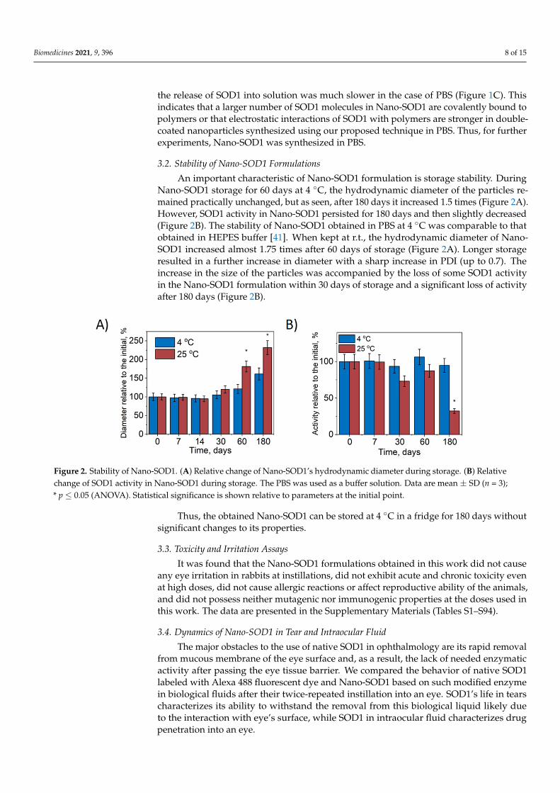
An important characteristic of Nano-SOD1 formulation is storage stability. DuringNano-SOD1 storage for 60 days at 4 ◦C, the hydrodynamic diameter of the particles re-mained practically unchanged, but as seen, after 180 days it increased 1.5 times (Figure 2A).However, SOD1 activity in Nano-SOD1 persisted for 180 days and then slightly decreased(Figure 2B). The stability of Nano-SOD1 obtained in PBS at 4 ◦C was comparable to thatobtained in HEPES buffer [41]. When kept at r.t., the hydrodynamic diameter of Nano-SOD1 increased almost 1.75 times after 60 days of storage (Figure 2A). Longer storageresulted in a further increase in diameter with a sharp increase in PDI (up to 0.7). Theincrease in the size of the particles was accompanied by the loss of some SOD1 activityin the Nano-SOD1 formulation within 30 days of storage and a significant loss of activityafter 180 days (Figure 2B).
Figure 2. Stability of Nano-SOD1. (A) Relative change of Nano-SOD1’s hydrodynamic diameter during storage. (B) Relativechange of SOD1 activity in Nano-SOD1 during storage. The PBS was used as a buffer solution. Data are mean ± SD (n = 3);* p ≤ 0.05 (ANOVA). Statistical significance is shown relative to parameters at the initial point.
Thus, the obtained Nano-SOD1 can be stored at 4 ◦C in a fridge for 180 days withoutsignificant changes to its properties.
3.3. Toxicity and Irritation Assays
It was found that the Nano-SOD1 formulations obtained in this work did not causeany eye irritation in rabbits at instillations, did not exhibit acute and chronic toxicity evenat high doses, did not cause allergic reactions or affect reproductive ability of the animals,and did not possess neither mutagenic nor immunogenic properties at the doses used inthis work. The data are presented in the Supplementary Materials (Tables S1–S94).
3.4. Dynamics of Nano-SOD1 in Tear and Intraocular Fluid
The major obstacles to the use of native SOD1 in ophthalmology are its rapid removalfrom mucous membrane of the eye surface and, as a result, the lack of needed enzymaticactivity after passing the eye tissue barrier. We compared the behavior of native SOD1labeled with Alexa 488 fluorescent dye and Nano-SOD1 based on such modified enzymein biological fluids after their twice-repeated instillation into an eye. SOD1’s life in tearscharacterizes its ability to withstand the removal from this biological liquid likely dueto the interaction with eye’s surface, while SOD1 in intraocular fluid characterizes drugpenetration into an eye.

Biomedicines 2021, 9, 396 9 of 15
It was shown that exogenous native SOD1 was washed out completely by the thir-tieth minute after the last instillation (SOD1 level in a tear corresponded to the level ofendogenous SOD1). Nano-SOD1, however, continued to function in the tear much longer(Figure 3A). As seen, its activity in the tear after 30 min was 2.5–3 times higher than theendogenous SOD1 level coming to that level only after an hour.
Figure 3. Dynamics of SOD1 and Nano-SOD1 in tears after instillation: (A) SOD1 activity; (B) SOD1 concentration(percentage of labeled SOD1 fluorescence relative to the initial protein fluorescence). The dashed line corresponds to theactivity of endogenous SOD in tears. Symbols indicate the significance levels of differences according to the Mann–WhitneyU-test: ** p < 0.01; * p < 0.05; NS—no difference.
The dynamics of tear fluorescence that correlates with the amount of labeled SOD1 intears (Figure 3B) corresponded to the measured SOD1 activity and, thus, confirmed thatNano-SOD1 stayed in the tear much longer than the native one.
The results, indeed, evidenced that Nano-SOD1 retains much better on the surface ofeye cornea in comparison to native enzyme, and, thus, Nano-SOD1 could be considered aspotential long-acting antioxidant drug.
Further, we evaluated the ability of Nano-SOD1 to penetrate into the anterior chamberof the eye. Figure 4A shows that in both cases, native SOD1 and Nano-SOD1, some enzymepassed through ocular barrier. However, Nano-SOD1 penetrated into the anterior chamberbetter than the native one. It is noteworthy that SOD1 activity in the intraocular fluidincreased above the background level 30 min after the instillation of the Nano-SOD1,remaining increased even after an hour (Figure 4A). Labeled SOD1 fluorescence dataalso confirmed that Nano-SOD1 was able to penetrate the anterior chamber of the eye(Figure 4B).
Figure 4. Dynamics of SOD1 and Nano-SOD1 in intraocular fluid after instillation: (A) SOD1 activity;(B) SOD1 concentration (fluorescence of labeled protein). The dashed lines correspond to the activityof endogenous SOD in tears. The symbols denote the significance levels of differences according tothe Mann–Whitney U-test: * p < 0.05; NS—no difference.
It is noteworthy that while Nano-SOD1 activity was high in tears 30 min after instilla-tion, its activity in the intraocular fluid was rather low. However, at 60 min after instillation,

Biomedicines 2021, 9, 396 10 of 15
Nano-SOD1 activity in the intraocular fluid increased significantly, while activity in tearsdecreased (Figures 3 and 4). This result indicates that Nano-SOD1 passes from the anteriorsurface of the eye into the inner region, this process definitely requiring time.
In addition, we determined the level of H2O2 in tear and intraocular liquid as anindicator of Nano-SOD1 functioning in these liquids. It should be mentioned that SOD1catalyzes the dismutation of the superoxide radical into molecular oxygen and hydrogenperoxide. Therefore, after the instillation of Nano-SOD1, the level of hydrogen peroxide inanimal’s tears increased, reaching a maximum at 30 min (Figure 5A). Apparently, this wasdue to a rather effective sorption of the Nano-SOD1 on the eye’s surface (see the dynamicsof SOD1 activity and fluorescence, Figure 3), leading to an increase in Н2О2 concentrationin 30 min. By 1 h, however, the Н2О2 concentration returned to its background level inaccordance with the abovementioned observations (Figure 3) that at this point all Nano-SOD1 was washed away by a tear.
Figure 5. Dynamics of the H2O2 level in rabbit tears and intraocular fluid after a single instillation of Nano-SOD1. The dataare presented as mean ± SEM for 5 rabbits. The current is proportional to hydrogen peroxide concentration. The H2O2
dynamics were calculated as the difference between the left (experimental) and right (control) eyes of each animal in tear(A) and intraocular fluid (B).
Unlike a tear, a noticeable increase in the level of hydrogen peroxide in the intraocularfluid was observed only 30 min after the instillation of Nano-SOD1 (Figure 5B). Obviously,it takes some time for Nano-SOD1 to penetrate into the anterior chamber of the eye. WhileSOD1 levels remained constant for an hour after the preparation instillation (Figure 4), thelevel of hydrogen peroxide in the intraocular fluid continuously increased for at least 2 h(Figure 5B). One can assume that this fact indicates continued functioning of Nano-SOD1in the anterior eye chamber.
Thus, while the maximum SOD1 level in a tear was reached immediately after instilla-tion of Nano-SOD1, it took time for the enzyme to enter the intraocular fluid.
3.5. Effects of Topical Instillations of Nano-SOD1 on Clinical Manifestations of ImmunogenicUveitis in Rabbits
At 24 h after intravitreal injection of equine serum, rabbits in all experimental groups(placebo-treated, native SOD1-treated, and Nano-SOD1-treated) showed typical clinicalsigns of acute uveitis: corneal and conjunctival edema, hyperemia of the iris, and fibrinprecipitation on the iris surface. Over 3–4 days, these clinical signs intensified, but the de-velopment of uveitis in SOD1-treated and Nano-SOD1-treated groups remarkably differedfrom that in the placebo group.
Rabbits without any treatment (placebo) exhibited pronounced corneal and conjuncti-val edema and iris hyperemia. There was a lot of fibrin clots in the anterior part of the eye,which, in several cases, formed massive clouds.

Biomedicines 2021, 9, 396 11 of 15
The SOD1-treated rabbits showed less pronounced corneal and conjunctival edema,as well as iris hyperemia. However, there was no difference between the amount of fibrinclots observed in SOD1-treated and placebo-treated rabbits.
In the group treated with Nano-SOD1, clinical manifestations of uveitis were even lesspronounced than in placebo- and SOD1-treated groups (Figure 6). Most importantly, notonly corneal and conjunctival edema and iris hyperemia were less pronounced than in twoother groups, but we revealed a statistically different intensity of fibrin clots in the anteriorchamber (p < 0.05 at 3, 4, and 7 days) between SOD1 and Nano-SOD1-treated groups.
Figure 6. The effect of topical instillations of SOD1 and Nano-SOD1 on the development of inflam-matory signs in rabbits with acute uveitis. Clinical scores for all signs of uveitis in each eye weresummed up and expressed as mean ± SEM. * p < 0.05 (Mann–Whitney U-test).
Comparison of the effect of SOD1 and Nano-SOD1 on the total grade for intraocularinflammation (Figure 6) revealed statistically significant difference among the groups at 4,7, and 8 days of the disease (p < 0.05).
Leukocyte counting in the intraocular fluid showed that both SOD1 and Nano-SOD1had a remarkable influence on the inflammation in the anterior chamber of the eye. Theaverage cell amount in both groups was significantly lower than that in the placebo group(p < 0.05). The leukocyte amount in the Nano-SOD1-treated group (5.6 pcs for five visualfields) was also less than in SOD1-treated group (4 pcs for five visual fields), but thedifference was not statistically significant due to the high data variation.
Analysis of intraocular fluid showed that uveitis was accompanied by a dramaticincrease in the protein level (p < 0.01 for all groups) compared to the normal one of1.06 ± 0.07 mg/mL, especially for the placebo group for which the protein level increasedup to 23.4 ± 1.7 mg/mL. Both treated groups, however, showed significantly lower proteinlevels: 17.1 ± 1.3 mg/mL for SOD1-treated and 12.7 ± 0.5 mg/mL for Nano-SOD1-treatedgroups. So, treatment of animals with Nano-SOD1 decreased the protein level significantly,more effectively than the treatment with native SOD1 (p < 0.01) (Figure 7).

Biomedicines 2021, 9, 396 12 of 15
Figure 7. Total protein concentration in the intraocular fluid of rabbits on the 8th day of experimentalimmunogenic uveitis (mean ± SD) * p < 0.05 (Mann–Whitney U-test).
Uveitis was also accompanied by the exhaustion of endogenous antioxidant system ofthe eye. The AOA in the intraocular fluid on the 8th day of uveitis decreased drastically. Inthe placebo group, the AOA value was only 76 ± 13 µM Trolox, while the normal level ofAOA was 1100 ± 54 µM Trolox. Treatment with SOD1 and Nano-SOD1, however, helpedto maintain the AOA level, 136 ± 11 µM Trolox and 270 ± 30 µM Trolox for SOD1-treatedand Nano-SOD1-treated groups, respectively. It is worth noting that Nano-SOD1 wassignificantly more effective than native enzyme (p < 0.01) in maintaining protective AOA(Figure 8).
Figure 8. Antioxidant activity in the intraocular fluid of rabbits on the 8th day of experimentalimmunogenic uveitis (mean ± SD). ** p < 0.01; * p < 0.05 (Mann–Whitney U-test).
4. Conclusions
The main results of the study are as follows: (i) A new formulation based on multi-layer polyion SOD1 nanoparticles, Nano-SOD1, was specifically manufactured for local

Biomedicines 2021, 9, 396 13 of 15
topical use in ophthalmology. Technique optimization allowed for more effective SOD1interactions with polymers in nanoparticles and increased the residual SOD1 activity yieldwithin Nano-SOD1 formulation; (ii) Nano-SOD1 storage stability studies showed that boththe hydrodynamic diameter and SOD1 activity of the Nano-SOD1 formulation remainedpractically unchanged for at least 60 days at 4 ◦C; (iii) NanoSOD1’s ability to reduce inflam-matory processes in the eye was demonstrated in vivo in rabbits with immunogenic uveitisrepresenting the inner vascular eye tract inflammation. Topical Nano-SOD1 instillationsshowed high efficiency in decreasing uveitis manifestations, such as fibrin clots in theanterior chamber of the eye, corneal and conjunctival edema, and iris hyperemia. (iv)Nano-SOD1 did not exhibit any toxicity (eye irritation, acute, chronic and reproductivetoxicity, allergenicity, immunogenicity, mutagenicity) in a wide concentration range andeven at extremely high doses used; (v) compared to the native enzyme, Nano-SOD1 retainson the surface of eye’s cornea much better; it penetrates into interior eye structures moreeffectively and retains the enzyme activity in the eye for a much longer time; (vi) Nano-SOD1 not only decrease inflammation more effectively than the native enzyme, but alsorestore antioxidant potential of eye tissues in the model of immunogenic uveitis
Thus, the obtained Nano-SOD1 can be considered as a promising therapeutic agentfor the treatment of eye diseases associated with the inflammatory processes.
Supplementary Materials: The following are available online at https://www.mdpi.com/article/10.3390/biomedicines9040396/s1.
Author Contributions: Conceptualization, A.N.V., O.A.K., and N.L.K.; methodology, O.A.K., O.V.B.,N.B.C. and N.L.K.; software, A.N.V.; validation, A.N.V., O.A.K. and O.V.B.; formal analysis, A.N.V.and O.V.B.; investigation, A.N.V., O.V.B., A.V.A., P.V.G., and A.S.E.; resources, N.B.C., P.V.G., N.L.K.;data curation, N.L.E.; writing—original draft preparation, A.N.V., O.A.K., N.L.E.; writing—reviewand editing, O.A.K., N.B.C., A.V.K., N.L.K.; visualization, A.N.V.; supervision, O.A.K., N.L.E., P.V.G.and N.L.K.; project administration, N.L.K.; funding acquisition, N.L.K., P.V.G., A.S.E. All authorshave read and agreed to the published version of the manuscript.
Funding: This research was funded by Lomonosov Moscow State University (Registration ThemeAAAA-A21-121011290089-4). The work, in part, was funded by the Russian Foundation for BasicResearch (grants numbers 17-54-33027 and 18-29-09154) and the Russian Science Foundation (grantnumber 20-63-46029). The authors also acknowledge Lomonosov Moscow State University Develop-ment Program PNR 5.13. The Pt-nanoelectrode measurements were supported by the Ministry ofEducation and Science of the Russian Federation in the Framework of Increased CompetitivenessProgram of NUST “MISIS”, implemented by a governmental decree dated 16 March 2013, No 211.In vivo and toxicology experiments were supported by the Ministry of Education and Science of theRussian Federation (Contract No. 14.N08.11.0079).
Institutional Review Board Statement: The study was conducted according to the guidelines of theDeclaration of Helsinki and approved by the Committee on the Ethics of Animal Experiments of theHelmholtz National Medical Research Center of Eye Diseases (Permit number 22/2, date approval5.09.2016).
Informed Consent Statement: Not applicable.
Data Availability Statement: Not applicable.
Conflicts of Interest: The authors declare no conflict of interest.
References1. Papaharalambus, C.A.; Griendling, K.K. Basic mechanisms of oxidative stress and reactive oxygen species in cardiovascular
injury. Trends Cardiovasc. Med. 2007, 17, 48–54. [CrossRef]2. Lugrin, J.; Rosenblatt-Velin, N.; Parapanov, R.; Liaudet, L. The role of oxidative stress during inflammatory processes. Biol. Chem.
2014, 395, 203–230. [CrossRef] [PubMed]3. Yamada, M.; Shichi, H.; Yuasa, T.; Tanouchi, Y.; Mimura, Y. Superoxide in ocular inflammation: Human and experimental uveitis.
J. Free Radic. Biol. Med. 1986, 2, 111–117. [CrossRef]4. Fattman, C.L.; Schaefer, L.M.; Oury, T.D. Extracellular superoxide dismutase in biology and medicine. Free Radic. Biol. Med. 2003,
35, 236–256. [CrossRef]

Biomedicines 2021, 9, 396 14 of 15
5. Yasui, K.; Baba, A. Therapeutic potential of superoxide dismutase (SOD) for resolution of inflammation. Inflamm. Res. 2006, 55,359–363. [CrossRef]
6. Valdivia, A.; Pérez-Álvarez, S.; Aroca-Aguilar, J.D.; Ikuta, I.; Jordán, J. Superoxide dismutases: A physiopharmacological update.J. Physiol. Biochem. 2009, 65, 195–208. [CrossRef] [PubMed]
7. Alzoghaibi, M.A. Concepts of oxidative stress and antioxidant defense in Crohn’s disease. World J. Gastroenterol. 2013, 19,6540–6547. [CrossRef] [PubMed]
8. Li, S.; Case, A.J.; Yang, R.-F.; Schultz, H.D.; Zimmerman, M.C. Over-expressed copper/zinc superoxide dismutase localizes tomitochondria in neurons inhibiting the angiotensin II-mediated increase in mitochondrial superoxide. Redox Biol. 2014, 2, 8–14.[CrossRef]
9. Watanabe, K.; Shibuya, S.; Ozawa, Y.; Nojiri, H.; Izuo, N.; Yokote, K.; Shimizu, T. Superoxide dismutase 1 loss disturbs intracellularredox signaling, resulting in global age-related pathological changes. BioMed Res. Int. 2014, 2014, 140165. [CrossRef]
10. Ratliff, B.B.; Abdulmahdi, W.; Pawar, R.; Wolin, M.S. Oxidant mechanisms in renal injury and disease. Antioxid. Redox Signal.2016, 25, 119–146. [CrossRef]
11. Kanzaki, H.; Wada, S.; Narimiya, T.; Yamaguchi, Y.; Katsumata, Y.; Itohiya, K.; Fukaya, S.; Miyamoto, Y.; Nakamura, Y. Pathwaysthat regulate ROS scavenging enzymes, and their role in defense against tissue destruction in periodontitis. Front. Physiol.2017, 8, 351. [CrossRef]
12. Srivastava, S.; Singh, D.; Patel, S.; Singh, M.R. Role of enzymatic free radical scavengers in management of oxidative stress inautoimmune disorders. Int. J. Biol. Macromol. 2017, 101, 502–517. [CrossRef]
13. Mizushima, Y.; Hoshi, K.; Yanagawa, A.; Takano, K. Topical application of superoxide dismutase cream. Drugs Exp. Clin. Res.1991, 17, 127–131.
14. Vorauer-Uhl, K.; Fürnschlief, E.; Wagner, A.; Ferko, B.; Katinger, H. Topically applied liposome encapsulated superoxidedismutase reduces postburn wound size and edema formation. Eur. J. Pharm. Sci. 2001, 14, 63–67. [CrossRef]
15. Reddy, M.K.; Wu, L.; Kou, W.; Ghorpade, A.; Labhasetwar, V. Superoxide dismutase-loaded PLGA nanoparticles protect culturedhuman neurons under oxidative stress. Appl. Biochem. Biotechnol. 2008, 151, 565–577. [CrossRef]
16. Hangaishi, M.; Nakajima, H.; Taguchi, J.-I.; Igarashi, R.; Hoshino, J.; Kurokawa, K.; Kimura, S.; Nagai, R.; Ohno, M. LecithinizedCu, Zn-superoxide dismutase limits the infarct size following ischemia-reperfusion injury in rat hearts in vivo. Biochem. Biophys.Res. Commun. 2001, 285, 1220–1225. [CrossRef]
17. Corvo, M.L.; Jorge, J.C.S.; Hof, R.V.; Cruz, M.E.M.; A Crommelin, D.J.; Storm, G. Superoxide dismutase entrapped in long-circulating liposomes: Formulation design and therapeutic activity in rat adjuvant arthritis. Biochim. Biophys. Acta Biomembr.2002, 1564, 227–236. [CrossRef]
18. Redmond, T.M.; Duke, E.; Coles, W.H.; Simson, J.A.V.; Crouch, R.K. Localization of corneal superoxide dismutase by biochemicaland histocytochemical techniques. Exp. Eye Res. 1984, 38, 369–378. [CrossRef]
19. Garg, U.; Jain, A.; Singla, P.; Beri, S.; Garg, R.; Saili, A. Free radical status in retinopathy of prematurity. Indian J. Clin. Biochem.2011, 27, 196–199. [CrossRef]
20. Yuki, K.; Yoshida, T.; Miyake, S.; Tsubota, K.; Ozawa, Y. Neuroprotective role of superoxide dismutase 1 in retinal ganglion cellsand inner nuclear layer cells against N-methyl-d-aspartate-induced cytotoxicity. Exp. Eye Res. 2013, 115, 230–238. [CrossRef]
21. González, M.M.; Solano, M.M.; Porco, T.C.; Oldenburg, C.E.; Acharya, N.R.; Lin, S.C.; Chan, M.F. Epidemiology of uveitis in a USpopulation-based study. J. Ophthalmic Inflamm. Infect. 2018, 8, 1–8. [CrossRef] [PubMed]
22. Tsirouki, T.; Dastiridou, A.; Symeonidis, C.; Tounakaki, O.; Brazitikou, I.; Kalogeropoulos, C.; Androudi, S. A Focus on theepidemiology of uveitis. Ocul. Immunol. Inflamm. 2018, 26, 2–16. [CrossRef] [PubMed]
23. Miserocchi, E.; Fogliato, G.; Modorati, G.; Bandello, F. Review on the worldwide epidemiology of uveitis. Eur. J. Ophthalmol. 2013,23, 705–717. [CrossRef] [PubMed]
24. Nirankari, V.S.; Varma, S.D.; Lakhanpal, V.; Richards, R.D. Superoxide radical scavenging agents in treatment of alkali burns: Anexperimental study. Arch. Ophthalmol. 1981, 99, 886–887. [CrossRef]
25. Alio, J.; Ayala, M.; Mulet, M.; Artola, A.; Ruiz, J.; Bellot, J. Antioxidant therapy in the treatment of experimental acute cornealinflammation. Ophthalmic Res. 1995, 27, 136–143. [CrossRef]
26. Rao, N.A.; Calandra, A.J.; Sevanian, A.; Bowe, B.; Delmage, M.; Marak, G.E., Jr. Modulation of lens-induced uveitis by superoxidedismutase. Ophthalmic Res. 1986, 18, 41–46. [CrossRef]
27. De Kozak, Y.; Nordman, J.-P.; Faure, J.-P.; Rao, N.A.; Marak, G.E., Jr. Effect of antioxidant enzymes on experimental uveitis in rats.Ophthalmic Res. 1989, 21, 230–234. [CrossRef]
28. Chesnokova, N.B.; Neroev, V.V.; Beznos, O.V.; Beı̆shenova, G.A.; Nikol’skaia, I.I.; Kost, O.A.; Binevskiı̆, P.V.; Shekhter, A.B.Oxidative stress in uveitis and its correction with superoxide dismutase antioxidative enzyme (experimental study). Vestn.Oftalmol. 2014, 130, 30–34.
29. Chesnokova, N.B.; Neroev, V.V.; Beznos, O.V.; Beyshenova, G.A.; Panova, I.G.; Tatikolov, A.S. Effects of dexamethasone andsuperoxide dismutase instillations on clinical course of uveitis and local biochemical processes (experimental study). Vestn.Oftalmol. 2015, 131, 71–75. [CrossRef]
30. Diebold, Y.; Calonge, M. Applications of nanoparticles in ophthalmology. Prog. Retin. Eye Res. 2010, 29, 596–609. [CrossRef]31. Sharma, R.K.; Yassin, A.E.B. Nanostructure-based platforms-current prospective in ophthalmic drug delivery. Indian J. Ophthalmol.
2014, 62, 768–772. [CrossRef]

Biomedicines 2021, 9, 396 15 of 15
32. Andonova, V.Y. A new direction in ophthalmic development: Nanoparticle drug delivery systems. Curr. Pharm. Des. 2016, 22,6313–6329. [CrossRef]
33. Reimondez-Troitiño, S.; Csaba, N.; Alonso, M.; de la Fuente, M. Nanotherapies for the treatment of ocular diseases. Eur. J. Pharm.Biopharm. 2015, 95, 279–293. [CrossRef]
34. Lalu, L.; Tambe, V.; Pradhan, D.; Nayak, K.; Bagchi, S.; Maheshwari, R.; Kalia, K.; Tekade, R.K. Novel nanosystems for thetreatment of ocular inflammation: Current paradigms and future research directions. J. Control. Release 2017, 268, 19–39. [CrossRef]
35. Gote, V.; Sikder, S.; Sicotte, J.; Pal, D. Ocular drug delivery: Present innovations and future challenges. J. Pharmacol. Exp. Ther.2019, 370, 602–624. [CrossRef] [PubMed]
36. Klyachko, N.L.; Manickam, D.S.; Brynskikh, A.M.; Uglanova, S.V.; Li, S.; Higginbotham, S.M.; Bronich, T.K.; Batrakova, E.V.;Kabanov, A.V. Cross-linked antioxidant nanozymes for improved delivery to CNS. Nanomedicine 2012, 8, 119–129. [CrossRef]
37. Savalia, K.; Manickam, D.S.; Rosenbaugh, E.G.; Tian, J.; Ahmad, I.M.; Kabanov, A.V.; Zimmerman, M.C. Neuronal uptake ofnanoformulated superoxide dismutase and attenuation of angiotensin II-dependent hypertension after central administration.Free Radic. Biol. Med. 2014, 73, 299–307. [CrossRef]
38. Saraswathi, V.; Ganesan, M.; Perriotte-Olson, C.; Manickam, D.S.; Westwood, R.A.; Zimmerman, M.C.; Ahmad, I.M.; Des-ouza, C.V.; Kabanov, A.V. Nanoformulated copper/zinc superoxide dismutase attenuates vascular cell activation and aorticinflammation in obesity. Biochem. Biophys. Res. Commun. 2016, 469, 495–500. [CrossRef]
39. Natarajan, G.; Perriotte-Olson, C.; Casey, C.A.; Donohue, T.M., Jr.; Talmon, G.A.; Harris, E.N.; Kabanov, A.V.; Saraswathi, V. Effectof nanoformulated copper/zinc superoxide dismutase on chronic ethanol-induced alterations in liver and adipose tissue. Alcohol2019, 79, 71–79. [CrossRef]
40. Kost, O.A.; Beznos, O.V.; Davydova, N.G.; Manickam, D.S.; Nikolskaya, I.I.; Guller, A.E.; Binevski, P.V.; Chesnokova, N.B.;Shekhter, A.B.; Klyachko, N.L.; et al. Superoxide dismutase 1 nanozyme for treatment of eye inflammation. Oxid. Med. Cell.Longev. 2016, 2016, 1–13. [CrossRef]
41. Nukolova, N.V.; Aleksashkin, A.D.; Abakumova, T.O.; Morozova, A.Y.; Gubskiy, I.L.; Kirzhanova, Е.A.; Abakumov, M.A.;Chekhonin, V.P.; Klyachko, N.L.; Kabanov, A.V. Multilayer polyion complex nanoformulations of superoxide dismutase 1 foracute spinal cord injury. J. Control. Release 2018, 270, 226–236. [CrossRef]
42. Amatore, C.; Arbault, S.; Bouton, C.; Coffi, K.; Drapier, J.-C.; Ghandour, H.; Tong, Y. Monitoring in real time with a microelectrodethe release of reactive oxygen and nitrogen species by a single macrophage stimulated by its membrane mechanical depolarization.ChemBioChem 2006, 7, 653–661. [CrossRef] [PubMed]
43. Vaneev, A.N.; Gorelkin, P.V.; Garanina, A.S.; Lopatukhina, H.V.; Vodopyanov, S.S.; Alova, A.V.; Ryabaya, O.O.; Akasov, R.A.;Zhang, Y.; Novak, P.; et al. In vitro and in vivo electrochemical measurement of reactive oxygen species after treatment withanticancer drugs. Anal. Chem. 2020, 92, 8010–8014. [CrossRef] [PubMed]
44. Erofeev, A.; Gorelkin, P.; Garanina, A.; Alova, A.; Efremova, M.; Vorobyeva, N.; Edwards, C.; Korchev, Y.; Majouga, A. Novelmethod for rapid toxicity screening of magnetic nanoparticles. Sci. Rep. 2018, 8, 1–11. [CrossRef]
45. Neroev, V.V.; Davydova, G.A.; Perova, T.S. Model of immunogenic uveitis in rabbits. Bull. Exp. Biol. Med. 2006, 142, 649–650.[CrossRef] [PubMed]
46. Fang, C.-B.; Zhou, D.-X.; Zhan, S.-X.; He, Y.; Lin, Z.; Huang, C.; Li, J. Amelioration of experimental autoimmune uveitis byleflunomide in Lewis rats. PLoS ONE 2013, 8, e62071. [CrossRef] [PubMed]
47. Gulidova, O.V.; Lyubitskii, O.B.; Klebanov, G.I.; Chesnokova, N.B. Antioxidant activity of tear fluid in experimental alkali eyeburns. Bull. Exp. Biol. Med. 1999, 128, 1155–1158. [CrossRef]
48. Lowry, O.H.; Rosebrough, N.J.; Farr, A.L.; Randall, R.J. Protein measurement with the Folin phenol reagent. J. Biol. Chem. 1951,193, 265–275. [CrossRef]
49. Faqi, A.S. A Comprehensive Guide to Toxicology in Nonclinical Drug Development; Academic Press: London, UK, 2016;ISBN 0128036214.
50. Lepe-Zuniga, J.L.; Zigler, J.; Gery, I. Toxicity of light-exposed Hepes media. J. Immunol. Methods 1987, 103, 145. [CrossRef]