Changes of superoxide dismutase, catalase and glutathione ... of superoxide dismutase... ·...
8
Histol Histopathol (2000) 15: 1043-1050 001 : 10.14670/HH-15.1043 http://www.hh.um.es Histology and Histopathology Cellular and Molecular Biology Changes of superoxide dismutase, catalase and glutathione peroxidase in the corneal epithelium after UVB rays. Histochemical and biochemical study J.tejkova 1 , J. Crkovska 2 and T. Ardan1 1 Department of Eye Histochemistry, Institute of Experimental Medicine, Academy of Sciences of Czech Republic, Prague, and 2First Institute of Medical Chemistry and Biochemistry, First Faculty of Medicine, Charles University, Prague, Czech Republic Summary. In this study, the effects of UVA and UVB rays on antioxidant enzymes (superoxide dismutase , glutathione peroxidase, catalase) were examined in the corneal epithelium. The corneas of albino rabbits were irradiated with a UV lamp generating UVA (365 nm wavelength) or UVB rays (312 nm wavelength), 1 x daily for 5 min, from a distance of 0.03 m, over 4 days (shorter procedure) or 8 days (longer procedure). In contrast to UVA rays, which did not evoke significant disturbances , UVB rays changed the activities of antioxidant enzymes. The longer repeated irradiation with UVB rays was performed, the deeper the observed decrease in antioxidant enzymes. The shorter procedure evoked a more profound decrease of glutathione peroxidase and catalase (the enzymes cleaving hydrogen peroxide) than of superoxide dismutase, an enzyme scavenging superoxide radical and producing hydrogen peroxide during the dismutation reaction of a superoxide free radical. This may contribute to an insufficient hydrogen peroxide cleavage at the corneal surface and danger to the cornea from oxidative damage. After the longer procedure (UVB ra ys), the activities of all antioxidant enzymes were very low or completely absent. In conclusion, repeated irradiation of the cornea with UVB rays evokes a deficiency in antioxid ant enzymes in the corneal epithelium, which very probably contributes to the damage of the cornea (and possibly also deeper parts of the eye) from UVB rays and the reactive oxygen products generated by them. Key words : Antioxidant enzymes, Corneal epithelium, UVrays Offprint requests to . Prof . Jitka Cejkova , MD, PhD, DSc, Head , Department of Eye Histochemistry, Institute of Experimental Medicine, Academy of Sciences of Czech Republic Videiiska 1083, 14220, Prague 4, CR. Fax : (4202) 4752692. e-mail : [email protected] Introduction Recently, the influence of UV rays on the eye has been extensively studied. The increased danger of UV rays to the eye is related to a decrease in stratospheric ozone. A 10% ozone reduction increases non-melanoma skin cancer by 26% and cataract (the loss of intraocular lens transparency) by 6 to 8% (Ambach and Blumthaler, 1993). The health risks associated with ozone depletion are due to increased UV radiation in the environment a nd the penetration of UV radiation of shorter wavelengths (between 280 nm and 320 om, UVB rays). Chronic eye conditions likely to increase with ozone depletion and increased UV radiation include cataract, squamous cell carcinoma , ocular melanoma and a variety of corneal and conjunctival pathologies, e. g. pterygium and pinguecula (for review see Longstreth et aI. , 1998). Moreover, recently great attention has been devoted to the photochemical damage related to ultra violet lasers and corneal surgery (e.g. Zuclich, 1989; Nagy et a1. , 1997; Renard, 1997). A series of ecological , clinical as well as experimental studies point to the suspicion that UV rays, particularly UV of shorter wavelength (UVB rays), are harmful to the eye, and from this point of view the association between UVB rays and corneal disorders (Clarke et a1. , 1990; Riley and Elgebaly, 1990; Borderie et aI., 1996; Estil et aI. , 1997; Kennedy et aI. , 1997; Podskochy and Fagelholm, 1998) and cataract formation (Hightower and McCready, 1992; Dillon, 1994; Hightower et aI., 1994; Vrensen, 1994-95; Wolff, 1994- 95; Hightower, 1995; Reddy and Bhat, 1998; Reddy et a1., 1998; West et aI., 1998) has been described as the most significant ocular damage associated with UVB exposure. In spite of this information, little is known about the mechanisms of the development of ocular pathologies. It is known that a large portion of UVB rays is absorbed in the cornea (Ringvold, 1998) and lens and only a small amount reaches the retina (Oguni et aI. , 1996). It is also known that UVB radiation may have
Transcript of Changes of superoxide dismutase, catalase and glutathione ... of superoxide dismutase... ·...
Histol Histopathol (2000) 15: 1043-1050
001 : 10.14670/HH-15.1043
http://www.hh.um.es
Histology and Histopathology
Cellular and Molecular Biology
Changes of superoxide dismutase, catalase and glutathione peroxidase in the corneal epithelium after UVB rays. Histochemical and biochemical study J.tejkova1, S.~tipek2, J. Crkovska2 and T. Ardan1 1 Department of Eye Histochemistry, Institute of Experimental Medicine, Academy of Sciences of Czech Republic, Prague, and
2First Institute of Medical Chemistry and Biochemistry, First Faculty of Medicine, Charles University, Prague, Czech Republic
Summary. In this study, the effects of UVA and UVB rays on antioxidant enzymes (superoxide dismutase, glutathione peroxidase, catalase) were examined in the corneal epithelium. The corneas of albino rabbits were irradiated with a UV lamp generating UVA (365 nm wavelength) or UVB rays (312 nm wavelength) , 1 x daily for 5 min, from a distance of 0.03 m, over 4 days (shorter procedure) or 8 days (longer procedure). In contrast to UVA rays, which did not evoke significant disturbances , UVB rays changed the activities of antioxidant enzymes. The longer repeated irradiation with UVB rays was performed, the deeper the observed decrease in antioxidant enzymes. The shorter procedure evoked a more profound decrease of glutathione peroxidase and catalase (the enzymes cleaving hydrogen peroxide) than of superoxide dismutase, an enzyme scavenging superoxide radical and producing hydrogen peroxide during the dismutation reaction of a superoxide free radical. This may contribute to an insufficient hydrogen peroxide cleavage at the corneal surface and danger to the cornea from oxidative damage. After the longer procedure (UVB rays), the activities of all antioxidant enzymes were very low or completely absent. In conclusion, repeated irradiation of the cornea with UVB rays evokes a deficiency in antioxidant enzymes in the corneal epithelium, which very probably contributes to the damage of the cornea (and possibly also deeper parts of the eye) from UVB rays and the reactive oxygen products generated by them.
Key words : Antioxidant enzymes, Corneal epithelium, UVrays
Offprint requests to. Prof. Jitka Cejkova, MD, PhD, DSc, Head , Department of Eye Histochemistry, Institute of Experimental Medicine,
Academy of Sciences of Czech Republic Videiiska 1083, 14220, Prague
4, CR. Fax : (4202) 4752692. e-mail : [email protected]
Introduction
Recently, the influence of UV rays on the eye has been extensively studied. The increased danger of UV rays to the eye is related to a decrease in stratospheric ozone. A 10% ozone reduction increases non-melanoma skin cancer by 26% and cataract (the loss of intraocular lens transparency) by 6 to 8% (Ambach and Blumthaler, 1993). The health risks associated with ozone depletion are due to increased UV radiation in the environment and the penetration of UV radiation of shorter wavelengths (between 280 nm and 320 om, UVB rays). Chronic eye conditions likely to increase with ozone depletion and increased UV radiation include cataract, squamous cell carcinoma , ocular melanoma and a variety of corneal and conjunctival pathologies, e.g. pterygium and pinguecula (for review see Longstreth et aI. , 1998). Moreover, recently great attention has been devoted to the photochemical damage related to ultra violet lasers and corneal surgery (e.g. Zuclich, 1989; Nagy et a1. , 1997; Renard, 1997).
A series of ecological , clinical as well as experimental studies point to the suspicion that UV rays, particularly UV of shorter wavelength (UVB rays), are harmful to the eye, and from this point of view the association between UVB rays and corneal disorders (Clarke et a1. , 1990; Riley and Elgebaly, 1990; Borderie et aI., 1996; Estil et aI. , 1997; Kennedy et aI. , 1997; Podskochy and Fagelholm, 1998) and cataract formation (Hightower and McCready, 1992; Dillon, 1994; Hightower et aI., 1994; Vrensen, 1994-95; Wolff, 1994-95; Hightower, 1995; Reddy and Bhat, 1998; Reddy et a1., 1998; West et aI., 1998) has been described as the most significant ocular damage associated with UVB exposure. In spite of this information, little is known about the mechanisms of the development of ocular pathologies. It is known that a large portion of UVB rays is absorbed in the cornea (Ringvold, 1998) and lens and only a small amount reaches the retina (Oguni et aI. , 1996). It is also known that UVB radiation may have

Antioxidant enzymes in the corneal epithelium after UVrays
direct effects on free radical generation (Riley, 1988), which is an important mediator of tissue injury (e.g. Mittag, 1984; Kehrer, 1993). The corneal epithelium (which is directly exposed to atmospheric oxygen and environmental influences, including UV rays) contains antioxidant enzymes such as superoxide dismutase, catalase and glutathione peroxidase (Bhuyan and Bhuyan, 1977, 1978; Redmond et al., 1984; Atalla et al., 1987, 1988, 1990; Behndig et al., 1998), protecting the cornea against oxidative damage (Rao et al., 1985, reviewed by Rose et al., 1998). We described in previous histochemical studies in rabbits that irradiation of the eye with UVB rays changed the activities of various enzymes in the cornea, accompanied _by the development of tissue damage (for re_ferences see Cejkovh and Lojda, 1994, 1995a,b, 1996; Cejkovh et al., 1999, for review see Cejkovh, 1998). The decrease of catalase activity (an enzyme cleaving hydrogen peroxide) in the corneal epithelium seemed to be very important in the initiation of corneal disorders. To obtain more information about the effects of UV rays on the distribution and levels of the antioxidant enzymes in the corneal epithelium, in this study we have examined the antioxidant enzymes in this location biochemically as well as histochemically. To investigate the influence of the duration of repeated irradiation on the antioxidant protective enzymatic mechanism in the corneal epithelium, rabbit corneas were evaluated after 4 or 8 days of repeated irradiation. The last aim of this study was to indentify differences between the effects of UVA and UVB rays. Therefore, similar biochemical and histochemical investigations of antioxidant enzymes were performed following irradiation with both shorter and longer wavelengths of UV rays.
Materials and methods
Corneas of albino rabbits (body weight, 2.5-3.0 kg) were used for comparative histochemical and biochemical studies. The investigation was conducted according to the ARVO Statement for the Use of Animals in Ophthalmic and Vision Reseach. The animals were anesthetized by i.m. injection of Rometar (Xylazinum hydrochloricum, 2%) (Spofa) (0.2 mlllkg body weight) and Narkamon (Ketaminum hydro- chloricum, 5%) (Spofa) ( lml/ lkg body weight). The open eyes were irradiated with a UVB lamp (Illkirch Cedex, France; 312 nm wavelength, 6W) or with a UVA lamp (UVGL-25, CVP, Inc, San Gabriel, Calif., USA; 365 nm wavelength, 6W). In both cases the eyes were irradiated from a distance of 0.03 m. Only the corneas were irradiated; the rest of the corneal surface was protected from the UV rays. The animals were irradiated for five minutes with UVB rays or UVA rays once daily, for 4 days (shorter procedure) or 8 days (longer procedure). Every day a microbiological examination was performed on all eyes, and only sterile eyes or eyes with non-pathogenic microbes were used for further investigations. After finishing the experiments, the
animals were sacrificed (under thiopental anesthesia) and the enucleated eyes were immediately examined. For histochemical analysis each group (shorter-term, longer-term irradiation) contained 12 corneas. For the biochemical examination, the scraped corneal epithelium was used; the corneal epithelium was carefully scraped off - limbus to limbus - with a Graefe's knife. For both the histochemical as well as biochemical investigations normal corneas served as controls.
Histochemical investigation
The isolated corneas were frozen in light petroleum chilled with an acetone-dry ice mixture. Sections were cut in a cryostat at 8 p m and kept at -20 "C until use.
Superoxide dismutase activity was detected using the method of Frederiks and Bosch (1997). Xanthine oxidase was prepared on clean glass slides by spreading 10 p1 of an aqueous solution containg 50 mIU xanthine oxidase (phosphate-free, isolated from cow milk; Boehringer, Manheim, Germany) onto the slide (over an area approximately 1 cm2). These enzyme films on the slides were air-dried for 5 min and afterwards the sections were processed (for 15 min to 30 min) at 37 OC in the incubation medium for the histochemical detection of xanthine oxidase activity (see Frederiks et al., 1994 for details). The incubation medium contained 10% (Wlv) polyvinyl alcohol (PVA, weight average M, 70.000-100.000, Sigma, St. Louis, MO, USA). 100 mM Tris-maleate buffer, pH 7.6, 10 mM cerium chloride (Fluka Chemie A.G., Buchs, Switzerland), and 0.1-1.0 mM hypoxanthine (Merck, Darmstadt, Germany). After incubation in the PVA-hypoxanthine medium, sections were rinsed in distilled water (50 "C) to stop the reaction and to remove the PVA. The second step was visualization, performed by incubating the tissue sections for 30 min (at laboratory temperature) in 50 mM sodium acetate buffer at pH 5.3 with 42 mM cobalt chloride, 100 mM sodium azide, 1.4 mM 3,3'- diaminobenzidine (Sigma), and 0.6 mM H202. After rinsing, the sections were mounted in glycerol-jelly and immediately analyzed and photographed using a light microscope. Controls were performed in the incubation medium in the absence of hypoxanthine, in the absence of exogenous xanthine oxidase, or in the absence of hypoxanthine and xanthine oxidase. In other controls the incubation medium lacked hypoxanthine, and exogenous xanthine oxidase was replaced by bovine serum albumin. The inhibitor diethyldithiocarbamate (10 mM) was added to the incubation medium in the presence of hypoxanthine and xanthine oxidase. Furthermore, in other controls 3-amino-1,2,4-triazole (20 mM) was added to the incubation medium to avoid the possible involvement of catalase activity.
The procedure for the demonstration of catalase activity was performed in unfixed cryostat sections by the modified DAB (3,3'-diaminobenzidine) method of Novikoff and Goldfischer as given by h j d a et al. (1979) for peroxisomes and also by the method of Angermiiller

Antioxidant enzymes in the corneal epithelium after UVrays
and Fahimi (1981) modified as described by Frederiks and Bosch (1995). The sections were fixed in 0.3% (vlv) glutaraldehyde for 5 min (at room temperature). Incubation of sections took place for 30 min at 37 "C in a medium containing 2% (Wlv) PVA, 100 mM glycine- NaOH buffer, pH 10, 15 mM hydrogen peroxide and 5 mM diaminobenzidine. Controls were performed by incubation without hydrogen peroxide or in the presence of 20 mM 3-aminotriazole.
Biochemical investigation
Preparation of tissue extract
Samples of the epithelium were obtained by pooling epithelial cells from 3-4 corneas (after the irradiation with UVB rays) and 1-2 corneas (controls or after the irradiation with UVA rays). The samples were homogenized in deionized water with a teflon-glass homogenizer in an ice cold water bath. The protein content was determined according to the method of Bradford (1976) modified by Read and Northcote (1981).
Superoxide dismutase assay
Superoxide dismutase activity was estimated by a modified method of Sun et al. (1988) using the xanthinelxanthine oxidase reaction as a source of substrate (superoxide) and reduced nitroblue tetrazolium as an indicator of superoxide. In brief, 0.7 m1 of tissue homogenate was added to 0.4 m1 extracting mixture (chloroform/ethanol 0.6:l vlv), vortexed three times for 20 sec, and the mixture was then centrifuged at 20 000 X
g for 45 rnin at 4 T. The enzyme activity in the supernatant (extract of
superoxide dismutase) was measured at 25 T in 1 m1 of reaction mixture, containing (final concentrations): 0.075M phosphate buffer (pH 7.0), 1.0 mM EDTA, bovine serum albumin (0.2 gll), nitroblue tetrazolium (NBT) (0.2 gll), 0.05-0.150 m1 of sample, and xanthine oxidase (Boehringer, Germany), the concentration of which was adjusted to an activity causing an increase of absorbance of 0.01 per rnin at 540 nm. The reaction was started with xanthine added to a final concentration of 0.05 mM. The increase of the absorbance at 540 nm (Ultrospec 111, LKB, England) in 20 min was compared with the kinetics of the xanthine oxidase reaction when the superoxide dismutase sample was omitted. The linear fit of the data collected at 2 min intervals (least-squares method) and an evaluation of per cent inhibition comparing the slopes of xanthine oxidase with xanthine oxidase-superoxide dismutase kinetics were performed using our own PC-program developed in LabWindows version 2.3 (National Instruments) (Stipek et al., 1995 a,b). The inhibition of the xanthine oxidase reaction was calibrated with commercial superoxide dismutase (CuZn, bovine erythrocytes, SERVA, Germany; 3125 U/mg). (One Serva unit catalyzes a 50% inhibition in the
rate of reduction of Cytochrome C at 25 "C, pH 7.8 according to McCord and Fridowich, 1969. Our callibration curve was used in the range of 10-50% inhibition of NBT reduction; see Stipek et al. (1995a).
Glutathione peroxidase assay
The glutathione peroxidase activity was measured by the assay of Beutler (1975). Glutathione peroxidase catalyzes the oxidation of reduced glutathione (GSH) to GSSG by t-butyl hydroperoxide (t-BHP). The rate of formation of GSSG was measured by means of the glutathione reductase reaction (the oxidization of NADPH was followed at 340 nm). The reaction mixture (1.0 ml) contained (final concentrations): 0.1 M Tris- HCl buffer pH 8, EDTA (0.5 mM), reduced glutathione (20 mM), NADPH (2.0 mM), glutathione reductase (1000 Ull), t-butyl hydroperoxide (0.07 mM) and 0.1 m1 of tissue homogenate. The decrease of the absorbance (Ultrospec 111, LKB, England) in 10 rnin was evaluated using a PC-program developed in LabWindows version 2.3 (National Instruments).
Chemicals
All unspecified chemicals were obtained from Aldrich (Steinheim, Germany), Boehringer (Manheim, Germany), Sigma (Munich, Germany) and Serva (Heidelberg, Germany) and were all of analytical, high or the highest purity grade.
Results
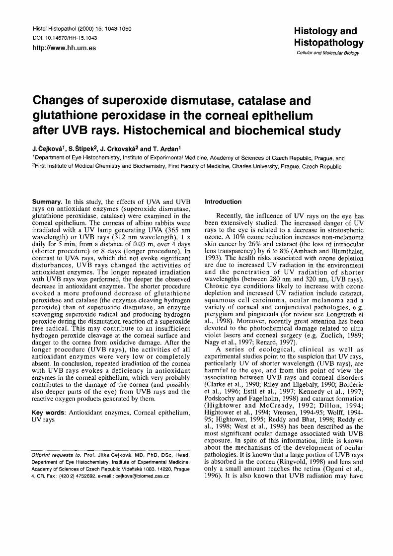
Histochemical examination (Fig. 1)
Normal corneal epithelium: Catalase activity
In the central parts of the corneal epithelium the activity of catalase was high (Fig. l a , e-epithelium). From the corneal center to the corneal periphery the catalase activity decreased, and in the limbal region (Fig. lg) the higher activity was located superficially (arrow). Superoxide dismutase: The activity was high in the whole corneal epithelium; in the central epithelial region (Fig. I d , e-epithelium) the activity was slightly decreased in the basal cell layers which was better seen in the limbal region (Fig. lch, e-epithelium) - white arrows point to slightly decreased activity in basal cell layers.
Shorter irradiation procedure with UVB rays
In the central parts of the corneal epithelium, catalase activity was lacking in the superficial epithelial layers; residual activity remained in the basal cell layers - arrows point to the residual enzyme activity (Fig. Ib). The superoxide dismutase activity was equally decreased throughout the entire corneal epithelium, in the central epithelial region (Fig. l e ) as well as at the corneal
Antioxidant enzymes in the corneal epithelium after UVrays
periphery (Fig. li); the black arrows point to the higher activity in the upper parts of the corneal epithelium and the white arrows point to the basal cell layers where the activity was less pronounced.
Longer irradiation procedure with UVB rays evoked the disappearance of catalase and superoxide dismutase in the flat corneal epithelium (e) (Fig. l c - catalase, Fig. If - su~e rox ide dismutase - both enzvmes in central epitheli'al region). After the longer irrad;Gion procedure, superoxide dismutase was also absent at the corneal periphery (Fig. l j ) . This was in contrast to catalase activity, which was already absent in the limbal region after the shorter irradiation procedure.
The efficacy of UVA rays (shorter and longer irradiation procedure). In contrast to UVB rays, UVA rays did not cause significant changes in the activities of catalase or superoxide dismutase.
Remark: Both methods for the detection of catalase activity used in this study (as described by Lojda et al., 1979 for peroxisomes or according to Frederiks and Bosch, 1995) revealed similar results in the normal corneal epithelium as well as after UV irradiation.
Biochemical investigation
. . % *.&*&?. , .
3 -;*$c, m. " S";%:*- -.
b dt- " "
" '.W\' " --- .+. * C " " ,
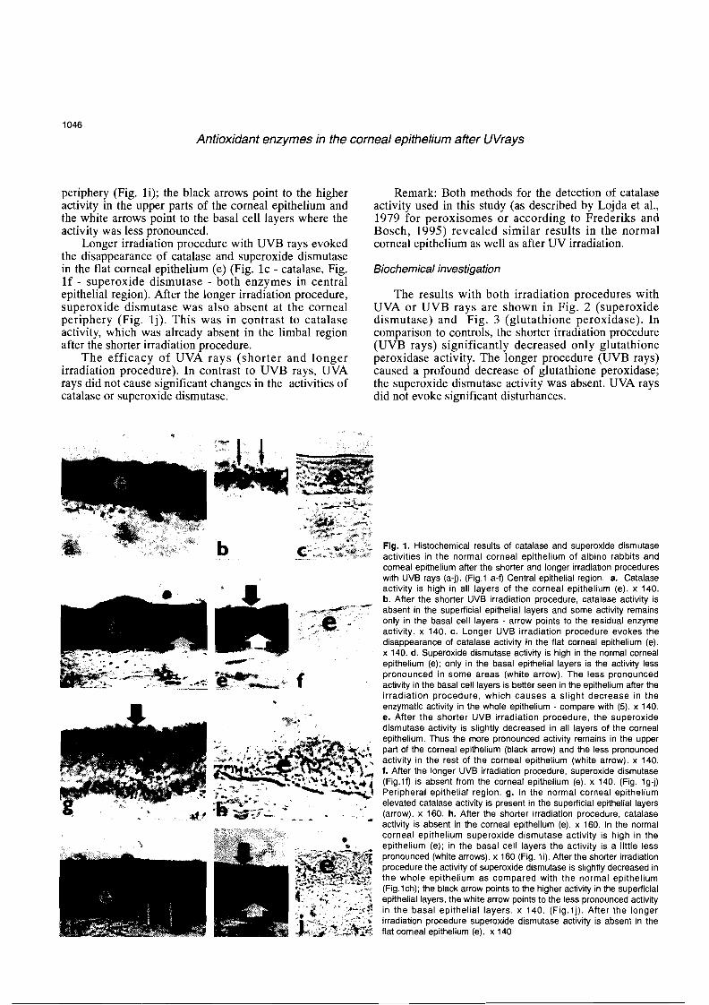
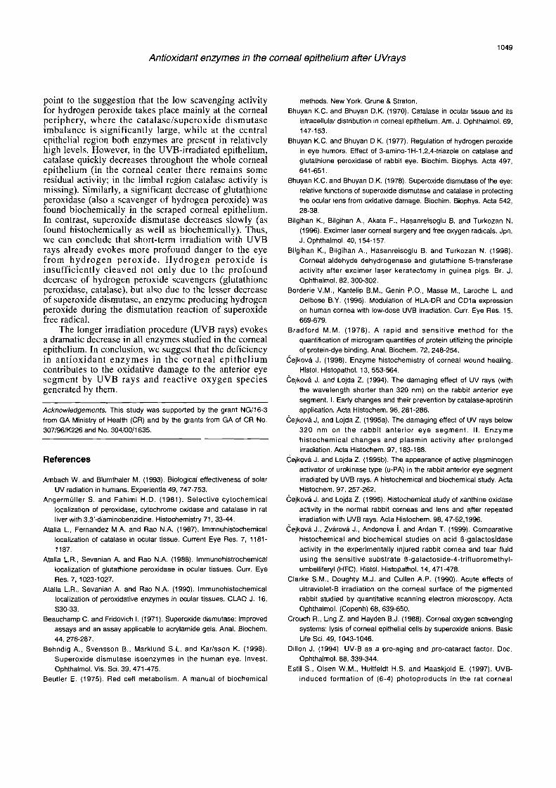
The results with both irradiation procedures with UVA or UVB rays are shown in Fig. 2 (superoxide dismutase) and Fig. 3 (glutathione peroxidase). In comparison to controls, the shorter irradiation procedure (UVB rays) significantly decreased only glutathione peroxidase activity. The longer procedure (UVB rays) caused a profound decrease of glutathione peroxidase; the superoxide dismutase activity was absent. UVA rays did not evoke significant disturbances.
;*sf, %%",G S- .$l:
.-.-g :* ,C'. .V $S$,**
F., 4 - lc -+4! ;g%*:; L; <-
W:" " - - - - . S a
Fig. 1. Histochemical results of catalase and superoxide dismutase activities in the normal corneal epithelium of albino rabbits and corneal epithelium after the shorter and longer irradiation procedures with UVB rays (a-j). (Fig.1 a-f) Central epithelial region. a. Catalase activity is high in all layers of the corneal epithelium (e). X 140. b. After the shorter UVB irradiation procedure, catalase activity is absent in the superficial epithelial layers and some activity remains only in the basal cell layers - arrow points to the residual enzyme activity. X 140. c. Longer UVB irradiation procedure evokes the disappearance of catalase activity in the flat corneal epithelium (e). X 140. d. Superoxide dismutase activity is high in the normal corneal epithelium (e); only in the basal epithelial layers is the activity less pronounced in some areas (white arrow). The less pronounced activity in the basal cell layers is better seen in the epithelium after the irradiation procedure, which causes a slight decrease in the enzymatic activity in the whole epithelium - compare with (5). X 140. e. After the shorter UVB irradiation procedure, the superoxide dismutase activity is slightly decreased in all layers of the corneal epithelium. Thus the more pronounced activity remains in the upper part of the corneal epithelium (black arrow) and the less pronounced activity in the rest of the corneal epithelium (white arrow). X 140. f. After the longer UVB irradiation procedure, superoxide dismutase (Fig.19 is absent from the corneal epithelium (e). X 140. (Fig. Ig-j) Peripheral epithelial region. g. In the normal corneal epithelium elevated catalase activity is present in the superficial epithelial layers (arrow). X 160. h. After the shorter irradiation procedure, catalase activity is absent in the corneal epithelium (e). X 160. In the normal corneal epithelium superoxide dismutase activity is high in the epithelium (e); in the basal cell layers the activity is a little less pronounced (white arrows). X 160 (Fig. li). After the shorter irradiation procedure the activity of superoxide dismutase is slightly decreased in the whole epithelium as compared with the normal epithelium (Fig.lch); the black arrow points to the higher activity in the superficial epithelial layers, the white arrow points to the less pronounced activity in the basal epithelial layers. X 140. (Fig.1~). After the longer irradiation procedure superoxide dismutase activity is absent in the flat corneal epithelium (e). X 140

Antioxidant enzymes in the corneal epithelium after UVrays
Discussion
The eye is exposed to the full range of the solar spectrum and due to the ozone depletion not only to an increased amount of UVA rays, but also to UVB rays, which may have direct effects on reactive oxygen species generation (superoxide and hydroxyl radicals, hydrogen peroxide) (Riley, 1988). Although it has been clearly shown that the cornea contains antioxidant enzymes such as catalase, glutathione peroxidase and superoxide dismutase (Bhuyan and Bhuyan, 1977, 1978; Redmond et al., 1984; Atalla et al., 1987, 1988, 1990; -Cejkovi and Lojda, 1996; Behndig et al., 1998), which play an integral role in controlling reactive oxygen species that can harm cells (e.g. Mittag, 1984), little is known about the influence of UV rays (and reactive oxygen species generated by them) on corneal antioxidant enzymes. Jain et al. (1995) and Bilgihan et al. (1996, 1998) described that the free radical balance of the eye may be changed by excimer laser keratectomy and ultraviolet radiation produced by a 193 nm argon fluoride excimer laser. We found previously that UVB rays evoked changes of various enzymes, including catalase, in the cornea. During the repeated irradiation of the rabbit cornea with UVB, rays the activity of catalase was decreased first in the corneal epithelium, then also in the endothelium and lens epithelium. As for other enzymatic disturbances, the activities of y-glutamyl transpeptidase and Na+-K+- dependent adenosintriphosphatase were decreased in the same corneal layers, and the activities of lysosomal hydrolases (acid glycosidases and proteases) were increased in the cells of all corneal layers ( Cejkovi and
Superoxide dismutase
l I Shor ter-irradiation Longer-irradiation
procedure procedure
Control UVA UVB Control UVA UVB"
Fig. 2. Biochemical results of superoxide dismutase activity in the scraped corneal epithelium: The effects of shorter and longer irradiation procedures with UVA and UVB rays. The shorter repeated UV irradiation of the cornea caused some decrease of superoxide dismutase in the corneal epithelium, however, it was not significant. Longer repeated irradiation (UVB rays) evokes the disappearance of superoxide dismutase activity from the corneal epithelium (UVB 1). UV A rays do not evoke significant disturbances. *: P<0.001 (Bonferroni multiple comparison test - ANOVA); mean ? SD of 8 samples. SOD: superoxide dismutase; U: Sewa Units - see Material and Methods for details.
Lojda, l994,1995a,b, 1966; -cejkovi, 1998; -Cejkovi et al., 1999). This was accompanied by increased corneal hydration, changes of corneal transparency and later the development of corneal inflammation. Because the decrease of catalase activity seemed to play the key role in the initiation of corneal disorders, in this study we systematically analyzed the relationship between UV rays and antioxidant enzymes in the corneal epithelium. However, until now, superoxide dismutase has not been detected within the corneal epithelium. Therefore, great attention was devoted to both the detection of the enzyme activities in the normal corneal epithelium and in the epithelium of the cornea irradiated with UV rays. The qualitative findings of enzyme activities were compared with quantitative investigations in the scraped corneal epithelium.
Superoxide dismutase (SOD; EC 1.15.1.1 .) catalyzes the superoxide radical dismutation to hydrogen peroxide and molecular oxygen (Fridovich, 1978). At least three different forms of superoxide dismutase have been described in mammalian tissues: cytosolic copper-zinc superoxide dismutase, mitochondrial superoxide dismutase (Weisinger and Fridovich, 1973) and the high molecular weight extracellular superoxide dismutase (Marklung, 1982). This enzyme has been identified in the normal rabbit corneal epithelium (and also corneal endothelium, lens, iris, ciliary body and retina) by Bhuyan and Bhuyan (1978) with the method of Marklung and Murklung (1974). Using biochemical methods (as described by Beauchamp and Fridowich, 1971) as well as immunohistochemical techniques Redmond et al. (1984) found superoxide dismutase
Glutathione peroxidase
Shorter- i r radiat ion Longer-irradiation procedure procedure
250 T
Control UVA UVB Control UVA UVB
Fig. 3. Biochemical results of glutathione peroxidase activity in the scraped corneal epithelium: The effects of shorter and longer irradiation procedures with UVA and UVB rays. After shorter irradiation of the cornea with UVB rays, the glutathione activity is significantly decreased in the corneal epithelium; after longer irradiation with UVB rays, enzyme activity is nearly absent. UVA rays do not evoke significant changes in glutathione peroxidase activity (shorter as well as longer irradiation procedures). "': P<0.001 (Bonferroni multiple comparison test - ANOVA); mean ? SD of 8 samples. GPX: glutathione peroxidase; U: one Unit corresponds to oxidation of 1 mmol NADPH per minute.

Antioxidant enzymes in the corneal epithelium after UVrays
(identical to the Cu-Zn superoxide dismutase) in the corneal epithelium of rats, dogs, rabbits and humans. The enzyme quantities detected by radioimmune assay and bioactivity were similar, providing evidence that the enzyme is present as biologically functional. Immuno- histochemical techniques demonstrated the association of the enzyme with the cytoplasm of corneal layers. Immunohistochemically, the distribution of superoxide dismutase in the corneal epithelium (and also in other eye structures) was described in rats by Rao et al. (1985). Behding et al. (1998) describing superoxide dismutase isoenzymes in the human eye found that the cornea and sclera contained several t imes more superoxide dismutase activity than the lens, and the extracellular superoxide dismutase contents were equal or greater than those of CuZn superoxide dismutase.
In this paper, the activity of superoxide dismutase (CuZn superoxide dismutase) was detected biochemically using the method of Sun et al. (1988) modified by Stipek et al. (1995a,b). Results in the normal and irradiated rabbit corneal epithelium (Fig. 2) were compared with the histochemical localization of superoxide dismutase within corneal epithelium (Fig. 1).
Until Frederiks and Bosch (1997), no techniques were available to detect superoxide dismutase activity within tissues. The method developed by these authors is based on the use of a thin film of xanthine oxidase between the glass slide and the unfixed cryostat section and a medium containing hypoxanthine as a source of electrons for the production of superoxide anions. The incubation medium also contains cerium ions to precipitate the hydrogen peroxide product and polyvinyl alcohol to prevent the leakage of soluble andlor loosely bound enzymes from the sections into the incubation medium. The cerium perohydroxides that are formed are visualized with the light microscope in a second step using an incubation medium consisting of 3 ,3 ' - diaminobenzidine, cobalt ions, and hydrogen peroxide, which results in oxidation of the diaminobenzidine to the final insoluble blue reaction product. This methology was first used in this study for investigating the superoxide dismutase activity within the normal corneal epithelium and then following UV irradiation (Fig. 1).
Glutathione peroxidase (GSH: H 2 0 2 oxido- reductase, EC 1.11.1.6.) is a very important enzyme scavenging hydrogen peroxide. Glutathione peroxidase was detected immunohistochemically by Atalla et al. (1988, 1990) in rats in the corneal epithelium (and corneal endothelium, choroid, inner segment of photoreceptors and retina1 pigment epithelium). Biochemically, glutathione peroxidase activity has been described in various eye tissues of rabbits (including the corneal epithelium) by Bhuyan and Bhuyan (1977). Currently, no histochemical method exists for the detection of glutathione peroxidase activity within tissues. Therefore, in the present study we investigated the activity of glutathione peroxidase only bio- chemically, using the method of Beutler (1975). Its activity in the normal scraped corneal epithelium (Fig. 3)
corresponds with the enzyme levels obtained in the rabbit corneal epithelium by Bhuyan and Bhuyan (1977) using the same method.
Catalase ( H 2 0 Z : H 2 0 2 2 oxidoreductase, EC 1.11.1.9) was described biochemically in the normal rabbit corneal epithelium (also corneal endothelium, lens epithelium, ciliary body and retina) by Bhuyan and Bhuyan (1970), and immunohistochemically by Atalla et al. (1987). Ocular catalase was also investigated by Mayer (1980) using Warburg's respirometer, and the results were given in microliter O2 per mg soluble protein. The detection of catalase activity within the normal corneal epithelium and-endothelium of pigmented rabbits was described by Cejkova and Lojda (1995a) using the modified method of Novikoff and Goldfischer as given by Lojda et al. (1979) for peroxisomes. In the present study, for the detection of the enzymatic activity in the corneal epithelium of albino rabbits, we also employed the method of Angermiiller and Fahimi (1981) modified as described by Frederiks and Bosch (1995). The results obtained by both methods were similar. In the albino rabbits used in this study, catalase activity was present in relatively high levels in all epithelial layers in the central epithelial region, whereas towards the corneal periphery the catalase activity was less pronounced. Reddan et al. (1996) described regional differences in catalase activity in the epithelium of the ocular lens. However, in the lens e~ithelium the reaction ~roduct for catalase activitv (and
d \
aiso its immunopero;idase localization) was more intense in peripheral epithelial layers than in cells throughout the central epithelium. Further studies are necessary for the elucidation of the importance of regional differences of catalase activity in the corneal and lens epithelium.
As mentioned above, various methods were employed for the biochemical investigation of superoxide dismutase activity in the normal cornea and therefore the precise comparison of the enzyme activity in individual corneal layers is difficult. However, our present histochemical results with superoxide dismutase activity within the normal corneal epithelium obtained using the method of Frederiks and Bosch (1997) (which allows the analysis of all three types of known superoxide dismutase-Cu/Zn, Mn and extracellular) are in accordance with authors describing the relatively high superoxide dismutase activity in the normal corneal epithelium as compared with the activity of glutathione peroxidase and catalase. In this connection, Behndig et al. (1998) suggested that the large amounts of superoxide dismutase in the cornea (and sclera) may be related to the risk for photochemical production of superoxide in these tissues. Based on the low concentrations of catalase and especially of glutathione peroxidase, Crouch et al. (1988) suggested that the scavenging system for hydrogen peroxide is minimal in the normal cornea. We are in agreement with this opinion; however, our histochemical findings with superoxide dismutase and catalase activities within the corneal epithelium
Antioxidant enzymes in the corneal epithelium after UVrays
point to the suggestion that the low scavenging activity for hydrogen peroxide takes place mainly at the corneal periphery, where the catalase/superoxide dismutase imbalance is significantly large, while at the central epithelial region both enzymes are present in relatively high levels. However, in the UVB-irradiated epithelium, catalase quickly decreases throughout the whole corneal epithelium (in the corneal center there remains some residual activity; in the limbal region catalase activity is missing). Similarly, a significant decrease of glutathione peroxidase (also a scavenger of hydrogen peroxide) was found biochemically in the scraped corneal epithelium. In contrast, superoxide dismutase decreases slowly (as found histochemically as well as biochemically). Thus, we can conclude that short-term irradiation with UVB rays already evokes more profound danger to the eye from hydrogen peroxide. Hydrogen peroxide is insufficiently cleaved not only due to the profound decrease of hydrogen peroxide scavengers (glutathione peroxidase, catalase), but also due to the lesser decrease of superoxide dismutase, an enzyme producing hydrogen peroxide during the dismutation reaction of superoxide free radical.
The longer irradiation procedure (UVB rays) evokes a dramatic decrease in all enzymes studied in the corneal epithelium. In conclusion, we suggest that the deficiency in antioxidant enzymes in the corneal epithelium contributes to the oxidative damage to the anterior eye segment by UVB rays and reactive oxygen species generated by them.
Acknowledgements. This study was supported by the grant NGl16-3 from GA Ministry of Health (CR) and by the grants from GA of CR No. 307196lK226 and No. 30410011635.
References
Ambach W. and Blumthaler M. (1993). Biological effectiveness of solar UV radiation in humans. Experientia 49, 747-753.
Angermiiller S. and Fahimi H.D. (1981). Selective cytochemical localization of peroxidase, cytochrome oxidase and catalase in rat liver with 3,3'-diaminobenzidine. Histochemistry 71, 33-44.
Atalla L., Fernandez M.A. and Rao N.A. (1987). lmmnuhistochemical localization of catalase in ocular tissue. Current Eye Res. 7, 1181- 1187.
Atalla L.R., Sevanian A. and Rao N.A. (1988). lmmunohistrochemical localization of glutathione peroxidase in ocular tissues. Curr. Eye Res. 7, 1023-1027.
Atalla L.R., Sevanian A. and Rao N.A. (1990). lmmunohistochemical localization of peroxidative enzymes in ocular tissues. C M 0 J. 16, S30-33.
Beauchamp C. and Fridovich 1. (1971). Superoxide dismutase: improved assays and an assay applicable to acrylamide gels. Anal. Biochem. 44, 276-287.
Behndig A., Svensson B.. Marklund S.L. and Karlsson K. (1998). Superoxide dismutase isoenzymes in the human eye. Invest. Ophthalmol. Vis. Sci. 39, 471-475.
Beutler E. (1975). Red cell metabolism. A manual of biochemical
methods. New York. Grune & Straton. Bhuyan K.C. and Bhuyan D.K. (1970). Catalase in ocular tissue and its
intracellular distribution in corneal epithelium. Am. J. Ophthalmol. 69, 147-153.
Bhuyan K.C. and Bhuyan D.K. (1977). Regulation of hydrogen peroxide in eye humors. Effect of 3-amino-1 H- l ,2,4-triazole on catalase and glutathione peroxidase of rabbit eye. Biochim. Biophys. Acta 497, 641 -651.
Bhuyan K.C. and Bhuyan D.K. (1978). Superoxide dismutase of the eye: relative functions of superoxide dismutase and catalase in protecting the ocular lens from oxidative damage. Biochim. Biophys. Acta 542, 28-38.
Bilgihan K., Bilgihan A., Akata F.. Hasanreisoglu B. and Turkozan N. (1996). Excimer laser corneal surgery and free oxygen radicals. Jpn. J. Ophthalmol. 40, 154-157.
Bilgihan K., Bilgihan A., Hasanreisoglu B. and Turkozan N. (1998). Corneal aldehyde dehydrogenase and glutathione S-transferase activity after excimer laser keratectomy in guinea pigs. Br. J. Ophthalmol. 82, 300-302.
Borderie V.M., Kantelip B.M., Genin P.O., Masse M., Laroche L. and Delbose B.Y. (1996). Modulation of HIA-DR and CDla expression on human cornea with low-dose UVB irradiation. Curr. Eye Res. 15, 669-679.
Bradford M.M. (1976). A rapid and sensitive method for the quantification of microgram quantities of protein utilizing the principle of protein-dye binding. Anal. Biochem. 72, 248-254.
kejkova J. (1998). Enzyme histochemistry of corneal wound healing. Histol. Histopathol. 13, 553-564.
eejkova J. and Lojda Z. (1994). The damaging effect of UV rays (with the wavelength shorter than 320 nm) on the rabbit anterior eye segment. I. Early changes and their prevention by catalase-aprotinin application. Acta Histochem. 96, 281 -286.
kejkova J, and Lojda Z. (1 995a). The damaging effect of UV rays below 320 nm on the rabbit anterior eye segment. II. Enzyme histochemical changes and plasmin activity after prolonged irradiation. Acta Histochem. 97, 183-188.
kejkova J. and Lojda Z. (1995b). The appearance of active plasminogen activator of urokinase type (U-PA) in the rabbit anterior eye segment irradiated by UVB rays. A histochemical and biochemical study. Acta Histochem. 97, 257-262.
kejkova J. and Lojda Z. (1996). Histochemical study of xanthine oxidase activity in the normal rabbit corneas and lens and after repeated irradiation with UVB rays. Acta Histochem. 98, 47-52,1996.
kejkova J., Zvarova J., Andonova i. and Ardan T. (1999). Comparative histochemical and biochemical studies on acid B-galactosidase activity in the experimentally injured rabbit cornea and tear fluid using the sensitive substrate B-galactoside-4-trifluoromethyl- umbelliferyl (HFC). Histol. Histopathol. 14. 471 -478.
Clarke S.M., Doughty M.J. and Cullen A.P. (1990). Acute effects of ultraviolet-B irradiation on the corneal surface of the pigmented rabbit studied by quantitative scanning electron microscopy. Acta Ophthalmol. (Copenh) 68, 639-650.
Crouch R., Ling Z. and Hayden B.J. (1988). Corneal oxygen scavenging systems: lysis of corneal epithelial cells by superoxide anions. Basic Life Sci. 49, 1043-1046.
Dillon J. (1994). UV-B as a pro-aging and pro-cataract factor. Doc. Ophthalmol. 88, 339-344.
Estil S., Olsen W.M., Huitfeldt H.S. and Haaskjold E. (1997). UVB- induced formation of (6-4) photoproducts in the rat corneal
Antioxidant enzymes in the corneal epithelium after UVrays
epithelium. Acta Ophthalmol. Scand. 75, 120-123. Frederiks W.M., Bosch K.S., Van den Munckhof R.J. and Van der
Noorden C.J. (1994). A quantitative histochemical study of xanthine oxidase activity in rat liver using the cerium capture method in the
presence of polyvinyl alcohol. J. Histochem. Cytochem. 42, 1091- 1096.
Frederiks W.M. and Bosch K.S. (1995). The role of xanthine oxidase in ischemia/reperfusion damage of rat liver. Histol. Histopathol. 10, 111-116.
Frederiks W.M. and Bosch K.S. (1997). Localization of superoxide
dismutase activity in rat tissues. Free Rad. Biol. Med. 22, 241-248. Fridovich 1. (1978). The biology of oxygen radicals. Science 201, 875-
880. Hightower K. and McCready J. (1992). Physiological effects of UVB
irradiation on cultured rabbit lens. Invest. Ophthalmol. Vis. Sci. 33, 1783-1 787.
Hightower K.R., Reddan J.R., McCready J.P. and Dziedzic D.C. (1994). Lens epithelium: a primary target of UVB irradiation. Exp. Eye Res. 59, 557-564.
Hightower K.R. (1995). The role of lens epithelium in development of UV
cataract. Curr. Eye Res. 14, 71 -78. Jain S., Hahn T.W., McCally R.L. and Azar D.T. (1995). Antioxidants
reduce corneal light scattering after excimer keratectomy in rabbits. Lasers Surg. Med. 17, 160-165.
Kehrer J.P. (1993). Free radicals as mediators of tissue injury and disease. Crit. Rev. Toxicol. 23, 21 -48.
Kennedy M., Kim K.H., Harten B., Brown J., Planek S., Meshul C., Edelhauser H., Rosenbaum J.T., Armstrong C.A. and Ansel J.C. (1997). Ultraviolet irradiation induces the production of multiple cytokines by human corneal cells. Invest. Ophthalmol. Vis. Sci. 38. 2483-2491.
Lojda Z., Gossrau R. and Schiebler T.H. (1979). Enzyme histochemistry. A laboratory manual. Springer. Berlin, Heidelberg, New York.
Longstreth J., de Gruijl F.R., Kripke M.L., Abseck S., Arnold F., Slaper H.I., Velders G., Takizawa Y. and van der Leun J.C. (1998). Health risks. J. Photochem. Photobiol. B 46, 20-39.
Marklund S.L. (1982). Human copper-containing superoxide dismutase of high molecular weight. Proc. Natl. Acad. Sci. USA 79, 7634-7638.
Marklund S. and Marklund G. (1974). Involvement of the superoxide anion radical in the autooxidation of pyrogallol and a convenient assay for superoxide dismutase. Eur. J. Biochem. 16, 469-474.
McCord J.M. and Fridovich 1. (1969). Superoxide dismutase. An enzymic finction for erythrocuprein (hemocuprein). J. Biol. Chem. 244, 6049-6055.
Mayer U. (1980). Comparative investigations of catalase activity in different ocular tissues of cattle and man. Albrecht Von Graefes Arch. Klin. Exp. Ophthalmol. 213, 261-265.
Mittag T. (1984). Role of oxygen radicals in ocular inflammation and cellular damage. Exp. Eye Res. 39, 759-769.
Nagy Z.Z., Hiscon P,, Seitz B., Schlotzer-Schrehardt U,, Suveges I. and Naumann G.O. (1997). Clinical and morphological response to UV-B irradiation after excirner laser photorefractive keratectorny. Surv. Ophthalmol. 42, Suppl. 1, S 64-76.
Oguni M., Tamura H., Kato K. and Setogawa T. (1996). Chronic retina1 effects by ultraviolet irradiation, with special reference to superoxide dismutase. Histol. Histopathol. 11, 695-702.
Podskochy A. and Fagerholm P. (1 998). Cellular response and reactive hyaluronan production in UV-exposed rabbit corneas. Cornea 17, 640-645.
Rao N.A., Thaete L.G., Delmage J.M. and Sevanian A. (1985). Superoxide dismutase in ocular structures. Invest. Ophthalmol. Vis. Sci. 26, 1778-1 781.
Read S.M. and Northocote D.H. (1981). Minimization of variation in the response to different proteins of the Coomasie blue G dye-binding assay for protein. Anal. Biochem. 11 6, 53-64.
Reddan J.R., Steiger C.A., Dziedzic D.C. and Gordon S.R. (1996). Regional differences in the distribution of catalase in the epithelium of the ocular lens. Cell Mol. Biol. 42, 209-219.
Reddy G.H. and Bhat K.S. (1998). Synergistic effect of UVB radiation and age on HMPS enzymes in rat lens homogenate. J. Photochem.
Photobiol. B 43, 56-60. Reddy V.N., Giblin F.J., Lin L.R. and Chakrapani B. (1998). The effect of
aqueous humor ascorbate on ultraviolet-B-induced DNA damage in lens epithelium. Invest. Ophthalmol. Vis. Sci. 39, 344-350.
Redmond T.M., Duke E.J., Coles W.H., Simson J.A. and Crouch R.K. (1984). Localization of corneal superoxide dismutase by biochemical and histocytochemical techniques. Exp. Eye Res. 38, 369-378.
Renard G. (1997). Laser and corneal surgery: pathological anatomy. Bull. Soc. Belge Ophthalmol. 266, 23-25.
Riley M.V. (1988). Response to the corneal endothelium to oxidative stress. In: The Cornea. Transactions of the World Congress on the Cornea Ill. Cavamagh H.D. (ed). Raven Press. New York. pp 211- 21 6.
Riley M.V. and Elgebaly S.A. (1990). The release of a neutrophil chemotactic factor from UV-B irradiated rabbit corneas in vitro. Curr. Eye Res. 9, 677-682.
Ringvold A. (1998). Corneal epithelium and UV-protection of the eye. Acta Ophthalmol. Scand. 76. 149-153.
Rose R.C., Richer S.P. and Bode A.M. (1998). Ocular oxidants and antioxidant protection. Proc. Soc. Exp. Biol. Med. 217, 397-407.
Stipek S., Crkovska J. and Dvorak J. (1 995a). Spectrophotometric assay for superoxide dismutase controlled by PC-Programme developed in Lab Windows System. Klin. Biochem. Metab. 3, 93-97 (In Czech).
Stipek S., Mechurova A., Crkovska J., Zima T. and Platenik J. (1995b). Lipid peroxidation and superoxide dismutase activity in umbilical and maternal blood. Biochem. Mol. Biol. Int. 35, 705-71 1.
Sun Y., Oberley L.W. and Li Y. (1988). A simple method for clinical assay of superoxide dismutase. Clin. Chem. 34, 497-500.
Vrensen G.F. (1994-95). UV-B and early cortical and nuclear changes in the human lens. Doc. Ophthalmol. 88, 255-261.
Weisinger R.A. and Fridovich 1. (1973). Mitochondrial superoxide dismutase. Site of synthesis and mitochondrial localisation. J. Biol. Chem. 248, 4793-4796.
West S.K., Duncan D.D., Munoz B., Rubin G.S., Fried L.P., Bandeen- Roche K. and Schein O.D. (1998). Sunlight exposure and risk of lens opacities in a population-based study: the Salisbury Eye Evaluation project. JAMA 280, 71 4-71 8.
Wolff S.P. (1994-95). Cataract and UV radiation. Doc. Ophthalmol. 88, 201 -204.
Zuclich J.A. (1989). Ultraviolet-induced photochemical damage in ocular
tissues. Health Phys. 56, 671-682.
Accepted April 25, 2000


