Superior Mesenteric Artery Originating from the Celiac
3
CASE REPORT Open Access Superior Mesenteric Artery Originating from the Celiac Axis: A Rare Vascular anomaly Michael G Wayne * , Rahul Narang, Suzanne Verzosa and Avram Cooperman Abstract The knowledge of the vascular anatomy of the concerned region is an important prerequisite for planning surgical intervention. The awareness of the existing vascular anomalies enhances the insight regarding that region. We report a patient undergoing preoperative evaluation with CTA finding of Superior Mesenteric Artery (SMA) originating from the celiac artery. This celiac-mesenteric trunk is rare (<1%). Case Presentation A 74-year-old woman was referred by her gastroenterol- ogist with painless jaundice. She presented with several months of decreased appetite and a three week history of light colored stool with dark urine. An endoscopic ultrasound was performed and revealed a hypoechoic, irregular, 3.4 cm mass in the head of the pancreas. The common bile duct and pancreatic duct were obstructed from the mass. No vascular invasion, celiac or peri- celiac lymph nodes were noted. Two biliary stents were placed and no biopsies were taken during the procedure. Prior to considering the patient a candidate for sur- gery, a high resolution computed tomography (CT) scan was performed with pancreatic protocol in non-contrast, arterial and venous phase to determine resectablity. CT scan was consistent with a double duct sign with mark- edly dilated pancreatic and common bile duct and intra- hepatic biliary dilation secondary to mass on the pancreatic head. An interesting variant in anatomy was also identified, which was important for proper surgical planning. The superior mesenteric artery was found to be originating from the celiac axis. (Figure 1, 2, 3) Pancreaticoduodenectomy is utilized selectively in the management of patients with neoplastic lesions of the pancreas and periampullary region. In these patients, the role of CT angiography (CTA) is important in determin- ing tumor respectability and it allows one to evaluate for variant arterial anatomy. Preoperative knowledge of var- iant anatomy can assist in selection of treatment options and facilitate in surgical dissection and avoid iatrogenic injury. The celiac artery supplies the liver, spleen, pancreas, and some of the stomach and duodenum. The superior mesenteric artery (SMA) supplies the small intestine, ascending colon, and a large portion of the transverse colon. Variation of arterial anatomy is common and occurs in nearly half of the population [1]). We report a patient undergoing preoperative evalua- tion with CTA finding of Superior Mesenteric Artery (SMA) originating from the celiac artery. This celiac- mesenteric trunk is rare (<1%), however has been described [2]. In the embryo, the three paired arteries of the trunk ori- ginate from the aorta. Posterior arteries are parietal, lateral arteries are urogenital, and anterior arteries are intestinal. In human embryos the primitive intestinal arteries (vitel- line arteries) are connected by a Tandler’s anterior longitu- dinal anastomosis [3]. When the connection between celiac trunk and SMA remains presents, it tends to form a small vertical arch just behind the body of the pancreas. The rarely reported arterial anastomosis between the celiac trunk and SMA is known as the arc of Bühler’s according to McNulty et al. [4]. An arc of Bühler was identified in 4 patients (3.3%) out of 120 combined celiac and superior mesenteric artery angiograms, in a study by Saad et al. [5]. In one study the arc of Bühler was identified in 14 cases among 340 selective celiac and superior mesenteric arter- iographic studies [6]. They also stated that the arc of Büh- ler between the celiac and superior mesenteric arteries has to be considered as an embryological persistence 10th and 13th primitive arteries, which is associated with the persis- tence of ventral longitudinal anastomosis [2,6]). * Correspondence: [email protected] Pancreas Center at Beth Israel Medical Center, NY 37 Union Square West, 4 th floor, NY 10003, USA Wayne et al. World Journal of Surgical Oncology 2011, 9:71 http://www.wjso.com/content/9/1/71 WORLD JOURNAL OF SURGICAL ONCOLOGY © 2011 Wayne et al; licensee BioMed Central Ltd. This is an Open Access article distributed under the terms of the Creative Commons Attribution License (http://creativecommons.org/licenses/by/2.0), which permits unrestricted use, distribution, and reproduction in any medium, provided the original work is properly cited.
Transcript of Superior Mesenteric Artery Originating from the Celiac

CASE REPORT Open Access
Superior Mesenteric Artery Originating from theCeliac Axis: A Rare Vascular anomalyMichael G Wayne*, Rahul Narang, Suzanne Verzosa and Avram Cooperman
Abstract
The knowledge of the vascular anatomy of the concerned region is an important prerequisite for planning surgicalintervention. The awareness of the existing vascular anomalies enhances the insight regarding that region. Wereport a patient undergoing preoperative evaluation with CTA finding of Superior Mesenteric Artery (SMA)originating from the celiac artery. This celiac-mesenteric trunk is rare (<1%).
Case PresentationA 74-year-old woman was referred by her gastroenterol-ogist with painless jaundice. She presented with severalmonths of decreased appetite and a three week historyof light colored stool with dark urine. An endoscopicultrasound was performed and revealed a hypoechoic,irregular, 3.4 cm mass in the head of the pancreas. Thecommon bile duct and pancreatic duct were obstructedfrom the mass. No vascular invasion, celiac or peri-celiac lymph nodes were noted. Two biliary stents wereplaced and no biopsies were taken during the procedure.Prior to considering the patient a candidate for sur-
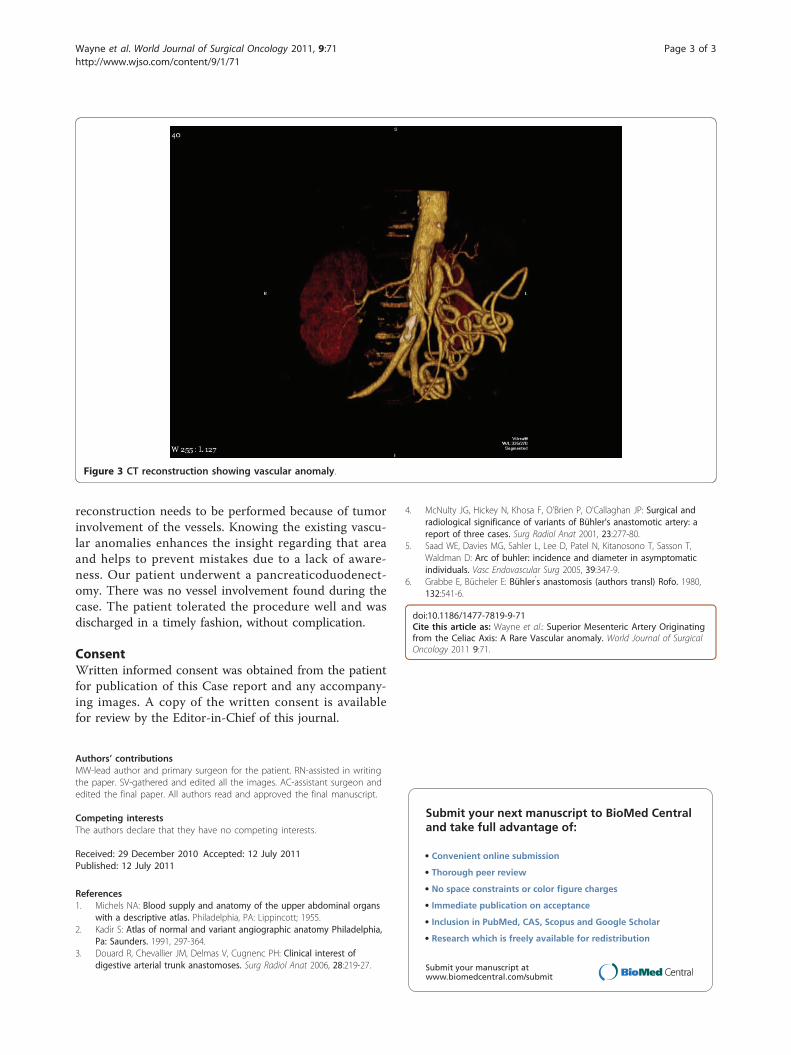
gery, a high resolution computed tomography (CT) scanwas performed with pancreatic protocol in non-contrast,arterial and venous phase to determine resectablity. CTscan was consistent with a double duct sign with mark-edly dilated pancreatic and common bile duct and intra-hepatic biliary dilation secondary to mass on thepancreatic head. An interesting variant in anatomy wasalso identified, which was important for proper surgicalplanning. The superior mesenteric artery was found tobe originating from the celiac axis. (Figure 1, 2, 3)Pancreaticoduodenectomy is utilized selectively in the
management of patients with neoplastic lesions of thepancreas and periampullary region. In these patients, therole of CT angiography (CTA) is important in determin-ing tumor respectability and it allows one to evaluate forvariant arterial anatomy. Preoperative knowledge of var-iant anatomy can assist in selection of treatment options
and facilitate in surgical dissection and avoid iatrogenicinjury.The celiac artery supplies the liver, spleen, pancreas,
and some of the stomach and duodenum. The superiormesenteric artery (SMA) supplies the small intestine,ascending colon, and a large portion of the transversecolon. Variation of arterial anatomy is common andoccurs in nearly half of the population [1]).We report a patient undergoing preoperative evalua-
tion with CTA finding of Superior Mesenteric Artery(SMA) originating from the celiac artery. This celiac-mesenteric trunk is rare (<1%), however has beendescribed [2].In the embryo, the three paired arteries of the trunk ori-
ginate from the aorta. Posterior arteries are parietal, lateralarteries are urogenital, and anterior arteries are intestinal.In human embryos the primitive intestinal arteries (vitel-line arteries) are connected by a Tandler’s anterior longitu-dinal anastomosis [3]. When the connection betweenceliac trunk and SMA remains presents, it tends to form asmall vertical arch just behind the body of the pancreas.The rarely reported arterial anastomosis between the celiactrunk and SMA is known as the arc of Bühler’s accordingto McNulty et al. [4]. An arc of Bühler was identified in 4patients (3.3%) out of 120 combined celiac and superiormesenteric artery angiograms, in a study by Saad et al. [5].In one study the arc of Bühler was identified in 14 casesamong 340 selective celiac and superior mesenteric arter-iographic studies [6]. They also stated that the arc of Büh-ler between the celiac and superior mesenteric arteries hasto be considered as an embryological persistence 10th and13th primitive arteries, which is associated with the persis-tence of ventral longitudinal anastomosis [2,6]).
* Correspondence: [email protected] Center at Beth Israel Medical Center, NY 37 Union Square West, 4th
floor, NY 10003, USA
Wayne et al. World Journal of Surgical Oncology 2011, 9:71http://www.wjso.com/content/9/1/71 WORLD JOURNAL OF
SURGICAL ONCOLOGY
© 2011 Wayne et al; licensee BioMed Central Ltd. This is an Open Access article distributed under the terms of the Creative CommonsAttribution License (http://creativecommons.org/licenses/by/2.0), which permits unrestricted use, distribution, and reproduction inany medium, provided the original work is properly cited.
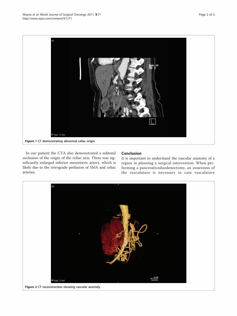
In our patient the CTA also demonstrated a subtotalocclusion of the origin of the celiac axis. There was sig-nificantly enlarged inferior mesenteric artery, which islikely due to the retrograde perfusion of SMA and celiacarteries.
ConclusionIt is important to understand the vascular anatomy of aregion in planning a surgical intervention. When per-forming a pancreaticoduodenectomy, an awareness ofthe vasculature is necessary in case vasculature
Figure 1 CT demonstrating abnormal celiac origin.
Figure 2 CT reconstruction showing vascular anomaly.
Wayne et al. World Journal of Surgical Oncology 2011, 9:71http://www.wjso.com/content/9/1/71
Page 2 of 3
reconstruction needs to be performed because of tumorinvolvement of the vessels. Knowing the existing vascu-lar anomalies enhances the insight regarding that areaand helps to prevent mistakes due to a lack of aware-ness. Our patient underwent a pancreaticoduodenect-omy. There was no vessel involvement found during thecase. The patient tolerated the procedure well and wasdischarged in a timely fashion, without complication.
ConsentWritten informed consent was obtained from the patientfor publication of this Case report and any accompany-ing images. A copy of the written consent is availablefor review by the Editor-in-Chief of this journal.
Authors’ contributionsMW-lead author and primary surgeon for the patient. RN-assisted in writingthe paper. SV-gathered and edited all the images. AC-assistant surgeon andedited the final paper. All authors read and approved the final manuscript.
Competing interestsThe authors declare that they have no competing interests.
Received: 29 December 2010 Accepted: 12 July 2011Published: 12 July 2011
References1. Michels NA: Blood supply and anatomy of the upper abdominal organs
with a descriptive atlas. Philadelphia, PA: Lippincott; 1955.2. Kadir S: Atlas of normal and variant angiographic anatomy Philadelphia,
Pa: Saunders. 1991, 297-364.3. Douard R, Chevallier JM, Delmas V, Cugnenc PH: Clinical interest of
digestive arterial trunk anastomoses. Surg Radiol Anat 2006, 28:219-27.
4. McNulty JG, Hickey N, Khosa F, O’Brien P, O’Callaghan JP: Surgical andradiological significance of variants of Bühler’s anastomotic artery: areport of three cases. Surg Radiol Anat 2001, 23:277-80.
5. Saad WE, Davies MG, Sahler L, Lee D, Patel N, Kitanosono T, Sasson T,Waldman D: Arc of buhler: incidence and diameter in asymptomaticindividuals. Vasc Endovascular Surg 2005, 39:347-9.
6. Grabbe E, Bücheler E: Bühler’s anastomosis (authors transl) Rofo. 1980,132:541-6.
doi:10.1186/1477-7819-9-71Cite this article as: Wayne et al.: Superior Mesenteric Artery Originatingfrom the Celiac Axis: A Rare Vascular anomaly. World Journal of SurgicalOncology 2011 9:71.
Submit your next manuscript to BioMed Centraland take full advantage of:
• Convenient online submission
• Thorough peer review
• No space constraints or color figure charges
• Immediate publication on acceptance
• Inclusion in PubMed, CAS, Scopus and Google Scholar
• Research which is freely available for redistribution
Submit your manuscript at www.biomedcentral.com/submit
Figure 3 CT reconstruction showing vascular anomaly.
Wayne et al. World Journal of Surgical Oncology 2011, 9:71http://www.wjso.com/content/9/1/71
Page 3 of 3