Subject category: Proteins/Cellular metabolism
19
Q D site menasemiquinone in nitrate reductase A 1 Determination of the proton environment of the high stability menasemiquinone intermediate in Escherichia coli nitrate reductase A by pulsed EPR* Stéphane Grimaldi ‡1 , Rodrigo Arias-Cartin §2 , Pascal Lanciano ‡3 , Sevdalina Lyubenova ¶4 , Rodolphe Szenes ‡ , Burkhard Endeward ¶ , Thomas F. Prisner ¶ , Bruno Guigliarelli ‡ , and Axel Magalon § From the ‡ Unité de Bioénergétique et Ingénierie des Protéines (UPR9036), Institut de Microbiologie de la Méditerranée, CNRS & Aix-Marseille Univ, 13009 Marseille, France, § Laboratoire de Chimie Bactérienne (UPR9043), Institut de Microbiologie de la Méditerranée, CNRS & Aix-Marseille Univ, 13009 Marseille, France, ¶ Institut für Physikalische und Theoretische Chemie, Univ Frankfurt, 60438 Frankfurt, Germany * Running title: Q D site menasemiquinone in nitrate reductase A To whom correspondence should be addressed: Stéphane Grimaldi, Unité de Bioénergétique et Ingénierie des Protéines, Institut de Microbiologie de la Méditerranée, CNRS & Aix-Marseille Université, 31, chemin Joseph Aiguier 13009 Marseille, France, Phone: (33) 491 164 557, Fax: (33) 491 164 097, E-mail: [email protected] Keywords : Bioenergetics; Semiquinones; Metalloenzymes; Electron paramagnetic resonance (EPR) Background: Escherichia coli nitrate reductase A highly stabilizes a semiquinone catalytic intermediate Results: Three proton hyperfine couplings to this radical with atypical characteristics are characterized Conclusion: Semiquinone binding is strongly asymmetric and occurs via a single short in-plane H-bond Significance: Learning how the protein environment tunes the semiquinone properties is crucial for understanding the quinol utilization mechanism by energy-transducing enzymes SUMMARY Escherichia coli nitrate reductase A (NarGHI) is a membrane-bound enzyme that couples quinol oxidation at a periplasmically- oriented Q-site (Q D ) to proton release into the periplasm during anaerobic respiration. To elucidate the molecular mechanism underlying such a coupling, endogenous menasemiquinone- 8 intermediates stabilized at the Q D site (MSQ D ) of NarGHI have been studied by high- resolution pulsed EPR methods in combination with 1 H 2 O/ 2 H 2 O exchange experiments. One of the two non exchangeable proton hyperfine couplings resolved in HYSCORE spectra of the radical displays characteristics typical from quinone methyl protons. However, its unusually small isotropic value reflects a singularly low spin density on the quinone carbon carrying the methyl group, which is ascribed to a strong asymmetry of the MSQ D binding mode and consistent with single-sided hydrogen bonding to the quinone oxygen O1. Furthermore, a single exchangeable proton hyperfine coupling is resolved, both by comparing HYSCORE spectra of the radical in 1 H 2 O and 2 H 2 O samples, and by selective detection of the exchanged deuterons using Q-band 2 H Mims ENDOR spectroscopy. Spectral analysis reveals its peculiar characteristics i.e. a large anisotropic hyperfine coupling together with an almost zero isotropic contribution. It is assigned to a proton involved in a short ~ 1.6 Å in-plane hydrogen bond between the quinone O1 oxygen and the N of the His66 residue, an axial ligand of the distal heme b D . Structural and mechanistic implications of these results for the electron-coupled proton translocation mechanism at the Q D site are discussed, in light of the unusually high thermodynamic stability of MSQ D . Quinones are small lipophilic organic molecules found in energy-transducing membranes of all living organisms except methanogens (1). Due to their ability to transfer up to two electrons and two protons, they are widely used in photosynthetic and respiratory electron-transfer chains. Quinones can freely diffuse in the hydrophobic core of lipid membranes. They can http://www.jbc.org/cgi/doi/10.1074/jbc.M111.325100 The latest version is at JBC Papers in Press. Published on December 21, 2011 as Manuscript M111.325100 Copyright 2011 by The American Society for Biochemistry and Molecular Biology, Inc. by guest on April 3, 2018 http://www.jbc.org/ Downloaded from
Transcript of Subject category: Proteins/Cellular metabolism

QD site menasemiquinone in nitrate reductase A
1
Determination of the proton environment of the high stability menasemiquinone intermediate in
Escherichia coli nitrate reductase A by pulsed EPR*
Stéphane Grimaldi‡1
, Rodrigo Arias-Cartin§2
, Pascal Lanciano‡3
, Sevdalina Lyubenova¶4
, Rodolphe
Szenes‡, Burkhard Endeward
¶, Thomas F. Prisner
¶, Bruno Guigliarelli
‡, and Axel Magalon
§
From the ‡Unité de Bioénergétique et Ingénierie des Protéines (UPR9036), Institut de Microbiologie de la
Méditerranée, CNRS & Aix-Marseille Univ, 13009 Marseille, France, §Laboratoire de Chimie
Bactérienne (UPR9043), Institut de Microbiologie de la Méditerranée, CNRS & Aix-Marseille Univ,
13009 Marseille, France, ¶Institut für Physikalische und Theoretische Chemie, Univ Frankfurt, 60438
Frankfurt, Germany
* Running title: QD site menasemiquinone in nitrate reductase A
To whom correspondence should be addressed: Stéphane Grimaldi, Unité de Bioénergétique et Ingénierie
des Protéines, Institut de Microbiologie de la Méditerranée, CNRS & Aix-Marseille Université, 31,
chemin Joseph Aiguier 13009 Marseille, France, Phone: (33) 491 164 557, Fax: (33) 491 164 097, E-mail: [email protected]
Keywords : Bioenergetics; Semiquinones; Metalloenzymes; Electron paramagnetic resonance (EPR)
Background: Escherichia coli nitrate reductase A
highly stabilizes a semiquinone catalytic intermediate
Results: Three proton hyperfine couplings to this
radical with atypical characteristics are
characterized Conclusion: Semiquinone binding is strongly
asymmetric and occurs via a single short in-plane
H-bond Significance: Learning how the protein
environment tunes the semiquinone properties is
crucial for understanding the quinol utilization
mechanism by energy-transducing enzymes
SUMMARY
Escherichia coli nitrate reductase A
(NarGHI) is a membrane-bound enzyme that
couples quinol oxidation at a periplasmically-
oriented Q-site (QD) to proton release into the
periplasm during anaerobic respiration. To
elucidate the molecular mechanism underlying
such a coupling, endogenous menasemiquinone-
8 intermediates stabilized at the QD site (MSQD)
of NarGHI have been studied by high-
resolution pulsed EPR methods in combination
with 1H2O/
2H2O exchange experiments. One of
the two non exchangeable proton hyperfine
couplings resolved in HYSCORE spectra of the
radical displays characteristics typical from
quinone methyl protons. However, its unusually
small isotropic value reflects a singularly low
spin density on the quinone carbon carrying
the methyl group, which is ascribed to a strong
asymmetry of the MSQD binding mode and
consistent with single-sided hydrogen bonding
to the quinone oxygen O1. Furthermore, a
single exchangeable proton hyperfine coupling
is resolved, both by comparing HYSCORE
spectra of the radical in 1H2O and
2H2O
samples, and by selective detection of the
exchanged deuterons using Q-band 2H Mims
ENDOR spectroscopy. Spectral analysis reveals
its peculiar characteristics i.e. a large
anisotropic hyperfine coupling together with an
almost zero isotropic contribution. It is assigned
to a proton involved in a short ~ 1.6 Å in-plane
hydrogen bond between the quinone O1 oxygen
and the N of the His66 residue, an axial ligand
of the distal heme bD. Structural and
mechanistic implications of these results for the
electron-coupled proton translocation
mechanism at the QD site are discussed, in light
of the unusually high thermodynamic stability
of MSQD.
Quinones are small lipophilic organic
molecules found in energy-transducing membranes
of all living organisms except methanogens (1). Due to their ability to transfer up to two electrons
and two protons, they are widely used in
photosynthetic and respiratory electron-transfer chains. Quinones can freely diffuse in the
hydrophobic core of lipid membranes. They can
http://www.jbc.org/cgi/doi/10.1074/jbc.M111.325100The latest version is at JBC Papers in Press. Published on December 21, 2011 as Manuscript M111.325100
Copyright 2011 by The American Society for Biochemistry and Molecular Biology, Inc.
by guest on April 3, 2018
http://ww
w.jbc.org/
Dow
nloaded from

QD site menasemiquinone in nitrate reductase A
2
therefore bind into specific quinone-reactive sites
(Q-sites) of membrane proteins in which they
function as two-electron and proton carriers and are responsible for exchange of reducing
equivalents between different electron-transport
complexes. In this case, the quinones leave the protein after completion of the redox cycle.
Typical examples are the QB site of bacterial
reaction center (RC5) or photosystem II and the Q-
sites (Qo and Qi) of bc1 complex. In contrast, non dissociable quinones can be tightly bound at
specific quinone-reactive sites of proteins in which
they can be involved in electron transfer processes as a prosthetic group. Well-known representatives
of this type include quinones in the QA site in RCs
of purple bacteria and in photosystem II, or the A1
sites in photosystem I (2,3). The different redox states of quinones may
also adopt different conformations in the quinone-
binding pockets, as evidenced for ubiquinone and ubisemiquinone at the QB site of bacterial RC (4).
The functional diversity of Q-sites arises from a
particular tuning of the protein environment. Despite the fact that high-resolution structural data
are available for several Q-sites, how protein-
cofactor interactions relate to and control the
functional properties of the bound quinone is largely unknown. In particular, understanding the
molecular mechanism underlying the coupling
between electron transfer and proton translocation that occurs at dissociable Q-sites requires
obtaining structural information on all three forms,
quinone (Q), semiquinone (SQ), quinol (QH2). For this purpose, high-resolution EPR methods such as
ENDOR (electron nuclear double resonance) and
ESEEM (electron spin echo envelope modulation)
spectroscopies were proven to be valuable by giving detailed structural information on protein-
bound semiquinone intermediates, provided that
this paramagnetic state can be trapped for spectroscopic studies.
E. coli nitrate reductase A (NarGHI) is a
membrane-bound heterotrimeric enzyme induced
by anaerobiosis and the presence of nitrate. Involved in the nitrate respiratory pathway, a
major alternative to the bacterial oxidative
phosphorylation, it couples the oxidation of menaquinols or ubiquinols at a periplasmically
oriented Q-site (named QD) to the cytoplasmic
reduction of nitrate. Thus, both substrate turnovers contribute to the generation of a proton motive
force across the cytoplasmic membrane. NarGHI
contains eight redox-active metal centers (5-9): a
molybdenum cofactor and a Fe4S4 cluster (FS0) in
the nitrate-reducing subunit NarG; one Fe3S4
cluster (FS4) and three Fe4S4 clusters (FS1-3) in
the electron transfer subunit NarH; two low-spin hemes b in the membrane-anchor subunit NarI,
termed bD and bP to indicate their distal and
proximal position to the catalytic site. Importantly, NarI stabilizes an EPR-detectable semiquinone
intermediate of both natural substrates at its quinol
oxidation site QD close to heme bD (10-12).
Remarkably, the resulting menasemiquinone species herein referred to as MSQD has the largest
thermodynamic stability measured so far in
respiratory complexes stabilizing semiquinone intermediates. These peculiar properties render
NarGHI ideally suited for investigating the
molecular factors responsible for the reactivity of
respiratory enzymes towards quinols. While no high-resolution structural data
revealing the binding mode of the natural
quinol/quinone substrate is available, we recently utilized high-resolution EPR techniques on
endogenous MSQD and USQD stabilized in
NarGHI-enriched inner membrane vesicles (IMVs) of E. coli to explore their environment using the
unpaired electron as a probe. The use of ESEEM
and HYSCORE (Hyperfine sublevel correlation)
spectroscopies on either the wild-type enzyme or the enzyme uniformly enriched with
15N nuclei
provided direct evidence for nitrogen ligation to
MSQD and USQD. On the basis of the direct determination of the quadrupolar parameters of the
corresponding interacting 14
N by S-band (~3 GHz)
HYSCORE experiments, we assigned the later to a
N imidazole nitrogen and proposed it to arise
from the heme bD axial ligand His66 (13). The non zero isotropic hyperfine coupling of this nitrogen
suggests that the interaction occurs via a hydrogen
bond, allowing electron spin density to be transferred from the radical to the interacting
nucleus. Interestingly, these experiments did not
support a direct H-bond between MSQD (or USQD)
and Lys86, a residue in the QD site that was previously shown to be essential for quinol
oxidation and menasemiquinone detection (11,14).
Indeed, no evidence for the transfer of a measurable spin density on any other nuclei than
that mentioned above was found. Thus, we
tentatively proposed that a water-mediated interaction is formed between MSQD (or USQD)
and Lys86, consistent with the later being involved
in reactivity towards quinols (13). Moreover, we
have recently shown that a cardiolipin molecule specifically bound to the complex is necessary for
by guest on April 3, 2018
http://ww
w.jbc.org/
Dow
nloaded from

QD site menasemiquinone in nitrate reductase A
3
quinol substrate fixation at the QD site, probably
through the action of one of its acyl chains located
in the vicinity of His66 (15). Clearly, additional information is required to improve our
understanding of the semiquinone binding mode in
the QD site and of its functional tuning by the protein environment.
In this work, high-resolution EPR
techniques have been used to map the environment
and the binding mode of MSQD via the detection of proton hyperfine couplings to the radical. Using
a combination of X-band (~9 GHz)
ESEEM/HYSCORE and Q-band (~34 GHz) Mims ENDOR experiments on MSQD prepared in either
a protonated or a deuterated solvent, one
exchangeable and two non-exchangeable protons
magnetically coupled to the radical are detected. Their detailed characterization allows their
assignment to specific protons in the vicinity of the
radical. Implications of these results for deciphering the semiquinone binding mode and the
catalytic mechanism at the QD site are discussed.
EXPERIMENTAL PROCEDURES
Sample preparation - NarGHI was expressed in
an E. coli nitrate reductase-deficient strain
LCB3063 (RK4353, ΔnapA-B, narG::ery,
narZ::Ω, SpcR) (16) using pVA700 plasmid
(AmpR) (6), which encodes for the narGHJI
operon under control of the tac promoter. Cells
were grown in Terrific Broth under semi-anaerobic
conditions at 37 °C as described in (11) with
ampicillin (100 g.ml-1
) and spectinomycin (50
g.ml-1
) included in the growth medium.
Purified E. coli NarGHI-enriched inner membrane vesicules (IMVs) were used for this
study, allowing to maintain an unmodified lipid
environment and to study the interaction of NarGHI with its endogenous menaquinol
substrate. For this purpose, purified E. coli
NarGHI-enriched IMVs were isolated by differential centrifugation and sucrose gradient
step as described in (11) using a buffer containing
100 mM MOPS and 5 mM EDTA at pH 7.5.
Deuterium-exchanged samples were prepared using the same membrane extraction protocol with
a buffer containing 2H2O (99.9 % atom
2H) instead
of 1H2O. The functionality of NarGHI in our
samples was confirmed spectrophotometrically by
measuring the quinol:nitrate oxidoreductase
activity. Stabilization of the semiquinone at the QD site was achieved through redox titrations under
the same conditions as those used in our previous
works (10,11,13). Redox potentials are given in
the text with respect to the standard hydrogen
electrode. The semiquinone concentration in our samples estimated from the double integration of
their corresponding EPR spectra and by
comparison with a standard (1 mM CuSO4) was
estimated in the range of 10-12 M.
Pulsed EPR/ENDOR Experiments- X-band (~9 GHz) and Q-band (~34 GHz) pulsed
EPR/ENDOR experiments were performed using
a Bruker EleXsys E580-Q spectrometer equipped with an Oxford Instruments CF 935 cryostat.
Spectra were measured at 90 K to avoid
contamination from fast relaxing metal centers
such as FeS centers in NarGHI (13).
For the two-pulse experiments (/2 - -
), the echo intensity was measured as a function
of magnetic field at fixed time interval between the two microwave pulses for field sweep ESE, or
as a function of at fixed magnetic field value for
two-pulse ESEEM.
Two-pulse and four-pulse (/2 - - /2 –
T/2 - - T/2 - /2) ESEEM and HYSCORE (/2 -
- /2 - t1 - - t2 - /2) experiments were performed at a magnetic field corresponding to the maximum intensity of the MSQD two-pulse field
sweep ESE spectrum where all orientations of the
semiquinone with respect to the external magnetic
field contribute, giving rise to powder ESEEM/HYSCORE spectra (see supplemental
data). Spectra were processed using Bruker Xepr
software. Relaxation decays were subtracted (fitting by polynomial functions) followed by zero-
filling and tapering with a Hamming window,
before Fourier transformation which finally gives the spectrum in frequency-domain. All spectra are
shown in absolute value mode. HYSCORE spectra
are presented as contour plots.
Q-band pulsed 2H-ENDOR spectra were
obtained using the Mims (/2--/2-t-/2--echo)
sequence (17). A radio frequency (RF5) pulse
was applied during the time interval t. The RF power was delivered by a 2 kW Dressler solid state
RF amplifier. It was optimized for RF pulse
lengths of 40 s for deuterium Mims ENDOR experiments. Pulsed ENDOR spectra were
recorded at a magnetic field corresponding to the
g position of the nearly axial Q-band EPR signal of MSQD (10). Pulsed EPR/ENDOR spectra were
simulated in the MATLAB environment using the Easyspin software package (release 3.1.0) (18).
Hyperfine and quadrupole interactions- A
hyperfine coupling between a S = ½ radical and a
by guest on April 3, 2018
http://ww
w.jbc.org/
Dow
nloaded from

QD site menasemiquinone in nitrate reductase A
4
nucleus with nuclear spin value I consists in
general of (i) the isotropic contribution Aiso =
20gegnen|0(0)|2/3h where |0(0)|2 is the
electron spin density at the nucleus, ge and gn are
electron and nuclear g-factors, respectively, e and
n are Bohr and nuclear magnetons, respectively, h is Planck’s constant, and (ii) the anisotropic
contribution described by the traceless dipolar
coupling tensor T~
. In most cases, T~
can be assumed to be axial, with principal values (– T, –
T, 2T).
The hyperfine couplings of different isotopes of the same element are proportional to a
very good approximation to the corresponding gn
values. In this study, the direct and simultaneous determination of Aiso and T of the protons
interacting with MSQD were derived from the
analysis of HYSCORE cross-peak contours as
detailed in the supplemental data (19). A
2H nucleus has a quadrupole moment
which interacts with the electric field gradient
(EFG) at the nucleus. The components of the EFG tensor are defined in its principal axis system and
ordered according to qZZ qYY qXX. This traceless tensor can then be fully described by only
two parameters: (i) the 2H nuclear quadrupole
coupling constant = |e2qZZQ/h|, where e is the
charge of electron, Q is the 2H nuclear electric
quadrupole moment, (ii) the asymmetry parameter
= |qYY-qXX/qZZ|. is a measure of the strength of the interaction between the nuclear quadrupole
moment and the EFG at the 2H nucleus site due to
anisotropic charge distribution around the nucleus
whereas is a measure of the deviation of this
distribution from axial symmetry. Thus, the EFG is related to the specific binding geometry. Its
components can, therefore, be used to obtain
detailed information on hydrogen bonds (20-26).
In this study the parameters and of the 2H
interacting with MSQD were estimated by simulation of the Q-band
2H Mims ENDOR
spectrum.
RESULTS
X-band pulsed EPR (Field sweep, two-pulse
ESEEM) - X-band field sweep ESE spectra of
NarGHI-enriched IMVs were recorded at 90 K in samples redox poised at ~ -100 mV prepared either
in 1H2O or
2H2O. They show a single line from the
MSQ stabilized at the QD site of NarGHI with g ~ 2.0045 and the width ~ 0.8 mT in
1H2O (13).
Replacement of 1H2O by
2H2O decreases the line
width by less than 0.1 mT (Fig. 1A). The weakness
of this effect is due to the primary contribution to
line shape of the g-tensor anisotropy, which was previously resolved using numerical simulation of
the MSQD Q-band EPR spectrum (10). The two-
pulse spin echo decay of the radical measured in 1H2O at 90 K is depicted in Figure 1B. It mainly
shows the modulation associated with weakly
coupled protons in the immediate environment,
with Zeeman frequencies I(1H) around 14.7 MHz.
A characteristic deep additional modulation of the echo intensity appears in the sample prepared in 2H2O (Fig. 1B). Fourier transformation of this echo
envelope reveals that the major contribution to the
deep variations occurs at the frequency ~ 2.3 MHz, corresponding to the Zeeman frequency of
deuterium (not shown). These results give a first
indication of solvent accessibility and 1H/
2H
exchange around MSQD. To increase spectral
resolution and thus provide more detailed
information about the proton environment of MSQD, HYSCORE experiments were carried out
and are shown below.
X-band 1H HYSCORE - The low-frequency
part of the X-band HYSCORE spectra of MSQD were previously shown and analyzed in details.
They revealed cross-peaks arising from a single 14
N hyperfine coupling assigned to the heme bD ligand His66 residue (12,13). In addition to these 14
N signals, several cross-features from protons
symmetrically positioned with respect to the 1H
Zeeman frequency (I(1H) ~ 14.7 MHz) are clearly
resolved in the 10-20 MHz frequency range in the (+,+) quadrant of these spectra (Fig. 2A). This
indicates that several protons are magnetically
coupled to the radical. Appearance of these correlations in the (+,+) quadrant indicates that the
corresponding hyperfine couplings for a given
proton satisfy the relationships T+2Aiso <<
4I(1H) (27). To further analyze the spectrum and
discriminate between exchangeable and non
exchangeable features, HYSCORE experiments
were also performed under the same conditions in the sample prepared in
2H2O. Figure 2 shows the
proton region of the corresponding HYSCORE
spectra recorded with = 204 ns in 1H2O (Fig. 2A)
or 2H2O (Fig. 2B). In addition to the diagonal peak
at I(1H) ~ 14.7 MHz, four pairs of cross-features
located symmetrically relative to the diagonal are well resolved in the spectrum shown in Figure 2A.
They are designated 1, 1’, 2, 2’, 3, 3’, 4 and 4’.
The ridges 2-2’ exhibit the smallest resolved
hyperfine splitting, of the order of ~ 2 MHz while
by guest on April 3, 2018
http://ww
w.jbc.org/
Dow
nloaded from

QD site menasemiquinone in nitrate reductase A
5
the largest one is observed for cross-peaks 1-1’.
Cross-ridges 3-3’ possess the most extended
anisotropic contour, with the largest deviation from the diagonal, while cross-peaks 4-4’ deviate
significantly from the normal to the diagonal.
These two features indicate a significant anisotropic hyperfine component. Contours 1-1’
and 2-2’ are approximately normal to the diagonal,
suggesting a smaller anisotropy. Cross-peaks 1 and
4 partially overlap. Cross-peaks 3-3’ and 4-4’ completely
disappear in the proton HYSCORE spectrum
measured in 2H2O, demonstrating that they are
produced by at least one exchangeable proton (Fig.
2B). In contrast, cross-peaks 1-1’ and 2-2’ still
appear in the spectrum measured in 2H2O, showing
that they arise from non exchangeable (i.e. covalently bound) protons.
Quantitative analysis of the cross-peak
contour line-shapes indicates that cross-peaks 3, 3’, 4, 4’ are produced by a single exchangeable
proton (Supplemental Fig. S2 and Supplemental
Table S1). Hence, HYSCORE signals derive from three protons coupled to MSQD: H1 (1-1’), H2 (2-
2’) and H3 (3-3’-4-4’). Among them, H3 is
exchangeable. The isotropic (Aiso) and anisotropic
(T) components of the three 1H hyperfine tensors
are given in Table 1. The magnitude of the
hyperfine couplings deduced from the analysis of
HYSCORE spectra are consistent with our previous preliminary observations of
1H
continuous wave ENDOR resonances with
corresponding estimated hyperfine couplings A1 ~ 5.7 MHz (H3) and A2 ~ 3.3 MHz (H2) (10,11).
X-band 1H four-pulse ESEEM - Additional
information about the interacting protons was obtained from one-dimensional four-pulse ESEEM
spectra, which are particularly useful for the
observation of proton sum combination lines with improved resolution (27,28). The four-pulse
ESEEM spectrum of MSQD in 1H2O buffer
contains two well resolved lines in the region of
the proton around 2I(1H) as shown in Fig. 3A.
The most intense line appears exactly at the
2I(1H) frequency and represents the contribution
of weakly coupled protons from the protein environment. In addition, the spectrum exhibits a
peak of lower intensity shifted from 2I(1H) to
higher frequencies by ~ 1.2 MHz. This shifted
peak completely disappears in the spectra of the sample prepared in
2H2O (Fig. 3B). This indicates
that the line shifted from 2I(1H) arises from an
exchangeable proton. The shift observed in the
four-pulse ESEEM is well described by
= 9T2/16I(
1H) (Eq. 1)
from which the anisotropic component T of the
hyperfine coupling can be estimated (see
Supplemental data). The shift of ~ 1.2 MHz corresponds to T = 5.7 MHz, which is in very good
agreement with the corresponding value
determined for H3 from the analysis of the HYSCORE spectra. The expected shifts from H1
and H2 (0.06 and 0.05 MHz, respectively) are too
small to be resolved in a four-pulse ESEEM
spectrum (Supplemental Table S2). The proton sum combination peak therefore confirms the
assignment made in the HYSCORE spectra for the
exchangeable proton and the hyperfine coupling determined from these spectra.
Q-band 2H Mims ENDOR – Further details
concerning exchangeable protons coupled to
MSQD were obtained through the use of pulsed 2H
ENDOR spectroscopy. Figure 4 shows the Q-band 2H Mims ENDOR spectrum of MSQD in NarGHI-
enriched IMVs prepared in 2H2O. It has been
recorded at a magnetic field value corresponding
to the maximum intensity of the nearly axially symmetric Q-band EPR spectrum of MSQD with g-
tensor principal values gx = 2.0061, gy = 2.0051, gz
= 2.0023 ± 0.0001 (Fig. 4, top) (10). The ENDOR
spectrum exhibits two pairs of well resolved intense lines located symmetrically with respect to
the 2H nuclear Larmor frequency (I(
2H) ~ 7.84
MHz), and subject to the nuclear quadrupole
interaction. The splitting of their center away from the Larmor frequency (~ 0.8 MHz) is determined
by the hfc and the splitting within the pair (~ 0.13
MHz) is given by the quadrupole interaction. The
same holds for the two pairs of less intense lines resolved in the spectrum shown in Figure 4,
separated by ~ 1.5 MHz and splitted each by ~
0.26 MHz. The hyperfine coupling values of ~ 0.8
MHz and ~ 1.5 MHz are first estimates of the A
and A components of an almost purely dipolar hyperfine tensor. They are very close to those
found for H3 and scaled to the 2H nucleus, i.e. A
= 0.87 and A = 1.77 MHz. Similarly, a nuclear
quadrupole coupling constant k ~ /4/3 0.13 ~
0.173 MHz can be estimated from the 0.13 MHz splitting of the two most intense doublets. A
numerical simulation of the spectrum is shown in
Figure 4. It was obtained using the proton
by guest on April 3, 2018
http://ww
w.jbc.org/
Dow
nloaded from

QD site menasemiquinone in nitrate reductase A
6
hyperfine coupling values of H3 deduced from the
analysis of the HYSCORE spectra and rescaled by
the factor gn(1H)/gn(
2H) ~ 6.5. This best simulation
was obtained assuming that the g-, A- and Q-
tensors are collinear, with quadrupole parameters
= (0.176 ± 0.004) MHz and = 0.20 ± 0.05. Finally, this procedure allowed us to select
unambiguously the right set (Aiso, T) for H3 from the two alternatives (see Table 1). Overall, the data
show that a single exchangeable proton is coupled
to the radical, in agreement with the analysis above.
DISCUSSION
A single exchangeable proton coupling with
peculiar hyperfine coupling characteristics - Our
data clearly show the presence of a single exchangeable proton in the vicinity of MSQD
characterized by Aiso = 0.06 MHz and T = 5.73 MHz. Such hyperfine coupling constants are in the
range of those measured for exchangeable protons
coupled to protein-bound semiquinones and assigned to protons hydrogen-bonded to the
quinone carbonyl oxygens. Typical examples
include exchangeable proton couplings to the menasemiquinone stabilized at the QH site of the
aa3 menaquinol oxidase from Bacillus subtilis (T
= 5.6 MHz and Aiso = 5.4 MHz) or to the photoaccumulated phyllosemiquinone A1 in
Thermococcus elongatus photosystem I (T ~ 3.7
MHz and Aiso ~ 0.1 MHz) (29,30). Thus, we assign H3 to a proton involved in H-bonding to
one of the MSQD carbonyl oxygens. However, the
hyperfine coupling tensor to H3 has atypical properties as it combines both an almost zero
isotropic hyperfine coupling constant Aiso and a
large anisotropic part T. The magnitude of the H-
bond tensor is determined by the geometry of the H-bond. A small Aiso is expected when the H-bond
lies in the molecular plane due to small overlap
between the hydrogen 1s orbital and the oxygen 2pz orbital forming part of the semiquinone
SOMO. Almost purely anisotropic hyperfine
tensors have thus been observed for in-plane hydrogen-bonded protons to unsubstituted
quinones measured in alcoholic solvent, while the
corresponding T values do not exceed 3 MHz (23,24,31,32). The T value measured for H3 is one
of the largest measured so far for a proton hydrogen bonded to a semiquinone. According to
DFT calculations, the large T value of H3 is
predicted to account for a short hydrogen-bond
length, typically in the range of 1.3-1.4 Å (22). In
this case, Sinnecker et al. have shown that the
point dipole model does not work due to the increased covalent character of the H-bond that is
not covered in the point dipole approximation. A
more reliable alternative approach is to evaluate H-bond distances from the nuclear quadrupole
coupling constant of 2H (20-22,24). As empirically
proposed by Soda and Chiba (25) and Hunt and
Mackay (26), it has been shown that the nqc of 2H
nuclei H-bonded to semiquinones follows a r-3
(O--
-2H) dependence of the form
kHz
HOr
ba
23 (Eq. 2)
where a and b are empirical parameters (22). Using a = 319 kHz and b = 607 kHz·Å
3 (20) and
the value of k = 176 ± 4 kHz deduced from our
work, we obtain from Eq. 2 a bond length of r(O---2H) = 1.62 ± 0.02 Å. This value is in the range of
short hydrogen bonds for biological systems. For
instance, it is similar to that formed from the carbonyl oxygen O4 of the ubisemiquinone QA
.- in
the RC from Rhodobacter sphaeroides R-26 to the
imidazole nitrogen N of His M219 (r(O---2H) =
1.60 ± 0.04 Å) (20).
Assignment of the non exchangeable proton couplings - In addition to H3, two non
exchangeable proton couplings H1 and H2 are
clearly resolved in the HYSCORE spectra of MSQD measured in
2H2O. For their assignment, we
rely on previous experimental and theoretical
studies on phylloquinones (called also vitamin K1)
and menaquinones (vitamin K2) radicals examined in liquid and solid organic solvents or in proteins
(23,29,30,33-37). Indeed, these quinones share the
same naphtoquinone ring structure methylated at the second position but differ in their aliphatic side
chain attached at the 3-position (see Fig. S0). It has
been shown both experimentally and theoretically
that the aliphatic side chain properties have only a weak influence on the proton hyperfine coupling
tensors measured in organic solvents
(23,33,34,38,39). From these previous studies, it is evident that the non exchangeable proton
couplings from H1 and H2 > 2 MHz originate
either from the ring methyl protons, the -
methylene isoprenyl protons or from -protons directly attached to the quinone ring. Due to rapid
rotation of the methyl group even at low temperature, methyl protons of vitamin K
molecules have equal hyperfine tensors and give
by guest on April 3, 2018
http://ww
w.jbc.org/
Dow
nloaded from

QD site menasemiquinone in nitrate reductase A
7
prominent ENDOR/ESEEM signals. They are
characterized by an almost axial hyperfine tensor,
a predominant isotropic hyperfine coupling value in the range of 6.8-12.3 MHz, and a characteristic
relative hyperfine anisotropy (A||-A)/Aiso in the
range of 0.26-0.45 with A|| > A > 0. The later value increases up to 0.76 for the methyl protons
of the asymmetrically bound ubisemiquinone QA
in the RC from Rhodobacter sphaeroides (2). Based on these results, we assign H1 to the methyl
protons of MSQD, with Aiso = +5.53 MHz and T =
+1.25 MHz leading to a (A||-A)/Aiso value of ~ 0.68 (Fig. 5).
Hyperfine data for -methylene protons
and ring protons in semiquinones are less abundant. Unlike the methyl protons, the
methylene protons are not expected to rotate freely (40). They are also characterized by a nearly axial
hyperfine tensor with A|| > A > 0 (40,41). In contrast, a high degree of anisotropy is expected
for a ring proton hyperfine tensor due to the short distance of the proton and the spin density
(30,40). Because of the axial symmetry of its
hyperfine coupling tensor, we tentatively assign
H2 to one of the -methylene isoprenyl protons with Aiso = +0.96 MHz and T = +1.18 MHz (Fig.
5). Overall, the 1H hyperfine coupling constants
determined in this work are consistent with those
estimated from our previous cw X-band 1H
ENDOR studies of MSQD (10,11).
A strongly asymmetric spin distribution in MSQD - The
1H hyperfine coupling constants are an
excellent probe of the asymmetry of the spin
density distribution in the quinone. In particular, the predominant isotropic component, Aiso, of
hyperfine coupling to the methyl protons is
directly proportional to the unpaired spin density
in the orbital on the adjacent -carbon (C), as described by the McConnell relation, Aiso =
CB2/2, where B2 has been taken in the range from
120 to 212 MHz (42). The corresponding value for the methyl protons of MSQD (Aiso = 5.53 MHz) is
the smallest one ever reported for these protons for
vitamin K molecules bound to proteins or in protic solvents (23,29,30,33,34,36,38,39). In particular,
the spin density on the C at the 2-position which is sensed by the methyl protons is reduced by ~ 30
% in MSQD as compared to the symmetrically
hydrogen bonded MSQ-4 prepared in 2-propanol (39). This decrease can be explained by a strong
asymmetry of hydrogen bonding to the carbonyl
oxygens of MSQD in NarGHI, which leads to a
redistribution of both the spin density and charges
within the quinone ring (2,43,44). Indeed, a
stronger hydrogen bond to the carbonyl oxygen O1, compared to oxygen O4, is expected to lead to
an increase of spin density on carbon 3 but a
decrease of the spin density on carbon 2 as observed here for MSQD. A similar but less
pronounced spin density shift has been proposed
for the menasemiquinone-9 in the QA site of the
RC from Rhodopseudomonas viridis for which the methyl protons isotropic hyperfine coupling
constant is about 6.8 MHz (39). In contrast, the
large isotropic constants for methyl protons of the MSQ-7 in the aa3 menaquinol oxidase from
Bacillus subtilis (Aiso ~ 11.0 MHz) (29) or of the
radical anion of phylloquinone in the A1 site of
photosystem I from Thermosynechococcus elongatus (Aiso = 9.8 MHz) (30) suggest that a
stronger hydrogen-bond to oxygen O4 is formed
compared to oxygen O1, a strongly asymmetric binding mode reverse to that observed in NarGHI.
Model for MSQD binding to NarGHI and mechanistic implications - Altogether, the data
inferred from the present work allow to refine the
MSQD binding model previously proposed (13).
We conclude that the asymmetrical spin density distribution in MSQD is primarily due to the strong
hydrogen-bond formed to the O1 oxygen of MSQD
involving the exchangeable H3 proton coupling (Fig. 5). Hence, the later appears mostly
responsible for the transfer of spin density from
the radical to the interacting nitrogen nucleus that was deduced from the measurement of a small
14N
isotropic hyperfine coupling of Aiso ~ 0.8 MHz to
MSQD using HYSCORE spectroscopy. Based on
the measurement of its nuclear quadrupole
parameters by S-band HYSCORE spectroscopy (
= 0.49, = 0.50), this nucleus was assigned to the
N imidazole nitrogen from the heme bD axial ligand His66 (13). In addition, this model is
consistent with the relatively small value found
for this imidazole nitrogen that is close to that predicted by DFT calculations on imidazoles
forming a strong in-plane hydrogen bond to one of
the benzosemiquinone oxygen atoms (45). Finally, the presence of the positive charges on the nearby
heme Fe2+
ion may also contribute to the observed
asymmetrical spin distribution in MSQD.
Interestingly, a single strong and highly ordered H-bond to MSQD is detected in the present
work by using both 1H HYSCORE and
2H Q-band
ENDOR spectroscopies. In the most recent studies
by guest on April 3, 2018
http://ww
w.jbc.org/
Dow
nloaded from
QD site menasemiquinone in nitrate reductase A
8
of protein-bound semiquinones, the radicals appear
to be coupled to at least two exchangeable protons
assigned to those involved in H-bonds (20,29,46-49). One-sided H-bond was resolved only for the
photoaccumulated phyllosemiquinone A1 in
photosystem I (30,50). Based on their experimental and theoretical results, Niklas et al.
have shown that the single detected short ~ 1.64 Å
H-bond can fully account for the observed
asymmetry in the spin density distribution of the SQ in the A1 site (30). Remarkably, both the H-
bond length and the ~ 30 % variation of the spin
density on the C at the 2-position of the phyllosemiquinone in the A1 site with respect to
the symmetrically hydrogen-bonded radical are comparable to the corresponding values measured
for MSQD. Thus, our results indicate that MSQD
most likely binds to the protein via a one-sided H-bond.
Consequently, our present work indicates
that the O4 carbonyl oxygen of MSQD is not protonated in this intermediate state, showing that
at least one proton has to be released to the
periplasm consecutively to the first electron
transfer step to heme bD. This step likely involves Lys86, a residue located at the protein surface, at
the entrance of the QD cavity, and which is
required for correct binding of quinol analogues and for semiquinone detection at the QD site of
NarGHI (11,13,14). Thus, we speculate that Lys86
could be a direct hydrogen bond donor to the
quinol molecule, facilitating proton abstraction at the quinol O4 oxygen and, concomitantly, the first
electron transfer step. This would be accompanied
by a movement of Lys86 away from the substrate, leading to an asymmetric binding mode of the
semiquinone intermediate and allowing for proton
release toward the periplasm (Fig. 5). Whether quinol deprotonation at the O1 oxygen is coupled
to the first or the second electron transfer step
remains unclear. Additional studies aimed at
measuring the pH dependence of the redox reactions occurring at the QD site will be useful to
further understanding how quinol deprotonation
and electron transfer are synchronized at the QD site. Such studies are currently being performed in
our laboratories
Role of the protein environment in quinol
utilization and semiquinone stabilization - The
high stabilization of MSQD in NarGHI is directly
related to the redox potentials of the redox transitions MQH2/MSQ (Em,7.5 = -150 mV) and
MSQ/MQ (Em,7.5 = -40 mV), which are both
thermodynamically favourable for electron transfer
to the bD heme (10). We questioned whether the unusually high stabilization of the semiquinone
state at the NarGHI QD site can be related to the
strongly asymmetric binding mode of MSQD mainly due to the short in-plane H-bond formed to
the radical. Remarkably, the two other protein
bound semiquinones with redox properties most
strongly affected by the protein environment compared to the corresponding species in alcoholic
solvents are the very low potential (Em ~ -750 mV)
phyllosemiquinone anion A1.- in photosystem I and
the high affinity ubisemiquinone at the QH site of
cytochrome bo3 which has a high stability, albeit
10 times smaller than that measured for MSQD. In
both cases, the semiquinones interact with the protein environment in a very asymmetric manner
(46,51-53). This contrasts to the much lower
stability of the more symmetrically bound semiquinones at the QB site of photosynthetic
bacterial RC (48,54) or at the Qi site in R.
sphaeroides bc1 complex (47,55). In addition, the strong H-bond to MSQD is expected to withdraw
electron density and stabilize the semiquinone
form, thereby raising the redox potential of the
second oxidation step from semiquinone to quinone, as experimentally measured.
Consequently, we hypothesize that the atypical
binding mode of MSQD could strongly contribute to its unusual redox properties. In addition, other
effects such as the electrostatic environment of the
nearby protein should also be taken into account. Evaluating their respective contribution to the
MSQD redox properties requires further work.
Finally, the functional implications of this high
stabilization remain to be established. It has been shown both experimentally and
theoretically that the presence of bulky substituents
on the quinone ring force hydrogen bond formation out-of-plane, thereby increasing
simultaneously the isotropic and anisotropic
coupling of the hydrogen-bonded protons (32,56).
Remarkably, measurement of a small Aiso and a simultaneous large T value for the proton
hydrogen-bonded to MSQD indicates that the
protein environment around the radical strongly constraints the geometry of the hydrogen bond by
maintaining a short in-plane H-bond to the radical,
thus leading to the observed peculiar hyperfine coupling characteristics. We have recently shown
that an endogenous ubisemiquinone radical
(USQD) can also be stabilized at the NarGHI QD
site (12). It binds to the protein via a H-bond to the
by guest on April 3, 2018
http://ww
w.jbc.org/
Dow
nloaded from

QD site menasemiquinone in nitrate reductase A
9
same nitrogen as menasemiquinone does, i.e. most
likely His66 N. The similar 14
N HYSCORE pattern observed for both radicals suggests that the
H-bond involved in binding USQD has similar
characteristics to that detected in the present study. This provides further support for its involvement
in ubisemiquinone stabilization at the QD site.
Finally, our work indicates that the protein environment counteracts the effect of the presence
of bulky substituents to impose an atypical binding
mode. Further work is in progress in our laboratories to evaluate the importance of the
quinone substituents to accommodate and utilize
various substrates at this Q-site and to stabilize
semiquinone intermediates.
Concluding Remarks
From the experiments reported in this
work, we conclude that MSQD is involved in a
single strong in-plane and highly ordered H-bond with a solvent exchangeable proton. This strongly
asymmetric binding causes a shift of the electron
spin density over the quinone ring consistent with
the formation of a strong hydrogen bond to the quinone carbonyl oxygen O1. This peculiar
binding mode could strongly contribute to the
unusual redox properties of MSQD.
by guest on April 3, 2018
http://ww
w.jbc.org/
Dow
nloaded from

QD site menasemiquinone in nitrate reductase A
10
REFERENCES
1. Nicholls, D. G., and Ferguson, S. J. (2002) Bioenergetics 3, Academic press, London 2. Lubitz, W., and Feher, G. (1999) Appl. Magn. Reson. 17, 1-48
3. Srinivasan, N., and Golbeck, J. H. (2009) Biochim. Biophys. Acta 1787, 1057-1088
4. Stowell, M. H., McPhillips, T. M., Rees, D. C., Soltis, S. M., Abresch, E., and Feher, G. (1997) Science 276, 812-816
5. Lanciano, P., Savoyant, A., Grimaldi, S., Magalon, A., Guigliarelli, B., and Bertrand, P. (2007) J.
Phys. Chem. B 111, 13632-13637 6. Guigliarelli, B., Magalon, A., Asso, M., Bertrand, P., Frixon, C., Giordano, G., and Blasco, F.
(1996) Biochemistry 35, 4828-4836
7. Blasco, F., Guigliarelli, B., Magalon, A., Asso, M., Giordano, G., and Rothery, R. A. (2001) Cell
Mol. Life Sci. 58, 179-193 8. Rothery, R. A., Blasco, F., Magalon, A., and Weiner, J. H. (2001) J. Mol. Microbiol. Biotechnol.
3, 273-283
9. Bertero, M. G., Rothery, R. A., Palak, M., Hou, C., Lim, D., Blasco, F., Weiner, J. H., and Strynadka, N. C. (2003) Nat. Struct. Biol. 10, 681-687
10. Grimaldi, S., Lanciano, P., Bertrand, P., Blasco, F., and Guigliarelli, B. (2005) Biochemistry 44,
1300-1308 11. Lanciano, P., Magalon, A., Bertrand, P., Guigliarelli, B., and Grimaldi, S. (2007) Biochemistry 46,
5323-5329
12. Arias-Cartin, R., Lyubenova, S., Ceccaldi, P., Prisner, T., Magalon, A., Guigliarelli, B., and
Grimaldi, S. (2010) J. Am. Chem. Soc. 132, 5942-5943 13. Grimaldi, S., Arias Cartin, R., Lanciano, P., Lyubenova, S., Endeward, B., Prisner, T. F.,
Magalon, A., and Guigliarelli, B. (2010) J. Biol. Chem. 285, 179-187
14. Bertero, M. G., Rothery, R. A., Boroumand, N., Palak, M., Blasco, F., Ginet, N., Weiner, J. H., and Strynadka, N. C. (2005) J. Biol. Chem. 280, 14836-14843
15. Arias-Cartin, R., Grimaldi, S., Pommier, J., Lanciano, P., Schaefer, C., Arnoux, P., Giordano, G.,
Guigliarelli, B., and Magalon, A. (2011) Proc. Natl. Acad. Sci. U S A 108, 7781-7786
16. Potter, L. C., Millington, P., Griffiths, L., Thomas, G. H., and Cole, J. A. (1999) Biochem. J. 344
Pt 1, 77-84
17. Mims, W. B. (1965) Proc. R. Soc. London, Ser. A 283, 452-457
18. Stoll, S., and Schweiger, A. (2006) J. Magn. Reson. 178, 42-55 19. Dikanov, S. A., and Bowman, M. K. (1995) J. Magn. Reson. 116, 125-128
20. Flores, M., Isaacson, R., Abresch, E., Calvo, R., Lubitz, W., and Feher, G. (2007) Biophys. J. 92,
671-682 21. Sinnecker, S., Flores, M., and Lubitz, W. (2006) Phys. Chem. Chem. Phys. 8, 5659-5670
22. Sinnecker, S., Reijerse, E., Neese, F., and Lubitz, W. (2004) J. Am. Chem. Soc. 126, 3280-3290
23. Epel, B., Niklas, J., Sinnecker, S., Zimmermann, H., and Lubitz, W. (2006) J. Phys. Chem. B 110,
11549-11560 24. Flores, M., Isaacson, R. A., Calvo, R., Feher, G., and Lubitz, W. (2003) Chem. Phys. 294, 401-
413
25. Soda, G., and Chiba, T. (1969) J. Chem. Phys. 50, 439-455 26. Hunt, M. J., and Mackay, A. L. (1974) J. Magn. Reson. 15, 402-414
27. Schweiger, A., and Jeschke, G. (2001) Principles of pulse electron paramagnetic resonance,
Oxford university press, New York
28. Reijerse, E. J., and Dikanov, S. A. (1991) J. Chem. Phys. 95, 836-845 29. Yi, S. M., Narasimhulu, K. V., Samoilova, R. I., Gennis, R. B., and Dikanov, S. A. (2010) J. Biol.
Chem. 285, 18241-18251
30. Niklas, J., Epel, B., Antonkine, M. L., Sinnecker, S., Pandelia, M. E., and Lubitz, W. (2009) J. Phys. Chem. B 113, 10367-10379
31. O'Malley, P. J., and Babcock, G. T. (1986) J. Am. Chem. Soc. 108, 3995-4001
32. MacMillan, F., Lendzian, F., and Lubitz, W. (1995) Magn. Reson. Chem. 33, S81-S93
by guest on April 3, 2018
http://ww
w.jbc.org/
Dow
nloaded from

QD site menasemiquinone in nitrate reductase A
11
33. Das, M. R., Connor, H. D., Leniart, D. S., and Freed, J. H. (1970) J. Am. Chem. Soc. 92, 2258-
2268
34. Teutloff, C., Bittl, R., and Lubitz, W. (2004) Appl. Magn. Reson. 26, 5-21 35. O'Malley, P. J. (1999) Biochim. Biophys. Acta 1411, 101-113
36. Hastings, S. F., Heathcote, P., Ingledew, W. J., and Rigby, S. E. (2000) Eur. J. Biochem. 267,
5638-5645 37. Rigby, S. E., Evans, M. C., and Heathcote, P. (2001) Biochim. Biophys. Acta 1507, 247-259
38. Rigby, S. E., Evans, M. C., and Heathcote, P. (1996) Biochemistry 35, 6651-6656
39. Gardiner, A. T., Zech, S. G., MacMillan, F., Kass, H., Bittl, R., Schlodder, E., Lendzian, F., and Lubitz, W. (1999) Biochemistry 38, 11773-11787
40. Zheng, M., and Dismukes, G. C. (1996) Biochemistry 35, 8955-8963
41. Kevan, L., and Kispert, L. D. (1976) Electron spin double resonance spectroscopy, John Wiley &
sons Ed., Wiley-Interscience 42. McConnell, H. M. (1956) J. Chem. Phys. 24, 764-766
43. Lubitz, W., Abresch, E. C., Debus, R. J., Isaacson, R. A., Okamura, M. Y., and Feher, G. (1985)
Biochim. Biophys. Acta 808, 464-469 44. Feher, G., Isaacson, R. A., Okamura, M. Y., and Lubitz, W. (1985) in Antennas and Reaction
Centers of Photosynthetic Bacteria (Michel-Beyerle, M. E., ed) Vol. 42, pp. 174-189, Springer-
Verlag, Berlin Heidelberg 45. Fritscher, J. (2004) Phys. Chem. Chem. Phys. 6, 4950-4956
46. Yap, L. L., Samoilova, R. I., Gennis, R. B., and Dikanov, S. A. (2006) J. Biol. Chem. 281, 16879-
16887
47. Dikanov, S. A., Samoilova, R. I., Kolling, D. R., Holland, J. T., and Crofts, A. R. (2004) J. Biol. Chem. 279, 15814-15823
48. Martin, E., Samoilova, R. I., Narasimhulu, K. V., Lin, T. J., O'Malley, P. J., Wraight, C. A., and
Dikanov, S. A. (2011) J. Am. Chem. Soc. 133, 5525-5537 49. Chatterjee, R., Milikisiyants, S., Coates, C. S., and Lakshmi, K. V. (2011) Biochemistry 50, 491-
501
50. Srinivasan, N., Chatterjee, R., Milikisiyants, S., Golbeck, J. H., and Lakshmi, K. V. (2011)
Biochemistry 50, 3495-3501 51. Grimaldi, S., Ostermann, T., Weiden, N., Mogi, T., Miyoshi, H., Ludwig, B., Michel, H., Prisner,
T. F., and MacMillan, F. (2003) Biochemistry 42, 5632-5639
52. Kacprzak, S., Kaupp, M., and MacMillan, F. (2006) J. Am. Chem. Soc. 128, 5659-5671 53. MacMillan, F., Kacprzak, S., Hellwig, P., Grimaldi, S., Michel, H., and Kaupp, M. (2011)
Faraday Discuss. 148, 315-344
54. Rutherford, A. W., and Evans, M. C. (1980) FEBS Lett. 110, 257-261 55. Robertson, D. E., Prince, R. C., Bowyer, J. R., Matsuura, K., Dutton, P. L., and Ohnishi, T. (1984)
J. Biol. Chem. 259, 1758-1763
56. O'Malley, P. J. (1998) Chem. Phys. Letters 291, 367-374
Acknowledgments- We thank Guillaume Gerbaud and Emilien Etienne for maintenance of the Aix-
Marseille EPR facility, Patrick Bertrand and Frédéric Biaso for helpful discussions.
FOOTNOTES
*This work was supported by the CNRS, the ANR and Aix-Marseille Université. The access to Research Infrastructures Activity in the 6
th Framework Program of the European Community (Contract # RII3-
026145, EU-NMR) is gratefully acknowledged for financial support to SG, as well as the European
Cooperation in Science and Technology Action (COST P15) “Advanced Paramagnetic Resonance Methods in Molecular Biophysics” for Short-Term Scientific Mission funding to SG. 1To whom correspondence may be addressed: Unité de Bioénergétique et Ingénierie des Protéines (BIP –
CNRS UPR9036), Institut de Microbiologie de la Méditerranée, CNRS & Aix-Marseille Université, 31,
chemin Joseph Aiguier 13402 Marseille cedex 20, France, Tel.: (33) 491 164 557 ; Fax: (33) 491 164 097 ; E-mail: [email protected]
by guest on April 3, 2018
http://ww
w.jbc.org/
Dow
nloaded from

QD site menasemiquinone in nitrate reductase A
12
2Present address : Department of Molecular, Cellular & Developmental Biology, Yale University, New
Haven, CT 06520
3Present address : Department of Biology, University of Pennsylvania, Philadelphia, PA 19104
4Present address : EPR Division, Bruker BioSpin GmbH, 76287 Rheinstetten, Germany
5The abbreviations used are: DFT, density functional theory; ENDOR, electron nuclear double resonance;
ESE, electron spin echo; ESEEM, electron spin echo envelope modulation; EFG, electric field gradient; hfc, hyperfine coupling constant; HYSCORE, hyperfine sublevel correlation; IMVs, inner membrane
vesicules; Moco, molybdenum cofactor; MSQD, menasemiquinone stabilized at the QD site of NarGHI;
NarGHI, membrane-bound form of the native enzyme complex; nqc, nuclear quadrupole coupling constant; NQR, nuclear quadrupole resonance; RC, photosynthetic reaction center; RF, radio frequency;
SOMO, singly occupied molecular orbital.
FIGURE LEGENDS
FIGURE 1. Two pulse experiments of MSQD. A) Field sweep ESE spectra in redox-poised samples prepared in
1H2O (-107 mV, solid line) and
2H2O (-106 mV, dotted lines). B) ESEEM patterns of the
corresponding samples in 1H2O (top) and
2H2O (bottom). For the sample in
1H2O, the microwave
frequency was 9.6912 GHz and the magnetic field was 345.2 mT. For the sample in 2H2O, these were
9.6899 GHz and 345.3 mT, respectively.
FIGURE 2. Proton part of HYSCORE spectra of MSQD in 1H2O (A) or in
2H2O (B) with time =
204 ns. Microwave frequency was 9.6944 GHz (A) and 9.6934 GHz (B), magnetic field was 345.2 mT.
For both spectra, the durations of the /2 and pulses were 12 ns and 24 ns respectively, with equal amplitude. 256 points were recorded in each dimension. t1 and t2 were incremented in steps of 16 ns from
their initial value.
FIGURE 3. Stacked presentations of the two-dimensional set of the four-pulse ESEEM spectra of
MSQD in 1H2O (A) and
2H2O (B). The spectra show modulus Fourier transforms along the time T/2 axis
(1024 points with a 4 ns step) at 30 different times . The initial is 96 ns and was increased by 8 ns in
successive traces. The microwave frequency was 9.6912 GHz (A) and 9.6898 GHz, and the magnetic field was 345.2 mT (A) and 345.4 mT (B).
FIGURE 4. Q-band 2H Mims ENDOR spectrum of MSQD in
2H2O. Microwave pulse length, 24 ns,
microwave frequency, 33.684926 GHz, magnetic field value, 1.1999 T, measurement time, 20 hours. The
simulated spectrum (dotted lines) was generated from the following parameters: a single hyperfine tensor
with components (Aiso = 0.06/6.5 ~ 0.009 MHz , T = 5.73/6.5 ~ 0.88 MHz) and with nuclear quadrupole
coupling parameters (k = 0.18 MHz, = 0.2), with g-, A- and Q- collinear to each other. The Q-band
field-swept ESE spectrum of MSQD is shown on top. Its simulation, shown as dotted lines, has been
performed using the g-tensor principal values given in the text, an isotropic convolutional Gaussian
linewidth with full width at half maximum of 0.76 mT. Experimental conditions: microwave pulse
lengths, 24 ns and 48 ns for /2 and pulses, respectively, microwave frequency, 33.68504 GHz.
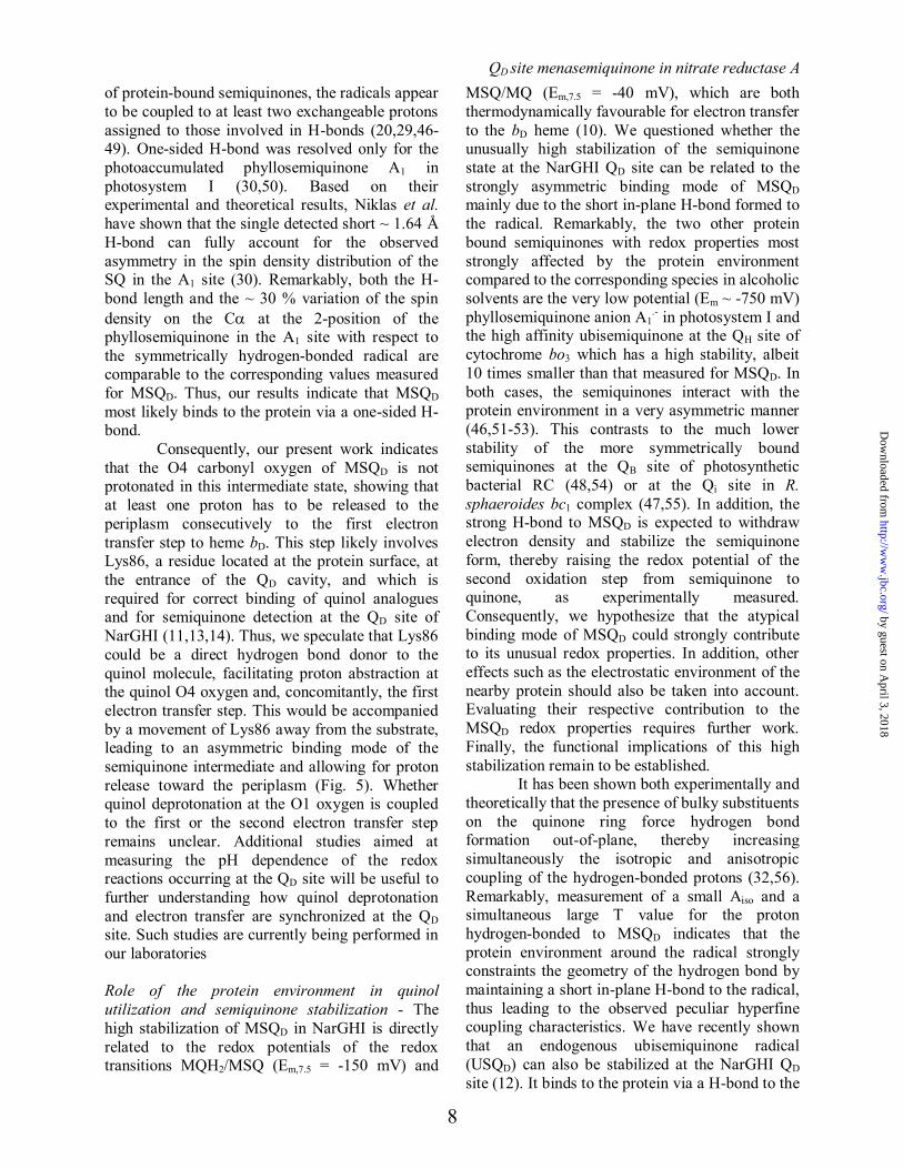
FIGURE 5. Working model of the MSQD binding mode in E. coli NarGHI based on our
spectroscopic work. Strongly asymmetric binding of MSQD occurs via a short in-plane H-bond to the N of His66, while Lys86 does not appear to be a direct H-bond donor to the radical in the semiquinone state
(13). The MSQD O4 oxygen is deprotonated. The protons H1, H2 and H3 discussed in the text are
indicated by arrows. R = NHCCO(CH2)3.
by guest on April 3, 2018
http://ww
w.jbc.org/
Dow
nloaded from

QD site menasemiquinone in nitrate reductase A
13
TABLES
Table 1. Hyperfine tensors derived from contour line shape analysis of HYSCORE spectra. A is the component of the hyperfine tensor where the direction of magnetic field is perpendicular to the symmetry
axis of the axially symmetric hyperfine tensor, and A, where the direction of magnetic field is parallel to
it (see the Supplemental Data). Aiso = 1/3(2A+A). The preferred sets are indicated in bold (see Discussion).
(Aiso, T) MHz |A| = |Aiso-T| |A| = |Aiso+2T| |(A-A)/Aiso| Assignment
H1a H1b
6.78, ±1.25 ±5.53, ±1.25
8.03 4.28
4.28 8.03
0.55 0.68
methyl protons
H2a H2b
2.14, ±1.18 ±0.96, ±1.18
3.32 0.22
0.22 3.32
1.3 3.2
-methylene proton
H3a H3b
5.79, ±5.73 ±0.06, ±5.73
11.52 5.67
5.67 11.52
1.01 97.5
H-bond proton
by guest on April 3, 2018
http://ww
w.jbc.org/
Dow
nloaded from

H3C
CH3
H8
His66
Heme bD
MSQD
12 3
4
R
CH2
H2N
Lys86
1.6 Ao
H
H1
H3
-Nε
Nδ
Fe2+
H2
O
Figure 5
by guest on April 3, 2018
http://ww
w.jbc.org/
Dow
nloaded from

MagalonRodolphe Szenes, Burkhard Endeward, Thomas F. Prisner, Bruno Guigliarelli and Axel
Stephane Grimaldi, Rodrigo Arias-Cartin, Pascal Lanciano, Sevdalina Lyubenova, nitrate reductase A by pulsed EPREscherichia coliintermediate in
Determination of the proton environment of the high stability menasemiquinone
published online December 21, 2011J. Biol. Chem.
10.1074/jbc.M111.325100Access the most updated version of this article at doi:
Alerts:
When a correction for this article is posted•
When this article is cited•
to choose from all of JBC's e-mail alertsClick here
Supplemental material:
http://www.jbc.org/content/suppl/2011/12/21/M111.325100.DC1
by guest on April 3, 2018
http://ww
w.jbc.org/
Dow
nloaded from