STUDY OF SOME ANATOMICAL AND … OF SOME ANATOMICAL AND HISTOLOGICAL CHARACTERISTICS IN LIVER OF...
8
ISI Impact Factor:3.461 Bas.J.Vet.Res.Vol.14,No.2,2015 150 STUDY OF SOME ANATOMICAL AND HISTOLOGICAL CHARACTERISTICS IN LIVER OF MALE INDIGENOUS TURKEY (Meleagris gallopava) Ahmed Saad Al-A´Aaraji Department of Anatomy and Histology ,College of Veterinary Medicine, University Baghdad ,Baghdad,Iraq (Received 8 April 2015 ,Accepted 18 August 2015) Key words: Hepatocytes ,Liver, Turkey. ABSTRACT The present study was performed to illustration some anatomical and histological characteristics of the liver of adult male local turkey. The present study was conducted on 20 turkeys divided into two groups (10 for anatomical study and 10 for histological study) mature male healthy turkey were used. The anatomical results showed that the liver of male turkey located in the right and left hepatoperitoneal cavity, it has red- brown to dark brown in color and it consisted of right and left lobes, left lobe subdivided into dorsal part and ventral part. The present study revealed that the weight of the liver was (1.89 ± 0.112)% in relation with the body weight. From the result of the histological study was noted there was no clear distinct limited between lobules in the hepatic parenchyma, the hepatocytes are usually arranged as two cell thickness between the liver sinusoids, the sinusoids lined by endothelial cellscontract with the kupffer’s cells. The portal area contained branch of portal vein, branch of hepatic artery and 2-3 inter lobar bile ducts. INTRODUCTION The liver in birds is one of the largest, most important organ in the body. It has numerous functions, as in other vertebrates, including digestive functions, metabolism of proteins, fats, and carbohydrates, synthesizing and secreting bile which contains two bile pigments, bilirubin and biliverdin into the small intestine (1,2). Theavian liver is suspended by peritoneum that is connected with overlying air sac and surrounded by hepatic celomic cavities (3,4). The avian liver has two lobes, it's in most avian have the right lobe larger than the left one for example, in pigeon, bustards and ostrich (5,6,7), while the two lobes can be of equal size, for example, in Galliformes (8). However, the left lobe of the domestic fowl divided into the dorsal and ventral parts (3,9) The avian liver is covered by a peritoneal layer of mesothelium, under this layer the dense connective tissue. The liver of avian similar to that in mammals but there is some differences such as absent connective tissue septa between lobules except in portal area (8). The principal cell of avian liver is the hepatocyte. Avian hepatocytes are polyhedral cells with a large rounded, oval and centrally located nucleus, the sheets of hepatocytes are separated by sinusoids.( 6, 10 and 11 ). In both birds and mammals, the sinusoids lined by endothelial cells and Kupffer’s cells, and the perisinusoidal spaces may be linked directly by penetrating
Transcript of STUDY OF SOME ANATOMICAL AND … OF SOME ANATOMICAL AND HISTOLOGICAL CHARACTERISTICS IN LIVER OF...

ISI Impact Factor:3.461 Bas.J.Vet.Res.Vol.14,No.2,2015
150
STUDY OF SOME ANATOMICAL AND HISTOLOGICAL
CHARACTERISTICS IN LIVER OF MALE INDIGENOUS
TURKEY (Meleagris gallopava)
Ahmed Saad Al-A´Aaraji Department of Anatomy and Histology ,College of Veterinary Medicine, University
Baghdad ,Baghdad,Iraq (Received 8 April 2015 ,Accepted 18 August 2015)
Key words: Hepatocytes ,Liver, Turkey.
ABSTRACT
The present study was performed to illustration some anatomical and histological characteristics of the liver of adult male local turkey. The present study was conducted on 20 turkeys divided into two groups (10 for anatomical study and 10 for histological study) mature male healthy turkey were used. The anatomical results showed that the liver of male turkey located in the right and left hepatoperitoneal cavity, it has red- brown to dark brown in color and it consisted of right and left lobes, left lobe subdivided into dorsal part and ventral part. The present study revealed that the weight of the liver was (1.89 ± 0.112)% in relation with the body weight. From the result of the histological study was noted there was no clear distinct limited between lobules in the hepatic parenchyma, the hepatocytes are usually arranged as two cell thickness between the liver sinusoids, the sinusoids lined by endothelial cellscontract with the kupffer’s cells. The portal area contained branch of portal vein, branch of hepatic artery and 2-3 inter lobar bile ducts.
INTRODUCTION The liver in birds is one of the largest, most important organ in the body. It has
numerous functions, as in other vertebrates, including digestive functions, metabolism of proteins, fats, and carbohydrates, synthesizing and secreting bile which contains two bile pigments, bilirubin and biliverdin into the small intestine (1,2).
Theavian liver is suspended by peritoneum that is connected with overlying air sac and surrounded by hepatic celomic cavities (3,4). The avian liver has two lobes, it's in most avian have the right lobe larger than the left one for example, in pigeon, bustards and ostrich (5,6,7), while the two lobes can be of equal size, for example, in Galliformes (8). However, the left lobe of the domestic fowl divided into the dorsal and ventral parts (3,9) The avian liver is covered by a peritoneal layer of mesothelium, under this layer the dense connective tissue. The liver of avian similar to that in mammals but there is some differences such as absent connective tissue septa between lobules except in portal area (8).
The principal cell of avian liver is the hepatocyte. Avian hepatocytes are polyhedral cells with a large rounded, oval and centrally located nucleus, the sheets of hepatocytes are separated by sinusoids.( 6, 10 and 11 ).
In both birds and mammals, the sinusoids lined by endothelial cells and Kupffer’s cells, and the perisinusoidal spaces may be linked directly by penetrating

ISI Impact Factor:3.461 Bas.J.Vet.Res.Vol.14,No.2,2015
151
traversing the endothelial cells or by intercellular gaps in the sinusoidal linings, forming a mass of branching and hollow cords, their lumina called the bile (12).
The hepatocytes drain the bile substance into bile canaliculi that located on opposite side of the hepatocytes from Disse᾿s space. The bile canaliculi drain this substance into the interlobular ducts of the portal area, the portal area has also hepatic portal veins which drain into the hepatic sinusoids and hepatic arteries also connected with the sinusoids by arterioles (8). The aim of this study was to carry out some anatomical and histological study
description of the liver of turkey in order to show some possible difference with
respect to other avian species.
MATERIALS AND METHODS Ten adult male turkey were obtained from commercial market of Al-kut and
Baghdad district at winter season. All studied birds were free of any diseases or lesions, therefore; they were considered to be apparently normal.The birds were anesthetized by intramuscular injection of a mixture of ketamine and diazepam at dose 25,5 mg/kg of body weight then killed(13). Then specimens were cut from different areas of liver. The processing of Fixation, dehydration, clearing, embedding and cutting were made, H&E were used (14). Statistical analysis was obtained by used to spss to calculate as standard deviation and standard error for anatomical parameters in liver (15).
RESULTS AND DISCUSSIONS The anatomical result showed that the liver of the male turkey located in the
right and left hepatoperitoneal cavity, it has red- brown to dark brown color (Fig. 1). This result agreed with (16 ) who said that the normal color of the adult male geese was red-brown to dark brown and disagree with (3) who remained that the normal color of the avian liver depends on the nutritional state of the bird and it is red-brown or it may be light brown but it is yellow if the bird is on a high-fat diet.
The liver of male turkey consisted of right and left lobes, left lobe subdivided into dorsal part and ventral part while right lobe un divided (Fig. 2), this result agreed with (5,10, 17) whom mentioned that the liver of domestic fowl consist of two lobes the left one subdivided into the dorsal and ventral parts, while there were no further lobular subdivisions in the liver of Houbara Bustards (5) and in contrast with (18 and 7) whom described that the left lobe in ostrich is subdivided into a small caudodorsal part, a large caudoventral part and a small left intermediate part.
The liver weight of the male turkey was (1.89 ± 0.112 )% in relation with the body weight. This is less than in geese that the liver weight (2.105 ± 0.071 )% relation with the body weight which reported by (16). This variation in weight was explain by (5) who remained that the type of food reception appears to have an important correlation with liver size relative to body size. Behind this, (19) was indicate the metabolism of proteins, fats , and carbohydrates is high in liver of snow geese and the weight of liver changes in relation to the level of food consumed which responsive to dietary changes.
The histological results were declared there was no clear distinct limited between lobules in the hepatic parenchyma of turkey's liver, due to paucity or absence of the interlobular connective septa, except in the portal area; each lobule has central vein (Fig. 3 and 4), this is similar to the results pointed by (20, 21) in chicken and (8) in psittacine.

Bas.J.Vet.Res.Vol.14,No.2,2015
The histology picture of the liver in turkey was composed of plates of hepatocytes which are usually arranged as two cell thickness between the liver sinusoids, these are hepatic cords, these cells ordered radially around the central vein (Fig. 5).This finding was in agreement with that of (22) in duck, (20) in chicken (8) and in domestic birds. (22) referred that the hepatic cords and blood sinusoids run beside each other in a continuously branching and interlocking structure, this is give the standard suitable for metabolic interchange between the two systems.
The hepatocytes are polyhedral in shape and it had cytoplasm contain many
granules with their ovoid nucleus which had distinct nucleolus. The sinusoids lined
by endothelial cells that are nearly flattened in shape with presence of the kupffer’s
cells contract with endothelial cells, last cells had large nucleus with some debris in
cytoplasm, the sinusoids continued with the hepatic vein and portal vessels (Fig.6).
The similar findings were previously reported by (12) in chicken and
The portal area contained the inter lobular connective septa which composed
the branches of the portal vein, branch of hepatic artery and usually
bile ducts (Fig. 4). The bile duct lined by simple cuboidal epithelium, while the
hepatic artery lined by endothelial cells of the simple squamous epithelium protruded
into the lumen containing RBCs and a layer of smooth muscle fiber more t
that of the branch of the portal vein which is lined by endothelial cells, protruded into
the large lumen (Fig.4, 6), this
(4) in duck, (8) in pssitacine and (20) in chicken.
Figure (1): Photograph illustrate position of Trukeyliver . A
righthepatoperitoneal cavity. B
heart. D- gizzard. E
ISI Impact Factor:3.461 Bas.J.Vet.Res.Vol.14,No.2,2015
152
The histology picture of the liver in turkey was composed of plates of hepatocytes which are usually arranged as two cell thickness between the liver sinusoids, these are hepatic cords, these cells ordered radially around the central vein
finding was in agreement with that of (22) in duck, (20) in chicken (8) and in domestic birds. (22) referred that the hepatic cords and blood sinusoids run beside each other in a continuously branching and interlocking structure, this is give
suitable for metabolic interchange between the two systems. The hepatocytes are polyhedral in shape and it had cytoplasm contain many
granules with their ovoid nucleus which had distinct nucleolus. The sinusoids lined
cells that are nearly flattened in shape with presence of the kupffer’s
cells contract with endothelial cells, last cells had large nucleus with some debris in
cytoplasm, the sinusoids continued with the hepatic vein and portal vessels (Fig.6).
r findings were previously reported by (12) in chicken and (23
The portal area contained the inter lobular connective septa which composed
the branches of the portal vein, branch of hepatic artery and usually
bile ducts (Fig. 4). The bile duct lined by simple cuboidal epithelium, while the
hepatic artery lined by endothelial cells of the simple squamous epithelium protruded
into the lumen containing RBCs and a layer of smooth muscle fiber more t
that of the branch of the portal vein which is lined by endothelial cells, protruded into
the large lumen (Fig.4, 6), thisresult correspond with the reported of some authors as
(4) in duck, (8) in pssitacine and (20) in chicken.
Figure (1): Photograph illustrate position of Trukeyliver . A
righthepatoperitoneal cavity. B- righthepatoperitoneal cavity. C
gizzard. E- liver.
ISI Impact Factor:3.461
The histology picture of the liver in turkey was composed of plates of hepatocytes which are usually arranged as two cell thickness between the liver sinusoids, these are hepatic cords, these cells ordered radially around the central vein
finding was in agreement with that of (22) in duck, (20) in chicken (8) and in domestic birds. (22) referred that the hepatic cords and blood sinusoids run beside each other in a continuously branching and interlocking structure, this is give
suitable for metabolic interchange between the two systems. The hepatocytes are polyhedral in shape and it had cytoplasm contain many
granules with their ovoid nucleus which had distinct nucleolus. The sinusoids lined
cells that are nearly flattened in shape with presence of the kupffer’s
cells contract with endothelial cells, last cells had large nucleus with some debris in
cytoplasm, the sinusoids continued with the hepatic vein and portal vessels (Fig.6).
(23) in ostrich.
The portal area contained the inter lobular connective septa which composed
the branches of the portal vein, branch of hepatic artery and usually 2-3 branch of
bile ducts (Fig. 4). The bile duct lined by simple cuboidal epithelium, while the
hepatic artery lined by endothelial cells of the simple squamous epithelium protruded
into the lumen containing RBCs and a layer of smooth muscle fiber more thicker than
that of the branch of the portal vein which is lined by endothelial cells, protruded into
result correspond with the reported of some authors as
Figure (1): Photograph illustrate position of Trukeyliver . A-
righthepatoperitoneal cavity. C-

Bas.J.Vet.Res.Vol.14,No.2,2015
Figure (2): Photograph show parietal surface of livershow A
dorsal part of left part . C
Figure (3): hepatic parenchyma of liver in turky
B- central vein.
ISI Impact Factor:3.461 Bas.J.Vet.Res.Vol.14,No.2,2015
153
Figure (2): Photograph show parietal surface of livershow A- right lobe . B
dorsal part of left part . C- ventral part of left lobe .
hepatic parenchyma of liver in turkyshow A- no distinct lobules .
central vein. H & E ( 4 X)
ISI Impact Factor:3.461
right lobe . B-
no distinct lobules .

Bas.J.Vet.Res.Vol.14,No.2,2015
Figure (4): Portal area of the liver of turkey show A
B- branch of hepatic artery . C
H & E ( 20 X).
Figure (5): Histological section at liver of male turkey show A
B- sinusoids C
ISI Impact Factor:3.461 Bas.J.Vet.Res.Vol.14,No.2,2015
154
Portal area of the liver of turkey show A- branch of portal vein.
branch of hepatic artery . C- branch of bile duct. D-connective tissue septa
Histological section at liver of male turkey show A-
sinusoids C-Kupffer cells. D-central vein.H & E ( 40 X)
ISI Impact Factor:3.461
branch of portal vein.
connective tissue septa .
hepatocytes .
H & E ( 40 X)

Bas.J.Vet.Res.Vol.14,No.2,2015
دراسة بعض الصفات التشریحیة والنسیجیة لكبد ذكر الدیك الرومي المحلي
.، بغداد ، العراق
. اجریت الدراسة الحالیة لتوضیح بعض الصفات التشریحیة والنسیجیة في كبد الدیك الرمي المحلي البالغ
من الدیك ) عشرة للدراسة التشریحیة وعشرة للدراسة النسیجیة
ظھرت النتائج التشریحیة لكبد الدیك الرومي انھ یقع في تجاویف الكبد
الفص االیسر ، الخلبیة الیمنى والیسرى وانھ ذات لون بني محمر إلى بني قاني ویتكون من فص ایمن وفص ایسر
مقارنتا الى وزن % )٠,١١٢± ١,٨٩
وان الخالیا الكبدیة ، تبین من الدراسة النسیجیة ان الحدود بین الفصیصات غیر واضحة في متن الكبد
تصطف بسمك خلیتین بین الجیبانیات والجیبانیات مبطنة بخالیا یطانیة وبتماس مع خالیا كوفر وان المنطقة
.من القنوات الصفراء بین الفصوص
Figure (6): Histological section at liver of male turkey show A
hepatocytes. B- nucleus of Kupffer´s cells. C
D-endothelial of bile duct.
ISI Impact Factor:3.461 Bas.J.Vet.Res.Vol.14,No.2,2015
155
دراسة بعض الصفات التشریحیة والنسیجیة لكبد ذكر الدیك الرومي المحلي
(Meleagrisgallopava)
أحمد سعد األعرجي
، بغداد ، العراق بغداد كلیة الطب البیطري ، جامعة ،فرع التشریح
الخالصة
اجریت الدراسة الحالیة لتوضیح بعض الصفات التشریحیة والنسیجیة في كبد الدیك الرمي المحلي البالغ
عشرة للدراسة التشریحیة وعشرة للدراسة النسیجیة(في ھذه الدراسة تم استخدام عشرین طیرا
ظھرت النتائج التشریحیة لكبد الدیك الرومي انھ یقع في تجاویف الكبد ا. الرومي البالغ الخالي من االمراض
الخلبیة الیمنى والیسرى وانھ ذات لون بني محمر إلى بني قاني ویتكون من فص ایمن وفص ایسر
١,٨٩(بینت الدراسة بان وزن الكبد ھو . مقسم الى جزء ظھري وجزء بطني
تبین من الدراسة النسیجیة ان الحدود بین الفصیصات غیر واضحة في متن الكبد
تصطف بسمك خلیتین بین الجیبانیات والجیبانیات مبطنة بخالیا یطانیة وبتماس مع خالیا كوفر وان المنطقة
من القنوات الصفراء بین الفصوص ٣-٢و البوابیة تحتوي على فرع الورید البوابي وفرع من الشریان الكبدي
Histological section at liver of male turkey show A-nucleus of
nucleus of Kupffer´s cells. C-endothelial of portal vein.
endothelial of bile duct. H & E (100X)
ISI Impact Factor:3.461
دراسة بعض الصفات التشریحیة والنسیجیة لكبد ذكر الدیك الرومي المحلي
اجریت الدراسة الحالیة لتوضیح بعض الصفات التشریحیة والنسیجیة في كبد الدیك الرمي المحلي البالغ
في ھذه الدراسة تم استخدام عشرین طیرا
الرومي البالغ الخالي من االمراض
الخلبیة الیمنى والیسرى وانھ ذات لون بني محمر إلى بني قاني ویتكون من فص ایمن وفص ایسر
مقسم الى جزء ظھري وجزء بطني
تبین من الدراسة النسیجیة ان الحدود بین الفصیصات غیر واضحة في متن الكبد . لجسما
تصطف بسمك خلیتین بین الجیبانیات والجیبانیات مبطنة بخالیا یطانیة وبتماس مع خالیا كوفر وان المنطقة
البوابیة تحتوي على فرع الورید البوابي وفرع من الشریان الكبدي
nucleus of
endothelial of portal vein.
ISI Impact Factor:3.461 Bas.J.Vet.Res.Vol.14,No.2,2015
156
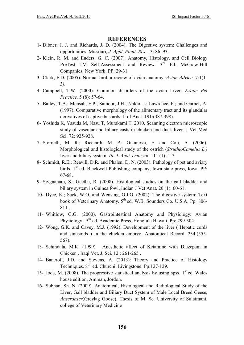
REFERENCES 1- Dibner, J. J. and Richards, J. D. (2004). The Digestive system: Challenges and
opportunities. Missouri, J. Appl. Poult. Res. 13: 86–93.
2- Klein, R. M. and Enders, G. C. (2007). Anatomy, Histology, and Cell Biology
PreTest TM Self-Assessment and Review. 3rd Ed. McGraw-Hill
Companies, New York. PP: 29-31.
3- Clark, F.D. (2005). Normal bird, a review of avian anatomy. Avian Advice. 7:1(1-
3).
4- Campbell, T.W. (2000): Common disorders of the avian Liver. Exotic Pet
Practice. 5 (8): 57-64.
5- Bailey, T.A.; Mensah, E.P.; Samour, J.H.; Naldo, J.; Lawrence, P.; and Garner, A.
(1997). Comparative morphology of the alimentary tract and its glandular
derivatives of captive bustards. J. of Anat. 191:(387-398).
6- Yoshida K, Yasuda M, Nasu T, Murakami T. 2010. Scanning electron microscopic
study of vascular and biliary casts in chicken and duck liver. J Vet Med
Sci. 72: 925-928.
7- Stornelli, M. R.; Ricciardi, M. P.; Giannessi, E. and Coli, A. (2006).
Morphological and histological study of the ostrich (StruthioCamelus L.)
liver and biliary system. Iit. J. Anat. embryol. 111 (1): 1-7.
8- Schmidt, R.E.; Reavill, D.R. and Phalen, D. N. (2003). Pathology of pet and aviary
birds. 1st ed. Blackwell Publishing company, Iowa state press, Iowa. PP:
67-68.
9- Sivgnanam, S.; Geetha, R. (2008). Histological studies on the gall bladder and
biliary system in Guinea fowl, Indian J Vet Anat. 20 (1): 60-61.
10- Dyce, K.; Sack, W.O. and Wensing, G.J.G. (2002). The digestive system: Text
book of Veterinary Anatomy. 5th ed. W.B. Sounders Co. U.S.A. Pp: 806-
811 .
11- Whitlow, G.G. (2000). Gastrointestinal Anatomy and Physiology: Avian
Physiology . 5th ed. Academic Press ,Honoiula.Hawaii. Pp: 299-304.
12- Wong, G.K. and Cavey, M.J. (1992). Development of the liver ( Hepatic cords
and sinusoids ) in the chicken embryo. Anatomical Record. 234:(555-
567).
13- Schindala, M.K. (1999) . Anesthetic affect of Ketamine with Diazepam in
Chicken . Iraqi Vet. J. Sci. 12 : 261-265 .
14- Bancroft, J.D. and Stevens, A. (2013): Theory and Practice of Histology
Techniques. 8th ed. Churchil Livingstone. Pp:127-129.
15- Joda, M. (2008). The progressive statistical analysis by using spss. 1st ed. Wales
house edition, Amman, Jordon.
16- Subhan, Sh. N. (2009). Anatomical, Histological and Radiological Study of the
Liver, Gall bladder and Biliary Duct System of Male Local Breed Geese,
Anseranser(Greylag Goose). Thesis of M. Sc. University of Sulaimani.
college of Veterinary Medicine

ISI Impact Factor:3.461 Bas.J.Vet.Res.Vol.14,No.2,2015
157
17- Caceci , T. (2006). Avian digestive system. 3th ed. Academic Press ,Itheca ,New
York. Pp;1-94.
18- Bezuidenhout, A.J. (1999). Anatomy of Ostrich: The Ostrich biology, Production
and Health.1st ed. UK CABI Publishing, Oxon. Pp: 29- 41.
19- Ankney, C. D.(1977). Feeding and digestive organ size in breeding lesser snow
geese. The Auk. 94 (2): 275-282.
20- Mclleland, J. (1993). Pericardium, pluera and peritoneum. In: Baumel, J. J.; King,
A.S.; Breazile, J. E.; Evans, H. E. and Berge, J.C.V.(eds.). Handbook of
avian.
21- Bacha, W. J. and Wood, G.L.M. (2006). Avian Digestive System .Color Atlas of
Veterinary Histology .William and Wikins .Waverlly Company .Hong
Kong Pp:113-150.
22- Abdelwahab, E.M.(1987). Ultrastructure and arrangement of hepatocyte cords in
the duckling's liver. J. of Anat. 150: (181-189).
23- Chagas, M.A.; SILVA, B.X.; BATH, F.V.; Babinski, M.A. &Figuefredo, M.A.
2007. Histologic structure of the parenchyma and stroma of the young
ostrich (Struthiocamelus) liver. RevistaBrasileiraMedicinaVeterinaria J.,
29: 61-64.