STUDIES ON PATHOGENESIS AND RECOVERY IN ERYSIPELAS
16
STUDIES ON PATHOGENESIS AND RECOVERY IN ERYSIPELAS By THOMAS FRANCIS, JR. (From the Department of Internal Medicine, Yale University School of Medicine and the Medical Serice of the New Haven Hospital, New Haven, Conn.) (Received for publication June 27, 1928) During the past three years Birkhaug (1-5) has reported the results of an extensive investigation of many phases of erysipelas both in experimental animals and in man. His observations may be sum- marized briefly. Hemolytic streptococci from erysipelas were found to form an immunologically specific group of streptococci. Ninety- one per cent of such organisms tested by the agglutinin absorption technique fell into this group. No cross agglutination with Strepto- coccus scarlatinae was noted (1). Serum obtained by immunizing rabbits against strains of erysipelas streptococci protected the skin of normal rabbits from infection with the homologous and heterologous erysipelas strains when the serum and bacteria were injected together. Passive immunity was also obtained by intravenous or subcutaneous injection of the serum twenty-four hours before the inoculation of a supralethal dose of organisms. Under these conditions the results with heterologous organisms were not so regular as with the homol- ogous strains (2). The immune serum caused a blanching of the erysipelatous lesion in patients or tended to prevent its spread. This effect was not obtained with scarlatinal antitoxin; nor was blanching of the scarlatinal rash obtained with anti-erysipelas serum. Blanch- ing of the erysipelatous lesion was also obtained with convalescent erysipelas serum (2). Broth cultures of erysipelas streptococci uni- formly gave toxic filtrates which in dilutions of 1: 1000 produced a skin reaction in susceptible individuals similar to a positive Dick test. Some strains produced filtrates which contained 30,000 to 50,000 S.T.D. of toxin per cubic centimeter. It was noted that the maximum production of toxin was obtained with 48 hours' incubation and that a 221
Transcript of STUDIES ON PATHOGENESIS AND RECOVERY IN ERYSIPELAS
STUDIES ON PATHOGENESIS AND RECOVERY INERYSIPELAS
By THOMAS FRANCIS, JR.
(From the Department of Internal Medicine, Yale University School of Medicine and theMedical Serice of the New Haven Hospital, New Haven, Conn.)
(Received for publication June 27, 1928)
During the past three years Birkhaug (1-5) has reported the resultsof an extensive investigation of many phases of erysipelas both inexperimental animals and in man. His observations may be sum-marized briefly. Hemolytic streptococci from erysipelas were foundto form an immunologically specific group of streptococci. Ninety-one per cent of such organisms tested by the agglutinin absorptiontechnique fell into this group. No cross agglutination with Strepto-coccus scarlatinae was noted (1). Serum obtained by immunizingrabbits against strains of erysipelas streptococci protected the skin ofnormal rabbits from infection with the homologous and heterologouserysipelas strains when the serum and bacteria were injected together.Passive immunity was also obtained by intravenous or subcutaneousinjection of the serum twenty-four hours before the inoculation of asupralethal dose of organisms. Under these conditions the resultswith heterologous organisms were not so regular as with the homol-ogous strains (2). The immune serum caused a blanching of theerysipelatous lesion in patients or tended to prevent its spread. Thiseffect was not obtained with scarlatinal antitoxin; nor was blanchingof the scarlatinal rash obtained with anti-erysipelas serum. Blanch-ing of the erysipelatous lesion was also obtained with convalescenterysipelas serum (2). Broth cultures of erysipelas streptococci uni-formly gave toxic filtrates which in dilutions of 1: 1000 produced askin reaction in susceptible individuals similar to a positive Dick test.Some strains produced filtrates which contained 30,000 to 50,000S.T.D. of toxin per cubic centimeter. It was noted that the maximumproduction of toxin was obtained with 48 hours' incubation and that a
221
ERYSIPELAS
definite decrease in the amount of toxin occurred after 90 hours. Thecutaneous reaction produced by 1 S.T.D. could be completely neutral-ized "by mixing the skin test dose with an equal amount of conva-lescent erysipelas serum, or with 0.001 cc. of erysipelas antistrepto-coccic rabbit or donkey sera" (3). Eighteen patients with erysipelasgave positive reactions to 1 S.T.D. of toxin on admission to the hospitaland as the erysipelatous lesion cleared and convalescence was estab-lished the reactivity of the skin to a similar dose was lost. Thisoccurred from 5 to 38 days after the original test. During the acutestage of erysipelas when skin reactions were positive there wasdemonstrable in the patient's serum a toxic substance which caused alocal cutaneous reaction in normal susceptible individuals. Thisreaction could be neutralized by immune or convalescent erysipelasserum. A toxic substance occurred in large amounts in the urine.With the disappearance of the cutaneous susceptibility to the toxin,following recovery from the disease, the presence of antitoxin in theserum of patients was demonstrated by means of toxin neutralizationtests in the skin of susceptible individuals. Cross neutralization ofthe erysipelas toxin by scarlatinal antitoxin was not obtained (3).Anti-erysipelas donkey or rabbit serum produced favorable thera-peutic results when given early in the disease. The impressionobtained was that the serum was antitoxic in nature. The amount ofserum required was determined by the inoculation of 1 S.T.D. of toxininto the skin of the patient simultaneously with the intramuscular orintravenous administration of the therapeutic serum. A positive ornegative reaction was considered to indicate incomplete or completeneutralization of the circulating toxin, respectively (4). In patientswith recurrent attacks of erysipelas the presence of antitoxin in theblood and the insusceptibility of the skin to the toxin were replacedafter an interval by the return of skin sensitivity and the absence ofantitoxin in the circulating blood. Active immunization by meansof the toxic filtrate was considered to be highly efficacious in preventingrecurrences. In the group of cases reported, immunization caused adecrease in the frequency of recurrences, induced a loss of skin reac-tivity and increased the neutralizing capacity of the patient's serum(5). In normal individuals tested, 21 per cent of 272 school children,ranging in age from 7 to 17 years, and 27 per cent of 135 hospital
222
THOMAS FRANCIS, JR.
patients (other than those with erysipelas), of ages from 18 to 72 years,gave positive skin reactions to 1 S.T.D. of toxin (3).Kaplan and Singer (6) also noted disappearance or diminution of
the cutaneous reactivity during convalescence from the disease.With an antitoxin produced by repeated injections of culture filtrateof erysipelas streptococci into human subjects they obtained com-plete neutralization of the homologous undiluted toxin in 73.7 per centof the susceptible individuals in whom tests were made, whereas,with convalescent erysipelas serum it was obtained in only 38.1 percent. Definite cross neutralization of the toxin by scarlet feverantitoxin and of scarlet fever toxin by the erysipelas antitoxin orconvalescent erysipelas serum was noted. Blanching of the erysipe-latous lesion occurred in 50 per cent of those tested with the authors'antitoxin, but was uniformly absent when convalescent erysipelasserum was used.As Birkhaug has pointed out (5), the results of his studies on ery-
sipelas present a striking parallelism with the observations on scarletfever which have been reported during the last few years by Dick andDick (7), Dochez (8), Blake and Trask (9), and numerous others; somuch so, in fact, that one would be led to believe that in its patho-genesis erysipelas is essentially comparable to scarlet fever, and,consequently, amenable to treatment by a specific method similar tothat which has proved successful in the case of the latter disease.But on clinical grounds this parallelism does not obtain. Scarletfever is most prevalent between the ages of six months and twentyyears; erysipelas is most prevalent under six months and over twentyyears. An attack of scarlet fever almost invariably gives a lastingimmunity; one attack of erysipelas would appear to render manyindividuals, at least, more prone to subsequent attacks. It is alsonoteworthy that recurrent erysipelas tends to involve the same areasaffected in previous attacks. In scarlet fever the essential and mostevident clinical phenomena are removed from the local focus of infec-tion and have been shown to be due to a toxin absorbed from the localfocus; in erysipelas the essential and most evident phenomenon is thelocal spreading lesion of the skin due to actual infection of the skinby streptococci and dispersed specific lesions comparable to the toxiclesions of scarlet fever are not present. Clinically, scarlet fever pre-
223
ERYSIPELAS
sents three relatively distinct phases: the early toxic phase, the septicphase, and the late sequelae. In its pathology and its clinical aspectserysipelas would appear to simulate more closely the septic than thetoxic phase of scarlet fever. Like scarlet fever, of course, it may befollowed by late sequelae.
Inability to correlate satisfactorily the experimental observations ofBirkhaug with the well established clinical phenomena of erysipelashas led to the present study. Three aspects of the subject have beeninvestigated: (1) the reactivity of the skin of erysipelas patients tointracutaneous injections of filtrates of erysipelas streptococci; (2) thepresence of a toxic substance in the blood of patients acutely ill witherysipelas; and (3) the capacity of the serum of erysipelas patients toneutralize the toxic action of culture filtrates.
SKIN REACTIVITY OF PATIENTS WITH ERYSIPELAS TO FILTRATES OF
ERYSIPELAS STREPTOCOCCI
The skin reactions to sterile filtrates from broth cultures of erysipe-las streptococci were studied in 30 patients with erysipelas at intervalsduring the acute and convalescent stages of the disease. With oneexception the patients were all on the adult medical wards of the NewHaven Hospital, their ages ranging from 13 to 70 years. The filtratesemployed in most instances were prepared in our laboratory fromcultures of hemolytic streptococci cultivated directly from the lesionsof typical cases of erysipelas. For comparison a standardizederysipelas toxin obtained from the Squibb Laboratories through thecourtesy of Dr. J. F. Anderson was used in some of the cases.
Preparation and standardization of filtrates. Flasks containing 250cc. of 1 per cent defibrinated rabbit's blood, buffered, meat infusitonbroth, pH 7.4 to 7.6, were inoculated with 2.5 cc. of an 18 hour brothculture of erysipelas streptococci. After incubation for 48 hours at37°C. the culture was passed through a Berkefeld filter, the filtratetested for sterility and bottled. The filtrates were standardized interms of skin test doses per cubic centimeter by injecting 0.1 cc. ofgraded dilutions in the skin of the most reactive individual available.One-tenth cubic centimeter of that dilution which caused a localerythema approximately 10 mm. in diameter 24 hours after injection
224
THOMAS FRANCIS, JR.
was considered to be 1 S.T.D. Titrated in this way the filtrates werefound to contain from 2,000 to 3,000 S.T.D. per cubic centimeter. TheSquibb toxin had been standardized by the makers, 1 cc. containing5,000 S.T.D. One-tenth cubic centimeter of a 1: 500 dilution of thistoxin caused a positive reaction approximately 10 mm. in diameter inthe same subject used in the standardization of our filtrates. Nega-tive heat controls were obtained only by boiling the filtrates two tofour hours.
Technique of skin tests. Graded dilutions of filtrate containing from1 to 50 S.T.D. of toxin in 0.1 cc. volume were injected intradermally onthe flexor surfaces of the forearms, the doses employed being 1, 3 or5, 10, and 30 or 50 S.T.D. The complete series of doses was not usedin each patient. In 12 cases 10 S.T.D. was the largest dose employed,while in a moderate number 3 S.T.D. was the smallest dose used, andin a few an intermediate dose was omitted. In general, the same doseswere used in the same patient throughout the course of his illness.Heat controls for the largest doses, at least, were used. Readingswere made at 24 hours and the size and intensity of the reactionrecorded. All reactions which were less than 6 mm. in diameter wereconsidered negative.The results of the observations on the skin reactivity of erysipelas
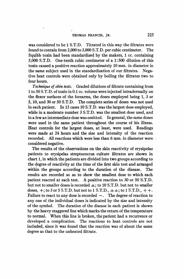
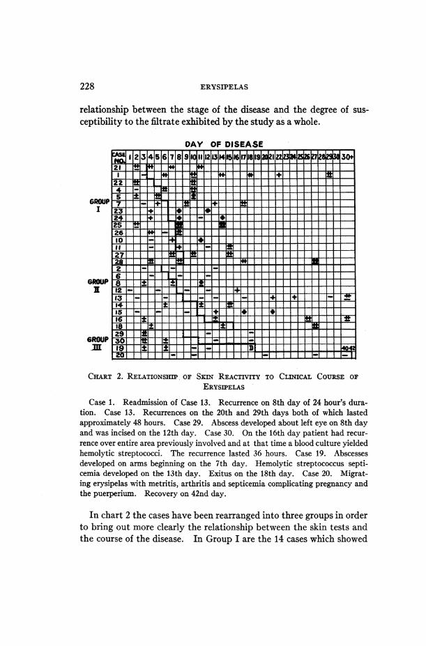
patients to erysipelas streptococcus culture filtrates are shown inchart 1, in which the patients are divided into two groups according tothe degree of reactivity at the time of the first skin test and arrangedwithin the groups according to the duration of the disease. Theresults are recorded so as to show the smallest dose to which eachpatient reacted at each test. A positive reaction to 30 or 50 S.T.D.but not to smaller doses is recorded i; to 10 S.T.D. but not to smallerdoses, +; to 3 or 5 S.T.D. but not to 1 S.T.D., i 4; to 1 S.T.D., + +.Failure to react to any dose is recorded -. The degree of reaction toany one of the individual doses is indicated by the size and intensityof the symbol. The duration of the disease in each patient is shownby the heavy staggered line which marks the return of the temperatureto normal. When this line is broken, the patient had a recurrence ordeveloped a complication. The reactions to heat controls are notincluded, since it was found that the reaction was of about the samedegree as that to the unheated filtrate.
225

226 ERYSIPELAS
The results as charted demonstrate that 20 cases (Group I) showedon the first test during the active stage of the disease either no reactionor only a very slight degree of reactivity of somewhat doubtful
GROUPI
P
DAY OF D ISEASE
Z 3 4 5 6 7 8 9 10i 111213l14116l17l18l192Dl21 MMBsl5lk7l 30
13 -_ ++ - -________
6 _+ - s - t I ]]]1l,_ ____
17_L-_ _-_-II__A__tO______+____8_______19 IIII__l___120_______3 - .1Z4 _24 ~_________+_ +.65_1li7-__6
TOrL23 27_ 28__+l_|]24 _ 7 a. IIII__25_5_____"_____"I_1 7 7
CHART 1. SKIN REACTIVITY OF ERYSIPELAS PATIENTS- = non-reactive; + = reactive to 30 or 50 S.T.D.; + = reactive to 10 S.T.D.;
4 4 = reactive to 3 or 5 S.T.D.; ++ = reactive to 1 S.T.D. The broken stag-gered lines mark the end of 7 and 15 day periods, respectively, after return oftemperature to normal.
significance, since the 30 to 50 S.T.D. required the use of a 1:10 dilu-tion of the filtrate. They furthermore show that 10 cases (Group II)possessed some degree of reactivity. Of the patients in this group 2

THOMAS FRANCIS, JR.
-reacted to 10 S.T.D. but not to less, 7 to 3 or 5 S.T.D., and only 1 to 1S.T.D. As shown at the bottom of the chart only 7 of the 26 patientstested during the first four days of the disease, at a time when sus-ceptibility to the filtrate should be greatest according to Birkhaug,were sufficiently susceptible to give a positive skin reaction to 3 or5 S.T.D., 2 more were sufficiently susceptible to react to 10 S.T.D.,and 17 were highly insusceptible.The repeated tests in the later acute period of the disease and during
convalescence show a general, though not uniform, tendency for thereactivity of the skin to increase rather than to decrease and becomenegative as reported by Birkhaug. This is particularly conspicuousin Group I. Between the fourth day of the disease and the end of thefirst week of convalescence cases 1, 4, 5, 7, and 17 became reactive to1, 3, or 5 S.T.D., cases 3, 9, 10, 11, and 15 to 10 S.T.D., and cases 8 and14 showed a more marked reaction to 30 and 50 S.T.D., respectively.The relatively fewer observations later in convalescence show that cases12, 13, 16 and 18 eventually became reactive, and that cases 11 and 15were more susceptible to the filtrate than they had been on earliertests. Case 3, on the other hand, lost the moderate degree of reac-tivity exhibited toward the end of the first week of convalescence.In Group II all but 3 cases (26, 29, and 30) either maintained theiroriginal reactivity or showed increased susceptibility in the convales-cent period. In the whole series of cases there are only three thatremained completely negative throughout,-cases 2 and 6, who werenot retested late in convalescence, and case 20, who ran a prolongedcourse of migrating erysipelas complicated by metritis, arthritis andsepsis and was not retested after her final recovery on the forty-secondday. - There were also three cases which showed a diminishingreactivity with the progress of the disease. Case 19, slightly sensitiveearly in the disease, subsequently became completely non-reactiveconcurrently with the development of a complicating and fatal sepsis.Cases 29 and 30, one of whom developed a complicating abscess, theother recurrent erysipelas with sepsis, also became non-reactive,though originally susceptible to 5 and 3 S.T.D., respectively. Thereare four cases which showed a negative skin test between two positivetests. Occasional irregularities in tests of this type are to be expectedand their occurrence would not appear to invalidate the general
227
228 ERYSIPELAS
relationship between the stage of the disease and the degree of sus-ceptibility to the filtrate exhibited by the study as a whole.
DAY OF DISEASEA 123RL 1T 6 7S 1 9OF S121314KIN5REAV1* TO CIN 30O
tinCas 13 Recurrence son __th 20t an 29t day bot of whichlaste
GROUP 7 _-_+___ ___+_________IIIII11
26 _ ______X
Z112 _ +
65 I I_ ___ ___+
andwasicson the 12t dayCase_ . On e h y p n hd rc-ur-20_-_-1_-_ ______IB
ROima 4__ 2 Abssdevelopedon te 1h d. Eu o te 1_ 8thday.C 2 Migat
an was incse on th ___ __day Cas 30 On th da ptien had recur-
CHemolTi stepoociTENheP reurec laste 36 hours.T CIase COURSAbcese
ing erysipelas with metritis, arthritis and septicemia complicating pregnancy andthe puerperium. Recovery on 42nd day.
In chart 2 the cases have been rearranged into three groups in orderto bring out more clearly the relationship between the skin tests andthe course of the disease. In Group I are the 14 cases which showed

THOMAS FRANCIS, JR.
positive skin reactions to 10 S.T.D. or less by the eighth day ofthe disease and maintained positive reactions throughout conva-lescence. In all of these the duration of the disease was eight days orless, the average duration being 5.9 days. In Group II are the ninecases showing negative skin tests to 10 S.T.D. up to the twelfth day ofthe disease inclusive. Five of these had a duration of eight days ormore, the average duration being 8.8 days. In Group III are the threecases which showed decreasing skin reactivity and case 20 whichshowed negative tests throughout a long period. -All four of thesecases developed septic complications with positive blood cultures inthree and a fatal outcome in one.
TOXIN IN THE BLOOD OF ERYSIPELAS PATIENTS
Samples of blood were collected from a series of patients with ery-sipelas at frequent intervals during the course of the disease. Theblood was allowed to clot, centrifuged, and with sterile precautions,the serum was pipetted off and bottled. The initial samples, takenearly in the acute stage, were tested for the presence of a toxin byinjecting 0.1 or 0.3 cc. of the serum into the skin of normal individualsand patients known to be reactive to erysipelas streptococcus culturefiltrates. Of 29 such sera tested only 3 caused positive local reactionswith any constancy, while 4 others produced a reaction on one occasion,although repeatedly negative on other tests: From these results itwould appear very improbable that the majority of patients witherysipelas have any demonstrable or significant amount of specifictoxin in the circulating blood during the acute stage of the disease, atleast in amounts equivalent to 3 to 10 S.T.D. per cubic centimeter.
NEUTRALIZATION OF CULTURE FILTRATE BY THE SERUM OF ERYSIPELAS
PATIENTS
In order to study the development of an antitoxin in the blood ofpatients recovering Irom erysipelas, neutralization tests were donewith the serial sera obtained from 27 cases of erysipelas of varyingduration and severity. Twenty-three of these were patients in whomthe skin reactivity was studied and reported upon above.
Technique of neutralization tests. To 0.5 cc. of each serum from each
229
ERYSIPELAS
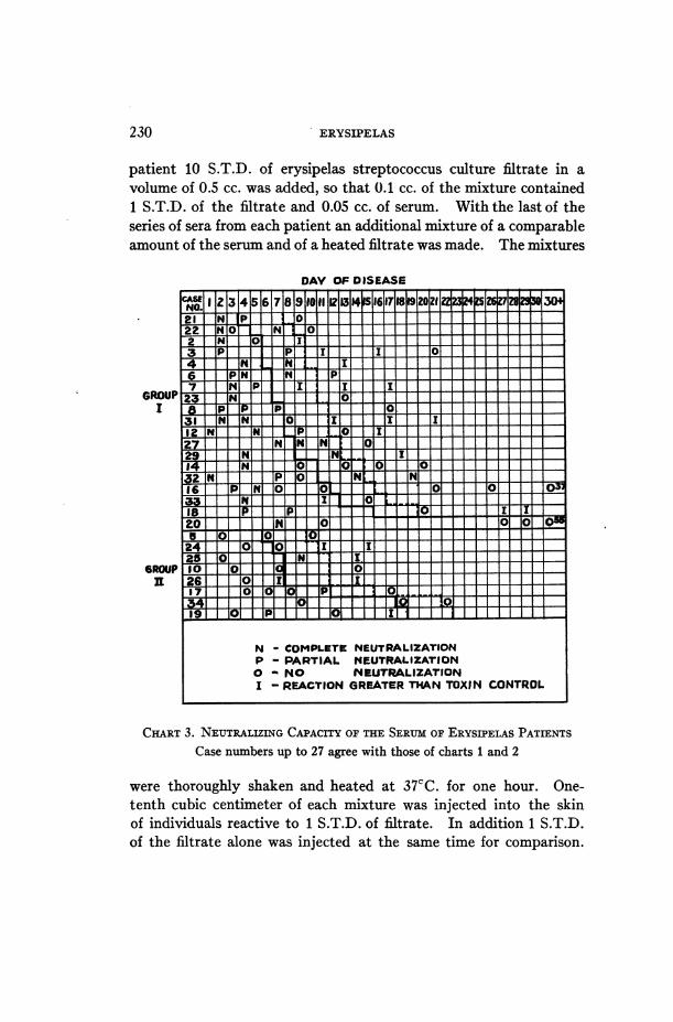
patient 10 S.T.D. of erysipelas streptococcus culture filtrate in avolume of 0.5 cc. was added, so that 0.1 cc. of the mixture contained1 S.T.D. of the filtrate and 0.05 cc. of serum. With the last of theseries of sera from each patient an additional mixture of a comparableamount of the serum and of a heated filtrate was made. The mixtures
DAY OF DISEASE
NI 2 g3456 7
21 T7IN PI I -
I
GROUPit
22 N lO' N I191 L1L 1___2 INI 101 113 I I3 _IP II _Pi t II _ oI
1_ _ " I67
8
-12:12729114216
24 _ 0 0I-O I- 1125 0O T 11 I_g10 0 _ 1 I 026 _ 0_ IO_X L
370_ 0 _T r
N - COMPLETE NEUTRALIZATIONP - PARTIAL NEUTRALIZATION0 - NO NEUTRALIZATIONI - REACTION GREATER THAN TOXIN CONTROL
CHART 3. NEUTRALIZING CAPACITY OF THE SERUM OF ERYSIPELAS PATIENTSCase numbers up to 27 agree with those of charts 1 and 2
were thoroughly shaken and heated at 37CC. for one hour. One-tenth cubic centimeter of each mixture was injected into the skinof individuals reactive to 1 S.T.D. of filtrate. In addition 1 S.T.D.of the filtrate alone was injected at the same time for comparison.
230
0s-
THOMAS FRANCIS, JR.
The reactions were observed and measured at 8, 18, and 24 hours.It was found that the most satisfactory time for recording the resultswas at 24 hours, and this was adhered to throughout.The results of the neutralization tests are presented in chart 3, in
which the patients have been divided into two groups according towhether the serum from the early bleedings neutralized or not. Theduration of the acute stage of the disease in each case is indicated asin the previous charts. The results obtained from the mixture of thelast serum from each patient and the heated filtrate are not recorded,since these uniformly failed to cause a reaction, nor are the reactions tothe filtrate alone included since these were uniformly positive.From chart 3 it will be seen that the great majority of the cases fall
into the first group, in which the sera collected early in the acutestage of the disease completely or partially neutralized the toxicaction of the filtrate, and that in only 8 cases (Group II) did the earlybleedings fail to neutralize. Another very striking result is the failureof the late convalescent sera to neutralize at all. In only 2 cases(Numbers 6 and 32) did convalescent sera collected more than fourdays after return of the temperature to normal show any capacity toneutralize the filtrate. Not only was there a failure of the convales-cent sera to neutralize, but in many instances a more marked reactionoccurred than with the filtrate alone. It is furthermore noteworthythat there is no case in which the early sera failed to neutralize and thelate convalescent sera neutralized. As with the skin tests, there are afew apparenitly irregular results, but this is not surprising in view ofthe nature of the observations.
Correlation of the initial skin and neutralization tests fails to showany consistent relationship. While it is true that 11 of the patientswhose sera completely or partially neutralized the filtrate failed toreact to 10 S.T.D. of the filtrate, there are 5 cases, whose sera neutral-ized, who reacted to 10 S.T.D. or less. Of the 7 patients whose earlysera failed to neutralize, 3 gave positive skin reactions to 10 S.T.D.or less, but 4 failed to react to 10 S.T.D. It is obvious, therefore, thatno simple explanation of the skin reactions on the basis of the presenceor absence of a neutralizing antitoxin in the blood is satisfactory ininterpreting the results obtained.
231

ERYSIPELAS
DISCUSSION
The three important phenoniena brought to light in the precedingobservations, namely, the tendency for the cutaneous reactivity oferysipelas patients to become more marked during convalescence.the absence of a demonstrable toxin in the circulating blood of patientsin the acute stage of the disease, and the neutralization of erysipelasstreptococcus culture filtrates by the serum of most patients in theacute phase of the disease with apparent loss of this power duringconvalescence, fail to support Birkhaug's concept of erysipelas as aspecific toxemia, recovery from which is due to the development ofan antitoxic immunity.The results of the skin tests suggest, rather, that an increasing
sensitiveness or allergy to streptococcus products, presumably liber-ated at the site of the erysipelatous lesion, develops with the progressof the disease to convalescence and recovery. The fairly definitecorrelation between the rapidity with which skin reactivity developedand the clinical course of the disease, as shown in chart 2, furthermoresuggests that the development of tissue allergy may play at least apart, perhaps an important part, in the mechanism of recovery fromthe infection. That the skin reactions were neutralizable not only insensitive normal individuals but also in sensitive patients in the conva-lescent period, as proved to be the case on a number of tests, is notsurprising, since it has been shown by Dochez and Stevens (10) thatthe skin reactions of rabbits rendered allergic to undiluted culturefiltrates of erysipelas streptococci, are neutralizable during the earlyphase of sensitivity.The negative results of the tests for toxin in the circulating blood
early in the disease indicate that a specific toxemia in the sense inwhich it has been shown to occur in scarlet fever (9), plays little or nosignificant part in erysipelas. This view is further supported by theneutralization tests, in which it is shown that the majority of adultpatients with erysipelas already possess early in the disease sufficientcirculating antibody to neutralize 20 S.T.D. of filtrate per cubiccentimeter of blood. The toxigenic activity of erysipelas strepto-cocci, in most instances relatively poorly developed as compared withthat of most scarlatinal streptococci, is probably counteracted at the
232
THOMAS FRANCIS, JR.
site of the local lesion from the onset of the disease by the alreadyexisting antitoxic immunity. In the case of the few patients whoseearly sera failed to neutralize, antitoxin may have been present, butin amounts too small to be demonstrated by the method used. Fourof these cases (Numbers 5, 10, 17, and 19) failed to give positive skinreactions to 10 S.T.D. of filtrate. In fact, there are but three cases inthe whole series (Numbers 24, 25, and 26) with positive skin testsand simultaneous absence of demonstrable antitoxin in the blood in theearly acute stage of the disease, who would lend themselves to thesupport of a simple toxin-antitoxin concept of the disease, and eventhese fail through the apparent absence of antitoxin and the presenceof positive skin tests (Number 26 not skin tested) about the end of thefirst week of convalescence.The failure of the later convalescent sera to neutralize demands an
explanation which at present, without further study, can be onlyhypothetical. It does not seem probable that this phenomenon is dueto an actual disappearance of the neutralizing antibody from theblood, since this would be contrary to general immunological princi-ples. It is suggested, therefore, that there appears in the blood of thepatient an antibody to the absorbed products of the growth and disso-lution of the streptococci distinct from the antitoxin, which underproper conditions will combine with similar products in culturefiltrates to produce a demonstrable reaction. This antibody maytentatively be termed an antiallergen and the phase of immunitywhich it represents termed humoral allergy. On the basis of thishypothesis not only the apparent failure of the convalescent serum toneutralize the toxic action of the filtrate, but also the frequent occur-rence of an intensified reaction might be explained as due to the reac-tion resulting from the combination of the antiallergen with bacterialproducts in the filtrate. Under these circumstances the passiveneutralization of the toxin in the filtrate by antitoxin in the convales-cent serum would be obscured and it would not be necessary to adoptthe highly improbable view that antitoxin had disappeared from theblood in the convalescent stage of the disease.
Finally, it is necessary to attempt to correlate the results of the skintests with those of the neutralization tests. Although the generaltrend of the two tests is in the same direction, it is obvious, as pointed
233
ERYSIPELAS
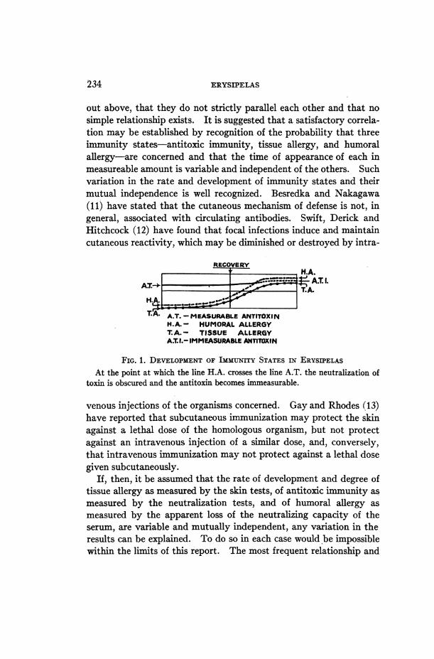
out above, that they do not strictly parallel each other and that nosimple relationship exists. It is suggested that a satisfactory correla-tion may be established by recognition of the probability that threeimmunity states-antitoxic immunity, tissue allergy, and humoralallergy-are concerned and that the time of appearance of each inmeasureable amount is variable and independent of the others. Suchvariation in the rate and development of immunity states and theirmutual independence is well recognized. Besredka and Nakagawa(11) have stated that the cutaneous mechanism of defense is not, ingeneral, associated with circulating antibodies. Swift, Derick andHitchcock (12) have found that focal infections induce and maintaincutaneous reactivity, which may be diminished or destroyed by intra-
RECOVERYM.A.
H,H.A.
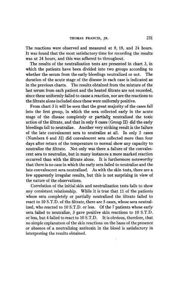
TA A.T. -MEASURABLE ANTITOXINH.A.- HUMORAL ALLERGYT.A.- TISSUE ALLERGYA.T.I.- IMMEASURABLE ANTITOXIN
FIG. 1. DEVELOPMENT OF IMMUNITY STATES IN ERYSIPELASAt the point at which the line H.A. crosses the line A.T. the neutralization of
toxin is obscured and the antitoxin becomes immeasurable.
venous injections of the organisms concerned. Gay and Rhodes (13)have reported that subcutaneous immunization may protect the skinagainst a lethal dose of the homologous organism, but not protectagainst an intravenous injection of a similar dose, and, conversely,that intravenous immunization may not protect against a lethal dosegiven subcutaneously.
If, then, it be assumed that the rate of development and degree oftissue allergy as measured by the skin tests, of antitoxic immunity asmeasured by the neutralization tests, and of humoral allergy asmeasured by the apparent loss of the neutralizing capacity of theserum, are variable and mutually independent, any variation in theresults can be explained. To do so in each case would be impossiblewithin the limits of this report. The most frequent relationship and
'234

THOMAS FRANCIS, JR.
general trend of the three immunity phases, however, are presentedschematically in figure 1. Independent alterations in the slopes of thelines, which represent the rates of development of the three immunityphases, will serve to explain the different combinations of skin andneutralization tests at different stages of the disease in all of the pa-tients studied.
CONCLUSIONS
As a result of the observations here presented it seems justifiable tocondude (1) that the pathogenesis of erysipelas is not comparable tothe pathogenesis of the specific toxic phase of scarlet fever and (2) thatthe mechanism of recovery from erysipelas, in adults at least, is not asimple neutralization of a circulating toxin through the developmentof an antitoxin, but rather that it is more intimately related to thedevelopment of allergy to products of the growth and dissolution ofstreptococci in the erysipelatous lesion. Furthermore, the failure tofind toxin in the blood and the usual presence of a neutralizing anti-body in the early acute stage of the disease provide little basis for theview that antitoxin treatment in adults should be of particular value inerysipelas.
BIBLIOGRAPHY
1. Birkhaug, K. E., Bull. Johns Hopkins Hosp., 1925, xxxvi, 248. Studies onthe Biology of the Streptococcus Erysipelatis. I. Agglutination and Agglu-tinin Absorption with the Streptococcus Erysipelatis.
2. Birkhaug, K. E., Bull. Johns Hopkins Hosp., 1925, xxxvii, 307. Studies onthe Biology of the Streptococcus Erysipelatis. III. Experimental Pro-duction of Erysipelas in Rabbits and Demonstration of Protective Powerof Immune Erysipelas Sera.
3. Birkhaug, K. E., Proc. Soc. Exp. Biol. and Med., 1925, xxiii, 201. Studies onthe Biology of the Streptococcus Erysipelatis. IV. Toxin Production ofthe Streptococcus Erysipelatis.
4. Birkhaug, K. E., J. Am. Med. Assn., 1926, lxxxvi, 1411. Erysipelas. V.Observations on the Etiology and Treatment with Erysipelas Antistrepto-coccus Serum.
5. Birkhaug, K. E., J. Am. Med. Assn., 1927, lxxxviii, 885. Erysipelas. VI.Immunization with Soluble Toxin from Streptococcus Erysipelatis AgainstRecurrent Attacks of Erysipelas.
6. Singer, H. A., and Kaplan, B., J. Am. Med. Assn., 1926, lxxxvii, 2141. Strep-tococcus Erysipelatis Toxin and Antitoxin.
THE JOURNAL OF CLINICAL INVESTIGATION, VOL. VI, NO. 2
235
236 ERYSIPELAS
7. Dick, G. F., and Dick, G. H., J. Am. Med. Assn., 1924, lxxxii, 265. A SkinTest for Susceptibility to Scarlet Fever.
8. Dochez, A. R., Trans. Assoc. Amer. Phys., 1924, xxxix, 136. Studies inScarlet Fever.
9. Blake, F. G., and Trask, J. D., J. Clin. Invest., 1926, iii, 397. Studies inScarlet Fever. II. The Relation of the Specific Toxemia of Scarlet Feverto the Course of the Disease.
10. Dochez, A. R., and Stevens, F. A., J. Exp. Med., 1927, xlvi, 487. Studieson the Biology of Streptococcus. VII. Allergic Reactions with Strainsfrom Erysipelas.
11. Besredka, A., and Nakagawa, S., Ann. Inst. Pasteur, 1927, xli, 607. Im-munization Passive Contre le Tetanos par la Voie Cutan6e.
12. Swift, H. F., Derick, C. L., and Hitchcock, C. H., J. Am. Med. Assn., 1928,xc, 906. Bacterial Allergy to Non-hemolytic Streptococci in its Relationto Rheumatic Fever.
13. Gay, F. P., and Rhodes, B., J. Inf. Dis., 1922, xxxi, 101. ExperimentalErysipelas. Studies in Streptococcus Infection and Immunity. IV.