Studies on Amanita Basidiomycetes Amanitaceae) in Northern ... · 98 A. pudibunda, which was...
27
Fungal Diversity 97 Studies on Amanita (Basidiomycetes: Amanitaceae) in Northern Thailand Sanmee, R. 1 , Tulloss, R.E. 2* , Lumyong, P. 3 , Dell, B. 4 and Lumyong, S. 1 1 Department of Biology, Faculty of Science, Chiang Mai University, Chiang Mai 50200, Thailand 2 P.O. Box 57, Roosevelt, New Jersey 08555-0057, USA. [Res. Assoc. (hons.), the New York Botanical Garden, Bronx, New York, USA] 3 Department of Plant Pathology, Faculty of Agriculture, Chiang Mai University, Chiang Mai 50200, Thailand 4 School of Biological Sciences and Biotechnology, Murdoch University, Perth 6150, Australia Sanmee, R., Tulloss, R.E., Lumyong, P., Dell, B. and Lumyong, S. (2008). Studies on Amanita (Basidiomycetes: Amanitaceae) in Northern Thailand. Fungal Diversity 32: 97-123. Specimens of the genus Amanita, collected from northern Thailand and deposited in the fungal herbarium of Chiang Mai University and some overseas herbaria were studied. Twenty-five taxa, including eighteen new to Thailand, are described briefly. A key to species is provided. The new records for Thailand are A. alboflavescens, A. avella- neosquamosa, A. chepangiana, A. clarisquamosa, A. flavipes sensu lato, A. fritillaria, A. fuliginea, A. hongoi, A. japonica, A. manginiana sensu W.F. Chiu, A. obsita, A. ovalispora, A. pseudoporphyria, A. rubrovolvata, A. sinensis, A. sinocitrina, A. subglobosa, and A. virgineoides. A type study of A. pudibunda is included. Amanita frostiana sensu R. Heim is proposed to be properly diagnosed as A. rubrovolvata. Key words: Agaricales, southeast Asia, taxonomy Article information Received 31 March 2008 Accepted 1 May 2008 Published online 30 September 2008 *Corresponding author: Rodham E. Tulloss; e-mail: [email protected] Introduction The forests in northern Thailand have a high diversity of Agaricales (Nuytinck et al., 2006; Le et al., 2007a,b), but knowledge of species in the genus Amanita Pers., many of which are ectomycorrhizal with forest trees, is limited; and only a few species have been reported from Thailand in the literature (Heim, 1962; Høiland and Schumacher, 1982; Chansri- kul et al., 1984; Soytong, 1994; Rachabun- ditayasathan, 1996; Chansrikul, 1998; Ruksawong and Flegel, 2001). Two taxa covered in this paper were originally described from Thailand: A. pudibunda and A. siamensis. The presence of two previously reported taxa is confirmed: A. princeps and A. sculpta. Three previously reported taxa (A. angustilamellata, A. mira and A. hemibapha) may be present and are phenetically similar to taxa that were collected and revised in prepration of this paper. Materials and methods For four years (1999-2002), Amanita taxa growing in forests dominated by members of the Dipterocarpaceae, Fagaceae, or Pinaceae were surveyed during the rainy season in five provinces (Chiang Mai, Chiang Rai, Nakhon Panom, Phayao, and Sakhon Nakhon) of northern and northeastern Thailand. Colour names were coded according to Petersen (1996). Basidiocarps were placed in size categories following Bas (1969). Microscopic data is presented in the format of Yang (1997), for the most part. The specimens of Thai collectors were deposited in the fungal herba- rium of the Department of Biology, Faculty of Science, Chiang Mai University (CMU). Throughout this paper, when stipe length is given and length of the stipe’s bulb is not, it should be assumed that bulb length is included in stipe length. Additional material collected by, or communicated to, Roger Heim (deposited in PC) and collected by David Arora (deposited in SFSU and the private herbarium of Tulloss) was reviewed and is reported using the same format, with the exception of the type study of
Transcript of Studies on Amanita Basidiomycetes Amanitaceae) in Northern ... · 98 A. pudibunda, which was...

Fungal Diversity
97
Studies on Amanita (Basidiomycetes: Amanitaceae) in Northern Thailand Sanmee, R.1, Tulloss, R.E.2*, Lumyong, P.3, Dell, B.4 and Lumyong, S.1
1Department of Biology, Faculty of Science, Chiang Mai University, Chiang Mai 50200, Thailand 2P.O. Box 57, Roosevelt, New Jersey 08555-0057, USA. [Res. Assoc. (hons.), the New York Botanical Garden, Bronx, New York, USA] 3Department of Plant Pathology, Faculty of Agriculture, Chiang Mai University, Chiang Mai 50200, Thailand 4School of Biological Sciences and Biotechnology, Murdoch University, Perth 6150, Australia Sanmee, R., Tulloss, R.E., Lumyong, P., Dell, B. and Lumyong, S. (2008). Studies on Amanita (Basidiomycetes: Amanitaceae) in Northern Thailand. Fungal Diversity 32: 97-123. Specimens of the genus Amanita, collected from northern Thailand and deposited in the fungal herbarium of Chiang Mai University and some overseas herbaria were studied. Twenty-five taxa, including eighteen new to Thailand, are described briefly. A key to species is provided. The new records for Thailand are A. alboflavescens, A. avella-neosquamosa, A. chepangiana, A. clarisquamosa, A. flavipes sensu lato, A. fritillaria, A. fuliginea, A. hongoi, A. japonica, A. manginiana sensu W.F. Chiu, A. obsita, A. ovalispora, A. pseudoporphyria, A. rubrovolvata, A. sinensis, A. sinocitrina, A. subglobosa, and A. virgineoides. A type study of A. pudibunda is included. Amanita frostiana sensu R. Heim is proposed to be properly diagnosed as A. rubrovolvata. Key words: Agaricales, southeast Asia, taxonomy Article information Received 31 March 2008 Accepted 1 May 2008 Published online 30 September 2008 *Corresponding author: Rodham E. Tulloss; e-mail: [email protected] Introduction
The forests in northern Thailand have a high diversity of Agaricales (Nuytinck et al., 2006; Le et al., 2007a,b), but knowledge of species in the genus Amanita Pers., many of which are ectomycorrhizal with forest trees, is limited; and only a few species have been reported from Thailand in the literature (Heim, 1962; Høiland and Schumacher, 1982; Chansri-kul et al., 1984; Soytong, 1994; Rachabun-ditayasathan, 1996; Chansrikul, 1998; Ruksawong and Flegel, 2001). Two taxa covered in this paper were originally described from Thailand: A. pudibunda and A. siamensis. The presence of two previously reported taxa is confirmed: A. princeps and A. sculpta. Three previously reported taxa (A. angustilamellata, A. mira and A. hemibapha) may be present and are phenetically similar to taxa that were collected and revised in prepration of this paper. Materials and methods
For four years (1999-2002), Amanita taxa
growing in forests dominated by members of the Dipterocarpaceae, Fagaceae, or Pinaceae were surveyed during the rainy season in five provinces (Chiang Mai, Chiang Rai, Nakhon Panom, Phayao, and Sakhon Nakhon) of northern and northeastern Thailand. Colour names were coded according to Petersen (1996). Basidiocarps were placed in size categories following Bas (1969). Microscopic data is presented in the format of Yang (1997), for the most part. The specimens of Thai collectors were deposited in the fungal herba-rium of the Department of Biology, Faculty of Science, Chiang Mai University (CMU).
Throughout this paper, when stipe length is given and length of the stipe’s bulb is not, it should be assumed that bulb length is included in stipe length.
Additional material collected by, or communicated to, Roger Heim (deposited in PC) and collected by David Arora (deposited in SFSU and the private herbarium of Tulloss) was reviewed and is reported using the same format, with the exception of the type study of

98
A. pudibunda, which was carried out according to the methods of, and is reported using the notation of, Tulloss et al. (1992) as amended by Tulloss (1993, 1994, 1998b, 2000) and restated by Tulloss and Lindgren (2005). Format of the type study conforms to that of Tulloss (1994) in order to maintain Tulloss’ type studies in a constant format for purposes of comparison.
Words describing spore shapes are used according to the definitions of Bas (1969): A spore is ‘globose’ if its length/width ratio (Q) falls between 1 and 1.05; ‘subglobose’ if its Q falls between 1.05 and 1.15; ‘broadly ellipsoid’ if its Q falls between 1.15 and 1.3; ‘ellipsoid’ if its Q falls between 1.30 and 1.6; ‘elongate’ if its Q falls between 1.6 and 2; ‘cylindric’ if its Q falls between 2 and 3; and ‘bacilliform’ if its Q falls above 3.
Biometric variables used in Tulloss’ type study are defined most recently in (Tulloss and Lindgren, 2005) and on the Amanita Studies web site (Tulloss, 2007b). The letter ‘R’ represents the length of a pileus radius.
Unless otherwise stated, determinations of material were based on: (1) protologs; (2) monographs or monographic-style works of Corner and Bas (1962), Bas (1969), Yang (1997), and Tulloss et al. (2001); and (3) additional keys such as Tulloss (2007a).
National Park names are abbreviated as follows: DLNP (Doi Luang National Park), DSPNP (Doi Suthep-Pui National Park), and KCNP (Khun Chae National Park). Herbarium codes follow Holmgren, Holmgren and Barnett (1992) with the exception of the following: HKAS—Herbarium of Cryptograms, Kunming Institute of Botany, Academia Sinica, Kunming, Yunnan Province, China. RET—personal herba-rium of Tulloss. Abbreviations in author citations follow Kirk and Ansell (1992) with the exception of ‘E.-J. Gilbert’, in which case we use the author’s initials in the order in which they appear on the vast majority of his publications including his magnum opus (Gilbert, 1940-41).
Names used for supraspecific taxa in Amanita follow Corner and Bas (1962) and Bas (1969) as modified by Yang (1997).
We take this opportunity to emphasize the importance of including modern Asian sources when researching Amanita in Asia. An over-
emphasis on European and North American literature has led unavoidably to misdetermi-nation of endemic taxa. This tendency should become a thing of the past. We strongly recom-mend (Yang, 1997) and other works of Yang and his co-authors as models for modern, Asian, mycological work in the genus Amanita. The encounter with an unfamiliar Amanita should lead to its being worked up as if it were an undescribed taxon. The chances are that it will be endemic and, often, new to science. Results
Organized by section, the species now recognized for northern Thailand are as follows—with newly reported taxa marked by an asterisk (*): Amanita [subg. Amanita] sect. Amanita
A. affin. mira A. obsita* A. rubrovolvata* A. siamensis A. sinensis* A. subglobosa*
Amanita sect. Caesareae A. chepangiana* A. hemibapha sensu lato A. princeps
Amanita sect. Vaginatae A. affin. angustilamellata A. ovalispora* A. pudibunda
Amanita [subg. Lepidella] sect. Amidella
A. avellaneosquamosa* A. clarisquamosa*
Amanita sect. Lepidella A. alboflavescens* A. hongoi* A. japonica* A. sculpta A. virgineoides*
Amanita sect. Phalloideae A. fuliginea* A. manginiana sensu W.F. Chiu* A. pseudoporphyria*
Amanita sect. Validae A. flavipes sensu lato* A. fritillaria* A. sinocitrina*
There are strong climatic and vegetational
links between the Thai region of study and other upland regions of southern and eastern Asia. With the exception of A. pudibunda and the recently described A. siamensis, the species in this list were originally described from Japan, China, Indonesia, peninsular Malaya, Nepal, and Singapore. The habitats from which these species were described include the same families of mycorrhizal trees associated with the species in Thailand (e.g., Tulloss and Bhandary, 1992; Yang, 1997; Bhatt et al., 2003). In the cited countries combined, there are approximately 125 named taxa of Amanita

99
known at this writing (Yang, 1997, 2005; Tulloss, 2005). Therefore, it seems very likely that some previously described Amanita taxa would be found in northern Thailand.
We expect that additional undescribed Amanita taxa will be found in the region of study. For example, given the size of Amanita sect. Vaginatae regionally (Yang, 1997; Tulloss, 2005) and worldwide (Tulloss and Yang, 2007a), it is unlikely that the count of taxa for northern Thailand in that section will remain as low as three species in the face of continued explora-tion and study of herbarium material.
Not surprisingly, given the close geogra-phic proximity, 84% of Amanita taxa recorded for northern Thailand also occur in southern and southwestern China (Yang, 1997; Yang et al., 2000; Yang et al., 2001). However, in China there appears to be much greater diversity of Amanita than in northern Thailand, probably due to the more diverse vegetational types especially at higher elevations.
All members of Amanita play an impor-tant role in forest ecology in northern Thailand, due to mycorrhizal associations with trees. In this regard, more taxa are reported herein from possible association with taxa of Fagaceae (22 or 88% of reported taxa) than with taxa of Dipterocarpaceae (10 or 40%). Only 5 reported taxa (20%) are known from possible association with a member of the Pinaceae. In the region of study, mixed Fagaceae/Pinaceae forests at higher elevations and dry Dipterocarpaceae forests at lower elevations require more extensive collecting in the future; and their Amanita taxa should be compared with collections from other areas of southeast Asia.
A number of the available collections were not in a good state of preservation or for another reason bore atypical spores. Throughout the paper, we provide spore measurements from other sources in the ‘Notes’ sections of our descriptions in order to provide additional support for future workers in southeast Asia. Key to known Amanita species of northern Thailand
1. Basidiospores amyloid (Amanita subg. Lepidella)....... .....................................................................................2 1. Basidiospores inamyloid (Amanita subg. Amanita) ..... ....................................................................................14
2. Margin of pileus short striate, appendiculate at first; stipe base bearing thick, membranous, saccate volva with distinct layers (three to five) ranging from floccose to farinose adjacent to pileipellis to membraneous on exterior (Amanita sect. Amidella)......................................................................................... 3
2. Margin of pileus non-striate, appendiculate only in sect. Lepidella; universal veil friable, submembranous, or membranous, sometimes limbate, but never saccate
..................................................................................... 4 3. Pileus whitish sometimes covered only with cream to
yellowish brown farinose to floccose patches representing the lower level of the universal veil, when upper layers are present they may be fibrillose (browning on exposure) or membranous (white to sordid); basidiospores (8.5-)9-11(-12) × (4.5-)5-6.5(-7) µm, with Q = (1.35-)1.5-1.88(-2.5) ...........................................................................A. avellaneosquamosa
3. Pileus whitish, with volval remnants brown or brownish, patchy, fibrillose, rarely with membranous universal veil outer layer present; basidiospores (10-)11-12.5(-16) × (7-)7.5-8 (11) µm, with Q = (1.15-)1.44-1.63(-2.07).............................. A. clarisquamosa
4. Margin of pileus appendiculate (Amanita sect.
Lepidella) .................................................................... 5 4. Margin of pileus non-appendiculate........................... 9 5. Volval remnants on pileus floccose to squamulose;
context of basidiocarp white, becoming yellow to lemon-chrome when cut; basidiospores 8-10.5(-11) × 5-6(-6.5) µm, with Q = 1.47-1.78(-2) .................................................................................... A. alboflavescens
5. Volval remnants on pileus as conic to subconic warts; context of basidiocarp not becoming yellow or lemon-chrome when cut; basidiospores differing in range of size or range of Q........................................................ 6
6. Pileus reddish brown with chocolate coloured conic
volval remnants; lamellae white when young, turning pinkish to violet when mature; context white, turning pinkish to purplish when cut; basidiospores (8-)9-11(-11.5) × (8-)9-10.5(-11) µm, with Q = 1-1.04(-1.08) ..........................................................................A. sculpta
6. Pileus and volval remnants not so deeply pigmented; lamellae white to cream, unchanging; context white, unchanging; basidiospores differing in range of size or range of Q ................................................................... 7
7. Pileus white to brownish, with brown or pinkish
brown volval remnants; basidiospores 7-9(-10) × (5.5-)6.5-7(-8) µm, with Q = (1.06-)1.12-1.25(-1.36); basal septa of basidia lacking clamps...........................................................................................................A. hongoi
7. Pileus grey to greyish or white, with similarly coloured volval remains; 95% of basidiospores having Q exceeding 1.25; basal septa of basidia often bearing clamps ......................................................................... 8

100
8. Pileus and universal veil grey or greyish; basal bulb of stipe slender, radicating; basidiospores 8-9(-11.5) × (4.5-)5.5-6.5(-7.5) µm, with Q = 1.34-1.64(-2)................................................................................A. japonica
8. Pileus and universal veil white; basal bulb of stipe not slender, not radicating; basidiospores (7.5-)8-9 × (5-)6-7 µm, with Q = (1.11-)1.25-1.47(-1.69) ................................................................................A. virgineoides
9. Volval remnants limbate, membranous, mostly present
on basal bulb of stipe (Amanita sect. Phalloideae).........................................................................................10
9. Volval remnants floccose, subconic or granular, or patch-like, mostly present on pileus (Amanita sect. Validae) .....................................................................12
10. Pileus dark brown to blackish over umbo, paler
toward margin; annulus grey, persistent; basidiospores 7-8.5(-10) × (6.5-)7-8(-9.5) µm, with Q = 1-1.06 (-1.11)...........................................................A. fuliginea
10. Pileus differently coloured; annulus not grey, although sometimes sordid white; basidiospores globose to elongate. ....................................................................11
11. Pileus pallidly sordid to grey; annulus white,
persistent; basidiospores (5-)7-8(-) × (4-)5-6(-7) µm, with Q = (1.20-)1.27-1.71(-1.89)........................................................................................A. pseudoporphyria
11. Pileus greyish brown; annulus white to sordid white, fragile; basidiospores (5.5-)6-7 × 5-6(-) µm, with Q = 1-1.20(-.50)...............A. manginiana sensu W.F. Chiu
12. Stipe with marginate bulbous base; basidiospores
(6.5-)7-8(-9) × 6.5-7.5(-8.5) µm, with Q = 1-1.06 (-1.1) .......................................................A. sinocitrina
12. Stipe without marginate basal bulb; 95% of basidio-spores having Q equal to or greater than 1.06.................................................................................................13
13. Pileus olivaceous buff to honey-yellow, paler toward
margin, decorated with yellow floccose to patch-like volval remnants; basidiospores (6.5-)7-8(-9) × (5-)5.5-7(-8) µm, with Q = 1.06-1.38(-1.47) ................................................................................A. flavipes sensu lato
13. Pileus greyish brown (sometimes distinctly paler at margin) covered with dark brown to fuscous volval remnants as powder or small warts; volval remnants on upper surface of stipe bulb powdery in broken rings; basidiospores (6.5-)7-9(-10) × (5-)5.5-7(-8.5) µm, with Q = 1.06-1.43(-1.56) .................A. fritillaria
14. Basidiocarps developing eccentrically in primordium;
hence, stipe usually with bulbous base (Amanita sect. Amanita) ....................................................................15
14. Basidiocarps developing centrally in primordium; stipe totally elongating and, hence, without bulbous base ............................................................................20
15. Basidial clamps absent or infrequent; basidiocarps
often small to medium-sized.....................................16 15. Basidial clamps present and common; basidiocarps
usually medium-sized to large; occasionally small ..........................................................................................9
16. Annulus present (at least until broken by expanding pileus)........................................................................ 17
16. Annulus lacking from prior to expansion of basidio-carp............................................................................ 18
17. Pileus reddish orange, paler toward margin; volval
remnants red to orange to yellow; basidiospores (6.5-)7-8(-9) × (6.5-)7-8(-8.5) µm, with Q = 1-1.06(-.24) .................................................................A. ubrovolvata
17. Pileus greenish yellow to olivaceous buff, volval remnants cinnamon buff; basidiospores (9-)9.5-11.5(-3) × (5-)5.5-7(-8) µm, with Q = (1.38-) 1.53-1.88 (93)....
.................................................................. A. siamensis 18. Pileus greyish brown, paler toward margin, volval
remnants farinose; upper part of bulbous base covered with greyish brown, farinose volval remnants; basidiospores (5.5-)6.5-7 × 5.5-6.5(-7) µm, with Q = 1-1.07(-1.14) ...................................................A. obsita
18. Pileus cinnamon buff with yellowish pyramidal warts; upper part of bulbous base with scattered yellow volval remnants; basidiospores (7-)8-9 × 7-8(-9) µm, with Q = 1-1.05 (-22)..................A. affin. mira
19. Volval remnants on pileus dirty white to cream;
pileus brownish to tea-brown, darker over disc; upper part of bulb on stipe base covered with whitish floccose volval remnants; basidiospores (7.5-)8-10(-11) × (6.5-)7-8.5(-9) µm, with Q = (1.05-)1.1-1.32(-1.33) .......................................................A. subglobosa
19. Volval remnants on pileus dark grey; pileus brownish grey; upper part of bulb and stipe base covered with grey floccose volval remnants; basidiospores 9.5-12.5(-13.5) × 7-9(-5) µm, with Q = (1.22-)1.27-1.50 (-.56) .............................................................. A. sinensis
20. Membranous annulus present; clamps present and
common (Amanita sect. Caesareae) ........................ 21 20. Annulus lacking; clamps usually rare or absent
(Amanita sect. Vaginatae) ....................................... 23 21. Pileus bearing bright colours such as yellow, orange
or red over disc, paler or differently coloured toward margin; annulus some shade of yellow, orange or red; stipe bearing yellow, orange or red fibrils or patches; basidiospores (7.5-)8-11(-13.5) × (5.5-)6-7(-9) µm, with Q = (1.16-)1.31-1.5(-1.81) ................................................................................. A. hemibapha sensu lato
21. Pileus lacking orange, red or yellow regions (or with minimal pigmentation only over disc); basidiospores with lower Q values.................................................. 23
22. Pileus dominantly white; basidiospores 9-11(-13) ×
(7-)8-10.5(-1) µm, with Q = (1-)1.04-1.2(-1.33) ......................................................................A. chepangiana
22. Pileus brownish over disc, paler toward margin; basidiospores (9-)9.5-11(-13) × 9-11(-13) µm, with Q = 1-1.08(-.22)............................................. A. princeps
23. Pileus white, bearing a layer of plentiful remains
from inner surface of volva; basidiospores (6.5-)6.8-10.4(-10.5) × (5.1-)5.2-7.4(-7.9) µm, with Q = (1.25-)1.27-1.65(-1.74).................................... A. pudibunda

101
23. Pileus greyish brown to ash-grey, not bearing such volval remains; basidiospores have 95% of Q values less than 1.38.............................................................24
24. Pileus greyish brown; basidiospores (9-)9.5-11(-12)
× (8-)9-11(-11.5) µm, with Q = 1-1.04(-1.14) ......................................................... A. affin. angustilamellata
24. Pileus ash-grey; basidiospores (8-)9-10.5(-13.5) × (6-)7-9(-10) µm, with Q = (1.09-)1.15-1.37(-1.7) .............................................................................A. ovalispora
Amanita [subgenus Amanita] section Amanita Amanita affin. mira Corner & Bas, Persoonia 2: 290 (1962).
Basidiocarps small. Pileus 4-5 cm wide, cinnamon-buff, paler toward margin, plane with depressed centre, with margin striate (approx. 0.5 R), non-appendiculate, with volval remnants as yellowish pyramidal warts (ca. 1 × 1 mm). Lamellae free, subcrowded, white to whitish, thin; lamellulae truncate. Stipe 6.5-8 × 0.5-0.6 cm, cream, tapering upward, with apex slightly expanded, hollow, with 1.3 cm wide solid subbulbous base, with yellow warts on upper surface of bulb; context white to whitish. Exannulate.
Basidia 28-35 × 7-13 µm, clavate, 4-spored; sterigmata 2.5-4.5 µm long; basal septa without clamps. Basidiospores [32/1/1] (7-)8-9 × 7-8(-9) µm [Q = 1-1.05(-1.22), Q = 1.05 ± 0.05], globose to subglobose, rarely broadly ellipsoid, inamyloid, colourless, hyaline, thin-walled, smooth, with contents guttulate.
Habitat: Solitary on ground in forest of Fagaceae.
Material examined: THAILAND, Chiang Mai Prov., DSPNP-Huay Kog Ma, 28 September 1999, R. Sanmee (CMU ecmu0016).
Notes: While this species seems similar to A. mira, it differs in pigment of the pileus and stipe and in having larger spores than have been reported for that species (Corner and Bas, 1962; Yang, 1997). In particular, Corner and Bas describe the species as having a pileus that is orange or red-orange over the disc with a gradual transition toward a more yellowish pigmentation on the margin. In age, the pileus darkens considerably according to the protolog, becoming ‘dingy fuliginous olive or bistre from the centre outward’. It is probable that ecmu0016 is a specimen of an undescribed species, which requires further collecting and study.
The most recent detailed treatment of A. mira is by Yang (1997). The true A. mira is
known from Singapore and southwestern China.
Amanita obsita Corner & Bas, Persoonia 2: 292 (1962).
Basidiocarps very small to small. Pileus 2.5-3 cm wide, convex to plane, concave, greyish brown, darker over disc, paler toward striate (0.4-0.5R) margin, non-appendiculate, slightly depressed in centre, with volval remnants farinose. Lamellae white, free, subcrowded, 0.3-0.4 cm broad; lamellulae truncate. Stipe 2.5-3 × 0.2-0.3 cm, brownish, equal, apex slightly expanded, subbulbous at base, with bulb 0.4-0.5 cm wide and with upper part decorated with greyish brown farinose material in few irregular rings; context white. Exannulate.
Basidia 28-34 × 8-10 µm, clavate, 4-spored; sterigmata 3-5.5 µm long; basal septa without clamps. Basidiospores [30/1/1] (5.5-)6.5-7 × 5.5-6.5(-7) µm [Q = 1-1.07(-1.14), Q = 1.05 ± 0.05], globose to subglobose, inamyloid, colourless, hyaline, thin-walled, smooth, with contents guttulate.
Habitat: Solitary on ground in forest dominated by Shorea.
Material examined: THAILAND, Chiang Rai Prov., Ban Mae Sard, 11 July 1999, R. Sanmee, S. & P. Lumyong, B. Dell (CMU e0162).
Notes: The material cited from Ban Mae Sard is a rather good match to the protolog of the present species. Amanita farinosa Schwein. is somewhat similar to Amanita obsita; however, the former has a brownish grey pileus and slightly larger, broadly ellipsoid spores (Corner and Bas, 1962; Tulloss et al., 1995; Yang, 1997; Yang, 2000; Tulloss, 2007c). For comparative purposes, spore measurements from the protolog are provided: 5.8-6.7 × 5.2-6.1 µm (in preserved material) and 6.5-7.5(-8) × 6-7(-7.5) µm (in fresh material). Previous to this report, A. obsita was known from Singapore (protolog).
Amanita rubrovolvata S. Imai, Botanical Magazine (Tokyo) 53: 392 (1939).
Basidiocarps small. Pileus 2.2-4.2 cm wide, convex, plano-convex to plane, reddish orange over disc, paler (straw-yellow to yellow) toward margin, densely covered with reddish orange floccose volval remnants, with volval remnants also sometimes as floccose patches, with margin striate (approx. 0.5 R) and non-

102
appendiculate. Lamellae free, white to whitish, crowded; lamellulae truncate, approximately 17/cm. Stipe 4-5.5 × 0.3-0.5 cm, pale yellow to yellow with concolourous pulverulence, tapering upward, with 0.7-0.9 cm wide bulbous base, with volva present as ring around upper part of bulb; context white to whitish, hollow in mature specimens. Annulus ascending, superior to median, membranous, pale yellow, with orange margin. Spore print white to cream.
Basidia 25-44 × 10-14 µm, clavate, 4-spored, rarely 2-spored; sterigmata 3-4(-5.5) µm long; basal septa without clamps. Lamella trama bilateral. Subhymenium with (1-) 2-3 layers of globose to subglobose, ellipsoid, or doliform inflated cells (6-22 × 5-18 µm). Basidiospores [90/3/3] (6.5-)7-8(-9) × (6.5-)7-8 (-8.5) µm [Q = 1-1.06(-1.24), Q = 1.04 ± 0.04], globose to subglobose, rarely broadly ellipsoid, inamyloid, colourless, hyaline, thin-walled, smooth, with contents guttulate. Volva with remnants on pileus comprising more or less vertically arranged elements: inflated cells fairly abundant, globose to subglobose (14-50 × 12-40 µm) to ellipsoid (16-44 × 12-26 µm) to ovoid (20-46 × 12-24 µm), single and terminal, or in chains of 2-3, thin-walled, colourless, hyaline; hyphae abundant, 2-6 µm wide, thin-walled, colourless, hyaline. Volva with remnants on stipe base and upper bulb made up of irregularly arranged elements—inflated cells abundant, globose to subglobose (12-30 × 10-24 µm) to long-ellipsoid to subfusiform (30-50 × 8-12 µm), often in chains of 4-6 cells.
Habitat: Gregarious on ground in forest of Fagaceae.
Material examined: THAILAND, Chiang Mai Prov., DSPNP-Sun Gu, 29 July 2000, R. Sanmee, S. & P. Lumyong (CMU 2215); ibid., 11 July 2002, R. Sanmee, S. & P. Lumyong, R. Kodsueb (CMU 4527); ibid., 3 July 2002, R. Sanmee, Z.L. Yang, S. & P. Lumyong, B. Dell, K.D. Hyde (CMU 45138).
Notes: The eastern North American A. frostiana Peck has been reported from Thailand, but this appears to be in error. The latter species resembles A. rubrovolvata slightly, but has short striations at the margin of a yellow-orange pileus, slightly larger spores, a volva that is yellow from the button stage onward, common basidial clamps, and other differences (Tulloss et al., 1995, 2007d; Yang, 1997). There follows a translation of the description
Heim (1962: 149) gave for his Thai material of ‘Amanita frostiana’:
Basidiocarps small. Pileus 25-35 mm wide, orangish citrine yellow, very fragile, quickly becoming planar, then centrally depressed; context white, membrane-like (except, possibly, over stipe); margin profoundly striate-sulcate; volval remnants covering disc as orange tomentum and away from disc as soft orange warts. Lamellae free, cream, with serrate and farinose edge; lamellulae not described. Stipe 50-70 × 3-4 mm, colour rang-ing from whitish at base to cream in mid-portion to yellowish at apex, narrowing upward, flaring at apex, farinose in upper part; bulb globose, 8 mm wide, pronounced; annulus simple, thin, membranous, citrine, subsuperior to submedian, fugacious; volval remnants present on bulb as orange-raspberry warts (‘pustules’) arranged in few broken rings above widest point. Odour lacking. Taste insipid.
Basidiospores 7.1-8.2 × 6.8-7.3 µm, [approx. Q = 1.1], inamyloid, smooth; apiculus at least sometimes proportionately narrow; contents not recorded; white in deposit. [N.B.: Heim’s spore drawings do not illustrate spores in lateral view. Therefore, measurements made only from lateral views of spores may produce greater lengths and a slightly different value of Q.]
Habitat: On ground, in old Dipterocar-pus forest, ca. 1200 m elev.
Collection cited by Heim: THAILAND, Chiang Mai Prov., DSPNP, s.d. R. Heim Th. 47 (location of deposit unknown, mat’l. possibly lost).
In his protolog of the species A. subfros-tiana Zhu L. Yang (1997), the author provided a thorough description of both his new species and A. frostiana. The Thai material of Heim differs from both and more strongly suggests A. rubrovolvata, which we propose is the correct determination for it. Unfortunately, Heim’s southeast Asian material in PC has often not been maintained in preservation. Bottles once containing a specimen in liquid now may contain useless dried lumps. Amanita pudibunda, below, is a welcome exception. Also, Heim is said to have kept material from the herbarium in several apartments around Paris. The last communication on this subject from PC to Tulloss indicated that some exsiccata, notebooks, paintings, etc. were missing.

103
For comparative purposes, spore measure-ments for A. rubrovolvata from (Yang, 1997) are provided: [300/15/15] (7-)7.5-9(-11) × (6.5-)7-8.5(-10.5) µm [Q = 1-1.1(-1.23), Q = 1.06 ± 0.04].
The most recent detailed treatment of A. rubrovolvata is by Yang (1997) who reported the species from southwestern China. It was originally described from Japan. It has been reported from as far north as Korea (Kim et al., 1993), as far west as Nepal (Tulloss and H.R. Bhandary, unpub. data) and northern India (Bhatt et al., 2003; Semwal et al., 2007), and as far south as southern peninsular Malaya (Corner and Bas, 1962). Amanita siamensis Sanmee et al., Mycotaxon 88: 225 (2003).
Basidiocarps medium-sized. Pileus 7.5-9.5 cm wide, convex to plano-convex, with low broad umbo, greenish yellow to olivaceous buff, with a greyish brown disc; volval remnants farinose or as floccose patches, cinnamon buff; margin striate (0.2-0.4R), slightly reflexed at maturity, non-appendiculate; context white, unchanging. Lamellae free, white, crowded, with edge farinose; lamellulae truncate to subtruncate. Stipe 9-15 × 1-1.5 cm, subcylindric or slightly tapering upward, with apex slightly expanded, greenish yellow to olivaceous, densely covered with cinnamon buff, farinose, squamules; context white; bulb at stipe base subglobose, 1.5-2.5 cm wide. Annulus membranous, easily broken during expansion of pileus, upper surface white with fine subradial striations, lower surface cinnamon buff, with edge cinnamon buff viewed from above. Spore print white.
Basidia 36-48 × 9.5-14.5 µm, clavate, 4-spored; sterigmata 4-7.5 (-9.5) µm long; basal septa without clamps. Basidiospores [60/2/1] (9-)9.5-11.5(-13) × (5-)5.5-7(-8) µm [Q = (1.38-)1.53-1.88(-1.93), Q = 1.62 ± 0.15], elongate, sometimes ellipsoid, inamyloid, colour-less, hyaline, thin-walled, smooth; apiculus small.
Habitat: Gregarious on ground in a forest dominated by Fagaceae. Presently known only from the type locality.
Material examined: THAILAND, Chiang Mai Prov., Khun Chang-Kian, 11 June 2002, R. Sanmee, P. Lumyong, S. Lumyong, R. Kodsueb & W. Chittrong (CMU 4528, holotype; HKAS 41153, isotype).
Notes: Since its original publication, the present species has not been encountered again. Of east Asian taxa, A. rufoferruginea Hongo is somewhat similar to the present species. However, it can be distinguished from A. siamensis by its having yellowish brown basidiocarps with reddish brown volva remnants on the pileus and stipe, and globose to subglobose basidiospores (Hongo, 1966; Imazeki et al., 1988; Imazeki and Hongo, 1995; Yang, 1997).
Amanita sinensis Zhu L. Yang, Bibliotheca Mycologica 170: 23 (1997).
Basidiocarps medium to large. Pileus 12-14 cm wide, convex to plane, margin striate (0.15-0.25R), non-appendiculate, brownish grey, darker at centre, paler toward margin, volval remnants as fibrillose, floccose patches, dark grey, diminishing in size toward margin, with margin of mature cap upturned. Lamellae free, white to dirty white, crowded, thick; lamellulae truncate. Stipe 9-11 × 1.2-1.5 cm, brownish grey, slightly tapering upward, with ovoid bulb farinose; context white to whitish. Annulus white, membranous, apical. Spore print white to cream.
Lamella trama bilateral. Mediostratum ca. 40 µm wide, consisting of ellipsoid to fusiform cells, 10-20 µm wide, mixed with abundant, branching hyphae, 2-8 µm wide; vascular hyphae rare. Lateral strata comprising long ellipsoid to fusiform cells. Subhymenium 30-50 µm thick, with 3-4 layers of globose to subglobose to pyriform cells, 8-24 × 8-18 µm. Basidia 44-68 × 9-16 µm, clavate, 4-spored; sterigmata 4-9 µm long; basal septa often bearing clamps. Basidiospores [33/1/1] 9.5-12.5(-13.5) × 7-9(-9.5) µm [Q = (1.22-)1.27-1.5(-1.56), Q = 1.39 ± 0.09], broadly ellipsoid to ellipsoid, inamyloid, colourless, hyaline, thin-walled, smooth, with contents guttulate. Lamella edge sterile, as somewhat gelatinized strip, 160 µm wide in side view, made up of ovoid to subglobose (12-20 × 10-20 µm) to ellipsoid (28-30 × 12-14 µm) to clavate (30-34 × 10-12 µm) to fusiform (35-40 × 8-10 µm) cells in chains of 2-4, thin-walled, colourless. Volval remnants on pileus with plentiful inflated cells subglobose to broadly ellipsoid to elongate-ellipsoid, 20-80 × 20-40 µm.

104
Habitat: Subgregarious. Terrestrial under Pinus kesiya Royle ex Gordon.
Material examined: THAILAND, Phayao Prov., DLNP-Jumpathong, 28 May 2000, S. & P. Lumyong and students (CMU 2135).
Notes: The colour and habit of A. sinensis are unusual for a taxon in sect. Amanita. In the field, the general appearance suggests a species of sect. Lepidella (e.g., A. griseofarinosa Hongo). However, simply testing the spores for amyloidity can quickly resolve such a confusion.
Spore measurements from the protolog are provided for comparative purposes: [140/5/4] (8-)9.5-12.5(-13.5) × 7-8.5(-9.5) [Q = (1.14-) 1.25-1.53(-1.63); Q = 1.38 ± 0.09].
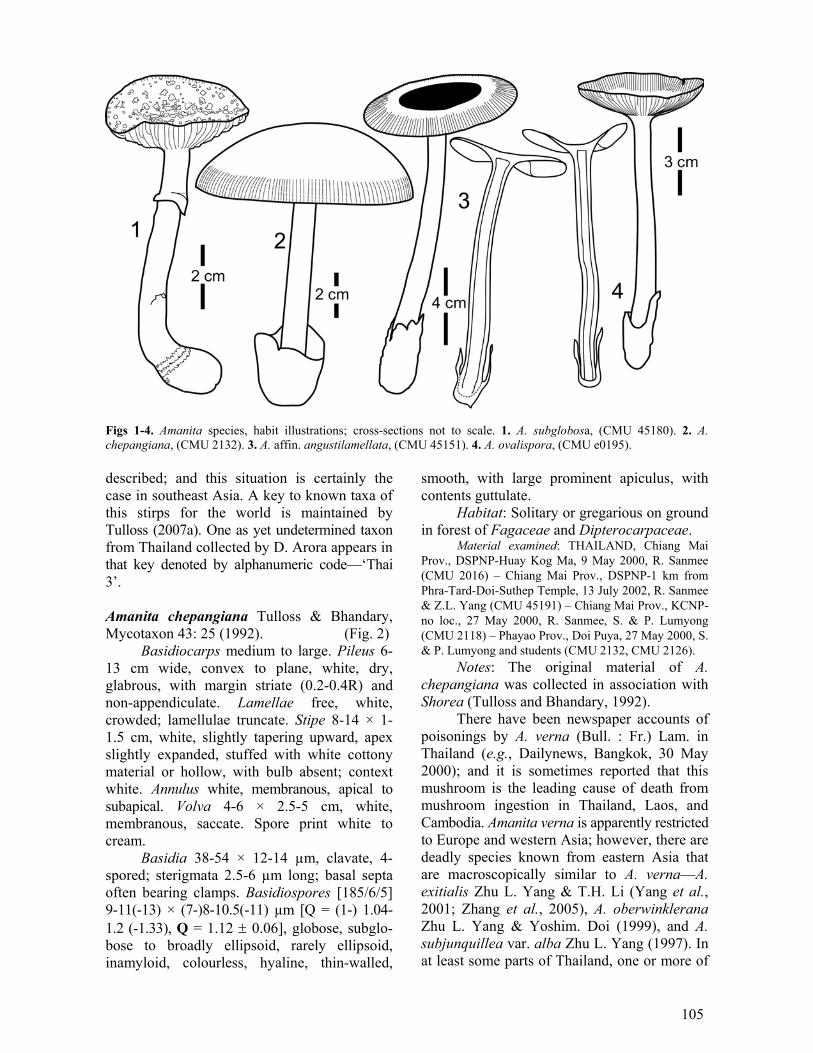
The most recent detailed treatment of the present species is its protolog. The material cited therein is from southwestern China. More recently, Oda et al. (2000) have reported A. sinensis from Japan and Nepal. Amanita subglobosa Zhu L. Yang, Bibliotheca Mycologica 170: 18 (1997). (Fig. 1)
Basidiocarps medium-sized. Pileus 3-5 cm wide, convex to plane, brownish to tea-brown, darker over disc, volval remnants as subpyramidal warts, 1-2 mm wide, up to 1.5 mm high, dirty white to cream, scattered; margin short striate (0.15-0.3 R), non-appendiculate; context white. Lamellae free, white; lamellulae truncate. Stipe 5-7 × 0.4-1.3 cm, white, equal, with apex slightly expanded, bulb at stipe base subglobose, ca. 0.8-2.5 cm wide, with juncture of stipe and bulb surrounded with whitish floccose ring of volva. Annulus white, submem-branous.
Lamella trama bilateral. Subhymenium 2-3 layers of subglobose to cuneiform to pyri-form to broadly clavate cells, 6-20 × 4-15 µm. Basidia 36-48 × 9-13 µm, clavate, 4-spored; sterigmata 2.5-5 µm long; basal septa often bearing clamps. Basidiospores [60/1/1] (7.5-) 8-10(-11) × (6.5-)7-8.5(-9) µm [Q = (1.05-) 1.1-1.32(-1.33), Q = 1.19 ± 0.07], subglobose to broadly ellipsoid, inamyloid, colourless, hyaline, thin-walled, smooth, with contents guttulate. Lamella edge as sterile and some-what gelatinized strip, 120-190 µm wide in side view, made up of globose to subglobose (6-16 µm wide), ovoid (16-18 × 10-12 µm) to clavate (18-26 × 10-14 µm) to bacilliform (20-25 × 8-10 µm) cells in chains of 4-8, thin-walled,
colourless. Volval remnants on pileus made up of irregularly arranged elements: filamentous hyphae fairly abundant, 3-6 (-8) µm wide, hyaline, thin-walled, with septa sometimes bearing clamps; inflated cells abundant to very abundant, globose to subglobose to ovoid to ellipsoid to (occasionally) fusiform, 18-60 × 8-44 µm, often in short chains, thin-walled, colourless. Annulus dominantly comprising irregularly arranged, filamentous hyphae (2-9 µm wide) mixed with abundant inflated cells (globose to subglobose to ellipsoid to clavate to fusiform, often in terminal chains, sometimes single and terminal, 14-88 × 8-52 µm, colourless, hyaline, thin-walled); clamps common.
Habitat: Solitary on ground in a forest with Castanopsis, Quercus, and species of Dipterocarpaceae.
Material examined: THAILAND, Chiang Mai Prov., DSPNP-1 km from Phra-Tard-Doi-Suthep Temple, 13 July 2002, R. Sanmee (CMU 45179, CMU 45180).
Notes: Amanita kwangsiensis Y.C. Wang (=A. sychnopyramis f. subannulata Hongo) and A. parvipantherina Zhu L. Yang et al. are somewhat similar to A. subglobosa, but they both lack clamps; and, in addition, the former has much smaller, globose to subglobose spores, and (Yang, 1994; Yang, 1997; Yang et al. 2004). Rarity or lack of clamps is common in pantherinoid taxa.
For comparative purposes the spore measurements from the protolog are provided: [110/4/3] (8.5-)9.5-12(-15) × (6.5-)7-9.5(-12.5) µm, [Q = (1.1-)1.18-1.44(-1.62); Q = 1.31 ± 0.09].
The most recent detailed treatment of the present species is provided by Yang (2005). The most recent detailed treatment in English is its protolog (southwestern Chinese material). A description of this taxon based on collections from northern India has appeared recently (Semwal et al., 2007). Amanita ibotengutake T. Oda, C. Tanaka & Tsuda (2002), described from Japan, is strikingly similar to A. subglobosa (Yang, 2005, 2007a); it is said to be distin-guishable on the basis of molecular phylogeny. Amanita section Caesareae Singer
All of the known taxa of this section from southeast Asia are assignable to the rather large group of species presently called Amanita stirps Hemibapha (Tulloss, 1998a, 2007a). Many taxa of this stirps are still to be
105
Figs 1-4. Amanita species, habit illustrations; cross-sections not to scale. 1. A. subglobosa, (CMU 45180). 2. A. chepangiana, (CMU 2132). 3. A. affin. angustilamellata, (CMU 45151). 4. A. ovalispora, (CMU e0195). described; and this situation is certainly the case in southeast Asia. A key to known taxa of this stirps for the world is maintained by Tulloss (2007a). One as yet undetermined taxon from Thailand collected by D. Arora appears in that key denoted by alphanumeric code—‘Thai 3’. Amanita chepangiana Tulloss & Bhandary, Mycotaxon 43: 25 (1992). (Fig. 2)
Basidiocarps medium to large. Pileus 6-13 cm wide, convex to plane, white, dry, glabrous, with margin striate (0.2-0.4R) and non-appendiculate. Lamellae free, white, crowded; lamellulae truncate. Stipe 8-14 × 1-1.5 cm, white, slightly tapering upward, apex slightly expanded, stuffed with white cottony material or hollow, with bulb absent; context white. Annulus white, membranous, apical to subapical. Volva 4-6 × 2.5-5 cm, white, membranous, saccate. Spore print white to cream.
Basidia 38-54 × 12-14 µm, clavate, 4-spored; sterigmata 2.5-6 µm long; basal septa often bearing clamps. Basidiospores [185/6/5] 9-11(-13) × (7-)8-10.5(-11) µm [Q = (1-) 1.04-1.2 (-1.33), Q = 1.12 ± 0.06], globose, subglo-bose to broadly ellipsoid, rarely ellipsoid, inamyloid, colourless, hyaline, thin-walled,
smooth, with large prominent apiculus, with contents guttulate.
Habitat: Solitary or gregarious on ground in forest of Fagaceae and Dipterocarpaceae.
Material examined: THAILAND, Chiang Mai Prov., DSPNP-Huay Kog Ma, 9 May 2000, R. Sanmee (CMU 2016) – Chiang Mai Prov., DSPNP-1 km from Phra-Tard-Doi-Suthep Temple, 13 July 2002, R. Sanmee & Z.L. Yang (CMU 45191) – Chiang Mai Prov., KCNP-no loc., 27 May 2000, R. Sanmee, S. & P. Lumyong (CMU 2118) – Phayao Prov., Doi Puya, 27 May 2000, S. & P. Lumyong and students (CMU 2132, CMU 2126).
Notes: The original material of A. chepangiana was collected in association with Shorea (Tulloss and Bhandary, 1992).
There have been newspaper accounts of poisonings by A. verna (Bull. : Fr.) Lam. in Thailand (e.g., Dailynews, Bangkok, 30 May 2000); and it is sometimes reported that this mushroom is the leading cause of death from mushroom ingestion in Thailand, Laos, and Cambodia. Amanita verna is apparently restricted to Europe and western Asia; however, there are deadly species known from eastern Asia that are macroscopically similar to A. verna—A. exitialis Zhu L. Yang & T.H. Li (Yang et al., 2001; Zhang et al., 2005), A. oberwinklerana Zhu L. Yang & Yoshim. Doi (1999), and A. subjunquillea var. alba Zhu L. Yang (1997). In at least some parts of Thailand, one or more of
106
these species goes under the local name of ‘hed kai kao teen ton’. Because of the presence of these deadly species, it is important to be able to distinguish the poisonous white species of sect. Phalloideae from the very common, edible A. chepangiana (with the local name ‘hed kai kao’).
Amanita verna (Breitenbach and Kränzlin, 1995; Neville and Poumarat, 2004) and its Asian look-alikes can be segregated from A. chepangiana by characters observable in the field, by their lacking striations along the pileal margin and having a bulb at the stipe base with a comparatively short limbate (not saccate) volva. If appropriate reagents are available, all species of section Phalloideae can also be segregated from all species of stirps Hemiba-pha by the amyloid reaction of a pile of spores scraped from a spore print. Under a microscope, specimens of all species of section Phalloideae will also be found to lack clamps at the bases of basidia in contrast with all taxa of stirps Hemibapha.
The most recent detailed description of the present species was published by Yang (1997); in his description he corrects several mistakes made in the protolog of A. chepangiana.
For comparative purposes, spore measure-ments from (Yang, 1997) are provided: [120/4/4] (8.5-)10-13(-16) × (7.5-)8.5-11(-11.5) µm [Q = (1.04-) 1.09-1.33 (-1.52), Q = 1.19 ± 0.08].
The species was originally described from Nepal; Yang’s material is from south-western China. The species has repeatedly been reported from as far north as Korea under the misapplied name Amanita caesarea var. alba Gillet (Yang, 1997, 2000) or A. hemibapha subsp. alba Kim et al. nom. inval. (1993). Amanita hemibapha (Berk. & Broome) Sacc. sensu lato, Sylloge Fungorum 5: 13 (1887).
The following is a general description that probably covers several taxa that may or may not have been recognized in the literature. More work is necessary in order to separate them.
Basidiocarps medium to large. Pileus 5-16 cm wide, convex to plane, concave, margin striate (0.3-0.4R), non-appendiculate, orange-red over centre, paler toward curry-yellow margin, glabrous, viscid when wet. Lamellae
free, creamy to yellow, crowded (approx. 7 lamellae/cm); lamellulae truncate. Stipe 6-19 × 0.6-3 cm, equal or slightly tapering upwards, apex slightly expanded, yellow to pale yellow, bearing pale orange fibrillose zones below annulus, stuffed with white to yellowish cottony material or hollow, with bulb absent; context white, creamy, or yellowish. Annulus yellow to pale orange, thin, membranous, apical, pendant, easily collapsing. Volva 2-6 × 1.5-2.5 cm, white, saccate, membranous. Spore print white.
Basidia 44-68 × 11-13 µm, clavate, 4-spored; sterigmata 3-4.5 µm long; basal septa often with clamps. Basidiospores [279/9/7] (7.5-)8-11(-13.5) × (5.5-)6-7(-9) µm [Q = (1.16-)1.31-1.5(-1.81), Q = 1.41 ± 0.09], ellipsoid, sometimes broadly ellipsoid, rarely elongate, inamyloid, colourless, hyaline, thin-walled, smooth, with contents guttulate.
Habitat: Gregarious on ground in forests of Dipterocarpaceae and Fagaceae.
Material examined: THAILAND, Chiang Mai Prov., DSPNP-Quinine Garden, 17 May 2000, R. Sanmee (CMU 2001) – Chiang Mai Prov., DSPNP-Sun Gu, 11 May 2002, R. Sanmee (CMU 4525) – Chiang Mai Prov., DSPNP-1 km from Phra-Tard-Doi-Suthep Temple, 13 July 2002, R. Sanmee & Z.L. Yang (CMU 45183) – Chiang Mai Prov., DSPNP-Huay Kog Ma, 28 August 2002, R. Sanmee (CMU 45252) – Chiang Mai Prov., KCNP–no loc., 27 May 2000, R. Sanmee, S. & P. Lumyong (CMU 2121) – Chiang Mai Prov., Khun Chang Kian, 11 June 2002, S. Lumyong (CMU 4523) – Phayao Prov., Doi Kum Phra, 27 May 2000, S. & P. Lumyong and students (CMU 2123).
Notes: Brightly coloured taxa of Amanita stirps Hemibapha make up one of the most popular groups of wild edible mushrooms in northern Thailand. The local name is ‘hed kai laung’.
Worldwide, there are many taxa in stirps Hemibapha (Corner and Bas, 1962; Yang, 1997; Tulloss, 1998a; 1998b, 2007a; Tulloss et al., 2001). They vary in colour, bruising reactions, size, and spore characters; many have a pronounced umbo and rather long marginal striations. However, the basidiocarps of some, including A. hemibapha sensu stricto [known with greatest confidence from Sri Lanka and southern India (Berkeley and Broome, 1871; Vrinda et al., 2005; Tulloss, 2007g)], may often lack an umbo. In our Thai collections, a dominantly yellow entity of stirps Hemibapha is more plentiful than a red one. Chansrikul et al. (1984) and Rachabundi-

107
tayasathan (1996) reported two taxa in Thailand—A. hemibapha subsp. javanica Corner & Bas (1962) [=A. javanica (Corner & Bas) T. Oda et al. (1999)], a predominantly yellow to orange-yellow entity and A. hemibapha subsp. similis (Boedijn) Corner & Bas (1962) [=A. similis Boedijn (1951)], a brown-capped entity having a yellow stipe decorated with orange fibrillose patches. A species of the same stirps with a completely bright orange-red cap is depicted on the worldwide web in a photograph labelled ‘Amanita caesarea [sic] (Thailand)’ by Taylor Lockwood (2004); this photograph strongly suggests A. caesareoides Lj. N. Vassiljeva, presently known from northern India to eastern Siberia.
The present entry uses the name A. hemi-bapha in a broad sense—covering the brightly coloured taxa (of whatever rank they may be assigned) of stirps Hemibapha in Thailand. Amanita chepangiana, above, and A. princeps, below, are both taxa of stirps Hemibapha that lack the bright colours of taxa treated within the present entry; moreover, the two cited species have spores tending to be more nearly globose or subglobose than spores of many of the brightly coloured taxa. Amanita princeps Corner & Bas, Persoonia 2: 297 (1962).
Basidiocarps medium to large. Pileus 8-15 cm wide, convex to plane, concave, brownish, darker over disc, paler toward a striate margin (0.2-0.4R), non-appendiculate, glabrous, viscid when wet. Lamellae free, white, approx. 1.2 cm broad, crowded, averaging 6-7 per cm; lamellulae truncate. Stipe 8-23 × 1.5-2.5 cm, slightly tapering upward, white to whitish, stuffed with white cottony material or hollow; bulb absent; context white, moist. Annulus white, apical, membranous, thin, easily collapsed. Volva saccate, 4-8 × 2-3.5 cm, membranous, white to whitish. Spore print white.
Basidia 33-58 × 9-17(-25) µm, clavate, 4-spored; sterigmata (2.5-)4-7 µm long; basal septa often with clamps. Basidiospores [90/3/3] (9-)9.5-11(-13) × 9-11(-13) µm [Q = 1-1.08(-1.22), Q = 1.06 ± 0.05], globose to subglo-bose, rarely broadly ellipsoid, inamyloid, colour-less, hyaline, thin-walled, smooth, with large and prominent apiculus, with contents guttulate.
Habitat: Solitary or gregarious on ground under Castanopsis.
Material examined: THAILAND, Chiang Mai Prov., DSPNP-Quinine Garden, 17 May 2000, R. Sanmee (CMU 2000) – ibid., 6 June 2002, R. Sanmee & R. Kodsueb (CMU 4504).
Notes: Amanita princeps is a common edible wild mushroom in northern Thailand during the rainy season. The local name is ‘hed kai kao’.
The current species was originally describ-ed from Singapore. It seems to be common in tropical China (Yang et al., 2001; Yang 2005, 2007b).
Amanita section Vaginatae (Fr.) Quél. Amanita affin. angustilamellata (Höhn.) Boedijn, Sydowia 5: 318 (1951). (Fig. 3)
Basidiocarps medium-sized. Pileus 3.5-9.5 cm wide, convex to umbonate-convex to plane, often subumbonate or lacking an umbo, with margin long striate (0.4-0.5R), non-appendiculate, greyish brown, darker at disc, paler toward margin, glabrous. Lamellae free, white to whitish, subcrowded, thick; lamellulae truncate. Stipe 10-14 × 0.5-1 cm, slightly taper-ing upward, apex slightly expanded, white to brownish, bearing grey to greyish fibrils; context white, hollow. Exannulate. Volva 2.5-4 × 1-2 cm, white, with very pale greyish tinge, saccate, membranous.
Basidia 42-62 × 13-16 µm, clavate, 4-spored; sterigmata 4.5-7 µm long; basal septa without clamps. Basidiospores [151/5/5] (9-)9.5-11(-12) × (8-)9-11(-11.5) µm [Q = 1-1.04 (-1.14), Q = 1.04 ± 0.03], globose to subglo-bose, inamyloid, colourless, hyaline, thin-walled, smooth, with contents guttulate.
Habitat: Solitary or gregarious on ground in forests including Dipterocarpaceae or Fagaceae.
Material examined: THAILAND, Chiang Mai Prov., DSPNP-Huay Kog Ma, 24 July 1999, R. Sanmee (CMU e0209) – Chiang Mai Prov., Doi Saked, Huay Hong Krai, 6 July 2002, R. Sanmee, Z.L. Yang & K.D. Hyde (CMU 45151) – Chiang Mai Prov., KCNP-no loc., 10 July 1999, R. Sanmee, S. & P. Lumyong & B. Dell (CMU e0152) – Chiang Mai Prov., Sanpathong, Mae Wang, 5 October 2002, S. & P. Lumyong, R. Sanmee (CMU 45265) – ibid., 8 December 2002, S. & P. Lumyong, R. Sanmee (CMU 45333).
Notes: FH 4712 is sole syntype and, therefore, lectotype of the present species; and it is in good condition (Zhu L. Yang, pers. comm.) except that the base of the stipe and the

108
volva were not collected. The present day understanding of the taxon is based largely on the protolog, the drawing von Höhnel deposited with his exsiccatum (FH), and interpretations by subsequent authors—Boedijn (1951), Corner and Bas (1962) and Yang (1997). A brief summary of current knowledge of the present species is maintained by Tulloss (2007e).
Z.L. Yang (pers. corresp.) generously provided us with his unpublished data on the spores of the type. They are included here for comparative purposes: [35/1/1]11-14(-15) × (10-) 10.5-13 (-14.5) µm, Q = 1-1.1 (-1.15), Q = 1.05 ± 0.04.
The region of interest is not at all thoroughly explored, and species of sect. Vaginatae are among the undetermined exsiccata awaiting further research. In the present case, at least three concerns remain as open issues with regard to determination of the Thai material here listed as A. affin. angustilamellata. The original drawing of von Höhnel shows the length:breadth ratio of the lamellae to be in the range of 6.5-6.75 while the same ratio taken from the habit illustration accompanying the present description is 2.9. Tulloss’ on-line summary [based largely on the descriptions of von Höhnel, Boedijn, and Yang (1997)] mentions that the saccate volva of A. angustilamellata is attached only at the very base of the stipe. This character is also shown in Corner’s watercolour of the present species (Corner and Bas, 1962; Tulloss, 2007e). The habit on Thai material examined shows a more extensive region of volval attachment to the stipe. Spore measurements (possibly affected by the quality of preservation of the Thai material?) are not a close match to those reported from the type (e.g., ranges of spore length in the two data sets are nearly disjunct). Additional, well-annotated and illustrated collections of material considered to be A. angustilamellata are very much needed from sites throughout the supposed range of the species, in order to get a more definitive understanding of it.
Amanita angustilamellata has been previously reported from Indonesia (protolog), Singapore (Corner and Bas, 1962), and south-western China (Yang, 1997).
Amanita ovalispora Boedijn, Sydowia 5: 320 (1951). (Fig. 4)
Basidiocarps medium-sized. Pileus 4-5 cm wide, convex to plane, glabrous, viscid, dark grey at centre, paler (e.g., ash-grey) toward nonappendiculate, striate (0.4-0.6 R) margin. Lamellae free, white, crowded; lamel-lulae truncate. Stipe 8-10.5 × 0.6-0.8 cm, slightly tapering upward, with apex slightly expanded, white, fibrillose to floccose; context white, hollow. Exannulate. Volva saccate, 2.5-3 × 1.5-2 cm, membranous, white to dirty white.
Basidia 33-50 × 12-15 µm, clavate, 4-spored; sterigmata 2.5-5 µm long; basal septa without clamps. Basidiospores [120/4/4] (8-)9-10.5(-13.5) × (6-)7-9(-10) µm [Q = (1.09-) 1.15-1.37(-1.7), Q = 1.3 ± 0.13], broadly ellipsoid to ellipsoid, rarely subglobose or elongate, inamy-loid, colourless, hyaline, thin-walled, smooth, with contents guttulate.
Habitat: Solitary on ground in forest of Fagaceae.
Material examined: THAILAND, Chiang Mai Prov., DSPNP-Huay Kog Ma, 20 July 1999, R. Sanmee, S. & P. Lumyong (CMU e0195) – ibid., 24 July 1999, R. Sanmee (CMU e0211) – ibid., 9 May 2000, R. Sanmee, S. & P. Lumyong (CMU 2036) – Chiang Mai Prov., Chiang Mai Univ., Ang Kaew, 27 July 2002, R. Sanmee, Z.L. Yang & H.Y. He (CMU 45238).
Notes: The most recent detailed treatment of this taxon is by Yang (1997) who described southwestern Chinese material. In the same work, Yang provides a useful comparison to A. pseudovaginata Hongo.
This species is often called A. vaginata (Bull. : Fr.) Lam. in the Thai literature; however, A. vaginata is a European and west Asian species that has globose basidiospores (e.g., Breitenbach and Kränzlin, 1995). Unfortunขately, the latter species is poorly understood, and its name is misapplied throughout the world.
The current species was originally described from Indonesia. Amanita pudibunda R. Heim ex R. Heim, Revue de Mycologie 30: 235 (1965).
≡ Amanita pudibonda [sic] R. Heim, Revue de Mycologie 27: 146 (1962) nom. inval. (Lacking Latin diagnosis.) (Figs 5-7)
Pileus: 35-45 mm wide, white, gibbous to convex at first, then convex to pulvinate, flat over disc, with tomentose to farinose surface decoration; context white; margin short striate (faint, but 0.25R in preserved material),

109
Figs 5-7. A. pudibunda (holotype). Scale bars on microscopic anatomy indicate 20 µm. 5. Habit. 6. Elements of hymenium and subhymenial tree. 7. Elements of universal veil from stipe base. farinose, slightly incurved at least at first; universal veil not described in original descrip-tions, but depicted as scattered irregular fragments, probable source of surface decora-tion reported with microanatomical characters (below).
Lamellae: spacing from stipe not noted, crowded as preserved, white with flesh-coloured tint in mass, white in side view, rather broad, rather thick and fleshy, with smooth and concolourous edge; lamellulae not described.
Stipe: 80-100 × 9-12 mm, white, narrow-ing upward evenly, having series of concolour-ous rings or broken rings in apical third (fragile, white, farinose), pulverulent, with pulverulence easily lost below apical third, quickly becoming nearly smooth below, with subtle browning below apical portion (from handling?); context white, with ample central cylinder (width not recorded); universal veil as saccate volva, ample, with limb of roughly even height to slightly bilobate, membranous, rather thick, whitish with pinkish brown spots, 32 × 20 mm, 2 mm thick as preserved,
apparently lacking limbus internus in preserved state.
Odour subtle, agreeable; taste sapid. Macrochemical tests: none reported. Pileipellis: comprising colourless supra-
pellis (445-450 µm thick) and subpellis (235± µm thick) with hyphae having brown tinted walls, overall 680-685 µm thick, overlaid by nearly uniform layer from inner surface of universal veil (see below); suprapellis compri-sing gelatinized matrix including loosely interwoven, narrow, filamentous, undifferen-tiated hyphae seeming to curve up into matrix from subpellis; subpellis with filamentous, undifferentiated hyphae up to 9.6 µm wide, branching, often constricted at septa, ungelati-nized; vascular hyphae not observed. Pileus context: filamentous, undifferentiated hyphae 4.8-14.4 µm wide, branching, plentiful; acro-physalides narrowly clavate, plentiful to dominant, with many subradially arranged, up to 145 × 32 µm or larger, with walls thin or commonly up to 0.8 µm thick, terminal or occasionally in chains; vascular hyphae 6.4-

110
12.8 µm wide, scattered, sinuous, orange-brown. Lamella trama: bilateral; wcs = 30-35 µm;
subhymenial base including plentiful inflated cells (clavate to broadly clavate to elongate to ovoid, thin-walled, apparently all intercalary, up to 73 × 30 µm), with angle of divergence from shallow to 60° or more; central stratum containing plentiful partially inflated intercalary segments up to 14.4 µm wide; filamentous, undifferentiated hyphae 2.1-6.4 µm wide, branching, often with granular contents; terminal, divergent inflated cells occasional, of the same form as intercalary cells of subhymenial base; vascular hyphae not observed. Subnymenium: wst-near = 70-90 µm; wst-far = 95-100 µm; as branching structure comprising short uninflated and partially inflated hyphal segments and small inflated cells, with 10-30 µm between bases of largest basidia/-oles and subhymenial base, with 45-50 µm between bases of shortest basidia/-oles and subhymenial base, with basidia arising from cells of all types, least frequently from inflated cells. Basidia: 39-61 × 8.0-11.6 µm, 4-sterig-mate; clamps not observed. Universal veil: On pileus, lower surface, attached to pileipellis: nearly uniform layer 20-50 µm thick of brownish walled partially gelatinized elements, with all types of elements often having granular contents (see description of pileipellis, above); filamentous, undifferentiated hyphae up to 7.2 µm wide, branching; inflated cells (up to 47 × 11.2 µm or larger), collapsed, narrowly clavate to allantoid, possibly in chains. On stipe base, exterior surface: as rather dense layer to depth of up to 85 µm; filamentous, undifferentiated hyphae 1.0-8.0 µm wide, branching, dominantly fasciculate and often in rather broad fascicles, also appearing singly, interwoven in open lattice, but denser than in interior, with many partially gelatinized; inflated cells not observed; vascular hyphae 3.2 µm wide, scattered to rare, fragmented. On stipe base, interior: filamentous, undifferentiated hyphae 3.2-11.2 µm wide, branching, plentiful, often fasciculate, thin-walled, with septa occasionally constricted, very loosely inter-woven in open lattice, with tip cells sometimes slightly expanded; inflated cells plentiful to locally dominating, thin-walled, up to 105 × 79 µm, occasionally irregularly clavate, usually
broadly ellipsoid to subglobose; vascular hyphae 3.2-15.2 µm wide, scattered, sinuous; clamps not observed. On stipe base, inner surface: partially gelatinized in some regions, very similar to dark layer on pileipellis surface, from comparatively thin to up to 110 µm thick; filamentous, undifferentiated hyphae 2.4-9.6 µm wide, with some intercalary segments up to 14.4 µm wide, frequently branching, dominating, densely interwoven, with almost all in fascicles, frequently septate, with yellow granular contents in 20% NH4OH, with dark brown pigment apparently restricted to cell walls; inflated cells apparently terminal (singly or in short chains), narrowly clavate or allantoid (up to 123 × 32 µm, thin-walled) to pyriform or subglobose (up to 43 × 40 µm or larger, with walls up to 1.6 µm thick), plentiful, often with granular contents as in hyphae; vascular hyphae not observed. Stipe context: longitudinally acro-physalidic; filamentous, undifferentiated hyphae 2.4-9.6 µm wide, branching, plentiful; acrophy-salides dominating, thin-walled, up to 298 × 32 µm; vascular hyphae not observed.
Basidiospores: [20/1/1] (6.5-)6.8-10.4 (-10.5) × (5.1-)5.2-7.4(-7.9) µm, (L = 8.7 µm; W = 6 µm; Q = (1.25-)1.27-1.65(-1.74); Q = 1.46), hyaline, colourless, with walls very slightly thickened (< 0.5 µm), smooth, inamyloid, broadly ellipsoid to ellipsoid to elongate; apiculus sublateral; contents multiguttulate; colour in deposit unknown.
Habitat: In humus of old Dipterocarpus forest, 1200 m elev.
Material examined: THAILAND, Chiang Mai Prov., DSPNP, [5] December 1957, R. Heim [Th.] 57 (PC, holotype).
Notes: Heim (1962: 148) states that this species was most closely related to the varieties of Amanita annulatovaginata Beeli (from the Republic of Congo) due to the presence of fragile decoration on the upper stipe (suggestive of an annulus) and the shape and size of the spores. However, the gracile African species was not reported to have, and from its illustrations does not appear to have, the curious covering of volval material that A. pudibunda bears on its pileus; and there seems little reason to judge the two taxa to be closely related phenetically.
Yang (pers. corresp.) has suggested to Tulloss that this taxon might well be placed in

111
sect. Amidella except for its inamyloid spores. Weak amyloid reaction in spores of one collection of Amidella was reported by Bas (1969: 342) as Yang noted. Yang (pers. corresp.) also reports that he has reviewed dried material that seems to belong to sect. Amidella with the exception of having inamyloid spores.
This species is known only from a single specimen. The holotype of A. pudibunda is preserved in liquid. Generally speaking, Tulloss found the tissues to be in very good condition. Parts of the collection data in brackets do not appear on the herbarium label in PC. ‘Th.’ stands for the name of the actual collector.
New material of A. pudibunda with good notes on its fresh state and in condition to support molecular studies is very much needed. Amanita [subgenus Lepidella] section Amidella (E.-J. Gilbert) Konrad & Maubl. Amanita avellaneosquamosa (S. Imai) S. Imai in Ito, Mycological Flora of Japan 2(5): 250 (1959).
Basidiocarps medium-sized. Pileus 4-9 cm wide, convex to plane, slightly depressed in centre, margin striate (0.25-0.35R), non-appen-diculate when collected, whitish, with volval remnants cream to yellowish brown, farinose or floccose small patches. Lamellae free, white to whitish, subcrowded; lamellulae truncate. Stipe 5-7 × 0.7-2 cm, slightly tapering upward, white to cream, decorated with white farinose to floccose material; context white to whitish. Annulus fugacious. Volva 2-3 × 1.5-3 cm, yellowish brown, saccate, membranous. Odour strong, unpleasant (possibly due to decay). Spore print white.
Basidia 40-50 × 8-14 µm, 4-spored; sterig-mata 3-5 µm long; basal septa without clamps. Basidiospores [109/3/3] (8.5-)9-11(-12) × (4.5-)5-6.5(-7) µm [Q = (1.35-)1.5-1.88(-2.5), Q = 1.74 ± 0.19], ellipsoid to elongate, rarely cylindric, amyloid, colourless, hyaline, thin-walled, smooth; apiculus small.
Habitat: Solitary on ground in forests of Fagaceae and Pinaceae.
Material examined: THAILAND, Chiang Mai Prov., DSPSP-Suan Sone, 11 June 1999, R. Sanmee, S. & P. Lumyong (CMU e0051) – Chiang Mai Prov., DSPSP-Sun Gu, 29 July 2000, S. & P. Lumyong, R. Sanmee (CMU 2220) – Chiang Mai Prov., KCNP, 27 May 2000, S. & P. Lumyong, R. Sanmee (CMU 2119).
Notes: Species of sect. Amidella represent notable exceptions to the belief that the presence of truncate lamellulae in an Amanita correlates with the presence of inamyloid spores (Tulloss and Yang, 2007b). Amanita volvata (Peck) Lloyd (a North American taxon) is similar to A. avellaneosquamosa but differs by pinkish to red-brown staining after injury to the context, having a fibrillose-floccose inner layer of volval remnants (made up of abundant, fusiform, broadly clavate to ellipsoid, loosely and irregularly arranged inflated cells) almost always remaining in part or whole on the pileus, having a thicker subhymenium with (2-) 3-4 (-5) layers of inflated cells, and having somewhat larger spores. This is according to Yang (2000), who reported the present species from southwestern China (Yang, 1997). This species was reported from northern India as A. volvata according to Bhatt et al. (2003).
The present species differs from A. clari-squamosa, below, by having more elongate spores (higher average Q), having a volval sac that is often more laterally compressed, longer marginal striations on the pileus, less crowded lamellae, and smaller spores—as reported by Yang (1997).
For comparative purposes, the spore measurements from Yang’s description of material from southwestern China are provided: [110/4/2] (8-)9-11(-12) × 5.5-6.5(-7) µm [Q = (1.33-)1.43-1.87(-2), Q = 1.65 ± 0.14]. In addition Tulloss’ spore measurements from material of China, Japan, and northern India are as follows: [125/5/5] (5.8-)7-10.5 (-11.8) × (4-)4.8-6.5(-7.8) µm [Q = (1.28-)1.38-1.91(-2), avg. Q per specimen = 1.48-1.71; Q = 1.61 ± 0.16].
Extralimital habitat: India: At 1900±-3250 m elev., in forest of Quercus leucotri-chophora A. Camus and Rhododendron arbo-reum Sm., with scattered Cedrus deodara (Roxb. ex Lambert) G. Don. or under Pinus roxburghii Sarg. or in mixed forest including Abies pindrow (Royle ex D. Don) Royle, Picea smithiana (Wall.) Boiss., Pinus wallichiana A.B. Jacks., Q. dilatata Royle, Q. semecarpifolia Sm. [latter known to be mycorrhizal with current species (Kumar et al., 1990)], and Taxus. Japan: At ca. 300 m elev., in evergreen forest with Castanopsis and some Abies and Pinus.

112
Extralimital material examined: CHINA, Jiangsu Prov., Nanjing, Ling-ku-sze Woods, 14 July 1936 S.C. Li 112 (CUP-CH) – INDIA, Himachal Pradesh, Shimla, Narkanda, Hattoo Peak, 20 August 1986, T.N. Lak-hanpal & A. Kumar s.n. (HPUB 4430 (n.v.); BPI 71990) – Uttarakhand, Mussooree, 21 September 1964 C. Bas 4442 (L) – Uttarakhand, Pauri Garhwal, Dandapani, 8 August 2001, K.C. Semwal & R.P. Bhatt 378 (GUH; RET) – JAPAN, Chiba Pref., Kiyosumi, 25 August 1983 C. Bas 9002 (L) – Shiga Pref., Seta-chô, 2 October 1949, T. Hongo 6 (herb. T. Hongo).
The current species has been reported previously from Japan (protolog), southwestern China (Yang, 1997), and northern India (Bhatt et al., 2003). Amanita clarisquamosa (S. Imai) S. Imai in Ito, Mycol. Fl. Jap. 2: 250 (1959).
Field notes for the single collection available were limited. The description is largely based on the description of Yang (1997).
Pileus 7 cm wide, convex at first, planar when expanded, slightly depressed over disc, white to whitish, slightly yellow-brownish over disc, viscid (or tacky), decorated with grey-brown, brownish to brown, irregular, membra-nous to fibrillose volval remains, with margin shortly and lightly striate (0.1-0.15R) and often appendiculate; context white, 4-6 mm thick over stipe, thinner toward margin, not or indistinctly discolouring. Lamellae free to nearly free, 5-8 mm broad, white, tending to become greyish, grey-brownish, brownish or chocolate brown when dried slowly or in humid environment, crowded to moderately crowded, with edge finely floccose, greyish to brownish; lamellulae truncate to rounded truncate, with 0-2 between each pair of lamellae. Stipe 12 × 1.2-1.8 cm, nearly cylin-dric or tapering slightly toward apex, with whitish ground colour under grey-brown scales or flocculence; context white to whitish; basal bulb lacking. Volva saccate, fleshy, 4-5 cm high, 3.5-4 cm wide, 2-9 mm thick, adhering to stipe surface, with outer surface white to sordid white, with inner surface whitish, with limbus internus placed high on limb and somewhat difficult to distinguish. Annulus fragile or ephemeral, often remaining as grey-brown scales on upper portion of stipe. Odour and taste not conspicuous. Spore deposit ‘white’.
Basidia 4-sterigmate; basal septa without clamps. Basidiospores [34/3/1] (10-)11-12.5(-16) × (7-)7.5-8(-11) µm, [Q = (1.15-)1.44-1.63
(-2.07), Q = 1.52 ± 0.17], amyloid, broadly ellipsoid to ellipsoid, occasionally elongate, rarely cylindric, colourless, hyaline, thin-walled; apiculus small, cylindric, sublateral.
Habitat: In mixed forest including Pinus and Castanopsis.
Material examined: THAILAND, Chiang Mai Prov., Om Koi, 12 September 1999, D. Arora 99281 (RET; SFSU).
Notes: Our material conforms well to the description of the present species by Yang (1997) as well as with unpublished data of Tulloss. For comparison to A. avellaneosqua-mosa, see observations under that species. In Arora 99-281, ‘giant spores’ were present—probably spores from basidia with less than 4 sterigmata. Bhatt et al. (2003) note that this species has been reported in northern India as A. peckiana Kauffman. Tulloss’ spore data for A. clarisquamosa (using notation of the present paper) based on collections from India are as follows: [150/7/3] (7-)10-12(-15) × (5.5-)6-7(-9) µm, [Q = (1.18-)1.45-1.77(-1.88), avg. Q per specimen = 1.50-1.69, Q = 1.59 ± 0.13].
Extralimital habitat: India: At 1850-2800± m elev., on slope in pure Abies pindrow forest or on N slope in coniferous forest including A. pindrow, Picea smithiana, and Pinus wallichiana or scattered on rich humus under Myrica esculenta Buch.-Ham. ex D. Don, Quercus leucotrichophora, and Rhodo-dendron arboreum.
Extralimital material examined: INDIA, Himachal Pradesh, Narkanda, 6 August 1964, C. Bas 4066 (L) – ibid., 11 August 1964 C. Bas 4118 (L) – Uttarakhand, Garhwal, Dandapani, 20 September 1993, V.K. & R.P. Bhatt s.n. (GUH M-20168; RET).
The current species has been reported previously from Japan (protolog), southwestern China (Yang, 1997), and northern India (Bhatt et al., 2003). Amanita section Lepidella sensu Bas (1969) Amanita alboflavescens Hongo, Memoirs of the Shiga University, Natural Science 20: 50 (1970).
Basidiocarps medium-sized. Pileus 6 cm wide, convex to plane, pale yellow, volval remnants floccose to squamulose, yellowish to brownish, becoming lemon-chrome when touched, margin non-striate, appendiculate. Lamellae free, whitish, cream to yellowish, edges serrate, crowded; lamellulae nearly truncate. Stipe 12 × 0.9-1.2 cm, slightly tapering

113
upward, apex slightly expanded, yellowish, floccose to squamulose, with ovoid, bulbous base 2.2 cm wide; context white. Annulus yellowish, apical, floccose-membranous. Volva yellowish, floccose-felted, fugacious. Spore print white to cream.
Basidia clavate, 4-spored, sterigmata 5-6.5 µm long. Basidiospores [30/1/1] 8-10.5 (-11) × 5-6(-6.5) µm [Q = 1.47-1.78 (-2), Q = 1.66 ± 0.12], ellipsoid to elongate, rarely cylindric, amyloid, colourless, hyaline, thin-walled, smooth; apiculus small.
Habitat: Solitary on ground in forest of Fagaceae.
Material examined: THAILAND, Chiang Mai Prov., DSPNP-Huay Kog Ma, 25 May 2000, S. & P. Lumyong, R. Sanmee (CMU 2113).
Notes: The North American species, A. crassifolia Bas nom. prov., is similar to A. alboflavescens, but the former has thicker lamellae and common clamps (Bas, 1969). The Thai A. alboflavescens specimen is in poor condition, and no clamps were observed. According to Yang’s observation of Chinese specimens, A. alboflavescens has no clamps.
Tulloss (2000) has expressed the view that some yellow-staining North American species belonging to sect. Lepidella are apparently specimens of non-yellowing taxa that have been infected by one or more patho-gens. Bas considered the possibility that A. crassifolia was an unusual specimen of A. subsolitaria (Murrill) Murrill; and Tulloss (2007f) now states this to be the case. This situation suggests that A. alboflavescens should be investigated for the cause of its yellowing reaction. If the variability of spore width (and, consequently, of spore shape) reported in the above description and these notes is supported by examination of additional collections, this may be further evidence of a nongenetic factor’s having an effect on the basidiocarps of A. alboflavescens. Corner (1947) observed that spore width of a given species is usually less variable than spore length of that species; and, based on Tulloss’ nearly 30 years of research on the genus, this appears to be true in Amanita.
Since the Thai material may not have typical spores, for purposes of comparison, we provided three other sources of data. In its protolog, the spores of the present species were described as follows: ‘8-12 × 4.5-6.5 µm’ (esti-
mated Q = 1.82). Yang (1997: 159) provides the following data from a southwestern Chinese collection: [33/1/1] (7.5-)8-10.5(-11.5) × (4.5-) 5-6 µm [Q = (1.52-)1.58-1.91(-2.3), Q = 1.72 ± 0.14]. Tulloss’ spore measurements from a Chinese collection are as follows: [55/3/1] (7.7-)8-10.3(-11) × (3.6-)4-5.5(-6) µm [Q = (1.4-)1.62-2.24(-2.39), avg. Q per specimen = 1.88-2, Q = 1.92 ± 0.21].
Extralimital material examined: CHINA, Yunnan Prov., Lu Feng, 2 August 2002, D. Arora 02-149 (RET, SFSU).
Amanita alboflavescens was originally described from Japan. In addition to China, it has been reported from Korea (Kim et al., 1993).
Amanita hongoi Bas, Persoonia 5: 410 (1969). (Fig. 8)
Basidiocarps medium to large. Pileus 5-8 cm wide, convex to plano-convex; margin non-striate, appendiculate, brownish to dull yellow-ish over centre, paler toward margin (whitish to pale brown), dry; volval remnants as conic warts 1.5-4 mm high, 1.5-3.5 mm wide, whitish to dull yellowish to brownish, diminishing in size (with colour paler) towards margin. Lamellae free, close to crowded, white to cream, up to 2.5 cm broad; lamellulae attenuate. Stipe 7-16 × 1-2 cm, with apex often brownish, floccose to squamulose, with base clavate-bulbous, 2.5-7.5 cm wide, sometimes splitting longitudinally, dirty white to reddish, with conic volval warts on at least upper part of bulbous base if not also on lower stipe and more of bulb. Annulus fragile, with upper surface white and radially striate, with lower surface white to dirty white, verrucose. Context white, unchanging, solid.
Basidia 53-63 × 11-14 µm, clavate, 4-spored; sterigmata 4-7 µm long; basal septa without clamps. Basidiospores [123/4/2] 7-9 (-10) × (5.5- 6.5-7(-8) µm [Q = (1.06-) 1.12-1.25 (-1.36), Q = 1.19 ± 0.07], subglobose to broadly ellipsoid, rarely globose or ellipsoid, amyloid, colourless, hyaline, thin-walled, smooth.
Habitat: Gregarious on ground in forest of Fagaceae.
Material examined: THAILAND, Chiang Mai Prov., Khun Chang Kian, 29 July 2002, Z.L. Yang, R. Sanmee, S. & P. Lumyong (CMU 45216) – Chiang Mai

114
Figs 8-10. Amanita species, habit illustrations; cross-sections not to scale. 8. A. hongoi, (CMU 45216). 9. A. fuliginea, (CMU 45215). 10. A. sinocitrina, (CMU 45230). Prov., 50 km S of Om Koi, ca. Lahu village, 11 September 1999, D. Arora 99-280 (RET; SFSU).
Notes: This species is somewhat similar to Amanita castanopsis Hongo. However, the basidia of the latter have common clamp connections on their basal septa; and A. castanopsis has elongate spores (Hongo, 1974; Yang, 1997; Yang and Doi, 1999).
For comparative purposes, spore measure-ments from the protolog are provided: [20/1/1] (7-)8-10(-11.5) × 7-9(-10) µm [Q = 1-1.2, Q = 1.1].
Amanita hongoi was originally described from Japan. It has been reported from Korea and China (Kim et al., 1993; Yang, 2005) Amanita japonica Hongo ex Bas, Persoonia 5: 399 (1969).
Because of lack of data on the single collection obtained in the region of study, the following macroscopic description is excerpted
from the monograph of Bas (1969). Basidiocarps ‘medium-sized, slender’.
Pileus ‘55-80 mm wide, convex then plane, with appendiculate, non-sulcate margin, dry, at first moderately dark grey to pale buffy grey [‘drab-grey’ to ‘light drab’ of Ridgway (1912)], felted-subflocculose, rather densely set with somewhat paler, floccose-felted, subpyra-midal, adnate warts, up to about 2 mm wide and 1.5 mm high; later coloured surface of cap breaking up into vaguely delimited, thin patches between which whitish flesh showing, in which case every pale wart situated at centre of a minutely radially fibrillose, practically circular, darker grey spot; warts and spots diminishing in size and less distinct towards margin’. Lamellae ‘close to subdistant, nearly free, sometimes with slight decurrent teeth, rather broad, 7-8 mm broad, subventricose, white, with subflocculose edge’; lamellulae ‘subtrun-cate to attenuate’. Stipe 80-170 × 7-15 mm,

115
attenuate upward, with fusiform-rooting to subclavate base up to 25 mm wide, solid, white, floccose, with flocculose-pulverulent to small, vague, scale-like, pale buffy grey (‘light drab’ of Ridgway) remnants of volva on lower half, with fugacious floccose-fibrillose remnants of white ring at top. Flesh soft. Taste and smell indistinct’.
Basidia bearing clamps. Basidiospores [23/1/1] 8-9(-11.5) × (4.5-)5.5-6.5(-7.5) µm [Q = 1.34-1.64(-2), Q = 1.52 ± 0.14], colourless, hyaline, thin-walled, smooth, amyloid, ellipsoid to elongate; apiculus sublateral; contents granular to guttulate.
Habitat: On ground under Quercus and Dipterocarpaceae.
Material examined: THAILAND, Chiang Mai Prov., Om Koi, 1999 D. Arora s.n. (RET; SFSU).
Notes: The single specimen available is not in good condition, and the spore size and value of Q are probably somewhat depressed because of its condition prior to, or during, drying. Nevertheless, clamps were not difficult to find in the stipe context. Among Asian taxa, A. japonica is most similar to A. miculifera Bas & Hatan. (Bas & Hatanaka, 1984) and A. griseoverrucosa Zhu L. Yang. According to Yang (1997), A. miculifera differs from A. japonica in having a conic to flattened-conic pileus with volval remains in the form of friable warts, a stipe with a notably radicating basal bulb, larger spores, and a disordered microscopic structure in volval remnants. Its authors placed A. miculifera in Bas’ stirps Virgineoides, not with A. japonica in Bas’ stirps Solitaria. Amanita griseoverrucosa has a much less strongly rooting bulb, lighter coloured volval remnants on the pileus, and no clamps (Yang, 2004). Amanita eijii Zhu L. Yang (2002a) is a recently described species of stirps Solitaria that should be compared with the present species. Yang’s species is distin-guished among other things by its often larger basidiocarp, becoming pinkish to brownish in part after in situ aging or cutting, an annulus that is membranous or submembranous and sometimes persistent with fibrils largely free from the undersurface attaching that surface to the stipe, distinctly reflexed scales (with volval material on their tips) arranged in incomplete or complete rings on the lower portion of the stipe and the upper part of its bulb, and slightly
longer and significantly broader spores with Q = 1.36 ± 0.08.
Spore measurements from southwestern Chinese material of A. japonica (Yang, 1997) are provided for purposes of comparison: [70/2/2] (7.5-)8.5-10.5× (5-)5.5-7 µm [Q = (1.31-)1.36-1.67(-1.81), Q = 1.52 ± 0.1].
Amanita japonica was originally described from Japan. Amanita sculpta Corner & Bas, Persoonia 2: 255 (1962).
Basidiocarps large to very large, taking on a dark wine colour when dried. Pileus 10-22 cm wide, convex to plane, margin non-striate, appendiculate, reddish brown, decorated with chocolate brown, 6-13 mm high, 6-12 mm wide, conic warts; mature cap margin upturned, warts easily breaking off. Lamellae free, crowded, white when young, turning pinkish to violet when mature, up to 2 cm broad; lamel-lulae truncate. Stipe 28-35 × 2.5-4 cm, equal with bulbous base, 5.5-6.5 cm wide, reddish brown, fibrillose, floccose to squamulose, with somewhat recurved squamulose warts around stipe base. Context solid, white, turning pinkish to purplish when cut or bruised. Spore print white.
Lamella trama bilateral. Subhymenium 40-48 µm thick, with 2-3 layers of subglobose, ovoid, or doliform cells, 13-24 × 10-15 µm. Basidia 40-60 × 12-17 µm, clavate, 4-spored; sterigmata 4-7 (-9) µm long; basal septa with-out clamps. Basidiospores [30/1/1] (8-)9-11(-11.5) × (8-)9-10.5(-11) µm [Q = 1-1.04(-1.08), Q = 1.02 ± 0.02], globose to subglobose, amyloid, colourless, hyaline, thin-walled, smooth; apiculus small. Volval remnants on pileus comprising inflated cells, subglobose, ovoid, ellipsoid to pyriform, 24-130 × 16-80 µm, thin-walled, colourless, or sometimes with yellow-brown content; filamentous hyphae fairly abundant, 2.5-8(-12) µm wide; vascular hyphae rare, ca. 4 µm wide.
Habitat: Solitary on ground in forest of Fagaceae.
Material examined: THAILAND, Chiang Mai Prov., DSPNP-Huay Kog Ma, 30 July 1999, S. & P. Lumyong, R. Sanmee (CMU 0031) – Chiang Mai Prov., DSPNP-Quinine Garden, 9 July 2002, Z.L. Yang & R. Sanmee (CMU 45160) – Nakohn Panom Prov., loc. unkn., in market, July 1999, D. Arora 99-185 (RET; SFSU).

116
Notes: This species has a thick, submem-branous, floccose-arachnoid, disrupting, pale dingy vinaceous ring (Corner and Bas, 1962). Annuli of the specimens cited above were fugacious. This species is similar to the North American A. westii (Murrill) Murrill as noted by Bas (1969) and Tulloss and Lewis (1994).
For purposes of comparison, spore measurements from southwestern Chinese material (Yang, 1997) are provided: [70/2/2] (7.5-)8-11(-15.5) × (7.5-)8-10.5(-14.5) µm [Q = 1-1.1(-1.13), Q = 1.05 ± 0.03].
Amanita sculpta was originally described from North Borneo and Singapore. Amanita virgineoides Bas, Persoonia 5: 435 (1969).
Basidiocarps medium to large. Pileus 7-13.5 cm wide, convex to plane, margin non-striate, appendiculate, white, densely covered with white conic to subconic warts, 1-3 mm high, easily removed when touched. Lamellae free, white, crowded, 10 lamellae/cm, approx. 0.9 cm broad; lamellulae attenuate. Stipe 14-19 × 1.5-2 cm, equal with bulbous base 4-4.5 cm wide, white, covered with white squamules. Context white, solid. Annulus white, membran-ous, descending, fragile, soon falling to pieces. Spore print white.
Lamella trama bilateral. Subhymenium 2-3 layers of subglobose, ellipsoid, cuneiform to broadly pyriform cells, 8-22 × 6-15 µm. Basidia 32-48 × 9.5-12 µm, clavate, 4-spored; sterigmata 3-5 µm long; basal septa often with clamps. Basidiospores [30/1/1] (7.5-)8-9 × (5-)6-7 µm [Q = (1.11-)1.25-1.47(-1.69), Q = 1.35 ± 0.11], broadly ellipsoid to ellipsoid, infrequently subglobose, amyloid, colourless, sometimes guttulate, hyaline, thin-walled, smooth. Volval remnants on pileus comprising irregularly arranged elements: inflated cells fairly abundant to abundant, ellipsoid to subglobose, 16-48 × 13-36 µm, thin-walled, colourless, filamentous hyphae, 3-6 µm wide.
Habitat: Solitary or gregarious. Terres-trial in forest of Fagaceae.
Material examined: THAILAND, Chiang Mai Prov., DSPNP-Quinine Garden, 6 June 2002, R. Sanmee, R. Kodsueb, W. Chittrong & W. Bhilabut (CMU 4502).
Notes: The present species is somewhat similar to A. eijii, however the pinkish staining of lamellae and trama of the latter are not seen in A. virgineoides. Moreover, A. eijii has larger
spores (Yang, 2002a). See notes under A. japonica, above.
Data from a larger sampling of spores (Yang, 1997) based on southwestern Chinese material is provided for comparison: [160/7/6] (7.5-)8-10(-11.5) × (5.5-)6-7.5(-8.5) µm [Q = (1.07-)1.18-1.5(-1.67), Q = 1.34 ± 0.11].
Amanita virgineoides was originally described from Japan. It has been reported from Korea (Kim et al., 1993). Amanita section Phalloideae (Fr.) Quél. Amanita fuliginea Hongo, The Journal of Japanese Botany 28: 69 (1953). (Fig. 9)
Basidiocarps medium-sized. Pileus 3-4 cm wide, umbonate-convex to plane, margin non-striate, non-appendiculate, dark brown to blackish over umbo, paler toward margin, appearing innately fibrillose, with broad low umbo, glabrous. Lamellae free, white, 0.4 cm broad, subcrowded; lamellulae attenuate. Stipe 7 × 0.5 cm, equal, slightly tapering upward, apex slightly expanded, dirty white, decorated with grey or brownish grey fibrils or squamules, subbulbous at base, approx. 1 cm wide, solid; context white. Annulus grey, apical, membra-nous. Volva limbate, white, membranous, 1.5 cm from bottom of bulb to upper edge of limb.
Basidia 31-35 × 11-15 µm, clavate, 4-spored; sterigmata 4-5.5 µm long; basal septa without clamps. Basidiospores [30/1/1] 7-8.5(-10) × (6.5-)7-8(-9.5) µm [Q = 1-1.06 (1.11), Q = 1.04 ± 0.04], globose to subglobose, amy-loid, colourless, hyaline, thin-walled, smooth.
Habitat: Solitary on ground in forest of Fagaceae.
Material examined: THAILAND, Chiang Mai Prov., Khun Chang Kian, 29 July 2002, P. Lumyong (CMU 45215).
Notes: This mushroom is similar to A. elephas Corner & Bas, but the latter has a white ring and smaller subglobose to broadly ellipsoid spores (Corner and Bas, 1962).
Concerning A. fuliginea, Yang (2007c) writes, “This species is deadly poisonous. Many disasters have happened in central China in the last few years due to people's eating this mush-room.”
Spore measurements from southwestern Chinese material (Yang, 1997) are supplied for comparative purposes: [30/1/1] (7.5-)8-10 (-11) × (6.5-)7-9.5 (-10) µm [Q = 1-1.13(-1.21), Q =

117
1.07 ± 0.05]. Amanita fuliginea was originally descri-
bed from Japan. Amanita manginiana sensu W.F. Chiu, Science Reports of National Tsing Hua University, Series B, Biological & Psychological Sciences 3: 166 (1948).
Basidiocarps medium-sized to large. Pileus 9-13 cm wide, convex to plane, greyish brown, appearing innately fibrillose, glabrous, subviscid; margin smooth, non-striate, non-appendiculate. Lamellae free, cream, crowded, averaging 9 per cm; lamellulae attenuate. Stipe 12-13.5 × 2-2.5 cm, white, slightly tapering upward; context white, solid. Annulus white, apical, membranous, fragile, soon falling to pieces. Volva 5-6 × 3-4 cm, limbate, membra-nous, white.
Basidia 29-36 × 8-11 µm, clavate, 4-spored; sterigmata 3.5-4.5 µm long; basal septa without clamps. Basidiospores [30/1/1] (5.5-)6-7 × 5-6(-7) µm [Q = 1-1.2 (-1.5), Q = 1.16 ± 0.13], globose, subglobose, broadly ellipsoid, sometimes ellipsoid, amyloid, colourless, hya-line, thin-walled, smooth.
Habitat: Solitary on ground in forest of Fagaceae.
Material examined: THAILAND, Chiang Mai Prov., Khun Chang-Kian, 11 June 2002, R. Sanmee (CMU 4530).
Notes: Amanita manginiana sensu W.F. Chiu is an edible species known in China (Yang, 1997), but until now unknown in Thailand. According to the original description of Hariot and Patouillard (1914) and the coloured illustrations published by Gilbert (1941), A. manginiana Har. & Pat. differs from A. manginiana sensu W.F. Chiu by its castaneous brown pileus with castaneous brown fimbriate pileal margin, and a brownish limbate volva on the stipe’s bulb (Yang, 1997).
For comparative purposes, spore measure-ments from southwestern Chinese material (Yang, 1997) are provided: [200/9/5] (5.5-)6-8 (-9) × (4.5-)5-7 (-7.5) µm [Q = (1-)1.07-1.3(-1.44), Q = 1.20 ± 0.07].
The current species was reported previ-ously from China and Japan (Yang and Doi, 1999; Yang, 2005). Amanita pseudoporphyria Hongo, The Journal of Japanese Botany 32: 141 (1957).
Basidiocarps medium-sized to large. Pileus 5-10 cm wide, grey to pale grey or pale greyish brown, pigment more concentrated over disc in older material, smooth, with margin nonstriate and sometimes bearing fragments of annulus. Lamellae white, adnexed, sometimes yellowish in age; lamellulae plentiful, of diverse lengths, unevenly distributed, round-ed truncate to subtruncate (with narrow tooth along pileus context) to attenuate. Stipe 10-20 × 1-1.6 cm, white, cylindric; context white, stuffed; bulb subfusiform, white. Annulus very thin, membranous, skirt-like, apical, with floccose underside occasionally separable. Volva thin, white, limbate, with distance from bottom of bulb to top of limb 1.9-5 cm.
Basidia 4-sterigmate; basal septa without clamps. Basidiospores [40/2/2] (5-)7-8(-9) × (4-)5-6(-7) µm [Q = 1.19-1.62(-1.88), Q = 1.35 ± 0.13], broadly ellipsoid to ellipsoid, rarely elongate, colourless, hyaline, thin-walled, smooth, amyloid; apiculus cylindric; contents guttulate.
Habitat: On ground in forest of Diptero-carpaceae; in one case, with forest dominated by young trees.
Material examined: THAILAND, Chiang Mai Prov., Hang Dong, 20 August 1995, D. Arora s.n. (RET; SFSU) – Sakhon Nakhon Prov., Poo Pan, August 1998, D. Arora 98-43 (RET; SFSU).
Notes: Bhatt et al. (2003) proposed that A. indica R.P. Bhatt, Locq. & T.N. Lakh. (Kumar et al., 1990) is a synonym of the present species. The species is eaten in the Thai provinces from which material is reported. Arora (pers. comm.) reports market names for this species that he transliterated as ‘hed mahn ung’, ‘hed man’, and ‘hed lai’.
For comparative purposes, spore measure-ments from plentiful southwestern Chinese material (Yang, 1997) are provided: [405/16/12] (6-)7-9(-10.5) × (4.5-)5-6.5(-7.5) µm [Q = (1.09-)1.21-1.58(-1.8), Q = 1.39 ± 0.11].
Amanita pseudoporphyria was originally described from Japan. In addition to China (Yang, 1997; 2005), this species has been reported from northern India (Bhatt et al., 2003) and Korea (Kim et al., 1993). Amanita section Validae (Fr.) Quél. Amanita flavipes S. Imai sensu lato, Botanical Magazine (Tokyo) 47: 428 (1933).
Basidiocarps small to medium-sized.

118
Pileus 3.5-5.5 cm wide, umbonate-convex to plane, olivaceous buff to honey-yellow, paler toward margin, non-striate, non-appendiculate, volval remnants as floccose patches, yellow, diminishing toward margin. Lamellae white to cream, free, subcrowded, 12-13 lamellae/cm; lamellulae attenuate. Stipe 6-7 × 1.5-2 cm, yellowish, slightly tapering upward, floccose, bulbous at base, 1.2-1.3 cm wide, upper part of bulb sometimes decorated with yellowish, floccose patches or occasionally with yellowish, floccose ring; context white to whitish, hollow. Annulus yellowish, median, sometimes ascen-ding, membranous, thin, persistent.
Basidia 30-40 × 11-12 µm, clavate, 4-spored; sterigmata 3-5 µm long; basal septa without clamps. Basidiospores [90/3/2] (6.5-)7-8(-9) × (5-)5.5-7(-8) µm [Q = 1.06-1.38 (-1.47), Q = 1.2 ± 0.09], subglobose to broadly ellipsoid, sometimes ellipsoid, amyloid, colour-less, hyaline, thin-walled, smooth.
Habitat: Subgregarious on ground in forest of Fagaceae.
Material examined: THAILAND, Chiang Mai Prov., DSPNP-Sun Gu, 11 June 2002, S. & P. Lumyong, R. Sanmee (CMU 4529) – ibid., 3 July 2002, R. Sanmee (CMU 45111).
Notes: Tulloss et al. (2001) proposed that A. watlingii Ash. Kumar & T.N. Lakh. is a taxonomic synonym of A. flavipes.
Comparative spore measurements are found in two recent papers. Spores from south-western Chinese material come from (Yang, 1997): [210/8/6] (6.5-)7-9(-10) × (5-)5.5-7 (-8.5) µm [Q = (1.06-)1.2-1.48(-1.56), Q = 1.31 ± 0.07]. Very similar spore measurements from Indian and Pakistani material can be found in Tulloss et al. (2001).
This species has been reported to have one of the most extended ranges known for an Asian Amanita species—extending from Japan (protolog) and Korea in the northeast and Thailand in the south to northwestern Pakistan in the west (Kim et al., 1993; Yang, 1997; Tulloss et al., 2001; Tulloss, 2005). This range description may need to be revised because Zhang et al. (2004) reported the presence of genetically and morphologically distinguish-able taxa within A. flavipes sensu lato. Amanita fritillaria (Berk.) Sacc., Sylloge Fungorum 9: 2 (1891).
Basidiocarps medium-sized. Pileus 6-9.5 cm wide, convex to plane, greyish brown;
margin non-striate, non-appendiculate; volval remnants as floccose patches, dark brown to fuscous, diminishing in size and frequency toward margin. Lamellae free, crowded, approx. 17-21 lamellae/cm, white; lamellulae attenuate. Stipe 7-12.5 × 0.5-1.5 cm, slightly tapering upward, paler brown, decorated with greyish brown fibrils, subbulbous at base, 1.5-2 cm wide; upper part of bulb decorated with blackish brown, floccose warts in one or more rings; context white to whitish. Annulus greyish brown, membranous, apical. Spore print white.
Basidia 33-37 × 7-11 µm, clavate, 4-spored; sterigmata 3.5-4.5 µm long; basal septa without clamps. Basidiospores [160/5/5] (6.5-) 7-9(-10) × (5-)5.5-7(-8.5) µm [Q = 1.06-1.43(-1.56), Q = 1.22 ± 0.11], broadly ellipsoid, sometimes subglobose or ellipsoid, amyloid, colourless, hyaline, thin-walled, smooth.
Habitat: Solitary or gregarious on ground in forests including Fagaceae, Dipterocarpa-ceae, and Pinus.
Material examined: THAILAND, Chiang Mai Prov., DSPNP-Huay Kog Ma, 30 July 1999, S. & P. Lumyong, R. Sanmee (CMU 0038) – ibid., 9 May 2000, S. & P. Lumyong, R. Sanmee (CMU 2050) – Chiang Mai Prov., DSPNP-Quinine Garden, 6 June 2002, R. Sanmee (CMU 4503) – Chiang Mai Prov., Khun Chang Kian, 14 June 2002, R. Sanmee & R. Kodsueb (CMU 4562) – Chiang Mai Prov., Sanpathong, Mae Wang, 5 October 2002, R. Sanmee, S. & P. Lumyong (CMU 45270) – Phayao Prov., DLNP-Jampathong, 28 May 2000, S. & P. Lumyong & students (CMU 2136).
Notes: Yang (2004, 2005) regards Amanita spissacea S. Imai (1933) as a synonym of A. fritillaria.
For comparative purposes spore measure-ments from plentiful southwestern Chinese material (Yang, 1997) are provided: [395/23/17] 7-9(-12) × 5.5-7(-8.4) µm, [Q = (1.03-)1.18-1.4 (-1.54); Q = 1.28 ± 0.07].
Amanita fritillaria was originally described from northern India. In addition to China, this rather common species has been reported from Singapore (Corner and Bas, 1962), Japan (e.g., as A. spissacea), and Korea (Kim et al., 1993, as A. spissacea). Amanita sinocitrina Zhu L. Yang, Zuo H. Chen & Z.G. Zhang, Mycotaxon 79: 275 (2001). (Fig. 10)
Basidiocarps medium-sized. Pileus 4-5 cm wide, convex to plano-convex, greyish brown, darker over disc, appearing innately fibrillose, with low umbo, becoming depressed at centre; margin non-striate, non-appendiculate;

119
context cream. Lamellae free, crowded, white to cream; lamellulae attenuate. Stipe 8-12 × 0.4-0.7 cm, dirty white, tapering upward, with apex slightly expanded, decorated with greyish brown fibrils; marginate, bulbous base ca. 1-1.3 cm wide. Annulus dirty white, membra-nous.
Lamella trama bilateral. Subhymenium ca. 20-40 µm thick, with 2-4 layers of globose to subglobose, ellipsoid to pyriform cells, 7-38 × 7-20 µm. Basidia 22-36 × 9-12 µm, clavate, 4-spored; sterigmata 2.5-4(-5) µm long; basal septa without clamps. Basidiospores [30/1/1] (6.5-)7-8(-9) × 6.5-7.5(-8.5) µm [Q = 1-1.06 (-1.1), Q = 1.03 ± 0.03], globose to subglobose, amyloid, colourless, hyaline, thin-walled, smooth.
Habitat: Solitary on ground in Casta-nopsis forest.
Material examined: THAILAND, Chiang Mai Prov., Khun Chang-Kian, 29 July 2002, Z.L. Yang & H.Y. He (CMU 45230).
Notes: Amanita sinocitrina is rather similar to A. bulbosa var. citrina (Schaeff.) Gillet (≡ A. citrina (Schaeff.) Pers.). However, A. sinocitrina differs from A. bulbosa var. citrina by its differently coloured pileus with somewhat darker coloured volval remnants, smaller basidia, and significantly smaller spores (Chen et al., 2001).
Spore measurements from the protolog are provided for comparative purposes: [135/6/5] (5.5-)6-7.5(-8) × (5-)5.5-7(-7.5) µm, [Q = 1-1.15(-1.2); Q = 1.08 ± 0.05]. Discussion A few words on Amanita poisonings
Amanita is important as a food source for local people in northern Thailand (Sanmee et al., 2003; Dell et al., 2005). At least six species are edible (A. chepangiana, A. hemibapha sensu lato, A. princeps, A. manginiana sensu W.F. Chiu, A. pseudoporphyria and A. sinensis) and three (A. princeps, A. chepangiana, and A. hemibapha sensu lato) are very common in northern Thai markets during the wet season (July to September).
On the other hand, one must have concern over ingestion of toxic species of Amanita in the region of study. Care must be taken to avoid confusion of comestible and toxic species of Amanita when they are being
collected in the wild for food. Each rainy season in Thailand, there is a risk of consuming poisonous mushrooms because, unlike in parts of Western Europe, there is no reliable system for the certification of fungal identification in rural or city markets.
There are reports in newspapers each year of poisonings: (a) Dr. Vivatth Koviriyakamon, Public Health of Loei Province, released numbers of victims of mushroom poisoning—60 people in 1999, 27 people in 2000 (one death) and 84 people in 2001 (Dailynews, Bangkok, June, 10 2002); (b) Dr. Ittiphon Sungkaeng, Public Health of Yasothorn Province, warned people to be careful about collecting wild mushrooms since in 2001 there were three deaths in Ubonrachathanee Province because of mistaken identification of a deadly Amanita or ‘hed rangok hin’ in the local language (Dailynews, Bangkok, July, 8 2003); (c) the Ministry of Public Health released the number of victims of life-threatening mush-room poisoning during January to July of 2003 as 132 (7 deaths) (Dailynews, Bangkok, September, 10 2003). Mention of an additional poisoning can be found in the notes on A. chepangiana (Amanita sect. Caesareae), above. In spite of poisonings attributed to the genus, Amanita species remain popular table mush-rooms for people in northern Thailand.
The single species containing amatoxins that was found in this study is A. fuliginea of sect. Phalloideae. A number of toxic species previously reported from Thailand were not found during the present study: A. muscaria (L. : Fr.) Lam., A. pantherina (DC. : Fr.) Krombh., A. phalloides (Fr. : Fr.) Link, A. verna and A. virosa (Fr.) Bertill. (Soytong, 1994; Rachabun-ditayasathan, 1996; Chansrikul, 1998; Chaiear et al., 1999). Recent studies in eastern and southern Asia (e.g., northern India, north-western Pakistan, and south-western China) have demonstrated that collections identified in the past under the cited names were misidentified (e.g., Yang, 1997, 2002; Yang and Doi, 1999; Yang and Lee, 2001; Bhatt et al., 2003). Hence, the Thai reports need re-examination.
With regard to amatoxins, in the notes on A. chepangiana, above, we list a set of white species of sect. Phalloideae from the group called ‘destroying angels’ in English. Since the

120
listed species are present to the west and north of the study area, they may also occur in Thai forests, particularly in association with species of Fagaceae or Pinaceae. Based on data from India and China (Bhatt et al., 2003; Yang, 2005) and using the same reasoning, the deadly poisonous A. subjunquillea S. Imai (1933) (per-haps previously determined as ‘A. phalloides’) is likely to occur in Thailand.
With regard to the Pantherine Syndrome (Benjamin, 1995) in Thai amanitas, A. subglo-bosa is one of the muscarioid taxa of sect. Amanita and should be suspected of harboring toxins similar to those of A. muscaria and A. pantherina.
With regard to the so-called ‘amino-acid poisoning’ recently documented in species of sect. Lepidella such as A. smithiana Bas (Tulloss and Lindgren, 1992; Pelizzari et al., 1994; Benjamin, 1995), all taxa in sect. Lepidella should be suspect unless there is convincing ethnomycological and/or toxicological evidence to the contrary. This type of poisoning can cause life-threatening shutdown of both the kidney and liver. At least one species of sect. Amidella (A. proxima Dumée) has been reported to cause serious poisonings in Europe (Neville and Poumarat, 2001) with symptoms similar to those seen in A. smithiana intoxications.
Sufficient evidence from other regions of the world suggest that species of sect. Validae should be suspected of containing a heat-labile hemolytic compound causing gastrointestinal distress if eaten without cooking (e.g., Benjamin, 1995). Commonly, populations that regularly eat species of this section have a long ethnomycological history that teaches them never to eat their familiar species of sect. Validae raw.
Inquiries among local people who loving eating Amanita indicate that they will not renounce eating this genus, so there is a need for more public education concerning use of Amanita as food.
Acknowledgements
This project was supported by the Royal Golden
Jubilee Ph.D. Program. The authors are grateful to Dr. Zhu-Liang Yang (HKAS) and Dr. Dennis Desjardin (SFSU) for providing multiple, valued, pre-publication reviews of this paper. The Thai authors are grateful for
Dr. Yang’s visit to Thailand and his useful interactions during that visit. The following are gratefully acknow-ledged for supporting loans from their respective institutions in support of Tulloss’ research and/or hosting Tulloss at their respective institutions: Drs. Cornelis Bas (L), R.P. Bhatt (GUH), Bart Buyck (P), Richard Korf (CUP) and Amy Rossman (BPI). Mr. David Arora (California, USA) contributed duplicates of his Thai Amanita collections to Tulloss, which gift is very much appreciated. We are grateful to Mrs. Mary A. Tulloss (Roosevelt, NJ, USA) for assistance in final preparation of the manuscript. The first author is very grateful to the following persons for their kind help: Dr. Kevin D. Hyde provided much support in the program; Dr. Yoshito Shimono provided literature; her friends and students in the laboratory of Dr. Saisamorn Lumyong, provided help in the field; above all, she appreciates the continuing encouragement and support of her parents. References Bas, C. (1969). Morphology and subdivision of Amanita
and a monograph of its section Lepidella. Persoonia 5: 285-579.
Bas, C. and Hatanaka, S.I. (1984). An undescribed species of Amanita section Lepidella from Japan. Persoonia 12: 321-325.
Benjamin, D.R. (1995). Mushrooms. Poisons and Panaceas. W.H. Freeman, New York.
Berkeley, M.J. and Broome, C.E. (1871 [‘1870?’]). On some species of Agaricus from Ceylon. Transac-tions of the Linnean Society of London 27: 149-152, pl. 33-34.
Bhatt, R.P., Tulloss, R.E., Semwal, K.C., Bhatt, V.K., Moncalvo, J.M. and Stephenson, S.L. (2003). Amanitaceae reported from India. A critically annotated checklist. Mycotaxon 88: 249-270.
Boedijn, K.B. (1951). Notes on Indonesian Fungi. The genus Amanita. Sydowia 5: 317-327.
Breitenbach, J. and Kränzlin, F. (1995). Fungi of Switzer-land 4. Verlag Mycologia, Luzern.
Chaiear, K., Limpaiboon, R., Meechai, C. and Poovora-wan, Y. (1999). Fatal mushroom poisoning caused by Amanita virosa in Thailand. Southeast Asian Journal of Tropical Medicine and Public Health 30: 157-160.
Chansrikul, A. (1998). Mushrooms of Thailand. 6th edn. Thai Wattanphanich, Bangkok (in Thai).
Chansrikul, A., Saipha, Y. and Phanitchphon, T. (1984). The genus Amanita of Thailand. Warasarn-Vithayasard-Kased 17: 190-199 (in Thai).
Chen, Z.H., Yang, Z.L. and Zhang, Z.G. (2001). Three noteworthy Amanitae of subgenus Lepidella from China. Mycotaxon 79: 275-284.
Chiu, W.F. (1948). The Amanitaceae of Yunnan. The Science Reports of National Tsing Hua University, Series B, Biological and Psychological Sciences 3: 165-178.
Corner, E.J.H. (1947). Variation in the size and shape of spores, basidia, and cystidia in Basidiomycetes. New Phytologist 46: 195-228.

121
Corner, E.J.H. and Bas, C. (1962). The genus Amanita in Singapore and Malaya. Persoonia 2: 241-304.
Dell, B., Sanmee, R., Lumyong, P. and Lumyong, S. (2005). Ectomycorrhizal fungi in dry and wet dipterocarp forests in northern Thailand—diversity and use as food. Proceedings of the 8th Round Table Conference on Dipterocarps, Ho Chi Minh City, 15-17 November 2005. [www.apafri.org/ 8thdip/Session_3/S3_Dell.doc].
Gilbert, E.J. (1940-41). Amanitaceae. Iconographica Mycologica (Milan) 27: 1-427.
Hariot, P. and Patouillard, N. (1914). Champignons recueillis dans l'Annam par M. Eberhardt. Bulletin du Muséum National d'Histoire Naturelle Paris 20: 151-156.
Heim, R. (1962). Contribution à la flore mycologique de la Thaïland. Revue de Mycologie 27: 123-160.
Høiland, K. and Schumacher, T. (1982). Agarics, clavarioid and some heterobasidiomycetous fungi form Northern Thailand. Nordic Journal of Botany 2: 265-271.
Holmgren, P.K., Holmgren, N.H. and Barnett, L.C. (1992). Index herbariorum. Part I. The herbaria of the world, 8th edn. Regnum Vegetabile 120: i-x, 1-693.
Hongo, T. (1953). Larger fungi of the provinces of Omi and Yamashiro (4). The Journal of Japanese Botany 28: 69-75.
Hongo, T. (1966). Notes on Japanese larger fungi (18). The Journal of Japanese Botany 41: 165-172.
Hongo, T. (1970). Notulae Mycologicae 9. Memoirs of the Shiga University, Natural Science 20: 49-54.
Hongo, T. (1974). Two new species of Amanita from Castanopsis forests in Japan. Travaux mycologiques dédiès à R. Kühner. Bulletin de la Société Linnéenne de Lyon, Numero Spécial: 189-193.
Imai, S. (1933). Studies on the Agaricaceae of Japan I. Volvate Agarics in Hokkaido. Botanical Magazine (Tokyo) 47: 423-432.
Imai, S. (1939). Studia Agaricacearum Japonicarum. I. Botanical Magazine (Tokyo) 53: 392-399.
Imazeki, R. and Hongo, T. (1995). Coloured illustrations of mushrooms of Japan. Vol. 1. Hoikusha Publish-ing, Osaka (in Japanese).
Imazeki, R., Otani, Y. and Hongo, T. (1988). Fungi of Japan. Yama-kei Publishering, Tokyo (in Japanese).
Ito, S. (1959). Mycological flora of Japan. Vol. II. Basidiomycetes. No. 5. Yokendo, Tokyo (in Japanese).
Jenkins, D.T. (1986). Amanita of North America. Mad River Press, Eureka.
Kim, Y.S., Suck, S.J., Park, Y.H. and Cha, D.Y. (1993). Amanita in Korea. Proceedings of the First Korea-China Joint Seminar for Mycology (Seoul, Dec. 2-5): 114-127.
Kirk, P.M. and Ansell, A.E. (1992). Authors of fungal names. Index of Fungi, Supplement: 1-95.
Kumar, A., Bhatt, R.P. and Lakhanpal, T.N. (1990). The Amanitaceae of India. Bishen Singh Mehendra Pal Singh Publication, Dehra Dun.
Le, H.T., Nuytinck, J., Verbeken, A., Lumyong, S. and Desjardin, D. (2007a). Lactarius in Northern
Thailand: 1. Lactarius subgenus Piperites. Fungal Diversity 24: 173-224.
Le, H.T., Stubbe, D., Verbeken, A., Nuytinck, J., Lumyong, S. and Desjardin, D.E. (2007b). Lactarius in Northern Thailand: 2. Lactarius subgenus Plinthogali. Fungal Diversity 27: 61-94.
Lockwood, T.F. (2004). Amanita caesarea (Thailand). Treasures from the Kingdom of Fungi. http://www. fungiphoto.com/CTLG/D/pages/588-27.htm (accessed 7 January 2004).
Neville, P. and Poumarat, S. (2001). Amanita proxima Dumée, una specie tossica vicina a A. ovoidea (Bull. : Fr.) Link. Micologo 33: 12-21.
Neville, P. and Poumarat, S. (2004). Amaniteae. Amanita, Limacella and Torrendia. Fungi Europaei 9: 1-1120.
Nuytinck, J., Wang X.H. and Verbeken, A. (2006). Descriptions and taxonomy of the Asian repre-sentatives of Lactarius sect. Deliciosi. Fungal Diversity 22: 171-203.
Oda, T., Tanaka, C. and Tsuda, M. (1999). Molecular phylogeny of Japanese Amanita species based on nucleotide sequences of the internal transcribed spacer region of nuclear ribosomal DNA. Mycoscience 40: 57-64.
Oda, T., Tanako, C. and Tsuda, M. (2000). Amanita sinensis, new to Japan and Nepal. Mycoscience 41: 403-405.
Oda, T., Tanako, C. and Tsuda, M. (2002). Amanita ibotengutake sp. nov., a poisonous fungus from Japan. Mycological Progress 1(4): 355-365.
Pelizzari, V., Feifel, E., Rohrmoser, M.M., Gstraun-thaler, G., Moser, M. (1994). Partial purification and characterization of a toxic component of Amanita smithiana. Mycologia 86: 555-560.
Petersen, J.H. (1996). The Danish Mycological Society’s Colour-Chart.
Rachabunditayasathan. (1996). Edible and Poisonous Mushrooms in Thailand. Amarin Printing and Publishing (in Thai).
Ridgway, R. (1912). Color Standards and Nomenclature. Self-published, Washington, DC.
Ruksawong, P. and Flegel, T.W. (2001). Thai mush-rooms and other fungi. National Science and Technology Development Agency (in Thai).
Sanmee, R., Yang, Z.L., Lumyong, P. and Lumyong, S. (2003). Amanita siamensis, a new species of Amanita from Thailand. Mycotaxon 88: 225-228.
Semwal, K.C., Tulloss, R.E., Bhatt, R.P., Stephenson, S.L. and Upadhyay, R.C. (2007). New records of Amanita sect. Amanita from Garhwal Himalaya, India. Mycotaxon 101: 331-348.
Soytong, K. (1994). Mushrooms and Macro Fungi in Thailand. Sirithum Offset Press, Ubonrachthanee (in Thai).
Tulloss, R.E. (1993). Amanita pachysperma, Amanita subvirginiana, and Amanita virginiana (taxonomy and distribution) with notes on the description of lamella trama in Amanita. Mycotaxon 49: 449-475.
Tulloss, R.E. (1994). Type studies in Amanita section Vaginatae I: Some taxa described in this Century (studies 1-23) with notes on the description of

122
spores and refractive hyphae in Amanita. Mycotaxon 52: 305-396.
Tulloss, R.E. (1998a). Provisional world key to species closely related to Amanita hemibapha with notes on the Slender Caesar’s Mushrooms of eastern North America. McIlvainea 13: 46-53.
Tulloss, R.E. (1998b). Syllabus for a Seminar on Amanita. North America Mycological Association and Mycological Society of San Francisco.
Tulloss, R.E. (2000). Note sulla metodologia per lo studio del genere Amanita (Agaricales). Bollettino del Gruppo Micologico G. Bresadola 43: 41-59.
Tulloss, R.E. (2005). Amanita—distribution in the Americas with comparison to eastern and southern Asia and notes on spore character variation with latitude and ecology. Mycotaxon 93: 189-231.
Tulloss, R.E. (2007a). Notes on Amanita section Caesa-reae, Torrendia, and Amarrendia (Agaricales, Amanitaceae) with provisional division into stirpes In: Studies in the genus Amanita Pers. (eds. R.E. Tulloss and Z.L. Yang). http://eticomm. net/~ret/amanita/key.dir/hemibkey.html (accessed 4 December 2007).
Tulloss, R.E. (2007b). Meaning of certain biometric variables. In: Studies in the genus Amanita Pers. (eds. R.E. Tulloss and Z.L. Yang). http://eticomm. net/~ret/amanita/biomvari.html (accessed 2 Febru-ary 2007).
Tulloss, R.E. (2007c) Amanita farinosa Schwein. In: Studies in the genus Amanita Pers. (eds. R.E. Tulloss and Z.L. Yang). http://eticomm.net/~ret/ amanita/species/farinosa.html (accessed 3 Febru-ary 2007).
Tulloss, R.E. (2007d). Amanita frostiana Peck. In: Studies in the genus Amanita Pers. (eds. R.E. Tulloss, and Z.L. Yang). http://eticomm.net/~ret/ amanita/species/frostian.html (accessed 3 Febru-ary 2007).
Tulloss, R.E. (2007e). Amanita angustilamellata (Höhn.) Boedijn. In: Studies in the genus Amanita Pers. (eds. R.E. Tulloss, and Z.L. Yang). http://eticomm. net/~ret/amanita/species/angustil.html (accessed 3 February 2007).
Tulloss, R.E. (2007f). Amanita subsolitaria (Murrill) Murrill. In: Studies in the genus Amanita Pers. (eds. R.E. Tulloss, and Z.L. Yang). http://eticomm. net/~ret/amanita/species/subsolsh.html (accessed 3 February 2007).
Tulloss, R.E. (2007g). Amanita hemibapha (Berk. & Broome) Sacc. In: Studies in the genus Amanita Pers. (eds. R.E. Tulloss, and Z.L. Yang). http://eticomm.net/~ret/amanita/species/hemibaph.html (accessed 8 February 2007).
Tulloss, R.E. and Bhandary, H.R. (1992). Amanita chepangiana: a new species from Nepal. Mycotaxon 43: 25-31.
Tulloss, R.E., Iqbal, S.H., Khalid, A.N., Bhatt, R.P. and Bhatt, V.K. (2001). Studies in Amanita (Amanita-ceae) from Southern Asia. 1. Some species of Pakistan’s Northwest Frontier Province. Mycotaxon 77: 455-490.
Tulloss, R.E. and Lewis, D.P. (1994). Amanita westii: taxonomy and distribution. A rare species from states bordering the Gulf of Mexico. Mycotaxon 50: 131-138.
Tulloss, R.E. and Lindgren, J.E. (1992). Amanita smithiana—axonomy, distribution, and poisonings. Mycotaxon 45: 373-387.
Tulloss, R.E. and Lindgren, J.E. (2005). Amanita aprica—a new toxic species from western North America. Mycotaxon 91: 193-205.
Tulloss, R.E., Ovrebo, C.L. and Halling, R.E. (1992). Studies on Amanita (Amanitaceae) from Andean Colombia. Memoirs of the New York Botanical Garden 66: 1-46.
Tulloss, R.E., Stephenson, S.L., Bhatt, R.P. and Kumar, A. (1995). Studies on Amanita (Amanitaceae) in West Virginia and adjacent areas of the mid-Appalachians. Preliminary results. Mycotaxon 56: 243-293.
Tulloss, R.E. and Yang, Z.L. (2007a). Amanita sect. Vaginatae (Fr.) Quel. In: Studies in the genus Amanita Pers. (eds. R.E. Tulloss and Z.L. Yang). http://eticomm.net/~ret/amanita/sectvagi.html (accessed 2 February 2007).
Tulloss, R.E. and Yang, Z.L. (2007b). Studies in the genus Amanita Pers. (eds. R.E. Tulloss and Z.L. Yang) http://eticomm.net/~ret/amanita/ mainaman. html (accessed 7 February 2007).
Vrinda, K.B., Pradeep, C.K. and Kumar, S.S. (2005). Occurrence of a lesser known edible Amanita in the western ghats of Kerala. Mushroom Research 14: 5-8.
Yang, Z.L. (1994). Studies of the genus Amanita from southwestern China (1). Mycotaxon 51: 459-470.
Yang, Z.L. (1997). Die Amanita-Arten von Südwestchina. Bibliotheca Mycologica 170: 1-240.
Yang, Z.L. (2000). Revision of the Chinese Amanita collections deposited in BPI and CUP. Myco-taxon 75: 117-130.
Yang, Z.L. (2002a). Amanita eijii: a new name for Amanita cokeri f. roseotincta. Journal of Jilin Agriculture University 24: 32-34.
Yang, Z.L. (2004). Two new species of Amanita (Basi-diomycota) from China. In: Frontiers in Basidio-mycote Mycology. (eds. R. Agerer, M. Piepen-bring and P. Blanz). IHW-Verlag, Eching: 315-324.
Yang, Z.L. (2005). Amanitaceae. Flora Fungorum Sinicorum. 27. [1-4], i-xviii, 1-258. (In Chinese).
Yang, Z.L. (2007a). Amanita subglobosa Zhu L. Yang. In: Studies in the genus Amanita Pers. (eds. R.E. Tulloss, and Z.L. Yang). http://eticomm.net/~ret/ amanita/species/subglobo.html (accessed 8 Febru-ary 2007).
Yang, Z.L. (2007b). Amanita princeps Corner & Bas. In: Studies in the genus Amanita Pers. (eds. R.E. Tulloss, and Z.L. Yang). http://eticomm.net/ ~ret/amanita/species/princeps.html (accessed 8 February 2007).
Yang, Z.L. (2007c). Amanita subjunquillea Hongo. In: Studies in the genus Amanita Pers. (eds. R.E. Tulloss, and Z.L. Yang). http://eticomm.net/~ret/

123
amanita/species/princeps.html (accessed 31 Octo-ber 2007).
Yang, Z.L. and Doi, Y. (1999). A contribution to the knowledge of Amanita (Amanitaceae, Agaricales) in Japan. Bulletin of the National Science Museum. Series B, Botany 25: 107-130.
Yang, Z.L., Li, T.H. and Wu, X.L. (2001). Revision of Amanita collections made from Hainan, Southern China. Fungal Diversity 6: 149-165.
Zhang, L.F., Yang, J.B. and Yang, Z.L. (2004). Molecular phylogeny of eastern Asian species of Amanita
(Agaricales, Basidiomycota): Taxonomic and biogeographic implications. Fungal Diversity 17: 219-238.
Zhang, P., Chen, Z.H., Hu, J.S., Wei, B.Y., Zhang, Z.G. and Hu, W.Q. (2005). Production and characteri-zation of amanitin toxins from a pure culture of Amanita exitialis. Federation of European Micro-biological Societies. Microbiology Letters 252: 223-228.