Structures of the nucleoid occlusion protein SlmA bound to...
6
Structures of the nucleoid occlusion protein SlmA bound to DNA and the C-terminal domain of the cytoskeletal protein FtsZ Maria A. Schumacher a,1 and Wenjie Zeng a a Department of Biochemistry, Duke University School of Medicine, Durham, NC 27710 Edited by James M. Berger, Johns Hopkins University School of Medicine, Baltimore, MD, and approved March 16, 2016 (received for review February 10, 2016) Cell division in most prokaryotes is mediated by FtsZ, which polymerizes to create the cytokinetic Z ring. Multiple FtsZ-binding proteins regulate FtsZ polymerization to ensure the proper spatio- temporal formation of the Z ring at the division site. The DNA-binding protein SlmA binds to FtsZ and prevents Z-ring formation through the nucleoid in a process called “nucleoid occlusion” (NO). As do most FtsZ-accessory proteins, SlmA interacts with the conserved C-terminal domain (CTD) that is connected to the FtsZ core by a long, flexible linker. However, SlmA is distinct from other regulatory factors in that it must be DNA-bound to interact with the FtsZ CTD. Few structures of FtsZ regulator–CTD complexes are available, but all reveal the CTD bound as a helix. To deduce the molecular basis for the unique SlmA–DNA–FtsZ CTD regulatory interaction and provide insight into FtsZ–regulator protein complex formation, we determined structures of Escherichia coli, Vibrio cholera, and Klebsiella pneumonia SlmA–DNA–FtsZ CTD ternary complexes. Strikingly, the FtsZ CTD does not interact with SlmA as a helix but binds as an extended con- formation in a narrow, surface-exposed pocket formed only in the DNA-bound state of SlmA and located at the junction between the DNA-binding and C-terminal dimer domains. Binding studies are con- sistent with the structure and underscore key interactions in complex formation. Combined, these data reveal the molecular basis for the SlmA–DNA–FtsZ interaction with implications for SlmA’s NO function and underscore the ability of the FtsZ CTD to adopt a wide range of conformations, explaining its ability to bind diverse regula- tory proteins. cell division | nucleoid occlusion | FtsZ | SlmA | protein–protein interaction I n Escherichia coli cell division is directed by a cytoskeletal element called the “Z ring,” which is formed at the cell membrane by the tubulin-like protein, FtsZ (1–5). FtsZ is an ancient and highly conserved protein that mediates cell division in most bacteria, many archaea, chloroplasts, and the mitochondria of primitive eukaryotes (5). FtsZ consists of three main domains: a globular core that harbors the GTP-binding site, a flexible C-terminal linker (CTL) that is ∼50 residues long in E. coli but is of variable length among FtsZ homologs, and a C-terminal domain (CTD) comprised of a highly conserved set of residues followed by a short and less con- served, variable (CTV) region (Fig. S1A) (6–9). FtsZ self-assembles into linear protofilaments in a GTP-dependent manner by inter- actions between its globular domains, and data suggest that loosely arranged lateral contacts between protofilaments mediate the for- mation of the Z ring at the cell center (10–16). Notably, the in- tracellular levels of FtsZ remain largely unchanged during the cell cycle and exceed the critical concentration required for Z-ring formation (17). A diverse repertoire of FtsZ-binding regulatory proteins has evolved that affect FtsZ localization and polymeriza- tion to ensure that the Z ring is created at the correct place and time during cell division (2–4, 18–20). In the model bacteria E. coli and Bacillus subtilis, two FtsZ regulatory systems, the Min system (21– 25) and nucleoid occlusion (NO) (26– 28), play key roles in controlling temporal and spatial establishment of the Z ring. The Min system prevents Z-ring formation at the cell poles, and NO prevents Z rings from forming over chromosomal DNA (Fig. S1B). Although the Min system has been studied extensively for decades, NO- associated factors have been discovered only recently, and the mo- lecular mechanisms by which they function have remained unclear. In B. subtilis, a protein called Noc, which contains a putative ParB-like fold, participates in NO, whereas in E. coli the TetR family member SlmA has been shown to be involved in this process (29–38). Cellular studies on Noc and SlmA revealed that, although they belong to distinct structural families, they display similar NO properties in that both are synthetic lethal with a knockout of Min function and both localize to the nucleoid, where they inhibit Z-ring formation in their vicinity. Both SlmA and Noc also interact with specific DNA sequences, SlmA- binding sites (SBS) and Noc-binding sites (NBS), respectively. These DNA sites are located throughout the E. coli and B. subtilis nucleoids with the exception of the terminus-containing (Ter) re- gion (31–33). The Ter is the last chromosomal region to segregate (39, 40). Hence, the exclusion of Noc and SlmA, which inhibit Z-ring formation, at the Ter region allows the Z ring to form at the cell center concomitant with DNA segregation (Fig. S1B). In this way, Noc and SlmA function as timing devices to coordinate DNA segregation with cell division. However, despite these similarities, the details of the NO mechanisms used by SlmA and Noc appear to be distinct. In particular, SlmA binds directly to FtsZ and antago- nizes the formation of protofilaments. By contrast, Noc does not interact with FtsZ (29, 30, 33, 37, 38). Although data suggest that the FtsZ globular core may contribute to SlmA binding, the NO function of SlmA was shown to require its interaction with the Significance The bacterial protein FtsZ polymerizes into protofilaments to create the cytokinetic ring responsible for directing cell division. Cellular levels of FtsZ are above the concentration required for Z-ring formation. Hence, FtsZ-binding proteins have evolved that control its spatiotemporal formation. The SlmA protein is one such factor that, when bound to specific chromosomal DNA, inhibits FtsZ polymerization to prevent Z rings from forming through the bacterial chromosome. This inhibition depends on complex formation between SlmA-DNA and the FtsZ C-terminal domain (CTD). Here we describe SlmA–DNA–FtsZ CTD structures. These structures and complementary biochemistry unveil the molecular basis for the unique requirement that SlmA be DNA- bound to interact with FtsZ, a mechanism that appears to be conserved among SlmA-containing bacteria. Author contributions: M.A.S. designed research; M.A.S. and W.Z. performed research; M.A.S. analyzed data; and M.A.S. wrote the paper. The authors declare no conflict of interest. This article is a PNAS Direct Submission. Data deposition: Crystallography, atomic coordinates, and structure factors reported in this paper have been deposited in the Protein Data Bank, www.pdb.org (PDB ID codes 5HSZ, 5HBU, and 5HAW). 1 To whom correspondence should be addressed. Email: [email protected]. This article contains supporting information online at www.pnas.org/lookup/suppl/doi:10. 1073/pnas.1602327113/-/DCSupplemental. 4988–4993 | PNAS | May 3, 2016 | vol. 113 | no. 18 www.pnas.org/cgi/doi/10.1073/pnas.1602327113
Transcript of Structures of the nucleoid occlusion protein SlmA bound to...

Structures of the nucleoid occlusion protein SlmAbound to DNA and the C-terminal domain of thecytoskeletal protein FtsZMaria A. Schumachera,1 and Wenjie Zenga
aDepartment of Biochemistry, Duke University School of Medicine, Durham, NC 27710
Edited by James M. Berger, Johns Hopkins University School of Medicine, Baltimore, MD, and approved March 16, 2016 (received for review February 10, 2016)
Cell division in most prokaryotes is mediated by FtsZ, whichpolymerizes to create the cytokinetic Z ring. Multiple FtsZ-bindingproteins regulate FtsZ polymerization to ensure the proper spatio-temporal formation of the Z ring at the division site. The DNA-bindingprotein SlmA binds to FtsZ and prevents Z-ring formation through thenucleoid in a process called “nucleoid occlusion” (NO). As do mostFtsZ-accessory proteins, SlmA interacts with the conserved C-terminaldomain (CTD) that is connected to the FtsZ core by a long, flexiblelinker. However, SlmA is distinct from other regulatory factors in thatit must be DNA-bound to interact with the FtsZ CTD. Few structures ofFtsZ regulator–CTD complexes are available, but all reveal the CTDbound as a helix. To deduce the molecular basis for the uniqueSlmA–DNA–FtsZ CTD regulatory interaction and provide insightinto FtsZ–regulator protein complex formation, we determinedstructures of Escherichia coli, Vibrio cholera, and Klebsiella pneumoniaSlmA–DNA–FtsZ CTD ternary complexes. Strikingly, the FtsZ CTD doesnot interact with SlmA as a helix but binds as an extended con-formation in a narrow, surface-exposed pocket formed only in theDNA-bound state of SlmA and located at the junction between theDNA-binding and C-terminal dimer domains. Binding studies are con-sistent with the structure and underscore key interactions in complexformation. Combined, these data reveal the molecular basis for theSlmA–DNA–FtsZ interaction with implications for SlmA’s NO functionand underscore the ability of the FtsZ CTD to adopt a wide rangeof conformations, explaining its ability to bind diverse regula-tory proteins.
cell division | nucleoid occlusion | FtsZ | SlmA | protein–protein interaction
In Escherichia coli cell division is directed by a cytoskeletal elementcalled the “Z ring,” which is formed at the cell membrane by the
tubulin-like protein, FtsZ (1–5). FtsZ is an ancient and highlyconserved protein that mediates cell division in most bacteria, manyarchaea, chloroplasts, and the mitochondria of primitive eukaryotes(5). FtsZ consists of three main domains: a globular core thatharbors the GTP-binding site, a flexible C-terminal linker (CTL)that is ∼50 residues long in E. coli but is of variable length amongFtsZ homologs, and a C-terminal domain (CTD) comprised of ahighly conserved set of residues followed by a short and less con-served, variable (CTV) region (Fig. S1A) (6–9). FtsZ self-assemblesinto linear protofilaments in a GTP-dependent manner by inter-actions between its globular domains, and data suggest that looselyarranged lateral contacts between protofilaments mediate the for-mation of the Z ring at the cell center (10–16). Notably, the in-tracellular levels of FtsZ remain largely unchanged during the cellcycle and exceed the critical concentration required for Z-ringformation (17). A diverse repertoire of FtsZ-binding regulatoryproteins has evolved that affect FtsZ localization and polymeriza-tion to ensure that the Z ring is created at the correct place andtime during cell division (2–4, 18–20). In the model bacteria E. coliand Bacillus subtilis, two FtsZ regulatory systems, the Min system(21–25) and nucleoid occlusion (NO) (26–28), play key roles incontrolling temporal and spatial establishment of the Z ring. TheMinsystem prevents Z-ring formation at the cell poles, and NO prevents
Z rings from forming over chromosomal DNA (Fig. S1B). Althoughthe Min system has been studied extensively for decades, NO-associated factors have been discovered only recently, and the mo-lecular mechanisms by which they function have remained unclear.In B. subtilis, a protein called Noc, which contains a putative
ParB-like fold, participates in NO, whereas in E. coli the TetRfamily member SlmA has been shown to be involved in thisprocess (29–38). Cellular studies on Noc and SlmA revealed that,although they belong to distinct structural families, they displaysimilar NO properties in that both are synthetic lethal with aknockout of Min function and both localize to the nucleoid,where they inhibit Z-ring formation in their vicinity. Both SlmAand Noc also interact with specific DNA sequences, SlmA-binding sites (SBS) and Noc-binding sites (NBS), respectively.These DNA sites are located throughout the E. coli and B. subtilisnucleoids with the exception of the terminus-containing (Ter) re-gion (31–33). The Ter is the last chromosomal region to segregate(39, 40). Hence, the exclusion of Noc and SlmA, which inhibitZ-ring formation, at the Ter region allows the Z ring to form at thecell center concomitant with DNA segregation (Fig. S1B). In thisway, Noc and SlmA function as timing devices to coordinate DNAsegregation with cell division. However, despite these similarities,the details of the NOmechanisms used by SlmA and Noc appear tobe distinct. In particular, SlmA binds directly to FtsZ and antago-nizes the formation of protofilaments. By contrast, Noc does notinteract with FtsZ (29, 30, 33, 37, 38). Although data suggest thatthe FtsZ globular core may contribute to SlmA binding, the NOfunction of SlmA was shown to require its interaction with the
Significance
The bacterial protein FtsZ polymerizes into protofilaments tocreate the cytokinetic ring responsible for directing cell division.Cellular levels of FtsZ are above the concentration required forZ-ring formation. Hence, FtsZ-binding proteins have evolved thatcontrol its spatiotemporal formation. The SlmA protein is onesuch factor that, when bound to specific chromosomal DNA,inhibits FtsZ polymerization to prevent Z rings from formingthrough the bacterial chromosome. This inhibition depends oncomplex formation between SlmA-DNA and the FtsZ C-terminaldomain (CTD). Here we describe SlmA–DNA–FtsZ CTD structures.These structures and complementary biochemistry unveil themolecular basis for the unique requirement that SlmA be DNA-bound to interact with FtsZ, a mechanism that appears to beconserved among SlmA-containing bacteria.
Author contributions: M.A.S. designed research; M.A.S. and W.Z. performed research; M.A.S.analyzed data; and M.A.S. wrote the paper.
The authors declare no conflict of interest.
This article is a PNAS Direct Submission.
Data deposition: Crystallography, atomic coordinates, and structure factors reported inthis paper have been deposited in the Protein Data Bank, www.pdb.org (PDB ID codes5HSZ, 5HBU, and 5HAW).1To whom correspondence should be addressed. Email: [email protected].
This article contains supporting information online at www.pnas.org/lookup/suppl/doi:10.1073/pnas.1602327113/-/DCSupplemental.
4988–4993 | PNAS | May 3, 2016 | vol. 113 | no. 18 www.pnas.org/cgi/doi/10.1073/pnas.1602327113

CTD of FtsZ (37, 38). Hence, SlmA is a member of the expandinggroup of proteins that mediate their regulatory functions bybinding the FtsZ CTD (37, 38). Proteins that interact with theFtsZ CTD are structurally and functionally diverse and in E. coliinclude ZipA, FtsA, ClpXP, MinC, and ZapD (2, 18–20). To date,only two structures of FtsZ regulatory protein–CTD complexeshave been obtained: the E. coli FtsZ-binding domain of ZipAbound to the CTD and the Thermotoga maritima FtsA–CTDcomplex (41, 42). FtsA has an actin-like structure, whereas ZipAhas a split βαβ fold, but the FtsZ CTD binds both proteins as ahelix, suggesting that the FtsZ CTD may assume a helical statewhen binding to regulators. However, the CTDs of the E. coli andT. maritima FtsZ proteins are not well conserved, making it dif-ficult to predict regulatory protein–CTD interactions based onthese two structures alone. Therefore, how the FtsZ CTD canbind proteins that are structurally as well as functionally diverseremains unclear.SlmA is unique among the characterized FtsZ CTD-binding reg-
ulatory proteins in that it must be bound to specific DNA to enable itsinteraction with the FtsZ CTD. Structures of SlmA showed that itharbors a canonical TetR-like fold (34, 43). Unlike other TetR pro-teins, SlmA functions not as a transcription regulator but rather as anNO factor. Previous studies showed that SlmA binds DNA as a dimer-of-dimers and can spread along the DNA (34). This finding wasinteresting in light of recent studies by the Lutkenhaus laboratory,which revealed that binding of SlmA requires FtsZ oligomerizationbecause it converts the CTD into a multivalent ligand (38). Thecombined data, therefore, suggest that SlmA forms extended as-semblages on the DNA, allowing it to bind simultaneously multipleFtsZ CTDs present on an FtsZ protofilament and ultimately an-tagonize protofilament formation. Although the SlmA–DNA–CTD interaction is central to the NO mechanism, the molecularbasis for this interaction and, importantly, why SlmA must bebound to its SBS DNA sites to permit avid interactions with the FtsZCTD are unknown. To gain insight into these questions and to fur-ther our understanding of FtsZ CTD interactions with regulatoryproteins, we performed structural and biochemical analyses onSlmA–DNA–FtsZ CTD ternary complexes.
ResultsOverall Structures of SlmA Complexes Bound to the FtsZ CTD. Todeduce the molecular basis for the requirement that SlmA mustbe bound to DNA to enable its interaction with the FtsZ CTD,we performed structural studies on SlmA–DNA–FtsZ CTDcomplexes from E. coli, Vibrio cholera, and Klebsiella pneu-monia. The K. pneumonia and V. cholera SlmA proteins share95% and 67% sequence identity with the E. coli protein, re-spectively, and their CTDs differ in only two residues: the E. coli andK. pneumoniaCTD is DYLDIPAFLRKQAD, and that of V. cholera isGYLDIPAFLRRQAD. Previous analyses showed that these SlmAproteins all bind specifically to the SBS, 5′-GTGAGTACTCAC-3′(34). To obtain complexes of SlmA–DNAwith the CTD, SlmA–DNAsolutions were mixed 1:1 (SlmA:CTD) with their respective FtsZ CTD14-amino acid peptide (SI Materials and Methods), and crystallizationtrials were carried out. Structures of E. coli SlmA-12mer DNA,K. pneumonia SlmA-12mer DNA, and V. cholera SlmA-12merDNA bound to the CTD were obtained at 2.6-, 3.0-, and 1.9-Åresolution, respectively (Fig. 1 and Table S1).Each SlmA subunit in the SlmA–DNA–CTD ternary structures
is composed of an N-terminal DNA-binding domain, with a helix-turn-helix motif formed by helices 2 and 3, and a C-terminal dimerdomain that is comprised of six helices, α4–α9 (32, 34). The SlmAmolecules in the ternary complexes bind the SBS as a dimer-of-dimers. A comparison of the SlmA dimer-of-dimers in the ternarystructures with previous SlmA–DNA complexes revealed no sig-nificant structural differences; superimpositions of corresponding Cαatoms resulted in rmsds of 0.6–0.8 Å (Fig. S2A). The SlmA–DNAinteraction is allosteric, whereby binding of the first SlmA dimerinduces significant DNA distortion that enhances binding of thesecond dimer (34). The DNA distortion is a sharp kink mediated bythe insertion of the conserved residue Thr33 (Thr31 in V. choleraSlmA) between phosphate groups of the DNA backbone. ThisDNA deformation permits the specific pattern of contacts betweenSlmA and the DNA. These same contacts are observed in theSlmA–DNA–CTD ternary complexes (Fig. S2B). Thus, these resultsindicate that the binding of the FtsZ CTD to SlmA–DNA does not
Fig. 1. SlmA–DNA–FtsZ CTD ternary complexes.(A) Ribbon diagrams showing the overall structures ofthe SlmA–DNA–CTD complexes from V. cholera, E. coli,and K. pneumonia. SlmA dimers are colored cyan/yel-low and orange/purple and the bound CTDs areshown as red stick surface renderings. (B) Fo-Fc elec-tron density maps (white mesh) shown for eachstructure (labeled) calculated before the CTD hadbeen added and contoured at 2.5 σ. (C, Left) Locali-zation of the FtsZ CTD-binding site on SlmA. The FtsZCTD is shown as red sticks bound between regions ofthe SlmA C domain, α4, α5, α6, and the DNA-bindingdomain, α1. (Right) An electrostatic surface represen-tation of SlmA with bound peptide. Blue and redrepresent electropositive and electronegative regions,respectively. Shown is the V. cholera SlmA–DNA–FtsZCTD, complex but all the structures harbor the sameelectrostatic surface properties.
Schumacher and Zeng PNAS | May 3, 2016 | vol. 113 | no. 18 | 4989
BIOCH
EMISTR
Y
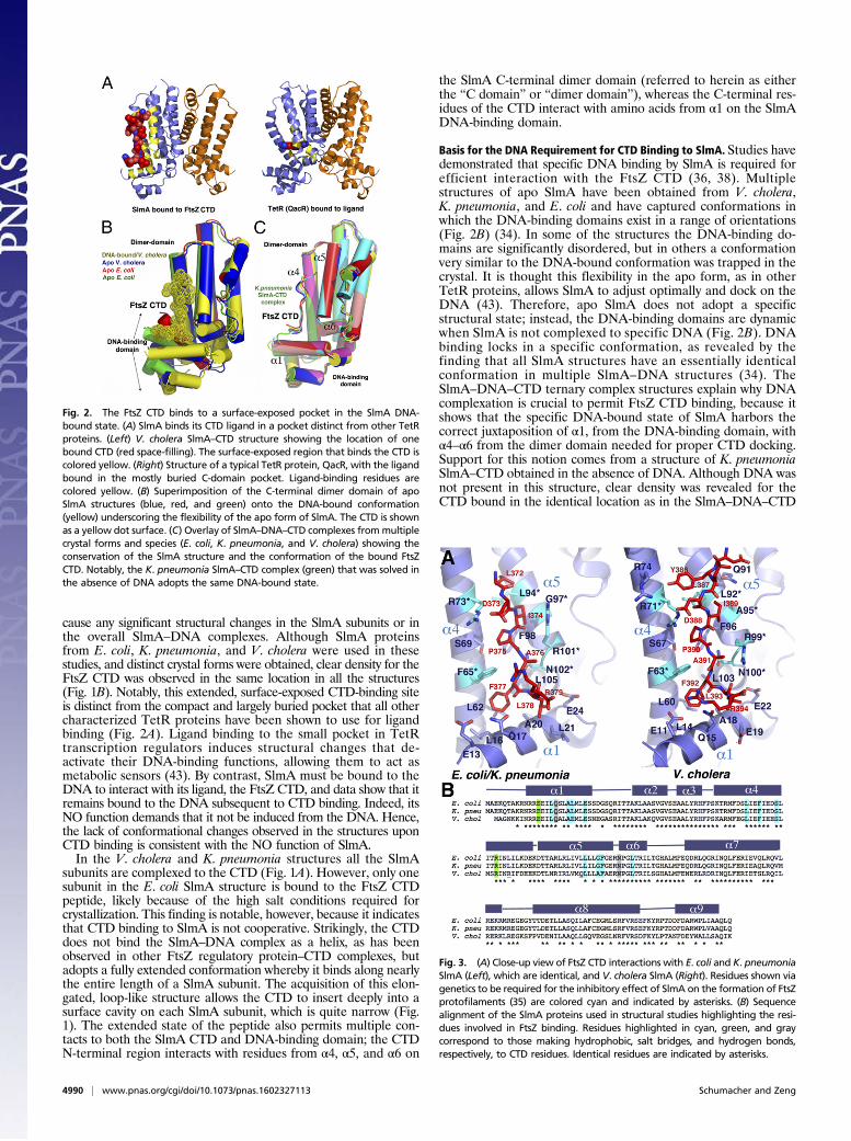
cause any significant structural changes in the SlmA subunits or inthe overall SlmA–DNA complexes. Although SlmA proteinsfrom E. coli, K. pneumonia, and V. cholera were used in thesestudies, and distinct crystal forms were obtained, clear density for theFtsZ CTD was observed in the same location in all the structures(Fig. 1B). Notably, this extended, surface-exposed CTD-binding siteis distinct from the compact and largely buried pocket that all othercharacterized TetR proteins have been shown to use for ligandbinding (Fig. 2A). Ligand binding to the small pocket in TetRtranscription regulators induces structural changes that de-activate their DNA-binding functions, allowing them to act asmetabolic sensors (43). By contrast, SlmA must be bound to theDNA to interact with its ligand, the FtsZ CTD, and data show that itremains bound to the DNA subsequent to CTD binding. Indeed, itsNO function demands that it not be induced from the DNA. Hence,the lack of conformational changes observed in the structures uponCTD binding is consistent with the NO function of SlmA.In the V. cholera and K. pneumonia structures all the SlmA
subunits are complexed to the CTD (Fig. 1A). However, only onesubunit in the E. coli SlmA structure is bound to the FtsZ CTDpeptide, likely because of the high salt conditions required forcrystallization. This finding is notable, however, because it indicatesthat CTD binding to SlmA is not cooperative. Strikingly, the CTDdoes not bind the SlmA–DNA complex as a helix, as has beenobserved in other FtsZ regulatory protein–CTD complexes, butadopts a fully extended conformation whereby it binds along nearlythe entire length of a SlmA subunit. The acquisition of this elon-gated, loop-like structure allows the CTD to insert deeply into asurface cavity on each SlmA subunit, which is quite narrow (Fig.1). The extended state of the peptide also permits multiple con-tacts to both the SlmA CTD and DNA-binding domain; the CTDN-terminal region interacts with residues from α4, α5, and α6 on
the SlmA C-terminal dimer domain (referred to herein as eitherthe “C domain” or “dimer domain”), whereas the C-terminal res-idues of the CTD interact with amino acids from α1 on the SlmADNA-binding domain.
Basis for the DNA Requirement for CTD Binding to SlmA. Studies havedemonstrated that specific DNA binding by SlmA is required forefficient interaction with the FtsZ CTD (36, 38). Multiplestructures of apo SlmA have been obtained from V. cholera,K. pneumonia, and E. coli and have captured conformations inwhich the DNA-binding domains exist in a range of orientations(Fig. 2B) (34). In some of the structures the DNA-binding do-mains are significantly disordered, but in others a conformationvery similar to the DNA-bound conformation was trapped in thecrystal. It is thought this flexibility in the apo form, as in otherTetR proteins, allows SlmA to adjust optimally and dock on theDNA (43). Therefore, apo SlmA does not adopt a specificstructural state; instead, the DNA-binding domains are dynamicwhen SlmA is not complexed to specific DNA (Fig. 2B). DNAbinding locks in a specific conformation, as revealed by thefinding that all SlmA structures have an essentially identicalconformation in multiple SlmA–DNA structures (34). TheSlmA–DNA–CTD ternary complex structures explain why DNAcomplexation is crucial to permit FtsZ CTD binding, because itshows that the specific DNA-bound state of SlmA harbors thecorrect juxtaposition of α1, from the DNA-binding domain, withα4–α6 from the dimer domain needed for proper CTD docking.Support for this notion comes from a structure of K. pneumoniaSlmA–CTD obtained in the absence of DNA. Although DNA wasnot present in this structure, clear density was revealed for theCTD bound in the identical location as in the SlmA–DNA–CTD
Fig. 2. The FtsZ CTD binds to a surface-exposed pocket in the SlmA DNA-bound state. (A) SlmA binds its CTD ligand in a pocket distinct from other TetRproteins. (Left) V. cholera SlmA–CTD structure showing the location of onebound CTD (red space-filling). The surface-exposed region that binds the CTD iscolored yellow. (Right) Structure of a typical TetR protein, QacR, with the ligandbound in the mostly buried C-domain pocket. Ligand-binding residues arecolored yellow. (B) Superimposition of the C-terminal dimer domain of apoSlmA structures (blue, red, and green) onto the DNA-bound conformation(yellow) underscoring the flexibility of the apo form of SlmA. The CTD is shownas a yellow dot surface. (C) Overlay of SlmA–DNA–CTD complexes frommultiplecrystal forms and species (E. coli, K. pneumonia, and V. cholera) showing theconservation of the SlmA structure and the conformation of the bound FtsZCTD. Notably, the K. pneumonia SlmA–CTD complex (green) that was solved inthe absence of DNA adopts the same DNA-bound state.
Fig. 3. (A) Close-up view of FtsZ CTD interactions with E. coli and K. pneumoniaSlmA (Left), which are identical, and V. cholera SlmA (Right). Residues shown viagenetics to be required for the inhibitory effect of SlmA on the formation of FtsZprotofilaments (35) are colored cyan and indicated by asterisks. (B) Sequencealignment of the SlmA proteins used in structural studies highlighting the resi-dues involved in FtsZ binding. Residues highlighted in cyan, green, and graycorrespond to those making hydrophobic, salt bridges, and hydrogen bonds,respectively, to CTD residues. Identical residues are indicated by asterisks.
4990 | www.pnas.org/cgi/doi/10.1073/pnas.1602327113 Schumacher and Zeng

complexes in a subunit that had clearly adopted the DNA-boundstate (Figs. 1 A and B and 2C).
The FtsZ CTD Binds SlmA in an Extended Conformation. The FtsZCTD-binding site on SlmA formed at the junction between theDNA-binding site and the C domain is surprisingly hydrophobicin character. It also is quite narrow and hence could not beaccessed by a helix. However, the extended conformation that isadopted by the SlmA-bound CTD allows hydrophobic residuesexposed on the CTD surface to fit perfectly into the elongatedSlmA cavity and evade solvent. Specifically, the CTD peptidemakes five hydrophobic contacts from residues (using E. colinumbering) Ile374, Pro375, Phe377, and Leu378 to residues inthe SlmA hydrophobic crevice (Fig. 3A). FtsZ CTD residuePhe377 (V. cholera residue Phe392) appears to play a central rolein binding to the specific DNA-bound state of SlmA, because itinserts in a hydrophobic cavity formed at the interface betweenthe DNA-binding domain and the C domain that is optimallyshaped to fit the phenyl side chain (Fig. 3A). Within this cleft,the FtsZ CTD Phe377 side chain interacts with Leu16 and Ala20and the aliphatic atoms of Gln17 from α1 in the DNA-bindingdomain, whereas its other face is encased by SlmA C-domainresidues Leu61, Phe65, and Leu105 (Fig. 3A). In addition toPhe377, Leu378 also contacts residues within the SlmA DNA-binding domain, including Ala20 and the aliphatic atoms ofresidues Glu21 and Glu24. The remainder of the bound FtsZCTD residues interacts with the SlmA C domain. These inter-actions include contacts between the CTD residue Pro375 andSlmA residue Phe65 and the aliphatic portions of the Ser69 andthe Phe98 side chains. SlmA residue Arg73 is anchored overPro375, thus shielding it from solvent, via a salt bridge with CTDresidue Asp373. Finally, CTD residue Ile374 embeds into thenarrow hydrophobic SlmA cavity and contacts Phe98 and Leu94.In addition to hydrophobic contacts, there are several hydro-
gen bonds and salt bridges in the SlmA–CTD complex. As noted,CTD residue Asp373 makes a salt bridge to SlmA residue Arg73.Residue Asn102, from the SlmA C domain, hydrogen bonds toamide nitrogen and carbonyl oxygen atoms of CTD residuesAla376 and Phe377. Similarly, SlmA residue Gln17, from the
DNA-binding domain, contacts the backbone atoms of FtsZCTD residues Phe377 and Arg379. These backbone contacts arekey, because they specify the extended nature of the peptide andanchor it into the pocket. The CTD Arg379 residue is also jux-taposed with the highly acidic side of α1 in the DNA-bindingdomain (Fig. 1C). In the E. coli, V. cholera, and K. pneumoniaSlmA–DNA–CTD complexes there is no density for residuesC-terminal to CTD residue Arg379, and only weak density isobserved in the K. pneumonia SlmA–CTD complex for Arg379and Lys380. This finding indicates that SlmA proteins do notinteract with the CTV region and bind CTD residues 370–380,which correspond to the conserved N-terminal region of theCTD, and suggests that SlmA proteins are active in bacteria thatcontain FtsZ proteins with CTVs that are highly variable inlength and sequence. Moreover, it shows that SlmA showsbinding selectivity for a relatively short sequence, LDIPAFL.BLAST searches indicate that this sequence is not found in otherE. coli proteins, ensuring that the SlmA–CTD interaction is unique.
Conserved Model for SlmA–FtsZ CTD Interaction in Bacterial NO.Residues that contact the FtsZ CTD are conserved amongE. coli, K. pneumonia, and V. cholera SlmA proteins. V. choleraSlmA contains an alanine (Ala95) in the place of E. coli/K. pneumonia residue Gly97 and a glutamate, Glu19, instead ofLeu21. These substitutions do not affect CTD binding, becausethe alanine side chain in V. cholera protein is small enough topermit docking of the CTD, and the Glu19/Leu21 side chainmediates contacts with the CTD via side-chain Cβ atoms. Fur-ther, although the V. cholera FtsZ CTD differs from the E. coliand K. pneumonia FtsZ CTDs at residues 370 and 380 (theresidues are glycine and arginine, respectively, in V. cholera andare aspartic acid and lysine, respectively, in E. coli/K. pneumo-nia), the structures show that these residues are not involved inSlmA binding. SlmA homologs appear to be widespread and arepresent in γ- and β-proteobacteria with the exceptions of Fran-cisella, Legionella, Pseudomonas, Stenotrophomonas, Xanthomo-nas, and Neisseria. Multiple sequence alignments of SlmAproteins (ranging from 98% to 48% identity) show that the keyresidues involved in CTD binding are conserved in the SlmA
Fig. 4. SlmA–DNA–CTD complex: CTD conformationaladaptability and insight into SlmA-mediated NO. (A) FPisotherms of SlmA–DNA binding to various p53 tetra-mer domain–FtsZ linker–FtsZ CTD chimeras to test theSlmA–DNA–FtsZ CTD structural model. (B) Comparisonof FtsZ CTD conformation in structures solved bound toFtsZ regulators. (Upper) Schematic of the FtsZ domainorganization. Residues D373 (D338 in T. maritima) arealigned for reference and underscore the dramaticrange of conformational states adopted by the CTD.(Lower) Sequence alignment of the FtsZ CTDs fromE. coli, K. pneumonia, V. cholera (analyzed in thisstudy), and T. maritima. CTT, conserved region of theCTD. Residues conserved in all CTDs are colored blue,and those conserved in the E. coli, K. pneumonia, andV. cholera CTDs are colored blue-green. Note: Only theconserved residues make contact with SlmA (Fig. 3A).(C) SlmA-mediated NO. This process involves SlmAbinding to the SBS as a dimer-of-dimers and nucleatingthe binding of adjacent SlmA dimers (34). Then FtsZfilaments are recruited to the nucleoid by binding SlmAvia their CTD (shown as loops) and subsequently FtsZcore regions (squares). DNA charge might contribute toprotofilament fracturing. Once the protofilaments aredisrupted into subunits, they no longer bind SlmA–DNAwith significant affinity and diffuse from the nucleoid.(D) Schematic of E. coli cell division with Min- and NO-mediated NO processes indicated. SlmA binding to thenucleoid non-Ter regions averts nucleoid fragmenta-tion by FtsZ, whereas the Min system prevents Z-ringformation at the poles, the net effect being to driveZ-ring formation at the cell center.
Schumacher and Zeng PNAS | May 3, 2016 | vol. 113 | no. 18 | 4991
BIOCH
EMISTR
Y

proteins and that residues that make important contacts are ei-ther conserved or contain conservative substitutions (Fig. S3).Sequence alignments of the corresponding FtsZ CTDs show thatSlmA-contacting residues are identical. Thus, these data indicatethat the SlmA–FtsZ CTD interaction, and hence its NO func-tion, are likely to be conserved across SlmA-containing bacteria.
Probing the SlmA–DNA–CTD Structure. Several studies have probedthe SlmA–FtsZ CTD interaction and its effect on FtsZ filamentformation (35, 37, 38). A genetic screen carried out by Cho et al.(35) revealed that residues Phe65, Arg73, Leu94, Gly97,Arg101, and Asn102 were critical for the antagonism of FtsZprotofilament formation by E. coli SlmA. These residues mapprecisely to the CTD portion of the FtsZ CTD-binding pocketrevealed in our structures (Fig. 3A). Interestingly, this study didnot identify residues in α1, likely because the side-chain aliphaticportion of α1 residues mediates most of the contacts to the CTD.In a separate study, FtsZ mutations L378E and I374K wereshown to abrogate the SlmA–FtsZ interaction (37). The SlmA–DNA–CTD structures show that these substitutions would result inthe insertion of large and charged residues into the hydrophobicCTD-binding crevice on SlmA; such an insertion would prohibitbinding. Furthermore, our structures show that SlmA interacts withthe same residues as ZipA, as is consistent with studies showingthat ZipA competes with SlmA for FtsZ CTD binding (38).However, to probe the model further, we sought to test two specificstructure-based predictions: that Phe377 is critical for the DNA-specific SlmA–CTD interaction and that the FtsZ CTV, which isnot visible in the structure and does not contribute to binding,would be dispensable for the interaction.Studies showed that the full-length FtsZ protein, which ex-
poses multiple CTDs as a multivalent ligand source, binds itsregulators with significantly enhanced affinity (38). Indeed, theFtsZ CTD must be present as a multivalent ligand to observeappreciable binding at the relatively low concentrations used inbiochemical measurements (38). Thus, we developed an assaythat used a chimeric construct in which the FtsZ 50-residuelinker–CTD region was connected to C terminus of the tetra-merization domain of human p53 to probe our structural model(44). This construct and mutant forms of the construct then weretitrated into preformed complexes of SlmA bound to fluo-rescently labeled DNA. Binding resulted in a saturable increasein fluorescence polarization (FP) that could be used to de-termine an apparent Kd. Following this strategy, a p53 chimeracontaining the wild-type FtsZ CTD sequence bound the E. coliSlmA–DNA complex with an apparent Kd of 25.6 ± 1.8 μM (Fig.4A). When the experiment was performed with a chimera lackingthe CTD (ΔCTD), i.e., with the p53 tetramerization domainalone, no binding was observed. Removal of the CTV (ΔCTV)resulted in binding similar to that observed for the wild-typechimera (apparent Kd = 27.0 ± 1.4 μM), whereas a single F377Amutation in the CTD resulted in only weak, nonsaturable binding(Fig. 4A). Thus, the combined results from the FP binding studiessupport the SlmA–DNA–CTD structural models. The wild-typeSlmA–DNA–CTD binding affinity obtained by this assay is com-parable to those previously measured for FtsZ CTD binding toother regulatory proteins via biosensor assays, which present mul-tiple proteins on a surface (41, 42). For example, ZipA and FtsAwere shown to bind the FtsZ CTD with Kds of 20 μM and 50 μM,respectively (41, 42).
The FtsZ CTD: An Intrinsically Disordered Region Capable of AdoptingMultiple Conformations upon Binding Diverse FtsZ RegulatoryProteins. The FtsZ CTD mediates the majority of its contactswith regulatory proteins. As a result, it has been called the FtsZ“landing pad” (18). How these proteins with diverse structuresand functions can bind the same FtsZ CTD region has been afascinating but unresolved question. Currently the only proteinswhose structures have been obtained in the presence of the CTDare E. coli ZipA and T. maritima FtsA, both of which function torecruit FtsZ to the membrane (41, 42, 45). The CTD binds both
these proteins as helices. In the ZipA–CTD structure most of thecontacts made by the CTD are hydrophobic. In contrast, in theFtsA–CTD complex there are only three contacts, and all are saltbridges (41, 42). It is possible that the differences in thesecomplexes might reflect the divergence in the FtsZ CTD be-tween these two organisms; the T. maritima and E. coli CTDshare only five residues in common, and the T. maritimaCTD harbors a longer CTV: the T. maritima CTD isPEGDIPAIYRYGLEGLL compared with the E. coli FtsZ CTD,DYLDIPAFLRKQAD (conserved residues are underlined). In-deed, although the helical segment of the CTD bound to bothE. coli ZipA and T. maritima FtsA begins with the conservedproline (Pro375 in E. coli numbering), the helix bound to FtsA hasa kink caused by a glycine that is not present in the E. coli CTD(Fig. 4B). Hence, the question of how the FtsZ CTD can bind somany diverse regulatory proteins has remained unanswered.Our structures of SlmA–DNA bound to the FtsZ CTD resolve
this issue. Importantly, these structures show that the CTD does notbind all its regulators as a helix but can adopt a striking range ofconformations depending on its binding partner (Fig. S4). Indeed,although the CTD contains a significant number of hydrophobicresidues, SlmA and ZipA use distinct strategies to shield theseresidues from solvent. The hydrophobic face of the amphipathicCTD helix bound to ZipA is docked into a large hydrophobicsurface on ZipA, whereas in SlmA an extended, narrow cavity withhydrophobic character allows the insertion of the hydrophobic CTDresidues. As noted, despite the differences in the conformation bywhich the CTD binds these regulators, the affinities of the CTD forthese proteins are similar (41, 42). As is consistent with this affinity,analysis of the buried surface areas showed that the ZipA–CTD andFtsA–CTD complexes bury 565 and 570 Å2, respectively, whereasthe SlmA–CTD interaction buries 633–727 Å2. However, thesecombined findings suggest the unlikelihood of predicting how theFtsZ CTD might bind each of its regulators, even if biochemicalinformation, such as binding affinities, is available, and indicate thatstructures likely will be required to deduce the binding mechanismsfor each complex.
DiscussionThe FtsZ protein mediates cell division in most prokaryotes and issubject to regulation at multiple levels to ensure that the Z ringforms at the correct time and place during cell division. NO, thephenomenon by which the chromosomal DNA is spared fromZ-ring bisection, is particularly important because Z-ring formationthrough the nucleoid would result in chromosome fragmentation.Thus, it is not surprising that failsafe mechanisms, including dedi-cated NO factors, have evolved to prevent this catastrophe. SlmAwas identified as the E. coli NO factor (30) and was shown to in-hibit FtsZ directly from forming Z rings through the DNA. Thisinhibition depends on an interaction between SlmA–DNA and theCTD of FtsZ (33, 37). Because FtsZ must be tethered to themembrane by interactions between its CTD and either ZipA orFtsA to form a Z ring, the SlmA–CTD interaction might preventZ-ring formation simply by competition. However, studies showedthat SlmA–DNA complexes actively disrupt FtsZ protofilaments,suggesting a more complex mechanism (33, 37). Data indicate thatthe FtsZ core also interacts weakly with SlmA–DNA subsequent toCTD binding. Interestingly, small-angle X-ray–scattering studiessuggested that the FtsZ core also may bind the SlmA C domain;however, the resolution of the analyses was too low to assign aprecise binding location (34). These interactions could collaborateto antagonize the formation of FtsZ protofilaments. SlmA canspread from its initial DNA site to form extended assemblages onthe DNA. Thus, the SlmA assemblies could interact with multipleFtsZ molecules on a protofilament to fragment and antagonize itsformation (Fig. 4 C and D). Once the protofilaments have beendisrupted into single or small protofilaments, they would no longerbind SlmA–DNA, because this binding requires a multivalent formof the FtsZ CTD found only on the protofilaments (Fig. 4C). Therequirement that SlmA be bound to the nucleoid to be active forthis recruitment step is vital, because it prevents any free SlmA,
4992 | www.pnas.org/cgi/doi/10.1073/pnas.1602327113 Schumacher and Zeng

from binding FtsZ protofilaments and disrupting Z-ring formation.The multiple SlmA–FtsZ interactions would bring FtsZ protofila-ments within proximity of the electronegative nucleoid DNA (Fig.4C). Hence, it is possible that the nucleoid could be involved in NO(Fig. 4 C and D). Indeed, charge has been shown to play a key role inthe formation and clustering of FtsZ protofilaments. For example,the electropositive B. subtilis FtsZ CTD participates in bundlingprotofilaments (46), and although the E. coli FtsZ CTD does notappear to play a role in protofilament bundling, multiple studieshave shown that cations and positively charged molecules enhanceits polymerization (5).Although more studies are needed to assess the specific roles
played by SlmA and the nucleoid in the complex process of NO, aclearly vital step in SlmA-mediated NO is the recruitment of FtsZand its tethering to the nucleoid by the SlmA–DNA–FtsZ CTDinteraction. However, the molecular basis behind this interactionhas been unknown. This interaction also is of interest because it isunusual among FtsZ regulators, in that it requires SlmA to bespecifically bound to DNA. Here we deciphered the molecularmechanism behind this unique regulatory function by determiningcrystal structures of multiple SlmA–DNA–FtsZ CTD ternarycomplexes. Notably, all the structures, obtained under diversecrystallization conditions and with SlmA proteins from differentorganisms, revealed the same binding mode in which the FtsZCTD interacts with the DNA-bound form of SlmA as an extendedloop-like structure. This mode of ligand interaction is completelydifferent from that used by other characterized TetR proteins tobind ligands, namely via a small, mostly buried cleft located in their
C domains (43). Importantly, the SlmA–DNA–CTD structures alsoreveal that SlmA must be in its DNA-bound form to bind the CTDbecause it is locked into a particular conformation that harbors thecorrect juxtaposition of the dimer and DNA-binding domains thatallows specific docking of the FtsZ CTD. Residues critical for CTDinteractions are shared among SlmA proteins, indicating that theyuse a conserved mode of CTD binding. Finally, these findings in-dicate that the FtsZ CTD is able to bind a diverse array of regulatoryproteins because it is able to adopt different conformational statesdepending on its regulatory binding partner. Thus, our structures notonly reveal the underlying molecular mechanism by which SlmA–DNA recruits FtsZ to the nucleoid but also unveil how the FtsZCTD acts as a landing pad for diverse regulatory proteins.
Materials and MethodsSI Materials and Methods provides details and additional methods notprovided below.
The genes encoding the E. coli, K. pneumonia, and V. cholera SlmA proteinswere purchased from GenScript Corporation and subcloned into pET15b so thatHis-tags were expressed for purification. For details of purification, crystallization,structure determination, and refinement (47), see SI Materials and Methods.
ACKNOWLEDGMENTS. This work was supported by NIH Grant GM115563 (toM.A.S.). Beamline 8.3.1 at the Advanced Light Source is operated by the Universityof California Office of the President Multicampus Research Programs and Initia-tives Grant MR-15-328599 and by the Program for Breakthrough BiomedicalResearch, which is partially funded by the Sandler Foundation.
1. Bi EF, Lutkenhaus J (1991) FtsZ ring structure associated with division in Escherichiacoli. Nature 354(6349):161–164.
2. Lutkenhaus J, Pichoff S, Du S (2012) Bacterial cytokinesis: From Z ring to divisome.Cytoskeleton (Hoboken) 69(10):778–790.
3. Margolin W (2005) FtsZ and the division of prokaryotic cells and organelles. Nat RevMol Cell Biol 6(11):862–871.
4. Adams DW, Errington J (2009) Bacterial cell division: Assembly, maintenance anddisassembly of the Z ring. Nat Rev Microbiol 7(9):642–653.
5. Erickson HP, Anderson DE, Osawa M (2010) FtsZ in bacterial cytokinesis: Cytoskeletonand force generator all in one. Microbiol Mol Biol Rev 74(4):504–528.
6. Löwe J, Amos LA (1998) Crystal structure of the bacterial cell-division protein FtsZ.Nature 391(6663):203–206.
7. Li Y, et al. (2013) FtsZ protofilaments use a hinge-opening mechanism for constrictiveforce generation. Science 341(6144):392–395.
8. Löwe J (1998) Crystal structure determination of FtsZ from Methanococcus jannaschii.J Struct Biol 124(2-3):235–243.
9. Löwe J, van den Ent F (2001) Conserved sequence motif at the C-terminus of thebacterial cell-division protein FtsA. Biochimie 83(1):117–120.
10. Rowlett VW, Margolin W (2014) 3D-SIM super-resolution of FtsZ and its membranetethers in Escherichia coli cells. Biophys J 107(8):L17–L20.
11. Biteen JS, Goley ED, Shapiro L, Moerner WE (2012) Three-dimensional super-resolu-tion imaging of the midplane protein FtsZ in live Caulobacter crescentus cells usingastigmatism. ChemPhysChem 13(4):1007–1012.
12. Li Z, Trimble MJ, Brun YV, Jensen GJ (2007) The structure of FtsZ filaments in vivosuggests a force-generating role in cell division. EMBO J 26(22):4694–4708.
13. Buss J, et al. (2013) In vivo organization of the FtsZ-ring by ZapA and ZapB revealed byquantitative super-resolution microscopy. Mol Microbiol 89(6):1099–1120.
14. Fu G, et al. (2010) In vivo structure of the E. coli FtsZ-ring revealed by photoactivatedlocalization microscopy (PALM). PLoS One 5(9):e12682.
15. Jennings PC, Cox GC, Monahan LG, Harry EJ (2011) Super-resolution imaging of thebacterial cytokinetic protein FtsZ. Micron 42(4):336–341.
16. Romberg L, Levin PA (2003) Assembly dynamics of the bacterial cell division proteinFTSZ: Poised at the edge of stability. Annu Rev Microbiol 57:125–154.
17. Weart RB, Levin PA (2003) Growth rate-dependent regulation of medial FtsZ ringformation. J Bacteriol 185(9):2826–2834.
18. Huang KH, Durand-Heredia J, Janakiraman A (2013) FtsZ ring stability: Of bundles,tubules, crosslinks, and curves. J Bacteriol 195(9):1859–1868.
19. Ortiz C, Natale P, Cueto L, Vicente M (2016) The keepers of the ring: Regulators ofFtsZ assembly. FEMS Microbiol Rev 40(1):57–67.
20. Kirkpatrick CL, Viollier PH (2011) New(s) to the (Z-)ring. Curr OpinMicrobiol 14(6):691–697.21. de Boer PAJ, Crossley RE, Rothfield LI (1989) A division inhibitor and a topological
specificity factor coded for by the minicell locus determine proper placement of thedivision septum in E. coli. Cell 56(4):641–649.
22. Hu Z, Gogol EP, Lutkenhaus J (2002) Dynamic assembly of MinD on phospholipidvesicles regulated by ATP and MinE. Proc Natl Acad Sci USA 99(10):6761–6766.
23. Raskin DM, de Boer PA (1999) Rapid pole-to-pole oscillation of a protein required for di-recting division to the middle of Escherichia coli. Proc Natl Acad Sci USA 96(9):4971–4976.
24. Raskin DM, de Boer PA (1999) MinDE-dependent pole-to-pole oscillation of divisioninhibitor MinC in Escherichia coli. J Bacteriol 181(20):6419–6424.
25. Lutkenhaus J (2007) Assembly dynamics of the bacterial MinCDE system and spatial
regulation of the Z ring. Annu Rev Biochem 76:539–562.26. Woldringh CL, et al. (1990) Role of the nucleoid in the toporegulation of division. Res
Microbiol 141(1):39–49.27. Woldringh CL, Mulder E, Huls PG, Vischer N (1991) Toporegulation of bacterial di-
vision according to the nucleoid occlusion model. Res Microbiol 142(2-3):309–320.28. Woldringh CL (2002) The role of co-transcriptional translation and protein translocation
(transertion) in bacterial chromosome segregation. Mol Microbiol 45(1):17–29.29. Wu LJ, Errington J (2004) Coordination of cell division and chromosome segregation
by a nucleoid occlusion protein in Bacillus subtilis. Cell 117(7):915–925.30. Bernhardt TG, de Boer PA (2005) SlmA, a nucleoid-associated, FtsZ binding protein required
for blocking septal ring assembly over Chromosomes in E. coli. Mol Cell 18(5):555–564.31. Wu LJ, et al. (2009) Noc protein binds to specific DNA sequences to coordinate cell
division with chromosome segregation. EMBO J 28(13):1940–1952.32. Tonthat NK, et al. (2011) Molecular mechanism by which the nucleoid occlusion
factor, SlmA, keeps cytokinesis in check. EMBO J 30(1):154–164.33. Cho H, McManus HR, Dove SL, Bernhardt TG (2011) Nucleoid occlusion factor SlmA is a
DNA-activated FtsZ polymerization antagonist. Proc Natl Acad Sci USA 108(9):3773–3778.34. Tonthat NK, et al. (2013) SlmA forms a higher-order structure on DNA that inhibits cyto-
kinetic Z-ring formation over the nucleoid. Proc Natl Acad Sci USA 110(26):10586–10591.35. Cho H, Bernhardt TG (2013) Identification of the SlmA active site responsible for blocking
bacterial cytokinetic ring assembly over the chromosome. PLoS Genet 9(2):e1003304.36. Adams DW, Wu LJ, Errington J (2015) Nucleoid occlusion protein Noc recruits DNA to
the bacterial cell membrane. EMBO J 34(4):491–501.37. Du S, Lutkenhaus J (2014) SlmA antagonism of FtsZ assembly employs a two-pronged
mechanism like MinCD. PLoS Genet 10(7):e1004460.38. Du S, Park KT, Lutkenhaus J (2015) Oligomerization of FtsZ converts the FtsZ tail motif
(conserved carboxy-terminal peptide) into a multivalent ligand with high avidity for
partners ZipA and SlmA. Mol Microbiol 95(2):173–188.39. Li Y, Sergueev K, Austin S (2002) The segregation of the Escherichia coli origin and
terminus of replication. Mol Microbiol 46(4):985–996.40. Deghorain M, et al. (2011) A defined terminal region of the E. coli chromosome shows
late segregation and high FtsK activity. PLoS One 6(7):e22164.41. Mosyak L, et al. (2000) The bacterial cell-division protein ZipA and its interaction with
an FtsZ fragment revealed by X-ray crystallography. EMBO J 19(13):3179–3191.42. Szwedziak P, Wang Q, Freund SMV, Löwe J (2012) FtsA forms actin-like protofila-
ments. EMBO J 31(10):2249–2260.43. Ramos JL, et al. (2005) The TetR family of transcriptional repressors. Microbiol Mol
Biol Rev 69(2):326–356.44. Jeffrey PD, Gorina S, Pavletich NP (1995) Crystal structure of the tetramerization
domain of the p53 tumor suppressor at 1.7 angstroms. Science 267(5203):1498–1502.45. Pichoff S, Lutkenhaus J (2002) Unique and overlapping roles for ZipA and FtsA in
septal ring assembly in Escherichia coli. EMBO J 21(4):685–693.46. Buske PJ, Levin PA (2013) A flexible C-terminal linker is required for proper FtsZ assembly
in vitro and cytokinetic ring formation in vivo. Mol Microbiol 89(2):249–263.47. Adams PD, et al. (2010) PHENIX: A comprehensive Python-based system for macro-
molecular structure solution. Acta Crystallogr D Biol Crystallogr 66(Pt 2):213–221.
Schumacher and Zeng PNAS | May 3, 2016 | vol. 113 | no. 18 | 4993
BIOCH
EMISTR
Y