Functional Domains of Moloney Murine Leukemia Virus Integrase

Vol. 29, No. 2JOURNAL OF VIROLOGY, Feb. 1979, p. 735-7430022-538X/79/02-0735/09$02.00/0
Structure of the Murine Leukemia Virus EnvelopeGlycoprotein Precursor
OWEN N. WITTE* AND DYANN F. WIRTHt
Department of Biology and Center for Cancer Research, Massachusetts Institute of Technology, Cambridge,Massachusetts 02139
Received for publication 22 August 1978
The glycosylated env gene precursor (Pr8Oenv) of Moloney murine leukemiavirus has been isolated by selective immunoprecipitation. Use of the drug tuni-camycin to inhibit nascent glycosylation or specific cleavage with endoglycosidaseH demonstrated that the precursor contained an apoprotein with a molecularweight of 60,000. The finished virion glycoprotein (gp7O) was largely resistant tothe action of endoglycosidase H. Chromatography of the glycopeptides of Pr8Oenuin conjunction with endoglycosidase H digestion studies suggested that theprecursor contained two distinct major glycosylation sites. Analysis of partialproteolytic cleavage fragments of Pr8Oenv before and after endoglycosidase Htreatment placed the two glycosylation sites within a 30,000-dalton region of theapoprotein sequence. Kinetic experiments showed that carbohydrate processingas well as proteolytic cleavage are late steps in the maturation of Pr8Oenv.
Genome mapping correlated with in vitrotranslation studies has shown that a 20 to 22SmRNA encoded from the 3' third of the murineleukemia virus (MuLV) genome is translated invivo into a glycosylated polyprotein with a mo-lecular weight of 80,000 to 90,000 designatedPrenv (4, 22, 34). Prenv is synthesized and proc-essed in cellular membranes by proteolysis andcarbohydrate alteration to the finished env geneproducts gp70 and pi5E (5, 13, 16, 18, 26, 37).Recent work on other viral and cellular gly-
coprotein systems has shown that the primaryglycosylation event involves the en bloc transferof a large, branched, preformed, lipid-linked,high-mannose-containing structure to the poly-peptide chain. This glycosylation can occur onnascent polypeptide chains. Subsequently, thesecore or high-mannose groups are processed inthe rough and smooth endoplasmic reticulum byinitial cleavage followed by sequential additionsof sugars including fucose and terminal sialicacid into distinct branched-chain patterns re-ferred to as complex carbohydrate groups (7, 8,19, 20, 29, 35).The elucidation of the carbohydrate struc-
tures of Pren' and gp7O, and of the relationshipof carbohydrate processing and proteolyticcleavage to the viral budding event, is a centralconcern of these studies. We have examined thestructure of the MuLV glycoprotein precursor(Pren"), using the drug tunicamycin (30, 35),which inhibits forrmation of the high-mannose
t Present address: The Biological Laboratories, HarvardUniversity, Cambridge, MA 02138.
carbohydrate groups, and the enzyme endogly-cosidase H (endo H) (31), which cleaves en blocthese high-mannose groups from the poly-peptide backbone. Our results indicate that thecarbohydrate processing of the MuLV Prenv issimilar to that described for other animal viralsystems (8, 20). In addition, these carbohydrategroups provide a useful marker in studying thestructure of the env gene precursor and its prod-ucts.
MATERIALS AND METHODSCeUs and viruses. Wild-type Moloney MuLV,
temperature-sensitive Moloney ts3 (38), and Rauscherts24 (27) strains were all cloned and grown in NIH/3T3cells as previously described (36). Cells were labeledwith [35S]methionine (New England Nuclear Corp.,Boston, Mass.) at 50 tiCi/ml in Dulbecco minimalessential medium containing 1/100 the normal methi-onine concentration for the times noted in the text.Cells were labeled with [2-3H]mannose (New EnglandNuclear Corp.) at 200 uCi/ml in glucose-free phos-phate-buffered saline for 60 min. In one experiment,to prepare labeled virions, cells were labeled with [2-3H]mannose (200 uCi/ml) for 2 h in media containing1/50 the normal glucose concentration; then the mediawere supplemented to the normal glucose level, andsupernatant fluid was collected over 16 h. The virionswere purified by sucrose gradient centrifugation aspreviously described (37).
Immunoprecipitation. Labeled cells were ex-tracted at 0 to 4°C into 0.01 M NaH2PO4-Na2HPO4(pH 7.5)-0.1 M NaCl containing 1% Triton X-100, 0.5%deoxycholate, and 0.1% sodium dodecyl sulfate (SDS)at 106 to 2 x 10⁶ cells/ml. Lysates were clarified at150,000 x g for 2 to 3 h, and samples were immuno-precipitated at 0 to 4°C as previously described, with
735
on February 11, 2018 by guest
http://jvi.asm.org/
Dow
nloaded from
736 WITTE AND WIRTH
the use of Staphylococcus aureus Cowen strain I tocollect antigen-antibody complexes (11, 37). Sera usedincluded rabbit anti-Moloney gp7O, rabbit anti-Molo-ney p30, and rabbit anti-Moloney virions previouslycharacterized (36).Washed precipitates were denatured in 1% SDS-1%
2-mercaptoethanol and subjected to electrophoresison a discontinuous stack, Tris-buffered polyacryl-amide gel system as previously describ3d (12, 37). Gelswere fluorographed by the procedure of Bonnar andLaskey (1). Standard proteins used as molecular-weight markers included rabbit immunoglobulinheavy (50,000) and light (25,000) chains, MuLV p30(30,000) and p12 (12,000), and the structural proteinsof vesicular stomatitis virus and adenovirus type 2.Enzyme digestions: endo H digestion. Endo 1-
N-acetylglucosaminidase H was prepared by themethod described by Tarentino and Maley (31). Thefinal protein concentration in the enzyme stock was30,g/ml. The enzyme activity was assayed with dan-sylated ovalbumin glycopeptides as substrate. Enzymeincubations were carried out as follows. Cell lysateswere precipitated as described above with anti-gp7O.The S. aureus pellets were resuspended in 0.1 M Tris-hydrochloride, pH 6.8, and incubated for 60 min at30°C with 3 ,ug of endo H/ml. The enzyme incubationwas stopped by the addition of SDS to a final concen-tration of 2% followed by heating for 2 min at 100°C.In other experiments, the immune precipitates wereresuspended in 1% SDS-0.05 M Tris, pH 6.8, heatedfor 2 min at 100°C, and centrifuged to remove the S.aureus. The supernatant fluid was diluted with anequal volume of 0.06 M citrate, pH 5.5, or Tris (0.05M), pH 6.8, and was incubated for various times with0.3 ,ug of endo H/ml. The samples were precipitatedby the addition of 2 volumes of ice-cold acetone.Pronase digestion. Pronase digestions were per-
formed as described by Sefton and Keegstra (25).Briefly, the samples were digested with 1% (wt/vol)Pronase (Calbiochem, grade B) in 0.1 M Tris-hydro-chloride (pH 8.0)-0.01 M CaCl2 for 72 h at 500C.Additional Pronase in amounts of 1% (wt/vol) eachwere added at 24 and 48 h. The final Pronase concen-tration was 3% (wt/vol). Virions were prepared asdescribed above. Cell lysates were immunoprecipi-tated with anti-gp7O before digestion. Samples whichwere digested with endo H were first boiled for 5 minto inactivate the Pronase, diluted with one-third vol-ume of 0.3 M citrate, pH 5.5, and digested with 0.3 Mugof endo H/ml for 12 h. The Pronase-derived glycopep-tides were resolved on a Bio-Gel P6 column (1 cm by100 cm) in 0.01 M Tris, pH 8.0. The fractions (1 ml)were counted in Handifluor (Beckman Instruments,Inc.) in a Beckman LS-230 scintillation counter. Eachcolumn run contained [14C]mannose-labeled, in vitro-synthesized, lipid-linked oligosaccharide containing 6to 9 mannose units (21) as an internal standard, agenerous gift of P. Robbins.Tunicamycin inhibitor. Tunicamycin was a gift
from W. Lennarz (Johns Hopkins University). It wasdissolved in 0.01 M NaOH and subsequently dilutedinto tissue culture medium to final concentrations of10 and 100 ,ug/ml. Cells were preincubated with tuni-camycin (14) for 3 h prior to metabolic labeling.
Peptide cleavage maps. The basic approach of
Cleveland et al. (3) was followed. Immunoprecipitatedsamples released from S. aureus were digested in 0.05M Tris, pH 6.8, 0.5% SDS, and 10% glycerol with aspecific protease for the time and temperature notedin the text. Preliminary dose response and kinetics ofcleavage with each protease were evaluated (data notshown) on preparations of Moloney Pr8Oen. Sampleswere reconstituted into gel sample buffer, boiled for 2to 3 min, and analyzed by SDS-gel electrophoresis andfluorography. Proteases used included S. aureus V8(Miles Laboratories), trypsin-tolylsulfonyl phenalanylchloromethyl ketone and a-chymotrypsin (Worthing-ton Biochemical Corp.), and thermolysin (SigmaChemical Co.). In some experiments samples weredigested with endo H prior to or after protease diges-tion as described above.Each digestion contained unlabeled rabbit immu-
noglobulin at approximately 2 mg/ml.
RESULTSSize of the Prenv apoprotein. Previous stud-
ies using 2-deoxyglucose, glucosamine, or cyto-chalasin to inhibit glycosylation in Rauscherleukemia virus-infected cells demonstrated anapparent molecular weight shift from 90,000 to70,000 for the glycosylated and nonglycosylatedforms of Pren", respectively (26, 32). To confirmand extend this observation, we used the drugtunicamycin, an inhibitor of glycosylationknown to act at the first step of glycoproteincarbohydrate addition. Tunicamycin inhibitsthe formation of a lipid-linked, high-mannosecore structure added en bloc to polypeptidechains (14, 19, 28, 30, 35).When cells infected with Moloney MuLV were
treated with tunicamycin, there was a shift inthe apparent molecular weight of Prenv (Fig. 1).Control cells pulse-labeled for 45 min with[35S]methionine showed Pr8Oenv with the normalmolecular weight of 80,000 when immunoprecip-itated with antisera specific for gp7O (Fig. 1A,lane 1). Cells pretreated for 3 h with 10 jg (Fig.1B) or 100 ,ug (Fig. 1C) of tunicamycin/ml andthen pulse-labeled and immunoprecipitatedshowed a shift in the size of the glycoproteinprecursor from a molecular weight of 80,000(Pr8Oenv) to 60,000 (P60e"v). This result indicatedthat a significant fraction of the primary glyco-sylation of Pr8Oenu occurred by transfer of tuni-camycin-sensitive, lipid-linked, high-mannosestructures similar to that described for severalother animal viruses (7, 8, 20). The size of thecore precursor (Pr65505) and its cleavage prod-ucts was unaffected (Fig. 1A, B and C, lane 2).A significant decrease in the rate of proteinsynthesis was observed (Fig. 1B and C) withhigh doses of this tunicamycin preparation.As an alternative method for removing car-
bohydrate from Prenv, we used the enzyme endoH (see Materials and Methods). This endogly-
J. VIROL.
on February 11, 2018 by guest
http://jvi.asm.org/
Dow
nloaded from

MuLV ENVELOPE GLYCOPROTEIN PRECURSOR 737
A1 2
B C1 2 1 2
--Pr8Oenv
a --Pr659a9XP60env
_m - p30
>)cxgp7O 2) MuLVFIG. 1. Effect of tunicamycin on the size of the env
gene precursor. Monolayers of cloned NIH/3T3 cellschronically infected with Moloney MuLV were nottreated (control, panel A) or were treated with 10 pg(panel B) or 100 pg (panel C) of tunicamycin/ml for 3h at 37°C and then were pulse-labeled (30 min) withf35Smethionine (50 sCi/ml) (see Materials andMeth-ods). Cells were extracted, and a sample was immu-noprecipitated with either anti-gp7O (lane 1) or anti-Moloney MuLV virions (lane 2). Immune complexeswere collected with S. aureus Cowen strain I disso-ciated with 1% SDS, 1% mercaptoethanol, and heatand then were subjected to electrophoresis on aSDS-10% polyacrylamide gel. The gel was fluoro-graphed and exposed to Kodak XR5 film at - 70°Cfor 1 day.
cosidase cleaves at the distal side of the first N-acetylglucosamine residue attaching the large,branched-chain, high-mannose structure to theprotein moiety, leaving only a single N-acetyl-glucosamine residue behind. Endo H is activeover a wide pH and temperature range, willfunction in the presence of moderate concentra-tions of denaturing detergents such as SDS, andcan be extensively purified (31).When pulse-labeled ([35S]methionine) cell ex-
tracts from Moloney MuLV-infected cells wereimmunoprecipitated with anti-gp7O serum andtreated with endo H (Fig. 2A, + lane), the Pr8Oenvhad an increased electrophoretic mobility (des-ignated P60"). Determinations of glycoproteinmolecular weight by SDS electrophoresis aregenerally overestimates (24). The shift of molec-ular weight from Pr8Oenv to p60env does not in-dicate that 20,000 daltons of carbohydrate wasremoved, but only that the carbohydrate groupsadded to an apoprotein with a molecular weightof 60,000 cause it to move at an apparent molec-ular weight of 80,000 in SDS-gels.
Digestion with endo H had no effect on thesize of the Pr65`9 precursor or its cleavage prod-ucts immunoprecipitated with anti-p30 serum.If the Pr8Oe`v was labeled with [3H]mannose,however, all of the [3H]mannose label was re-moved from the protein (data not shown).When cells infected with other strains of
MuLV were treated in the same manner, a sim-ilar increase in electrophoretic mobility of Prenvwas observed. Moloney ts3 Pr80env was shiftedto p60env (Fig. 2B), and Rauscher ts24 Pr90envwas shifted to P70"l (Fig. 2C) by endo H treat-ment. Wild-type Rauscher leukemia virusshowed a similarly sized Pr90env (36; P. Trakt-man and 0. Witte, unpublished data). Thus,different common laboratory strains of ecotropicMuLV differed by as much as 10,000 in themolecular weight of their Pre'v apoprotein. Theposition of this additional 10,000 molecularweight of protein within the precursor moleculewas not determined. We did not discern anydifference in the size of the finished gp70 andpl5E gene products when strains of Moloney orRauscher MuLV were compared (data notshown).Estimate of the number of carbohydrate
side chains in Pr8OefV. By assuming that eachhigh-mannose core oligosaccharide added at adifferent site would contribute equally and ad-ditively to the established molecular weight inSDS gels (23; J. Rothman, personal communi-cation), one can estimate the number of glyco-sylation sites from the number of discrete inter-mediates between the fully glycosylated Pr8Oenvand deglycosylated p60env during a time courseof digestion with endo H. Figure 3 demonstratessuch a time course for Pr8Oenv immunoprecipi-tated from cells labeled with [35S]methionine for30 min. A single intermediate with a molecularweight of 70,000 was found, suggesting that atleast two sites per Pr80env are glycosylated.Glycopeptides of Prnv and gp7O. To char-
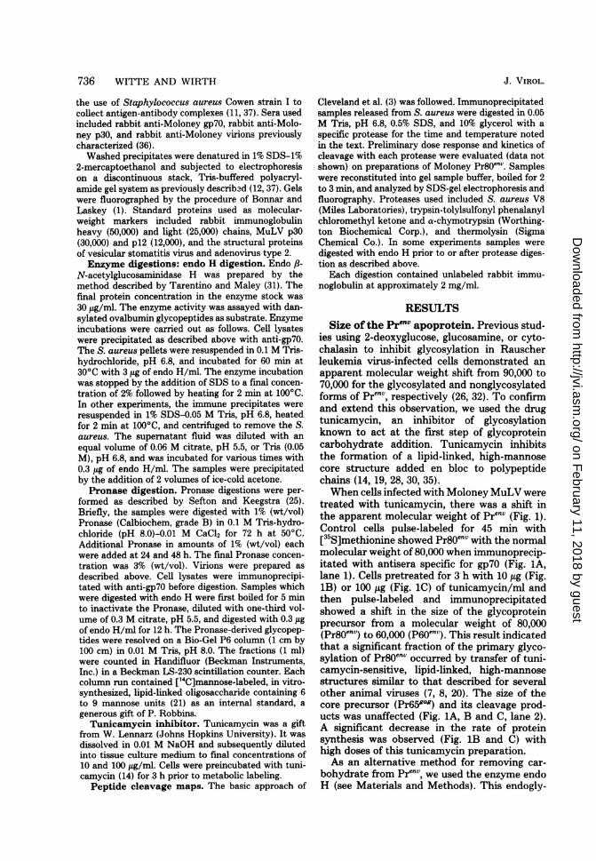
acterize further the env gene carbohydrate struc-tures, we compared the glycopeptides releasedfrom Pr8Oenv and virion gp70 after exhaustivePronase digestion by chromatography on Bio-Gel P-6. This technique separates on the basisof molecular weight, but terminal charged sugaradditions such as sialic acid can have pro-nounced effects (25). Pulse-labeled ([3H]man-nose, 45 min) Pr8Oenv recovered by immunopre-cipitation of Moloney MuLV-infected cells orMoloney MuLV virions released from cells la-beled during a 2-h interval with [3H]mannoseand collected over a 16-h chase period served assubstrates. Both were digested with Pronaseover 72 h (see Materials and Methods). Theglycopeptides of Pr80en` (Fig. 4A) resolved intotwo major peaks. This difference in mobility
VOL. 29, 1979
on February 11, 2018 by guest
http://jvi.asm.org/
Dow
nloaded from

738 WITTE AND WIRTH
AA
2 2
B +2 2
Pr8Oenv m_Pr659"g -P60env
p30
*.
_op a, - --Pr9Oe'z
P7envto \-- Pr65 g
_&
I)x gp70 2) cxp30FIG. 2. Effect ofendo H on the size of the env gene precursor. Monolayers of cloned NIH/3T3 cells infected
with wild-type Moloney MuLV (panel A), ts3 Moloney MuLV (panel B), or ts24 Rauscher MuLV (panel C)were pulse-labeled with P5S]methionine (50 ttCi/ml) at 39°C for 45 min. Cells were extracted and immunopre-cipitated with anti-gp7O (lane 1) or anti-p30 (lane 2). Antigen-antibody S. aureus complexes were incubatedovernight in citrate buffer (pH 5.5; see Materials and Methods) with endo H, 0.3 pg/ml (+ lanes), or withoutenzyme (- lanes). Samples were recovered by acetone precipitation and subjected to electrophoresis on
SDS-10% polyacrylamide gels developed by fluorography as in Fig. 1.
could have reflected differences in carbohydratestructure or residual number of amino acids notremoved by Pronase digestion. To distinguishbetween these possibilities, a parallel sample ofPr80env was first Pronase digested and subse-quently treated with endo H prior to chromatog-raphy (Fig. 4B). The carbohydrate now migratedas a single component oflower molecular weight.This suggested that Pr80env contained at leasttwo identically sized oligosaccharides located atsites which differ in their amino acid composi-tion.The glycopeptides derived from virion gp7O
resolved into several components as shown inFig. 4C. Endo H digestion of the gp70 glycopep-tides indicated that most of the glycopeptideswere resistant to cleavage by endo H (Fig. 4D).This was in contrast to the glycopeptides derivedfrom the Pr80env, which were sensitive to theaction of endo H. A fraction of the gp70 glyco-peptides (15 to 20%) were sensitive to cleavageby endo H. This is similar to the Sindbis virusglycoprotein complex, which contains a partiallyprocessed high-mannose group susceptible toendo H cleavage in the finished virion (2, 20).
It is interesting to note that treatment ofintact virions with endo H did not detectablyalter the apparent molecular weight of gp7O inSDS-gel electrophoresis (unpublished data).
Pren" carbohydrate processing and pro-teolytic cleavage appear temporally linked.It was previously shown that Pr80env was rapidlylabeled intracellularly but that the appearanceof mature gp70 and p15 was delayed by 45 to 60min (37).
If Pr8Oenv oligosaccharide was processed be-fore proteolytic cleavage, one might expect tofind a population of Pr80env molecules largelyresistant to endo H action. Conversely, if prote-olytic cleavage of pl5E from Pr8Oenv precededcarbohydrate modification, molecules of 65,000to 70,000 molecular weight sensitive to endo Hshould be found intracellularly. Pulse-chaseanalysis coupled with endo H treatment (Fig. 5and unpublished data) failed to reveal either ofthese putative intermediate forms accumulatingintracellularly, suggesting that carbohydratemodification may be closely timed to the prote-olytic processing of Pr8OenvStructural map of Prl"V by partial prote-
C +2 2
J. VIROL.
14
4m4.
qw
on February 11, 2018 by guest
http://jvi.asm.org/
Dow
nloaded from
MuLV ENVELOPE GLYCOPROTEIN PRECURSOR 739
Endo H Digestion(min)
0 5 10 20 40
Pr8OenvP60env_
a
FIG. 3. Estimation of the number ofhigh-mannosecarbohydrate groups on Pr8`nV by partial endo Hdigestion. Moloney MuLV-infected NIH/3T3 cellswere labeled with I5S]methionine (30 min). Cellswere extracted and immunoprecipitated with anti-gp7O serum. The antigen-antibody complexes wereresuspended in citrate (pH 5.5) buffer, and 20-1L sam-ples were digested with endo H (3 ptg/ml) for 0, 5, 10,20, or 40 min at 30°C. The reaction was stopped withSDS (final concentration, 2%, wt/vol) and boiling for2 min. Samples were subjected to electrophoresis onSDS-10% acrylamide gels developed by fluorography.
olysis in SDS. Cleveland et al. (3) have de-scribed a peptide mapping technique based onpartial enzymatic proteolysis in SDS and sub-sequent separation of fragments by SDS-gelelectrophoresis. We have applied this techniquein combination with differential labeling of car-bohydrate and protein and the use of endo H toprepare a preliminary structural map of Prenv.Pulse-labeled Prenv recovered from cellular ly-sates by immunoprecipitation with anti-gp7O se-rum was sufficiently pure to serve as substratefor these cleavages. Unlabeled immunoglobulinreleased from the immune complex served ascarrier.Immunoprecipitated Moloney Pr8Oenv labeled
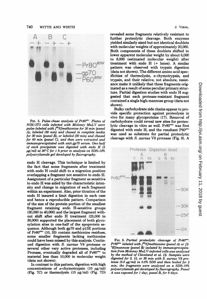
with [35S]methionine was digested for 0, 15, or30 min (Fig. 6, panel A) with S. aureus V8protease (5.0 utg/ml) in 0.5% SDS and then wasanalyzed on SDS-10% acrylamide gels. Theseconditions generated fairly large fragments withmolecular weights from 70,000 to 27,000. Pr8Oenvlabeled with [2-3H]mannose was similarlytreated (Fig. 6, panel B). The fragment with amolecular weight of 27,000 was deficient in car-bohydrate label, showing that both sugar sidechains are confined to a region less than or equalto one-half of the apoprotein length.As an alternative to [3H]mannose labeling, we
used endo H to identify sugar-containing frag-
ments. Pr8Oenv labeled with [35S]methionine wasfirst digested with a pretitered amount of pro-tease. The proteolysis was stopped by boilingthe reaction mixture, and one-half was subse-quently digested with endo H.
Pr8Oenv control, not treated with any protease,is shown in Fig. 7A (- lane). Treatment withendo H (+ lane) shifted the apparent molecularweight to P60en'. A minor component precipi-tated with anti-gp7O serum with a molecularweight of approximately 50,000 was also sensi-tive to treatment with endo H. This may haverepresented a precursor form for the minor gly-coprotein gp45 described for MuLV and shownto overlap with the amino-terminal end of gp7Oin its primary structure (6).
Digestion with S. aureus V8 protease (5.0,ug/ml) (Fig. 7B) for 10 or 40 min generatedfragments from full length (80,000) to material-10,000 migrating at the buffer front. Subse-quent treatment with endo H (+ lanes) shiftedmany, but not all, fragments to lower molecularweight. These fragments presumably containedat least one high-mannose group sensitive to
Fraction NumberFIG. 4. Analysis oftheglycopeptides ofPr8O?nv and
gp70. (A) NIH/3T3 cells infected with MoloneyMuLVwere labeled with [2-3H]mannose (200 ,uCi/ml) for 60min at 37C. Cells were extracted, and Pr80Onv wasisolated by immunoprecipitation with anti-gp70 se-rum. Glycopeptides wereprepared by exhaustive Pro-nase digestion and halfchromatographed on Bio-GelP6 as described by Sefton and Keegstra (25). [14C]-mannose-labeled, in vitro-synthesized oligosaccha-rides (21) were used as an internal standard. (B)After Pronase digestion, one-half of the Pr8Oen sam-ple was boiled for 5 min and subsequently digestedwith endo H (0.3 pg/lu) over a 12-hperiod; it was thenanalyzed on Bio-Gel P6 with the same internal stan-dards. (C) [2-3H]mannose-labeled gp7O harvestedfrom virions was Pronase digested and analyzed asin A. (D) After Pronase digestion, gp7O was subse-quently digested with endo H and analyzed as in B.
VOL. 29, 1979
on February 11, 2018 by guest
http://jvi.asm.org/
Dow
nloaded from
740 WITTE AND WIRTH
A B C
@ @ " - Pr80envI II g9p7OP60env
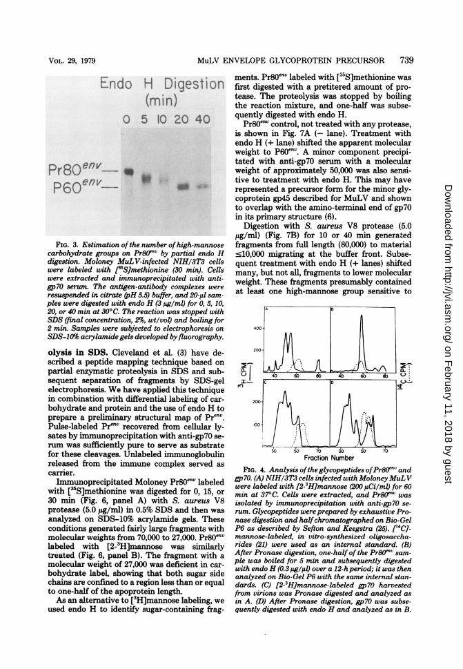
FIG. 5. Pulse-chase analysis of Pr8Oenv. Plates ofNIH/3T3 cells infected with Moloney MuLV werepulse-labeled with J5S]methionine for 30 min (panelA), labeled (30 min) and chased in complete mediafor 30 min (panel B), or labeled (30 min) and chasedfor 60 min (panel C), and then were extracted andimmunoprecipitated with anti-gp70 serum. One-halfof each precipitate was digested with endo H (3jg/ml) at 30°C for 1 h prior to analysis on SDS-10%Spolyacrylamide gel developed by fluorography.
endo H cleavage. This technique is limited bythe fact that some fragments after treatmentwith endo H could shift to a migration positionoverlapping a fragment not sensitive to endo H.Assignment of a particular fragment as sensitiveto endo H was aided by the characteristic inten-sity and change in migration of each fragmentwithin an experiment. Also, prior titration of theendo H insured a limit digestion in each caseand hence a reproducible pattern. Comparisonof the size of the protein portion of the smallestfragment retaining endo H-sensitive groups(30,000 to 40,000) and the largest fragment with-out shift after endo H treatment (25,000 to30,000) supported the placement of both glyco-sylation sites in one-half of the apoprotein se-quence. Although both gp70 and pl5E portionsof Pr8Oenv (10, 33) contain methionine residues,some smaller fragments lacking methioninecould have been missed by this analysis. Contin-ued digestion with S. aureus V8 protease orseveral other very active proteases, includingPronase, eventually degraded all of Pr8Oenv tomaterial less than 10,000 in molecular weight(data not shown).In contrast to this pattern, digestion with high
concentrations of a-chymotrypsin (10 ,ug/ml)(Fig. 7C) or thermolysin (15 ,ug/ml) (Fig. 7D)
J. VIROL.
revealed some fragments relatively resistant tofurther proteolytic cleavage. Both enzymesyielded similarly sized but not identical doubletswith molecular weights of approximately 20,000.Both components of these doublets shifted tolower apparent molecular weight by about 6,000to 8,000 (estimated molecular weight) aftertreatment with endo H (+ lanes). A similarpattern was observed with trypsin digestion(data not shown). The different amino acid spec-ificities of thermolysin, a-chymotrypsin, andtrypsin, and their relative, not absolute, resist-ance make it unlikely that these fragments orig-inated as a result ofsome peculiar primary struc-ture. Partial digestion studies with endo H sug-gested that each protease-resistant fragmentcontained a single high-mannose group (data notshown).Bulky carbohydrate side chains appear to pro-
vide specific protection against proteolysis invivo for many glycoproteins (17). Removal ofcarbohydrate could reveal new sites for proteo-lytic cleavage in vitro as well. Pr8Oenv was firstdigested with endo H, and the resultant P60envwas used as substrate for partial proteolyticcleavage with S. aureus V8 protease (Fig. 8). A
*Ij:'-%
:.I
_U; som
_wW-5CO
--0K
I--25<
L...
FIG. 6. Partial proteolytic cleavage of Pr80"nv.Pr80env labeled with I1S]methionine (panel A) or [2-3H]mannose (panel B) isolated by immunoprecipita-tion from MoloneyMuL V-infected cells was analyzedby the method of Cleveland et al. (3). Samples weredigested for 0, 15, or 30 min with S. aureus V8 pro-tease (5.0 pg/ml) in 0.5% SDS and then boiled for 2min; the fragments were analyzed on a SDS-10%opolyacrylamide gel developed by fluorography. PanelA was exposed for 1 day; panel B, for 8 days.
- Af A
on February 11, 2018 by guest
http://jvi.asm.org/
Dow
nloaded from

MuLV ENVELOPE GLYCOPROTEIN PRECURSOR 741
Protease Digestion mrn,!4 B C 90 10 40: ,0 40 .340
1- - XL __
-
r.;)env.~
r3C env_P6qefl
' It
(37), combined with pulse-chase data of Fig. 5,suggests that carbohydrate modification andproteolytic cleavage are both late events in theintracellular processing.The precise position of the high-mannose car-
bohydrate additions along the apoprotein se-quence is not known. However, several con-straints on the structure of Pren" can be deducedfrom the peptide fragment data of Fig. 6, 7, and8. Glycosylated fragments with molecular
_-; ; weights of 45,000 to 50,000 which shift to 30,000to 35,000 after endo H treatment (Fig. 7 and 8)
-2^5K and a fragment which has a molecular weight of9
Y
Endo H Trecitment (-'-'
FIG. 7. Use of endo H to identify carbohydrate-containing fragments of Pr8oTnv. f35SJmethionine-la-beled Pr8O'nv isolated by immunoprecipitation wasused as substrate for partial proteolytic cleavage bythe method of Cleveland et al. (3). (A) Control, noprotease treatment; (B) S. aureus V8 protease (5.0pg/ml) for 10 or 40 min at 37°C in 0.5% SDS; (C) a-chymotrypsin (10 jig/ml); (D) thermolysin (15 pg/ml).After protease digestion, samples were boiled andone-half was subsequently digested (+) with endo H(3 pg/ml) for 1 h at 30°C. Fragments were analyzedon a SDS-10%polyacrylamide gel developed by fluo-rography. Panels A and B were exposed for 3 days;panels C and D, for 1 day.
shift to lower molecular weight for the deglyco-sylated fragments of p60env was seen (Fig. 8D)when compared to those generated from Pr8Oenv(Fig. 8B). In addition, several new fragments oflower molecular weight (15,000 to 20,000) (Fig.8D) appeared in the fragments cleaved fromP60env, suggesting that some cleavage sites be-come more readily available after removal ofcarbohydrate. The particular region from whichthese fragments arose has not been mapped.
DISCUSSIONThe data presented demonstrate that the Mo-
loney MuLV glycoprotein precursor (Pr8Oenv) isa molecule with a apoprotein portion having amolecular weight of 60,000 and at least two largehigh-mannose carbohydrate groups. The inabil-ity to find pulse-labeled intracellular env precur-sors with molecular weights of less than 80,000(16, 26, 37) suggests that these primary carbo-hydrate additions occur during protein elonga-tion in a manner similar to that in other animalvirus models (23). The stable nature of Pr80env
pri0efivaP60envZ Z
a-50K
S
-30K
-25K
*--1K
FIG. 8. Proteolytic cleavage of Pr8Oenv after re-moval of carbohydrate. Pr80enL pulse-labeled with35S]methionine (30 min) was isolated by immunopre-cipitation as in Fig. 5. (A) No treatment; (B) treatedwith S. aureus V8 protease (5.0 pg/ml), 30 min, 37°C;(C) digested with endo H (3 pg/ml), 1 h, 300C; (D)digested first with endo H as in C and then boiled for2 min and treated with S. aureus V8 protease as inB. All samples were analyzed on SDS-10%polyacryl-amide gel developed by fluorography.
VOL. 29, 1979
I \A H c .),I
0f
on February 11, 2018 by guest
http://jvi.asm.org/
Dow
nloaded from

742 WITTE AND WIRTH
27,000 and lacks mannose label (Fig. 6) demon-strate that both sugar groups are confined to aregion of one-half the apoprotein length.The reported orientation of NH2gp7O-pl5E-
COOH for Prenv and lack of carbohydrate onpl5E (5, 9, 10, 15, 33) would limit the carbohy-drate sites to an N-terminal molecular-weightregion of 45,000, within the gp7O sequence. If thenaturally occurring fragment with a molecularweight of 50,000 to 55,000 cross-reactive withgp7O which shifts to a molecular weight of 35,000after endo H treatment (Fig. 7) does representa precursor form to gp45, then a further con-straint on the structure of Pren" can be noted.Since gp45 and gp7O share the same amino-terminal sequence (6), this fragment would placethe glycosylation sites within the amino one-halfof the apoprotein sequence. Further work isnecessary to confirm this working hypothesis.The two protease-resistant endo H-sensitive
fragments with molecular weights of 15,000 to20,000 (Fig. 7) suggest that each high-mannosegroup may protect a peptide region of 12,000 to15,000 in molecular weight. This relative protec-tion may result from an actual structural blockof the proteolytic sites by the carbohydrate oralternately could reflect a secondary structure ofthe peptide region surrounding the carbohy-drate.
Detailed knowledge of the primary structureof the MuLV env gene precursor and its intra-cellular processing will require further analysis.In particular, the precise relationship of the con-servation of peptide sequences in Pr8OenU to thevirions gp7O and pl5E is unknown. Peptide map-ping with uniformly labeled protein or primarysequence data will be necessary to approachsuch questions. The ability to prepare deglyco-sylated (tunicamycin-treated) or poorly glyco-sylated (endo H-treated) env gene apoproteinand its fragments should be useful in this regard.
ACKNOWLEDGMENTS
We are grateful to Phillips Robbins and David Baltimorefor their interest, advice, and review of the manuscript. Wethank David Steffen for his review of the manuscript and forthoughtful discussions.
This work was supported by Public Health Service grantsCA-14142 (to Phillips Robbins) and CA-14051 (to S. E. Luria)from the National Cancer Institute and by American CancerSociety grant VC-4I (to David Baltimore). O.N.W. is a HelenHay Whitney Foundation fellow.
LITERATURE CITED1. Bonnar, W. M., and R. A. Laskey. 1974. A film detection
method for tritium-labeled proteins and nucleic acids inpolyacrylamide gels. Eur. J. Biochem. 46:83-88.
2. Burke, D. J., and K. Keegstra. 1976. Purification andcomposition of the proteins from Sindbis virus grown inchick and BHK cells. J. Virol. 20:676-684.
3. Cleveland, D. W., S. G. Fischer, M. W. Kirschner,and U. Laemmli. 1977. Peptide mapping by limited
J. VIROL.
proteolysis in sodium dodecyl sulfate and analysis bygel electrophoresis. J. Biol. Chem. 252:1102-1106.
4. Faller, D. V., J. Rommelaere, and N. Hopkins. 1978.The large T1 oligonucleotides of Moloney leukemiavirus that are missing in an env gene recombinant, HIX,are present on an intracellular 21S Moloney viral RNAspecies. Proc. Natl. Acad. Sci. U.S.A. 75:2964-2968.
5. Famulari, N. G., D. L. Buchhogen, H.-D. Klenk, andE. Fleissner. 1976. Presence of murine leukemia virusenvelope proteins gp7O and pl5E in a common polypro-tein of infected cells. J. Virol. 20:501-508.
6. Henderson, L. E., T. D. Copeland, G. W. Smythers,H. Marquardt, and S. Oroszlan. 1978. Amino termi-nal amino acid sequence and carboxyl terminal analysisof Rauscher murine leukemia virus glycoproteins. Vi-rology 85:319-322.
7. Hunt, L., J. R. Etchison, and D. F. Summers. 1978.Oligosaccharide chains are trimmed during synthesis ofthe envelope glycoprotein of vesicular stomatitis virus.Proc. Natl. Acad. Sci. U.S.A. 75:754-758.
8. Hunt, L. A., and D. F. Summers. 1976. Glycosylation ofvesicular stomatitis virus glycoproteins in virus-infectedHeLa cells. J. Virol. 20:646-657.
9. Ikeda, H., W. Hardy, E. Tress, and E. Fleissner. 1975.Chromatographic separation and antigenic analysis ofproteins of the oncornaviruses. V. Identification of anew murine viral protein p15(E). J. Virol. 16:53-61.
10. Karshin, W. L., L. J. Arcement, R. B. Naso, and R. B.Arlinghaus. 1977. Common precursor for Rauscherleukemia virus gp69/71 pl5E and pl2E. J. Virol. 23:787-798.
11. Kessler, S. W. 1975. Rapid isolation of antigens from cellswith a Staphylococcus protein-A-antibody adsorbent:parameters of the interaction of antibody-antigen com-plexes with protein A. J. Immunol. 115:1617-1624.
12. Laemmli, U. K. 1970. Cleavage of structural proteinsduring the assembly of the head of bacteriophage T4.Nature (London) 227:680-685.
13. Leamnson, R. N., H. M. Shander, and M. S. Halpern.1977. A structural protein complex in Moloney leukemiavirus. Virology 76:437-439.
14. Leavitt, R., S. Schlesinger, and S. Kornfeld. 1977.Tunicamycin inhibits glycosylation and multiplicationof Sindbis and vesicular stomatitis virus. J. Virol. 21:375-385.
15. McLellan, W. L., and J. T. August. 1976. Analysis ofthe envelope of Rauscher murine oncornavirus: in vitrolabeling of glycopeptides. J. Virol. 20:627-636.
16. Naso, R. B., L. J. Arcement, W. L. Karshin, G. A.Jamjoom, and R. B. Arlinghaus. 1976. A fucose-deficient glycoprotein precursor to Rauscher leukemiavirus gp69/71. Proc. Natl. Acad. Sci. U.S.A. 73:2326-2330.
17. Olden, K., R. M. Pratt, and K. M. Yamada. 1978. Roleof carbohydrate in protein secretion and turnover: ef-fects of tunicamycin on the major cell surface glycopro-tein of chick embryo fibroblasts. Cell 13:461-474.
18. Pinter, A., and E. Fleissner. 1977. The presence ofdisulfide-linked gp70-pl5(E) complexes in AKR murineleukemia virus. Virology 83:417-422.
19. Pless, D. D., and W. J. Lennarz. 1977. Enzymatic con-version of proteins to glycoproteins. Proc. Natl. Acad.Sci. U.S.A. 74:134-138.
20. Robbins, P. W., S. C. Hubbard, S. J. Turco, and D. F.Wirth. 1977. Proposal for a common oligosaccharideintermediate in the synthesis of membrane glycopro-teins. Cell 12:893-900.
21. Robbins, P. W., S. S. Krag, and T. LAu. 1977. Effects ofUDP-glucose addition on the synthesis of mannosyllipid-linked oligosaccharides by cell-free fibroblastpreparations. J. Biol. Chem. 252:1780-1785.
22. Rothenberg, E., D. J. Donoghue, and D. Baltimore.1978. Analysis of a 5' leader sequence on murine leu-
on February 11, 2018 by guest
http://jvi.asm.org/
Dow
nloaded from

MuLV ENVELOPE GLYCOPROTEIN PRECURSOR 743
kemia virus 21S RNA: heteroduplex mapping with longreverse transcriptase products. Cell 13:435-451.
23. Rothman, J. E., and H. F. Lodish. 1977. Synchronisedtransmembrane insertion and glycosylation of a nascentmembrane protein. Nature (London) 269:775-780.
24. Russ, G., and K. Polackova. 1973. The molecular weightdetermination of proteins and glycoproteins of RNAenveloped viruses by polyacrylamide gel electrophoresisin SDS. Biochem. Biophys. Res. Commun. 55:666-672.
25. Sefton, B. M., and K. Keegstra. 1974. Glycoproteins ofSindbis virus: preliminary characterization of the oli-gosaccharides. J. Virol. 14:522-530.
26. Shapiro, S. Z., M. Strand, and J. T. August. 1976. Highmolecular weight precursor polypeptides to structuralproteins of Rauscher murine leukemia virus. J. Mol.Biol. 107:459-477.
27. Stephenson, J. R., and S. A. Aaronson. 1973. Charac-terization of temperature-sensitive mutants of murineleukemia virus. Virology 54:53-59.
28. Struck, D. K., and W. J. Lennarz. 1977. Evidence forthe participation of saccharide-lipids in the synthesis ofthe oligosaccharide chain of ovalbumin. J. Biol. Chem.252:1007-1013.
29. Tabas, I., S. Schlessinger, and S. Kornfield. 1978.Processing of high mannose oligosaccharides to formcomplex type oligosaccharides on the newly synthesizedpolypeptides of the vesicular stomatitis virus G proteinand the Ig G heavy chain. J. Biol. Chem. 253:716-722.
30. Takatsuki, A., K. Kohno, and G. Tamura. 1975. Inhi-bition of biosynthesis of polyisoprenol sugars in chickembryo microsomes by tunicamycin. Agric. Biol. Chem.39:2089-2091.
31. Tarentino, A. L., and F. Maley. 1974. Purification andproperties of an Endo B-N-acetyl-glucosaminidase fromStreptomyces griseus. J. Biol. Chem. 249:811-816.
32. Van den Ven, W. J. M., C. Onnehink, A. J. M. Ver-morken, and H. P. J. Bloemers. 1977. Effect ofimpaired glycosylation on the synthesis of envelopeproteins of Rauscher murine leukemia virus. Virology82:334-344.
33. Van Zaane, D., A. Dekker-Michielsen, and H. P. J.Bloemers. 1976. Virus specific precursor polypeptidesin cells infected with Rauscher leukemia virus: synthe-sis, identification and processing. Virology 75:113-129.
34. Van Zaane, D., A. L J. Gielkens, W. G. Hesselink,and H. P. J. Bloemers. 1977. Identification ofRauscher leukemia virus specific mRNAs for the syn-thesis of the gag and env gene products. Proc. Natl.Acad. Sci. U.S.A. 74:1855-1859.
35. Waechter, C. J., and W. J. Lennarz. 1976. The role ofpolyprenol-linked sugars in glycoprotein synthesis.Annu. Rev. Biochem. 45:95-112.
36. Witte, 0. N., and D. Baltimore. 1978. Relationship ofretrovirus polyprotein cleavages to virion maturationstudied with temperature sensitive MuLV mutants. J.Virol. 26:750-761.
37. Witte, 0. N., A. Tsukamoto-Adey, and I. L. Weiss-man. 1977. Cellular maturation of oncornavirus glyco-proteins: topological arrangement of precursor andproduct forms in cellular membranes. Virology 76:539-553.
38. Wong, P. K. Y., and J. A. McCarter. 1974. Studies oftwo temperature sensitive mutants of Moloney murineleukemia virus. Virology 58:396-408.
VOL. 29, 1979
on February 11, 2018 by guest
http://jvi.asm.org/
Dow
nloaded from


