Functional Domains of Moloney Murine Leukemia Virus Integrase
13
JOURNAL OF VIROLOGY, July 1996, p. 4585–4597 Vol. 70, No. 7 0022-538X/96/$04.0010 Copyright q 1996, American Society for Microbiology Functional Domains of Moloney Murine Leukemia Virus Integrase Defined by Mutation and Complementation Analysis COLLEEN B. JONSSON,² GEORGE A. DONZELLA, ELENA GAUCAN, CHRISTINE M. SMITH, AND MONICA J. ROTH* Department of Biochemistry, Robert W. Johnson Medical School, University of Medicine and Dentistry of New Jersey, Piscataway, New Jersey 08854 Received 16 January 1996/Accepted 15 April 1996 Retroviral integrases perform two catalytic steps, 3* processing and strand transfer, that result in the stable insertion of the retroviral DNA into the host genome. Mutant M-MuLV integrases were constructed to define the functional domains important for 3* processing, strand transfer, and disintegration by in vitro assays. N-terminal mutants had no detectable 3* processing activity, and only one mutant which lacks the HHCC domain, ND105, had strand transfer activity. Strand transfer mediated by ND105 showed preference for one site in the target DNA. Disintegration activity of N-terminal mutants decreased only minimally. In contrast, all C-terminal mutants truncated by more than 28 amino acids had no integration or disintegration activity. Activity on a single-strand disintegration substrate did not require a functional HHCC domain but did require most of the C-terminal region. Complementation analysis found that the HHCC region alone was able to function in trans to a promoter containing only the DD(35)E and C-terminal regions and to enhance integra- tion site selection. Increasing the reducing conditions or adding the HHCC domain to ND105 reaction mixtures restored the wild-type strand transfer activity and range of target sites. The reducing agent affected Cys-209 in the DD(35)E region. The presence of C-209 was required for complementation of ND105 by the HHCC region. To persist in an eukaryotic cell, retroviruses require the stable insertion of a double-stranded copy of their genome into the host chromosome by the retroviral integrase (IN). This requires a complex series of replication and integration pro- cesses in the early stages of their life cycle (7, 23, 47). The virus-encoded enzymes responsible for these events are reverse transcriptase and IN, respectively. Replication of the single- stranded Moloney murine leukemia virus (M-MuLV) RNA genome by reverse transcriptase to a double-stranded DNA occurs within 2.5 h postinfection (39) within the host cyto- plasm. During the replication process, viral sequences unique to the 39-terminal (U3) and 59-terminal (U5) regions are du- plicated and transposed to opposing ends of the linear viral DNA to generate long terminal repeats (LTRs). Each LTR serves as a substrate for the retroviral IN. As few as 7 bp of the terminal M-MuLV LTR sequences are required for a produc- tive integration event and, therefore, recognition by IN (33). The specific protein-DNA contacts responsible for LTR rec- ognition by IN have not been clearly defined. Prior to formation of the integrated M-MuLV provirus, IN processes both 39 ends of the viral DNA by an endonucleolytic cleavage of the terminal dinucleotide (39 processing) (39). Analysis of M-MuLV in vivo has suggested that the two viral termini must be coordinated by M-MuLV IN for 39 processing to occur (34). Following 39 processing, the viral DNA interme- diate and IN form a stable complex that localizes to the host chromatin, where the second IN-dependent catalytic step re- quired for retroviral integration occurs, strand transfer (1). In the murine retroviruses, strand transfer follows mitosis (31, 36). It is postulated that the IN and viral DNA complex enters into the nucleus after breakdown of the nuclear membrane (36). Strand transfer occurs precisely at the termini of the LTR following the ubiquitous 59-CA-39 sequence found in all retro- viral and retrotransposon LTRs. The reaction is a one-step transesterification whereby each end of the viral DNA is inte- grated into a random site in the host’s genome (16). The viral ends are positioned several base pairs apart (4–6), depending on the retrovirus. Repair of the gap created by the staggered integration event generates a duplication of the internal se- quences that flank the provirus. The 39 processing and strand transfer reactions have been reconstituted in vitro with assays that use oligonucleotide du- plexes containing the DNA sequences of the LTR termini (9, 24). These assays demonstrate that IN is the only protein required for integration. The retroviral IN is also active on an integration intermediate or disintegration substrate (6). The integration and disintegration activities have been character- ized for the M-MuLV IN proteins produced in bacteria (5, 11, 12, 21). Although the activities of the retroviral INs have re- action similarities, we have observed apparent differences in the disintegration substrate requirements of M-MuLV IN and human immunodeficiency virus type 1 (HIV-1) IN (11, 22). This is not unexpected, since M-MuLV IN and HIV-1 IN are proteins with quite different compositions. These differences also extend to the macromolecular composition of their pre- integration complexes and their abilities to infect nondividing and dividing cells (1, 17, 36). IN proteins from different retroviral species vary in size and amino acid homology (20, 25, 29). Two regions of strong sim- ilarity are shared between retroviral IN proteins, a proposed zinc finger motif or HHCC region in the amino terminus, and a central core region containing the DD(35)E motif. Linker * Corresponding author. Mailing address: Department of Biochem- istry, University of Medicine and Dentistry of New Jersey, Robert W. Johnson Medical School, 675 Hoes Ln., Piscataway, NJ 08854. Phone: (908) 235-5048. Fax: (908) 235-4783. Electronic mail address: [email protected]. ² Present address: Department of Chemistry and Biochemistry, New Mexico State University, Las Cruces, NM 88003. 4585 Downloaded from https://journals.asm.org/journal/jvi on 01 January 2022 by 42.3.194.72.
Transcript of Functional Domains of Moloney Murine Leukemia Virus Integrase
JOURNAL OF VIROLOGY, July 1996, p. 4585–4597 Vol. 70, No. 70022-538X/96/$04.0010Copyright q 1996, American Society for Microbiology
Functional Domains of Moloney Murine Leukemia Virus IntegraseDefined by Mutation and Complementation Analysis
COLLEEN B. JONSSON,† GEORGE A. DONZELLA, ELENA GAUCAN, CHRISTINE M. SMITH,AND MONICA J. ROTH*
Department of Biochemistry, Robert W. Johnson Medical School, University ofMedicine and Dentistry of New Jersey, Piscataway, New Jersey 08854
Received 16 January 1996/Accepted 15 April 1996
Retroviral integrases perform two catalytic steps, 3* processing and strand transfer, that result in the stableinsertion of the retroviral DNA into the host genome. Mutant M-MuLV integrases were constructed to definethe functional domains important for 3* processing, strand transfer, and disintegration by in vitro assays.N-terminal mutants had no detectable 3* processing activity, and only one mutant which lacks the HHCCdomain, ND105, had strand transfer activity. Strand transfer mediated by ND105 showed preference for onesite in the target DNA. Disintegration activity of N-terminal mutants decreased only minimally. In contrast, allC-terminal mutants truncated by more than 28 amino acids had no integration or disintegration activity.Activity on a single-strand disintegration substrate did not require a functional HHCC domain but did requiremost of the C-terminal region. Complementation analysis found that the HHCC region alone was able tofunction in trans to a promoter containing only the DD(35)E and C-terminal regions and to enhance integra-tion site selection. Increasing the reducing conditions or adding the HHCC domain to ND105 reaction mixturesrestored the wild-type strand transfer activity and range of target sites. The reducing agent affected Cys-209 inthe DD(35)E region. The presence of C-209 was required for complementation of ND105 by the HHCC region.
To persist in an eukaryotic cell, retroviruses require thestable insertion of a double-stranded copy of their genome intothe host chromosome by the retroviral integrase (IN). Thisrequires a complex series of replication and integration pro-cesses in the early stages of their life cycle (7, 23, 47). Thevirus-encoded enzymes responsible for these events are reversetranscriptase and IN, respectively. Replication of the single-stranded Moloney murine leukemia virus (M-MuLV) RNAgenome by reverse transcriptase to a double-stranded DNAoccurs within 2.5 h postinfection (39) within the host cyto-plasm. During the replication process, viral sequences uniqueto the 39-terminal (U3) and 59-terminal (U5) regions are du-plicated and transposed to opposing ends of the linear viralDNA to generate long terminal repeats (LTRs). Each LTRserves as a substrate for the retroviral IN. As few as 7 bp of theterminal M-MuLV LTR sequences are required for a produc-tive integration event and, therefore, recognition by IN (33).The specific protein-DNA contacts responsible for LTR rec-ognition by IN have not been clearly defined.Prior to formation of the integrated M-MuLV provirus, IN
processes both 39 ends of the viral DNA by an endonucleolyticcleavage of the terminal dinucleotide (39 processing) (39).Analysis of M-MuLV in vivo has suggested that the two viraltermini must be coordinated by M-MuLV IN for 39 processingto occur (34). Following 39 processing, the viral DNA interme-diate and IN form a stable complex that localizes to the hostchromatin, where the second IN-dependent catalytic step re-
quired for retroviral integration occurs, strand transfer (1). Inthe murine retroviruses, strand transfer follows mitosis (31,36). It is postulated that the IN and viral DNA complex entersinto the nucleus after breakdown of the nuclear membrane(36). Strand transfer occurs precisely at the termini of the LTRfollowing the ubiquitous 59-CA-39 sequence found in all retro-viral and retrotransposon LTRs. The reaction is a one-steptransesterification whereby each end of the viral DNA is inte-grated into a random site in the host’s genome (16). The viralends are positioned several base pairs apart (4–6), dependingon the retrovirus. Repair of the gap created by the staggeredintegration event generates a duplication of the internal se-quences that flank the provirus.The 39 processing and strand transfer reactions have been
reconstituted in vitro with assays that use oligonucleotide du-plexes containing the DNA sequences of the LTR termini (9,24). These assays demonstrate that IN is the only proteinrequired for integration. The retroviral IN is also active on anintegration intermediate or disintegration substrate (6). Theintegration and disintegration activities have been character-ized for the M-MuLV IN proteins produced in bacteria (5, 11,12, 21). Although the activities of the retroviral INs have re-action similarities, we have observed apparent differences inthe disintegration substrate requirements of M-MuLV IN andhuman immunodeficiency virus type 1 (HIV-1) IN (11, 22).This is not unexpected, since M-MuLV IN and HIV-1 IN areproteins with quite different compositions. These differencesalso extend to the macromolecular composition of their pre-integration complexes and their abilities to infect nondividingand dividing cells (1, 17, 36).IN proteins from different retroviral species vary in size and
amino acid homology (20, 25, 29). Two regions of strong sim-ilarity are shared between retroviral IN proteins, a proposedzinc finger motif or HHCC region in the amino terminus, anda central core region containing the DD(35)E motif. Linker
* Corresponding author. Mailing address: Department of Biochem-istry, University of Medicine and Dentistry of New Jersey, Robert W.Johnson Medical School, 675 Hoes Ln., Piscataway, NJ 08854. Phone:(908) 235-5048. Fax: (908) 235-4783. Electronic mail address:[email protected].† Present address: Department of Chemistry and Biochemistry, New
Mexico State University, Las Cruces, NM 88003.
4585
Dow
nloa
ded
from
http
s://j
ourn
als.
asm
.org
/jour
nal/j
vi o
n 01
Jan
uary
202
2 by
42.
3.19
4.72
.
insertion and point mutations in the HHCC domain are lethalin vivo because of their inability to mediate 39 processing of theviral termini (10, 37). The central core domain is proposed tocontain a single active site responsible for integration anddisintegration activities (23). This region features three invari-ant amino acids essential for catalysis, two aspartic acids and aglutamic acid (15, 25, 26, 45). The C-terminal portion of ret-roviral INs is the least conserved and has been functionallycharacterized to be the site of nonspecific DNA binding (32,35, 44, 48, 51, 52). The terminal 28 amino acids of the carboxylterminus of IN are nonessential for viability of M-MuLV inRat 2 cells (38). The M-MuLV IN is approximately 14 kDalarger than HIV-1 and avian sarcoma-leukosis virus INs. Thislarger size of M-MuLV IN is accounted for by differences in itsN and C termini. Approximately 50 amino acids precede theHHCC domain of the M-MuLV IN, and the C terminus con-tains a unique 36-amino-acid insertion (20).In this study, we sought to define the protein domains of
M-MuLV IN required for catalysis by a mutation and comple-mentation approach. Mutant INs were examined for enzymaticactivity in vitro by assays for integration, disintegration, andsingle-strand disintegration. N- and C-terminal mutants werehindered more in their ability to catalyze integration thandisintegration reactions. The minimal domain for enzymaticactivity for M-MuLV IN included a complete DDE region andmost of the C terminus. While not required for minimal enzy-matic activity, the HHCC domain restored wild-type (WT)strand transfer activity and target site selection in complemen-tation experiments. Mixing inactive IN mutant proteins morenarrowly defined the overlap between complementing pairs ofM-MuLV IN than that found for HIV-1 IN (14, 46). Theseresults support a model for the M-MuLV IN in which HHCCforms an independent domain distinct from the DD(35)E andC-terminal regions. Further, the HHCC region can function intrans to directly influence target site selection.
MATERIALS AND METHODS
Materials. Crude [g-32P]adenosine 59-triphosphate (7,000 Ci/mmol) was pur-chased from ICN. [a-32P]TTP (800 Ci/mmol) was obtained from Amersham. T4polynucleotide kinase was obtained from GIBCO BRL. Restriction enzymeswere purchased from New England Biolabs.Oligonucleotides. DNA oligonucleotides were purchased from the University
of Medicine and Dentistry of New Jersey Biochemistry Department SynthesisFacility. Oligonucleotides used in this study are referred to by their synthesisnumbers. Oligonucleotide substrates were 32P labeled at the 59 end with T4polynucleotide kinase (GIBCO BRL) and hybridized with a fivefold excess of thecomplementary strand. Excess radioactivity was removed by using G-25 spincolumns (Boehringer Mannheim) or by electrophoresis with 15% native poly-acrylamide gels. For the Y substrates used in disintegration assays, the LTRDNA sequence is underlined. The standard Y substrates are as follows: oligo-nucleotide 3152, 59-CAGCAACGCAAGCTTG; oligonucleotide 3192, 59-AATGAAAGACCCCGCTGACTA; oligonucleotide 3154, 59-TAGTCAGCGGGGTCTTTCAGGCTGCAGGTCGAC; and oligonucleotide 3527, 59-GTCGACCTGCAGCCCAAGCTTGCGTTGCTG. The dumbbell Y substrate, YDB, is oligo-nucleotide 3808, 59-AATGAAAGTTCTTTCAGGCTGTTCAGCCCAAGCTTGCTTG, which is self complementary. Oligonucleotide 3152 was 59 end labeled,purified, and annealed to oligonucleotides 3192, 3527, and 3154 to produce thestandard Y substrate or annealed to oligonucleotides 3527 and 3154 to producethe YSS substrate as previously described (11). YDB was 59 end labeled, self-annealed, and purified as previously described (11). YCD was made from strands3152 and 3527. For integration substrates, oligonucleotides 2784/2785 were pre-pared as described previously (22). The 39-processing substrate sequence is 59AATGAAAGACCTTGGTCTTTCA-39 and represents a new substrate thatforms a hairpin LTR molecule.Construction of M-MuLV IN mutants. Cloning of the M-MuLV IN into the
expression vector pET11C (Novagen, Madison, Wis.) has been previously de-scribed and was used as the parent vector (pETINH1) for many of the subse-quent manipulations (22). This construct has a Met introduced prior to the firstamino acid of the M-MuLV IN; however, this residue is not counted in thenumbering scheme of our mutant IN proteins. The first amino acid of theM-MuLV IN constructs presented here corresponds to nucleotide 4610 of the
M-MuLV RNA (42). A second parent vector used in these constructions ispETINH3. pETINH3 contains additional nucleic acid sequence at the 59 end ofthe gene, before the first M-MuLV amino acid and after the first methionineATG, which encodes for a hexahistidine tag. The HindIII site in the vectorportion of pETINH3 was removed to facilitate cloning of the C-terminal mu-tants. The site was destroyed by digestion of the vector withHindIII, filled in withKlenow fragment, and religated with T4 DNA ligase. Construction of C97Y(M-MuLV IN mutant with tyrosine at position 97 instead of cysteine) and in5247has been previously reported (22, 37). ND105 was constructed by introducing aMet prior to amino acid 106 by PCR. The 59-end PCR primer 3807, 59-GATATACATATGGCCGTTAAACAGGGA-39, adds an NdeI site (underlined) priorto nucleotide 316; the primer sequence for IN is shown in bold type. The 39-endPCR primer, 2350, 59-GGATCCAGTACTGACCCCTCTG-39 adds a BamHIsite (underlined) outside the stop codon of the gene. The PCR product was di-gested with NdeI, and the resulting 475-bp fragment replaced with WT 791-bpNdeI fragment of pETINH1. ND176 was constructed in a similar manner byreplacing the WT NdeI fragment with a 259-bp PCR-generated NdeI fragment.The 39-end primer, 2350, was used in conjunction with a 59-end primer, oligo-nucleotide 3904 (59-CATATGCCTCAGGTATTGGGA) to create the deletionfragment. The Cys-to-Ala mutation, C209A, was created by oligonucleotide-directed mutagenesis using overlap PCR (19). The complementary primers,3811, 59-GGATTGATTGGAAATTGCATGCTGGATACAGACC-39, and 3811C,59-GGTCTGTATCCAGCATGCAATTTCCAATCAATCC-39 introduced a muta-tion at the underlined sequence. PCRs were made with primer combinations, prim-ers 3811 and 2350 and primers 3811C and 2351 (59-end primer [21]). Individual PCRproducts were separated on a 1.2% agarose gel and glass powder purified (50). Thetwo products were then added to a PCR with primers 2350 and 2351. All PCRs wereperformed with Vent (New England Biolabs) orPfuDNA polymerase (Stratagene).K227A and CD311/346 (deletion of the C-terminal 36-amino-acid insertion) werecreated in pHS by the method of Kunkel (18, 27). The DNA oligonucleotidecorresponding to K227A, oligonucleotide 2076 (59-GAACCATCGCGGAGACTTTAAC-39), generates a BstU1 site (underlined) that allowed screening for themutation by the appearance of one additional BstU1 site. The oligonucleotide forCD311/346, 59-CGAAGTCTGGAGACCTCTGTGGAAAGGACCTTACACAGTCC-39, resulted in the loss of a KpnI site. Following construction of the mutation,a 750-bp fragment was digested with SacII-PmlI and exchanged into pNCA-C (8,18). The entire coding region was then moved into pETINH1. The construct en-coding the mutant protein CD232 was made in pETINH3 (38). pETINH3 was firstpartially digested with SphI. Linearized plasmid DNA was isolated, and the DNAtermini were blunt ended with T4 DNA polymerase in the presence of deoxynucleo-side triphosphates and ligated in the presence of 100-fold molar excess of NheIlinkers (catalog no. 1060; New England Biolabs). The same protocol was followedfor CD144; a partialNdeI digest of pETINH3 was made, the full-length linear DNAwas isolated and filled in with T4 polymerase, andNhe linkers were ligated into thelinearized plasmid. Carboxyl-terminal deletions were subcloned from previouslyreported plasmid constructs by isolation of the HindIII-BamHI fragment, and re-placement of the HindIII-BamHI fragment in the pETINH3 vector (38). The nu-cleotide sequence of each construct was verified by dideoxy sequencing with Seque-nase version 2.0 (United States Biochemical) (40).Protein purification.Mutant and WTM-MuLV IN proteins were expressed in
Escherichia coli and purified by nickel nitriloacetate (NTA) chromatography(Qiagen), using methods described previously (21). One additional chromatog-raphy step was added to increase the purity of the preparation. Briefly, thematerial eluted from the nickel-NTA resin in buffer A (10 mM Tris base, 0.1 MNa2HPO4, 0.1% Nonidet P-40, 10 mM b-mercaptoethanol, 100 mM NaCl, 10%glycerol) at pH 4.5 was pooled, dialyzed for 2 h against buffer A at pH 8.0, andreapplied to the same 1 ml of reactivated nickel-NTA resin reequilibrated to pH8.0. The column was then washed stepwise with 10 ml each of buffer A at pH 8.0,buffer A at pH 6.3, and buffer A at pH 5.9. M-MuLV IN was finally elutedfrom the resin with 10 ml of buffer A at pH 4.5 and refolded by a stepwiseremoval of denaturant (21). Chromatography steps were followed by sodiumdodecyl sulfate-polyacrylamide gel electrophoresis and Western blot (immuno-blot) analysis for each protein fraction. The protein concentrations in the frac-tions were quantitated by the Bradford method (2). To assess the enzymaticactivity and stability of each mutant, proteins were prepared and isolated aminimum of three times. In vitro assays for 39 processing, strand transfer, anddisintegration activities were also measured for each protein for a minimum ofthree independent purifications. Two independent purifications of ND105/C209A were assayed and demonstrated consistent strand transfer and disinte-gration activities. Protein preparations that showed consistency in their activitiesare represented herein.Integration and disintegration assays. Integration substrates were prepared
from oligonucleotides, and assays were done as previously described at 30 or378C (21). Strand transfer activities were assayed in a reaction mixture containing20 mM MES (morpholineethanesulfonic acid) (pH 6.2), 10 mM dithiothreitol(DTT), 10 mM KCl, 10 mMMnCl2, 20% glycerol, 1 pmol of substrate, and 12 to30 pmol of M-MuLV IN in a final volume of 15 ml. Reaction mixtures for 39processing were identical to strand transfer except for the inclusion of 10%ethylene glycol. The 39 end of the substrate was filled in at room temperature for30 min using 40 pmol of substrate and 80 pmol of [a-32P]TTP (800 Ci/mmol) with5 to 8 U of exonuclease-free Klenow fragment of E. coli DNA polymerase I. Theunincorporated deoxynucleoside triphosphate label was removed by using a spin
4586 JONSSON ET AL. J. VIROL.
Dow
nloa
ded
from
http
s://j
ourn
als.
asm
.org
/jour
nal/j
vi o
n 01
Jan
uary
202
2 by
42.
3.19
4.72
.

column, and the labeled substrate was gel purified on 20% polyacrylamide gels,eluted, and resuspended in 10 ml of 0.1 M NaCl. The substrate was self annealedby slow cooling after heating to 958C and subsequently was diluted to 0.5 pmol/mland used as indicated in integration reactions. Preparation of Y substrates,enzymatic assays for disintegration activity, and product analysis were performedas previously described (11, 21). The standard Y substrate was Y3154, thesingle-stranded YSS substrate was Y3154 without the A strand, and the dumbbellsubstrate, YDB, was Y3808. All disintegration reactions were performed in avolume of 15 ml containing 20 mM piperazine-N,N9-bis(2-ethanesulfonic acid)(PIPES) (pH 6.4), 10 mM DTT, 25 mM MnCl2, 10 mM 3-[(3-cholamidopropyl)-dimethyl-ammonio]-1-propanesulfonate (CHAPS), and 0.05% Nonidet P-40 at30 or 378C for 1 h, typically with 0.5 to 1.0 pmol of Y substrate, and 5 to 20 pmolof M-MuLV IN. All integration and disintegration reactions were terminated bythe addition of 10 ml of stop buffer (95% formamide, 1 mM EDTA). Reactionmixtures were heated to 958C for 3 to 5 min and then separated on 20%sequencing gels. Gels were dried, exposed to Kodak X-Omat X-ray film at2708Cor room temperature from 30 min to overnight, and analyzed by autoradiogra-phy.Complementation assays. Equimolar amounts (7.5 pmol) of individual M-
MuLV IN proteins were mixed and incubated for 15 min on ice or at 378C inreaction buffer as indicated for integration assays in the absence of substrate.Following preincubation, substrate (1 pmol) for the 39 processing or strandtransfer reaction was added to each reaction mixture and placed at 30 or 378C.After 1 h, reactions were terminated with 10 ml of stop buffer. Reaction mixtureswere examined by the autoradiography method mentioned above.Complementation (DTT) assay. DTT treatment of WT, C209A, CD232,
ND105, and ND105/C209A M-MuLV IN proteins were performed as follows.Each protein (120 pmol) was diluted to a final concentration of 2 pmol/ml withice-cold 20 mM HEPES (N-2-hydroxyethylpiperazine-N9-2-ethanesulfonic acid)(pH 7.4)–20% glycerol and equilibrated on ice for 5 min. For complementa-tion assays, 60 pmol of each protein was mixed together and then diluted im-mediately to 2 pmol/ml (as above) with respect to total protein concentration.Untreated proteins were not subjected to this dilution step. The DTT concentra-tion was then adjusted to 40 mM, and the mixture was allowed to incubateon ice for 20 min. Reactions were initiated by the addition of 10 or 20 pmol ofthe untreated or DTT batch-treated protein as indicated to 1 pmol of oligo-nucleotides 2784/2785 or Y standard substrate in their respective reactionbuffer. The final DTT concentrations in the reaction mixtures were 10 mM forthe untreated proteins and 40 mM for the DTT-treated proteins. Untreatedcomplementation assays were conducted by adding 10 pmol of each protein pairto the sides of the microcentrifuge tube and mixing by a 1-s spin in the micro-centrifuge. Reaction mixtures were incubated at 378C for 1.5 h, and then thereactions were terminated. Both strand transfer and disintegration assays wereperformed concurrently with the same batch-treated M-MuLV IN preparations.Integration and disintegration assays were repeated three times with three sep-arate DTT treatments of the M-MuLV IN proteins and yielded consistent re-sults.Product analysis. Data were quantitated directly from dried sequencing gels
with a Molecular Dynamic PhosphorImager as previously described (22). Datawere collected from gels with ImageQuant 3.1 software. Percent conversion toproduct was determined by dividing the amount of product by the sum of thesubstrate and product and multiplying by 100.
RESULTS
Construction and purification of M-MuLV IN mutants.WTand mutant M-MuLV IN proteins were produced in E. coliBL21(DE3). Protein expression was under the control of theT7 lac promoter (21). The open reading frames of the WT andmutant M-MuLV IN genes were constructed so as to contain ahexahistidine tag at the 59 or 39 end of the gene coding se-quence. The hexahistidine tag facilitated the purification of INproteins on Ni21-NTA affinity chromatography resin as previ-ously described (21). No difference was noted in the specificactivities of amino- and carboxyl-hexahistidine-tagged WT M-MuLV INs during in vitro integration or disintegration assays(data not shown). A schematic representation of each mutantM-MuLV IN protein and the position of the hexahistidine tagfor each construct are presented in Fig. 1. In general, IN genesconstructed with C-terminal truncations have the hexahistidinetag at the 59 end, while genes with N-terminal truncations havethe tag at the 39 end. Mutants which alter the HHCC fingerinclude in5247, C97Y, ND105, and ND176. Mutants with singleamino acid substitutions within the catalytic core region in-clude C209A and K227A. The C terminus was progressively
deleted through the catalytic core region. Mutants with thesedeletions included CD22, CD28, CD34, CD40, CD45, CD86,CD144, and CD232. Mutants CD311/346 contains an internaldeletion of the unique 36-amino-acid sequence found in theM-MuLV IN protein (20).Figure 2 shows the protein products of whole-cell extracts
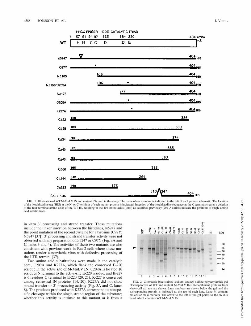
expressing the WT M-MuLV IN and the 14 mutants used inthis analysis. The protein products are expressed in large quan-tities, and they migrate to the positions predicted for the full-length product of each construct. A few of the IN proteinswere unstable during subsequent solubilization, extraction,and purification, resulting in variable levels of enzymatic ac-tivity and an increased background of nonspecific nucleaseactivity (CD311/346 and CD34). Proteolytic degradation ofthese mutants was noted from Western blot analysis of crudeprotein extracts from E. coli cultures as well as in the finalfractions eluted at pH 4.5 (data not shown). The poor yieldsand stabilities of CD34 and CD311/346 proteins precludedany further analysis. Activity of CD311/346 was noted in thestandard disintegration reaction but was variable (data notshown).Defining functional catalytic domains and amino acid resi-
dues for integration in M-MuLV IN by mutational analysis.Previous work with M-MuLV IN found that the HHCC regionwas necessary for 39 processing and integration, but not fordisintegration (22). In these experiments, we were interested infurther refinement of the domains of M-MuLV IN required forenzymatic activity. Mutations targeted the three major do-mains of the IN protein: the HHCC finger, the catalytic core,and the C-terminal region. Proteins were assayed for strandtransfer activity by using a 39 recessed oligonucleotide sub-strate, serving as both the viral and target DNA (Fig. 3A). 39processing activity was also measured with a blunt-ended, self-annealing oligonucleotide substrate (Fig. 3C). The abilities ofIN to remove the radiolabeled terminal dinucleotide and tomediate subsequent strand transfer reactions can be monitoredby separation of the products on polyacrylamide gels. Underthe assay conditions for 39 processing, three forms of the re-leased dinucleotide were identified previously (49).The amino-terminal deletion of M-MuLV IN, ND105, de-
letes the HHCC region to a position homologous to the ND105deletion of HIV-1 IN and Rous sarcoma virus IN (3, 4, 46).The increase in the size of this M-MuLV IN deletion is due tothe additional 55 amino acids preceding the HHCC domain.ND105 is truncated through the 5 amino acids following C-97.ND176 is truncated through the HHCC region and the first D(D-125) of the DDE motif. The ability of ND105 to performstrand transfer varied slightly among protein purifications (Fig.3A, lane 5, and Fig. 3B, lanes 3 to 5). ND105 had no detectable39 processing activity (FIg. 3C, lane 5). Upon further analysisof the strand transfer reactions, it was observed that strandtransfer selectively occurred at one site approximately 4 to 5nucleotides from the 39 end of the labeled LTR strand (Fig.3B). Several preparations of this protein were made to confirmthe preference for one integration site. Three other prepara-tions of ND105 are shown (Fig. 3B, lanes 3 to 5) in directcomparison with WT IN (Fig. 3B, lane 2). Two of these prep-arations (Fig. 3B, lanes 3 and 5) demonstrated marked speci-ficity for a site-specific integration, while one preparation ofND105 showed no activity (Fig. 3B, lane 4). We show theactivity of the inactive preparation because this protein wasused in the complementation analysis below (see Fig. 5).ND176 had no detectable strand transfer (Fig. 3A and C, lanes6) or 39 processing activity (Fig. 3C, lane 6).Additional mutants with mutations in the HHCC region,
which were previously described (22) were further analyzed for
VOL. 70, 1996 MUTATIONAL ANALYSIS OF M-MuLV IN 4587
Dow
nloa
ded
from
http
s://j
ourn
als.
asm
.org
/jour
nal/j
vi o
n 01
Jan
uary
202
2 by
42.
3.19
4.72
.
in vitro 39 processing and strand transfer. These mutationsinclude the linker insertion between the histidines, in5247 andthe point mutation of the second cysteine for a tyrosine (C97Y;bi5247 [37]). 39 processing and strand transfer activity were notobserved with any preparation of in5247 or C97Y (Fig. 3A andC, lanes 3 and 4). The activities of these two mutants are alsoconsistent with previous work in Rat 2 cells where these mu-tations render a nonviable virus with defective processing ofthe LTR termini (37).Two amino acid substitutions were made in the catalytic
core, C209A and K227A, which flank the conserved E-220residue in the active site of M-MuLV IN. C209A is located 10residues N terminal to the active-site E-220 residue, and K-227is 6 residues C terminal to E-220 (20, 25). K-227 is conservedamong retroviral IN proteins (14, 20). K227A did not showstrand transfer or 39 processing activity (Fig. 3A and C, lanes8). The products produced with K227A correspond to nonspe-cific cleavage within the single-strand region of the substrate;whether this activity is intrinsic to this mutant or is from a
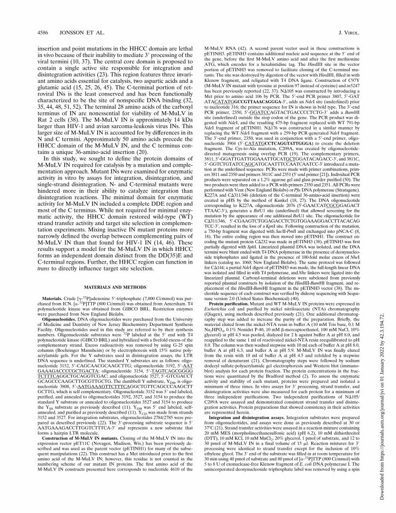
FIG. 1. Illustration of WT M-MuLV IN and mutant INs used in this study. The name of each mutant is indicated to the left of each protein schematic. The locationof the hexahistadine tag (HIS) at the N- or C terminus of each mutant protein is indicated. Insertion of the hexahistadine sequence at the C terminus creates a deletionof the four terminal amino acids of the WT IN, resulting in the 404 amino acids (total) as described previously (20). Asterisks indicate the positions of single aminoacid substitutions.
FIG. 2. Coomassie blue-stained sodium dodecyl sulfate-polyacrylamide gelelectrophoresis of WT and mutant M-MuLV INs. Recombinant proteins fromwhole-cell extracts are shown. Lane numbers are shown below the gel, and thecorresponding protein is indicated at the top of each lane. Lane M containsmolecular mass markers. The arrow to the left of the gel points to the 46-kDaband, which contains WT M-MuLV IN.
4588 JONSSON ET AL. J. VIROL.
Dow
nloa
ded
from
http
s://j
ourn
als.
asm
.org
/jour
nal/j
vi o
n 01
Jan
uary
202
2 by
42.
3.19
4.72
.
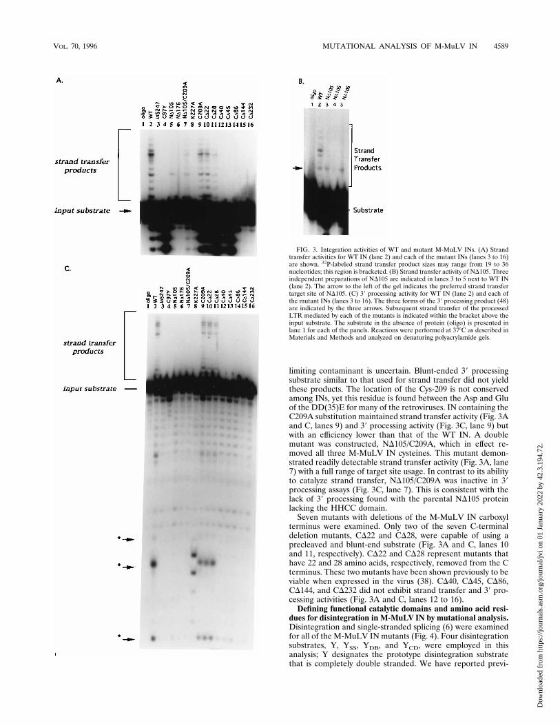
limiting contaminant is uncertain. Blunt-ended 39 processingsubstrate similar to that used for strand transfer did not yieldthese products. The location of the Cys-209 is not conservedamong INs, yet this residue is found between the Asp and Gluof the DD(35)E for many of the retroviruses. IN containing theC209A substitution maintained strand transfer activity (Fig. 3Aand C, lanes 9) and 39 processing activity (Fig. 3C, lane 9) butwith an efficiency lower than that of the WT IN. A doublemutant was constructed, ND105/C209A, which in effect re-moved all three M-MuLV IN cysteines. This mutant demon-strated readily detectable strand transfer activity (Fig. 3A, lane7) with a full range of target site usage. In contrast to its abilityto catalyze strand transfer, ND105/C209A was inactive in 39processing assays (Fig. 3C, lane 7). This is consistent with thelack of 39 processing found with the parental ND105 proteinlacking the HHCC domain.Seven mutants with deletions of the M-MuLV IN carboxyl
terminus were examined. Only two of the seven C-terminaldeletion mutants, CD22 and CD28, were capable of using aprecleaved and blunt-end substrate (Fig. 3A and C, lanes 10and 11, respectively). CD22 and CD28 represent mutants thathave 22 and 28 amino acids, respectively, removed from the Cterminus. These two mutants have been shown previously to beviable when expressed in the virus (38). CD40, CD45, CD86,CD144, and CD232 did not exhibit strand transfer and 39 pro-cessing activities (Fig. 3A and C, lanes 12 to 16).Defining functional catalytic domains and amino acid resi-
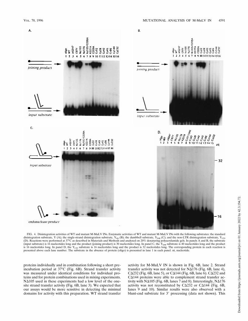
dues for disintegration in M-MuLV IN by mutational analysis.Disintegration and single-stranded splicing (6) were examinedfor all of the M-MuLV IN mutants (Fig. 4). Four disintegrationsubstrates, Y, YSS, YDB, and YCD, were employed in thisanalysis; Y designates the prototype disintegration substratethat is completely double stranded. We have reported previ-
FIG. 3. Integration activities of WT and mutant M-MuLV INs. (A) Strandtransfer activities for WT IN (lane 2) and each of the mutant INs (lanes 3 to 16)are shown. 32P-labeled strand transfer product sizes may range from 19 to 36nucleotides; this region is bracketed. (B) Strand transfer activity of ND105. Threeindependent preparations of ND105 are indicated in lanes 3 to 5 next to WT IN(lane 2). The arrow to the left of the gel indicates the preferred strand transfertarget site of ND105. (C) 39 processing activity for WT IN (lane 2) and each ofthe mutant INs (lanes 3 to 16). The three forms of the 39 processing product (48)are indicated by the three arrows. Subsequent strand transfer of the processedLTR mediated by each of the mutants is indicated within the bracket above theinput substrate. The substrate in the absence of protein (oligo) is presented inlane 1 for each of the panels. Reactions were performed at 378C as described inMaterials and Methods and analyzed on denaturing polyacrylamide gels.
VOL. 70, 1996 MUTATIONAL ANALYSIS OF M-MuLV IN 4589
Dow
nloa
ded
from
http
s://j
ourn
als.
asm
.org
/jour
nal/j
vi o
n 01
Jan
uary
202
2 by
42.
3.19
4.72
.
ously that the prototypic splicing substrate, YSS, for WT M-MuLV IN had a strict requirement for the presence of 17 ormore nucleotides on the single-stranded LTR portion of thesubstrate (11). This activity is substantially different from thatreported for HIV-1 IN, which is active even with a single-nucleotide overhang (6). Two alternative disintegration sub-strates, YDB and YCD, were also included in the analysis. Thedumbbell substrate, YDB, is a self-complementary substratethat contains a 5-bp duplex of the LTR region. Variation inM-MuLV IN catalysis with the YDB substrate versus the stan-dard disintegration Y substrate has been observed (11). Fi-nally, a new substrate variant, YCD, which constitutes only theC and D strands of the disintegration substrate and lacks LTRsequences is introduced. No activity with the YCD substratewas expected. Surprisingly, a low level of activity was notedwith this substrate.Representative assays of the M-MuLV IN proteins for each
of the Y substrates are shown in Fig. 4. The results of quanti-tation of multiple assays are summarized in Table 1. The WTM-MuLV IN activity was set to 100%, and the activities statedfor the mutants are given as percentages of WT activity. TheWT M-MuLV IN activity is shown for the Y, YSS, YDB, andYCD substrates (Fig. 4, lanes 2). Mutant proteins with muta-tions in the HHCC region, in5247 and C97Y (lanes 3 and 4),retained activity with Y, YDB, and YSS, although with efficien-cies lower than that of the WT IN (Table 1). This was incontrast to the lack of activity noted for these enzymes with theintegration substrates (Fig. 3A and B, lanes 3 and 4).In contrast to mutations in the HHCC region, deletion of the
N-terminal HHCC region, ND105, resulted in product accu-mulation using the Y and YDB substrates at levels similar toWT IN (Fig. 4A and C, lane 5). Activity on YSS remained lowbut detectable (Table 1 and Fig. 4B, lane 5). No disintegrationactivity was retained in an N-terminal M-MuLV IN mutantND176 with any of the substrates examined (Fig. 4A to C, lanes6). K227A had minimal activity with the Y, YSS, and YDBsubstrates (Fig. 4A to C, lanes 8; Table 1). C209A showedactivity with all the substrates, Y, YSS, and YDB (Fig. 4A to C,lanes 9). The highest activity of C209A was noted with the Ysubstrate, which accumulated product to a level equivalent tothat of WT IN (Fig. 4A, lane 9). C209A showed lower levels ofactivity with YSS converting 24% of the substrate to productcompared with WT IN (Fig. 4B, lane 9). Similarly, a low levelof activity was also observed with C209A for the YDB substrate,which had converted 27% of the substrate to product (Fig. 4C,lanes 9). The double mutant ND105/C209A retained activitywith Y substrate (Fig. 4A, lane 7, and Fig. 7B, lanes 10 and 11)and YSS substrate (Fig. 4B, lane 7), both of which averaged lessthan 10% of WT IN (Fig. 4B, lane 7). ND105/C209A showedthe least activity with the YDB at 3% of WT IN (Fig. 4C, lane7). In general, the effects of the mutations and truncations atthe N-terminal portions of M-MuLV IN were observed toproduce a loss in 39 processing and strand transfer activity, butnot disintegration.The disintegration activities of all seven of the carboxyl-
terminus deletion mutants of M-MuLV IN were examined.The mutants displayed patterns of activity similar to that dem-onstrated for integration (Fig. 3A and C). Disintegration ac-tivity was readily detected for deletions CD22 and CD28 withthe standard Y, YSS, and YDB substrates (Fig. 4A to C, lanes10 and 11). No disintegration activity was noted for CD40,CD45, CD86, CD144, and CD232 (Fig. 4A, lanes 12 to 16).Enzymatic activity for CD40, CD45, CD86, CD144, and CD232was also not detected with any of the other disintegrationsubstrate analogs (Fig. 4B and C, lanes 12 to 16). Disintegra-tion activities of mutants with C-terminal truncations were
affected in manners similar to that observed for both integra-tion activities examined.The final enzymatic activity examined is one that has not
previously been reported. The substrate contains no LTR se-quences and consists of two strands, the 16-nucleotide C strandand a 30-nucleotide D strand that form a DNA molecule withdouble- and single-stranded regions. The 59 end of the C strandis 32P labeled. The reaction catalyzed by IN is a nucleophilicattack mediated by the 39 OH of the C strand on the D strand,which results in the formation of a 32-nucleotide product. Theability to recognize the YCD substrate was limited to the WTIN, C209A (data not shown), and CD22 and CD28 (Fig. 4D,lanes 2, 9, and 10). The level of activity was lower for thissubstrate than observed with the other Y substrates analyzedand was not quantified.Functional complementation of strand transfer activity us-
ing N- and C-terminal deletions of M-MuLV IN. The abilitiesof two of the seven C-terminal mutants to function in trans withND105 and regain integration activity were examined (Fig. 5).Three concentrations of ND105 were examined, while the con-centration of each mutant was held constant. ND105 used inthis study did not have integration activity (Fig. 3B, lane 4).Each protein component was mixed in reaction buffer on iceand incubated for 15 min prior to the addition of strand trans-fer substrate.In Fig. 5, the WT strand transfer activity of M-MuLV IN is
shown in lane 8, and the activity of ND105 at the lowest con-centration is presented in lane 1. The addition of ND105 toM-MuLV IN did not affect strand transfer activity, although aslight increase in product was noted (Fig. 5, lanes 8 to 11). Adramatic revival of strand transfer activity was noted in reac-tion mixtures with CD45 (Fig. 5, lanes 2 to 4) upon the additionof ND105. Mixing CD86 and ND105 did not reconstitute strandtransfer activity as significantly (Fig. 5, lanes 5 to 7). Theseresults indicate that a viral LTR one-end strand transfer reac-tion can be reconstituted by the assembly of one HHCC do-main, two DD(35)E domains, and one carboxyl-terminal do-main.Functional complementation of ND105 and ND176 strand
transfer activities by the HHCC domain. In the experimentsshown in Fig. 5, complementation was performed with HHCC,DD(35)E and portions of the C-terminal region on one mol-ecule and DD(35)E with the C terminus on the second mole-cule. In these experiments, we ask whether the HHCC regioncan function as an independent domain. The combinations ofproteins used in this complementation analysis are schemati-cally represented in Fig. 6A. The two C-terminal deletionswere tested: CD144, which contains the HHCC and DD(35)Eregion, and CD232, which contains the HHCC domain andsequence up to and including the first D of the DD(35)Eregion. The C-terminal deletions were mixed with ND105 [con-tains the DD(35)E region and the C terminus] or ND176 [firstD of the DD(35)E region and the C terminus]. The locationsof the HHCC and DD(35)E invariant residues are depicted forthe WT M-MuLV IN (Fig. 6A, #1). The terminal 72 aminoacids of CD232 overlap with the first 72 amino acids of ND105(Fig. 6A, #2). CD144 comprises the first 263 amino acids ofM-MuLV IN and has an overlap of 159 amino acids withND105 (Fig. 6A, #3). Again, these 159 amino acids comprisethe entire DD(35)E region. CD232 protein contains the first176 amino acids of M-MuLV IN. No overlap exists betweenCD232 and ND176 (Fig. 6A, #4). The ND176 protein startswhere CD232 stops. CD144 has an overlap of 87 amino acidswith ND176 (Fig. 6A, #5). These 87 amino acids comprise onlythe D(35)E portion.The strand transfer activity was assayed for each of the four
4590 JONSSON ET AL. J. VIROL.
Dow
nloa
ded
from
http
s://j
ourn
als.
asm
.org
/jour
nal/j
vi o
n 01
Jan
uary
202
2 by
42.
3.19
4.72
.
proteins individually and in combination following a short pre-incubation period at 378C (Fig. 6B). Strand transfer activitywas measured under identical conditions for individual pro-teins and for protein combinations used in mixing experiments.ND105 used in these experiments had a low level of the one-site strand transfer activity (Fig. 6B, lane 3). We expected thatour assays would be more sensitive in detecting the minimaldomains for activity with this preparation. WT strand transfer
activity for M-MuLV IN is shown in Fig. 6B, lane 2. Strandtransfer activity was not detected for ND176 (Fig. 6B, lane 4),CD232 (Fig. 6B, lane 5), or CD144 (Fig. 6B, lane 6). CD232 andCD144 proteins were able to complement strand transfer ac-tivity with ND105 (Fig. 6B, lanes 7 and 8). Interestingly, ND176activity was not reconstituted by CD232 or CD144 (Fig. 6B,lanes 9 and 10). Similar results were also observed with ablunt-end substrate for 39 processing (data not shown). This
FIG. 4. Disintegration activities of WT and mutant M-MuLV INs. Enzymatic activities of WT and mutant M-MuLV INs with the following substrates: the standarddisintegration substrate, Y (A); the single-strand disintegration substrate, YSS (B); the dumbbell substrate, YDB (C); and the non-LTR disintegration substrate, YCD(D). Reactions were performed at 378C as described in Materials and Methods and analyzed on 20% denaturing polyacrylamide gels. In panels A and B, the substrate(input substrate) is 16 nucleotides long and the product (joining product) is 30 nucleotides long. In panel C, the YDB substrate is 40 nucleotides long and the productis 16 nucleotides long. In panel D, the YCD substrate is 16 nucleotides long and the product is 32 nucleotides long. The corresponding protein in each reaction ispresented above each lane number. The substrate in the absence of protein (oligo) is presented in lane 1 in each panel. nt, nucleotide.
VOL. 70, 1996 MUTATIONAL ANALYSIS OF M-MuLV IN 4591
Dow
nloa
ded
from
http
s://j
ourn
als.
asm
.org
/jour
nal/j
vi o
n 01
Jan
uary
202
2 by
42.
3.19
4.72
.
analysis suggests that the central core functions along with theC terminus, rather than the N terminus, and does not need tobe present on both molecules. The 71-amino-acid region, res-idues 106 to 177, was required in cis with the C-terminal por-tion of the IN, and not the N-terminal portion for reconstitu-tion of integration activity.Effects of protein concentration and high reductant levels on
strand transfer and disintegration. IN mutants lacking theHHCC domain typically exhibit impaired strand transfer, al-though a low level of in vitro activity was noted with ND105(Fig. 3C). Several biochemical observations indirectly corre-lated the redox state of C209A to the level of strand transferactivity. These observations include the following: (i) activitiesof C209A and WT M-MuLV IN (the activity of C209A was
frequently higher than that of the WT M-MuLV IN), (ii) thesite specificity of ND105 strand transfer, and (iii) increasedyields of active ND105 and other M-MuLV IN proteins ob-tained when the protein concentration was limited to 2 to 5 mMduring renaturation. As C-209 is the sole Cys present inND105, these data suggested that the redox state of the M-MuLV IN proteins may affect activity. To address this possi-bility, the effects of elevated DTT and protein concentrationon strand transfer (Fig. 7A) and disintegration reactions (Fig.7B) were investigated.Standard assay conditions contain 10 mM DTT, and two
levels of protein (10 or 20 pmol) which served as standards forprotein concentrations used in complementation reactions.With WT M-MuLV IN, strand transfer was efficient (Fig. 7A,lanes 2 and 3). The CD232 finger-containing protein was inac-tive regardless of concentration (Fig. 7A, lanes 4 and 5).C209A, which contains the HHCC domain but lacks the sulf-hydryl group of C-209, was capable of strand transfer at bothprotein concentrations (Fig. 7A, lanes 6 and 7) with an effi-ciency slightly lower than that of WT M-MuLV IN. ND105demonstrated limited strand transfer activity, and integra-tion appeared restricted to a single site (Fig. 7A, lanes 8 and 9).The ND105/C209A double mutant also displayed limitedstrand transfer activity at a lower protein concentration (Fig.7A, lane 10); however, the sites of integration were moredisperse than observed with ND105 (Fig. 7A, compare lane 10with lane 8). This was more dramatic at the higher ND105/C209A protein concentration (Fig. 7A, compare lane 11 withlane 9), with the strand transfer pattern approximating that ofWT M-MuLV IN. In complementation assays, CD232 had anegligible effect on the strand transfer activity of intermediateconcentrations of WT and C209A M-MuLV IN proteins (Fig.7A, lanes 12 and 13, respectively). The complementation ofND105 by CD232 restored strand transfer activity to near WTlevels (Fig. 7A, lane 14). Significantly, the ND105/C209A dou-ble mutant could not be complemented by CD232 (Fig. 7A,lane 15).To determine the effects of reducing agents on activity, a
fixed concentration of each of the M-MuLV IN proteins wasbatch treated with 40 mM DTT. The DTT treatments areshown for both standard (10 pmol) and higher (20 pmol) levels
FIG. 5. Complementation of mutant M-MuLV INs for strand transfer activ-ity. Strand transfer activity for ND105 (lane 1) and WT IN (lane 8) are presented.Strand transfer activity for pairs are shown for ND105 and CD45 (lanes 2 to 4),ND105 and CD86 (lanes 5 to 7), and ND105 and WT (lanes 9 to 11). Protein pairswere preincubated in reaction buffer on ice for 15 min. Reactions were per-formed at 308C as described in Materials and Methods with 12 pmol of CD45,CD86, and WT IN and 0.5 pmol of oligonucleotide 2784/2785 strand transfersubstrate. ND105 was added to each of the reaction mixtures as indicated: 1, 5pmol; 2, 10 pmol; and 3, 15 pmol.
TABLE 1. Summary of M-MuLV IN activities and complementationa
M-MuLV INIntegration activity Disintegration activity with substrate: Complementation
39 processing Strand transfer Y YSS YDB YCD ND105 CD232 ND176
WT 111 111 111 111 111 111 111 111 NDin5247 2 2 1 1/2 1 2 2 ND 2C97Y 2 2 1 1/2 1 2 2 ND 2ND105 2 1/2 111 1 111 2 2 111 2ND176 2 2 2 2 2 2 2 ND 2K227A 2 2 1 1/2 1/2 2 1/2 ND 2CD311/346 2 2 1/2 2 2 2 ND ND 2C209A 111 11 111 1 1 111 ND 111 NDCD22 111 111 11 11 111 111 ND ND NDCD28 111 111 11 11 11 111 ND ND NDCD34 2 2 2 2 2 2 ND ND NDCD40 2 2 2 2 2 2 ND ND NDCD45 2 2 2 2 2 2 11 ND 2CD86 2 2 2 2 2 2 11 ND 2CD144 2 2 2 2 2 2 11 ND 2CD232 2 2 2 2 2 2 11 ND 2ND105/C209A 2 1 1 1 1/2 2 ND 2 ND
a Activity is based on WT IN activity. Symbols: 1/2, 0 to 5%; 1, 6 to 35%; 11, 36 to 75%, 111, 76 to 100%; 2, no activity. ND, not determined.
4592 JONSSON ET AL. J. VIROL.
Dow
nloa
ded
from
http
s://j
ourn
als.
asm
.org
/jour
nal/j
vi o
n 01
Jan
uary
202
2 by
42.
3.19
4.72
.

of protein. The DTT-treated WT M-MuLV IN (Fig. 7A, lanes16 and 17) and C209A proteins (Fig. 7A, lanes 20 and 21)demonstrated only slight enhancement of their strand transferactivity (compare these lanes with lanes 2 and 3 and lanes 6 and7, respectively). The strand transfer levels mediated by 10 pmolof DTT-treated ND105 also showed only slight enhancement(Fig. 7A, compare lane 22 with lane 8). However, at a higherenzyme concentration, the DTT-treated ND105 protein dis-played a dramatic enhancement in strand transfer activity andintegration site usage (Fig. 7A, lane 23) versus untreatedND105 (Fig. 7A, compare lane 23 with lane 9). The basalstrand transfer activity of DTT-treated ND105/C209A was es-sentially unaffected (Fig. 7A, lanes 24 and 25). The levels ofstrand transfer activity in complementation reaction mix-tures containing DTT-treated CD232 plus WT M-MuLV IN(Fig. 7A, lane 26), CD232 plus C209A (Fig. 7A, lane 27),CD232 plus ND105 (Fig. 7A, lane 28), and CD232 plus ND105/C209A (Fig. 7A, lane 29) remained unchanged in comparisonwith those in the untreated complementation reaction mix-tures.Since all of the protein domains in M-MuLV IN are more
stringently required for integration than for disintegration, itwas possible that the effect of reducing conditions on M-MuLVIN activity could be specific to the strand transfer reactions. Tomore directly address this question, the DTT batch-treatedM-MuLV IN proteins were simultaneously assayed in disinte-gration reactions (Fig. 7B). For reaction mixtures containingthe WT M-MuLV IN (Fig. 7B, lanes 2 and 3) and C209A (Fig.7B, lanes 6 and 7), ND105 (Fig. 7B, lanes 8 and 9), and ND105/C209A mutants, no significant differences were seen for disin-tegration reactions regardless of the reducing conditions. Inthe complementation reactions, mixtures of CD232 plus WT
M-MuLV IN (Fig. 7B, lane 12), CD232 plus C209A (Fig. 7B,lane 13), CD232 plus ND105 (Fig. 7B, lane 14), and CD232 plusND105/C209A (Fig. 7B, lane 15) did not show any stimulationof disintegration. Under stronger reducing conditions, comple-mentation mixtures containing DTT-treated CD232 plus WTM-MuLV IN (Fig. 7B, lane 26), CD232 plus C209A (Fig. 7B,lane 27), CD232 plus ND105 (Fig. 7B, lane 28), and CD232 plusND105/C209A (Fig. 7B, lane 29) also did not demonstratestimulation of disintegration.
DISCUSSION
Sixteen mutants were generated from the M-MuLV IN todefine the domains required for catalysis. Two of the C-termi-nal mutants exhibited proteolysis during expression and extrac-tion and were not examined further. Overall, both N- andC-terminal deletions drastically reduced integration activity,while disintegration was affected more by the C-terminal trun-cations. A deletion into the N terminus through amino acid 105was observed to retain partial strand transfer activity and nearWT disintegration activity. The strand transfer activity showeda target preference for one site. HIV-1 IN with a comparabledeletion does not exhibit integration activity (46), but Roussarcoma virus IN with the same deletion exhibits 39 processingand strand transfer activities (4). In the case of Rous sarcomavirus, the corresponding N-terminal deletion mutant retainsgreater activity when a short peptide is fused to its N terminus.The researchers suggest that the peptide may be providingstability to the folding of the protein or orientation of theactive multimer. Work with the M-MuLV IN mutants sug-gested that the redox state of Cys-209 may play a role in theactive state of ND105. To address the molecular basis for the
FIG. 6. Complementation of mutant M-MuLV INs for strand transfer activity. (A) Schematic illustration of protein pairs mixed for complementation analysis inpanel B. Protein pairs, #1, WT M-MuLV IN shown with HHCC and DDE motifs; #2, CD232 and ND105; #3, CD144 and ND105; #4, CD232 and ND176; #5, CD144and ND176. The ability of the protein pairs to complement is presented in brackets at the bottom of each set. 1, complements; 2, does not complement. Amino acidsare numbered as described in Materials and Methods. (B) Strand transfer activities are presented for WT IN (lane 2), ND105 (lane 3), ND176 (lane 4), CD232 (lane5), and CD144 (lane 6) individually and in pairs for CD232 and ND105 (lane 7), CD144 and ND105 (lane 8), CD232 and ND176 (lane 9), CD144 and ND176 (lane 10).Substrate without the addition of protein is shown in lane 1. Equimolar concentrations of protein pairs were preincubated at 378C in reaction buffer without substrate.Reactions were performed at 378C as described in Materials and Methods with the amounts of proteins indicated above and analyzed on 20% denaturing polyacrylamidegels.
VOL. 70, 1996 MUTATIONAL ANALYSIS OF M-MuLV IN 4593
Dow
nloa
ded
from
http
s://j
ourn
als.
asm
.org
/jour
nal/j
vi o
n 01
Jan
uary
202
2 by
42.
3.19
4.72
.
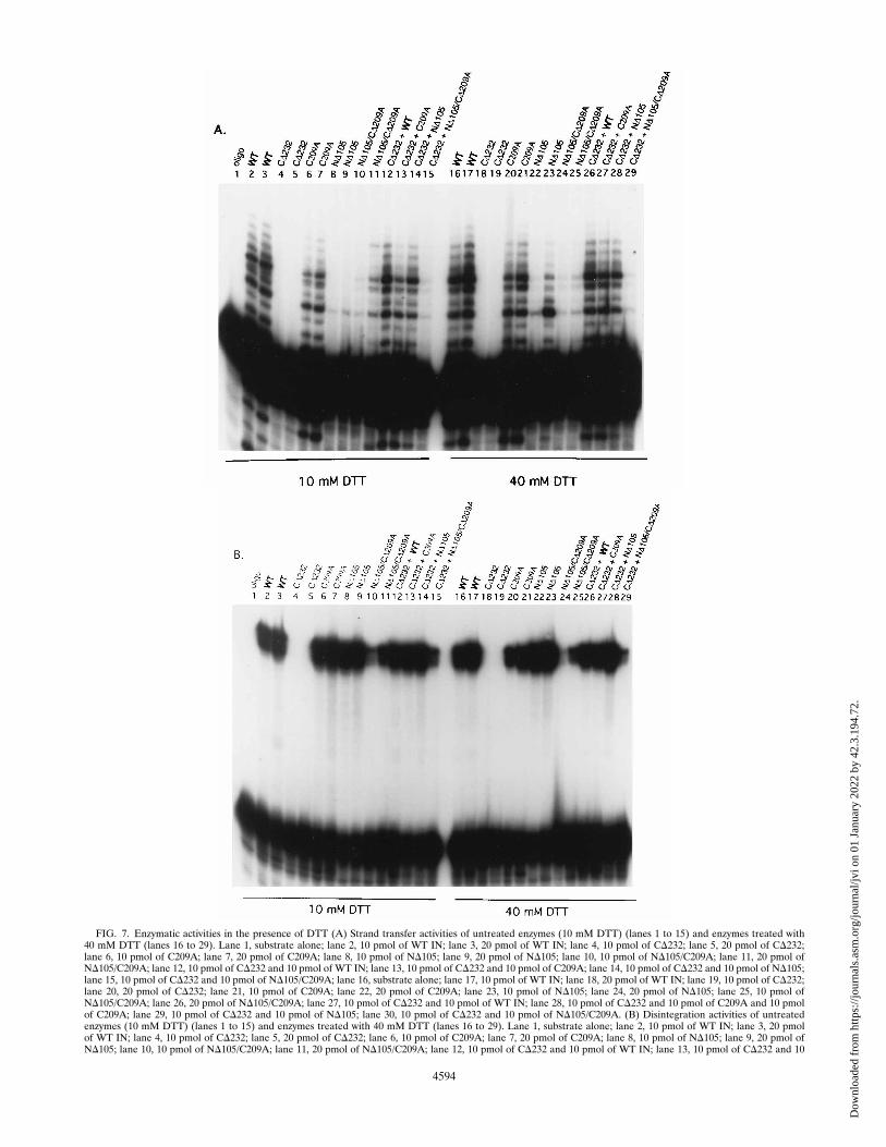
FIG. 7. Enzymatic activities in the presence of DTT (A) Strand transfer activities of untreated enzymes (10 mM DTT) (lanes 1 to 15) and enzymes treated with40 mM DTT (lanes 16 to 29). Lane 1, substrate alone; lane 2, 10 pmol of WT IN; lane 3, 20 pmol of WT IN; lane 4, 10 pmol of CD232; lane 5, 20 pmol of CD232;lane 6, 10 pmol of C209A; lane 7, 20 pmol of C209A; lane 8, 10 pmol of ND105; lane 9, 20 pmol of ND105; lane 10, 10 pmol of ND105/C209A; lane 11, 20 pmol ofND105/C209A; lane 12, 10 pmol of CD232 and 10 pmol of WT IN; lane 13, 10 pmol of CD232 and 10 pmol of C209A; lane 14, 10 pmol of CD232 and 10 pmol of ND105;lane 15, 10 pmol of CD232 and 10 pmol of ND105/C209A; lane 16, substrate alone; lane 17, 10 pmol of WT IN; lane 18, 20 pmol of WT IN; lane 19, 10 pmol of CD232;lane 20, 20 pmol of CD232; lane 21, 10 pmol of C209A; lane 22, 20 pmol of C209A; lane 23, 10 pmol of ND105; lane 24, 20 pmol of ND105; lane 25, 10 pmol ofND105/C209A; lane 26, 20 pmol of ND105/C209A; lane 27, 10 pmol of CD232 and 10 pmol of WT IN; lane 28, 10 pmol of CD232 and 10 pmol of C209A and 10 pmolof C209A; lane 29, 10 pmol of CD232 and 10 pmol of ND105; lane 30, 10 pmol of CD232 and 10 pmol of ND105/C209A. (B) Disintegration activities of untreatedenzymes (10 mM DTT) (lanes 1 to 15) and enzymes treated with 40 mM DTT (lanes 16 to 29). Lane 1, substrate alone; lane 2, 10 pmol of WT IN; lane 3, 20 pmolof WT IN; lane 4, 10 pmol of CD232; lane 5, 20 pmol of CD232; lane 6, 10 pmol of C209A; lane 7, 20 pmol of C209A; lane 8, 10 pmol of ND105; lane 9, 20 pmol ofND105; lane 10, 10 pmol of ND105/C209A; lane 11, 20 pmol of ND105/C209A; lane 12, 10 pmol of CD232 and 10 pmol of WT IN; lane 13, 10 pmol of CD232 and 10
4594
Dow
nloa
ded
from
http
s://j
ourn
als.
asm
.org
/jour
nal/j
vi o
n 01
Jan
uary
202
2 by
42.
3.19
4.72
.
observed activity of the M-MuLV N-terminal mutant ND105,reactions were titrated with ND105 protein pretreated with areducing agent. At the higher enzyme concentrations exam-ined, the strand transfer levels of the DTT-treated ND105protein demonstrated enhanced activity and integration siteusage. Further study of the aggregation state of the protein iswarranted. One interpretation of this data is that the ND105may form disulfide-linked multimers which are limited in ac-cess to the target DNA. High levels of DTT would break thecystine bonds and allow the assembly of active multimers.Similarly, evidence that a cysteine residue proximal to theHHCC domain of HIV-1 IN may assist in multimeric assemblyof IN-DNA complexes has been suggested (13). Alternatively,these data may suggest two conformation states of ND105brought about by either weak and strong reducing conditions.One can speculate that if the HHCC region were responsi-ble for a more ordered active multimer, then the reducedconformation could promote either strand transfer randomlyalong the DNA or random association with DNA. ND176,which has a larger N-terminal deletion of 176 amino acids,was completely inactivated for integration and disintegration.The lack of activity for ND176 is not surprising, since theregion deleted in this mutant includes the first D, D-125, ofthe invariant DD(35)E motif. This residue is absolutely re-quired for mediation of integration and disintegration activityin all retroviral INs examined to date (15, 26, 28). Comple-mentation studies showed that the M-MuLV D-125 residuewas essential as part of the central core and C terminus and notthe HHCC.Two specific amino acid substitutions in the catalytic core
region were examined, K227A and C209A. K-227 is conservedthroughout all retroviral INs. Substitutions at this position inRous sarcoma virus and HIV-1 IN retain some 39 processingand strand transfer activity (26). It was thus unexpected that amutation at this position was not tolerated in M-MuLV IN.This suggests that the K-227 residue plays a greater role in theintegration processes for M-MuLV IN. A cysteine (C-209 forM-MuLV) is located within the D35E region of the majorityof, but not all, INs. Given the reactive nature of Cys residues,studies addressed whether the residue afforded any specificityto the integration events. Surprisingly, C209A retained nearWT levels of integration and disintegration activity. And asdiscussed in the context of ND105 above, the reduction ofC-209 was required for a productive integration event in theabsence of a HHCC domain. For disintegration, the effect ofDTT treatment coupled to the mutation of C-209 to Ala is notas apparent as for integration. Disintegration is a unimolecularreaction, whereas strand transfer requires a bimolecular coor-dination of an LTR (donor) and target (acceptor) molecules.This suggests that the HHCC domain and redox state of C-209are more important for the multimeric assembly and stabilityof bimolecular IN-DNA complexes. In unimolecular reactionssuch as disintegration, complex assembly may be assisted bythe fixed structure of the Y substrate. This may diminish thereliance of M-MuLV IN on the more-specific interactions re-quired for assembly of integration complexes. The decreasedactivity of ND105/C209A in disintegration might simply be dueto protein-DNA complex instability.The C terminus of the M-MuLV IN was sensitive to dele-
tions and was observed to lose both integration and disinte-gration activities with deletions of 28 or more amino acids. Alinker insertion at amino acid 368 results in a temperature-sensitive protein with defective M-MuLV IN function and rep-lication (41). CD22 and CD28 functioned in ways similar to theWT IN and were active on all substrates including YCD. Thelack of LTR sequence in YCD substrate indicates a unimolecu-lar, nonspecific phosphoryl transfer reaction. This type of ac-tivity has been noted previously with M-MuLV IN disintegra-tion substrate variants (22). Recently, HIV-1 IN has also beenshown to catalyze a sequence-independent phosphoryl transfer(30, 43). Several of the M-MuLV IN mutants reported in thisstudy, CD40, CD45, CD86, CD232, and CD144, had no detect-able integration or disintegration activity. This is in contrastwith HIV-1 IN which shows disintegration activity in C-termi-nal mutants with deletions to amino acid 212, albeit at very lowlevels (3). Several of the M-MuLV C-terminal mutants wereable to function in trans with ND105. In the case of HIV-1 IN,efficient complementation does not occur if one molecule hasmore than 50 residues deleted from the N terminus and theother molecule has 70 residues deleted from the C terminus(46). In comparison to the results with HIV-1 IN, domainswithin the M-MuLV IN were able to act in trans in smallerfunctional units to promote integration activity. The results ofthe complementation analysis suggest that the HHCC regionfunctions as one domain and that the catalytic core and Cterminus function as a separate domain.The HHCC region has been proposed to be involved in the
recognition of the retroviral LTR termini, multimerization,positioning of the viral DNA into the catalytic core, and main-tenance of the architecture of the protomeric complex (4, 22,25). Studies of the strand transfer activity of ND105, whichlacks the HHCC region, offered several new insights into apossible function for the HHCC region. Several observationswere made. First, ND105 catalyzed strand transfer reactions ata low level; however, this activity was limited to one target sitein the DNA substrate. This site is 4 nucleotides closer to the 39end than the site primarily targeted for strand transfer with theWT M-MuLV IN enzyme. Second, reactions with a mixture ofND105 with either of the two mutant M-MuLV IN proteins,CD232 or CD144, produced integration levels similar to that ofWT IN. Thus, in these experiments, the HHCC domain func-tioned in trans to the core-C terminus to regain 39 processingand strand transfer activities. Third, increasing the amount ofDTT in the reaction environment of ND105 functioned in amanner similar to the addition of CD232, specifically, throughthe stimulation of integration activity. Last, while the integra-tion activity of ND105 could be increased through the additionof DTT or CD232, the double mutant, ND105/C209A, couldnot be complemented by CD232. This suggests that the C-209residue is in some manner required for IN-IN interactionsbetween the ND105 protein and the HHCC region of theCD232 protein. Similarly, Ellison et al. have proposed a modelfor HIV-1 IN multimerization on the basis of interactionsbetween the HHCC domain of one protomer and an N-ethyl-maleimide-sensitive site on a second protomer (13). The lackof complementation of ND105/C209A could indicate improperfolding of the active site. Although misfolding cannot be pre-cluded, the demonstrated low-level strand transfer activity and
pmol of C209A; lane 14, 10 pmol of CD232 and 10 pmol of ND105; lane 15, 10 pmol of CD232 and 10 pmol of ND105/C209A; lane 16, 10 pmol of WT IN; lane 17,20 pmol of WT IN; lane 18, 10 pmol of CD232; lane 19, 20 pmol of CD232; lane 20, 10 pmol of C209A; lane 21, 20 pmol of C209A; lane 22, 10 pmol of ND105; lane23, 20 pmol of ND105; lane 24, 10 pmol of ND105/C209A; lane 25, 20 pmol of ND105/C209A; lane 26, 10 pmol of CD232 and 10 pmol of WT; lane 27, 10 pmol of CD232and 10 pmol of C209A; lane 28, 10 pmol of CD232 and 10 pmol of ND105; lane 29, 10 pmol of CD232 and 10 pmol of ND105/C209A. Reactions were performed at378C as described in Materials and Methods and analyzed on 20% denaturing polyacrylamide gels.
VOL. 70, 1996 MUTATIONAL ANALYSIS OF M-MuLV IN 4595
Dow
nloa
ded
from
http
s://j
ourn
als.
asm
.org
/jour
nal/j
vi o
n 01
Jan
uary
202
2 by
42.
3.19
4.72
.
multiple sites of integration demonstrated by ND105/C209Aargue against this. These results suggest that the redox state ofC-209 affects M-MuLV IN integration activity, particularlystrand transfer, and that the HHCC domain assists in control-ling the redox state of the C-209 residue. Whether this effect isdue to a direct interaction of the HHCC and the core Cys or anindirect effect is an area of current investigation.
ACKNOWLEDGMENTS
We thank Kimberly C. Gray and Clarivick Macalalad for excellenttechnical assistance.This work was supported by American Cancer Society grant VM-134
and by National Science Foundation grant DMB-9105091. M.J.R. wasa Stohlmann Scholar of the Leukemia Society of America, Inc. C.B.J.was supported by NIH Postdoctoral Fellowship grant IF32CA09242.G.A.D. was supported by NIH training grant 5T32A107043-02.
REFERENCES
1. Bowerman, B., P. O. Brown, J. M. Bishop, and H. E. Varmus. 1989. Anucleoprotein complex mediates the integration of retroviral DNA. GenesDev. 3:469–478.
2. Bradford, M. M. 1976. A rapid and sensitive method for the quantitation ofmicrogram quantities of protein utilizing the principle of protein-dye bind-ing. Anal. Biochem. 72:248–254.
3. Bushman, F. D., A. Engelman, I. Palmer, P. Wingfield, and R. Craigie. 1993.Domains of the integrase protein of human immunodeficiency virus type Iresponsible for polynucleotidyl transfer and zinc binding. Proc. Natl. Acad.Sci. USA 90:3428–3432.
4. Bushman, F. D., and B. Wang. 1994. Rous sarcoma virus integrase protein:mapping functions for catalysis and substrate binding. J. Virol. 68:2215–2223.
5. Chow, S. A., and P. O. Brown. 1995. Juxtaposition of two viral DNA ends ina bimolecular disintegration reaction mediated by multimers of human im-munodeficiency virus type 1 or murine leukemia virus integrase. J. Virol.68:7869–7878.
6. Chow, S. A., K. A. Vincent, V. Ellison, and P. O. Brown. 1992. Reversal ofintegration and DNA splicing mediated by integrase of human immunode-ficiency virus. Science 255:723–726.
7. Coffin, J. M. 1990. Retroviridae and their replication, p. 645–708. In B. N.Fields and D. M. Knipe (ed.), Fundamental virology. Raven Press, NewYork.
8. Colicelli, J., and S. P. Goff. 1985. Mutants and pseudorevertants of Moloneymurine leukemia virus with alterations at the integration site. Cell 42:573–580.
9. Craigie, R., T. Fujiwara, and F. Bushman. 1990. The IN protein of Moloneymurine leukemia virus processes the viral DNA ends and accomplishes theirintegration in vitro. Cell 62:829–837.
10. Donehower, L. A. 1988. Analysis of mutant Moloney murine leukemia virusescontaining linker insertion mutations in the 39 region of pol. J. Virol. 62:3958–3964.
11. Donzella, G. A., C. B. Jonsson, and M. J. Roth. 1993. Influence of substratestructure on disintegration by Moloney murine leukemia virus integrase.J. Virol. 67:7077–7087.
12. Dotan, I., B. P. Scottoline, T. S. Heuer, and P. O. Brown. 1995. Character-ization of recombinant murine leukemia virus integrase. J. Virol. 69:456–468.
13. Ellison, V., J. Gerton, K. A. Vincent, and P. O. Brown. 1995. An essentialinteraction between distinct domains of HIV-1 integrase mediates assemblyof the active multimer. J. Biol. Chem. 270:3320–3326.
14. Engelman, A., F. D. Bushman, and R. Craigie. 1993. Identification of dis-crete functional domains of HIV-1 integrase and their organization within anactive multimeric complex. EMBO J. 12:3269–3275.
15. Engelman, A., and R. Craigie. 1992. Identification of conserved amino acidresidues critical for human immunodeficiency virus type 1 integrase functionin vitro. J. Virol. 66:6361–6369.
16. Engelman, A., K. Mizuuchi, and R. Craigie. 1991. HIV-1 DNA integration:mechanism of viral DNA cleavage and DNA strand transfer. Cell 67:1211–1221.
17. Farnet, C. M., and W. A. Haseltine. 1991. Determination of viral proteinspresent in the human immunodeficiency virus type 1 preintegration complex.J. Virol. 65:1910–1915.
18. Felkner, R. H., and M. J. Roth. 1992. Mutational analysis of the N-linkedglycosylation sites of the SU protein of Moloney murine leukemia virus.J. Virol. 66:4258–4264.
19. Ho, S. N., H. D. Huut, R. M. Horton, J. K. Pullen, and L. R. Pease. 1989.Site-directed mutagenesis by overlap extension using the polymerase chainreaction. Gene 77:51–59.
20. Johnson, M. S., M. A. McClure, D. F. Feng, J. Gray, and R. F. Doolittle.1986. Computer analysis of retroviral pol genes: assignment of enzymaticfunctions to specific sequences and homologies with nonviral enzymes. Proc.
Natl. Acad. Sci. USA 83:7648–7652.21. Jonsson, C. B., G. A. Donzella, and M. J. Roth. 1993. Characterization of the
forward and reverse integration reactions of the Moloney murine leukemiavirus integrase protein purified from Escherichia coli. J. Biol. Chem. 268:1462–1469.
22. Jonsson, C. B., and M. J. Roth. 1993. Role of the His-Cys finger of theMoloney murine leukemia virus integrase protein in integration and disin-tegration. J. Virol. 67:5562–5571.
23. Katz, R. A., and A. M. Skalka. 1994. The retroviral enzymes. Annu. Rev.Biochem. 63:133–173.
24. Katzman, M., R. A. Katz, A. M. Skalka, and J. Leis. 1989. The avianretroviral integration protein cleaves the terminal sequences of linear viralDNA at the in vivo sites of integration. J. Virol. 63:5319–5327.
25. Khan, E., J. P. G. Mack, R. A. Katz, J. Kulkosky, and A. M. Skalka. 1990.Retroviral integrase domains: DNA binding and the recognition of LTRsequences. Nucleic Acids Res. 19:851–860.
26. Kulkosky, J., K. S. Jones, R. A. Katz, J. P. G. Mack, and A. M. Skalka. 1992.Residues critical for retroviral integrative recombination in a region that ishighly conserved among retroviral/retrotransposon integrases and bacterialinsertion sequence transposases. Mol. Cell. Biol. 12:2331–2338.
27. Kunkel, T. A., J. D. Roberts, and R. A. Zakour. 1987. Rapid and efficientsite-specific mutagenesis without a phenotypic selection. Methods Enzymol.154:367.
28. Leavitt, A. D., L. Shiue, and H. E. Varmus. 1993. Site-directed mutagenesisof HIV-1 integrase demonstrates differential effects on integrase functions invitro. J. Biol. Chem. 268:2113–2119.
29. Lin, T. H., T. P. Quinn, D. Grandgenett, and M. T. Walsh. 1989. Secondarystructural analysis of retrovirus integrase: characterization by circular dichro-ism and empirical prediction methods. Proteins Struct. Funct. Genet. 5:156–165.
30. Mazumder, A., A. Engelman, R. Craigie, M. Fesen, and Y. Pommier. 1994.Intermolecular disintegration and intramolecular strand transfer activities ofwild type and mutant HIV-1 integrase. Nucleic Acids Res. 22:1037–1043.
31. Miller, D. G., M. A. Adam, and A. D. Miller. 1990. Gene transfer by retro-virus vectors occurs only in cells that are actively replicating at the time ofinfection. Mol. Cell. Biol. 10:4239–4242.
32. Mumm, S. R., and D. P. Grandgenett. 1991. Defining nucleic acid-bindingproperties of avian retrovirus integrase by deletion analysis. J. Virol. 65:1160–1167.
33. Murphy, J. E., T. de los Santos, and S. P. Goff. 1993. Mutational analysis ofthe sequences at the termini of the Moloney murine leukemia virus DNArequired for integration. Virology 195:432–440.
34. Murphy, J. E., and S. P. Goff. 1992. A mutation at one end of Moloneymurine leukemia virus DNA blocks cleavage of both ends by the viralintegrase in vivo. J. Virol. 66:5092–5095.
35. Puras-Lutzke, R., C. Vink, and R. H. A. Plasterk. 1994. Characterization ofthe minimal DNA-binding domain of the HIV integrase protein. NucleicAcids Res. 22:4125–4131.
36. Roe, T., T. C. Reynolds, G. Yu, and P. O. Brown. 1993. Integration of murineleukemia virus DNA depends on mitosis. EMBO J. 12:2099–2108.
37. Roth, M., P. Schwartzberg, N. Tanese, and S. P. Goff. 1990. Analysis ofmutations in the integration function of Moloney murine leukemia virus:effects on DNA binding and cutting. J. Virol. 64:4709–4717.
38. Roth, M. J. 1991. Mutational analysis of the carboxyl terminus of the Molo-ney murine leukemia virus integration protein. J. Virol. 65:2141–2145.
39. Roth, M. J., P. L. Schwartzberg, and S. P. Goff. 1989. Structure of the terminiof DNA intermediates in the integration of retroviral DNA: dependence onIN function and terminal DNA sequence. Cell 58:47–54.
40. Sanger, F., S. Nicklen, and A. R. Coulson. 1978. DNA sequencing withchain-terminating inhibitors. Proc. Natl. Acad. Sci. USA 74:5463–5467.
41. Schwartzberg, P. L., M. J. Roth, N. Tanese, and S. P. Goff. 1993. Analysis ofa temperature-sensitive mutation affecting the integration protein of Molo-ney murine leukemia virus. Virology 192:673–678.
42. Shinnick, T. M., R. A. Lerner, and J. G. Sutcliffe. 1981. Nucleotide sequenceof Moloney murine leukemia virus. Nature (London) 293:543–548.
43. van Den Ent, F. I., C. Vink, and R. H. A. Plasterk. 1995. DNA substrateactivities of the human immunodeficiency virus type 1 integrase protein.J. Virol. 68:7825–7832.
44. van Gent, D. C., Y. Elgersma, M. W. J. Bolk, C. Vink, and R. H. A. Plasterk.1991. DNA binding properties of the integrase proteins of human immuno-deficiency viruses types 1 and 2. Nucleic Acids Res. 19:3821–3827.
45. van Gent, D. C., A. A. M. O. Groenger, and R. H. A. Plasterk. 1992. Muta-tional analysis of the integrase protein of human immunodeficiency virustype 2. Proc. Natl. Acad. Sci. USA 89:9598–9602.
46. van Gent, D. C., C. Vink, A. A. M. O. Groeneger, and R. H. A. Plasterk. 1993.Complementation between HIV integrase proteins mutated in different do-mains. EMBO J. 12:3261–3267.
47. Varmus, H., and P. Brown. 1989. Retroviruses. American Society for Micro-biology, Washington, D.C.
48. Vink, C., A. A. M. O. Groenger, and R. H. A. Plasterk. 1993. Identification ofthe catalytic and DNA-binding region of the human immunodeficiency virustype 1 integrase protein. Nucleic Acids Res. 21:1419–1425.
4596 JONSSON ET AL. J. VIROL.
Dow
nloa
ded
from
http
s://j
ourn
als.
asm
.org
/jour
nal/j
vi o
n 01
Jan
uary
202
2 by
42.
3.19
4.72
.
49. Vink, C., E. Yeheskiely, G. A. van der Marel, J. H. van Boom, and R. H. A.Plasterk. 1993. Site specific hydrolysis and alcoholysis of human immunode-ficiency virus DNA termini mediated by the viral integrase protein. NucleicAcids Res. 19:6691–6698.
50. Vogelstein, B., and D. Gillespie. 1979. Preparative and analytical purificationof DNA from agarose. Proc. Natl. Acad. Sci. USA 76:615–619.
51. Woerner, A. M., M. Klutch, J. G. Levin, and C. J. Marcus-Sekura. 1992.Localization of DNA binding activity of HIV-1 integrase to the C-terminalhalf of the protein. AIDS Res. Hum. Retroviruses 8:297–304.
52. Woerner, A. M., and C. J. Marcus-Sekura. 1993. Characterization of a DNAbinding domain in the C-terminus of HIV-1 integrase by deletion mutagen-esis. Nucleic Acids Res. 21:3507–3511.
VOL. 70, 1996 MUTATIONAL ANALYSIS OF M-MuLV IN 4597
Dow
nloa
ded
from
http
s://j
ourn
als.
asm
.org
/jour
nal/j
vi o
n 01
Jan
uary
202
2 by
42.
3.19
4.72
.


![Molecular A byan - PNAS · 2868 Medical Sciences: Jansen et al. BRL's Moloney murine leukemia virus; 2 units in 5 A.l of H20]. Reaction mixtures were held at 420C for 30-60 min, afterwhich5A.l](https://static.fdocuments.net/doc/165x107/61034e96d1c440449245df66/molecular-a-byan-pnas-2868-medical-sciences-jansen-et-al-brls-moloney-murine.jpg)