Structural determinants and mechanism of action of a...
56
MOL #94516 1 Structural determinants and mechanism of action of a GluN2C- selective NMDA receptor positive allosteric modulator Alpa Khatri, Pieter B. Burger, Sharon A. Swanger, Kasper B. Hansen, Sommer Zimmerman, Erkan Karakas, Dennis C. Liotta, Hiro Furukawa, James. P. Snyder, Stephen F. Traynelis Emory University, Pharmacology Department: AK, SAS, SFT Emory University, Chemistry Department: SZ, PB, DCL, JPS University of Montana, Dept of Biomedical and Pharmaceutical Sciences, and Center for Biomolecular Structure and Dynamics: KBH Cold Spring Harbor Labs: EK, HF This article has not been copyedited and formatted. The final version may differ from this version. Molecular Pharmacology Fast Forward. Published on September 9, 2014 as DOI: 10.1124/mol.114.094516 at ASPET Journals on January 3, 2020 molpharm.aspetjournals.org Downloaded from
Transcript of Structural determinants and mechanism of action of a...

MOL #94516
1
Structural determinants and mechanism of action of a GluN2C-
selective NMDA receptor positive allosteric modulator
Alpa Khatri, Pieter B. Burger, Sharon A. Swanger, Kasper B. Hansen, Sommer
Zimmerman, Erkan Karakas, Dennis C. Liotta, Hiro Furukawa, James. P. Snyder,
Stephen F. Traynelis
Emory University, Pharmacology Department: AK, SAS, SFT
Emory University, Chemistry Department: SZ, PB, DCL, JPS
University of Montana, Dept of Biomedical and Pharmaceutical Sciences, and Center
for Biomolecular Structure and Dynamics: KBH
Cold Spring Harbor Labs: EK, HF
This article has not been copyedited and formatted. The final version may differ from this version.Molecular Pharmacology Fast Forward. Published on September 9, 2014 as DOI: 10.1124/mol.114.094516
at ASPE
T Journals on January 3, 2020
molpharm
.aspetjournals.orgD
ownloaded from

MOL #94516
2
Running Title: Analysis of a GluN2C NMDAR positive allosteric modulator
Corresponding author
Stephen F. Traynelis
1510 Clifton Road
RRC 5066
Atlanta GA, 30322
fax #: 404-727-0365
Number of text pages: 29
Number of Figures: 6
Numbers of Tables: 2
Number of words in the Abstract: 243
Number of words in the Introduction: 637
Number of words in the Discussion: 1259
List of non-standard abbreviations: NMDA: n-methyl-d-aspartate PYD: methyl 4-(3-acetyl-4-hydroxy-1-(2-(2-methyl-1H-indol-3-yl)ethyl)-5-oxo-2,5-dihydro-1H-pyrrol-2-yl)benzoate CNS: Central Nervous System GABAA: Gamma-Aminobutyric acid nAChR: Nicotinic Acetylcholine Receptor P2X: Purinergic Receptor DCS: D-cycloserine, CIQ: (3-chlorophenyl)(6,7-dimethoxy-1-((4- methoxyphenoxy)methyl)-3,4-dihydroisoquinolin-2(1H)-yl)methanone) 5-HT3A: Serotonin receptor NIMH-PDSP: National institute of Mental Health-psychoactive drug screening study HEK: Human Embryonic Kidney cell QNZ-46: (E)-4-(6-methoxy-2-(3-nitrostyryl)-4-oxoquinazolin-3(4H)-yl)-benzoic acid DQP-1105: 4-(5-(4-bromophenyl)-3-(6-methyl-2-oxo-4- phenyl-1,2-dihydroquinolin-3-yl)-4,5-dihydro-1H-pyrazol-1-yl)- 4-oxobutanoic acid AMPA: (α-Amino-3-hydroxy-5-methyl-4-isoxazolepropionic acid) SEM: Standard error of the mean ANOVA: Analysis of Variance ER: endoplasmic reticulum
This article has not been copyedited and formatted. The final version may differ from this version.Molecular Pharmacology Fast Forward. Published on September 9, 2014 as DOI: 10.1124/mol.114.094516
at ASPE
T Journals on January 3, 2020
molpharm
.aspetjournals.orgD
ownloaded from

MOL #94516
3
Abstract
NMDA receptors are tetrameric complexes of GluN1, GluN2A-D, and GluN3A-B
subunits and are involved in normal brain function and neurological disorders. We have
identified a novel class of stereo-selective pyrrolidinone (PYD) positive allosteric
modulators for GluN2C-containing NMDA receptors, exemplified by methyl 4-(3-acetyl-
4-hydroxy-1-(2-(2-methyl-1H-indol-3-yl)ethyl)-5-oxo-2,5-dihydro-1H-pyrrol-2-
yl)benzoate. Here we explore the site and mechanism of action of a prototypical
analogue PYD-106, which at 30 μM does not alter responses of NMDA receptors
containing GluN2A, GluN2B, and GluN2D, and has no effect on AMPA and kainate
receptors. Co-application of 50 μM PYD-106 with a maximally effective concentration of
glutamate and glycine increases the response of GluN1/GluN2C NMDA receptors in
HEK-293 cells to 221% of that obtained in the absence of PYD (taken as 100%).
Evaluation of the concentration-dependence of this enhancement revealed an EC50
value for PYD of 13 μM. PYD-106 increased opening frequency and open time of single
channel currents activated by maximally effective concentrations of agonist, but only
had modest effects on glutamate and glycine EC50. PYD-106 selectively enhanced the
responses of diheteromeric GluN1/GluN2C receptors, but not triheteromeric
GluN1/GluN2A/GluN2C receptors. Inclusion of residues encoded by GluN1-exon5
attenuated the effects of PYD. Three GluN2C residues (Arg194, Ser470, Lys470), at
which mutagenesis virtually eliminated PYD function, line a cavity at the interface of the
ligand binding and the amino terminal domains in a homology model of GluN1/GluN2C
built from crystallographic data on GluN1/GluN2B. We propose that this domain
interface constitutes a new allosteric modulatory site on the NMDA receptor.
This article has not been copyedited and formatted. The final version may differ from this version.Molecular Pharmacology Fast Forward. Published on September 9, 2014 as DOI: 10.1124/mol.114.094516
at ASPE
T Journals on January 3, 2020
molpharm
.aspetjournals.orgD
ownloaded from

MOL #94516
4
Introduction
N-Methyl-D-aspartate receptors (NMDARs) are ligand-gated cation channels that
mediate a slow Ca2+-permeable component of excitatory synaptic transmission. NMDA
receptors are involved in the development and normal function of the CNS. Dysfunction
of NMDARs is associated with epilepsy, pain, depression, Parkinson’s disease, and
schizophrenia, making these receptors an attractive therapeutic target (Hallett &
Standaert, 2004; Kalia et al., 2008; Lisman et al., 2008; Preskorn et al., 2008; Wu &
Zhuo, 2009; Endele et al., 2010; Traynelis et al., 2010; Balu & Coyle, 2011; Kostakis et
al., 2011). The majority of NMDARs in the central nervous systems are heteromeric
complexes formed by two GluN1 and two GluN2 subunits (Ulbrich & Isacoff, 2007), of
which there are four subtypes (GluN2A-D) with temporal and spatial variation in
expression (Watanabe et al., 1992; Ishii et al., 1993; Monyer et al., 1994). The
development of GluN2-selective modulators provides a therapeutic opportunity to target
NMDAR subtypes with anatomically restricted expression patterns, thereby minimizing
potential side effects (Kalia et al., 2008; Ogden & Traynelis, 2011; Collingridge et al.,
2013). Subunit-selective allosteric modulators exist for the GluN2A (TCN-201), GluN2B
(ifenprodil), and GluN2C/GluN2D subunits (CIQ, QNZ, UBP, DQP analogues) (Williams
et al., 1993; Bettini et al., 2010; Mullasseril et al., 2010; Acker et al., 2011; Hansen &
Traynelis, 2011; Costa et al., 2012; Hansen et al., 2012; Monaghan et al., 2012).
However, no modulators to date have been able to distinguish between GluN2C and
GluN2D subunits.
The GluN2C subunit is expressed in the cerebellum, amygdala, olfactory bulb,
retrosplenial cortex as well as in thalamic, cortical, and hippocampal interneurons
(Farrant et al., 1994; Monyer et al., 1994; Wenzel et al., 1997; Binshtok et al., 2006;
This article has not been copyedited and formatted. The final version may differ from this version.Molecular Pharmacology Fast Forward. Published on September 9, 2014 as DOI: 10.1124/mol.114.094516
at ASPE
T Journals on January 3, 2020
molpharm
.aspetjournals.orgD
ownloaded from

MOL #94516
5
Karavanova et al., 2007). Oligodendrocytes also express the GluN2C subunit
(Karadottir et al., 2005; Salter & Fern, 2005; Micu et al., 2006). GluN2C-containing
NMDARs have a lower sensitivity to voltage-dependent Mg2+ block, reduced Ca2+
permeability, and reduced conductance compared to GluN2A and GluN2B (Qian et al.,
2005; Clarke & Johnson, 2006; Siegler Retchless et al., 2012). Behavioral studies
evaluating the deletion of the GluN2C subunit suggest a possible role in working
memory (Hillman et al., 2011). In addition, studies with the GluN2C/GluN2D-selective
positive allosteric modulator CIQ suggest a possible role for enhancement of NMDAR
function in emotional learning, working memory, and sensorimotor gating (Ogden et al.,
2013; Suryavanshi et al., 2013). D-cycloserine (DCS), an antibiotic treatment used for
tuberculosis, acts as a partial agonist relative to glycine at GluN1/GluN2A,
GluN1/GluN2B, and GluN1/GluN2D receptors. By contrast, a maximally effective
concentration of DCS produces more current at GluN1/GluN2C receptors than the
endogenous agonist glycine, leading to selective enhancement of GluN1/GluN2C
receptors when DCS replaces glycine at the GluN1 agonist binding site (Sheinin et al.,
2001; Dravid et al., 2010). NMDAR hypofunction has been suggested to underlie some
aspects of schizophrenia (Krystal et al., 1994; Olney et al., 1999; Lisman, 2012) and
DCS has shown positive results in schizophrenic patients, suggesting increased
occupancy of the agonist binding site on GluN1 and thereby enhancement NMDAR
function (Goff et al., 1995; Goff et al., 2008; Gottlieb et al., 2011). The ability of DCS to
enhance the function of the GluN2C-containing receptors raises the possibility that
GluN2C modulation may contribute to clinically relevant actions of DCS in schizophrenia
(Norberg et al., 2008; Kaplan & Moore, 2011). Thus, better pharmacological tools are
This article has not been copyedited and formatted. The final version may differ from this version.Molecular Pharmacology Fast Forward. Published on September 9, 2014 as DOI: 10.1124/mol.114.094516
at ASPE
T Journals on January 3, 2020
molpharm
.aspetjournals.orgD
ownloaded from

MOL #94516
6
needed to evaluate the functional roles of GluN2C in neurological diseases such as
schizophrenia.
We describe here the mechanism of action and structural determinants for a
GluN2C-selective class of compounds exemplified by PYD-106, an analogue developed
during the study of the structure-activity relationship around a pyrrolidinone identified
from a high-throughput screen (Zimmerman et al., 2014). The PYD class of positive
allosteric modulators is the only series that selectivity enhances the response to
maximally effective concentrations of agonist for recombinant NMDARs containing two
copies of the GluN2C subunit.
This article has not been copyedited and formatted. The final version may differ from this version.Molecular Pharmacology Fast Forward. Published on September 9, 2014 as DOI: 10.1124/mol.114.094516
at ASPE
T Journals on January 3, 2020
molpharm
.aspetjournals.orgD
ownloaded from

MOL #94516
7
Material and Methods
Molecular biology
GluN1, GluN2A (D13211), GluN2B (U11419), GluN2C (M91563) and GluN2D
(L31611 ) cDNAs were provided by Drs. Heinemann (Salk Institute for Biological
Sciences, La Jolla, CA), Nakanishi (Kyoto University, Kyoto, Japan) and Seeburg
(University of Heidelberg, Heidelberg, Germany). The GenBank accession numbers for
GluN1-1a (hereafter GluN1), and splice variants GluN1-1b, -2a, -2b, -3a, -3b, -4a, -4b
were U08261, U08263, U08262, U08264, U08265, U08266, U08267, U08268,
respectively. The GluN2A-GluN2C chimeras and GluN2C point mutations were
generated as previously described (Chen et al., 2008). Supplemental Table S1 lists the
junctions for the chimeric receptors.
A GluN2C-2A C-terminal chimera was made from amino acids 1 - 851 of rat
GluN2C and amino acids 841 -1464 of rat GluN2A; these chimeric GluN2C-2A subunits
will be referred to hereafter as GluN2C*. Constructs GluN2AC1, GluN2AC2, GluN2C*C1,
and GluN2C*C2 were generated by adding a 23 residue synthetic linker followed by
coiled-coil regions of the GABAB1 and GABAB2 receptor subunits (C1 and C2,
respectively) plus a dilysine ER retention signal (KKTN) at the 3' end of rat GluN2A and
rat GluN2C*, as described previously (Hansen et al., 2014). Two point mutations were
introduced into the agonist binding domains by site-directed mutagenesis using the
Quikchange method and were GluN2A(R518K,T690I) and GluN2C*(R529K,T701I). All
chimeric subunits and mutations were verified by DNA sequencing.
Two-electrode voltage clamp recordings
This article has not been copyedited and formatted. The final version may differ from this version.Molecular Pharmacology Fast Forward. Published on September 9, 2014 as DOI: 10.1124/mol.114.094516
at ASPE
T Journals on January 3, 2020
molpharm
.aspetjournals.orgD
ownloaded from

MOL #94516
8
Xenopus laevis oocytes were obtained from EcoCyte (Austin, TX), injected with
cRNA created by in vitro transcription using the mMessage mMachine kits according to
the manufacturer’s instructions (Ambion), as previously described (Hansen et al., 2013).
Plasmids containing the genes for the GABAA (α1β2γ2s), GABAC (ρ1), glycine (α1),
serotonin (5-HT3A), nicotinic acetylcholine receptor (nAChR, α1β1δγ, α2β4, α4β3,
α9α10) and purinergic (P2X2 rat, P2X2 human) receptors were provided by Drs.
Heinemann (Salk), Weiss (Univ. of Texas, San Antonio), Papke (Univ. of Florida), and
Hume (Univ. of Michigan), linearized and used to synthesize cRNA for these receptors.
1-4 days after cRNA injection (5-15 ng), oocytes were recorded at room temperature
under two-electrode voltage clamp (VHOLD -30 to -60 mV) in a solution containing (in
mM) 90 NaCl, 1 KCl, 10 HEPES, 0.5 BaCl2 and 0.01 EDTA (pH 7.4). For triheteromeric
receptor experiments, the cRNA was prepared at a ratio of 1:6:6
(GluN1:GluN2C1:GluN2C2), and approximately 5-10 ng of total cRNA was injected into
each oocyte. Recordings were performed 3-4 days after injection. NMDAR currents
were evoked by bath application of 50-100 μM glutamate and 30-100 μM glycine.
Currents from the GluA1-4 and GluK1-2 receptors were evoked with 100 μM glutamate;
GluK2 was incubated in 1 mg/ml concanavalin A for 10 minutes prior to recording.
GluK2/GluK5 currents were evoked with 100 μM AMPA. Currents were evoked for
following receptors using the agonist concentrations indicated: GABAC (1 μM GABA),
GABAA (20 μM GABA), glycine α1 (50 μM glycine), 5-HT3A (1 μM serotonin), nicotinic
acetylcholine α1β1δγ (1 μM acetylcholine), α3β4 (10 μM acetylcholine), α4β2 (10 μM
acetylcholine), α9α10 (1 μM acetylcholine), α7 (300 μM acetylcholine), and the P2X2
receptors (9 μM ATP).
This article has not been copyedited and formatted. The final version may differ from this version.Molecular Pharmacology Fast Forward. Published on September 9, 2014 as DOI: 10.1124/mol.114.094516
at ASPE
T Journals on January 3, 2020
molpharm
.aspetjournals.orgD
ownloaded from

MOL #94516
9
Patch-clamp recordings
HEK-293 cells (ATCC CRL-1573, hereafter HEK cells) were maintained in
DMEM with GlutaMAX, 110 mg/ml sodium pyruvate and 4.5 gm/L glucose (Invitrogen,
Grand Island, NY) supplemented with 10% dialyzed fetal bovine serum, 10 U/ml
penicillin, and 10 μg/ml streptomycin. HEK cells were maintained in a 5% CO2
humidified 37°C incubator and transiently transfected using Fugene 6 with cDNAs
encoding NMDAR subunits and green fluorescent protein (GFP) at a ratio of 1:1:5 for
GluN1/GluN2A/GFP, 1:1:1 for GluN1/GluN2B/GFP, GluN1/GluN2C/GFP and
GluN1/GluN2D/GFP. Voltage clamp recordings were performed 12-36 hrs after
transfection (VHOLD -60 mV for whole cell, -80 mV for outside-out patches). The data
was filtered at 8 kHz (8-pole Bessel filter, -3dB) and digitized at 20-40 kHz. The
extracellular solution consisted of (in mM) 150 NaCl, 10 HEPES, 3 KCl, 0.5 CaCl2, 0.01
EDTA and 30 D-mannitol (pH 7.4 for whole cell and 8.0 for outside-out patches). The
intracellular solution contained (in mM) 110 D-gluconate, 110 CsOH, 30 CsCl2, 5
HEPES, 4 NaCl, 0.5 CaCl2, 2 MgCl2, 5 BAPTA, 2 Na-ATP and 0.3 Na-GTP; the pH was
adjusted to 7.35 with CsOH. Rapid solution exchange was achieved using a two-barrel
theta glass pipette controlled by a piezoelectric translator. The open tip junction currents
had a 10-90% rise time of less than 1 ms. The solution exchange around a whole cell
had a 10-90% rise time of 3.4 ± 0.3 ms (n=9) as determined by exchanging the
extracellular NaCl for KCl. Single channel recordings from outside-out patches were
made in response to steady-state agonist application at pH 8.0. All recordings were
performed at room temperature (23°C). The junction potential for HEK cell recording
This article has not been copyedited and formatted. The final version may differ from this version.Molecular Pharmacology Fast Forward. Published on September 9, 2014 as DOI: 10.1124/mol.114.094516
at ASPE
T Journals on January 3, 2020
molpharm
.aspetjournals.orgD
ownloaded from

MOL #94516
10
solution was +5.4 mV (Vance et al., 2011); single channel chord conductance values
were corrected for this value.
Recordings from outside-out patches were digitally filtered at 3-4 kHz (-3 dB) and
idealized using time course fitting (SCAN; Dr. David Colquhoun, University College
London; Colquhoun & Sigworth, 1983). An open resolution of 53 μs and shut resolution
of 31 μs was imposed on the data and open periods were combined for different
conductance levels. Both open and closed duration histograms were fitted with the sum
of 2-5 exponential functions using maximum likelihood methods. The open probability
was calculated as the total time the patch was in an open state divided by the total time
of the recording. Most outside-out patches contained multiple channels since double
and triple openings were occasionally observed. Multiple openings were excluded from
analysis of channel dwell times, and thus the reported open probabilities are
underestimates of the true open probability.
Molecular modeling
Amino acids are numbered with the initiating methionine set to 1. A protein
sequence alignment of the different GluN2A-D sequences was generated with Muscle
(Edgar, 2004) using the GluN1-1a (i.e. GluN1) and GluN2B sequences obtained from
the resolved GluN1/GluN2B crystal structure (pdb 4PE5). Five GluN1/GuN2C homology
models were generated with Modeler 9v12 (Sali & Blundell, 1993) using the
GluN1/GluN2B crystal structure as template (PDB 4PE5; Karakas & Furukawa, 2014)
No protein optimization was performed during model building. The models were
subjected to protein quality analysis and the model with the lowest discrete optimized
This article has not been copyedited and formatted. The final version may differ from this version.Molecular Pharmacology Fast Forward. Published on September 9, 2014 as DOI: 10.1124/mol.114.094516
at ASPE
T Journals on January 3, 2020
molpharm
.aspetjournals.orgD
ownloaded from

MOL #94516
11
protein energy (DOPE) score was selected. Side chain optimization and protonation
state assignment was performed with the protein preparation wizard (Sastry et al.,
2013) and monitored by visual inspection. This was followed by energy minimization
(heavy atom RMSD convergence of 0.3Å; Force field OPLS 2005) to relieve the
energetically unfavorable constraints. PYD-106 was prepared for docking using Ligprep
(Schrödinger Release 2014-2: LigPrep, version 3.0, Schrödinger, LLC, New York, NY,
2014). The docking grid was centered between the carbonyl oxygen of P428 and the
Cβ-atom of S472. The diameter midpoint of the docked ligands was required to remain
within a nested box (14 Å3) at the center of the grid. The extra precision (XP) scoring
algorithm of Glide (Friesner et al., 2006) was used to obtain the best scoring poses
during docking. PYD-106 was docked flexibly with docking poses being restricted to 10,
followed by post-docking minimization (OPLS 2005 force field) with an energy threshold
of 0.5 kcal/mol. A pdb file with PYD-106 docked into the homology model of
GluN1/GluN2C is included as supplemental data (see Data Supplement).
Synthesis of pyrrolidinone analogues
Compounds PYD-1 and PYD-106 were prepared as previously described (see
compounds in Zimmerman et al., 2014). Compounds were dissolved at 20 mM or 50
mM in 100% DMSO, and this stock was used for all solutions. Final DMSO
concentrations were between 0.005-0.5% (vol/vol). Both compounds were soluble up to
100 µM, as determined by nephelometry. Compound purity was greater than 95%.
Statistics
This article has not been copyedited and formatted. The final version may differ from this version.Molecular Pharmacology Fast Forward. Published on September 9, 2014 as DOI: 10.1124/mol.114.094516
at ASPE
T Journals on January 3, 2020
molpharm
.aspetjournals.orgD
ownloaded from

MOL #94516
12
All results are presented as the mean ± standard error of the mean. Statistical
significance was taken as p < 0.05 by t-test or one way ANOVA (Tukey, Bonferroni, or
Dunnett’s post hoc), as appropriate. For all tables and figures, n is the number of
observations.
This article has not been copyedited and formatted. The final version may differ from this version.Molecular Pharmacology Fast Forward. Published on September 9, 2014 as DOI: 10.1124/mol.114.094516
at ASPE
T Journals on January 3, 2020
molpharm
.aspetjournals.orgD
ownloaded from

MOL #94516
13
Results
Pyrrolidinones are selective positive allosteric modulators of GluN1/GluN2C receptors
We have studied the actions of the pyrrolidinone PYD-106 (Zimmerman et al.,
2014) on recombinant GluN1/GluN2C receptor function. Figure 1 shows concentration-
effect curves for glutamate in the absence and presence of PYD-106 as determined in
Xenopus oocytes injected with GluN1/GluN2C mRNA. PYD-106 prominently enhanced
the maximal fitted current response by over two-fold. In addition, the glutamate EC50
was modestly increased by 70 μM PYD-106, being 0.72 ± 0.05 μM (n = 13, Hill slope
1.45 ± 0.03) in the absence and 1.17 ± 0.08 μM (n = 13, Hill slope 1.39 ± 0.02) in the
presence of PYD-106 when studied in the same oocytes (p < 0.05; paired t-test; 30 μM
glycine present in all solutions). We also determined the glycine concentration-effect
curve independently in the absence or presence of 70 μM PYD-106. Coapplication of
PYD-106 with glycine modestly reduced the EC50 from 0.23 ± 0.004 μM (n = 6) in the
absence to 0.16 ± 0.006 μM (n = 5) in the presence of PYD-106 (p < 0.05, unpaired t-
test; 100 μM glutamate present in all solutions). PYD-106 showed no agonist activity
when applied alone, with the response amplitude being 1.1 ± 0.6% of control (n = 4).
Co-application of PYD-106 with either glutamate alone or glycine alone did not produce
inward currents (n = 5). These data suggest that PYD-106 is a positive allosteric
modulator that increases agonist efficacy, and also reveal significant negative
(glutamate) and positive (glycine) interactions with agonist potency.
We next evaluated the concentration-dependence of PYD-106 on recombinant
NMDAR activated by maximally effective concentrations of glutamate (100 μM) and
glycine (30 μM). The positive modulation produced by PYD-106 was both reversible and
This article has not been copyedited and formatted. The final version may differ from this version.Molecular Pharmacology Fast Forward. Published on September 9, 2014 as DOI: 10.1124/mol.114.094516
at ASPE
T Journals on January 3, 2020
molpharm
.aspetjournals.orgD
ownloaded from

MOL #94516
14
repeatable, with an EC50 value of 16 ± 0.5 μM and a Hill slope of 1.19 ± 0.03 in oocytes
(n = 31, Figure 2A,B). Co-application of 100 μM PYD-106 and saturating concentrations
of glutamate (100 μM) and glycine (30 μM) increased the maximal current response to
204 ± 3.9% of control (the response in the absence of PYD-106 was taken as 100%, n =
31). At sub-maximal concentrations of glutamate (1 μM) and glycine (0.3 μM), the
GluN1/GluN2C receptor response was increased to 223 ± 6.3% of control by 100
μM PYD-106 with an EC50 of 15 ± 1.9 μM (n = 5), which was not significantly different
compared to the data obtained with saturating concentrations of glutamate and glycine
(t-test, p > 0.05).
We subsequently tested the effect of PYD-106 at other recombinant ion channels
expressed in oocytes. We used a concentration of PYD-106 (30 μM) that strongly
enhanced the GluN1/GluN2C receptor response to maximally effective concentrations
of glutamate and glycine. PYD-106 (30 μM) did not alter the amplitude for
GluN1/GluN2A, GluN1/GluN2D, AMPA, kainate, GABAC and 5-HT3A receptor-mediated
currents in response to saturating agonist concentrations (Figure 2E). Responses of
GluN1/GluN2B, glycine α1, GABAA and nicotinic acetylcholine receptors were inhibited
to 73 - 88% of control by 30 μM PYD-106 (Figure 2E, where the response in the
absence of PYD is 100%). PYD-106 was also tested by the NIMH psychoactive drug
screening program (PDSP) using a binding assay to assess interactions with G-protein
coupled receptors, transporters, and voltage-gated ion channels. Out of 42 proteins that
were tested by the NIMH-PDSP, 10 µM PYD-106 showed inhibition in excess of 50% of
control for the kappa-opioid receptor, dopamine transporter (DAT) and the adrenergic
α2C receptor (Supplemental Table S2). A subsequent experiment determined the
This article has not been copyedited and formatted. The final version may differ from this version.Molecular Pharmacology Fast Forward. Published on September 9, 2014 as DOI: 10.1124/mol.114.094516
at ASPE
T Journals on January 3, 2020
molpharm
.aspetjournals.orgD
ownloaded from

MOL #94516
15
binding affinity (Ki) for PYD-106 to be 6.1 μM for the kappa-opioid receptor; the Ki
values for the α2C receptor and DAT were greater than 10 μM. These data suggest that
PYD-106 selectively enhances the GluN1/GluN2C response within the glutamate
receptor family, but produces significant inhibition at multiple targets (including GluN2B
at 100 μM).
Selectivity of pyrrolidinones for GluN1 splice variants
The GluN1 subunit RNA can be alternatively spliced with 8 different variants
described (Hollmann et al., 1993). GluN1 alternative exon-5 encodes 21 amino acids,
which are located near the ATD – S1 interface of GluN1. GluN1 exon5 is differentially
expressed throughout the CNS (Laurie & Seeburg, 1994a; Laurie et al., 1995), with
expression in cerebellar granule neurons, which also express GluN2C (Akazawa et al.,
1994; Prybylowski et al., 2000). Exon-5 has been shown to alter the effects of several
NMDA receptor modulators including neurosteroids, extracellular protons, Zn2+, and
polyamines, in addition to altering glutamate potency and the deactivation time course
(Traynelis et al., 1995; Traynelis et al., 1998; Rumbaugh et al., 2000; Kostakis et al.,
2011; Vance et al., 2012). We therefore evaluated the effects of RNA splicing of GluN1
on the activity of the positive allosteric modulator PYD-106. GluN1 subunits that contain
exon-5 are referred to as “b” and those lacking exon-5 as “a”; different combinations of
C-terminal splice variants that lack exon-21 and/or exon-22 are denoted by the suffix
2,3,4 (Hollmann et al., 1993). Comparison of PYD-106 activity (100 µM) at the different
GluN1 splice variants co-expressed with GluN2C revealed significant differences in the
maximal level of modulation compared to GluN1-1a/GluN2C (200 ± 5.6% of control, n =
This article has not been copyedited and formatted. The final version may differ from this version.Molecular Pharmacology Fast Forward. Published on September 9, 2014 as DOI: 10.1124/mol.114.094516
at ASPE
T Journals on January 3, 2020
molpharm
.aspetjournals.orgD
ownloaded from

MOL #94516
16
16). Modulation was significantly reduced for GluN1-1b (125 ± 2.8%, n = 18), GluN1-2b
(117 ± 0.6%, n = 7), GluN1-3a (173 ± 4.2%, n = 7), GluN1-3b (113 ± 0.4%, n = 9), and
GluN1-4b (118 ± 0.9%, n = 10) splice variants (one way ANOVA, Bonferroni’s post hoc,
p < 0.05), whereas there were no significant differences for GluN1-2a (202 ± 4.5%, n =
6) and GluN1-4a (188 ± 3.4%, n = 6). That is, PYD-106 produces a stronger
enhancement of the current response to maximal agonist concentrations for all GluN1
splice variants that lack exon-5 compared to those that contain exon-5. Concentration-
effect curves demonstrated that the PYD-106 EC50 for enhancement of the maximal
response of GluN1-1a (14 ± 1.1 μM, n=25) was significantly different from the EC50 for
GluN1-1b-containing receptors (5.6 ± 0.4 μM, n = 19, t-test, p < 0.05; Figure 2C).
To identify the structural determinants in the highly charged 21 amino acid
segment encoded by exon-5 that reduced positive allosteric modulation by PYD-106,
we first screened a series of triple charge neutralization mutations. The triple GluN1-1b
exon-5 mutation K207G, R208G, K211G restored the effects of PYD-106, which
enhanced the maximal current response to 206 ± 5.5% of control at 100 μM PYD-106
(EC50 of 21 ± 2.5 μM, n = 8, Figure 1C). By contrast, the triple GluN1-1b mutant K192G,
K193G, R194G only partially rescued modulation by 100 μM PYD-106 to 145 ± 3.2% of
control (n = 4) with an EC50 of 9.6 ± 0.9 μM (n = 4) (Figure 1C). Mutation of three
negatively charged GluN1-1b residues encoded by exon-5 (E197A, D200A, D205A) did
not restore the effects of 100 μM PYD-106 (116 ± 2.3% of control; n = 4).
A number of effects of exon-5 on allosteric modulators as well as the deactivation
time course are controlled by Lys211 in exon-5 (Traynelis et al., 1995; Traynelis et al.,
1998; Vance et al., 2012). We therefore assessed whether mutation of Lys211 to Gly or
This article has not been copyedited and formatted. The final version may differ from this version.Molecular Pharmacology Fast Forward. Published on September 9, 2014 as DOI: 10.1124/mol.114.094516
at ASPE
T Journals on January 3, 2020
molpharm
.aspetjournals.orgD
ownloaded from

MOL #94516
17
Arg restored the actions of PYD-106. The maximal current responses of GluN1-1b
K211G and GluN1-1b K211R mutations were only modestly enhanced by 100 μM PYD-
106 to 137 ± 2.3% (n = 9) and 129 ± 1.2% (n = 10), respectively (Figure 2C,
Supplemental Figure S1). The degree of modulation observed with GluN1-1b K211G
was significantly different compared to PYD-106 modulation of wild-type GluN1-1b (t-
test, p < 0.05). The EC50 values for PYD-106 enhancement of the maximal response of
GluN1-1b K211G and GluN1-1b K211R were 9.5 ± 0.9 μM (n = 3) and 9.0 ± 1.9 (n = 6)
μM, respectively, which were not significantly different compared to GluN1-1a (p >
0.05). These data suggest that the structural determinants within exon 5 responsible for
its effects on protons, polyamines, and Zn2+ are distinct from its effects on PYD-106.
Pyrrolidinone activity on triheteromeric NMDARs
NMDARs can form diheteromeric complexes that contain one type of GluN2
subunit or triheteromeric receptors containing two different GluN2 subunits. For
example, triheteromeric receptors containing GluN1/GluN2A/GluN2C have been
reported to form functional receptors in cerebellar neurons (Chazot et al., 1994; Cathala
et al., 2000; Lu et al., 2006). To determine whether PYD-106 enhanced the responses
of triheteromeric receptors that contain a single copy of GluN2C, we adapted a
recombinant expression system that promotes the surface expression of triheteromeric
receptors and limits the surface expression of diheteromeric receptors (Hansen et al.,
2014) to control expression of GluN1/GluN2A/GluN2C receptors. The GluN2C C-
terminus was replaced with that of GluN2A (referred to as GluN2C*), and heterodimeric
coiled-coil regions with ER retention signals (C1 and C2) were added to the C-terminus
This article has not been copyedited and formatted. The final version may differ from this version.Molecular Pharmacology Fast Forward. Published on September 9, 2014 as DOI: 10.1124/mol.114.094516
at ASPE
T Journals on January 3, 2020
molpharm
.aspetjournals.orgD
ownloaded from

MOL #94516
18
of both GluN2A and GluN2C* (hereafter named GluN2AC1, GluN2AC2, GluN2C*C1, and
GluN2C*C2). NMDAR tetramers containing one C1-tagged GluN2 subunit and one C2-
tagged GluN2 subunit are trafficked to the cell surface, whereas those complexes
containing only a single C1 or C2 tag are retained in the ER (Hansen et al., 2014). The
C-terminal domain of GluN2C was replaced with that of GluN2A because differences
between the C-terminal domains could lead to differing trafficking patterns or hinder the
C1-C2 interaction. To test how PYD-106 affects triheteromeric NMDAR responses,
Xenopus oocytes were co-injected with GluN1/GluN2AC1/GluN2AC2,
GluN1/GluN2C*C1/GluN2C*C2, or GluN1/GluN2AC1/GluN2C*C2, and receptors were
activated with 100 µM glutamate and 30 µM glycine. PYD-106 application increased the
maximal responses of GluN1/GluN2C*C1/GluN2C*C2 receptors to 212 ± 7.8 % (n = 12) of
control, whereas the responses of GluN1/GluN2AC1/GluN2AC2 and
GluN1/GluN2AC1/GluN2C*C2 receptors were modestly inhibited by PYD-106 (86 ± 0.9 %
and 87 ± 2.2 % of control, respectively, n = 13-14; Figure 2D). To confirm that
responses in oocytes expressing GluN1/GluN2AC1/GluN2C*C2 were mediated primarily
by triheteromeric receptors, oocytes were co-injected as above with one double-
mutated GluN2 that prevents glutamate binding, either GluN2C*C2(R529K,T701I) or
GluN2AC1(R518K,T690I) (Laube et al., 1997; Hatton & Paoletti, 2005; Erreger et al.,
2007; Hansen et al., 2014). The responses of oocytes co-injected with these mutated
subunits to 100 μM glutamate and 30 μM glycine were 8.6 ± 1.6 % (n = 15, GluN2AC1
escape) and 1.8 ± 1.1 % (n = 12, GluN2CC1 escape) of GluN1/GluN2AC1/GluN2C*C2
responses, indicating that approximately 90% of the NMDAR current was mediated by
This article has not been copyedited and formatted. The final version may differ from this version.Molecular Pharmacology Fast Forward. Published on September 9, 2014 as DOI: 10.1124/mol.114.094516
at ASPE
T Journals on January 3, 2020
molpharm
.aspetjournals.orgD
ownloaded from

MOL #94516
19
triheteromeric receptors. Together, these data suggest that PYD-106 selectively
enhances diheteromeric GluN1/GluN2C receptors.
We subsequently tested 18 additional PYD analogues that were active at
diheteromeric receptors containing GluN2C to determine whether the requirement of
two GluN2C subunits was a feature of the entire class of PYD compounds. None of the
PYD analogues with activity at diheteromeric GluN1/GluN2C receptors enhanced the
responses of triheteromeric GluN1/GluN2A/GluN2C* receptors, suggesting that the
ability to distinguish between the GluN2 composition of the receptors was a property of
this class of modulator (Supplemental Table S3).
Mechanism of action of pyrrolidinones in GluN2C modulation
We evaluated the voltage-dependence of PYD-106 positive allosteric modulation
to assess whether there are interactions with either the pore or process of ion
permeation. Evaluation of the current-voltage curve showed that the reversal potential
of GluN1/GluN2C receptor responses in oocytes was identical in the absence (-6.6 ±
1.9 mV, n = 4) and presence (-6.7 ± 1.9 mV, n = 4) of 100 μM PYD-106. Modulation was
voltage-independent, being 195 ± 4.2% of control at -40 mV and 236 ± 44% of control at
+40 mV (n = 4, p > 0.05, t-test). The EC50 value and degree of modulation by PYD-106
were compared in the absence and presence of 1 mM Mg2+, which exerts a voltage-
dependent block of the channel. The EC50 for PYD-106 modulation in the absence of
Mg2+ was 17 ± 1.2 μM (n = 7), only modestly different from the EC50 observed in the
presence of 1 mM Mg2+ (23 ± 1.2 μM, n = 7, p < 0.05, t-test). These data suggest that
the actions of PYD-106 are largely voltage-independent.
This article has not been copyedited and formatted. The final version may differ from this version.Molecular Pharmacology Fast Forward. Published on September 9, 2014 as DOI: 10.1124/mol.114.094516
at ASPE
T Journals on January 3, 2020
molpharm
.aspetjournals.orgD
ownloaded from

MOL #94516
20
In order to gain more insight into the functional mechanism by which PYD-106
enhances GluN1/GluN2C responses, we evaluated the time course of macroscopic
current responses recorded from NMDARs transiently expressed in HEK cells. PYD-
106 increased the GluN1/GluN2C whole cell current responses to maximally effective
concentrations of glutamate (100 μM) and glycine (30 μM) with an EC50 of 13 ± 1.0 μM
and a Hill slope of 1.30 ± 0.04 (n = 10, Figure 3A,D). Selective modulation of GluN2C-
containing NMDARs activated by a maximally-effective concentration of glutamate and
glycine was observed upon addition of 50 µM PYD-106 (224 ± 4.5% of control, n = 6),
compared to weak inhibition of GluN1/GluN2A (88 ± 2.7% of control, n = 5),
GluN1/GluN2B (81 ± 1.2% of control, n = 6), and GluN1/GluN2D (81 ± 1.0% of control,
n = 6) NMDARs expressed in HEK cells. Following rapid removal of glutamate in the
presence of 50 μM PYD-106, the time constants for a dual exponential function fitted to
the current deactivation time course (τfast = 67 ± 17 ms, %fast = 83 ± 3%, τslow = 260 ±
32 ms, n = 5) were not detectably different from those observed in the absence of PYD-
106 (τfast = 62 ± 15 ms %fast = 74 ± 10%, τslow = 295 ± 2 ms, Figure 3B,C, n = 5, paired
t-test, p > 0.05). There was no significant effect on the relative proportion of the two
components. However, τweighted was slightly slower in the presence of PYD-106 (τweighted
= 255 ± 9 ms) compared to control (τweighted = 204 ± 8 ms, n = 5, paired t-test, p < 0.05),
perhaps reflecting a combined effect of slightly slower tau and more prominent slow
component.
The time course for the onset of PYD-106 enhancement of GluN1/GluN2C was
rapid (τONSET= 9.0 ± 1.6 ms at 100 μM, n = 6-9), and could be well-described by a single
exponential function (Figure 3E). We determined the upper limit of the solution
This article has not been copyedited and formatted. The final version may differ from this version.Molecular Pharmacology Fast Forward. Published on September 9, 2014 as DOI: 10.1124/mol.114.094516
at ASPE
T Journals on January 3, 2020
molpharm
.aspetjournals.orgD
ownloaded from

MOL #94516
21
exchange time around the cell by measuring the leak current during exchange of NaCl
with KCl (Mott et al., 2001; Erreger & Traynelis, 2005). The time constant describing
solution exchange was 2.3 ± 0.3 ms, 4-fold faster than the onset of PYD-106 effects at
100 μM (Figure 3E). This indicates that solution exchange was not a limiting step in
assessing the association (kon) and dissociation (koff) rates of PYD-106. The reciprocal
of the time constant for modulation of PYD-106 was linearly related to the concentration,
with a slope corresponding to kon of 9.84 x 10⁵ M-1s-1. The dissociation rate koff was
estimated from koff = 1/τrecovery to be 32 s-1, which was similar to that determined from the
intercept of the relationship between concentration and 1/τONSET (koff-intercept = 29 ± 0.6 s-
1). The dissociation constant (KD) for PYD-106 was estimated from koff / kon to be 30 μM
(n = 6-9, Figure 3F).
We subsequently evaluated the single channel mechanism of PYD-106 positive
allosteric modulation of GluN1/GluN2C-containing NMDAR responses by analyzing
individual single channel openings recorded from excised outside-out patches (Figure
4). Outside-out patches containing rat recombinant GluN1/GluN2C receptors were
activated by 100 μM glutamate and 30 μM glycine in the absence and presence of 100
μM PYD-106. Because all patches contained more than one active channel, data
analysis was restricted to openings of individual channels. Two sublevels for the unitary
currents were observed under both the control conditions (2.58 ± 0.11 and 3.31 ± 0.16
pA) as well as in the presence of PYD-106 (2.40 ± 0.25 and 3.30 ± 0.16 pA; Table 1).
The chord conductance levels in the absence and presence of drug were not
significantly different (Table 1; n = 4, p < 0.05, paired t-test). The product of the number
of channels in the patch and open probability (n Po) under control conditions (0.032 ±
This article has not been copyedited and formatted. The final version may differ from this version.Molecular Pharmacology Fast Forward. Published on September 9, 2014 as DOI: 10.1124/mol.114.094516
at ASPE
T Journals on January 3, 2020
molpharm
.aspetjournals.orgD
ownloaded from

MOL #94516
22
0.015) was significantly increased in the presence of 100 µM PYD-106 (0.067±0.024, n
= 4, p < 0.05, paired t-test). The observed increase can be partly attributed to a
significant increase in the mean open time (0.35 ± 0.07 ms to 0.48 ± 0.07 ms) in the
presence of drug (n = 4, p < 0.05, paired t-test). This change in mean open time reflects
a shift towards longer duration openings, rather than a change in the time constants
describing the dual component open time histogram (Figure 4B, Table 1). These data
suggest that PYD-106 stabilizes the open state of the receptor. Opening frequency was
0.086 ± 0.045 Hz in control and 0.159 ± 0.069 Hz in the presence of PYD-106 (n = 4,
Table 1).
Interaction of pyrrolidinones with known modulator sites
We recently described three new classes of nonselective allosteric modulators,
which also act on the GluN1/GluN2C receptor, exemplified by CIQ (Mullasseril et al.,
2010), QNZ-46 (Mosley et al., 2010) and DQP-1105 (Acker et al., 2011). CIQ enhances
GluN1/GluN2C receptor function through actions at the GluN2 pre-M1/M1 helices
(Ogden & Traynelis, 2013), whereas QNZ-46 and DQP-1105 inhibit GluN1/GluN2C
function through actions that apparently involve residues within the GluN2 S2 region of
the ligand binding domain (Acker et al., 2011; Hansen & Traynelis, 2011). To assess
whether PYD-106 might interact with either of these two sites or downstream
mechanisms, we tested if positive allosteric modulation by PYD-106 would still be
observed in the presence of maximally effective concentrations of CIQ, QNZ-46, or
DQP-1105 on oocytes expressing the GluN1/GluN2C receptor. If the mechanisms of
allosteric modulation for PYD and CIQ are independent, the effects of CIQ (8 μM) or
This article has not been copyedited and formatted. The final version may differ from this version.Molecular Pharmacology Fast Forward. Published on September 9, 2014 as DOI: 10.1124/mol.114.094516
at ASPE
T Journals on January 3, 2020
molpharm
.aspetjournals.orgD
ownloaded from

MOL #94516
23
PYD-106 (100 μM) measured individually should add to give a combined enhancement
of the maximal response of 318% of control. The measured modulation by co-
application of CIQ and PYD-106 was 340 ± 14%, which was not significantly different
from the estimated modulation of 318% (one way ANOVA), suggesting that CIQ and
PYD-106 do not compete for the same molecular determinants or share downstream
mechanisms of action (Supplemental Figure S2). To further evaluate the possibility that
CIQ and PYD-106 might share a portion of their respective binding sites, we mutated
T578I in the GluN2C M1 domain, which is equivalent to the GluN2D T592I mutation that
eliminates CIQ modulation (Mullasseril et al., 2010). We found that the mutation
GluN2C(T578I) eliminates the actions of CIQ on the GluN1/GluN2C receptor (104 ±
2.0%, n = 4), but has no effect on PYD-1 (100 μM) enhancement of the maximal
response (249 ± 11.3% of control, n = 4, p < 0.05).
A similar approach was used to estimate the inhibition of QNZ-46 or DQP-1105
on oocytes when co-applied with PYD-106. The response when QNZ-46 (20 μM) or
DQP-1105 (20 μM) were co-applied with PYD-106 (100 μM) as a percent of control
should be 108% and 46% if these compounds do not interact. Consistent with this idea,
we observed 108 ± 2.3% and 39 ± 3.5% modulation compared to control for QNZ-46 or
DQP-1105 in the presence of PYD-106, respectively (p > 0.05 for both, one way
ANOVA with Tukey’s post hoc test). Together, these data suggest that the structural
determinants of PYD positive allosteric modulation are distinct from the elements
required for CIQ, QNZ-46, and DQP-1105 modulation (Supplemental Figure S2).
Only one other ligand, D-cycloserine (DCS), is known to show selectivity in its
actions as a co-agonist for NMDARs at the glycine site for GluN1/ GluN2C over
This article has not been copyedited and formatted. The final version may differ from this version.Molecular Pharmacology Fast Forward. Published on September 9, 2014 as DOI: 10.1124/mol.114.094516
at ASPE
T Journals on January 3, 2020
molpharm
.aspetjournals.orgD
ownloaded from

MOL #94516
24
GluN1/GluN2A, GluN1/GluN2B, and GluN1/GluN2D (Sheinin et al., 2001; Dravid et al.,
2010). A maximally effective concentration of DCS (100 μM) induces more current at
GluN1/GluN2C than a maximally effective concentration of glycine (IDCS/IGLYCINE=180 ±
4.9%, n=6). By contrast, a maximally effective concentration of DCS produces a smaller
response than glycine at GluN1/GluN2A, GluN1/GluN2B, GluN1/GluN2D receptors
(Sheinin et al., 2001). We recently described structural determinants of the subunit-
selective actions of DCS at the dimer interface between GluN1 and GluN2C ligand
binding domains (Dravid et al., 2010). To assess whether DCS and PYD share either
common structural determinants or similar mechanisms downstream of binding, we
determined whether the actions of the PYD analogues and DCS at GluN1/GluN2C were
additive. In this experiment, PYD-106 (100 μM) applied in the presence of both
glutamate (100 μM) and DCS (100 μM, a saturating concentration) increased the
maximal response to 172 ± 8.5% (n = 6). This reflects only a slight decrease of PYD-
106 modulation in DCS compared to glycine evaluated in the same oocytes (207 ± 5.7%
of control, n = 6, paired t-test, p < 0.05). Thus, the actions of PYD-106 and DCS are
largely additive, suggesting they do not share an overlapping site or mechanism of
action.
Molecular determinants of pyrrolidinones.
We constructed a series of GluN2A-GluN2C chimeric subunits in order to identify
the regions of GluN2C necessary for pyrrolidinone modulation (Supplemental Table S1).
For this extensive mutagenesis screen, we used a close analogue of PYD-106 that can
be more readily synthesized (PYD-1, EC50 19 ± 1.1 μM), which increased the maximal
This article has not been copyedited and formatted. The final version may differ from this version.Molecular Pharmacology Fast Forward. Published on September 9, 2014 as DOI: 10.1124/mol.114.094516
at ASPE
T Journals on January 3, 2020
molpharm
.aspetjournals.orgD
ownloaded from

MOL #94516
25
response to 231 ± 10% of control (n=14). Xenopus laevis oocytes were used to express
and test whether the chimeric receptors show altered responses to 100 μM PYD-1
compared to wild type receptors. Figure 5A shows that chimeras GluN2A(2C ATD),
GluN2A(2C ATD-L0), and GluN2A(2C L0-S2) did not transfer modulation of PYD-1 to
the GluN2A subunit. By contrast, the gain-of-function GluN2A(2C ATD-L0-S1) chimera
was sensitive to PYD-1 (151 ± 2.2% of control, n = 8), suggesting that the ATD, L0, and
S1 domains are required for PYD-1 modulation. To determine if all three regions are
necessary, potential loss-of-function chimeras were generated individually by
substituting the ATD, L0, and S1 of the GluN2A subunit into GluN2C. Replacement of
the GluN2C ATD or S1 individually with the corresponding region from GluN2A
eliminated modulation of GluN2C by PYD-1. Substitution of GluN2A L0 into GluN2C
significantly decreased modulation (Figure 5B). These data suggest that the ATD and
S1 domains of GluN2C-containing receptors might harbor a binding pocket for PYD-1.
To identify the residues necessary for modulation, 61 GluN2C point mutations
were constructed in the ATD, L0 and S1 domains of the GluN2C subunit (Table 2).
Sequence alignment of the GluN2A/C/D subunits was used to identify residues that
were candidates for mutation on the basis of unique identity in GluN2C or a position
near or within L0 (i.e. the ATD-S1 linker) (Figure 5C). Point mutations S393F, R401N
and K467S significantly decreased modulation by 50 μM PYD-1 (123 ± 1.5% of control,
148 ± 3.2% and 164 ± 2.2%, respectively, ANOVA p < 0.05, n = 7-11). Interestingly,
mutations K470G and S472T eliminated all effects of PYD-1. Several additional
mutations (Q395D, V397E, V468R, V469I) significantly enhanced the effects of PYD-1,
which increased the maximal responses up to 301-324% (n = 4-8, ANOVA, p < 0.0001,
This article has not been copyedited and formatted. The final version may differ from this version.Molecular Pharmacology Fast Forward. Published on September 9, 2014 as DOI: 10.1124/mol.114.094516
at ASPE
T Journals on January 3, 2020
molpharm
.aspetjournals.orgD
ownloaded from

MOL #94516
26
Table 2). Evaluation of the PYD-1 concentration-effect curves showed that the S393F
and R401N mutations decreased the potency to 90 ± 9.5 and 36 ± 0.7 μM and
decreased the enhancement of the maximal current response to 117 ± 1.2 % and 136 ±
1.5 %, respectively.
The glutamate EC50 for wild-type GluN1/GluN2C receptors is 0.75 ± 0.08 (n = 5),
which is not significantly different from that determined for the GluN2C mutations S393F
(0.68 ± 0.08 μM, n = 3), R401N (0.90 ± 0.05 μM, n = 7), and S472T (1.0 ± 0.1 μM, n =
8), but is modestly different from K470G (1.2 ± 0.1 μM, n = 7, p < 0.05 ANOVA,
Dunnett’s post hoc, Figure 5F). The glycine EC50 for wild-type GluN1/GluN2C receptors
is 0.23 ± 0.01 (n = 8), which is not significantly different from S393F (0.29 ± 0.01 μM, n
= 3), R401N (0.21 ± 0.01 μM, n = 7), K470G (0.22 ± 0.03 μM, n = 6), or S472T (0.26 ±
0.02 μM, n = 6, ANOVA post hoc Dunnett’s; Figure 5F). Thus, these mutations do not
cause substantive changes to agonist potency or receptor function.
Because K470G and S472T eliminated the effects of PYD analogues, we
substituted additional residues at these positions to further evaluate the effects of side
chain composition, size, polarity and hydrogen bonding capability. The GluN2C
mutations K470D, K470T, K470W, S472R and S472E all blocked PYD allosteric
modulation completely (Table 2). Only one mutation (S472A) permitted minimal
modulation (134 ± 2.8% of control, n = 6-8), suggesting that these two residues are
critically positioned to control the actions of PYD analogues.
We subsequently evaluated this region in a homology model of GluN1/GluN2C
built from the GluN1/GluN2B crystal structure, which is thought to represent an
inhibitory conformational state of the receptor (see Data Supplement) (Karakas &
This article has not been copyedited and formatted. The final version may differ from this version.Molecular Pharmacology Fast Forward. Published on September 9, 2014 as DOI: 10.1124/mol.114.094516
at ASPE
T Journals on January 3, 2020
molpharm
.aspetjournals.orgD
ownloaded from

MOL #94516
27
Furukawa, 2014; Lee et al., 2014). This suggests the structure of the modeled
GluN1/GluN2C receptor may also represent a similar inactive conformation. The
modeled structure shows extensive interactions between the ATD and the S1 region of
the ligand binding domain, which our mutagenesis data suggest harbors the PYD
binding site (Figure 6AB). Inspection of the structure between the ATD and S1 portion of
the ligand binding domain revealed a small cavity of sufficient volume to act as a
binding site for PYD analogues. A total of 35 residues form a pocket located between
the ATD (R2) and LBD (S1) domains (Karakas and Furukawa, 2014), which shows 43%
conservation between the GluN2A-D subunits. Interestingly, 10 of 24 of residues (44%,
Table 2) that were found to markedly alter the modulation by PYD analogues in the
mutagenesis screen formed part of the lining of this pocket (Figure 6C). Five additional
mutations with significant effects resided in the nearby ATD-S1 linker within 12-14 Å of
the cavity. These data support the idea that this cavity is involved in the actions of PYD.
To test whether PYD modulators can fit within this newly identified pocket, we
docked PYD-106 into this site on the GluN2C subunit (Figure 6D). The resulting docking
pose was then used to identify additional residues within the pocket that could be
central to the binding of PYD-106. The docked pose suggested that the mutation
GluN2C R194D should perturb PYD-106 binding, and thus we examined the effects of
this mutation on PYD106 modulation. We observed that GluN1/GluN2C R194D nearly
eliminated the actions of PYD-106 (112 ± 1.1%, n = 8). These data further support the
identification of a binding pocket in the homology model of GluN2C based on the crystal
structure of the GluN2B subunit (Karakas & Furukawa, 2014; Lee et al., 2014).
This article has not been copyedited and formatted. The final version may differ from this version.Molecular Pharmacology Fast Forward. Published on September 9, 2014 as DOI: 10.1124/mol.114.094516
at ASPE
T Journals on January 3, 2020
molpharm
.aspetjournals.orgD
ownloaded from

MOL #94516
28
Discussion
We describe here the site and mechanism of action of the first class of selective
positive allosteric modulators of GluN2C-containing NMDARs, which do not enhance
the maximal response at GluN2A, GluN2B or GluN2D. We also show this class of
modulator has the unique ability to sense the composition of the tetrameric assembly,
acting on receptors with two copies of GluN2C but not receptors with a single copy of
GluN2C. Furthermore, the compound series shows minimal activity when residues
encoded by the alternative exon-5 are included in the GluN1 ATD. The subunit
selectivity of this class of compounds suggests it should be useful in identifying
NMDARs that lack GluN1 exon-5 and contain two copies of GluN2C throughout the
CNS.
Recently, several new NMDAR modulators with novel candidate binding sites
have been identified (Ogden & Traynelis, 2011; Monaghan et al., 2012). Among these,
the GluN2C/D potentiator CIQ and related tetrahydroisoquinolines have been proposed
to interact with the extracellular end of the M1 transmembrane domain and pre-M1 cuff
helix (Ogden et al., 2013b). The GluN2C/D inhibitors QNZ46 and the DQP series
appear to involve residues in the GluN2 S2 region of the agonist binding domain (Acker
et al., 2011; Hansen & Traynelis, 2011). TCN-201 binds within the dimer interface
between GluN1 and GluN2A agonist binding domains (Hansen et al., 2012). In addition,
ifenprodil is a GluN2B-selective negative allosteric modulator that binds at the GluN1-
GluN2B ATD heterodimer interface (Karakas & Furukawa, 2014). Several classes of
positive and negative UBP ligands appear to interact with the ligand binding domain
(Costa et al., 2010). The neurosteroid pregnanolone sulfate inhibits all NMDARs, with
This article has not been copyedited and formatted. The final version may differ from this version.Molecular Pharmacology Fast Forward. Published on September 9, 2014 as DOI: 10.1124/mol.114.094516
at ASPE
T Journals on January 3, 2020
molpharm
.aspetjournals.orgD
ownloaded from
MOL #94516
29
the highest potency for GluN2C- and GluN2D-containing receptors (Malayev et al.,
2002; Petrovic et al., 2005). By contrast, pregnenolone sulfate shows positive allosteric
modulation of GluN2A- and GluN2B-containing NMDARs, and negative allosteric
modulation of GluN2C- and GluN2D-containing NMDARs (Malayev et al., 2002).
Pregnenolone sulfate enhancement of GluN2B receptor function has been suggested to
involve helices J/K in the S2 portion of the ligand binding domain in addition to residues
near the M4 transmembrane helix (Jang et al., 2004). Negative allosteric modulation by
pregnanolone sulfate has been suggested to involve the S2 region of the ligand binding
domain (Petrovic et al., 2005). Thus, the molecular determinants of both positive and
negative allosteric regulation of NMDAR function by neurosteroids are distinct from
regions of the receptor proposed to interact with PYD (e.g. Figure 6). In addition, our
data suggest that the selectivity and structural determinants for the PYD class of
NMDAR modulators are distinct from other known modulators.
The potential site for PYD function at the S1 – ATD interface has several
interesting features. For example, the ATD-S1 linker region is only weakly conserved
among GluN2 subunits, and not previously known as a site of action for any modulator
active within the glutamate receptor family. The site clearly exists in three crystal
structures independently solved (Karakas & Furukawa, 2014; Lee et al., 2014), and may
well be a feature of the subunit family, raising the possibility that new classes of ligands
may act at this pocket in other GluN2 subunits. The relatively fast time course for the
onset and recovery from positive allosteric modulation suggests that the extracellular
binding site for PYD is readily accessible. Moreover, the region between the ATD and
LBD is well positioned to influence a wide range of channel properties including agonist
This article has not been copyedited and formatted. The final version may differ from this version.Molecular Pharmacology Fast Forward. Published on September 9, 2014 as DOI: 10.1124/mol.114.094516
at ASPE
T Journals on January 3, 2020
molpharm
.aspetjournals.orgD
ownloaded from

MOL #94516
30
EC50, deactivation time course and open probability (Gielen et al., 2009; Yuan et al.,
2009; Hansen et al., 2013).
Within this new potential site, our data suggest specific roles for several of the
residues lining the pocket. The interactions described here are those proposed for the R
enantiomer of PYD-106, since its docking pose shows a more consistent correlation
with the mutational data than the S enantiomer (not shown). Whereas the two
enantiomers show significantly different biological activities (Zimmerman et al., 2014),
the absolute configuration of the most active enantiomer has yet to be determined. Our
models show that Ser472 (GluN2C) forms a hydrogen bond with His402, which in turn
hydrogen bonds with Asp474, perhaps stabilizing the ATD and LBD interface.
Moreover, these interactions may allow hydrogen bond formation between adjacent
residues Tyr473 and Ile475 and the acetyl and methyl ester moieties of PYD-106,
respectively. The backbone of three residues at which mutations perturb PYD-106
modulation (Pro428, Asn429 and Thr430) form part of the binding pocket of the indole
moiety in PYD-106. The latter is predicted to form a hydrogen bond with Pro428, which
is retained in models of GluN2C P428R, a result that may explain the modest reduction
in potency observed with this mutation. The similar modest decrease in modulation for
GluN2C N429D might be explained by the loss of hydrogen bonding with Asp460 of the
LBD. GluN2C A466S also reduces PYD modulation modestly, perhaps by influencing
the orientation of the phenol ring of Tyr473. Interestingly, one of the 10 GluN2C
mutations lining the pocket (V427I) increases the degree of modulation, which we
speculate may alter the interaction between the ATD and LBD due to steric effects of
the larger Ile side chain. The loss of PYD modulation by mutating Lys470 could be due
This article has not been copyedited and formatted. The final version may differ from this version.Molecular Pharmacology Fast Forward. Published on September 9, 2014 as DOI: 10.1124/mol.114.094516
at ASPE
T Journals on January 3, 2020
molpharm
.aspetjournals.orgD
ownloaded from

MOL #94516
31
to a loss of its interaction with Asp220 (ATD), which we suggest may stabilize the ATD
and LBD interface. It will be important in the future to determine the absolute
configuration of the more active enantiomer of PYD-106 as well as details of its binding
interactions within the ATD-LBD domain interface of GluN2C.
In terms of mechanism, single channel analysis of the GluN1/GluN2C shows that
PYD-106 increases the open probability and mean open time. This suggests that the
compound increases receptor-mediated currents and agonist efficacy in part by
stabilization of the open state, which prolongs the channel open periods. Enhancement
of receptor open frequency and open duration by molecules binding to the ATD-LBD
interface is consistent with emerging understanding of control of receptor function by the
ATD (Hansen et al., 2010; Zhu et al., 2013). The strong influence that the ATD has on
NMDAR gating can also now be understood through the extensive contacts between
the ATD and ligand binding domain, as observed in the NMDAR crystal structure
(Karakas & Furukawa, 2014; Lee et al., 2014). It is intriguing that the PYD modulators
that act at this site require two GluN2C subunits to be present in the receptor complex.
It remains unclear whether this reflects the need for modification of the receptor
conformation driven by the binding of molecules at two sites across the region of two-
fold symmetry, or whether the nature of the pocket itself is perturbed when a GluN2
subunit other than GluN2C is present in the receptor complex. This observation
emphasizes the need to consider the actions of allosteric modulators on triheteromeric
receptors, and provides a new precedent illustrating the potential for new
pharmacological probes that sense receptor stoichiometry. Such probes could become
useful tools for evaluating receptor composition. For example, we predict that PYD will
This article has not been copyedited and formatted. The final version may differ from this version.Molecular Pharmacology Fast Forward. Published on September 9, 2014 as DOI: 10.1124/mol.114.094516
at ASPE
T Journals on January 3, 2020
molpharm
.aspetjournals.orgD
ownloaded from

MOL #94516
32
not be active at cerebellar granule neurons, which express GluN1 exon-5 (Laurie &
Seeburg, 1994b; Laurie et al., 1995) in addition to GluN1/GluN2A/GluN2C
triheteromeric receptors (Chazot et al., 1994; Cathala et al., 2000; Lu et al., 2006).
However, this probe could be used to assess subunit stoichiometry of receptors
containing GluN2C in other neurons that lack GluN1 exon-5. In summary, the
identification of the site and mechanism of action of this series at the ATD-LBD interface
should spur new work evaluating whether this site in other NMDAR subunits can bind to
allosteric modulators, and provides new tools with which to study neuronal NMDAR
subunit composition.
This article has not been copyedited and formatted. The final version may differ from this version.Molecular Pharmacology Fast Forward. Published on September 9, 2014 as DOI: 10.1124/mol.114.094516
at ASPE
T Journals on January 3, 2020
molpharm
.aspetjournals.orgD
ownloaded from

MOL #94516
33
Acknowledgements
We thank the Custom Cloning Core Facility at Emory University for constructing
a subset of the chimeras and point mutations reported used in this study, and Phuong
Le, Jing Zhang, and Anel Tankovic for excellent technical assistance.
This article has not been copyedited and formatted. The final version may differ from this version.Molecular Pharmacology Fast Forward. Published on September 9, 2014 as DOI: 10.1124/mol.114.094516
at ASPE
T Journals on January 3, 2020
molpharm
.aspetjournals.orgD
ownloaded from

MOL #94516
34
Authorship Contributions
Participated in research design, Khatri, Burger, Swanger, Hansen, Zimmerman,
Karakas, Liotta, Furukawa, Snyder, Traynelis
Conducted experiments, Khatri, Burger, Hansen, Swanger, Zimmerman, Karakas
Contributed new reagents or analytic tools, Zimmerman, Hansen, Liotta, Karakas,
Furukawa
Performed data analysis, Khatri, Burger, Snyder, Traynelis
Wrote or contributed to the writing of the manuscript, Khatri, Burger, Swanger, Hansen,
Zimmerman, Karakas, Liotta, Furukawa, Snyder, Traynelis
This article has not been copyedited and formatted. The final version may differ from this version.Molecular Pharmacology Fast Forward. Published on September 9, 2014 as DOI: 10.1124/mol.114.094516
at ASPE
T Journals on January 3, 2020
molpharm
.aspetjournals.orgD
ownloaded from

MOL #94516
35
References
Acker TM, Yuan H, Hansen KB, Vance KM, Ogden KK, Jensen HS, Burger PB, Mullasseril P, Snyder JP, Liotta DC, Traynelis SF (2011). Mechanism for noncompetitive inhibition by novel GluN2C/D N-methyl-D-aspartate receptor subunit-selective modulators. Mol Pharmacol 80(5): 782-795. Akazawa C, Shigemoto R, Bessho Y, Nakanishi S, Mizuno N (1994). Differential expression of five N-methyl-D-aspartate receptor subunit mRNAs in the cerebellum of developing and adult rats. J Comp Neurol 347(1): 150-160. Balu DT, Coyle JT (2011). Neuroplasticity signaling pathways linked to the pathophysiology of schizophrenia. Neurosci Biobehav Rev 35(3): 848-870. Bettini E, Sava A, Griffante C, Carignani C, Buson A, Capelli AM, Negri M, Andreetta F, Senar-Sancho SA, Guiral L, Cardullo F (2010). Identification and characterization of novel NMDA receptor antagonists selective for NR2A- over NR2B-containing receptors. J Pharmacol Exp Ther 335(3): 636-644. Binshtok AM, Fleidervish IA, Sprengel R, Gutnick MJ (2006). NMDA receptors in layer 4 spiny stellate cells of the mouse barrel cortex contain the NR2C subunit. J Neurosci 26(2): 708-715. Cathala L, Misra C, Cull-Candy S (2000). Developmental profile of the changing properties of NMDA receptors at cerebellar mossy fiber-granule cell synapses. J Neurosci 20(16): 5899-5905. Chazot PL, Coleman SK, Cik M, Stephenson FA (1994). Molecular characterization of N-methyl-D-aspartate receptors expressed in mammalian cells yields evidence for the coexistence of three subunit types within a discrete receptor molecule. J Biol Chem 269(39): 24403-24409. Chen PE, Geballe MT, Katz E, Erreger K, Livesey MR, O'Toole KK, Le P, Lee CJ, Snyder JP, Traynelis SF, Wyllie DJ (2008). Modulation of glycine potency in rat recombinant NMDA receptors containing chimeric NR2A/2D subunits expressed in Xenopus laevis oocytes. J Physiol 586(1): 227-245. Clarke RJ, Johnson JW (2006). NMDA receptor NR2 subunit dependence of the slow component of magnesium unblock. J Neurosci 26(21): 5825-5834. Collingridge GL, Volianskis A, Bannister N, France G, Hanna L, Mercier M, Tidball P, Fang G, Irvine MW, Costa BM, Monaghan DT, Bortolotto ZA, Molnar E, Lodge D, Jane DE (2013). The NMDA receptor as a target for cognitive enhancement. Neuropharmacology 64: 13-26. Costa BM, Irvine MW, Fang G, Eaves RJ, Mayo-Martin MB, Laube B, Jane DE, Monaghan DT (2012). Structure-activity relationships for allosteric NMDA receptor inhibitors based on 2-naphthoic acid. Neuropharmacology 62(4): 1730-1736.
This article has not been copyedited and formatted. The final version may differ from this version.Molecular Pharmacology Fast Forward. Published on September 9, 2014 as DOI: 10.1124/mol.114.094516
at ASPE
T Journals on January 3, 2020
molpharm
.aspetjournals.orgD
ownloaded from
MOL #94516
36
Costa BM, Irvine MW, Fang G, Eaves RJ, Mayo-Martin MB, Skifter DA, Jane DE, Monaghan DT (2010). A novel family of negative and positive allosteric modulators of NMDA receptors. J Pharmacol Exp Ther 335(3): 614-621. Dravid SM, Burger PB, Prakash A, Geballe MT, Yadav R, Le P, Vellano K, Snyder JP, Traynelis SF (2010). Structural determinants of D-cycloserine efficacy at the NR1/NR2C NMDA receptors. J Neurosci 30(7): 2741-2754. Edgar RC (2004). MUSCLE: a multiple sequence alignment method with reduced time and space complexity. BMC bioinformatics 5: 113. Endele S, Rosenberger G, Geider K, Popp B, Tamer C, Stefanova I, Milh M, Kortum F, Fritsch A, Pientka FK, Hellenbroich Y, Kalscheuer VM, Kohlhase J, Moog U, Rappold G, Rauch A, Ropers HH, von Spiczak S, Tonnies H, Villeneuve N, Villard L, Zabel B, Zenker M, Laube B, Reis A, Wieczorek D, Van Maldergem L, Kutsche K (2010). Mutations in GRIN2A and GRIN2B encoding regulatory subunits of NMDA receptors cause variable neurodevelopmental phenotypes. Nat Genet 42(11): 1021-1026. Erreger K, Geballe MT, Kristensen A, Chen PE, Hansen KB, Lee CJ, Yuan H, Le P, Lyuboslavsky PN, Micale N, Jorgensen L, Clausen RP, Wyllie DJ, Snyder JP, Traynelis SF (2007). Subunit-specific agonist activity at NR2A-, NR2B-, NR2C-, and NR2D-containing N-methyl-D-aspartate glutamate receptors. Mol Pharmacol 72(4): 907-920. Erreger K, Traynelis SF (2005). Allosteric interaction between zinc and glutamate binding domains on NR2A causes desensitization of NMDA receptors. J Physiol 569(Pt 2): 381-393. Farrant M, Feldmeyer D, Takahashi T, Cull-Candy SG (1994). NMDA-receptor channel diversity in the developing cerebellum. Nature 368(6469): 335-339. Friesner RA, Murphy RB, Repasky MP, Frye LL, Greenwood JR, Halgren TA, Sanschagrin PC, Mainz DT (2006). Extra precision glide: docking and scoring incorporating a model of hydrophobic enclosure for protein-ligand complexes. J Med Chem 49(21): 6177-6196. Gielen M, Siegler Retchless B, Mony L, Johnson JW, Paoletti P (2009). Mechanism of differential control of NMDA receptor activity by NR2 subunits. Nature 459(7247): 703-707. Goff DC, Cather C, Gottlieb JD, Evins AE, Walsh J, Raeke L, Otto MW, Schoenfeld D, Green MF (2008). Once-weekly D-cycloserine effects on negative symptoms and cognition in schizophrenia: an exploratory study. Schizophr Res 106(2-3): 320-327. Goff DC, Tsai G, Manoach DS, Coyle JT (1995). Dose-finding trial of D-cycloserine added to neuroleptics for negative symptoms in schizophrenia. Am J Psychiatry 152(8): 1213-1215. Gottlieb JD, Cather C, Shanahan M, Creedon T, Macklin EA, Goff DC (2011). D-cycloserine facilitation of cognitive behavioral therapy for delusions in schizophrenia. Schizophr Res 131(1-3): 69-74.
This article has not been copyedited and formatted. The final version may differ from this version.Molecular Pharmacology Fast Forward. Published on September 9, 2014 as DOI: 10.1124/mol.114.094516
at ASPE
T Journals on January 3, 2020
molpharm
.aspetjournals.orgD
ownloaded from

MOL #94516
37
Hallett PJ, Standaert DG (2004). Rationale for and use of NMDA receptor antagonists in Parkinson's disease. Pharmacol Ther 102(2): 155-174. Hansen KB, Furukawa H, Traynelis SF (2010). Control of assembly and function of glutamate receptors by the amino-terminal domain. Mol Pharmacol 78(4): 535-549. Hansen KB, Ogden KK, Traynelis SF (2012). Subunit-selective allosteric inhibition of glycine binding to NMDA receptors. J Neurosci 32(18): 6197-6208. Hansen KB, Ogden KK, Yuan H, Traynelis SF (2014). Distinct functional and pharmacological properties of Triheteromeric GluN1/GluN2A/GluN2B NMDA receptors. Neuron 81(5): 1084-1096. Hansen KB, Tajima N, Risgaard R, Perszyk RE, Jorgensen L, Vance KM, Ogden KK, Clausen RP, Furukawa H, Traynelis SF (2013). Structural determinants of agonist efficacy at the glutamate binding site of N-methyl-D-aspartate receptors. Mol Pharmacol 84(1): 114-127. Hansen KB, Traynelis SF (2011). Structural and mechanistic determinants of a novel site for noncompetitive inhibition of GluN2D-containing NMDA receptors. J Neurosci 31(10): 3650-3661. Hatton CJ, Paoletti P (2005). Modulation of triheteromeric NMDA receptors by N-terminal domain ligands. Neuron 46(2): 261-274. Hillman BG, Gupta SC, Stairs DJ, Buonanno A, Dravid SM (2011). Behavioral analysis of NR2C knockout mouse reveals deficit in acquisition of conditioned fear and working memory. Neurobiol Learn Mem 95(4): 404-414. Hollmann M, Boulter J, Maron C, Beasley L, Sullivan J, Pecht G, Heinemann S (1993). Zinc potentiates agonist-induced currents at certain splice variants of the NMDA receptor. Neuron 10(5): 943-954. Ishii T, Moriyoshi K, Sugihara H, Sakurada K, Kadotani H, Yokoi M, Akazawa C, Shigemoto R, Mizuno N, Masu M, Nakanishi S (1993). Molecular characterization of the family of the N-methyl-D-aspartate receptor subunits. J Biol Chem 268(4): 2836-2843. Jang MK, Mierke DF, Russek SJ, Farb DH (2004). A steroid modulatory domain on NR2B controls N-methyl-D-aspartate receptor proton sensitivity. Proc Natl Acad Sci U S A 101(21): 8198-8203. Kalia LV, Kalia SK, Salter MW (2008). NMDA receptors in clinical neurology: excitatory times ahead. Lancet Neurol 7(8): 742-755. Kaplan GB, Moore KA (2011). The use of cognitive enhancers in animal models of fear extinction. Pharmacol Biochem Behav 99(2): 217-228.
This article has not been copyedited and formatted. The final version may differ from this version.Molecular Pharmacology Fast Forward. Published on September 9, 2014 as DOI: 10.1124/mol.114.094516
at ASPE
T Journals on January 3, 2020
molpharm
.aspetjournals.orgD
ownloaded from

MOL #94516
38
Karadottir R, Cavelier P, Bergersen LH, Attwell D (2005). NMDA receptors are expressed in oligodendrocytes and activated in ischaemia. Nature 438(7071): 1162-1166. Karakas E, Furukawa H (2014). Crystal structure of a heterotetrameric NMDA receptor ion channel. Science 344(6187): 992-997. Karavanova I, Vasudevan K, Cheng J, Buonanno A (2007). Novel regional and developmental NMDA receptor expression patterns uncovered in NR2C subunit-beta-galactosidase knock-in mice. Mol Cell Neurosci 34(3): 468-480. Kostakis E, Jang MK, Russek SJ, Gibbs TT, Farb DH (2011). A steroid modulatory domain in NR2A collaborates with NR1 exon-5 to control NMDAR modulation by pregnenolone sulfate and protons. J Neurochem 119(3): 486-496. Krystal JH, Karper LP, Seibyl JP, Freeman GK, Delaney R, Bremner JD, Heninger GR, Bowers MB, Jr., Charney DS (1994). Subanesthetic effects of the noncompetitive NMDA antagonist, ketamine, in humans. Psychotomimetic, perceptual, cognitive, and neuroendocrine responses. Arch Gen Psychiatry 51(3): 199-214. Laube B, Hirai H, Sturgess M, Betz H, Kuhse J (1997). Molecular determinants of agonist discrimination by NMDA receptor subunits: analysis of the glutamate binding site on the NR2B subunit. Neuron 18(3): 493-503. Laurie DJ, Putzke J, Zieglgansberger W, Seeburg PH, Tolle TR (1995). The distribution of splice variants of the NMDAR1 subunit mRNA in adult rat brain. Brain Res Mol Brain Res 32(1): 94-108. Laurie DJ, Seeburg PH (1994a). Regional and developmental heterogeneity in splicing of the rat brain NMDAR1 mRNA. J Neurosci 14(5 Pt 2): 3180-3194. Laurie DJ, Seeburg PH (1994b). Regional and developmental heterogeneity in splicing of the rat brain NMDAR1 mRNA. J Neurosci 14(5 Pt 2): 3180-3194. Lee C, Lu W, Michel J, Goehring A, Du J, Song X, Gouaux E (2014). NMDA receptor structures reveal subunit arrangement and pore architecture. Nature 511(7508):191-7. Lisman J (2012). Excitation, inhibition, local oscillations, or large-scale loops: what causes the symptoms of schizophrenia? Curr Opin Neurobiol 22(3): 537-544. Lisman JE, Coyle JT, Green RW, Javitt DC, Benes FM, Heckers S, Grace AA (2008). Circuit-based framework for understanding neurotransmitter and risk gene interactions in schizophrenia. Trends Neurosci 31(5): 234-242. Lu C, Fu Z, Karavanov I, Yasuda RP, Wolfe BB, Buonanno A, Vicini S (2006). NMDA receptor subtypes at autaptic synapses of cerebellar granule neurons. J Neurophysiol 96(5): 2282-2294.
This article has not been copyedited and formatted. The final version may differ from this version.Molecular Pharmacology Fast Forward. Published on September 9, 2014 as DOI: 10.1124/mol.114.094516
at ASPE
T Journals on January 3, 2020
molpharm
.aspetjournals.orgD
ownloaded from

MOL #94516
39
Malayev A, Gibbs TT, Farb DH (2002). Inhibition of the NMDA response by pregnenolone sulphate reveals subtype selective modulation of NMDA receptors by sulphated steroids. Br J Pharmacol 135(4): 901-909. Micu I, Jiang Q, Coderre E, Ridsdale A, Zhang L, Woulfe J, Yin X, Trapp BD, McRory JE, Rehak R, Zamponi GW, Wang W, Stys PK (2006). NMDA receptors mediate calcium accumulation in myelin during chemical ischaemia. Nature 439(7079): 988-992. Monaghan DT, Irvine MW, Costa BM, Fang G, Jane DE (2012). Pharmacological modulation of NMDA receptor activity and the advent of negative and positive allosteric modulators. Neurochem Int 61(4): 581-592. Monyer H, Burnashev N, Laurie DJ, Sakmann B, Seeburg PH (1994). Developmental and regional expression in the rat brain and functional properties of four NMDA receptors. Neuron 12(3): 529-540. Mosley CA, Acker TM, Hansen KB, Mullasseril P, Andersen KT, Le P, Vellano KM, Brauner-Osborne H, Liotta DC, Traynelis SF (2010). Quinazolin-4-one derivatives: A novel class of noncompetitive NR2C/D subunit-selective N-methyl-D-aspartate receptor antagonists. J Med Chem 53(15): 5476-5490. Mott DD, Erreger K, Banke TG, Traynelis SF (2001). Open probability of homomeric murine 5-HT3A serotonin receptors depends on subunit occupancy. J Physiol 535(Pt 2): 427-443. Mullasseril P, Hansen KB, Vance KM, Ogden KK, Yuan H, Kurtkaya NL, Santangelo R, Orr AG, Le P, Vellano KM, Liotta DC, Traynelis SF (2010). A subunit-selective potentiator of NR2C- and NR2D-containing NMDA receptors. Nat Commun 1(7): 1-8. Norberg MM, Krystal JH, Tolin DF (2008). A meta-analysis of D-cycloserine and the facilitation of fear extinction and exposure therapy. Biol Psychiatry 63(12): 1118-1126. Ogden KK, Khatri A, Traynelis SF, Heldt SA (2013). Potentiation of GluN2C/D NMDA Receptor Subtypes in the Amygdala Facilitates the Retention of Fear and Extinction Learning in Mice. Neuropsychopharmacology 39(3):625-37. Ogden KK, Traynelis SF (2011). New advances in NMDA receptor pharmacology. Trends Pharmacol Sci 32(12): 726-733. Ogden KK, Traynelis SF (2013). Contribution of the M1 transmembrane helix and pre-M1 region to positive allosteric modulation and gating of N-methyl-D-aspartate receptors. Mol Pharmacol 83(5): 1045-1056. Olney JW, Newcomer JW, Farber NB (1999). NMDA receptor hypofunction model of schizophrenia. J Psychiatr Res 33(6): 523-533.
This article has not been copyedited and formatted. The final version may differ from this version.Molecular Pharmacology Fast Forward. Published on September 9, 2014 as DOI: 10.1124/mol.114.094516
at ASPE
T Journals on January 3, 2020
molpharm
.aspetjournals.orgD
ownloaded from

MOL #94516
40
Petrovic M, Sedlacek M, Horak M, Chodounska H, Vyklicky L, Jr. (2005). 20-oxo-5beta-pregnan-3alpha-yl sulfate is a use-dependent NMDA receptor inhibitor. J Neurosci 25(37): 8439-8450. Preskorn SH, Baker B, Kolluri S, Menniti FS, Krams M, Landen JW (2008). An innovative design to establish proof of concept of the antidepressant effects of the NR2B subunit selective N-methyl-D-aspartate antagonist, CP-101,606, in patients with treatment-refractory major depressive disorder. J Clin Psychopharmacol 28(6): 631-637. Prybylowski K, Rumbaugh G, Wolfe BB, Vicini S (2000). Increased exon 5 expression alters extrasynaptic NMDA receptors in cerebellar neurons. J Neurochem 75(3): 1140-1146. Qian A, Buller AL, Johnson JW (2005). NR2 subunit-dependence of NMDA receptor channel block by external Mg2+. The J Physiol 562(Pt 2): 319-331. Rumbaugh G, Prybylowski K, Wang JF, Vicini S (2000). Exon 5 and spermine regulate deactivation of NMDA receptor subtypes. J Neurophysiol 83(3): 1300-1306. Sali A, Blundell TL (1993). Comparative protein modelling by satisfaction of spatial restraints. J Mol Biol 234(3): 779-815. Salter MG, Fern R (2005). NMDA receptors are expressed in developing oligodendrocyte processes and mediate injury. Nature 438(7071): 1167-1171. Sastry GM, Adzhigirey M, Day T, Annabhimoju R, Sherman W (2013). Protein and ligand preparation: parameters, protocols, and influence on virtual screening enrichments. J Comput Aided Mol Des 27(3): 221-234. Sheinin A, Shavit S, Benveniste M (2001). Subunit specificity and mechanism of action of NMDA partial agonist D-cycloserine. Neuropharmacology 41(2): 151-158. Siegler Retchless B, Gao W, Johnson JW (2012). A single GluN2 subunit residue controls NMDA receptor channel properties via intersubunit interaction. Nat Neurosci 15(3): 406-413, S401-402. Suryavanshi PS, Ugale RR, Yilmazer-Hanke D, Stairs DJ, Dravid SM (2013). GluN2C/GluN2D subunit-selective NMDA receptor potentiator CIQ reverses MK-801-induced impairment in prepulse inhibition and working memory in Y-maze test in mice. Br J Pharmacol. Traynelis SF, Burgess MF, Zheng F, Lyuboslavsky P, Powers JL (1998). Control of voltage-independent zinc inhibition of NMDA receptors by the NR1 subunit. J Neurosci 18(16): 6163-6175. Traynelis SF, Hartley M, Heinemann SF (1995). Control of proton sensitivity of the NMDA receptor by RNA splicing and polyamines. Science 268(5212): 873-876.
This article has not been copyedited and formatted. The final version may differ from this version.Molecular Pharmacology Fast Forward. Published on September 9, 2014 as DOI: 10.1124/mol.114.094516
at ASPE
T Journals on January 3, 2020
molpharm
.aspetjournals.orgD
ownloaded from

MOL #94516
41
Traynelis SF, Wollmuth LP, McBain CJ, Menniti FS, Vance KM, Ogden KK, Hansen KB, Yuan H, Myers SJ, Dingledine R (2010). Glutamate receptor ion channels: structure, regulation, and function. Pharmacological Review 62(3): 405-496. Ulbrich MH, Isacoff EY (2007). Subunit counting in membrane-bound proteins. Nat Methods 4(4): 319-321. Vance KM, Hansen KB, Traynelis SF (2012). GluN1 splice variant control of GluN1/GluN2D NMDA receptors. The J Physiol 590(Pt 16): 3857-3875. Vance KM, Simorowski N, Traynelis SF, Furukawa H (2011). Ligand-specific deactivation time course of GluN1/GluN2D NMDA receptors. Nat Commun 2: 294. Watanabe M, Inoue Y, Sakimura K, Mishina M (1992). Developmental changes in distribution of NMDA receptor channel subunit mRNAs. Neuroreport 3(12): 1138-1140. Wenzel A, Fritschy JM, Mohler H, Benke D (1997). NMDA receptor heterogeneity during postnatal development of the rat brain: differential expression of the NR2A, NR2B, and NR2C subunit proteins. J Neurochem 68(2): 469-478. Williams K, Russell SL, Shen YM, Molinoff PB (1993). Developmental switch in the expression of NMDA receptors occurs in vivo and in vitro. Neuron 10(2): 267-278. Wu LJ, Zhuo M (2009). Targeting the NMDA receptor subunit NR2B for the treatment of neuropathic pain. Neurotherapeutics 6(4): 693-702. Yuan H, Hansen KB, Vance KM, Ogden KK, Traynelis SF (2009). Control of NMDA receptor function by the NR2 subunit amino-terminal domain. J Neurosci 29(39): 12045-12058. Zhu S, Stroebel D, Yao CA, Taly A, Paoletti P (2013). Allosteric signaling and dynamics of the clamshell-like NMDA receptor GluN1 N-terminal domain. Nat Struct Mol Biol 20(4): 477-485. Zimmerman SS, Khatri A, Garnier-Amblard EC, Mullasseril P, Kurtkaya NL, Gyoneva S, Hansen KB, Traynelis SF, Liotta DC (2014). Design, synthesis, and structure-activity relationship of a novel series of GluN2C-selective potentiators. J Med Chem 57(6): 2334-2356.
This article has not been copyedited and formatted. The final version may differ from this version.Molecular Pharmacology Fast Forward. Published on September 9, 2014 as DOI: 10.1124/mol.114.094516
at ASPE
T Journals on January 3, 2020
molpharm
.aspetjournals.orgD
ownloaded from

MOL #94516
42
Footnotes
This work was supported by the National Institutes of Health [NS065371],
National Institute of Mental Health [MH094525], National Institute of General Medicine
[GM103546, GM105730], National Institute of Neurological Disorders and Stroke
[NS078873], and the Michael J Fox Foundation.
Additional Information Competing financial interests: Several of the authors
(DCL, SFT, SZ) are co-inventors on Emory University-owned intellectual property, board
members (DCL) or paid consultants for companies developing NMDAR modulators
(SFT, KBH).
This article has not been copyedited and formatted. The final version may differ from this version.Molecular Pharmacology Fast Forward. Published on September 9, 2014 as DOI: 10.1124/mol.114.094516
at ASPE
T Journals on January 3, 2020
molpharm
.aspetjournals.orgD
ownloaded from

MOL #94516
43
Figure Legends
Figure 1. PYD-106 enhances the maximal current response of GluN1/GluN2C
receptors. (A) Concentration-effect curves for glutamate are shown recorded in the
same oocyte with and without 70 μM PYD-106 (n = 13 oocytes); 30 μM glycine was
present in all solutions. All data in PYD-106 are expressed as a percentage of the
response to 10 μM glutamate without PYD-106. The smooth curve is a fit of the Hill
equation to the data
Response (%) = Maximal Response / ( 1 + ( EC50 / concentration ) N )
where EC50 is the concentration of agonist that produces half of the Maximal Response
and N is the Hill slope. The fitted maximal response was increased by PYD-106 to
236%. (B) The concentration-effect curves were normalized to the responses at 10 μM
glutamate for each and superimposed to more clearly illustrate the shift in glutamate
EC50, which was increased by PYD-106 from 0.72 ± 0.05 μM (Hill slope 1.45 ± 0.03) in
the absence of PYD-106 to 1.17 ± 0.08 μM (Hill slope 1.39 ± 0.02) in the presence of 70
μM PYD-106 (p < 0.05; t-test).
Figure 2.
Subunit-selectivity of PYD-106. (A) Two-electrode voltage-clamp recordings are shown
from Xenopus oocytes expressing recombinant NMDARs, which were activated by 100
μM glutamate and 30 μM glycine in the presence of PYD-106 (1, 3, 10, 30, 100 μM). (B)
Composite concentration-effect curves are shown for PYD-106 at GluN1/GluN2A,
GluN1/GluN2B, GluN1/GluN2C, and GluN1/GluN2D receptors. (C) PYD-106 is less
effective for receptors expressing GluN1-exon5. GluN1-1b K211G and GluN1-1b
This article has not been copyedited and formatted. The final version may differ from this version.Molecular Pharmacology Fast Forward. Published on September 9, 2014 as DOI: 10.1124/mol.114.094516
at ASPE
T Journals on January 3, 2020
molpharm
.aspetjournals.orgD
ownloaded from

MOL #94516
44
K192G,K193G,R194G (1b-KKR) did not fully restore PYD modulation of GluN1/GluN2C
receptors, whereas the triple mutation GluN1-1b K207G,R208G,K211G (1b-KRK) fully
restored PYD-106 modulation. (D) The ratio of the response to maximally effective
concentrations of glutamate (100 µM) and glycine (30 µM) with 50 µM PYD-106
compared to the maximal response in the absence of PYD (expressed as percent) was
increased in Xenopus oocytes expressing diheteromeric GluN1/GluN2C receptors
(2C*C1 + 2C*C2, see Methods). Maximal responses were slightly reduced in oocytes
expressing diheteromeric GluN1/GluN2A receptors (2AC1 + 2AC2) and triheteromeric
GluN1/GluN2A/GluN2C receptors (2AC1 + 2C*C2; * p < 0.001, n = 6-8 cells, one-way
ANOVA, post hoc Tukey's test. (E) 30 μM PYD-106 was tested for off-target effects at
wild-type NMDA, AMPA (GluA), kainate (GluK), γ-aminobutyric acid (GABA), nicotinic
acetylcholine (nACh), glycine (gly), and purinergic receptors (P2X), which were
activated by their respective agonists (see Methods for concentrations). The ratio of the
maximal current response in the presence and absence of PYD-106 is expressed as
percent. Modest inhibition observed at some receptors was statistically significant. (F)
The structure is shown for PYD-1(R=H) and PYD-106 (R=CH3).
Figure 3. Mechanism of action of PYD modulation (A) Whole-cell recordings of HEK
cells expressing GluN1/GluN2C receptors activated with 100 μM glutamate and 30 μM
glycine showed concentration-dependent modulation by PYD-106. (B) Normalized
current responses are superimposed to show the deactivation time course following
removal of glutamate in the absence (black trace) and presence (gray trace) of PYD-
106. (C) The deactivation time constant was fit with a dual exponential function. The
This article has not been copyedited and formatted. The final version may differ from this version.Molecular Pharmacology Fast Forward. Published on September 9, 2014 as DOI: 10.1124/mol.114.094516
at ASPE
T Journals on January 3, 2020
molpharm
.aspetjournals.orgD
ownloaded from

MOL #94516
45
time constants describing the independent fast and slow component were not
significantly different in the presence or absence of PYD-106 (n = 5, t-test, p > 0.05),
although the mean tau weighted by the relative amplitude of each tau was significantly
faster in the presence of PYD-106 (n = 5, t-test, p < 0.05). (D) Composite
concentration-effect curve for PYD-106 enhancement of maximal response in HEK cells
transiently expressing the GluN1/GluN2C receptor. (E) Expansion of the onset of PYD-
106 enhancement of the response at 10, 30, and 60 μM illustrates the normalized
concentration-dependent time course. The dashed line is the fluid exchange rate around
the cell, determined by switching between 150 mM NaCl with 150 mM KCl. Exchange
time around a cell had a tau of 2.3 ± 0.3 ms with a 10-90% rise-time of 3.4 ± 0.3 ms (n =
5), 3.8-fold faster than the onset of current increase at 100 μM PYD-106. (F) τONSET and
τRECOVERY were used to estimate the kon and koff, and KD for PYD-106 was determined as
koff / kon. The linear relationship between concentration and 1/τONSET suggest that the
compounds act directly on the receptor rather than, for example, through a multi-step
intracellular signaling pathway.
Figure 4. Representative excised outside-out patch recordings of HEK293 cells
expressing the GluN1/GluN2C NMDAR activated by 100 μM glutamate and 30 μM
glycine in the absence and presence of 100 μM PYD-106. The highlighted region is
expanded below demonstrating the open (o) and closed (c) levels of the recordings. (B)
The composite distributions of all the open and closed periods from four patches for the
control (black) and for PYD-106 (gray) were fitted with two (open times) and five (shut
times) exponentials components (Table 1).
This article has not been copyedited and formatted. The final version may differ from this version.Molecular Pharmacology Fast Forward. Published on September 9, 2014 as DOI: 10.1124/mol.114.094516
at ASPE
T Journals on January 3, 2020
molpharm
.aspetjournals.orgD
ownloaded from

MOL #94516
46
Figure 5. Structural determinants of PYD modulation. (A) Diagram of the gain-of-
function (GoF) and loss-of-function (LoF) chimeric GluN2 subunits that were evaluated.
(B) The change in the response to 100 μM glutamate and 30 μM glycine by 100 μM
PYD-1 is shown as a percent of the control response (taken as 100%). The data from
the chimera subunits suggest that the ATD, L0, and S1 regions of the GluN2C subunit
are required for PYD modulation (*p < 0.05 compared to GluN2A, ANOVA, Dunnett’s
post hoc, number of oocytes in parentheses). (C) Sequence alignment of the GluN2C,
GluN2A, and GluN2D subunits illustrates the identity between the three subunits. A total
of 64 mutations were constructed in the ATD, L0, and S1 regions, and the effects of
each mutation on PYD-1 modulation is indicated by a “+” (p < 0.05, ANOVA) or “–“ (not
statistically significant). All tested mutations except for R194D, E341D, G342N, and
F345L are indicated. (D) The modulation by 100 μM PYD-1 of the response in oocytes
to 100 μM glutamate and 30 μM glycine is shown as a percent of control for wild type
GluN1/GluN2C and GluN2C mutants that showed the strongest effects (p<0.05 against
wild type GluN2C, ANOVA, post hoc Dunnett’s). Table 2 gives the degree of PYD-1
modulation observed for all mutations. (E) PYD-1 concentration-effect curves show that
S393F and R401N decreased the maximum modulation and PYD-1 EC50, whereas
K470G and S472T mutations caused a complete loss of modulation by PYD-1 at all
concentrations tested. (F) Glutamate and (G) glycine concentration-effect curves for
these same mutations show minimal or no changes in EC50 values.
This article has not been copyedited and formatted. The final version may differ from this version.Molecular Pharmacology Fast Forward. Published on September 9, 2014 as DOI: 10.1124/mol.114.094516
at ASPE
T Journals on January 3, 2020
molpharm
.aspetjournals.orgD
ownloaded from

MOL #94516
47
Figure 6. Site of action of PYD positive allosteric modulators. (A,B) A homology model
of the GluN1/GluN2C receptor was based on the GluN1-1a/GluN2B crystal structure
(see Data Supplement) (Karakas and Furukawa, 2014, see Methods). (C) A depiction of
residues in close proximity to the proposed PYD binding pocket of GluN2C residing
between the ATD and LBD. Residues in blue and green were identified using a
mutational screen. Blue colored residues represent mutations that significantly
decreased PYD modulation of the maximal current response. Residues colored green
increased modulation by PYD of the maximal current response. The residue colored red
was selected from PYD binding poses to test; mutations at this site significantly
decrease the effect of PYD. (D) A docking pose of PYD-106 in the binding pocket is
shown. Panels (C) and (D) are viewed form the same angle.
PDB. Homology Model of the GluN2C subunit based on the GluN2B crystal structure
(See Karakas & Furukawa, 2014).
This article has not been copyedited and formatted. The final version may differ from this version.Molecular Pharmacology Fast Forward. Published on September 9, 2014 as DOI: 10.1124/mol.114.094516
at ASPE
T Journals on January 3, 2020
molpharm
.aspetjournals.orgD
ownloaded from

MOL #94516
48
Table 1. Effects of PYD-106 on single channel properties
Control + 100 μM PYD-106
nPo 0.032 ± 0.015 0.067 ± 0.024*
nPo (% control) 100 257 ± 45 %*
Opening frequency (Hz) 0.086 ± 0.045 0.159 ± 0.069
Opening frequency (% control) 100 217 ± 34 %*
Mean shut time 24.3 ± 10.6 10.0 ± 3.2
Mean open time 0.35 ± 0.07 0.48 ± 0.07*
Open tau 1, ms (%) 0.19 ± 0.07 (39) 0.25 ± 0.11 (42)
Open tau 2, ms (%) 0.46 ± 0.10 (61) 0.65 ± 0.16 (58)
Amplitude 1 (pA) 2.58 ± 0.11 2.40 ± 0.25
Amplitude 2 (pA) 3.31 ± 0.16 3.30 ± 0.16
Conductance 1 (pS) 32.3 ± 1.4 29.9 ± 3.1
Conductance 2 (pS) 41.4 ± 2.0 41.3 ± 2.0
All values are mean ± s.e.m, * p < 0.05, paired t-test. All patches contained 2 or more
channels (n=4 patches). Intervals either adjacent to or containing a double opening
were omitted from analysis. nPo is the product of the number of channels in the patch
and the open probability for an individual channel, which is the probability of at least one
channel being open.
This article has not been copyedited and formatted. The final version may differ from this version.Molecular Pharmacology Fast Forward. Published on September 9, 2014 as DOI: 10.1124/mol.114.094516
at ASPE
T Journals on January 3, 2020
molpharm
.aspetjournals.orgD
ownloaded from

MOL #94516
49
Table 2. Structural determinants of PYD modulation
GluN2C mutation ITEST/ ICONTROL (%) n GluN2C mutation ITEST/ ICONTROL (%) n
WT 247 + 2.3 63 P428R# 208 + 6.0* 8 R194D# 112 + 1.1* 8 N429D# 182 + 8.0* 5 E341D 217 + 4.7 4 T430S# 203 + 3.2* 7 G342N 259 + 4.2 4 R435K 244 + 3.8 5 F345L 262 + 1.4 3 Q436F 244 + 8.1 6 M371V 263 + 5.4 7 S437V 274 + 16 4 R374S 267 + 5.7 6 N438F 244 + 9.5 5 D376E 239 + 6.1 8 H439I 258 + 3.9 8 H377N 276 + 7.9* 6 T440N 242 + 4.7 3 G378Q 238 + 7.2 6 F441N 250 + 4.0 5 V379T 229 + 9.9 6 S443T 262 + 6.7 6
Y381S 263 +11.2 6 G444N 238 + 7.7 8 M382L 242 + 5.4 4 D445N 259 + 6.2 6 K383R 217 + 6.9* 6 L446E 242 + 6.5 6 S391K 277 + 2.8 3 T447G 283 + 6.9* 6 T392S 273 + 7.1 7 P448M 254 + 6.3 4 S393F 123 + 1.5* 7 Y449N 246 + 10.0 5 L394S 252 + 4.9 6 A466S# 184 + 4.3* 6 Q395D 314 +9.4* 6 K467S# 164 + 2.2* 1
P396C 215 + 8.8* 5 V468R 324 + 8.6* 6
V397E 301 + 11.1* 8 V469I 322 + 4.3* 4 V398P 257 + 3.4 7 K470G# 97 + 1.3* 7 S400D 194 + 4.9* 12 S472T# 96 + 1.6* 7 R401N# 147 + 3.2* 9 R486K 275 + 4.9* 8 T404S 233 + 8.2 8 R488N 276 + 5.3* 6 V405I 255 + 4.5 8 G489N 257 + 3.1 5 A406V 262 + 3.1 8 Y499V 265 + 3.3 7 R411A 238 + 6.6 6 K501Q 281 + 3.6* 8 S418D 253 + 8.2 7 K470D# 89 + 2.0* 8
P419I 190 + 3.5* 10 K470T# 102 + 4.0* 8 G422L 237 + 4.2 8 K470W# 102 + 1.3* 6 G424E# 189 + 2.8* 5 S472A# 134 + 2.8* 7 G425T 240 + 2.5 8 S472R# 90 + 3.1* 7 V427I# 294 + 5.2* 6 S472E# 98 + 2.2* 6
This article has not been copyedited and formatted. The final version may differ from this version.Molecular Pharmacology Fast Forward. Published on September 9, 2014 as DOI: 10.1124/mol.114.094516
at ASPE
T Journals on January 3, 2020
molpharm
.aspetjournals.orgD
ownloaded from

MOL #94516
50
Response as a percent of control are shown for 100 μM PYD-1 co-applied with 100 μM
glutamate and 30 μM glycine * p < 0.05 by ANOVA, Bonferroni post-hoc. # Residues
that form part of the proposed PYD-106 binding pocket and have a significant effect on
PYD-106 modulation.
This article has not been copyedited and formatted. The final version may differ from this version.Molecular Pharmacology Fast Forward. Published on September 9, 2014 as DOI: 10.1124/mol.114.094516
at ASPE
T Journals on January 3, 2020
molpharm
.aspetjournals.orgD
ownloaded from

This article has not been copyedited and formatted. The final version may differ from this version.Molecular Pharmacology Fast Forward. Published on September 9, 2014 as DOI: 10.1124/mol.114.094516
at ASPE
T Journals on January 3, 2020
molpharm
.aspetjournals.orgD
ownloaded from

This article has not been copyedited and formatted. The final version may differ from this version.Molecular Pharmacology Fast Forward. Published on September 9, 2014 as DOI: 10.1124/mol.114.094516
at ASPE
T Journals on January 3, 2020
molpharm
.aspetjournals.orgD
ownloaded from

This article has not been copyedited and formatted. The final version may differ from this version.Molecular Pharmacology Fast Forward. Published on September 9, 2014 as DOI: 10.1124/mol.114.094516
at ASPE
T Journals on January 3, 2020
molpharm
.aspetjournals.orgD
ownloaded from

This article has not been copyedited and formatted. The final version may differ from this version.Molecular Pharmacology Fast Forward. Published on September 9, 2014 as DOI: 10.1124/mol.114.094516
at ASPE
T Journals on January 3, 2020
molpharm
.aspetjournals.orgD
ownloaded from
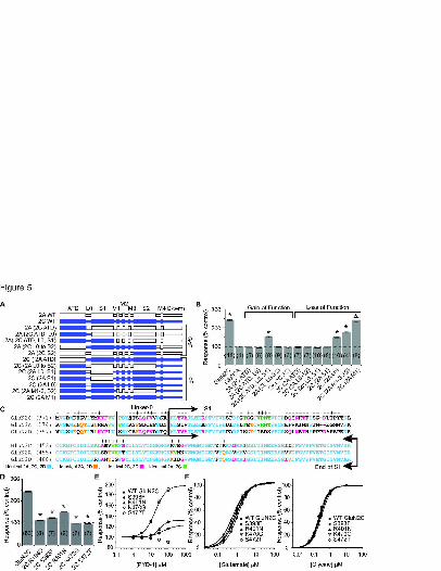
This article has not been copyedited and formatted. The final version may differ from this version.Molecular Pharmacology Fast Forward. Published on September 9, 2014 as DOI: 10.1124/mol.114.094516
at ASPE
T Journals on January 3, 2020
molpharm
.aspetjournals.orgD
ownloaded from

This article has not been copyedited and formatted. The final version may differ from this version.Molecular Pharmacology Fast Forward. Published on September 9, 2014 as DOI: 10.1124/mol.114.094516
at ASPE
T Journals on January 3, 2020
molpharm
.aspetjournals.orgD
ownloaded from


![molの話 [The Talk about "mol"]](https://static.fdocuments.net/doc/165x107/548129feb379595e2b8b5cb5/mol-the-talk-about-mol.jpg)