Stroke mimics transported by emergency medical services to ...
20
Stroke mimics transported by emergency medical services to a comprehensive stroke center: The magnitude of the problem Joel Neves Briard MD 1 , Rahel T. Zewude BSc 2 , Mahesh P. Kate MD 2 , Brian H. Rowe MD MSc 3 , Brian Buck MD MSc 2 , Ken Butcher MD PhD 2 , Laura C. Gioia MD MSc 1 This article has been published in a revised form in the Journal of Stroke and Cerebrovascular Diseases: https://doi.org/10.1016/j.jstrokecerebrovasdis.2018.05.046 © 2018. This manuscript version is made available under the CC-BY-NC-ND 4.0 license http://creativecommons.org/licenses/by-nc-nd/4.0/ Submitted: 7 April 2018. Accepted: 28 May 2018. Affiliations : 1. Department of Neurosciences, Université de Montréal, Quebec, Canada & Centre de recherche du Centre hospitalier de l’Université de Montréal, Quebec, Canada 2. Division of Neurology, University of Alberta, Alberta, Canada 3. Department of Emergency Medicine and School of Public Health, University of Alberta, Alberta, Canada Citation: Neves Briard J, Zewude RT, Kate MP, Rowe BH, Buck B, Butcher K, Gioia LC. Stroke mimics transported by emergency medical services to a comprehensive stroke center: The magnitude of the problem. J Stroke Cerebrovasc Dis. 2018;27(10):2738-45 Short Title: Stroke Mimics Transported by EMS Abstract (words): 249 Text (words): 3029 Figures: 1 Tables: 2 Corresponding author information: Laura C. Gioia MD MSc Centre Hospitalier de l’Université de Montréal (CHUM) Pavillon C, 13e Nord 1051 rue Sanguinet, porte C.13.6121 Montreal, Quebec, Canada H2X 0C1 Email: [email protected]
Transcript of Stroke mimics transported by emergency medical services to ...
Stroke mimics transported by emergency medical services to a comprehensive stroke center: The magnitude of the problem Joel Neves Briard MD1, Rahel T. Zewude BSc2, Mahesh P. Kate MD2, Brian H. Rowe MD MSc3, Brian Buck MD MSc2, Ken Butcher MD PhD2, Laura C. Gioia MD MSc1
This article has been published in a revised form in the Journal of Stroke and Cerebrovascular Diseases: https://doi.org/10.1016/j.jstrokecerebrovasdis.2018.05.046 © 2018. This manuscript version is made available under the CC-BY-NC-ND 4.0 license http://creativecommons.org/licenses/by-nc-nd/4.0/ Submitted: 7 April 2018. Accepted: 28 May 2018.
Affiliations :
1. Department of Neurosciences, Université de Montréal, Quebec, Canada & Centre de recherche du Centre hospitalier de l’Université de Montréal, Quebec, Canada
2. Division of Neurology, University of Alberta, Alberta, Canada
3. Department of Emergency Medicine and School of Public Health, University of Alberta, Alberta, Canada
Citation: Neves Briard J, Zewude RT, Kate MP, Rowe BH, Buck B, Butcher K, Gioia LC. Stroke mimics transported by emergency medical services to a comprehensive stroke center: The magnitude of the problem. J Stroke Cerebrovasc Dis. 2018;27(10):2738-45
Short Title: Stroke Mimics Transported by EMS
Abstract (words): 249 Text (words): 3029
Figures: 1 Tables: 2
Corresponding author information:
Laura C. Gioia MD MSc Centre Hospitalier de l’Université de Montréal (CHUM) Pavillon C, 13e Nord 1051 rue Sanguinet, porte C.13.6121 Montreal, Quebec, Canada H2X 0C1 Email: [email protected]
Abstract
Background: Despite the use of validated prehospital stroke screens, stroke mimics are frequent
among patients transported by Emergency Medical Services to the Emergency Department. We
aimed to describe the frequency and characteristics of neurological and non-neurological mimics
transported to a comprehensive stroke center for acute stroke evaluation.
Methods: This was a retrospective analysis of a database consisting of all consecutive patients with
suspected stroke transported to the Emergency Department of a comprehensive stroke center during
an 18-month period. Hospital charts and neuroimaging were utilized to adjudicate the final
diagnosis (acute stroke, stroke mimic, and specific underlying diagnoses).
Results: 950 patients were transported with suspected stroke, among whom 405 (42.6%) were
stroke mimics (age 66.9±17.1 years; 54% male). Neurological mimics were diagnosed in 223
(55.1%) patients and mimics were non-neurological in 182. The most common neurological
diagnoses were seizures (19.7%), migraines (18.8%) and peripheral neuropathies (11.2%).
Cardiovascular (14.6%) and psychiatric (11.9%) diagnoses were common non-neurological
mimics. Patients with neurological mimic were younger (64.1±17.3 years vs. 70.5±16.1 years,
p<0.001) and had less vascular risk factors than non-neurological mimics. The proportion of non-
neurological mimics remained high (38%) despite the use of a prehospital identification scale.
Conclusion: Stroke mimics are common among patients transported by Emergency Medical
Services to a comprehensive stroke center for suspected stroke, with a considerable proportion
being non-neurological in origin. Studies refining triage and transport of suspected acute stroke
may be warranted to minimize the number of mimics transported by to a comprehensive stroke
center for acute stroke evaluation.
1
Introduction
Rapid triage of acute stroke patients by Emergency Medical Services (EMS) is associated
with reduced transport delays to the Emergency Department (ED) for acute stroke evaluation (1),
higher use of thrombolytic therapy (2), and improved outcomes (3). EMS protocols diverting acute
stroke patients from primary care hospitals to comprehensive stroke centers (CSC) with
endovascular thrombectomy capabilities are increasingly popular in patients with suspected severe
stroke, although robust evidence supporting this practice is currently lacking (4). Mass bypass of
primary care sites to CSC is likely associated with several patients ineligible to endovascular
treatment being subjected to delays in hospital care, including intravenous tissue plasminogen
activator (tPA) administration.
Validated prehospital stroke identification scales are widely used by EMS professionals to
detect acute stroke in the field (5-10). Prehospital identification of acute stroke remains challenging
nonetheless as there are several non-stroke conditions (“stroke mimics”) that share similar clinical
features. As a result, prehospital stroke scales all tend to suffer from a relatively poor specificity
(24-99%) (11). Previous studies have reported that 10-44% of EMS-transported suspected stroke
patients are ultimately diagnosed with a stroke mimic in the ED (9, 12-15), yet little information
exists regarding prehospital stroke mimics, particularly those transported to CSC. Prehospital triage
of stroke mimics (including EMS diversion of primary hospitals to CSC) may be associated with
considerable increases in patient volume, risks, and resource utilization in CSC (12). Despite this,
little information exists regarding mimics in the prehospital setting. The aim of this study was to
describe the prevalence and clinical characteristics of stroke mimics transported by EMS for acute
stroke evaluation to a Canadian regional CSC.
Methods
2
Study Design
The methods of this study have been described previously (16). Briefly, this was a
secondary, cross-sectional, observational study of a prospectively-maintained centralized EMS
database of consecutive patients with suspected stroke transported by EMS to the ED of the
University of Alberta Hospital (UAH), in Edmonton, Canada during an 18-month period (January
1, 2012, to July 31, 2013). The UAH ED manages approximately 77,000 patients per year, is staffed
by full time certified emergency physicians, and functions as an academic teaching hospital for the
University of Alberta. The UAH is staffed 24-hours per day by a stroke service with a dedicated
on-call stroke fellow and neurologist.
The unified EMS service for the city of Edmonton, overseen by Alberta Health Services,
attends to more than 157,000 events annually and transports patients to one of 13 provincially
operated Edmonton Zone EDs. The UAH is a tertiary health care center and one of two CSC within
the Edmonton Zone, receiving the majority of patients transported by EMS for suspected stroke
and admitting 1,000 to 1,200 stroke patients annually. At the time of this study, EMS did not bypass
primary hospitals for the UAH in cases of suspected stroke.
During a 9-1-1 call for suspected stroke, the emergency dispatcher is trained to employ a
stroke-specific interview algorithm to assign a Medical Priority Dispatch System event code for
suspected stroke. This event code is subsequently communicated to EMS providers responding to
the call. The Cincinnati Prehospital Stroke Screen (CPSS) (7) was used on the field by EMS
providers to determine a primary paramedic impression of acute stroke. Dispatch and individual
electronic patient health care reports data were then prospectively uploaded to a centralized
provincial EMS database housed securely within Alberta Health Services. The research protocol
3
of this study was approved for waiver of consent by the local institutional Human Research Ethics
Review Board (University of Alberta).
Study Population and Data Collection
Eligible study participants were identified within the centralized Alberta Health Services
database by “suspected stroke” dispatch event and/or paramedic primary impression of acute
stroke. Once identified, data from individual electronic patient health care reports data were
extracted and linked to ED records through the Emergency Department Information Systems.
Electronic patient health care report data were also linked to in-hospital patient charts and
neuroimaging to determine the final diagnosis of ischemic stroke, transient ischemic attack (TIA),
intracerebral hemorrage (ICH) or stroke mimic, as per the treating physician’s discharge diagnosis.
Measures
Stroke mimics were defined after chart and neuroimaging review as any discharge diagnosis
– other than acute stroke – responsible for the initial symptoms that prompted EMS transport for
suspected stroke. Mimics were classified by the first author as being neurological or non-
neurological in origin, and then further divided into subcategories (neurological: seizure, delirium,
dementia, neoplasm, vertigo, peripheral neuropathy, migraine, weakness of undetermined origin,
and movement disorder; non-neurological: cardiovascular, metabolic, gastroenterological,
psychiatric and infectious).
Participant baseline characteristics – including age, sex, smoking, alcohol use, history of
hypertension, type 2 diabetes, dyslipidemia, coronary artery disease, atrial fibrillation or flutter,
congestive heart failure, cognitive disorder, prior ischemic stroke, ICH or TIA – as well as

4
paramedic primary impression, prehospital systolic blood pressure, prehospital Glasgow Coma
Scale score, reperfusion with tPA or endovascular treatment, and destination from the ED
(discharge home vs hospital admission, with admission length) were extracted for all eligible
patients.
Statistical Analyses
Continuous variables are reported as means and standard deviations (SD) or medians and
interquartile ranges (IQR), as appropriate. Dichotomous variables are reported as proportions.
Differences in baseline characteristics between neurological and non-neurological mimics were
assessed using independent t-tests (parametric data), Mann-Whitney tests (nonparametric) or chi-
square test, as appropriate. No specific sample size was determined for this study; however, the
sample provides 95% confidence intervals (CI) with a precision of +/- 3% for mid-point estimates.
Results
A total of 950 eligible patients meeting study inclusion criteria were identified. Among
these, 545 (57.3%; CI: 54.0-60.4) were diagnosed with acute stroke (367 (38.6%; CI: 35.5-41.8)
ischemic stroke, 117 (12.3%; CI: 10.3-15.3) transient ischemic attack, and 61 (6.3%; CI: 4.1-8.1)
acute intracerebral hemorrage) and 405 (42.6%; CI: 39.5-45.8) patients were diagnosed with a
stroke mimic. The mean age of all stroke mimics was 66.9 ± 17.1 years, and 54% of patients were
male. Compared to acute stroke patients, patients with stroke mimics were younger, and had lower
rates of vascular risk factors (such as hypertension, atrial fibrillarion, dyslipidemia and coronary
artery disease) than patients with acute stroke (Table 1). Nonetheless, baseline vascular risk factors
were frequently found in patients with stroke mimics (hypertension 53%, type 2 diabetes 28%,
dyslipidemia 33%, Table 1).
5
Neurological and Non-Neurological Stroke Mimics
Mimics considered neurological in origin occurred in 223 (55.1%; CI: 50.1-60.0) patients
and non-neurological mimics were diagnosed in 182 (44.9%; CI: 40.0-49.9). Patients with
neurological mimics were younger (64.1 ± 17.3 years vs. 70.5 ± 16.1 years, p<0.001) and had
slightly higher prehospital systolic blood pressures (147.8 ± 24.2 mmHg vs. 141.2 ± 26.2 mmHg,
p=0.01) than non-neurological mimics, respectively (Table 1). Median (IQR) prehospital Glasgow
Coma Scale scores were similar between groups (15 (1) vs. 15 (1), p=0.26). Patients with non-
neurological mimics more commonly exhibited previous diagnoses of hypertension (64% vs 44%),
type 2 diabetes (34% vs 24%), alcohol use (20% vs 9%) and congestive heart failure (10% vs 4%)
and than neurological mimics (p≤0.01). There were no others significant differences between the
two groups regarding other vascular risk factors. The only two mimic patients to have received tPA
were suffering from neurological stroke mimics (specifically, one seizure and one weakness of
undetermined origin). No stroke mimic patient was sent to the endovascular lab. Patient destination
from the ED was similar between neurological and non-neurological mimics, with approximately
40% of patients in both groups being admitted to the hospital (p=0.81). Admitted non-neurological
mimic patients required longer courses of hospitalisation than admitted neurological mimic patients
(5 days (IQR 11) vs 2 days (IQR 8), p=0.04).
Stroke Mimics by Specific Diagnosis
Table 2 describes the prevalence of neurological and non-neurological mimic final
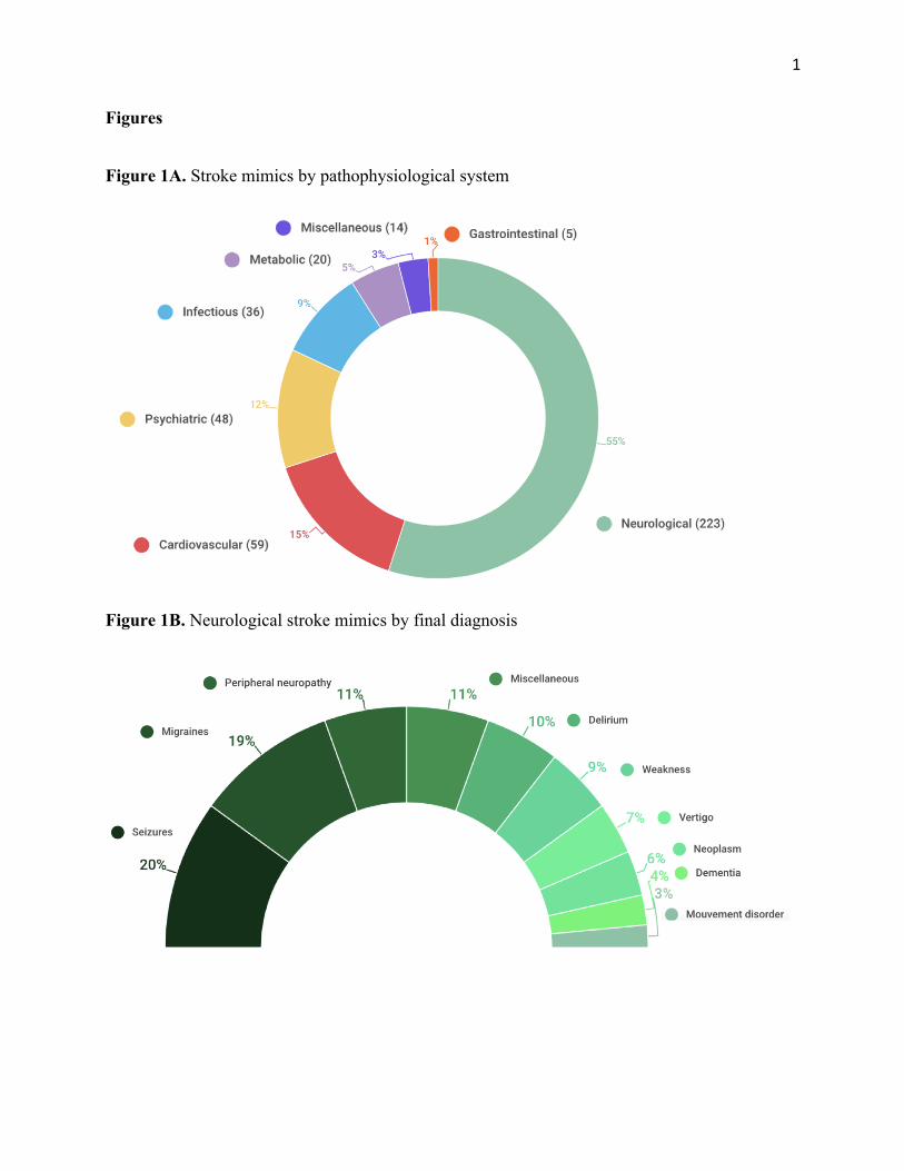
diagnoses. Cardiovascular (n=59, 15.9%), psychiatric (n=48, 11.9%), and infectious (n=36, 8.9%)
diagnoses were the most common causes of non-neurological stroke mimics (Figure 1A). Nearly
6
half of cardiovascular mimic cases were syncope/presyncope (n = 29, 7%). Among neurological
mimics, the most common final diagnoses were seizures (n=44, 19.7%), migraines (n=42, 18.8%),
and peripheral neuropathies (n=25, 11.2%) (Figure 1B).
Primary EMS Impression of Stroke
A primary EMS impression of acute stroke was specified in 210 out of the 405 mimic
patients (51.8%). The remaining 195 patients (48.1%), although flagged as a suspected acute stroke
by emergency medical dispatch, had an alternate primary EMS impression. Non-neurological
stroke mimics were less common but remained frequent (38.1%) in patients with an EMS
impression of stroke, compared with patients with an alternate impression (52.3%, Table 2).
Specifically, patients with an initial EMS impression of stroke had lower rates of cardiovascular
(10% vs 20%) and infectious (5% vs 13%) pathologies, but there was a slightly higher prevalence
of metabolic mimics (7% vs 3%).
Discussion
In an analysis of a centralized EMS database comprised of all consecutive patients
transported for suspected stroke to a regional CSC, we found that stroke mimics represent a
substantial number (>40%) of patients evaluated and transported by EMS. Although the majority
of stroke mimics were neurological in origin, a considerable and surprising number of stroke
mimics were ultimately diagnosed with a non-neurological condition. Non-neurological mimic
patients were older and more likely to have multiple vascular risk factors than neurological mimics.
Furthermore, the proportion of non-neurological stroke mimics remained high despite screening
with CPSS, a validated and widely implemented prehospital stroke identification scale. These
7
results highlight the important challenge faced by EMS providers when identifying acute stroke in
the prehospital field.
Our findings are consistent with previous studies showing that stroke mimics are common
among patients evaluated in the ED for suspected stroke (9, 12-15). In previous studies, stroke
mimicking pathologies were more prevalent in suspected stroke patients with the following
characteristics: young age (<50 years), female sex, absence of vascular risk factors, particularly
atrial fibrillation, and a history of epilepsy or psychiatric illness (15, 17, 18). In our cohort, many
of these mimic characteristics – younger age and absence of alcohol use, hypertension, type 2
diabetes and congestive heart failure – were common in stroke mimics, particularly neurological
mimics. To our knowledge, our study is the first to stratify stroke mimic pathologies in this detail
and identify differences in neurological and non-neurological stroke mimic patient characteristics.
The overtriage of stroke mimics to CSC may have negative impacts on both patients and
the health care system. Mass bypass of primary care sites to CSC is likely associated with several
patients ineligible to endovascular treatment being subjected to delays in hospital care, particularly
intravenous tPA administration. Moreover, overtriage of stroke mimics to CSC results in
overactivation of stroke protocols, requiring mobilization of a number of healthcare professionals
involved in acute stroke care (emergency physicians, triage and emergency nursing, stroke fellows
and neurologists, radiology technicians and neurointerventionalists). Finally, admitted mimic
patients are often subjected to extensive work-ups (19), which may expose them to unnecessary
radiation or interventions, and represent significant costs to the system.
False diagnosis in the prehospital setting is not unique to acute stroke management. Studies
have shown that 8-25% of cardiac catherterization lab activations following prehospital notification
for acute ST-elevation myocardial infarction (STEMI) result from false-positive EMS screening
8
(20-22). These false-positive cases for STEMI are regarded as acceptable, particularly since EMS
activation expedites care and translates into shorter door-to-balloon delays than ED activation (23,
24). Considering the significant burden of ischemic stroke on the individual and society in
association with the proven benefit of early stroke detection and overwhelming efficacy of acute
reperfusion therapies, a higher false positive rate may be acceptable at a societal level. Conversely,
overactivation of stroke teams and management of a high proportion of stroke mimics may put
considerable strain on the health system from a resource utilization perspective. Overactivation of
acute stroke may also be associated with negative outcomes for other ED patients for whom care
is delayed. An increase in the length of stay in ED patients overall has been described in designated
trauma centers with trauma activations (25-27). As the volume of patients eligible for endovascular
intervention rapidly evolves (28, 29), and as stroke mimic incidence is projected to increase and
possibly surpass acute stroke incidence in the future (19), it will be increasingly important to
address the problem of overtriage of stroke mimics to CSC.
Several prehospital stroke identification scales have been developed to improve acute
stroke detection in the out-of-hospital setting. Overall, these scales are associated with reasonably
high sensitivity and low specificity for acute stroke diagnosis (10). The Los Angeles Prehospital
Stroke Screen (LAPSS), the only scale that incorporates elements suggestive of stroke mimics
(such as seizure history), has been shown to be a more reliable and consistent prehospital scale for
acute stroke diagnosis when compared to other scales (6).
Other in-hospital scales have been developed to improve stroke mimic detection in the ED.
The Recognition of Stroke in the Emergency Room (ROSIER) scale was developed for use by
emergency physicians on all suspected stroke patients prior to neuroimaging (30). The presence of
focal neurological signs (e.g., face, arm or leg weakness; speech disturbance; visual field defect)
9
were scored favorably for acute stroke, while the presence of seizures, confusion, or loss of
consciousness were scored unfavourably (loss of points on the scale). A ROSIER score ≥1 favoured
the diagnosis of stroke with high diagnostic accuracy (92%). A second scale, the FABS, was
developed in order to help stratify the need for advanced neuroimaging (MRI) in suspected stroke
patients with a negative head CT scan in the ED (31). In the presence of three or more of the scale
elements (absence of facial droop, age<50 years old, absence of atrial fibrillation, SBP<150mmHg,
presence of isolated sensory deficit, or seizure history), the sensitivity (90%) and specificity (91%)
were high for stroke mimic. Indeed, our findings are in line with the elements predictive of stroke
mimics in the FABS scale such the absence of atrial fibrillation, history of seizure and lower blood
pressure. Given the absence of clinical information regarding the individual items of the CPSS that
triggered prehospital stroke activation in our study, we were not able to calculate FABS score in
our patient population.
Our findings suggest that prehospital stroke identification scales are associated with a high
rate of stroke mimics, with nearly half being non-neurological in origin. The inclusion of clinical
elements suggestive of stroke mimics (e.g., syncope, seizure) may help to decrease the rate of
stroke mimics transported to CSC for acute stroke evaluation, but whether these tools should be
employed in the EMS setting remains unknown. Future prospective studies are warranted to assess
whether the use of newly-developped prehospital scale incorporating elements predictive of stroke
mimics in the field is associated with fewer stroke mimics transported to ED. An alternative
approach could be a combined use of a sensitive EMS screening tool to trigger transport for acute
stroke followed by a second, more specific scale in order to grade stroke severity and help minimize
stroke mimics when considering EMS diversion directly to CSC, but future studies are needed.
Nonetheless, both approaches would require a systems-wide analysis of all patients transported for
10
suspected stroke by EMS to primary and comprehensive stroke centers in order to determine the
best approach.
Limitations
There are a number of limitations to our study. First, this was a retrospective study and is
subject to confounding. Patient data, however, was prospectively collected and uploaded to a
centralized database which may help to limit bias. Second, inclusion of all patients with suspected
stroke by dispatch code and/or EMS evaluation likely resulted in an overestimation of the
proportion of stroke mimics transported to the ED. We chose to include all consecutive patients
evaluated in the field for suspected stroke patients in order to better characterize stroke mimics in
the field as well as determine whether EMS impression of stroke was associated with lower rates
of non-neurological mimics transported to ED. Moreover, in a recent prospective field validation
of the Cincinnati Stroke Triage Assessment Tool, 44% of CPSS positive patients suffered from a
stroke mimic, so our reported 43% prevalence may not be significantly overestimated (9). Since
dispatcher recognition of stroke is associated with a poor positive predictive value (32), our study
results may not be generalizable to regions that rely solely on paramedic identification of stroke
using a prehospital stroke scale. Fourth, no adjustment was made for multiple statistical testing;
however, the differences are large so the statistical results are robust. Finally, the study was limited
to one Canadian academic centre, and the generalizability of these results warrants further scrutiny.
Conclusion
Stroke mimics are common and heterogenous in nature among patients transported by EMS
to the ED of a CSC for acute stroke evaluation, with a substantial number being non-neurological
11
in origin. Positive prehospital stroke identification by EMS may decrease the rate of non-
neurological stroke mimics; however, proportions remain high. Utilization of prehospital scales
incorporating elements suggestive of stroke mimics (such as syncope, seizures) may improve field
detection and decrease transport of stroke mimics to CSC, particularly in an increasingly
implemented model of EMS diversion to CSC, but prospective studies are needed.
Author Contributions
JNB: designed and conceptualized the study, performed analysis and interpretation of the data and
drafted the manuscript. RTZ: collected data, and revised the manuscript. MPK: designed and
conceptualized the primary study and revised the manuscript. BR: designed and conceptualized the
primary study and revised the manuscript. BB: performed analysis and interpretation of the data
and revised the manuscript. KB: designed and conceptualized the primary study and revised the
manuscript. LCG: designed and conceptualized the study, performed analysis and interpretation of
the data and revised the manuscript.
Sources of Funding
KSB holds a Canada Research Chair in Cerebrovascular Disease, a Heart and Stroke Foundation
of Alberta Professorship in Stroke Medicine and a New Investigator Award from Alberta Innovates
Health Solutions. LCG and MK are supported by Clinical Research Fellowship bursaries from the
Alberta Innovates Health Solutions. RZT received by a Summer Studentship Award from Alberta
Innovates Health Solutions. BHR is supported by CIHR as a Tier I Canada Research Chair in
Evidence-based Emergency Medicine through the Government of Canada (Ottawa, ON) and is the
Scientific Director of the Institute of Circulatory and Respiratory Health (ICRH) at CIHR. The
funding agencies take no responsibility for the conduct, results and interpretations of findings
reported.

12
Disclosure
The authors report no conflicts of interest.
13
References
1. Caceres JA, Adil MM, Jadhav V, Chaudhry SA, Pawar S, Rodriguez GJ, et al. Diagnosis of stroke by emergency medical dispatchers and its impact on the prehospital care of patients. J Stroke Cerebrovasc Dis. 2013;22(8):e610-4. 2. McKinney JS, Mylavarapu K, Lane J, Roberts V, Ohman-Strickland P, Merlin MA. Hospital prenotification of stroke patients by emergency medical services improves stroke time targets. J Stroke Cerebrovasc Dis. 2013;22(2):113-8. 3. Pérez de la Ossa N, Sanchez-Ojanguren J, Palomeras E, Millan M, Arenillas JF, Dorado L, et al. Influence of the stroke code activation source on the outcome of acute ischemic stroke patients. Neurology. 2008;70(15):1238-43. 4. Southerland AM, Johnston KC, Molina CA, Selim MH, Kamal N, Goyal M. Suspected Large Vessel Occlusion: Should Emergency Medical Services Transport to the Nearest Primary Stroke Center or Bypass to a Comprehensive Stroke Center With Endovascular Capabilities? Stroke. 2016;47:1965-7. 5. Bray JE, Martin J, Cooper G, Barger B, Bernard S, Bladin C. Paramedic Identification of Stroke: Community Validation of the Melbourne Ambulance Stroke Screen. Cerebrovascular Diseases. 2005;20(1):28-33. 6. Kidwell CS, Starkman S, Eckstein M, Weems K, Saver JL. Identifying Stroke in the Field: Prospective Validation of the Los Angeles Prehospital Stroke Screen (LAPSS). Stroke. 2000;31:71-6. 7. Kothari RU, Pancioli A, Liu T, Brott T, Broderick JP. Cincinnati Prehospital Stroke Scale: Reproducibility and validity. Ann Emerg Med. 1999;33(4):373-8. 8. Llanes JN, Kidwell CS, Starkman S, Leary MC, Eckstein M, Saver JL. The Los Angeles Motor Scale (LAMS): A New Measure to Characterize Stroke Severity in the Field. Prehosp Emerg Care. 2009;8(1):46-50. 9. McMullan JT, Katz B, Broderick J, Schmit P, Sucharew H, Adeoye O. Prospective Prehospital Evaluation of the Cincinnati Stroke Triage Assessment Tool. Prehosp Emerg Care. 2017;21(4):481-8. 10. Pérez de la Ossa N, Carrera D, Gorchs M, Querol M, Millan M, Gomis M, et al. Design and validation of a prehospital stroke scale to predict large arterial occlusion: the rapid arterial occlusion evaluation scale. Stroke. 2014;45(1):87-91. 11. Brandler ES, Sharma MS, Sinert RH, Levine SR. Prehospital stroke scales in urban environments: a systematic review. Neurology. 2014;82:2241-9. 12. Goyal N, Male S, Al Wafai A, Bellamkonda S, Zand R. Cost burden of stroke mimics and transient ischemic attack after intravenous tissue plasminogen activator treatment. J Stroke Cerebrovasc Dis. 2015;24(4):828-33. 13. Hand PJ, Kwan J, Lindley RI, Dennis MS, Wardlaw JM. Distinguishing between stroke and mimic at the bedside: the brain attack study. Stroke. 2006;37(3):769-75. 14. Sequeira D, Martin-Gill C, Kesinger MR, Thompson LR, Jovin TG, Massaro LM, et al. Characterizing Strokes and Stroke Mimics Transported by Helicopter Emergency Medical Services. Prehosp Emerg Care. 2016;20(6):723-8. 15. Vroomen PC, Buddingh MK, Luijckx GJ, De Keyser J. The incidence of stroke mimics among stroke department admissions in relation to age group. J Stroke Cerebrovasc Dis. 2008;17(6):418-22.
14
16. Gioia LC, Zewude RT, Kate MP, Liss K, Rowe BH, Jeerakathil T, et al. Prehospital systolic blood pressure is higher in acute stroke compared with stroke mimics. Neurology. 2016;86(23):2146-53. 17. Khan NI, Chaku S, Goehl C, Endris L, Mueller-Luckey G, Siddiqui FM. Novel Algorithm to Help Identify Stroke Mimics. J Stroke Cerebrovasc Dis. 2018;27(3):703-8. 18. Merino JG, Luby M, Benson RT, Davis LA, Hsia AW, Latour LL, et al. Predictors of Acute Stroke Mimics in 8187 Patients Referred to a Stroke Service. J Stroke Cerebrovasc Dis. 2013;22(8):e397-e403. 19. Faiz KW, Labberton AS, Thommessen B, Ronning OM, Dahl FA, Barra M. The Burden of Stroke Mimics: Present and Future Projections. J Stroke Cerebrovasc Dis. 2017. 20. Garvey JL, Monk L, Granger CB, Studnek JR, Roettig ML, Corbett CC, et al. Rates of cardiac catheterization cancelation for ST-segment elevation myocardial infarction after activation by emergency medical services or emergency physicians: results from the North Carolina Catheterization Laboratory Activation Registry. Circulation. 2012;125(2):308-13. 21. Potter BJ, Matteau A, Mansour S, Essiambre R, Montigny M, Savoie S, et al. Performance of a new "physician-less" automated system of prehospital ST-segment elevation myocardial infarction diagnosis and catheterization laboratory activation. Am J Cardiol. 2013;112(2):156-61. 22. Squire BT, Tamayo-Sarver JH, Rashi P, Koenig W, Niemann JT. Effect of prehospital cardiac catheterization lab activation on door-to-balloon time, mortality, and false-positive activation. Prehosp Emerg Care. 2014;18(1):1-8. 23. Bradley EH, Herrin J, Wang YW, Barton BA, Webster TR, Mattera JA, et al. Strategies for Reducing the Door-to-Balloon Time in Acute Myocardial Infarction. N Engl J Med. 2006;355:2308-20. 24. Rokos IC, French WJ, Koenig WJ, Stratton SJ, Nighswonger B, Strunk B, et al. Integration of pre-hospital electrocardiograms and ST-elevation myocardial infarction receiving center (SRC) networks: impact on Door-to-Balloon times across 10 independent regions. JACC Cardiovasc Interv. 2009;2(4):339-46. 25. Arya R, Dossantos F, Ohman-Strickland P, Merlin MA. Impact of trauma activation on the ED length of stay for nontraumatic patients. Am J Emerg Med. 2012;30(2):311-6. 26. Fishman PE, Shofer FS, Robey JL, Zogby KE, Reilly PM, Branas CC, et al. The impact of trauma activations on the care of emergency department patients with potential acute coronary syndromes. Ann Emerg Med. 2006;48(4):347-53. 27. Smith DC, Chapital A, Burgess Uperesa BM, Smith ER, Ho C, Ahana A. Trauma activations and their effects on non-trauma patients. J Emerg Med. 2011;41(1):90-4. 28. Albers GW, Marks MP, Kemp S, Christensen S, Tsai JP, Ortega-Gutierrez S, et al. Thrombectomy for Stroke at 6 to 16 Hours with Selection by Perfusion Imaging. N Engl J Med. 2018;378(8):708-18. 29. Nogueira RG, Jadhav AP, Haussen DC, Bonafe A, Budzik RF, Bhuva P, et al. Thrombectomy 6 to 24 Hours after Stroke with a Mismatch between Deficit and Infarct. N Engl J Med. 2018;378(1):11-21. 30. Nor AM, Davis J, Sen B, Shipsey D, Louw SJ, Dyker AG, et al. The Recognition of Stroke in the Emergency Room (ROSIER) scale: development and validation of a stroke recognition instrument. The Lancet Neurology. 2005;4(11):727-34. 31. Goyal M, Tsivgoulis G, Male S, Metter EJ, Iftikhar S, Kerro A, et al. FABS: An Intuitive Tool for Screening of Stroke Mimics in the Emergency Department. Stroke. 2016;47:2216-20.

15
32. Oostema JA, Carle T, Talia N, Reeves M. Dispatcher Stroke Recognition Using a Stroke Screening Tool: A Systematic Review. Cerebrovasc Dis. 2016;42:370-7.
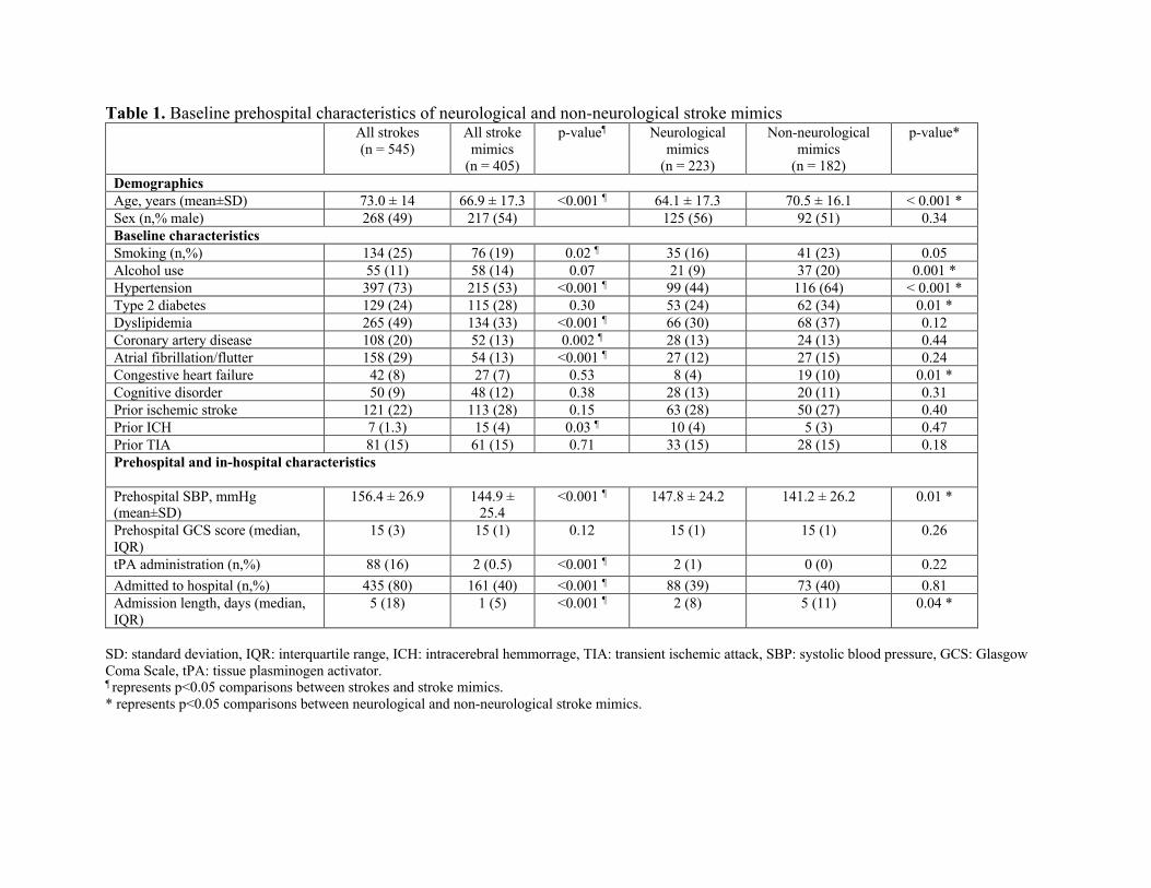
Table 1. Baseline prehospital characteristics of neurological and non-neurological stroke mimics All strokes
(n = 545) All stroke
mimics (n = 405)
p-value¶ Neurological mimics
(n = 223)
Non-neurological mimics
(n = 182)
p-value*
Demographics Age, years (mean±SD) 73.0 ± 14 66.9 ± 17.3 <0.001 ¶ 64.1 ± 17.3 70.5 ± 16.1 < 0.001 * Sex (n,% male) 268 (49) 217 (54) 125 (56) 92 (51) 0.34 Baseline characteristics Smoking (n,%) 134 (25) 76 (19) 0.02 ¶ 35 (16) 41 (23) 0.05 Alcohol use 55 (11) 58 (14) 0.07 21 (9) 37 (20) 0.001 * Hypertension 397 (73) 215 (53) <0.001 ¶ 99 (44) 116 (64) < 0.001 * Type 2 diabetes 129 (24) 115 (28) 0.30 53 (24) 62 (34) 0.01 * Dyslipidemia 265 (49) 134 (33) <0.001 ¶ 66 (30) 68 (37) 0.12 Coronary artery disease 108 (20) 52 (13) 0.002 ¶ 28 (13) 24 (13) 0.44 Atrial fibrillation/flutter 158 (29) 54 (13) <0.001 ¶ 27 (12) 27 (15) 0.24 Congestive heart failure 42 (8) 27 (7) 0.53 8 (4) 19 (10) 0.01 * Cognitive disorder 50 (9) 48 (12) 0.38 28 (13) 20 (11) 0.31 Prior ischemic stroke 121 (22) 113 (28) 0.15 63 (28) 50 (27) 0.40 Prior ICH 7 (1.3) 15 (4) 0.03 ¶ 10 (4) 5 (3) 0.47 Prior TIA 81 (15) 61 (15) 0.71 33 (15) 28 (15) 0.18 Prehospital and in-hospital characteristics Prehospital SBP, mmHg (mean±SD)
156.4 ± 26.9 144.9 ± 25.4
<0.001 ¶ 147.8 ± 24.2 141.2 ± 26.2 0.01 *
Prehospital GCS score (median, IQR)
15 (3) 15 (1) 0.12 15 (1) 15 (1) 0.26
tPA administration (n,%) 88 (16) 2 (0.5) <0.001 ¶ 2 (1) 0 (0) 0.22
Admitted to hospital (n,%) 435 (80) 161 (40) <0.001 ¶ 88 (39) 73 (40) 0.81 Admission length, days (median, IQR)
5 (18) 1 (5) <0.001 ¶ 2 (8) 5 (11) 0.04 *
SD: standard deviation, IQR: interquartile range, ICH: intracerebral hemmorrage, TIA: transient ischemic attack, SBP: systolic blood pressure, GCS: Glasgow Coma Scale, tPA: tissue plasminogen activator. ¶ represents p<0.05 comparisons between strokes and stroke mimics. * represents p<0.05 comparisons between neurological and non-neurological stroke mimics.
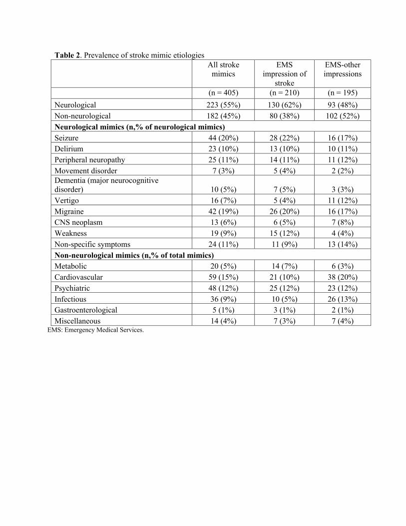
Table 2. Prevalence of stroke mimic etiologies
All stroke mimics
EMS impression of
stroke
EMS-other impressions
(n = 405) (n = 210) (n = 195) Neurological 223 (55%) 130 (62%) 93 (48%) Non-neurological 182 (45%) 80 (38%) 102 (52%) Neurological mimics (n,% of neurological mimics) Seizure 44 (20%) 28 (22%) 16 (17%) Delirium 23 (10%) 13 (10%) 10 (11%) Peripheral neuropathy 25 (11%) 14 (11%) 11 (12%) Movement disorder 7 (3%) 5 (4%) 2 (2%) Dementia (major neurocognitive disorder) 10 (5%) 7 (5%) 3 (3%) Vertigo 16 (7%) 5 (4%) 11 (12%) Migraine 42 (19%) 26 (20%) 16 (17%) CNS neoplasm 13 (6%) 6 (5%) 7 (8%) Weakness 19 (9%) 15 (12%) 4 (4%) Non-specific symptoms 24 (11%) 11 (9%) 13 (14%) Non-neurological mimics (n,% of total mimics) Metabolic 20 (5%) 14 (7%) 6 (3%) Cardiovascular 59 (15%) 21 (10%) 38 (20%) Psychiatric 48 (12%) 25 (12%) 23 (12%) Infectious 36 (9%) 10 (5%) 26 (13%) Gastroenterological 5 (1%) 3 (1%) 2 (1%) Miscellaneous 14 (4%) 7 (3%) 7 (4%)
EMS: Emergency Medical Services.
1
Figures Figure 1A. Stroke mimics by pathophysiological system
Figure 1B. Neurological stroke mimics by final diagnosis