
STAT3 IN EGF RECEPTOR-MEDIATED FIBROBLAST AND HUMAN PROSTATE
139
STAT3 IN EGF RECEPTOR-MEDIATED FIBROBLAST AND HUMAN PROSTATE CANCER CELL MIGRATION,INVASION AND APOPTOSIS by Weixin Zhou B.S., Zhongshan (Sun Yat-Sen) University, 1997 M.S., Zhongshan (Sun Yat-Sen) University, 2000 Submitted to the Graduate Faculty of The School of Medicine, Department of Cellular and Molecular Pathology In partial fulfillment of the requirements for the degree of Doctor of Philosophy University of Pittsburgh 2006
Transcript of STAT3 IN EGF RECEPTOR-MEDIATED FIBROBLAST AND HUMAN PROSTATE
STAT3 IN EGF RECEPTOR-MEDIATED FIBROBLAST AND HUMAN PROSTATE
CANCER CELL MIGRATION,INVASION AND APOPTOSIS
by
Weixin Zhou
B.S., Zhongshan (Sun Yat-Sen) University, 1997
M.S., Zhongshan (Sun Yat-Sen) University, 2000
Submitted to the Graduate Faculty of
The School of Medicine, Department of Cellular and Molecular Pathology
In partial fulfillment of the requirements for the degree of
Doctor of Philosophy
University of Pittsburgh
2006

UNIVERSITY OF PITTSBURGH
The School of Medicine
This dissertation was presented
by
Weixin Zhou
It was defended on
[August 8th, 2006]
and approved by
Jennifer Grandis, M.D., Department of Otolaryngology
Jianhua Luo, M.D., Ph.D., Department of Pathology
Reza Zarnegar, Ph.D., Department of Pathology
Tom Smithgall, Ph.D., Department of Molecular Genetics and Biochemistry
Dissertation Advisor: Alan Wells, M.D., D.M.Sc., Department of Pathology
ii

Copyright © by Weixin Zhou
2006
iii

STAT3 IN EGF RECEPTOR MEDIATED FIBROBLASTS AND HUMAN PROSTATE
CANCER CELLS MIGRATION AND INVASION AND APOPTOSIS
Weixin Zhou, Ph.D.
University of Pittsburgh, 2006
Growth factor-induced migration is a rate-limiting step in tumor invasiveness. The
molecules that regulate this cellular behavior would represent novel targets for limiting tumor
cell progression. Epidermal growth factor (EGF) receptor (EGFR)-mediated motility, present in
both autocrine and paracrine modes in prostate carcinomas, requires de novo transcription to
persist over times greater than a few hours. Therefore, we sought the specific signaling pathways
that directly alter cellular transcription. We confirmed that STAT3 directly associates with, and
is activated by EGFR in DU-145 and PC3 human prostate carcinoma cells in addition to the
model NR6 fibroblast cell line. This correlated with electrophoretic motility shift of
STAT3-selective oligonucleotides. Inhibition of STAT3 activity by antisense or siRNA
down-regulation or expression of a dominant-negative construct limited cell motility as
determined by an in vitro wound healing assay and invasiveness through a matrix barrier. The
expression of constitutively activated STAT3 in the absence of EGF did not increase the
migration. Together these data indicate that STAT3 is necessary but not sufficient for
EGFR-mediated migration. An initial gene array detected a number of candidate operative
molecules; the protein levels of both ENA/VASP, a repressor of cell motility, and caspase 3, a
nexus of apoptotic signaling, were down regulated by EGF in a STAT3-dependent manner.
Preliminary data show that EGF requires STAT3 functioning to inhibit the induction of apoptosis
iv

in the two human prostate cancer cell lines. This suggests that STAT3 signaling may be
contributing to tumor progression in a second manner by rendering the cells resistant to death.
Together, the sum of these findings suggest that STAT3 signaling may be a new target for both
limiting prostate tumor cell invasion and enabling the tumor cells to be killed.
v

TABLE OF CONTENTS
PREFACE....................................................................................................................................XII
1.0 INTRODUCTION ......................................................................................................... 1
1.1 PROSTATE CANCER.................................................................................................. 2
1.1.1 Background of Prostate Cancer .......................................................................... 2
1.1.2 Diagnosis of Prostate Cancer.............................................................................. 3
1.1.3 Therapy of Prostate Cancer ................................................................................ 4
1.1.4 EGFR mediated signalling pathway and Prostate Cancer .................................. 5
1.2 EGF RECEPTOR AS A MOLECULAR IN SIGNALLING........................................ 6
1.3 EGF RECEPTOR AND CANCER ............................................................................. 10
1.3.1 EGFR and Tumor Cell Invasion....................................................................... 14
1.3.2 EGFR and Tumor Cell Proliferation ................................................................ 16
1.4 SIGNALLING CELL MIGRATION INCLUDES ENA/VASP................................. 17
1.4.1 Signalling Cell Migration ................................................................................. 19
1.4.2 Arp2/3 Complex ............................................................................................... 22
1.4.3 Ena/VASP......................................................................................................... 22
1.4.4 Regulation of β-actin ........................................................................................ 26
1.5 SIGNAL TRANSDUCERS AND ACTIVATORS OF TRANSCRIPTION.............. 28
1.5.1 Background of STATs...................................................................................... 28
1.5.2 Signalling Pathways that activate STATs ........................................................ 31
1.5.3 STAT3 and Cancer ........................................................................................... 33
1.6 CASPASES AND APOPTOSIS ................................................................................. 35
1.6.1 Apoptosis .......................................................................................................... 36
1.6.2 Caspase proteins ............................................................................................... 38
2.0 MATERIAL AND METHODS ................................................................................... 40
2.1 CELL LINES & MEDIUM......................................................................................... 40
2.2 RNA PURIFICATION................................................................................................ 41
2.3 MICROARRAY ANALYSES .................................................................................... 42
2.4 MOLECULAR CLONING ......................................................................................... 43
2.5 ELECTROPORATION AND SELECTION OF THE CLONS ................................. 44
2.6 RNA INTERFERENCE .............................................................................................. 45
vi

2.7 NUCLEAR EXTRACT PREPARATION AND EMSA ............................................ 46
2.8 IMMUNOBLOT ......................................................................................................... 47
2.9 WOUND HEALING ASSAY..................................................................................... 47
2.10 INVASION ASSAY ................................................................................................ 48
2.11 ANNEXIN V STAINING FOR APOPTOSIS ASSAY .......................................... 48
2.12 PROLIFERATION ASSAY .................................................................................... 49
2.13 STATISTICAL ANALYSES .................................................................................. 49
3.0 STAT3 IS REQUIRED BUT NOT SUFFICIENT FOR EGFR MEDIATED
MIGRATION AND INVASION OF FIBROBLAST AND HUMAN PROSTATE
CARCINOMA CELLS................................................................................................................. 50
3.1 STAT3 ANTISENSE DECREASES EXPRESSION OF STAT3 IN NR6WT CELLS
50
3.2 STAT3 ANTISENSE DECREASES DNA BINDING ACTIVITY OF STAT3 IN
NR6WT CELLS ................................................................................................................... 52
3.3 STAT3 ANTISENSE DECREASES EXPRESSION OF STAT3 IN DU145WT
CELLS.................................................................................................................................. 54
3.4 STAT3 ANTISENSE DECREASES DNA BINDING ACTIVITY OF STAT3 IN
DU145WT CELLS............................................................................................................... 55
3.5 STAT3 SIRNA DECREASES EXPRESSION OF STAT3 IN DU145WT CELLS .. 57
3.6 STAT3 SIRNA DECREASES EXPRESSION OF STAT3 IN PC3 CELLS ............. 58
3.7 STAT3 SIRNA DECREASES DNA BINDING ACTIVITY OF STAT3 IN
DU145WT CELLS............................................................................................................... 60
3.8 STAT3 ANTISENSE INHIBITS EGF INDUCED MIGRATION IN NR6WT CELLS
61
3.9 STAT3 ANTISENSE INHIBITS EGF INDUCED MIGRATION IN DU145WT
CELLS.................................................................................................................................. 63
3.10 STAT3 SIRNA INHIBITS EGF INDUCED MIGRATION IN DU145WT AND
PC3 HUMAN PROSTATE TUMOR CELLS ..................................................................... 65
3.11 STAT3 ANTISENSE INHIBITS EGF INDUCED INVASION IN DU145WT..... 67
3.12 STAT3 SIRNA INHIBITS EGF INDUCED INVASION IN DU145WT AND PC3
HUMAN PROSTATE CANCER CELLS ........................................................................... 69
vii

3.13 STAT3 IS NOT SUFFICIENT FOR MIGRATION IN NR6WT CELLS .............. 71
3.14 STAT3 ANTISENSE DOES NOT INHIBIT EGFR MEDIATED
PROLIFERATION IN NR6WT FIBROBLAST CELLS .................................................... 73
3.15 STAT3 ANTISENSE DOES NOT INHIBIT EGFR MEDIATED
PROLIFERATION IN DU145WT CELLS ......................................................................... 75
4.0 STAT3 IS REQUIRED IN CASPASE 3 INDUCED HUMAN PROSTATE
CARCIMONA CELL APOPTOSIS............................................................................................. 77
4.1 MICROARRAY DATA SHOWS SIGNIFICANT CHANGES OF SOME
PROTEINS’ TRANSCRIPTION LEVELS AFTER EGF AND/OR STAT3 ANTISENSE
TREATMENT IN NR6WT CELLS..................................................................................... 77
4.2 EGF INHIBITS THE EXPRESSION OF VASP IN NR6WT CELLS, WHICH CAN
BE REVERSED BY STAT3 SIRNA................................................................................... 79
4.3 EGF INHIBITS THE EXPRESSION OF VASP AND CASPASE 3 IN DU145WT
CELLS AND CAN BE REVERSED BY STAT3 SIRNA .................................................. 81
4.4 STAT3 IS REQUIRED FOR RESISTANCE TO TNF-Α INDUCED APOPTOSIS OF
DU145WT CELLS............................................................................................................... 83
4.5 STAT3 IS REQUIRED FOR RESISTANCE TO TNF-Α INDUCED APOPTOSIS IN
PC3 CELLS.......................................................................................................................... 85
5.0 DISCUSSION CHAPTER........................................................................................... 87
6.0 CONCLUSIONS AND SPECULATION.................................................................... 91
6.1 CONCLUSIONS ......................................................................................................... 91
6.2 SPECULATION.......................................................................................................... 93
7.0 ABBREVIATIONS ..................................................................................................... 97
BIBLIOGRAPHY......................................................................................................................... 99
viii

LIST OF TABLES
Table 1. Mutations of the EGFR detected in tumor cells. ............................................................ 12
Table 2. Activation of STATs in human cancers.......................................................................... 35
Table 3. Subfamily of caspase proteins. ....................................................................................... 39
Table 4 Microarray in NR6WT cells shows some proteins’ mRNA expression level changed
significantly after EGF treatment and went back after blocking the STAT3 by antisense
nucleotide...................................................................................................................................... 79
ix

LIST OF FIGURES
Figure 1-1. Age distribution of deaths from prostate cancer in the United States.......................... 3
Figure 1-2. Biopsy pictures of normal and prostate cancer tissue. ................................................. 4
Figure 1-3. Schematics of EGFR signalling pathway..................................................................... 9
Figure 1-4. EGFR signalling pathway and approaches to inhibiting the EGFR........................... 13
Figure 1-5. The invasive growth program under physiological and pathological conditions. ..... 15
Figure 1-6. Cell motility events. ................................................................................................... 18
Figure 1-7. EGFR mediated cell motility pathways. .................................................................... 20
Figure 1-8. Domain structure of Ena/VASP family proteins........................................................ 24
Figure 1-9. Antagonism between Ena/VASP and capping protein............................................... 25
Figure 1-10. Regulation of localized β-actin mRNA translation in a polarized neural cell. ........ 27
Figure 1-11. STAT structure and family of proteins. ................................................................... 30
Figure 1-12. Signalling pathways of STATs. ............................................................................... 32
Figure 1-13. Schematic representing the core components of apoptosis pathways...................... 37
Figure 3-1. EGF increase the expression of STAT3 in NR6WT cells and it can be inhibited by
STAT3 antisense........................................................................................................................... 51
Figure 3-2. Electrophoretic mobility shift assay (EMSA) detects EGF-dependent STAT3 activity
in NR6WT fibroblasts cells. ......................................................................................................... 53
Figure 3-3. EGF increase the expression of STAT3 in DU145WT cells and it can be inhibited by
STAT3 antisense........................................................................................................................... 54
Figure 3-4. Electrophoretic mobility shift assay (EMSA) detects EGF-dependent STAT3 activity
in DU145WT human prostate tumor cells. ................................................................................... 56
Figure 3-5. EGF-mediated increase of STAT3 expression and activity is suppressed by STAT3
siRNA in DU145WT cells. ........................................................................................................... 57
Figure 3-6. EGF-mediated increase of STAT3 expression and activity is suppressed by STAT3
siRNA in PC3 cells. ...................................................................................................................... 59
Figure 3-7. The STAT3 siRNA negated EGF induced STAT3 activity in DU145WT prostate
tumor cells..................................................................................................................................... 60
x

Figure 3-8. STAT3 is required for EGF receptor mediated cell migration in NR6WT fibroblast
cells. .............................................................................................................................................. 62
Figure 3-9. STAT3 is required for EGF receptor mediated cell migration in human prostate
tumor cells..................................................................................................................................... 64
Figure 3-10. STAT3 is required for EGF receptor mediated cell migration in human prostate
tumor cells..................................................................................................................................... 66
Figure 3-11. STAT3 is critical for EGFR mediated prostate tumor cell invasiveness ................. 68
Figure 3-12. STAT3 is critical for EGFR mediated prostate tumor cell invasiveness. ................ 70
Figure 3-13. STAT3 is not sufficient for EGFR-mediated migration in NR6WT fibroblasts...... 72
Figure 3-14. STAT3 antisense does not inhibit EGFR mediated proliferation in NR6WT cells. 74
Figure 3-15. STAT3 antisense does not inhibit EGFR mediated proliferation in DU145WT cells.
....................................................................................................................................................... 76
Figure 4-1. EGF inhibits the expression of VASP and Caspase-3 in NR6WT cells, which can be
reversed by STAT 3 siRNA.......................................................................................................... 80
Figure 4-2. EGF inhibits the expression of VASP and Caspase-3 in DU145WT cells, which can
be reversed by STAT 3 siRNA. .................................................................................................... 82
Figure 4-3. Annexin-V staining data for apoptosis in DU145WT prostate tumor cells with the
treatment of EGF and STAT3 siRNA........................................................................................... 84
Figure 4-4. Annexin-V staining data for apoptosis in PC3 prostate tumor cells with the treatment
of EGF and STAT3 siRNA........................................................................................................... 85
xi

PREFACE
I dedicate this to my loving parents, Xiao-An Wu and Yi Zhou, for the encouragement
and support they have given me throughout my life.
I am greatly appreciative of my mentor Alan Wells, who has patiently taught and steered
me during my scientific training. For their insight and guidance, I would also like to thank the
members of my thesis committee at the University of Pittsburgh: Jennifer R Grandis, Jian Hua
Luo, Thomas Smithgall and Razor Zarnegar. I would especially like to thank past and present
members of the Wells Lab for their helpful advice and support over the past 5 years: Philip
Chang, Jeffrey Chou, Angela Glading, Akihiro Iwabu, Jareer Kassis, Priya Kulasekaran, Asmaa
Mamoune, Latha Satish, Hidenori Shiraha, Kate Sullivan, Kien Tran, Diana Whaley, Clayton
Yates, and Cecelia Yates.
xii

1.0 INTRODUCTION
Cancer progression results from complex and still riddling myriad of physiological and
pathological events. Among them, the ability to break tissue and matrix barriers and establish
growing masses within normal tissue represents the most fundamental characteristics of
dissemination. Such spread causes the vast majority of morbidity and mortality from cancer.
Histological analyses in de novo human tumor specimens and animal tumor models
shows cancer cells invading into adjacent healthy tissues or breaching a basement membrane to
access a vessel for dissemination; this local movement is termed tumor invasion [1]. The
processes of invasion includes de-adhesion and penetration of surrounding matrix. Metastasis,
which often has invasion as the first step, is the ability of the tumor cell to then move to, and
grow in a new site. The difference between normal invasive growth and invasion is that normal
cells end up with polarized structures while tumor cells infiltrate into surrounding tissue [2].
Central to invasion is the ability of the tumor cell to actively move into the surrounding
matrix and tissue. Cell motility is tightly controlled by growth factors and cytokines during
organogenesis, inflammation and wound healing, while it appears to become dysregulated during
tumorgenesis. This lack of control and direction results in invasion. The initiation and
maintenance of this aberrant motility is important to understanding the transition to tumor
invasion [1].
Many growth factors have had their increased levels in tumors correlated with tumor
invasiveness [3]. As an example, EGFR signaling is upregulated in over half of the invasive
gliomas compared to almost none of the non-invasive gliomas [4] [5]. EGFR is highly expressed
in a variety of tumors including bladder, breast, colorectal, esophageal, gastric, head and neck,
non-small cell lung cancer, ovarian, pancreatic and prostate cancers, and in most cases this
1

correlates with tumor invasion or metastasis [6]. How the EGFR signaling cascade works in
those cells is the key to understanding metastasis and invasiveness of tumor cells.
1.1 PROSTATE CANCER
Adenocarcinoma of the prostate is the most common malignancy of the male genitourinary
tract and is a significant health problem. In Europe the incidence of prostate cancer is 30 per
100, 000 males, whilst in the USA rates of 178 per 100 000 have been reported [7]. This makes it
the most common malignancy in American men. Localized prostate carcinomas exist in most of
elderly males, but the most of those carcinomas are asymptomatic and medically important [8].
For this reason, the research of tumor proliferation and dissemination are even more important of
this disease than of other cancers.
1.1.1 Background of Prostate Cancer
The major risk factors of prostate cancer including age, race, family history and maybe diet.
The chance of getting prostate cancer is highly correlated with a man’s age. Most patients get
prostate cancer when older than 55 [9] (Fig 1-1). Rates of prostate cancer vary widely across the
world. It is least common in South and East Asia, more common in Europe and most common in
the United States. In the United States, prostate cancer is more common in African American
men than in white men. The chance of getting prostate cancer is also found related to family
history [10]. Some studies suggest that dietary amounts of certain foods, vitamins, and minerals
can also contribute to prostate cancer risk.
2

Figure 1-1. Age distribution of deaths from prostate cancer in the United States.
(From American Family Physician, Lefevre, 1998, modified)
1.1.2 Diagnosis of Prostate Cancer
The most commonly used method to diagnose and evaluate prostate cancer is the PSA
(Prostate Specific Antigen) test though it is far from perfect. PSA is a serine protease produced
by prostate, its normal function is to liquify gelatinous semen after ejaculation. The only test
which can fully confirm the diagnosis of prostate cancer is a biopsy.
3

Figure 1-2. Biopsy pictures of normal and prostate cancer tissue.
Normal prostate (A) and prostate cancer (B). In prostate cancer, the regular glands of
the normal prostate are replaced by irregular glands and clumps of cells. (From NIH website)
1.1.3 Therapy of Prostate Cancer
Treatment for prostate cancer includes but not limited to surgery, radiation therapy,
cryosurgery and hormonal therapy. Surgical removal of the prostate (also called prostatectomy)
is a common treatment mainly for early stage prostate cancer. Radiotherapy is also widely used
in prostate cancer treatment. However, once the tumor has spread, such local tumor removal has
little impact on the overall outcome or course of disease. Thus, the disseminated tumors need
innovative and systemic approaches.
4
Hormonal therapy is to block prostate cancer cells from getting dihydrotestosterone
(DHT) by medicine or surgery. DHT is a prostate produced hormone and is required for the
growth and spread of most prostate cancer cells. Blocking DHT can inhibit prostate tumor
growth. Early in the prostate carcinoma, such physical or chemical castration leads to regression
of the tumor masses. However, this rarely cures prostate cancer as androgen independence
develops within a year or two. Thus, we have focused on the tumor-intrinsic events, in
androgen-independent prostate carcinomas, as novel approaches to halting the progression of this
disease.
1.1.4 EGFR mediated signalling pathway and Prostate Cancer
EGFR plays a pivotal role in the metastasis and proliferation of prostate cancer cells. Local
invasion leads to much morbidity and metastasis contributes most of the mortality in patients. It
has been long known that tumors originating in different sites metastasize to different locations.
Prostate cancer is usually clinically silent until metastatic disease produces symptoms. Prostate
tumor cells metastasize preferentially to bone marrow, especially to bone in the central spine
[11], and to the liver, though these are usually clinically silent [12] [13] [14]. The bone
metastases, being osteogenic cause pain, and are often the earliest clinical signs of tumor spread
[15] [16]. Prostate cancer is thought to spread by lymphatic and hematogenous vessels. The
larger and less differentiated the primary tumor, the higher the incidence of metastases. The
prostate cancer cells can spread to bone, lung liver, and kidney via blood vessels during the late
stage of the disease.
It is well known that growth factors stimulate the growth of tumors. In the case of
prostate cancer, TGF-α was found to stimulate the cancer cell growth via the cell surface EGFR
5

[17] [18]. In the normal prostate stromal-derived TGF-α supports the prostate epithelium in a
paracrine fashion. Both TGF-α and EGF are positive regulators for prostate cancer [19] [20],
promoting growth and invasiveness. Several human prostate cancer cell lines, including PC3,
DU145 and LNCaP cells, have autocrine loops consisting of EGFR and one or more of its
ligands [21] [22]. The autocrine activation of EGFR by TGF-α and/or EGF promotes prostate
tumor cell growth [23] [24] [25] and invasion and metastasis [26] [27].
1.2 EGF RECEPTOR AS A MOLECULAR IN SIGNALLING
Epidermal growth factor or EGF is a growth factor that plays an important role in a lot of cell
processes, which inclding growth, proliferation, migration, adhesion, apoptosis, angiogenesis and
differentiation. Human EGF is a 6045 Daltons single chain protein with 53 amino acid residues
derived from a large (1207 amino acids) integral membrane protein precursor [28]. EGF is the
first described member of a family of related but distinct ligands that bind to the same receptor.
Other members of this growth factor family which binds to EGFR include TGF-α
(transformation growth factor α), HB-EGF, vaccinia growth factor, amphiregulin and neuregulin
[29] [30] [31] [32] [33]. All of these ligands contain a conserved EGF-like domain and
synthesized as transmembrane precursor proteins [34]. Those ligands, especially TGF-α, were
found up regulated in many human cancers [35] [36] [37].
Epidermal growth factor receptor or EGFR is a member of ErbB receptor family, a subfamily
of four closely related receptor tyrosine kinases: EGFR (HER1 or ErbB-1), HER2/c-neu
(ErbB-2), Her 3 (ErbB-3) and Her 4 (ErbB-4) [38]. EGFR is a 170 KDa transmembrane
6

glycoprotein with 1186 amino acids [39]. It has an extracellular domain with ligand binding sites
and an intracellular domain with tyrosine kinase activity [40]; the other members of this family
are similarly constructed. These receptors can homo- and hetero-dimerize to transmit signals
(Figure 1.1). For instance HER2 has no known ligand and gets activated upon dimerizing with
other members. HER3 does not have an active kinase domain, but rather serves to be
phosphorylated by the other members of the EGFR family [41].
When the ligand binds to the EGFR, it induces a conformational change in the extracellular
domain of EGFR and the receptor assembles into dimers. This greatly increases the EGF
receptors’ intracellular tyrosine kinase activity. The activated EGFR kinase catalyses the transfer
of the γ-phosphate of bound ATP to its own or the other’s C-terminal domain on specific
tyrosine residues and also other substrates [42]. On activation, EGFR can pair with another
EGFR to create a homodimer or pair with another member of the ErbB receptor family to form a
heterodimer. Differences in the C-terminal domains of the ErbB receptors and the heterodimers
they make results in different repertoires of signaling molecules that activated (Fig 1-1). There
are also some compensating differences between the bindings of different ligands to EGFR [43].
The EGFR signaling affects apoptosis, differentiation, adhesion and, most evidently, cellular
migration and proliferation [44].
Recent structural work shows that the activated EGFR is an asymmetrical dimer, with one
kinase domain inducing allosteric activation of the other [45].
Various proteins can be complex with or phosphorylated by EGFR, which implies that
EGFR activation can results in simultaneous activation of multiple pathways. The most well
studied EGFR induced pathways include Ras/MAPK, STAT, and PLC-γ. Activated EGFR can
bind to Grb2/Sos complex, which constitutively binds to Ras and resulting in the exchange of
7
Ras-bound GDP for GTP and hence Ras activation [46]. Activated Ras in turn activates Raf-1
[47] and then Erk-1 and Erk-2. PLC-γ binds directly to the phosphorylated EGFR and is
activated by it [48]. Activated PLC-γ then moves to the membrane and cleaves PIP2
(phosphatidylinositol bisphosphate) into IP3 (inositol 1,3,5-trisphosphate) and DAG
(1,2-diacylglycerol). This hydrolysis of PIP2 releases actin binding proteins that alter the
cytoskeleton [49]. Further more, EGFR can also cross-talk with heterologous receptors activated
by other inducers [50].
After performing its function, the EGFR signal is inactivated two ways. If the ligand comes
off the receptor, phosphatases rapidly remove the phosphotyrosines which shuts off the kinase
activity. The greater part of inactivation likely occurs through endocytosis of the receptor-ligand
complex. The ligand binding receptor are then either degraded or recycled to the cell membrane
again [38]. Among the other ErbB family receptors, HER2 attracts most of the attention from
researchers. It is a major partner of EGFR in forming heterodimers [51]. Although HER2 is not a
receptor for EGF, it decreases the rate of ligand dissociation from EGFR, [52] and activated
heterodimers are more stable with HER2 in the complex [53]. This leads to prolonged and
heightened signaling [54].
8

Figure 1-3. Schematics of EGFR signalling pathway.
A) Input level. The ligands and the dimerized receptors. Numbers indicate the respective
high affinity HER receptor. HER1 (1), HER2 (2), HER3 (3) and HER4 (4). B) Signal
transduction level. Not all the signalling pathways are shown here. C) Output level. Some target
genes for EGFR signalling. (From The Oncologist, Arterga 2002)
9

1.3 EGF RECEPTOR AND CANCER
Aberrations in growth factor signaling pathways are strongly connected with cancer.
Extracellular growth factors/growth factor receptors are essential for tumor invasiveness and
metastasis as well as proliferation. The growth factor receptor most often found up-regulated in
human tumors that have progressed to the invasive and metastatic state is the EGF receptor [55].
It is already demonstrated that the EGFR family of receptor tyrosine kinases lies at the beginning
of many signal transduction pathways that regulate cell adhesion, migration, proliferation and
differentiation [44]. Previous studies demonstrated that EGFR signaling in tumor cells causes
enhanced motility/invasion, increased proliferation and decreased apoptosis, which are all critical
to carcinogenesis.
EGFR is found over expressed in many different solid human tumors, including
non-small cell lung, breast, gastric, head and neck, bladder, ovarian, esophageal, glioblastomas,
colorectal, pancreatic and prostate [56] [57] [58] .High EGFR expression correlates with severe
tumor stage, higher invasiveness of the tumor cells [59], resistance to normal therapies [60] [61],
and poor prognosis [62] [63] [64].
The dysregulation of EGFR expression may come from mutations. A number of EGFR
mutants have been found in tumors (Table 1) [65]. The most thoroughly studied EGFR mutant is
EGFR vⅢ, in which exon 2-7 are missing. This truncated receptor has constitutive if low level
activity and has defective downregulation [66] [67]. It has been detected in breast,
medulloblastomas, and ovarian and non-small cell lung cancer [68].
10

In addition to mutations, other mechanisms also cause aberrant EGFR regulation, such as
ligand autocrine/over expression, hetero-dimerization with other ErbB receptors, especially
HER2, and transactivation by heterologous signaling pathways. EGFR signals may be enhanced
by high levels of ligands. Co-expression of EGFR and its ligands can result in activation of an
autocrine loop which leading to dysregulation of EGFR [36] [69] [70].
During signal termination, activated EGFR is endocytosed. A ubiquitin ligase called Cbl
is required in this process. EGFR homodimers are more stable in the mildly acidic endosomal
environment and remain bound to Cbl, while EGFR-HER2 heterodimers are less stable and
cause Cbl to dissociate from the receptor complex, and the receptor is not degraded [71] [72].
Many data show that cancers with high expression of both EGFR and HER2 have a worse
prognosis than cancers that only have high expression of one of the receptors [73] [74] [75] [76].
Various methods have been applied to evaluate EGFR expression in human tumor and
normal tissues. None of them is consistently employed in all the research groups and this making
the comparison of results from different labs difficult. Some commonly used techniques
including immunohistochemistry (IHC), Western, Northern, RT-PCR, fluorescence in situ
hybridization and quantitative PCR.
From decades of work, a variety of data supports the view that the EGFR is a relevant
target for cancer therapy (Figure 1-2). So far, two main therapeutic approaches have been
exploited to inhibit the EGFR. The first is the monoclonal antibodies that against EGFR [77] [78].
Those antibodies bind to the EGFR with affinity similar to normal EGFR ligands like EGF and
TGF-α, compete with those ligands for receptor binding, and block EGF or TGF-α induced
activation of EGFR tyrosine kinase. A second method targeting EGFR is the small molecular
inhibitors of the EGFR tyrosine kinase enzymatic activity (TKIs) [79] [80]. Most of these
11

molecules are reversible competitors with ATP for binding to the intracellular catalytic domain
of the tyrosine kinase.
Type Alteration in sequence Reference
EGFR vI Translation starts at aa 543 [81]
EGFR vII Deletion of aa 521–603 [82]
EGFR vIII Deletion of aa 6–273 [83]
EGFR vIII/_12–13 Deletions of aa 6–273 and 409–520 [84]
EGFR vIV Deletion of aa 959–1030 [85]
EGFR vV Truncation at residue 958 [85]
EGFR.TDM/2–7 Tandem duplication of 6–273 [86]
EGFR.TDM/18–25 Tandem duplication of 664–1030 [86]
EGFR.TDM/18–26 Tandem duplication of 664–1014 [87]
Table 1. Mutations of the EGFR detected in tumor cells.
12

Figure 1-4. EGFR signalling pathway and approaches to inhibiting the EGFR.
Monoclonal antibodies against EGF receptor and small molecule inhibitors of tyrosine
kinase are the two main groups that targeting the blockade of EGFR signalling pathway. (From
Clinical Cancer Research, Ciardiello, 2001)
13

1.3.1 EGFR and Tumor Cell Invasion
Tumor invasion into surrounding tissues is the main reason for severe morbidity and
mortality in many cancers, especially prostate, bladder, head and neck and esophagus [88].
Metastases cause 90% of human cancer deaths [89]. The removal of the primary tumors is
accessible by surgery and radiological therapy, but local extension beyond the physiological
borders can make patients impotent or engender adverse effects. The major events of metastasis
include the invasion of adjacent tissues, intravasation, transport through the lymphatic and
hematic system, arrest at a remote site and growth in a secondary organ (Figure 1-3) [90].
Early studies found that a number of retroviral oncogenes are derived from peptide growth
factors and their receptors [88]. EGFR is the most frequently identified among those signaling
molecules that over expressed in a wide variety of human tumor cells. Most epithelial cells
express EGFR as well as its ligands like EGF and TGF-α. To prevent forming autocrine loop, the
cells segregate the growth factors and their receptors by releasing them from different polarities
[91] [92]. When the epithelium transformed with broken cell-cell junctions, the segregation
disappears and results in autocrine stimulation [93]. Studies on different human tumor cells
showed upregulated EGFR expression in invasive tumors compared to their non-invasive
counterparts, which including glioblastomas, bladder and gastric carcinomas [4] [5] [94] [95].
For instance, ErbB2 is an important biomarker that is over expressed in 15-30% of human breast
cancer and associated with poor prognosis [89]. More studies indicated that invasiveness of
many tumor cells is modulated by EGFR mediated signals at least in vitro and in animal models
[96] [26] [97] [98] [99].
14

Figure 1-5. The invasive growth program under physiological and pathological
conditions.
Invasive growth results from dissociation and migration, cell multiplication and survival.
Normal cells use invasive growth to colonize new territories and forming new organs in
development while tumor cells forming metastasis. (From JCI, Comoglio, 2002)
15

A critical protein needed in the EGFR mediated migration is PLC-γ. Wells’ group
demonstrated that overexpressing EGFR in DU145 human prostate carcinoma cells promoted
EGF receptor dependent invasiveness both in vitro and in vivo [26] [27]. In addition, PLC-γ is
necessary for the EGFR mediated motility [100] in human prostate tumor cells that over express
EGF receptors. PLC-γ actuates motility by hydrolyzing PIP2 into IP3 (inositol 1,4,5-triphosphate)
and DAG (diacylglycerol) and releasing actin binding proteins such as gelsolin, cofilin, and
profilin [49] [101]. A second key switch for EGFR mediated motility is m-calpain. Activation of
m-calpain is required for the deadhesion of tail retraction [102] [103]. How these molecules
function in cell motility will be discussed below.
1.3.2 EGFR and Tumor Cell Proliferation
It is well known that EGFR signaling is highly correlated with cell proliferation. The
EGFR autocrine pathway contributes not only to cancer cell migration/metastasis, but also to
proliferation, and decreased apoptosis [104]. In normal physiology, paracrine signaling of TGFα
from fibroblasts to the endothelial cells promotes angiogenesis and to soft organ epithelial cells
maintains the epithelial layer and heals any breaks. The aberrant activity of members of EGFR
family has been shown to play a critical role in the cancer development and progression. EGFR
combined with constitutive, elevated expression of c-myc leads to abrogated cell cycle regulation
in an in vitro mammary epithelial cell model system [105]. The involvement of EGFR was
recognized in breast cancer [106] [107], head and neck squamous cell carcinoma [108] [109],
and prostate cancer [110].
Normal cells require mitogenic growth signals for entering an active proliferative state.
These signals are transmitted into the cell by transmembrane receptors that bind to distinctive
16
classes of signalling ligands. Dependence on growth signalling offers a strict control method to
make cells only proliferate when in an appropriate environment. On the contrast, tumor cells
generate many of their own growth signals, which greatly reduce the dependence on a normal
tissue micro-environment and contribute to the unlimited growth of cancer cells. For instance, in
human prostate tumor cells, a TGF-α/EGFR autocrine loop is built and thus liberating the tumor
cells from normal growth regulation.
1.4 SIGNALLING CELL MIGRATION INCLUDES ENA/VASP
Directed cell migration is one of the most critical aspects of a functional living cell. It
choreographs the morphogenesis of the embryo during development, and in adult cell migration
is central to homeostatic processes such as immune response and the repair of injured tissues.
The dysregulation of cell motility can cause tumor invasion, chronic inflammatory diseases,
failure of wound healing and many other diseases [111] [112]. Cell migration is a dynamic,
cyclical process (Figure 1-6). A cell first extends a protrusion at its front which attaches to the
surface the cell is migrating, then moves the cell body forward toward the protrusion, and finally
it releases the attachments at the cell rear as the cell continues to move forward [112].
17

Figure 1-6. Cell motility events.
A schematic of the major steps involved in migration. (From Lauffenburge and Horowitz,
1996 and modified). The various aspects of cell migration are listed and given with their mainly
associated signaling molecules. First, a cell extends its lamellipods, and one of them becomes
the main extension, which adheres to an adjacent surface. Secondly, a rearrangement of
cytoskeleton happens, simultaneously with cellular morphological disruption and the flow of
activated kinases towards the extension. Thirdly, the rear side of the cell contracts to the
extension and is released from the surface.
18

1.4.1 Signalling Cell Migration
Cell migration is initiated by external signals, which are recognized by specialized
receptive proteins in the cell membrane. There are various signalling pathways and proteins
involved in the process of cell migration from polarization to extension to rear release.
EGFR signaling initiates migration in many cells. EGFR effects cell migration through
numerous downstream signaling pathways. When EGFR get activated, it dimerizes and
auto-phosphorylates the specific tyrosine residues that induce these pathways by binding via SH2
domains [113]. The phosphorylated tyrosines on EGFR are recognized by the SH2 domains of
PLC-γ and activate PLC-γ by phosphorylating it [114]. Activated PLC-γ then moves to the
membrane [115] and cleaves PIP2 (phosphatidyl inositol 3,4-bisphosphate) into IP3 (inositol
1,4,5-triphosphate) and DAG (diacylglycerol). When PIP2 is hydrolyzed, it releases many
actin-binding proteins like gelsolin, cofilin, destrin, profiling and capping proteins from
inhibition [116] [117] [118] [119] and thus making them available for the cytoskeletal
reorganization and protrusion formation [49]. On the other hand, DAG can signal for PKC
activation, which downregulates EGFR activity while IP3 signaling releases Ca2+ from
endoplasmic reticulum leading to the activation of Ca2+- dependent enzymes like PKC and
μ-calpain.
Another pathway from EGFR for cell migration is through ERK/MARK [120]. EGF
signaling goes through Ras-Raf-MEK-MAPK/ERK and the last activates m-calpain by
phosphorylating its Ser50 [121] [122]. Calpains are a family of calcium-dependent intracellular
cysteine proseases. They cleave several substrate proteins that connect the cytoskeleton to the
substratum [123] [124] [125] [126] [127] [128] [129], which demonstrates this pathway is
required for focal adhesion disassembly and decrease in adhesiveness to the substratum. [102]
19

Figure 1-7. EGFR mediated cell motility pathways.
A schematics of EGFR mediated pathways regulating cell migration. EGF receptors
phosphorylate PLC-γ and PI3K. Only surface EGF receptors mediate PIP2 hydrolysis by PLC
because PIP2 is not accessible to internalized receptors.
20

As we just mentioned, PLC-γ activation by EGFR produces DAG, which activates the
Ser/Thr kinase protein kinase C (PKC). PKC is a large family of at least 12 members [130]. PKC
proteins phosphorylate a variety of substrates including signal transductional proteins [131] and
motility-associated proteins [132] [133], making PKC an important player in migration signaling
pathways. Increased levels of PKC have been found associated with malignant transformation in
a number of cancers including lung, breast and gastric carcinomas. Previous work in our lab
clarified the EGF induced signaling pathway of migration in fibroblast cells. We demonstrated
that EGF stimulation increases myosin light chain (MLC) phosphorylation, which is a marker for
contractile force, concomitant with protein kinase C, particularly PKCδ [134]. Many recent data
suggest a crucial role that PKCδ may plays in breast cancer [135] [136], both for its invasion and
proliferation [137]. One of the interesting characteristics of PKC is that PKC attenuates signaling
from the EGF receptor [138]. It decreases the binding affinity of the EGFR and its ligands [139]
and diminishes tyrosine kinase activity of the receptor [140]. These data imply a feedback
attenuation mechanism in the EGFR induced cell migration regulation [141].
Lamellipod extension is the most obvious process in cell motility. This protrusion
extends from the cell body to create new, distal adhesion sites. The leading edge of a lamillipod
contains highly branched actin filaments [142]. At the leading edge, G-actin monomers are
added to the barbed ends of actin filaments and removed from the pointed end [143], which
pushes the membrane forward and leads to protursion at the leading edge. This cytoskeletal
growth is made possible by actin binding proteins released by PIP2 hydrolysis by PLC-γ. How
these are presented at the front of the cell, even in the absence of a gradient, involves Cdc42
binding to PLC-γ and directing towards the leading edge [144]. This then denudes the front of
21
PIP2. As m-calpain needs PIP2 to be fully activated by EGFR, this asymmetry of PLC-γ results in
de-adhesion occurring only at the cell’s rear [145].
1.4.2 Arp2/3 Complex
The polarizing of actin in the lamellipod is regulated by proteins that bind to and modify
actin. Arp2/3 (Actin-related protein 2/3) is a stable protein complex that responsible for
branching [146] and nucleation [147] of actin filaments. It is localized to the leading protrusions
of migrating cells [148] [149]. After being activated by WASPs (Wiskott-Aldrich Syndrome
proteins), Arp2/3 nucleates actin by binding profilin, a protein bound to monomeric actin-ATP
[150]. The actin-ATP is transported to the nucleated actin on Arp2/3, then loses its γ-phosphate
group and becomes actin-ADP. The Arp2/3 complex caps the pointed ends of actin filaments,
this is the molecular basis for the formation of highly branched actin filament network at the
leading edge [151]. Most recent data demonstrated that mRNAs for the seven subunits of Arp2/3
complex are localized to the protrusions in fibroblasts, which supporting the hypothesis that
Arp2/3 is targeted to its function site by mRNA localization [152].
1.4.3 Ena/VASP
Ena/VASP Proteins are actin-binding proteins that localize to actin stress fibres,
filopodial tips and to the lamellipodial leading edge [153]. Enabled (Ena) was first found in 1990
as a genetic suppressor of mutations in Drosophila Ableson tyrosine kinase [154]. Ena was also
found to function in several signaling pathways essential for axon guidance in the developing of
22

nervous system [155] [156]. Vertebrates have the other three members in this family, which are
VASP (Vasodilator-stimulated phosphoprotein), Mena (Mammalian Enabled) and EVL
(Ena-VASP like). They localize to focal adhesions, actin stress fibers, filopodial tips and
lamellipodial leading edge [157] [158] [159]. VASP was also identified as a major protein kinase
A substrate in platelets [160]. Ena/VASP family members share a conserved domain structure
consisting of an N-terminal Ena-VASP-homology-1 (EVH1) domain, a central polyproline-rich
core and a C-terminal EVH2 domain (Fig 1-9).
The studies on Ena/VASP these years got a number of seemingly conflict results.
Deletion of the Ena/VASP binding sites within the bacterial protein ActA decreased actin
dependent intracellular motility [161] [162], delocalization of all Ena/VASP proteins abolished
reorganization of the actin cytoskeleton and impaired actin dependent phagocytosis [163] [164],
and purified Ena/VASP proteins can stimulate the nucleation of actin filaments in vitro [165]
[166] [167]. This was all interpreted as indications of a positive role for Ena/VASP in actin
dependent process. On the other side, a dose-dependent decrease in movement is observed when
Ena/VASP proteins are over expressed in fibroblasts and deletion of them results in increased
cell movement [168]. Platelets without VASP expression shows increased collagen induced
platelet aggregation [169]. Neutralization of Ena/VASP functions in neurons in the developing
neocortex leads them migrate much farther than normal neurons [170]. All these data suggest a
negative role for Ena/VASP proteins in actin dependent processes.
Although all those processes mentioned are actin dependent, they are not directly
comparable. There is no persuasive evidence supporting Ena/VASP as nucleator of actin in
living cells [171]. Recent study shows Ena/VASP proteins antagonize capping protein to inhibit
actin polymerization at barbed ends in vitro [172], which suggested that Ena/VASP proteins
23

associate with actin filaments at or near the barbed end and protect them from being capped by
capping protein. The depletion of Ena/VASP making the lamellipodial protrudes slower but
persists longer, which actually makes cells move faster (Fig 1-9).
Figure 1-8. Domain structure of Ena/VASP family proteins.
Primary structure of Ena/VASP family proteins with their binding partners and functions.
(From: Krause M. et al. 2002 J Cell Sci)
24

Figure 1-9. Antagonism between Ena/VASP and capping protein.
The antagonism between Ena/VASP and capping protein regulates lamellipodial
protrusion and whole cell motility. Elevated Ena/VASP activity inhibits capping protein in the
leading edge, resulting in longer and less branched actin filaments. This leads to higher
lamellipodial protrusion velocity but shortened persistence. (From JCS, Krause, 2002)
25

1.4.4 Regulation of β-actin
Cells may also modulate migration by regulating localization of β-actin messenger RNA to
sites of active actin polymerization [173] [174] [175]. This monomer is the building block of the
actin cytoskeleton, and supply of the monomer regulates actin filament length and extension.
Asymmetric localization of specific mRNAs generates cell polarity by controlling sites of
translation and restricting the synthesis of its protein product to specific compartments of the cell
[176] [177]. β-actin mRNA is localized near the leading edge where actin polymerization is
actively promoting forward protrusion. A protein called ZBP1 (Zipcode binding protein 1) is
necessary for the localization. This protein binds to a conserved 54-nucleotide element in the
3’-untranslated region of β-actin mRNA known as “zipcode”. [178] [179] The β-actin zipcode
sequence is essential for correct targeting of β-actin mRNA [180]. The mRNAs are transported
on microtubules and actin filaments and anchored on actin filaments [181] [182] [183]. Most
recent research found ZBP1 associates with the β-actin transcript in the nucleus and blocks
initiation of translation in cytoplasm until the ZBP1-mRNA complex reaches its destination,
where the protein kinase Src phosphorylates a tyrosine residue in ZBP1 that is required for
binding to RNA and promotes translation [184] (Fig 1-11).
26
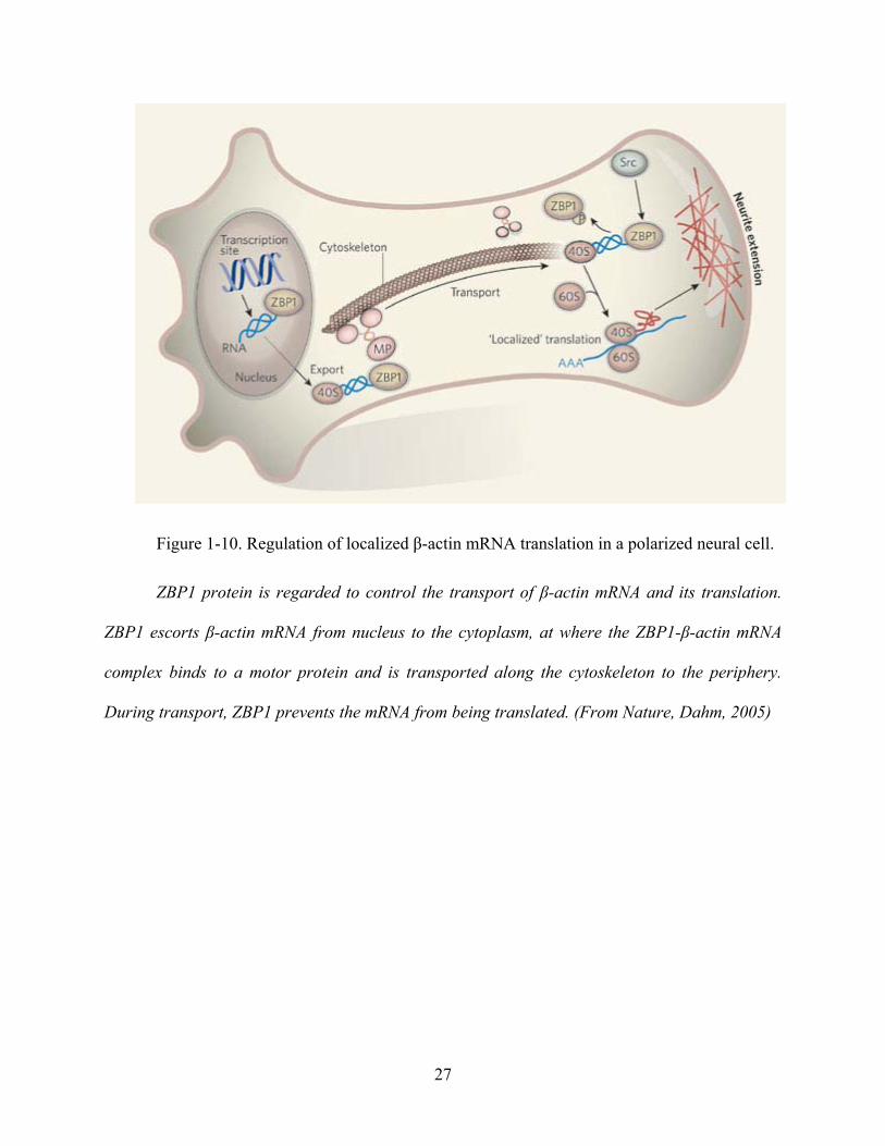
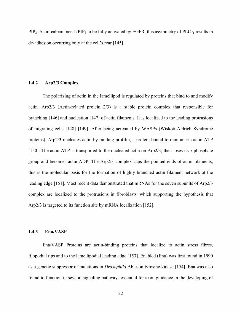
Figure 1-10. Regulation of localized β-actin mRNA translation in a polarized neural cell.
ZBP1 protein is regarded to control the transport of β-actin mRNA and its translation.
ZBP1 escorts β-actin mRNA from nucleus to the cytoplasm, at where the ZBP1-β-actin mRNA
complex binds to a motor protein and is transported along the cytoskeleton to the periphery.
During transport, ZBP1 prevents the mRNA from being translated. (From Nature, Dahm, 2005)
27

1.5 SIGNAL TRANSDUCERS AND ACTIVATORS OF TRANSCRIPTION
One of the major signal transduction pathways required for proliferation and migration
mediated by EGFR is the activation and translocation of STATs [185] [186]. De novo
transcription is required for proliferation, and also for migration [100]. It is still unknown
whether the transcriptional need in migration is for replacement of degraded proteins, increased
levels of the motility machinery components, or specific regulatory factors. Therefore, I decided
to ask whether this key transcriptional pathway was required in prostate cancer motility.
EGFR was found to directly activate STAT 1, STAT 3 and STAT 5. Our lab was among the
first to demonstrate that EGFR activates members of the STAT family [187]. Activation of ERK
MAP kinases and PI3-kinase are not sufficient for mitogenesis by growth factors, suggesting that
an additional pathway such as STAT is needed [188].
1.5.1 Background of STATs
STAT (Signal Transducer and Activator of Transcription) is a family of transcription factors
that are implicated in programming gene expression in biological events, including embryonic
development, programmed cell death, organogenesis, innate immunity, adaptive immunity and
cell growth regulation [189]. Seven mammalian STAT genes have been found now; they are
STAT1, 2, 3, 4, 5a, 5b and 6, which are structurally conserved (Fig 1-12). All the seven members
of the STAT family share several conserved domains. Those domains including an
amino-terminal domain (interacts with the transcriptional co-activator and regulates nuclear
translocation); a coiled-coil domain (forming predominantly hydrophilic surface, receptor
binding, tyrosine phosphorylation and nuclear export); the DNA binding domain; a linker
28

domain; an SH2 domain (binds to specific phospho-tyrosine motifs); a tyrosine activation
domain (circled P) and the transcriptional activation domain. The transcriptional activation
domain (TAD) is conserved in function but not in sequence [190].
29

Figure 1-11. STAT structure and family of proteins.
The domain structure of the seven STAT family members is shown. The N-terminal
domain mediates the interaction between two STAT dimers to form a tetramer. The coiled-coil
domain is involved in interactions with regulatory proteins and other transcription factors. The
DNA binding domain makes direct contact with STAT-binding sites in gene promoters with
consensus core sequence. Reciprocal interaction of SH2 domain of one STAT monomer and the
phosphotyrosine of another mediate dimer formation. (Nature Reviews of Cancer, Jove 2004)
30

1.5.2 Signalling Pathways that activate STATs
The Janus kinase-signal transducer and activator of transcription (JAK-STAT) pathway
transmits information received from extracellular polypeptide growth factors, through
transmembrane receptors, directly to target gene promoters in the nucleus, providing a
mechanism for transcriptional regulation without second messengers [191]. The STAT signaling
pathways were originally found in the cascade of normal cytokine receptors like interferon (IFN)
and interleukin-6 (IL-6). Unlike growth factor receptors, most of the cytokine receptors are
devoid of intrinsic tyrosine kinase activity. Instead, ligand engagement leads to activation of
Janus kinase (JAK) family members, a group of receptor associated tyrosine kinases [192]. In
some cases, SRC family kinases also involved [193]. Those kinases phosphorylate specific
tyrosine residues on the cytoplasmic tails of cytokine receptors, thus providing docking sites for
STAT monomer’s SH2 (SRC Homology 2) domain. The recruited STAT monomers are
phosphorylated on their specific tyrosine residues by the JAK or SRC kinases and form activated
dimers through reciprocal phosphotyrosine-SH2 interactions between two monomers [192].
STATs can be activated by growth factor receptors with intrinsic tyrosine kinase activity.
Those receptors include EGFR and PDGFR (platelet derived growth factor receptor) [194] [195].
These activate STAT directly without the intervention of JAK kinases [196]. However, the
intrinsic tyrosine kinases also may cooperate with JAK kinases in the phosphorylation of STATs
[197]. There is also non-receptor, cytoplasmic tyrosine kinases that signal through STATs, which
include ABL (Abelson Leukaemia Protein) and SRC related kinases (Fig 1-13) [198].
31

Figure 1-12. Signalling pathways of STATs.
Binding of growth factors or cytokines to their receptors results in the activation of
intracellular receptor tyrosine kinase activity or receptor associated kinases such as JAK or SRC.
These activated tyrosine kinases phosphorylate the receptor tails that in the cytoplasm, which
provide a docking sites for the mono-STAT protein. Non-receptor tyrosine kinases, such as ABL,
can phosphorylate STATs withorht receptor engagement. After been recruited, STAT monomers
phosphorylate each other on special tyrosine residues and form activated dimers. The dimers
translocate to the nucleus and directly regulate gene expression. (From Nature Reviews of
Cancer, Jove, 2004 and modified)
32

1.5.3 STAT3 and Cancer
In many cancer cells, both cytokine and growth factor receptors become constitutively
active due to autocrine or paracrine expression of their own ligands, mutations, failure of
degradation or simply over expression of themselves. These aberrant changes can all cause the
constitutive activation of STATs.
STAT3 activation is implicated in tumor invasion in head and neck squamous cell and
other carcinomas [185] [186]. A significant correlation has been reported between the expression
of nuclear STAT3 and breast cancer as compared to normal mammary tissues [199].
Interestingly, prostate tumor cells have been found to contain constitutively activated STAT3,
and blockade of this activated STAT3 significantly suppressing the tumor cell growth [200] [201]
[202]. A first clue implicating STATs in oncogenesis was the finding that STAT3 is
constitutively activated in SRC transformed cell lines and interrupting STAT3 signaling blocks
the transformation [203] [204] [205]. This is of key interest as a later study demonstrated that
constitutively activated STAT3 mediate cellular transformation [206]. A survey of
organ-confined prostate biopsies demonstrated a correlation between local aggressiveness and
phospho-STAT3 staining [207]. These reports support hypothesizing that STAT3 signaling
contributes to carcinogenic progression, in addition to increasing the cell number.
Constitutively activated STAT3 has been detected at high frequency in many human
cancers (Table 2). Various signaling molecules and their receptors like IL-6, TGF-α, EGF and
HGF (Hepatocyte Growth Factor) are involved in this process. Besides being a crucial part for a
variety of oncogenic signaling pathway, STAT3 also participates in tumor cell growth, survival,
angiogenesis and immune evasion. The first direct evidence that inhibition of STAT3 signaling
in human cancer cells apoptosis came from the finding that increased expression of BCL-XL, an
33

anti-apoptotic BCL-2 family gene, is dependent on constitutively activated STAT3 [208]. More
research data demonstrated that activated STAT3 increases the expression of c-Myc and cyclin
D1, which are both critical to cell proliferation and suppress the expression of TP53 gene, the
inducer of apoptosis [206] [209]. The same group also found STAT3 is a direct transcriptional
activator of the VEGF (Vascular Endothelial Growth Factor) gene. Blocking STAT3 signaling
inhibits the SRC and IL-6 induced VEGF up regulation [210]. Furthermore, inhibition of STAT3
in tumor cells was found to lead the production of inflammatory cytokines and chemokines,
which in turn activate immune cells [211].
34

TUMOR TYPE
SOLID TUMORS
ACTIVATED STAT
PROTEIN
REFERENCE
Breast cancer STAT1, 3, 5 [212]
Head and neck cancer STAT1, 3, 5 [186]
Melanoma STAT3 [213]
Lung cancer STAT3, 5 [214]
Ovarian cancer STAT3 [215]
Pancreatic cancer STAT3 [216]
Prostate cancer STAT3, 5 [217]
BLOOD TUMORS
Multiple myeloma STAT1, 3 [218]
HTLV-1-dependent leukemia STAT3, 5 [219]
Acute myelogenous leukemia STAT1, 3, 5 [220]
Chronic myelogenous leukemia STAT5 [221]
Large-granular-lymphocyte leukemia STAT3 [222]
Acute lymphoblastic leukemia STAT5 [223]
LYMPHOMA
EBV-related and Burkitt's lymphoma STAT3 [224]
Cutaneous T-cell lymphoma STAT3 [225]
B-cell non-Hodgkin's lymphoma STAT6 [226]
Anaplastic large-cell lymphoma STAT3 [227]
Table 2. Activation of STATs in human cancers.
1.6 CASPASES AND APOPTOSIS
Apoptosis is a programmed form of cell death, which is regulated in an orderly way by signal
pathways under certain situations. Apoptosis is critical to cell growth regulation, development,
35

immune response as well as keeping a constant amount of cells for organs. A hallmark of cancers
is the imbalance between cell proliferation and death, mainly by apoptosis, resulting in excessive
cell number. For the slow growing prostate cancer, there is often more a deficit of apoptosis
rather than rapid proliferation.
1.6.1 Apoptosis
So far, there are two main apoptosis pathways have been described: the death receptor
induced pathway and the mitochondrial mediated pathway, or called extrinsic and intrinsic
pathway according to the location of initial signaling [90]. Both of these pathways activate the
executioner intracellular proteases, caspases.
Generally, there are two pathways for caspase proteins to be activated: one is the death signal
induced and the other is mitochondrion mediated. In the first pathway, death signals as TNF
(tumor necrosis factor) or FasL can be specifically recognized by their receptors and activate
those receptors. Activated death receptors will interact with pro-caspases and induce their
activation through oligomerization and auto-cleavage, leading to apoptosis [228]. On the other
hand, procaspase can also be activated through a cytochrome C dependent pathway. After
cytochrome C is released from mitochondria under cellular stress like DNA damage, it can
directly activate some type of procaspase or form a complex called the apoptosome by
oligomerizing Apaf-1 (apoptotic protease activation factor-1), then activate caspases [229] [230].
There are many regulators of mitochondria mediated apoptosis pathway, including BCL-2 and
BCL-XL, which negative to apoptosis; and BAX and BAD, which is pro-apoptosis (Fig 1-14).
36

Figure 1-13. Schematic representing the core components of apoptosis pathways.
In the extrinsic pathway, TNF super family members including Fas Ligands binding to a
death receptor and forming a death inducing signalling complex (DISC), which activate
caspase-8. In the intrinsic pathway, cytochrome c released from mitochondria causes
apoptosome formation and caspase-9 activation. Both caspase-8 and caspase-9 activate down
stream caspases like caspase-3 and leading to apoptosis. (From Patric, 2006 and modified)
37

1.6.2 Caspase proteins
The initiation and execution of apoptosis requires a group of proteins that include signal
transducers, receptors, gene regulators and enzymes. Among them, the caspase proteins are vital
to the process of apoptosis [231].
Fourteen members of caspase family have been identified up to now, and all share some
common properties. Caspases are aspartate-specific cysteine proteases and they all come from a
zymogen precursor. Based on their function and homology in amino acid sequences, caspases are
divided into three groups: apoptosis activators like caspase 2, 8, 9, 10 that contain a long
prodomain at the N-terminus; apoptosis executioners such as caspase 3, 6, 7 that have a short
prodomain and inflammatory mediators with caspase 1, 4, 5, 11, 12, 13 and 14 (Table 3).
Among all the apoptosis-related caspase proteins, Caspase 3 acts at the effector’s stage of the
whole signal cascade. It cleaves various of targets like PARP (poly ADP-ribose polymerase) and
DFF (DNA fragmentation factor) that lead to cell death as well as cleaving Caspase 6, Caspase 7
and caspase 9 [232]. Also known as CPP32, Yama or apopain, Caspase 3 is a key factor in
apoptosis execution. It is activated by cleavage from procaspase 3. It was found that a small
molecule antagonists of XIAP (X-chromosome-linked inhibitor of apoptosis protein) that
overcome inhibition of caspase-3 directly induced cell death in tumor cells while having little
toxicity on normal cells, which indicates a critical role of caspase-3 in cancer cell apoptosis
[233].
38

Subfamily Role Members
I Apoptosis activator Caspase-2
Caspase-8
Caspase-9
Caspase-10
II Apoptosis executioner Caspase-3
Caspase-6
Caspase-7
III Inflammatory mediator Caspase-1
Caspase-4
Caspase-5
Caspase-11
Caspase-12
Caspase-13
Caspase-14
Table 3. Subfamily of caspase proteins.
39

2.0 MATERIAL AND METHODS
2.1 CELL LINES & MEDIUM
A murine fibroblast line was selected to define the transcriptional changes concomitant
with EGFR-mediated cell motility. This was chosen as such fibroblast lines avoid the autocrine
signaling through EGFR that is prevalent in carcinoma cells including those of the prostate [234].
The line chosen is a well characterized subline of NR6 cells that now expresses physiological
levels of the EGFR [235]. These are derived from murine Balb/c 3T3 cells that were selected to
not respond to EGF; these cells had silenced their EGFR transcription [236]. EGFR was
expressed upon MLV-based retroviral transduction of a single gene copy with the selectable
marker, neomycin phosphotransferase, driven from the env position using a separate promoter.
Use of these cells allowed for isolation of EGFR signaling by comparison to the parental NR6
cells.
I chose to use two human prostate carcinoma cell lines to explore the role of the
STAT3-driven transcription changes in tumor progression and survival. Androgen-independent
cell lines were used as these represent the state at which prostate carcinoma is refractory to
standard hormonal therapy [237] [238] and the situation that engenders the major part of prostate
cancer mortality [239]. DU145 cells were selected as their in vitro and in vivo invasive behavior
is well characterized [26] [27]. These were derived from a prostate adenocarcinoma metastasis to
the brain [238]. These cells are only moderately invasive and have limited dissemination from
orthotropic (intra-prostate) inoculation in mice. These cells were made more invasive and
metastatic by over expression of full length human EGFR [26] [27]. DU145 present a mutant
androgen receptor that is independent of exogenous testosterone [240]. The second cell line,
40

PC-3, was isolated from a prostate adenocarcinoma metastasis to the bone [237]. This line is
considered highly aggressive and metastatic when inoculated in mice.
Human DU145 prostate carcinoma cells [238] over-expressing EGFR, DU145WT [26],
were maintained in Dulbecco’s minimal essential media (DMEM) (Mediatech) supplemented
with 10% fetal bovine sera (FBS), L-glutamine (2mM), non-essential amino acids (0.1mM),
sodium pyruvate (1mM) and 100U/ml of penicillin. To maintain expression from the transduced
EGFR in the DU145WT cells, 350 mg/ml of G418 was added to the media. Human PC3 prostate
cancer cells [237] were maintained in F-12 medium (GIBCO) supplemented with 10% FBS,
L-glutamine (2mM), non-essential amino acids (0.1mM), and sodium pyruvate (1mM). NR6WT
mouse fibroblasts expressing human EGFR [235] were maintained in Eagle’s minimal essential
medium alpha modification (MEMα) (Mediatech) supplemented with 10% FBS, L-glutamine
(2mM), non-essential amino acids (0.1mM), sodium pyruvate (1mM), 100 U/ml of penicillin and
350 mg/ml of G418. All the cells were grown in 5% CO2 at 37˚C.
2.2 RNA PURIFICATION
To purify the RNA for microarray analyses, the treated cells were washed by ice cold 1 X
PBS twice. 1 ml of Trizol was added into each 10 cm plate and incubated at room temperature
for 5 minutes. After incubation, the cells were scraped and transferred into a 1.5 ml centrifugal
tube. Chloroform equal to 20% of the original volume of cells was added into the tube and shake
vigorously by hand for 15 seconds. The mixture was incubated at room temperature for 2
minutes before going centrifuge at 12,000 rpm for 15 minutes. The upper aqueous after
centrifuge was transferred into a fresh tube and 75% of the volume of isopropyl alcohol was
added. The mixture was incubated at room temperature for at least 10 minutes before going
41

centrifuge again at 12,000 rpm for 15 minutes. The pellets were washed with 70% and then
100% ethanol, ended with air dry for 10 minutes [241].
2.3 MICROARRAY ANALYSES
To determine the genes transcribed downstream from EGFR/STAT3 signaling pathway,
we performed transcription microarray analyses to monitor the gene expression changes in RNA
levels. The experimental design was to determine which transcripts were both up after EGF
exposure and then reverted to untreated levels when the cells were treated with STAT3 antisense
oligonucleotides prior to EGF treatment. I chose the NR6WT fibroblast cell, which expresses
physiological levels of the EGFR [235]. Use of these cells allowed for isolation of EGFR
signaling from the confounding influence of growth factors and their receptors’ autocrine loops
in human prostate tumor cell lines.
The NR6WT cells were starved in MEMα media containing 0.5% FBS for 24 hours to
quiescent the cells before treatment with 1nM of EGF and/or 10µM of mouse STAT3 antisense
nucleotide for another 24 hours. The sequence of it is 5’-GTT CCA CTG AGC CAT CCT GC-3’.
After the treatment, the RNA from those cells was purified with the protocol described in above.
5μg RNA from each sample was prepared for microarray analysis using the Affymetrix U74AV2
murine genome array chip that contains probe sets interrogating approximately 36,000
full-length mouse genes and EST clusters from the UniGene database of transcripts. 5 μg of
purified total RNA were added into a 20 μl first strand reaction with 200 U of SuperScript II
(Invitrogen) and 1 µg T7 primer [5’-GGCCAGTGAATTGTAATACGACTCACTATAGGGAG
GCGG(T)24] in 1X first strand buffer (Invitrogen) followed by a 42°C incubation for 1 hour.
Second strand synthesis was performed by adding 40 U of E. coli DNA polymerase, 2 U of E.
42

coli RNase H, and 10 U of E. coli DNA Ligase in 1 X second strand buffer followed by
incubation at 16°C for 2 hrs. The second strand synthesis reaction was purified according to the
manufacturer’s protocol (Affymetrix). The purified cDNA was amplified according to
manufacturer’s protocol to produce 70-120 μg of biotin labeled cRNA [242].
Murine U74Av2 GeneChip probe arrays were pre-hybridized in a GeneChip
Hybridization Oven 640 (Affymetrix) according to the manufacturer’s protocol. 15 μg of labeled
cRNA were fragmented in 30 μl 1X fragmentation buffer with 100 mM KOAc and 30 mM
MgOAc at 95°C for 35 minutes. The fragmented labeled cRNA was resuspended in 300 μl 1X
hybridization buffer containing 100 mM MES, 1 M [Na+], 20 mM EDTA, 0.01% Tween 20, 0.5
mg/mL Aceylated BSA, 0.1 mg/ml herring sperm DNA, control oligonucleotide B2, and control
transcripts bioB 1.5 pM, bioC 5 pM, bioD 25 pM, and cre 100 pM. 200 μl of the hybridization
cocktail (containing 10 μg of labeled cRNA) were hybridized to GeneChip probe arrays
according to manufacturer’s protocol (Affymetrix).
The raw data from microarray were normalized with the Affymetrix Microarray suite 5.0,
based on the housekeeping gene expression profile. Expression values were adjusted to the
intensity of the expression value of the 100 housekeeping genes. This allowed us to normalize
between runs.
2.4 MOLECULAR CLONING
To check if STAT3 effects alone are sufficient for EGFR mediated cell migration, I used a
constitutively activated STAT3 mutant to transfect the NR6WT cells. A dominant negative
mutant of STAT3 and empty vector were used as controls. In Stat3D, E434 and E435 of Stat3,
43

which is in the DNA binding domain, were replaced with alanines [243]. This construct
competes for upstream activation signals but cannot promote transcription [244] [245]. The
constitutively active mutant [206] of STAT3 was generated by a mutation of substituting
cysteine residues for A661 and N663 of the Stat3 molecule, which allowed for sulfhydryl bonds
to form between STAT3 monomers and render a STAT3 that dimerizes independent of upstream
activation [206]. Both constructs were kindly provided to me by Dr. Jennifer Grandis. The
mutant fragments were cut by Not I and Hind III endonucleases and inserted into pCEP4-MMTV
expression plasmid that induced by dexamethasone and has hygromycin resistance [246]. The
cloning was checked by DNA sequencing and immunoblot for the expression marker of HA-tag
in dominant negative STAT3 and Flag in constitutively active STAT3 mutants.
2.5 ELECTROPORATION AND SELECTION OF THE CLONS
The constructed plasmid with STAT3 mutants were transfected into NR6WT cells by
electroporation. Cells grown in a 10cm plate at around 50% ~ 60% confluence were trypsinized
and the pellet was collected in a 15 ml conical tube by centrifugation at 1,000 rpm for 5 minutes
at 4˚C. One milliliter of OptiMEM was added into the tube to resuspend the pellet. The
resuspended cells were mixed with 40 µg of plasmid DNA and transferred into a 0.4 cm cuvette.
(Bio-Rad, Hercules, CA) The electroporation was performed with a voltage 220 V and 950 µF of
capacitance.
After electroporation, cells were transferred to a new cell culture plate with 11 ml fresh
medium. Medium was changed after 24 hours to get rid of the dead cells and debris. Both the
STAT3 constitutively active mutant with Flag-tag and the STAT3 dominant negative mutant
44

with HA-tag were selected by hygromycin resistance. 100 µg/ml of hygromycin was added into
the medium, and multiple monoclones were picked up after at least one weeks culturing.
The expression of both STAT3 constitutively active and STAT3 dominant negative mutants
were controlled by 2 µg/ml of dexamethasone. Expression was determined by immunoblotting
for STAT3 and the tags after 24 hr exposure to dexamethasone. Expressing clones were tested.
2.6 RNA INTERFERENCE
RNA interference (RNAi) is a process in which double-stranded RNA triggers the
degradation of a homologous mRNA [247]. RNA interference is a technique that introduces
exogenous, double-stranded RNAs (dsRNAs) which are complimentary to known mRNA's into a
cell to specifically destroy that particular mRNA, thereby diminishing or abolishing gene
expression and translation. I use the siRNAs specifically against human and mouse STAT3 to
block the expression of the STAT3 gene in the target cell lines, which as a double confirmation
of STAT3 antisense oligonucleotides. The siRNAs of human and mouse STAT3 and control
siRNA were designed and ordered from the website of IDT. (http://www.idtdna.com) The
sequence for human STAT3 siRNA is 5’-AUCCUGAAGG UGCUGCUCCTT-3’; the sequence
for mouse STAT3 siRNA is 5’-UGCAUUCUCCUU GGCUCUUGAGGGUU-3’ and the
sequence for control eGFP siRNA is 5’-GACCCGCG CCGAGGUGAAGTT-3’. Cells for RNA
interference experiment were grown in an antibiotic-free medium before transfection. For a
6-well plate, 4 μl of siRNA (20 μM) was incubated with 100 μl OptiMEM (GIBCO) per well
(2ml medium) for 5 minutes, at the same time mix 4 μl of Lipofectamine 2000 (Invitrogen) with
100μl OptiMEM, then mix the siRNA and Lipofectamine dilution and incubate at room
45

temperature for 20 minutes before adding into the culturing cells. Incubate the cells at 37˚C for at
least 4 hours, change fresh antibiotic-free medium and let the cells recover for another 12 to 24
hours [246].
2.7 NUCLEAR EXTRACT PREPARATION AND EMSA
Electrophoretic mobility shift assay (EMSA) is used for studying the binding between
protein and specific DNA sequences. It can separate different types of complexes, such as
monomer and dimer. These complexes can be recognized by transcription factor-specific
antibodies that retard their mobility in gels for further identification.
For STAT3 analysis by EMSA [248], confluent cells were harvested and washed with ice
cold 1X PBS, with the pellets resuspended in 5-fold buffer A (10 mM HEPES, pH 7.9; 1.5 mM
MgCl2; 10 mM KCl; 1 mM NaF; 0.5 mM DTT; 0.2 mM PMSF; 1 µg/µl profilin; 5 µg/µl
aprotinin; 2 µg/µl leupeptin). After incubating on ice for 15 minutes, the nuclei were centrifuged
at 4℃ for 10 seconds to obtain the nuclear-localized active transcription factors. Pellets were
resuspended in 5-fold volumes (20μl ~ 100μl) cold buffer C (20 mM HEPES, pH 7.9; 1.5 mM
MgCl2; 420 mM NaCl; 10 mM NaF; 1 mM Na3VO4; 25% glycerol; 0.2 mM EDTA; 0.5 mM
DTT; 0.2 mM PMSF; 1 µg/µl profilin; 5 µg/µl aprotinin; 2 µg/µl leupeptin), incubated on ice for
30 minutes and microcentrifuged for 2 minutes at the maximum speed. Aliquots were stored at
-80℃. For EMSA, 10 µg of total extractions were used for each experimental point. EMSA was
performed using a γ-32P labeled double strand oligonucleotide probe m67 (sense: 5’-GAT TTC
CCG TAA ATC AT-3’) that binds STAT3 and STAT1 proteins [249]. Protein-DNA complexes
were resolved by non-denaturing PAGE gel and detected by autoradiography. Anti-STAT3
46

antibodies (Upstate Biotechnology, Waltham, MA) were used for super shift assay and a 50-fold
excess of unlabeled m67 probe was used in cold competition assay.
2.8 IMMUNOBLOT
Protein expression was determined by immunoblotting. Confluent cells were washed with
ice cold 1 X PBS twice followed by treated with lyses buffer (100mM Tris-HCl, pH 6.8; 4%
SDS; 20% glycerol and 5% B-mercapto-ethanol). Sample proteins were denatured at 100℃ for 5
minutes in water bath before loading to the gel. After electrophoresis, the proteins were
transferred to an Immobilon-P membrane. Membranes were blocked by 1% BSA. After blocking,
membranes were incubated with a primary antibody: mouse anti STAT3 (Zymed); mouse anti
GAPDH (abCam); mouse anti Flag (Stratagene); rabbit anti phosphor STAT3 (Cell Signaling)
and rabbit anti HA-tag (Cell Signaling) at 4℃ for over night or at room temperature for 1 hour.
The membranes were washed twice with 0.5 % Tween-20 (TBST) before incubated with
secondary antibodies (Goat anti-mouse Ig or Goat anti-rabbit Ig, Biosource) [250].
2.9 WOUND HEALING ASSAY
Wound healing assay was used in vitro to assess cell motility in two dimensions. I used
this assay to study the migration of both fibroblasts and human prostate tumor cells under the
treatment of EGF and/or STAT3 inhibitors. Cells were plated on a 12-well plate and grown to
confluence in their regular medium. Confluent cells were quiesced in 1% dialyzed FBS for 24
hours before each experiment. A rubber policeman was used to create a denuded area. Cells were
washed twice with PBS and treated with or without specific effectors for 24 hours. Photographs
47

were taken at hour 0 and hour 24, and the distance traveled was determined by subtracting the
values obtained at hour 0 from hour 24. Mitomycin C (0.5 µg/ml) was added to the medium to
prevent the confounding issue of cell proliferation [251].
2.10 INVASION ASSAY
Invasive potential was determined in vitro by transmigration of an ECM [252]. Matrigel
invasion chamber plates were purchased from Becton Dickinson/Biocoat (Bedford, MA). The
Matrigel was used to mimic the environmental extracellular matrix. The upper surface of the
matrix was challenged with 1.5 X 10⁴ cells, a number derived from empirical experimentation.
Cells were kept in serum-free medium containing 1% BSA for the first 24 h and then replaced
with only serum-free medium for the remaining 24 h; the lower chamber contained medium
containing 10% serum for the entire assay. Enumeration of the cells that invaded through the
matrix over a 48-h period was accomplished by visually counting cells on the bottom of the filter,
as per routine procedures, after any un-invaded cells were removed from the top of the filter with
a cotton swab. In all of the cases, individual experiments were performed in duplicate chambers.
2.11 ANNEXIN V STAINING FOR APOPTOSIS ASSAY
The Annexin V staining is based on the observation that soon after apoptosis initiation,
the membrane phosphatidyl-serine was translocated from the inner face of the plasma membrane
to be exposed on the cell surface. Once on the cell surface, the phosphatidyl-serine can easily be
detected by staining with a fluorescent conjugate of Annexin V, which is a protein that has high
affinity for it. Annexin V-Cy3 kit (BioVision) was used in the apoptosis assay. Cells were
48

washed with 1 X PBS and then digested by Tripsin-EDTA. The pellets of cells were washed
again by 1 X PBS twice and re-suspended in 500µl of 1 X Binding Buffer come with the kit.
After incubation at room temperature for 5 minutes, Annexin V was added into the cells at a ratio
of 1:100. The mixture was incubated in darkness for 10 minutes at room temperature and ready
for counting fraction of Annexin V-staining cells under a fluorescence microscope [253].
2.12 PROLIFERATION ASSAY
To study the viability and the toxicity of STAT3 inhibitors to the cell, the cells proliferation
assay was performed by direct cell enumeration. A Z-series Coulter Counter (Coulter Corp) was
used for the counting. Cells were counted by the Coulter Counter before being passed into new
culture plates, after the treatment and growth for 48 or 72 hours to allow for one or two rounds of
replication, cells were digested by Tripsin-EDTA (GIBCO) and then re-suspended in the original
medium. The cells were diluted for 100 times in Isoton Ⅱsolution (Beckman) before counting
[250].
2.13 STATISTICAL ANALYSES
All data from migration, proliferation, invasion and apoptosis staining were analyzed and
expressed as mean ± S.E.M. Evaluation of statistical significance was performed by use of T-test.
A value of P < 0.05 was considered significant.
49

3.0 STAT3 IS REQUIRED BUT NOT SUFFICIENT FOR EGFR MEDIATED
MIGRATION AND INVASION OF FIBROBLAST AND HUMAN PROSTATE
CARCINOMA CELLS
3.1 STAT3 ANTISENSE DECREASES EXPRESSION OF STAT3 IN NR6WT CELLS
EGFR signaling activates STAT proteins in fibroblasts [187] and a variety of carcinoma
cells [254] [255]. As our lab has previously shown, the prostate carcinoma cells present autocrine
EGFR signaling. I determined whether this also invoked STAT3 activation. To study this issue, I
used the motility model system, murine NR6WT fibroblasts, to examine EGF-triggered events as
autocrine signaling is absent in these cells in contrast to prostate carcinoma cells which present
autocrine EGFR-activating signaling loops [92], and thus the role of EGFR signaling can be
cleanly parsed. The two human prostate cancer cell lines, DU145WT and PC3, present autocrine
activation of endogenous and exogenous EGFR, as is the norm for prostate carcinoma cells.
However, to study the function of EGFR signaling in these cells, I tested our prostate cells under
conditions that minimize autocrine activation [187].
Initially to parse STAT3 mediated responses from EGFR even in the fibroblasts, I
downregulated the protein expression by using antisense oligonucleotides. This intervention
reduced whole cell levels of STAT3 significantly in NR6WT fibroblasts (Fig 3-1).
50
NoTx EGF EGF/STAT3AS EGF/Ctrl
Figure 3-1. EGF increase the expression of STAT3 in NR6WT cells and it can be inhibited
by STAT3 antisense.
In murine fibroblasts, NR6WT cells, EGF exposure increased the expression of STAT3 at
protein level, and this increase was inhibited by STAT3 antisense. Cells were quiesced in 0.5%
FBS medium for 24 hours before the treatment. After quiescing, the NR6WT cells were treated
with 1 nM EGF and/or 10 μM of oligonucleotides (STAT3 antisense or non-specific control) for
over night. NoTx: non-treatment control; EGF: 1 nM human EGF; EGF/STAT3AS: 1 nM human
EGF plus 10 μM mouse STAT3 antisense oligonucleotides; EGF/Ctrl: 1 nM human EGF plus 10
μM non-specific oligonucleotides. The treated cells were lysised in 2X loading buffer and
denatured at 100℃ in water bath for 5 minutes. Lysates were loaded to 10% acrylamide gel,
transferred to an Immobilon-P membrane and immunoblotted with antibody against mouse
STAT3. After being exposed, the membrane was stripped and immunoblotted with antibody
against GAPDH as a loading control.
51
3.2 STAT3 ANTISENSE DECREASES DNA BINDING ACTIVITY OF STAT3 IN
NR6WT CELLS
The signaling pathway of STAT3 is a complicated process, cytoplasm STAT3 must be
dimerized to get activated, and the performing of its transcriptional regulation function need the
binding of STAT3 to its target DNA sequence. I used the EMSA assay to check the activity of
STAT3 after STAT3 antisense treatment. A valuable aspect of EMSA is that is can separate
different types of complexes, such as monomer and dimer, and it is easier to see the bound
complex. In murine NR6WT fibroblasts cells, EGF exposure increased EMSA detection of the
STAT3 band, which was negated upon addition of an antibody to mouse STAT3 (Fig 3-2). A
competing binding of 50X concentration of cold nucleotide probe completely eliminated the
band, showing the specificity of the binding of probe DNA and STAT3.
52
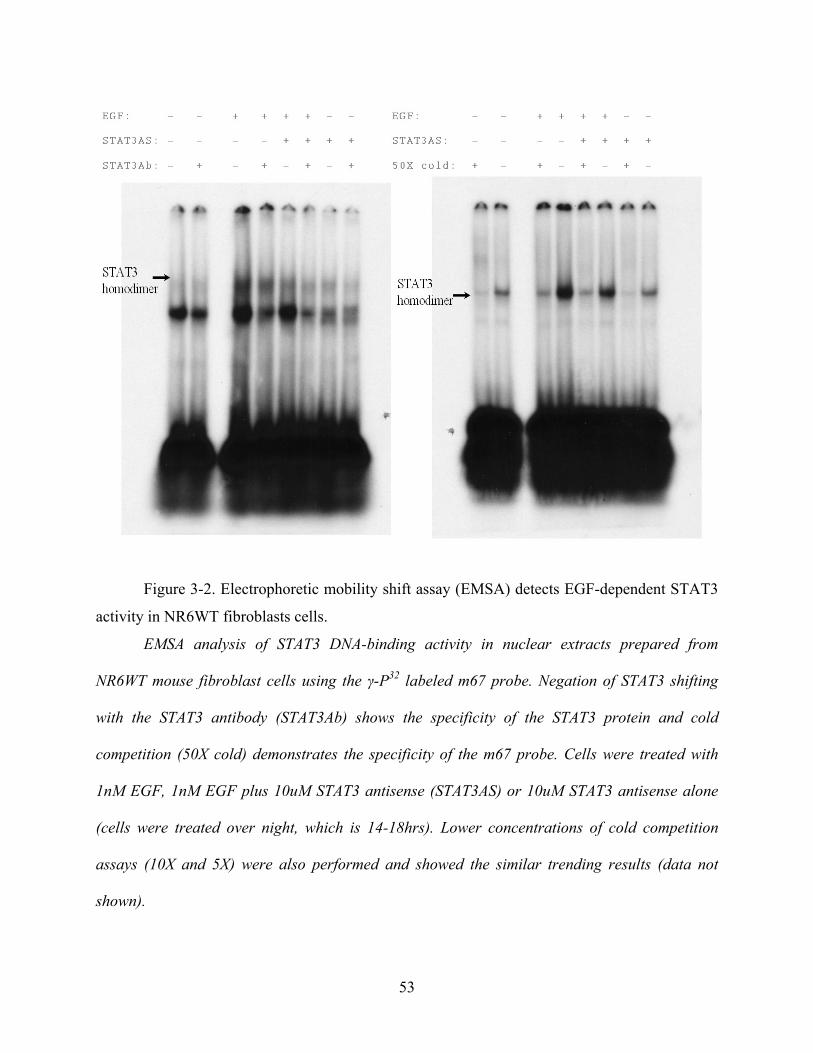
Figure 3-2. Electrophoretic mobility shift assay (EMSA) detects EGF-dependent STAT3
activity in NR6WT fibroblasts cells.
EMSA analysis of STAT3 DNA-binding activity in nuclear extracts prepared from
NR6WT mouse fibroblast cells using the γ-P32 labeled m67 probe. Negation of STAT3 shifting
with the STAT3 antibody (STAT3Ab) shows the specificity of the STAT3 protein and cold
competition (50X cold) demonstrates the specificity of the m67 probe. Cells were treated with
1nM EGF, 1nM EGF plus 10uM STAT3 antisense (STAT3AS) or 10uM STAT3 antisense alone
(cells were treated over night, which is 14-18hrs). Lower concentrations of cold competition
assays (10X and 5X) were also performed and showed the similar trending results (data not
shown).
53

3.3 STAT3 ANTISENSE DECREASES EXPRESSION OF STAT3 IN DU145WT CELLS
Now that I could downregulate STAT3 activity in the NR6WT model cell, I changed my
focus to the human prostate tumor cells, DU145WT cell. The DU145WT cells have a
TGF-α/EGFR autocrine loop, which implies its independence or insensitivity of extraneous EGF
stimulation. To overcome that disadvantage, a significant higher concentration of EGF and
shorter treatment were applied on the DU145WT cells. The result showed that the STAT3
antisense oligonucleotide reduced STAT3 levels in the DU145WT cells while no significant
inhibition happened in the non-specific oligonucleotides control (Fig 3-3).
Figure 3-3. EGF increase the expression of STAT3 in DU145WT cells and it can be
inhibited by STAT3 antisense.
DU145WT cells were quiesced in 0.5% FBS medium for 24 hours before the treatment.
After quiescing, the DU145WT cells were treated with 10 μM of oligonucleotides (STAT3
antisense or non-specific control – S3AS or CtrlAS, respectively) for over night followed by
and/or 10 nM EGF for 15 minutes. NoTx: non-treatment control; EGF: 10 nM human EGF;
EGF/STAT3AS: 10 nM human EGF plus 10 μM human STAT3 antisense oligonucleotides;
EGF/Ctrl: 10 nM human EGF plus 10 μM non-specific oligonucleotides. The treated cells were
lysised in 2 X loading buffer and denatured at 100oC in water bath for 5 minutes. Lysates were
54

loaded to 10% acrylamide gel, transferred to an Immobilon-P membrane and immunoblotted
with antibody against mouse STAT3. After being exposed, the membrane was stripped and
immunoblotted with antibody against GAPDH as a loading control.
3.4 STAT3 ANTISENSE DECREASES DNA BINDING ACTIVITY OF STAT3 IN
DU145WT CELLS
To check the activity of STAT3 after STAT3 antisense treatment, we performed the
EMSA assay. In human prostate tumor cell, DU145WT cells, EGF exposure increased EMSA
detection of the STAT3 band, which was up-shifted upon addition of an antibody to murine
STAT3 (Fig 3-4). A competing binding of 50X concentration of cold nucleotide probe
eliminated the band, showing the specificity of the binding of probe DNA and STAT3.
55
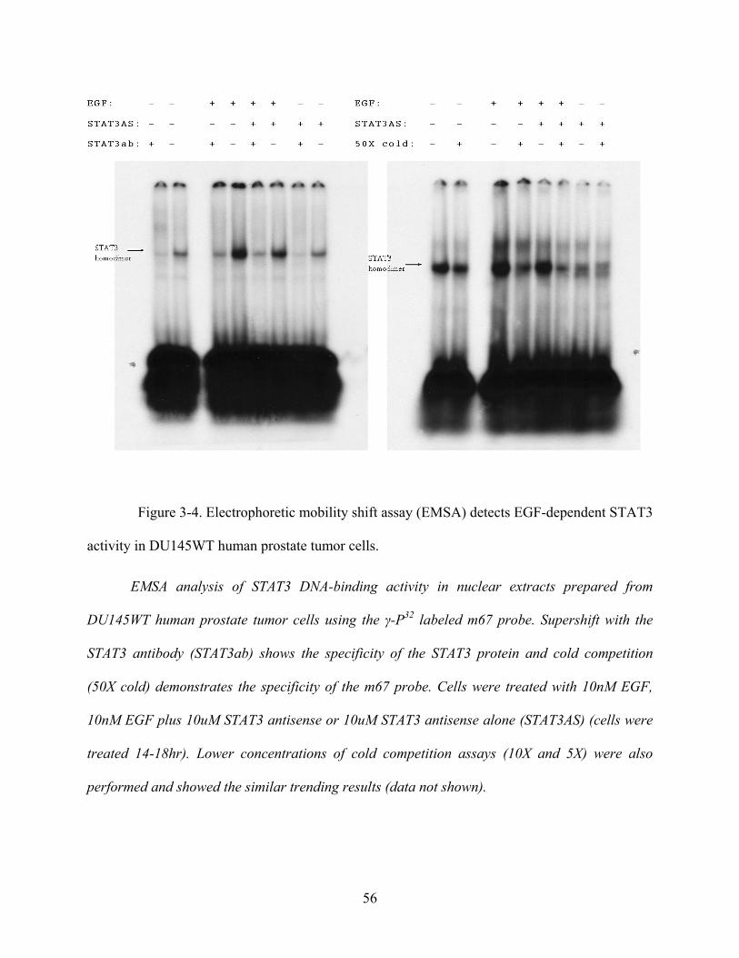
Figure 3-4. Electrophoretic mobility shift assay (EMSA) detects EGF-dependent STAT3
activity in DU145WT human prostate tumor cells.
EMSA analysis of STAT3 DNA-binding activity in nuclear extracts prepared from
DU145WT human prostate tumor cells using the γ-P32 labeled m67 probe. Supershift with the
STAT3 antibody (STAT3ab) shows the specificity of the STAT3 protein and cold competition
(50X cold) demonstrates the specificity of the m67 probe. Cells were treated with 10nM EGF,
10nM EGF plus 10uM STAT3 antisense or 10uM STAT3 antisense alone (STAT3AS) (cells were
treated 14-18hr). Lower concentrations of cold competition assays (10X and 5X) were also
performed and showed the similar trending results (data not shown).
56

3.5 STAT3 SIRNA DECREASES EXPRESSION OF STAT3 IN DU145WT CELLS
Thought the antisense interventions worked, I sought a second, confirmatory way to
downregulate STAT3 signaling. RNA interference (RNAi) is a process in which double-stranded
RNA triggers the degradation of a homologous mRNA [Bass, 2003]. Today, RNA interference is
a technique that introduces exogenous, double-stranded RNAs (dsRNAs) which are
complimentary to known mRNA's into a cell to specifically destroy that particular mRNA,
thereby diminishing or abolishing gene expression and translation., I used RNA interference to
further confirm the blocking effect got from STAT3 antisense nucleotide. I downregulated the
protein using siRNA directed against STAT3 in DU145WT cells (Fig 3-5).
NoTx EGF Si EGF/Si Ctrl EGF/Ctrl
Figure 3-5. EGF-mediated increase of STAT3 expression and activity is suppressed by
STAT3 siRNA in DU145WT cells.
EGF-mediated increase of STAT3 expression and activity is suppressed by STAT3 siRNA
(S3Si) in DU145WT cells, but not by a non-target siRNA (against eGFP)(CtrlSi). DU145WT
cells were grown in an antibiotic-free medium before being transfected by siRNA (20 μM) with
Lipofectamine 2000 to a final concentration of 100 pM. After been transfected and recovered,
DU145WT cells were quiesced in 0.5% FBS medium for 24 hours before the treatment. After
quiescing, the DU145WT cells were treated with 10 nM EGF for 15 minutes. NoTx:
57

non-treatment control; EGF: 10 nM human EGF; Si: 100 pM of STAT3 siRNA transfection; ESi:
10 nM human EGF plus 100 pM of human STAT3 siRNA; Ctrl: 100 pM of eGFP siRNA as
non-specific control; EGF/Ctrl: 10 nM human EGF plus 00 pM of eGFP siRNA. The treated
cells were lysised in 2 X loading buffer and denatured at 100℃ in water bath for 5 minutes.
Lysates were loaded to 10% acrylamide gel, transferred to an Immobilon-P membrane and
immunoblotted with antibody against human STAT3. After being exposed, the membrane was
stripped and immunoblotted with antibody against GAPDH as a loading control.
3.6 STAT3 SIRNA DECREASES EXPRESSION OF STAT3 IN PC3 CELLS
To inhibit the STAT3 protein expression in a second human prostate cell line, PC3, I
also used RNA interference. As with DU145WT cells, PC3 is a metastasis human prostate tumor
cell, the difference between them is that PC3 is separated from bone metastases [237], not from
brain as DU145 cells [238]. I also downregulated the STAT3 protein using siRNA directed
against STAT3 mRNA sequence in PC3 cells (Fig 3-6).
58

NoTx EGF Si EGF/Si Ctrl EGF/Ctrl
Figure 3-6. EGF-mediated increase of STAT3 expression and activity is suppressed by
STAT3 siRNA in PC3 cells.
EGF-mediated increase of STAT3 expression and activity is suppressed by STAT3 siRNA
(S3Si) in PC3 cells, but not by a nontarget siRNA (against eGFP)(CtrlSi). PC3 cells were grown
in an antibiotic-free medium before being transfected by siRNA (20 μM) with Lipofectamine
2000 to a final concentration of 100 pM. After been transfected and recovered, PC3 cells were
quiesced in 0.5% FBS medium for 24 hours before the treatment. After quiescing, the PC3 cells
were treated with 10 nM EGF for 15 minutes. NoTx: non-treatment control; EGF: 10 nM human
EGF; S3Si: 100 pM of STAT3 siRNA transfection; ES3Si: 10 nM human EGF plus 100 pM of
human STAT3 siRNA; CtrlSi: 100 pM of eGFP siRNA as non-specific control; EGF/CtrlSi: 10
nM human EGF plus 100 pM of eGFP siRNA. The treated cells were lysised in 2 X loading
buffer and denatured at 100oC in water bath for 5 minutes. Lysates were loaded to 10%
acrylamide gel, transferred to an Immobilon-P membrane and immunoblotted with antibody
against human STAT3. After being exposed, the membrane was stripped and immunoblotted with
antibody against GAPDH as a loading control.
59

3.7 STAT3 SIRNA DECREASES DNA BINDING ACTIVITY OF STAT3 IN DU145WT
CELLS
Inhibition of STAT3 protein expression of STAT3 siRNA was demonstrated by
immunoblotting. Now the next question is whether the DNA-binding activity of STAT3 protein
is also been suppressed by STAT3 siRNA. To get the answer, I also performed EMSA assay on
STAT3 siRNA transfected DU145WT cells. The result is as same as in the previous STAT3
antisense experiments. EGF significantly increases STAT3 binding activity while it can be
reversed by STAT3 siRNA (Fig 3-7).
NoTx EGF Si EGF/Si Ctrl EGF/Ctrl
STAT3
Figure 3-7. The STAT3 siRNA negated EGF induced STAT3 activity in DU145WT prostate
tumor cells.
EMSA analysis of STAT3 DNA-binding activity in nuclear extracts prepared from DU145WT
human prostate tumor cells using the γ-P32 labeled m67 probe. Cells were treated with 10nM
EGF, 10nM EGF plus 100 pM STAT3 siRNA or 100 pM STAT3 antisense alone. A non-specific
siRNA (eGFP siRNA) was used as control. NoTx: non-treatment control; EGF: 10 nM human
EGF; S3Si: 100 pM of STAT3 siRNA transfection; ES3Si: 10 nM human EGF plus 100 pM of
human STAT3 siRNA; CtrlSi: 100 pM of eGFP siRNA as non-specific control; EGF/CtrlSi: 10
nM human EGF plus 100 pM of eGFP siRNA.
60

3.8 STAT3 ANTISENSE INHIBITS EGF INDUCED MIGRATION IN NR6WT CELLS
Now that I could reproducibly limit STAT3 functioning, I asked what this did to
EGFR-mediated cell migration. EGFR signaling drives motility in fibroblasts and carcinoma
cells [256] and invasion of human prostate carcinoma cells [187] [257]. As such, we asked
whether STAT3 might be critical for sustained motility and invasion in response to EGFR
signaling. To quantitate cell motility, an in vitro wound healing assay was performed and
demonstrated that the addition of STAT3 antisense oligonucleotides and the EGFR signaling
pathway inhibitor PD153035 greatly decreased the migration distance of NR6WT fibroblast cell.
The extent of migration inhibition was similar to that achieved by blocking EGFR kinase activity
with the selective agent PD153035. Interestingly, the level of migration achieved with STAT3
down regulation, and with PD153035 was below that of basal motility, strongly suggesting that
either EGFR autocrine signaling via STAT3 or basal STAT3 was contributing to this cell
behavior, as has been suggested earlier for EGFR autocrine activation [187].
61
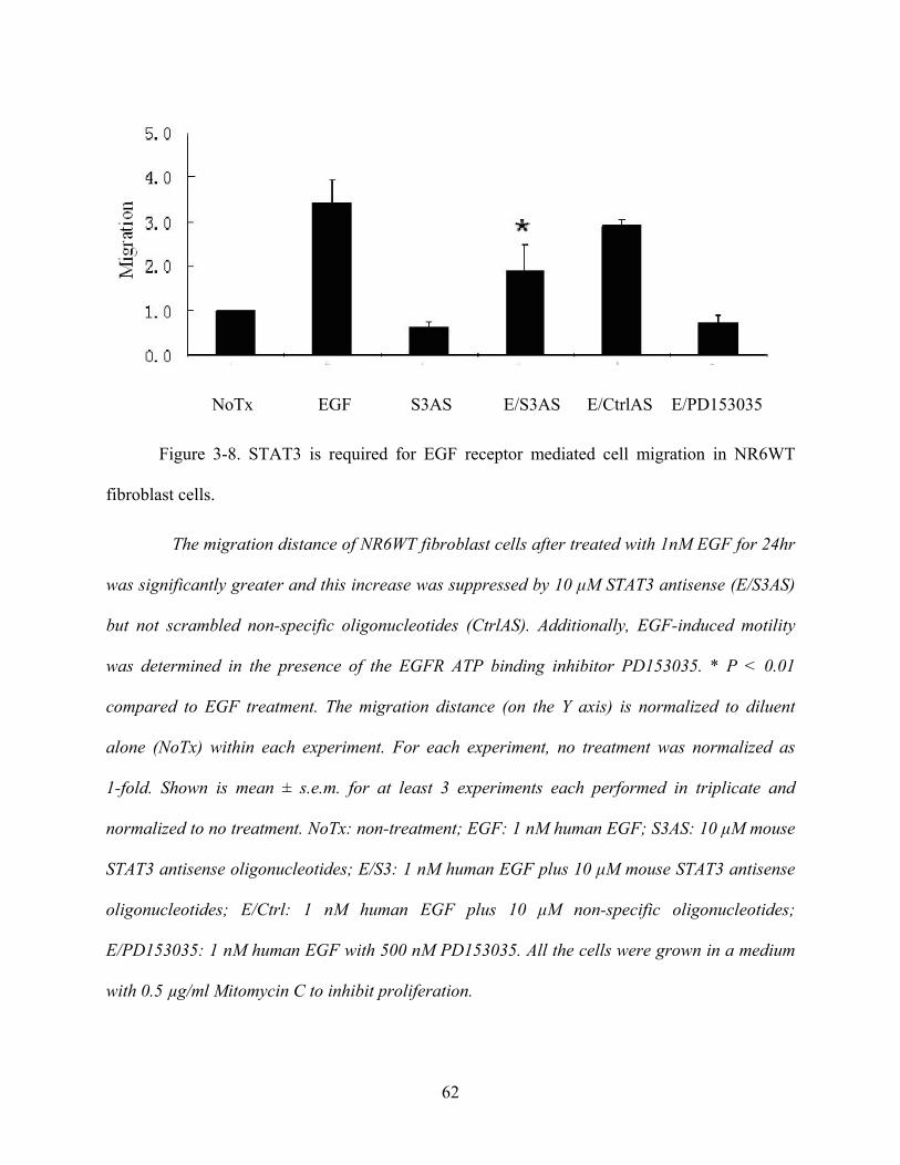
NoTx EGF S3AS E/S3AS E/CtrlAS E/PD153035
Figure 3-8. STAT3 is required for EGF receptor mediated cell migration in NR6WT
fibroblast cells.
The migration distance of NR6WT fibroblast cells after treated with 1nM EGF for 24hr
was significantly greater and this increase was suppressed by 10 μM STAT3 antisense (E/S3AS)
but not scrambled non-specific oligonucleotides (CtrlAS). Additionally, EGF-induced motility
was determined in the presence of the EGFR ATP binding inhibitor PD153035. * P < 0.01
compared to EGF treatment. The migration distance (on the Y axis) is normalized to diluent
alone (NoTx) within each experiment. For each experiment, no treatment was normalized as
1-fold. Shown is mean ± s.e.m. for at least 3 experiments each performed in triplicate and
normalized to no treatment. NoTx: non-treatment; EGF: 1 nM human EGF; S3AS: 10 μM mouse
STAT3 antisense oligonucleotides; E/S3: 1 nM human EGF plus 10 μM mouse STAT3 antisense
oligonucleotides; E/Ctrl: 1 nM human EGF plus 10 μM non-specific oligonucleotides;
E/PD153035: 1 nM human EGF with 500 nM PD153035. All the cells were grown in a medium
with 0.5 µg/ml Mitomycin C to inhibit proliferation.
62

3.9 STAT3 ANTISENSE INHIBITS EGF INDUCED MIGRATION IN DU145WT CELLS
Based on the inhbition of motility noted in the fibroblasts, I proceeded to ask whether
STAT3 might be critical for sustained motility and invasion in response to EGFR signaling in the
prostate carcinoma cells. STAT3 downregulation greatly decreased the migration distance of
DU145WT prostate cancer cell as well as in NR6WT fibroblast cell. The results showed that the
migration distance of DU145WT cells after treated with 10 nM EGF for 24hr was significantly
greater and this increase was suppressed by 10 μM STAT3 antisense (S3AS) but not scrambled
non-specific oligonucleotides (Ctrl). Additionally, EGF-induced motility was determined in the
presence of the EGFR inhibitor PD153035 (Fig 3-9).
63

NoTx EGF S3AS E/S3AS E/CtrlAS E/PD153035
Figure 3-9. STAT3 is required for EGF receptor mediated cell migration in human
prostate tumor cells.
The results showed that the migration distance of DU145WT cells after treated with 10 nM
EGF for 24hr was significantly greater and this increase was suppressed by 10 μM STAT3
antisense (S3AS) but not scrambled non-specific oligonucleotides (Ctrl). Additionally,
EGF-induced motility was determined in the presence of the EGFR inhibitor PD153035.* P <
0.01 compared to EGF treatment. The migration distance (on the Y axis) is normalized to diluent
alone (NoTx) within each experiment. Shown is mean ± s.e.m. for at least 3 experiments each
performed in triplicate and normalized to no treatment. NoTx: non-treatment; EGF: 1 nM
human EGF; S3AS: 10 μM mouse STAT3 antisense oligonucleotides; E/S3: 1 nM human EGF
plus 10 μM mouse STAT3 antisense oligonucleotides; E/Ctrl: 1 nM human EGF plus 10 μM
non-specific oligonucleotides; E/PD153035: 1 nM human EGF with 500 nM PD153035. All the
cells were grown in a medium with 0.5 μg/ml Mitomycin C to inhibit proliferation.
64

3.10 STAT3 SIRNA INHIBITS EGF INDUCED MIGRATION IN DU145WT AND
PC3 HUMAN PROSTATE TUMOR CELLS
Since the STAT3 siRNA inhibit STAT3 expression and its DNA binding activity as well
as STAT3 antisense oligonucleotides, it is reasonable to posit that STAT3 siRNA also inhibits
the EGF induced motility as the later. This would further confirm the specificity of the antisense
approach to that of disruption of STAT3 function. To study cell motility, an in vitro wound
healing assay was performed and demonstrated that the addition of human STAT3 siRNA
greatly decreased the migration distance of DU145WT prostate cancer cell (Fig 3-10).
The same experiment was performed in another aggressive, androgen-independent human
prostate tumor cell line, the PC3 cells. This was done to show that the results were not unique to
a single cell line. To study cell motility, an in vitro wound healing assay was performed and
demonstrated that the addition of human STAT3 siRNA greatly decreased the migration distance
of PC3 prostate cancer cell (Fig 3-10).
65
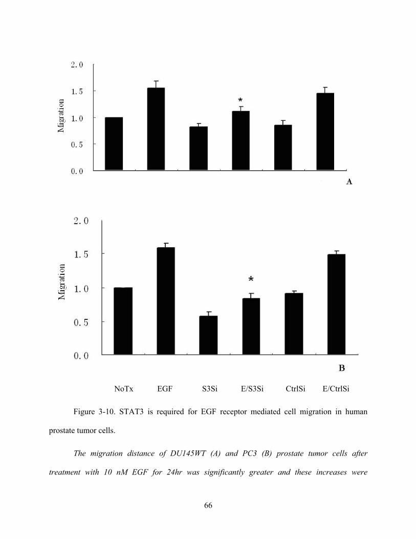
NoTx EGF S3Si E/S3Si CtrlSi E/CtrlSi
Figure 3-10. STAT3 is required for EGF receptor mediated cell migration in human
prostate tumor cells.
The migration distance of DU145WT (A) and PC3 (B) prostate tumor cells after
treatment with 10 nM EGF for 24hr was significantly greater and these increases were
66

suppressed by STAT3 siRNA (S3Si) but not siRNA against eGFP (CtrlSi). Shown is mean ± s.e.m.
for at least 3 experiments each performed in triplicate. * P < 0.01 compared to EGF treatment.
The migration distance (on the Y axis) is normalized to diluent alone (NoTx) within each
experiment. NoTx: non-treatment control; EGF: 10 nM human EGF; Si: 100 pM of STAT3
siRNA transfection; ESi: 10 nM human EGF plus 100 pM of human STAT3 siRNA; Ctrl: 100 pM
of eGFP siRNA as non-specific control; EGF/Ctrl: 10 nM human EGF plus 100 pM of eGFP
siRNA. All the cells were grown in a medium with 0.5 μg/ml Mitomycin C to inhibit proliferation.
3.11 STAT3 ANTISENSE INHIBITS EGF INDUCED INVASION IN DU145WT
As motility is a key component to tumor cell invasion [1], we determined whether this
inhibition affected tumor cell invasion by examining the transmigration of a Matrigel barrier.
Antisense oligonucleotides directed against STAT3 limited DU145WT tumor cell transmigration
to below the levels noted in the absence of added EGF.
This extent of inhibition is expected as both DU145WT and PC3 cells express EGFR
ligands [258], which generate autocrine stimulatory signals in the physical confines of a matrix,
and Matrigel contains competent levels of EGFR ligands [259]. That this is due to EGFR
signaling is shown by blockade using the selective inhibitor PD153035. This inhibitor reduced
not only EGF-enhanced invasiveness but also the invasiveness in the absence of exogenous EGF.
Thus, during transmigration of Matrigel the cells are in an active EGFR signaling mode. The
results showed that STAT3 antisense (S3AS) treated DU145WT cells have a much lower
invasion rate than those cells treated by EGF (Figure 3-11).
67

Figure 3-11. STAT3 is critical for EGFR mediated prostate tumor cell invasiveness
STAT3 antisense (S3AS) treated DU145WT cells have a much lower invasion rate than
those cells treated by EGF while non-specific control oligonucleotides had no effect on the
invasion. Shown is mean ± s.e.m. for at least 3 experiments each performed in triplicate. * P <
0.05 compared to EGF treatment. The invasiveness as determined by cell number transmigrated
through the Matrigel (on the Y axis) is normalized to diluent alone (NoTx) within each
experiment. NoTx: non-treatment; EGF: 10 nM human EGF; S3AS: 10 μM human STAT3
antisense oligonucleotides; E/S3: 10 nM human EGF plus 10 μM human STAT3 antisense
oligonucleotides; CtrlAS: 10 μM non-specific oligonucleotides; E/Ctrl: 10 nM human EGF plus
10 μM non-specific oligonucleotides.
68

3.12 STAT3 SIRNA INHIBITS EGF INDUCED INVASION IN DU145WT AND PC3
HUMAN PROSTATE CANCER CELLS
Since the STAT3 siRNA inhibit EGFR mediated migration as well as STAT3 antisense
oligonucleotides, it is reasonable to posit that STAT3 siRNA also inhibits the EGFR induced
invasion as the later. To study the invasion of human prostate cancer cells, examining the
transmigration of a Matrigel barrier was performed. The results showed that STAT3 antisense
(S3AS) treated DU145WT and PC3 cells have a much lower invasion rate than those cells
treated by EGF (Figure 3-12).
69
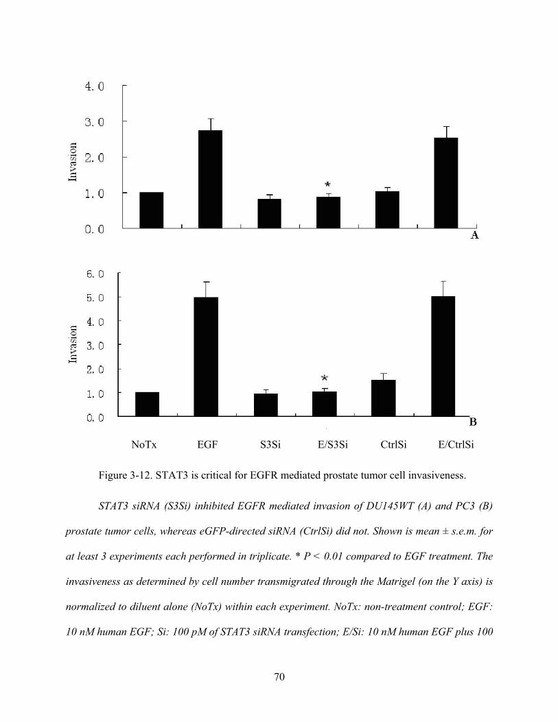
NoTx EGF S3Si E/S3Si CtrlSi E/CtrlSi
Figure 3-12. STAT3 is critical for EGFR mediated prostate tumor cell invasiveness.
STAT3 siRNA (S3Si) inhibited EGFR mediated invasion of DU145WT (A) and PC3 (B)
prostate tumor cells, whereas eGFP-directed siRNA (CtrlSi) did not. Shown is mean ± s.e.m. for
at least 3 experiments each performed in triplicate. * P < 0.01 compared to EGF treatment. The
invasiveness as determined by cell number transmigrated through the Matrigel (on the Y axis) is
normalized to diluent alone (NoTx) within each experiment. NoTx: non-treatment control; EGF:
10 nM human EGF; Si: 100 pM of STAT3 siRNA transfection; E/Si: 10 nM human EGF plus 100
70

pM of human STAT3 siRNA; Ctrl: 100 pM of eGFP siRNA as non-specific control; EGF/Ctrl: 10
nM human EGF plus 100 pM of eGFP siRNA.
3.13 STAT3 IS NOT SUFFICIENT FOR MIGRATION IN NR6WT CELLS
The foregoing data demonstrate that STAT3 activity is required for increased cell
migration and invasion upon EGFR signaling. Up regulation of STAT3 levels in various cancers
[206] [254] [255] might even suggest this functions as an ‘oncogenes’ or ‘tumor progression’
gene. Thus, we asked whether upregulation was sufficient to drive these behaviors. A
constitutively active (CA) STAT3 mutant failed [260] to drive motility of NR6WT cells in the
absence EGF (Figure 3-13). A dominant negative (DN) STAT3 construct did block EGF-induced
motility, as expected. The STAT3 constructs were induced by dexamethasone to avoid
adaptation or cellular modifications during prolonged selection; the dexamethasone by itself had
only a small negative effect on migration that was minimal compared to the effects of either EGF
or the constructs. These data are consistent with STAT3 being required, but not sufficient, for
transcriptional activation of either replacement proteins or modulation of the proteome at the
transcriptional to enable a locomotive state.
71
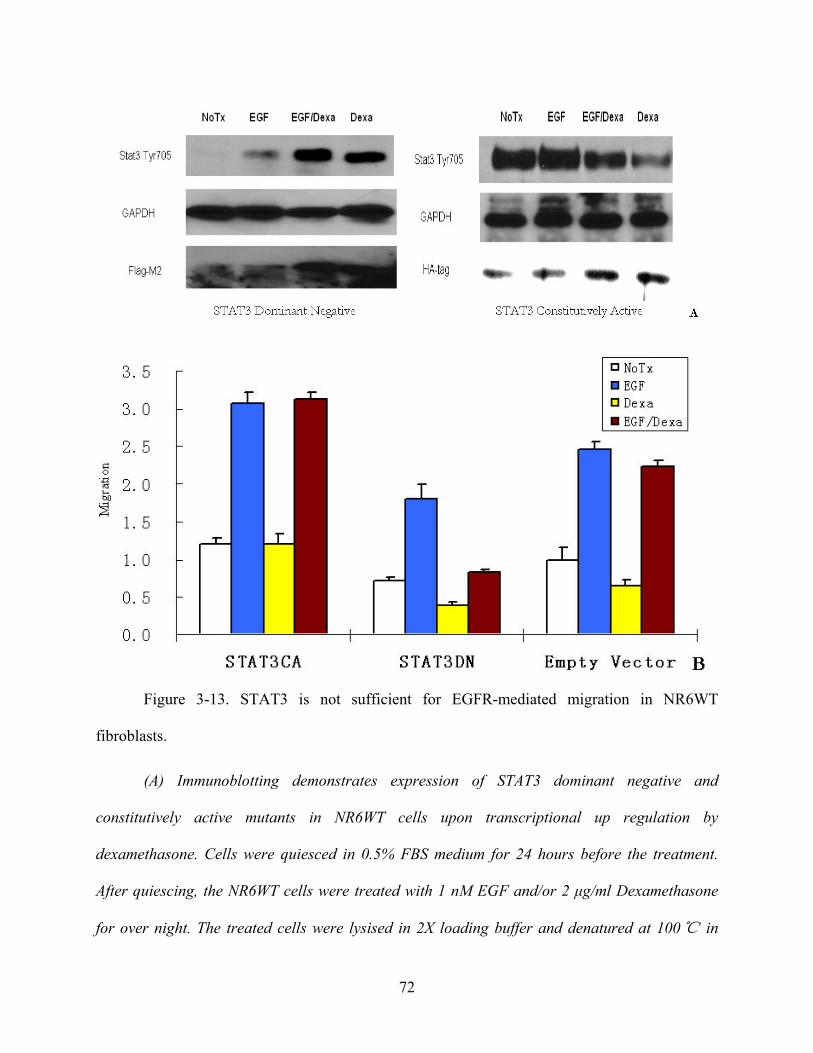
Figure 3-13. STAT3 is not sufficient for EGFR-mediated migration in NR6WT
fibroblasts.
(A) Immunoblotting demonstrates expression of STAT3 dominant negative and
constitutively active mutants in NR6WT cells upon transcriptional up regulation by
dexamethasone. Cells were quiesced in 0.5% FBS medium for 24 hours before the treatment.
After quiescing, the NR6WT cells were treated with 1 nM EGF and/or 2 μg/ml Dexamethasone
for over night. The treated cells were lysised in 2X loading buffer and denatured at 100℃ in
72

water bath for 5 minutes. Lysates were loaded to 10% acrylamide gel, transferred to an
Immobilon-P membrane and immunoblotted with antibody against phosphorylated STAT3
(STAT3 Tyr705) and the special expression tag on the STAT3 mutants (For STAT3 dominant
negative, HA-tag; for STAT3 constitutively active, Flag-M2). Shown are representative blots of
at least three experiments each. NoTx: non-treatment; EGF: 1 nM human EGF; EGF/Dexa: 1
nM human EGF with 2 μg/ml Dexamethasone; Dexa: 2 μg/ml Dexamethasone. After being
exposed, the membrane was stripped and immunoblotted with antibody against GAPDH as a
loading control. (B) The STAT3 constitutively active mutant did not drive motility of NR6WT
cells in the absence or presence EGF, while the dominant-negative construct blocked both basal
and EGF-induced motility, which indicates STAT3 is critical but not sufficient for the motility.
Shown is mean ± s.e.m. for at least 3 experiments each performed in triplicate. * P < 0.01
compared to EGF treatment. The migration distance (on the Y axis) is normalized to diluent
alone of the empty vector (NoTx) within each experiment. NoTx: non-Treatment; EGF: 1 nM
human EGF; EGF/Dexa: 1 nM human EGF with 2 μg/ml Dexamethasone; Dexa: 2 μg/ml
Dexamethasone.
3.14 STAT3 ANTISENSE DOES NOT INHIBIT EGFR MEDIATED
PROLIFERATION IN NR6WT FIBROBLAST CELLS
High expression of STAT3 is found correlated with tumor cells proliferation in many human
cancers [261] [262] [263]. If STAT3 blockade or downregulation limited proliferation, this
would confound longer term migration and invasion assessments. In the above, the time period
73

and manipulations precluded proliferation from substantively contributing to migration and
invasion, but in vivo, the situation would be reversed.
I also expected a decrease of NR6WT proliferation after the treatment of STAT3 antisense
oligonucleotides. Surprisingly, no significant change was found in the in vitro proliferation assay
between NR6WT cells treated with EGF and EGF plus STAT3 antisense oligonucleotides (Fig
3-14).
0
10
20
30
40
50
NoTx EGF EGF/STAT3AS STAT3AS
Nor
mal
ized
Cel
l Pro
lifer
atio
n
Figure 3-14. STAT3 antisense does not inhibit EGFR mediated proliferation in NR6WT
cells.
EGF plus STAT3 antisense treated NR6WT cells have no statistically significant change on
proliferation with those cells treated by EGF alone. The cell proliferation (on the Y axis) is
normalized to diluent alone (NoTx) within each experiment. NoTx: non-treatment control; EGF:
74

1 nM human EGF; EGF/STAT3AS: 1 nM human EGF plus 10 μM mouse STAT3 antisense
oligonucleotides; STAT3AS: 10 μM mouse STAT3 antisense oligonucleotides. Shown is mean ±
s.e.m. for at least 3 experiments each performed in triplicate.
3.15 STAT3 ANTISENSE DOES NOT INHIBIT EGFR MEDIATED
PROLIFERATION IN DU145WT CELLS
Cancer cells present unique signaling pathways and dependencies. As such we examined the
effect of STAT3 downregulation on the prostate carcinoma cells. It has been reported that many
carcinoma cells undergo apoptosis or proliferation arrest in the face of STAT3 downregulation
[246] [264] [265]. As such I expected a decrease of DU145WT proliferation after the treatment
of STAT3 antisense oligonucleotides. Surprisingly, again, no significant change was found in the
in vitro proliferation assay between DU145WT cells treated with EGF and EGF plus STAT3
antisense oligonucleotides (Fig 3-17). Similar experiments were performed with STAT3 siRNA
to block the STAT3 expression instead of STAT3 antisense and got the same results (Data not
shown).
75

0.00
0.20
0.40
0.60
0.80
1.00
1.20
1.40
NoTx EGF EGF/STAT3AS STAT 3 AS
Nor
mal
ized
Cel
l Pro
lifer
atio
n
Figure 3-15. STAT3 antisense does not inhibit EGFR mediated proliferation in DU145WT
cells.
EGF plus STAT3 antisense treated DU145WT cells have no statistically significant
change on proliferation with those cells treated by EGF alone. The cell proliferation (on the Y
axis) is normalized to diluent alone (NoTx) within each experiment. NoTx: non-treatment control;
EGF: 1 nM human EGF; EGF/STAT3AS: 1 nM human EGF plus 10 μM human STAT3 antisense
oligonucleotides; STAT3AS: 10 μM human STAT3 antisense oligonucleotides. Shown is mean ±
s.e.m. for at least 3 experiments each performed in triplicate.
76

4.0 STAT3 IS REQUIRED IN CASPASE 3 INDUCED HUMAN PROSTATE
CARCIMONA CELL APOPTOSIS
4.1 MICROARRAY DATA SHOWS SIGNIFICANT CHANGES OF SOME PROTEINS’
TRANSCRIPTION LEVELS AFTER EGF AND/OR STAT3 ANTISENSE TREATMENT
IN NR6WT CELLS
To further probe the role of the EGF/STAT3 pathway, an initial microarray analysis was
performed on NR6WT cells, thus avoiding variable amounts of autocrine signalling, to determine
transcripts that were altered by EGF in a STAT3-dependent manner. Interestingly, these did not
include the constituents of the well known epigenetic cascades but rather extracellular matrix or
cell cytoskeleton components. It is possible that the enzymatic epigenetic effects are regulated by
the other EGFR-induced transcription cascades, and that STAT3 primarily functions to provide
the motors and pathways for motility. Of note, we did not detect either collagenase-1 (MMP-1)
or stromelysin-2 (MMP-10) that was seen to be up regulated in T24 bladder carcinoma cells
[147]; this discrepancy is likely due to our examining the non-transformed NR6WT fibroblasts
presenting a more limited set of transcription changes, though the two changes noted in the
fibroblasts also were found in the cancer cells
77
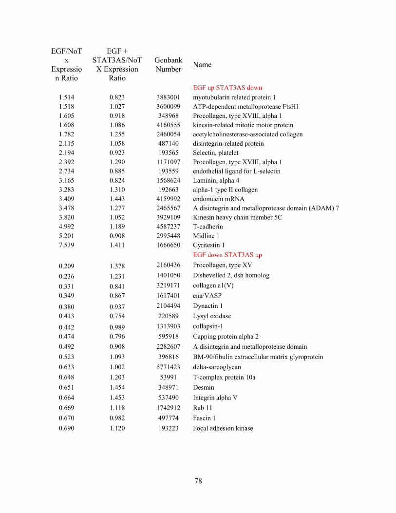
EGF/NoTx
Expression Ratio
EGF + STAT3AS/NoTX Expression
Ratio
Genbank Number Name
EGF up STAT3AS down 1.514 0.823 3883001 myotubularin related protein 1 1.518 1.027 3600099 ATP-dependent metalloprotease FtsH1 1.605 0.918 348968 Procollagen, type XVIII, alpha 1 1.608 1.086 4160555 kinesin-related mitotic motor protein 1.782 1.255 2460054 acetylcholinesterase-associated collagen 2.115 1.058 487140 disintegrin-related protein 2.194 0.923 193565 Selectin, platelet 2.392 1.290 1171097 Procollagen, type XVIII, alpha 1 2.734 0.885 193559 endothelial ligand for L-selectin 3.165 0.824 1568624 Laminin, alpha 4 3.283 1.310 192663 alpha-1 type II collagen 3.409 1.443 4159992 endomucin mRNA 3.478 1.277 2465567 A disintegrin and metalloprotease domain (ADAM) 7 3.820 1.052 3929109 Kinesin heavy chain member 5C 4.992 1.189 4587237 T-cadherin 5.201 0.908 2995448 Midline 1 7.539 1.411 1666650 Cyritestin 1
EGF down STAT3AS up
0.209 1.378 2160436 Procollagen, type XV
0.236 1.231 1401050 Dishevelled 2, dsh homolog
0.331 0.841 3219171 collagen a1(V) 0.349 0.867 1617401 ena/VASP
0.380 0.937 2104494 Dynactin 1 0.413 0.754 220589 Lysyl oxidase
0.442 0.989 1313903 collapsin-1 0.474 0.796 595918 Capping protein alpha 2 0.492 0.908 2282607 A disintegrin and metalloprotease domain 0.523 1.093 396816 BM-90/fibulin extracellular matrix glyroprotein 0.633 1.002 5771423 delta-sarcoglycan 0.648 1.203 53991 T-complex protein 10a 0.651 1.454 348971 Desmin 0.664 1.453 537490 Integrin alpha V 0.669 1.118 1742912 Rab 11 0.670 0.982 497774 Fascin 1 0.690 1.120 193223 Focal adhesion kinase
78
Table 4 Microarray in NR6WT cells shows some proteins’ mRNA expression level
changed significantly after EGF treatment and went back after blocking the STAT3 by antisense
nucleotide.
The NR6WT cells were quiesced in 0.5% FBS MEMα media for 24 hours before
treatment with 1nM of EGF and/or 10µM of mouse STAT3 antisense nucleotide (5’-GTT CCA
CTG AGC CAT CCT GC-3’) for another 24 hours. RNA from the treated cells was then purified.
and prepared for microarray analysis. Affymetrix U74AV2 murine genome array chip was
choosen for the microarray. 5 μg of purified total RNA were added into a 20 μl first strand
reaction with 200 U of SuperScript II (Invitrogen) and 1 µg T7 primer in 1X first strand buffer
(Invitrogen) followed by a 42°C incubation for 1 hour. Second strand synthesis was performed
followed by purification according to the manufacturer’s protocol (Affymetrix). The purified
cDNA was amplified to produce 70-120 μg of biotin labeled cRNA.
Murine U74Av2 GeneChip probe arrays were pre-hybridized in 200μl of the
hybridization cocktail (containing 10 µg of labeled cRNA) according to the manufacturer’s
protocol. 15 μg of labeled cRNA were fragmentedand and resuspended.
The raw data from microarray were normalized with the Affymetrix Microarray suite 5.0,
based on the housekeeping gene expression profile. Expression values were adjusted to the
intensity of the expression value of the 100 housekeeping genes.
4.2 EGF INHIBITS THE EXPRESSION OF VASP IN NR6WT CELLS, WHICH CAN
BE REVERSED BY STAT3 SIRNA
Previous research demonstrated VASP as a negative regulator of fibroblast cell motility
[168]. We also noticed significant decrease of VASP mRNA expression after EGF treatment in
79

NR6WT cells. I used immunoblot to examine if the protein level of VASP also changed. The
results showed that 1 nM EGF suppressed VASP protein expression in NR6WT cells 30 minutes
after treatment and continues for about 24 hours. (Data not shown) The inhibition of VASP
expression was reversed by STAT3 siRNA, which implicated STAT3 may play a role in EGF
mediated VASP expression (Fig 4-1).
Caspase 3 is a well known protein that plays a central role in effecting apoptosis. We also
found dramatically change of Caspase 3 mRNA after EGF and STAT3 antisense stimulation in
microarray screening. Preliminary data shows decreased Caspase 3 protein expression after EGF
stimulation. The inhibition of Caspase 3 expression was reversed by STAT3 siRNA, which
implicated STAT3 may play a role in EGF mediated Caspase 3 expression and apoptosis
inhibition (Fig 4-1).
Figure 4-1. EGF inhibits the expression of VASP and Caspase-3 in NR6WT cells, which can
be reversed by STAT 3 siRNA.
In murine fibroblasts, NR6WT cells, EGF exposure decreased the expression of VASP
and Caspase-3 at protein level, and this decrease was reversed by STAT3 siRNA. The NR6WT
80

cells were transfected with 20 nM of STAT3 siRNA or non-specific control (eGFP siRNA) for at
least 4 hours and recovered in fresh antibiotics-free medium for 24 hours. After that, cells were
quiesced in 0.5% FBS medium for another 24 hours before the addition of EGF. NoTx:
non-treatment control; EGF: 1 nM human EGF; EGF/STAT3si: 1 nM human EGF plus 20 nM
mouse STAT3 siRNA; EGF/Ctrl: 1 nM human EGF plus 20 nM eGFP siRNA. The treated cells
were lysised in 2X loading buffer and denatured at 100℃ in water bath for 5 minutes. Lysates
were loaded to 10% acrylamide gel, transferred to an Immobilon-P membrane and
immunoblotted with antibodies against mouse VASP and Caspase-3. After being exposed, the
membrane was stripped and immunoblotted with antibody against GAPDH as a loading control.
4.3 EGF INHIBITS THE EXPRESSION OF VASP AND CASPASE 3 IN DU145WT
CELLS AND CAN BE REVERSED BY STAT3 SIRNA
As mentioned above, VASP had been shown to be a negative regulator of fibroblast cell
motility [168]. We also found significant decrease of VASP mRNA expression after EGF
treatment in NR6WT cells and confirmed it by immunoblot. The inhibition of VASP and
Caspase-3 expression by EGF and reversion by STAT3 siRNA also was observed in DU145
human prostate tumor cells, which implicated STAT3 may plays a role in EGF mediated VASP
and Caspase 3 expression and the tumor cell resistance to apoptosis (Fig 4-2). Similar results
were observed in PC3 cells (data not shown).
81

Figure 4-2. EGF inhibits the expression of VASP and Caspase-3 in DU145WT cells,
which can be reversed by STAT 3 siRNA.
In DU145WT human prostate cancer cells, EGF exposure decreased the expression of
VASP and Caspase-3 at protein level, and this decrease was reversed by STAT3 siRNA. The
DU145WT cells were transfected with 40 nM of STAT3 siRNA or non-specific control (eGFP
siRNA) for at least 4 hours and recovered in fresh antibiotics-free medium for 24 hours. After
that, cells were quiesced in 0.5% FBS medium for another 24 hours before the addition of EGF.
NoTx: non-treatment control; EGF: 10 nM human EGF; EGF/STAT3si: 10 nM human EGF plus
40 nM human STAT3 siRNA; EGF/Ctrl: 10 nM human EGF plus 40 nM eGFP siRNA. The
treated cells were lysised in 2X loading buffer and denatured at 100℃ in water bath for 5
minutes. Lysates were loaded to 10% acrylamide gel, transferred to an Immobilon-P membrane
and immunoblotted with antibodies against VASP and Caspase-3. After being exposed, the
membrane was stripped and immunoblotted with antibody against GAPDH as a loading control.
82

4.4 STAT3 IS REQUIRED FOR RESISTANCE TO TNF-Α INDUCED APOPTOSIS OF
DU145WT CELLS
Since Caspase 3 plays a critical role in apoptosis and we found some relationship
between EGF, STAT3 and Caspase 3 expression, it is quite reasonable to querying if the first 2
proteins also involved in Caspase 3 mediated apoptosis. Most recent research indicated that EGF
protects prostate cancer cells from apoptosis [266], which put the role that EGFR mediated
signalling pathway plays in apoptosis under focus. An Annexin-V staining assay for apoptosis
induced by TNF-α was performed. Result shows EGF protect DU145WT human prostate tumor
cells from apoptosis, and the protection can be reversed by STAT3 siRNA, which indicated a
major role STAT3 maybe plays in Caspase 3 mediated apoptosis (Fig 4-4). I did not see major
cell death in the experiment, but still sensitivity to the TNF-α. A similar apoptosis assay that
induced by cisplatin instead of TNF-α was also performed and showed the same result. (Data not
shown)
83
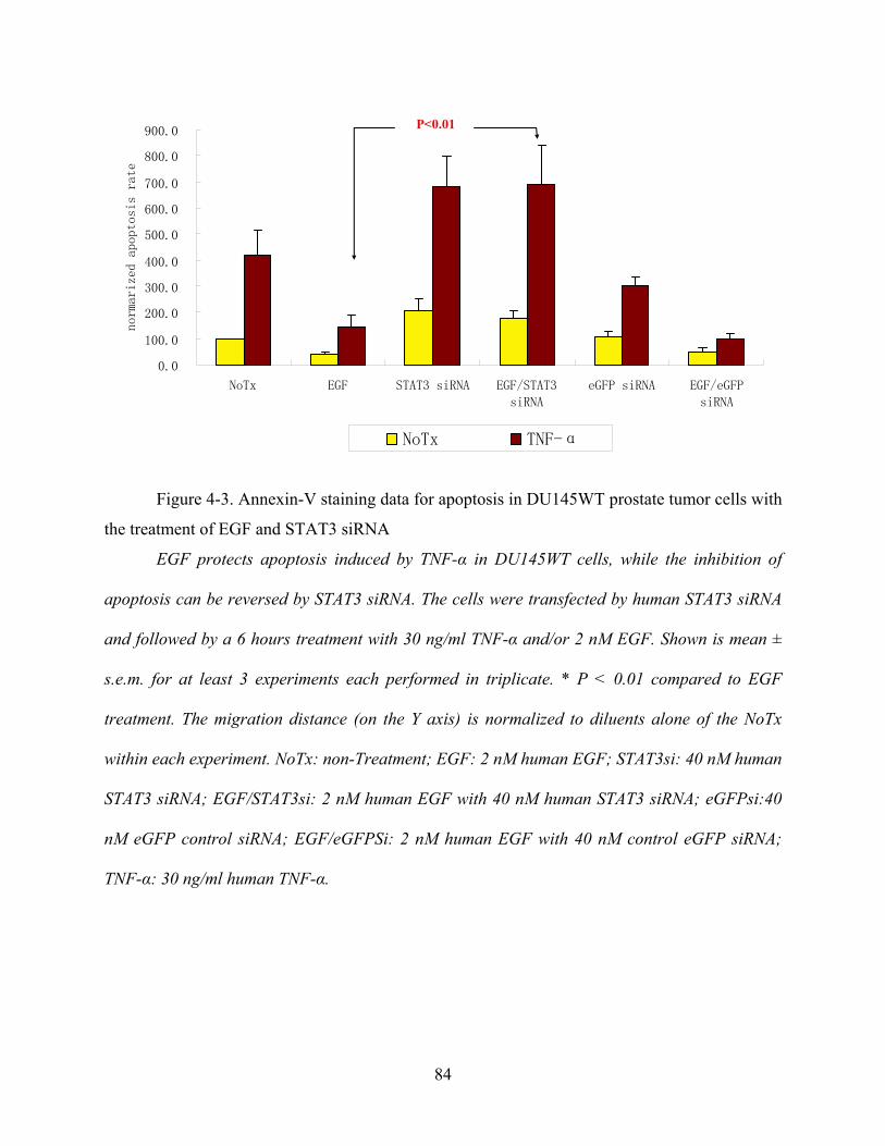
0.0
100.0
200.0
300.0
400.0
500.0
600.0
700.0
800.0
900.0
NoTx EGF STAT3 siRNA EGF/STAT3
siRNA
eGFP siRNA EGF/eGFP
siRNA
normarized apop
tosis rate
NoTx TNF-α
P<0.01
Figure 4-3. Annexin-V staining data for apoptosis in DU145WT prostate tumor cells with
the treatment of EGF and STAT3 siRNA
EGF protects apoptosis induced by TNF-α in DU145WT cells, while the inhibition of
apoptosis can be reversed by STAT3 siRNA. The cells were transfected by human STAT3 siRNA
and followed by a 6 hours treatment with 30 ng/ml TNF-α and/or 2 nM EGF. Shown is mean ±
s.e.m. for at least 3 experiments each performed in triplicate. * P < 0.01 compared to EGF
treatment. The migration distance (on the Y axis) is normalized to diluents alone of the NoTx
within each experiment. NoTx: non-Treatment; EGF: 2 nM human EGF; STAT3si: 40 nM human
STAT3 siRNA; EGF/STAT3si: 2 nM human EGF with 40 nM human STAT3 siRNA; eGFPsi:40
nM eGFP control siRNA; EGF/eGFPSi: 2 nM human EGF with 40 nM control eGFP siRNA;
TNF-α: 30 ng/ml human TNF-α.
84

4.5 STAT3 IS REQUIRED FOR RESISTANCE TO TNF-Α INDUCED APOPTOSIS IN
PC3 CELLS
The same Annexin-V staining assay for apoptosis is also performed in another human
prostate cancer cell line, the PC3 cells. Result shows EGF protect PC3 human prostate tumor
cells from TNF-α induced apoptosis, and the protection can be reversed by STAT3 siRNA,
which is the same as what I got from DU145WT cells. (Fig 4-5) I did not see major cell death in
the experiment, but still sensitivity to the TNF-α. A similar apoptosis assay, which induced by
cisplatin instead of TNF-α, was also performed and showed the same result. (Data not shown)
0.0
100.0
200.0
300.0
400.0
500.0
600.0
NoTx EGF STAT3 siRNA EGF/STAT3siRNA
eGFP siRNA EGF/eGFPsiRNA
normarized apoptosis rate
NoTx TNF-α
P<0.01
Figure 4-4. Annexin-V staining data for apoptosis in PC3 prostate tumor cells with the
treatment of EGF and STAT3 siRNA.
The EGF induced suppression of apoptosis was turn over by STAT3 siRNA in PC3 human
prostate tumor cells. The cells were transfected by human STAT3 siRNA and followed by a 6
85

hours treatment with 30 ng/ml TNF-α and/or 2 nM EGF. Shown is mean ± s.e.m. for at least 3
experiments each performed in triplicate. * P < 0.01 compared to EGF treatment. The migration
distance (on the Y axis) is normalized to diluents alone of the NoTx within each experiment.
NoTx: non-Treatment; EGF: 2 nM human EGF; STAT3si: 40 nM human STAT3 siRNA;
EGF/STAT3si: 2 nM human EGF with 40 nM human STAT3 siRNA; eGFPsi:40 nM eGFP
control siRNA; EGF/eGFPsi: 2 nM human EGF with 40 nM control eGFP siRNA; TNF-α: 30
ng/ml human TNF-α.
86

5.0 DISCUSSION CHAPTER
Carcinogenesis is a multi-step process, in which cancer cells disseminate from a localized
primary tumor mass to both invade local tissue and metastasize to distant organs. It has been well
known that tumor invasiveness is a distinct character of tumor progression involving induced
motility in addition to dysregulated proliferation. Since there is no catholicon for the current
anti-cancer treatments, disrupting or at least inhibiting the tumor invasiveness would greatly
ameliorate the morbidity and mortality. In this study, we investigated whether activated STAT3
protein is involved in the EGF receptor mediated migration in fibroblasts and human prostate
cancer cells and the invasiveness of the latter. EGF increased STAT3 activity along with cell
motility and invasiveness of these cells. Decreased expression and activation of STAT3 by
treatment with STAT3 antisense oligo nucleotides or siRNA or in the presence of a dominant
negative mutant of STAT3 abrogated migration and invasion. The results together, indicated that
STAT3 could be a targeted critical element in the EGF- mediated cell migration and invasion of
prostate carcinoma cells.
Epigenetic events, in addition to transcriptome changes signaled through STAT3, also
contribute to tumor cell motility and invasion. Comparing the migration of NR6WT cells
expressing constitutively activated STAT3 to those expressing the STAT3 dominant negative
mutants, it is interesting to find that constitutively activated STAT3 alone does not increase cell
motility, nor does it increase EGF-stimulated migration. This result suggests that STAT3 is not
sufficient for cell migration but that it provides for end effectors that require additional signals
for either activation or full phenotypic expression. EGF stimulation of a cell results in the
activation of multiple pathways that lead to transcriptional control including Src, PLC-γ, PI3-K,
Ras/MAPK and JAK/STAT [38]. These pathways are often functionally interlinked [267]. Thus,
87

the need for de novo transcription and translation that one notes during growth factor induced
motility [268] [269], can be accomplished by a number of downstream transcription factors but
requires STAT3 in addition to the previously determined PLCγ pathway [270]. Recently, it has
been noted that EGFR signalling promotes progression of T24 bladder carcinoma cells via
STAT3 upregulation of transcription, including that of matrix metaloproteinases [271].
To further probe the role of the EGF/STAT3 pathway, an initial microarray analysis was
performed on NR6WT cells, thus avoiding variable amounts of autocrine signalling, to determine
transcripts that were altered by EGF in a STAT3-dependent manner. (The subset of these
transcripts that are classified as related motility and cell adhesion are provided in the
Supplemental Table). Interestingly, these did not include the constituents of the well known
epigenetic cascades but rather extracellular matrix or cell cytoskeleton components. It is possible
that the enzymatic epigenetic effects are regulated by the other EGFR-induced transcription
cascades, and that STAT3 primarily functions to provide the motors and pathways for motility.
Of note, we did not detect either collagenase-1 (MMP-1) or stromelysin-2 (MMP-10) that was
seen to be upregulated in T24 bladder carcinoma cells [271]; this discrepancy is likely due to our
examining the non-transformed NR6WT fibroblasts presenting a more limited set of
transcription changes, though the two changes noted in the fibroblasts also were found in the
cancer cells. Obviously, these early hints are under active further investigation as these questions
lie beyond the scope of the current communication.
Recent studies suggest that aberrant STAT3 signalling may play an important role in the
carcinogenesis of prostate cancer. Previous data from other groups shows higher
phospho-STAT3 expression in prostate tumor tissues than in adjacent normal tissues [272] [273],
and that this correlates with invasiveness [252]. We also found increased expression of STAT3
88

protein in prostate tumor tissues compared to normal tissue from the same patient (data not
shown, as similar data have been previously published). It is quite reasonable that the high
expressed pSTAT3 in prostate tumor tissues is from the increased expression of STAT3 as well
as activation by autocrine growth factor signalling [274]. Our study indicating that STAT3 is
critical for invasion of prostate cancer cells DU145WT and PC3, secondary to increased cell
motility is consistent with these findings and provide for a mechanism by which phosphorulated
STAT exerts its effects.
Contrary to other studies [275] [276], we didn’t find decreased tumor cell numbers upon
STAT3 down regulation; this may be due to the incomplete nature of such interventions (though
the other studies suffered from similar non-quantitative abrogation), high levels of autocrine
EGFR signalling overcoming this limitation via other pathways, or likely the shorter time course
of our experiments. However, in accord with these other studies, when we overexpressed the
dominant-negative STAT3 construct in DU145WT cells, we noted a high degree of apoptosis in
these cells (for the experiments in Figure 19, we achieved a lower, non-apoptotic level of DN
STAT3); our failure to established stable clones expressing this dominant-negative construct
likely relates to this inhibition of proliferation and/or increased apoptosis. In addition, when we
stress the cells using apoptosis inducers the blockade of STAT3 does increase tumor cell
apoptosis. As such, we feel that this discrepancy is more likely a quantitative rather than a
qualitative effect.
VASP is known as a negative regulator of fibroblast motility [80]. The result from
microarray indicated significant change of VASP mRNA after the treatment of EGF and STAT 3
antisense (Table 4). Immunoblot on NR6WT fibroblast cells showed the expression of VASP
protein is also inhibited by EGF. The inhibition can be observed 30 minutes after the EGF
89

treatment and last for about 24 hours. After adding STAT 3 antisense or STAT 3 siRNA, the
expression of VASP goes back to normal level. This result supports the previous finding, as EGF
suppressed VASP expression, it is quite reasonable to understand that EGF induced fibroblast
cell motility can be turned over by blocking the expression of STAT 3, which is needed for
VASP expression. Further experiments also showed that EGF can suppress VASP expression in
DU145WT and PC3 human prostate tumor cells, though not as significant as in NR6WT
fibroblast cells. The reason could be high autocrine level of EGF in those cancer cells.
90

6.0 CONCLUSIONS AND SPECULATION
6.1 CONCLUSIONS
Growth factor-induced migration is a rate-limiting step in tumor invasiveness. The
molecules that regulate this cellular behavior would represent novel targets for limiting tumor
cell progression. Epidermal growth factor (EGF) receptor (EGFR)-mediated motility, present in
both autocrine and paracrine modes in prostate carcinomas, requires de novo transcription to
persist over times greater than a few hours. Therefore, we sought to define specific signalling
pathways that directly alter cellular transcription. STAT3 (Signal Transducer and Activator of
Transcription 3) is activated, as determined by electrophoretic motility shift assays, by EGFR in
DU145 and PC3 human prostate carcinoma cells in addition to the motility model NR6 fibroblast
cell line. Downregulation of STAT3 activity by antisense oligonucleotides or siRNA or
expression of a dominant-negative construct limited cell motility as determined by an in vitro
wound healing assay and invasiveness through a extracellular matrix barrier. The expression of
constitutively activated STAT3 did not increase the migration, which indicates that STAT3 is
necessary but not sufficient for EGFR-mediated migration. These findings suggest that STAT3
signalling may be a new target for limiting prostate tumor cell invasion.
The inhibition of STAT3 expression does not decrease the basal or EGFR mediated
proliferation of cells studied in this project, NR6WT murine fibroblasts and DU145WT and PC3
human prostate tumor cells. These results contrasted with many of the previous research from
other groups suggesting a positive regulation of tumor cell proliferation from EGF and STAT3
[186] [261] [277] [278] [279]. On the other hand, other publications demonstrated that EGF
91

suppresses tumor growth [280] [281], or has different results between tumor cells in vivo and in
vitro [282]. The most likely explanations for my proliferation data is that these cell lines either
do not require STAT3 for in vitro proliferation (possibly by the presence of redundant inductive
pathways) or the level of STAT3 inhibition attained is insufficient for proliferation and survival
suppression. We prefer this second explanation as the apoptosis studies suggest that lacking
STAT3 predisposes to cell death. This latter explanation also would suggest that motility is the
more sensitive to transcriptional perturbations, in line with the laboratory’s earlier findings [100].
In a microarray analysis of what transcription units are altered by EGF in a
STAT3-dependent manner in NR6WT fibroblast cells, we found that the expression of
motility-limiting VASP protein and the apoptosis nexus Caspase-3 were both down regulated
upon EGF exposure. The microarray data were confirmed by preliminary immunoblot
experiments.
As recent study shows, Ena/VASP proteins antagonize capping protein to inhibit actin
polymerization at barbed ends in vitro [172], which suggested that Ena/VASP proteins associate
with actin filaments at or near the barbed end and protect them from being capped by capping
protein. The depletion of Ena/VASP causes the lamellipodia to protrude more slowly but to
persist longer, yielding a faster moving cell [153]. Immunoblot on NR6WT fibroblast cells
showed the expression of VASP protein is inhibited by EGF 30 minutes after the EGF treatment
and last for about 24 hours. After adding STAT 3 antisense or STAT 3 siRNA, the expression of
VASP goes back to normal level. This result supports the previous finding in microarray. Since
EGF suppresses VASP expression, it is quite reasonable to ask in future studies if STAT3 plays a
role in the VASP regulated-aspects of cell motility.
92

Further experiments showed that EGF could suppress VASP expression in DU145WT
and PC3 human prostate tumor cells, though not as significantly as in NR6WT fibroblast cells.
The reason could be high autocrine level of EGF in those cancer cells.
Caspase-3 plays an essential role in the process of apoptosis as an executor of the caspase
cascade. Recent publications demonstrated decreased caspase-3 level in various human tumor
cells, which suggests the dire pathology of losing its effect on apoptosis; overcoming the
inhibition of caspase-3-induced apoptosis in tumor cells and have little toxicity on normal cells
[259]. My preliminary data from microarray and immunoblot sheds some light on the
relationship between EGF/STAT 3 pathway and caspase-3. In sum, EGF can inhibit the
activation and expression of caspase-3, which save the cells from apoptosis, while STAT 3
siRNA overcomes the effect and make the cells vulnerable to apoptosis inducer. More
experiments to focus on the EGF/STAT 3/caspase-3 apoptosis regulation pathway are ongoing.
As a conclusion, the model that emerges is one in which STAT3 signaling downstream
from EGFR is required for persistent cell motility and invasion, and that partial abrogation of this
pathway hinders this tumor progression. Only more extensive abrogation of STAT3 signalling
compromises carcinoma cell survival and proliferation. Thus, STAT3 inhibition, even if
suboptimal, would slow tumor progression.
6.2 SPECULATION
There are still many questions about the EGF/STAT3 signalling pathway and its branches
in human prostate cancer. One of the most crucial questions is the role STAT3 plays in the
prostate tumor in vivo. Previous data showed aberrant STAT3 expression in many cancers
93

including prostate, as described in the introduction, and STAT3 activation is required for the
proliferation of many cancer cell lines; though my in vitro proliferation data did not find such a
connection in DU145WT and PC3 prostate tumor cells. Considering the complexity of signal
transduction pathways in vivo, it is too early to say that STAT3 does not related to prostate tumor
growth. At least, an in vivo assessment is necessary for determining this discrepancy.
Early embryonic death in STAT3 knockout mice indicated that STAT3 is essential for
early embryonic development [283]. Unforunately, this also excludes the possibility of adult
STAT3-/- mice in which to induce de novo prostate tumor formation; crossbreeding with the
TRAMP mice would be an example. The attempt of getting STAT3 dominant negative mutants
in DU145WT prostate cancer cells was also performed as in NR6WT fibroblasts. Unfortunately,
there were no stable clones survived from transfection and the following selection. The most
likely explanation is that STAT3 is critical to the survival or viability of this cell line, and that
the dominant negative was more efficient at downregulating STAT3 function than siRNA or
antisense; these were only partially abrogating at best. A second possibility is that the STAT3
DN also inhibited the functioning of other STAT isoforms through heterodimerization. The
high-level background expression of STAT3 dominant negative even in the absence of
dexamethasone may also contribute to this fatal result. To overcome the toxicity, I am planning
an alternatively induced STAT3 mutant to avoid the highest levels of STAT3 signaling blockade.
The lethality of STAT3 dominant negative expressing DU145WT human prostate cancer
cells also attracts my attention to the cell death. Combined with previous microarray data, an
EGF-STAT3-Caspase-3 signalling pathway emerged. Caspase-3 is a pivotal executor of
apoptosis and the crosslink of the two main apoptosis pathways. Both Caspase-8 activated by the
death receptor pathway and Caspase-9 activated by the mitochondria pathway will cleave
94

pro-Caspase-3 to then activate the downstream proteins to finish the program cell death. I used
TNF-α to stimulate and activate the death receptor pathway of apoptosis, which can be
suppressed by EGF and recovered by STAT3 abrogation by siRNA. Cisplatin is another
apoptosis inducer, which initiates the mitochondria pathway by binding to the DNA of target
cells [284]. It was also used in the experiment and obtained similar data. Those results suggested
a critical role for STAT3 in apoptosis. Many other experiments are needed to confirm that
EGF/STAT3 signaling pathway is involved in Caspase-3 regulation. For example, the
measurement of Caspase-3 activity on its substrate with or without STAT3, is an essential
complement for previous microarray and immunoblot data. I also need some other method like
flow assay to check the cell apoptosis besides Annexing-V staining.
The next step of studying STAT3 signaling in apoptosis would be the define of the whole
signal transduction pathway. The intersection of STAT3 target genes and apoptosis related genes
offers me a promising lode to mine. A well known such protein is BCL-XL [208]: increased
expression of BCL-XL, an anti-apoptotic BCL-2 family gene, is dependent on constitutively
activated STAT3.
Since the STAT3 is a transcription factor, questions will be asked on how STAT3
regulates the expression of those genes involved in apoptosis. From previous publications, it
looks like STAT3 performs its mission by activating the expression of anti-apoptosis genes. Is
the activation from direct STAT3 binding on the target DNA sequence, or from indirect
mechanisms? Is the STAT3 activation also suppress some pro-apoptosis genes, such as Caspase
3 herein? This was one of the unexpected aspects of the microarray, that STAT3 signaling
appeared to repress gene transcription. As STAT3 is generally considered a transcription induced
and not repressor, the question is whether this repression is direct by STAT3 or secondary to
95

STAT3 induction of a transcription repressor. The answer of those questions will be found in a
more detailed study on the molecular level.
While my findings reported herein suggest a molecular basis for the STAT3 dependence
of EGFR-mediated prostate tumor progression, many more questions have been raised than
answered. I hope that others can find value in addressing these issues.
96
7.0 ABBREVIATIONS
ABL Abelson Leukemia Protein
Arp Actin-related protein
ATP Adenosine triphosphate
BAD Bcl Associated Death promoter
BAX BCL-2 Associated X protein
BCL-2 B-Cell Lymphoma 2
DAG Diacylglycerol
DFF DNA fragmentation factor
DHT Dihydrotestosterone
EGF Epidermal Growth Factor
eGFP Enhanced Green Fluorescent Protein
EGFR Epidermal Growth Factor Receptor
EMSA Electrophoretic Mobility Shift Assay
Ena Enabled
ERK Extracellular Signal-Regulated Kinase
EVH Ena-VASP-Homology
EVL Ena-VASP like
FBS Fetal Bovine Sera
GAPDH Glyceraldehyde 3-phosphate dehydrogenase
HGF Hepatocyte Growth Factor
IFN Interferon
IHC Immunohistochemistry
97

IL-6 Interleukin-6
IP3 Inositol 1,4,5-triphosphate
JAK Jauns Kinase
MAPK Mitogen-Activated Protein Kinase
Mena Mammalian Enabled
PARP Poly ADP-Ribose Polymerase
PCR Polymerase Chain Reaction
PIP2 Phosphatidyl Inositol 3,4-bisphosphate
PKC Protein Kinase C
PLC-γ Phospholipase C-γ
PSA Prostate Specific Antigen
RNAi RNA Interference
SH2 SRC Homology 2
STAT Signal Transducer and Activator of Transcription
TAD Transcriptional Activation Domain
TGF-α Transformation Growth Factor-α
TKI Tyrosine Kinase Inhibitor
TNF Tumor Necrosis Factor
VASP Vasodilator-stimulated phosphoprotein
VEGF Vascular Endothelial Growth Factor
XIAP X-Chromosome-Linked Inhibitor Of Apoptosis Protein
ZBP Zipcode Binding Protein
98

BIBLIOGRAPHY
1. Wells, A. 2000. Tumor invasion: role of growth factor-induced cell motility. Adv Cancer Res.
78:31-101.
2. Comoglio, P.M., Trusolino, L. 2002. Invasive growth: from development to metastasis. J Clin
Invest. 109(7):857-862.
3. Vande Woude, G. F., Jeffers, M., Cortner, J., Alvord, G., Tsarfaty, I., and Resau, J. 1997.
Met-HGF/SF: tumorigenesis, invasion and metastasis. Ciba Found. Symp. 212:119-130.
4. Libermann, T. A, Razon, N., Bartal, A. D., Yarden, Y., Schelessinger, J., Soreq, H. 1984.
Expression of epidermal growth factor receptors in human brain tumors. Cancer Res.
44:753-760.
5. Schlegel, J., Merdes, A., Stumm, G. 1994. Amplification of the epidermal growth factor
receptor gene correlates with different growth behavior in human glioblastoma. Int J Cancer.
56:72-77.
6. Arteaga C. 2002. Epidermal growth factor receptor dependence in human tumors: more than
just expression? The Oncologist. 7(suppl 4):31-39.
7. Wilkinson, B.A. and Hamdy, F.C., 2001 State-of-the-art staging in prostate cancer. British
Journal of Urology 87: 5.
8. Whittermore, A. S., Keller, J. B., and Betensky, R. 1991. Low-grade, latent prostate cancer
volume: predictor of clinical cancer incidence. Journal of the National Cancer Institute
83:1321-1325.
9. Lefevre, M. L. 1998. Prostate cancer screening: more harm than good? Am Fam Physician
58(2):432-438.
10. Lichtenstein, P., Holm, N. V., Verkasalo, P. K., Iliadou, A., Kaprio, J., Koskenvuo, M.,
99

Pukkala, E., Skytthe, A., Hemminki, K. 2000. Environmental and heritable factors in the
causation of cancer--analyses of cohorts of twins from Sweden, Denmark, and Finland. N
Engl J Med. 343(2): 78-85.
11. Karr, J. P. and Yamanaka H. 1992. Prostate cancer and bone metastasis. Adv. in Exp Med
and Biol. 324: 39-45.
12. Haddad, J. R., Reyes, E. C. 1970. Carcinosarcoma of the prostate with metastasis of both
elements: case report. J Urol 103(1): 80-83.
13. Bubendorf, L., Schopfer, A., Wagner, U., Sauter, G., Moch, H., Willi, N., Gasser, T. C.,
Mihatsch, M. J. 2000. Metastatic patterns of prostate cancer: an autopsy study of 1,589
patients. Hum Pathol 31(5): 578-583.
14. Guice, S. L., 3rd, Glenn, J. F., Paulson, D. F. 1978. Liver and prostate adenocarcinoma: an
"estrogen-free" environment? Urology. 11(2): 175-178.
15. Ritchie, C. K., Andrews, L. R., Thomas, K. G., Tindall, D. J., Fitzpatrick, L. A. 1997. The
effects of growth factors associated with osteoblasts on prostate carcinoma proliferation and
chemotaxis: implications for the development of metastatic disease. Endocrinology. 138(3):
1145-1150.
16. Hall, C. L., Bafico, A., Dai, J., Aaronson, S. A., Keller, E. T. 2005. Prostate cancer cells
promote osteoblastic bone metastases through Wnts. Cancer Res. 65(17): 7554-7560.
17. Putz, T., Culig, Z., Eder, I. E., Nessler-Menardi, C., Bartsch, G., Grunicke, H., Uberall, F.,
Klocker, H. 1999. Epidermal growth factor (EGF) receptor blockade inhibits the action of
EGF, insulin-like growth factor I, and a protein kinase A activator on the mitogen-activated
protein kinase pathway in prostate cancer cell lines. Cancer Res. 59(1): 227-233.
18. Liu, X. H., Wiley, H. S., Meikle, A. W. 1993. Androgens regulate proliferation of human
prostate cancer cells in culture by increasing transforming growth factor-alpha (TGF-alpha)
and epidermal growth factor (EGF)/TGF-alpha receptor. J Clin Endocrinol Metab. 77(6):
1472-1478.
19. Culig, Z., Hobisch, A., Hittmair, A., Cronauer, M. V., Radmayr, C., Bartsch, G., Klocker, H.
1997. Androgen receptor gene mutations in prostate cancer. Implications for disease
progression and therapy. Drugs Aging. 10(1): 50-58.
100

20. Seth, D., Shaw, K., Jazayeri, J., Leedman, P. J. 1999. Complex post-transcriptional
regulation of EGF-receptor expression by EGF and TGF-alpha in human prostate cancer cells.
Br J Cancer. 80(5-6): 657-669.
21. Carruba, G., Leake, R. E., Rinaldi, F., Chalmers, D., Comito, L., Sorci, C., Pavone-Macaluso,
M., Castagnetta, L. A. 1994. Steroid-growth factor interaction in human prostate cancer. 1.
Short-term effects of transforming growth factors on growth of human prostate cancer cells.
Steroids. 59(7): 412-420.
22. Jones, H. E., Barrow, D., Dutkowski, C. M., Goddard, L., Smith, C., Harper, M. E.,
Nicholson, R. I. 2001. Effect of an EGF-R selective tyrosine kinase inhibitor and an
anti-androgen on LNCaP cells: identification of divergent growth regulatory pathways.
Prostate. 49(1): 38-47.
23. Khazaie, K., Schirrmacher, V., Lichtner, R. B. 1993. EGF receptor in neoplasia and
metastasis. Cancer Metastasis Rev. 12(3-4): 255-274.
24. Wilding, G., Valverius, E., Knabbe, C., Gelmann, E. P. 1989. Role of transforming growth
factor-alpha in human prostate cancer cell growth. Prostate. 15(1): 1-12.
25. Connolly, J. M., Rose, D. P. 1991. Autocrine regulation of DU145 human prostate cancer
cell growth by epidermal growth factor-related polypeptides. Prostate. 19(2): 173-180.
26. Xie H, Turner T, Wang MH, Singh RK, Siegal GP, Wells A. 1995. In vitro invasiveness of
DU-145 human prostate carcinoma cells is modulated by EGF receptor-mediated signals.
Clin Exp Metastasis. 13: 407-419.
27. Turner, T., Chen, P., Goodly, L. J., Wells, A. 1996. EGF receptor signaling enhances in vivo
invasiveness of DU-145 human prostate carcinoma cells. Clin Exp Metastasis. 14(4):
409-418.
28. Carpenter G, Cohen S. 1990. Epidermal growth factor. J Biol Chem. 265(14): 7709-7712.
29. Kudlow, J. E., Khosravi, M. J., Kobrin, M. S., Mak, W. W. 1984. Inability of anti-epidermal
growth factor receptor monoclonal antibody to block "autocrine" growth stimulation in
transforming growth factor-secreting melanoma cells. J Biol Chem. 259 (19): 11895-11900.
30. Higashiyama, S., Abraham, J. A., Miller, J., Fiddes, J. C., Klagsbrun, M. 1991. A heparin
101

binding growth factor secreted by macrophage-like cells that is related to EGF. Science. 251:
936-939.
31. Plowman, G. D., Whitney, G. S., Neubauer, M. G., Green, J. M., McDonald, V. L., Todaro,
G. J., Shoyab, M. 1990. Molecular cloning and expression of an additional epidermal growth
factor receptor-related gene. Proc Natl Acad Sci U S A. 87(13): 4905-4909.
32. Twardzik, D. R., Ranchalis, J. E., Moss, B., Todaro, G. J. 1987. Vaccinia growth factor:
newest member of the family of growth modulators which utilize the membrane receptor for
EGF. Acta Neurochir Suppl (Wien). 41: 104-109.
33. Peles, E., Bacus, S. S., Koski, R. A., Lu, H. S., Wen, D., Ogden, S. G., Levy, R. B., Yarden,
Y. 1992. Isolation of the neu/HER-2 stimulatory ligand: a 44 kd glycoprotein that induces
differentiation of mammary tumor cells. Cell. 69(1): 205-216.
34. Heldin, C. H. 1996 Protein tyrosine kinase receptors. Cancer Surv 27: 7-24. 35. Barrett-Lee, P., Travers, M., Luqmani, Y., Coombes, R. C. 1990. Transcripts for
transforming growth factors in human breast cancer: clinical correlates. Br J Cancer. 61(4):
612-617.
36. Gorgoulis, V., Aninos, D., Mikou, P., Kanavaros, P., Karameris, A., Joardanoglou, J.,
Rasidakis, A., Veslemes, M., Ozanne, B., Spandidos, D. A. 1992. Expression of EGF,
TGF-alpha and EGFR in squamous cell lung carcinomas. Anticancer Res. 12(4): 1183-1187.
37. Grandis, J. R., Tweardy, D. J. 1992. The role of peptide growth factors in head and neck
carcinoma. Otolaryngol Clin North Am. 25(5): 1105-1115.
38. Wells A. 1999. Moleculars in focus EGFR receptor. Int. J. Biochem. Cell Biol. 31: 637-643.
39. Favoni RE, Cupis A. 2000. The role of polypeptide growth factors in human carcinomas:
new targets for a novel pharmacological approach. Pharmacological Reviews. 52: 179-206.
40. Ushiro H, Cohen S. 1980. Identification of phosphotyrosine as a product of epidermal growth
factor-activated protein kinase in A-431 cell membrane. J Biol Chem. 255: 8363-8365.
41. Wallasch, C., Weiss, F. U., Niederfellner, G., Jallal, B., Issing, W., Ullrich, A. 1995.
Heregulin-dependent regulation of HER2/neu oncogenic signaling by heterodimerization
with HER3. EMBO J. 14(17): 4267-4275.
102

42. Heldin, C. H. 1995 Dimerization of cell surface receptors in signal transduction. Cell. 80:
213-223.
43. Garrett, T. P., McKern, N. M., Lou, M., Elleman, T. C., Adams, T. E., Lovrecz, G. O., Zhu,
H. J., Walker, F., Frenkel, M. J., Hoyne, P. A., Jorissen, R. N., Nice, E. C., Burgess, A. W.,
Ward, C. W. 2002. Crystal structure of a truncated epidermal growth factor receptor
extracellular domain bound to transforming growth factor alpha. Cell. 110 (6): 763-773.
44. Yarden Y. 2001 The EGFR family and its ligands in human cancer: signalling mechanisms
and therapeutic opportunities. Euro J Cancer 37 Suppl 4:S3-8.
45. Zhang, X., Gureasko, J., Shen, K., Cole, P. A., Kuriyan, J. 2006. An allosteric mechanism for
activation of the kinase domain of epidermal growth factor receptor. Cell. 125(6):
1137-1149.
46. Hashimoto, S., Maruoka, S., Gon, Y., Matsumoto, K., Takeshita, I., Horie, T. 1999.
Mitogen-activated protein kinase involves neutrophil elastase-induced morphological
changes in human bronchial epithelial cells. Life Sci. 64(16): 1465-1471.
47. Hallberg, B., Rayter, S. I., Downward, J. 1994. Interaction of Ras and Raf in intact
mammalian cells upon extracellular stimulation. J Biol Chem. 269(6): 3913-3916.
48. Chattopadhyay, A., Vecchi, M., Ji, Q., Mernaugh, R., Carpenter, G. 1999. The role of
individual SH2 domains in mediating association of phospholipase C-gamma1 with the
activated EGF receptor. J Biol Chem. 274(37): 26091-26097.
49. Chen P, Murphy-Ullrich JE and Wells A. 1996. A role for gelsolin in actuating epidermal
growth factor receptor-mediated cell motility. J Cell Biol. 134:689-698.
50. Gschwind, A., Zwick, E., Prenzel, N., Leserer, M., Ullrich, A. 2001. Cell communication
networks: epidermal growth factor receptor transactivation as the paradigm for interreceptor
signal transmission. Oncogene. 20(13): 1594-1600.
51. Tzahar, E., Waterman, H., Chen, X., Levkowitz, G., Karunagaran, D., Lavi, S., Ratzkin, B. J.,
Yarden, Y. 1996. A hierarchical network of interreceptor interactions determines signal
transduction by Neu differentiation factor/neuregulin and epidermal growth factor. Mol Cell
Biol. 16(10): 5276-5287.
103

52. Karunagaran, D., Tzahar, E., Beerli, R. R., Chen, X., Graus-Porta, D., Ratzkin, B. J., Seger,
R., Hynes, N. E., Yarden, Y. 1996. ErbB-2 is a common auxiliary subunit of NDF and EGF
receptors: implications for breast cancer. EMBO J. 15(2): 254-264.
53. Lenferink, A. E., Pinkas-Kramarski, R., van de Poll, M. L., van Vugt, M. J., Klapper, L. N.,
Tzahar, E., Waterman, H., Sela, M., van Zoelen, E. J., Yarden, Y. 1998. Differential
endocytic routing of homo- and hetero-dimeric ErbB tyrosine kinases confers signaling
superiority to receptor heterodimers. EMBO J. 17(12): 3385-3397.
54. Haugh, J. M., Schooler, K., Wells, A., Wiley, H. S., Lauffenburger, D. A. Effect of epidermal
growth factor receptor internalization on regulation of the phospholipase C-gamma1
signaling pathway. J Biol Chem. 274(13): 8958-8965.
55. Aaronson, S. A. 1991 Growth factors and cancer. Science 254:1146-1153.
56. Woodburn, J. R. 1999. The epidermal growth factor receptor and its inhibition in cancer
therapy. Pharmacol Ther. 82: 241-250.
57. Sung, T., Miller, D. C., Hayes, R. L., Alonso, M., Yee, H., Newcomb, E. W. 2000.
Preferential inactivation of the p53 tumor suppressor pathway and lack of EGFR
amplification distinguish de novo high grade pediatric astrocytomas from de novo adult
astrocytomas. Brain Pathol. 10(2): 249-259.
58. Salomon, D. S., Normanno, N., Ciardiello, F., Brandt, R., Shoyab, M., Todaro, G. J. 1995.
The role of amphiregulin in breast cancer. Breast Cancer Res Treat. 33: 103-114.
59. Fielding, A. K., Maurice, M., Morling, F. J., Cosset, F. L., Russell, S. J. 1998. Inverse
targeting of retroviral vectors: selective gene transfer in a mixed population of hematopoietic
and nonhematopoietic cells. Blood. 91(5): 1802-1809.
60. Sartor, C. I. 2000. Biological modifiers as potential radiosensitizers: targeting the epidermal
growth factor receptor family. Semin Oncol. 27: 15-20.
61. Newby, J. C., Johnston, S. R., Smith, I. E., Dowsett, M. 1997. Expression of epidermal
growth factor receptor and c-erbB2 during the development of tamoxifen resistance in human
breast cancer. Clin Cancer Res. 3(9): 1643-1651.
62. Klijn, J. G., Look, M. P., Portengen, H., Alexieva-Figusch, J., van Putten, W. L., Foekens, J.
A. 1994. The prognostic value of epidermal growth factor receptor (EGF-R) in primary
104

breast cancer: results of a 10 year follow-up study. Breast Cancer Res Treat. 29: 73-83.
63. Fischer-Colbrie, J., Witt, A., Heinzl, H., Speiser, P., Czerwenka, K., Sevelda, P., Zeillinger,
R. 1997. EGFR and steroid receptors in ovarian carcinoma: comparison with prognostic
parameters and outcome of patients. Anticancer Res. 17: 613-619.
64. Magne, N., Pivot, X., Bensadoun, R. J., Guardiola, E., Poissonnet, G., Dassonville, O.,
Francoual, M., Formento, J. L., Demard, F., Schneider, M., Milano, G. 2001. The
relationship of epidermal growth factor receptor levels to the prognosis of unresectable
pharyngeal cancer patients treated by chemo-radiotherapy. Eur J Cancer. 37: 2169-2177.
65. Kuan, C. T., Wikstrand, C. J., Bigner, D. D. 2001. EGF mutant receptor vIII as a molecular
target in cancer therapy. Endocr Relat Cancer. 8: 83-96.
66. Huang, H. S., Nagane, M., Klingbeil, C. K., Lin, H., Nishikawa, R., Ji, X. D., Huang, C. M.,
Gill, G. N., Wiley, H. S., Cavenee, W. K. 1997. The enhanced tumorigenic activity of a
mutant epidermal growth factor receptor common in human cancers is mediated by threshold
levels of constitutive tyrosine phosphorylation and unattenuated signaling. J Biol Chem.
272(5): 2927-2935.
67. Nishikawa, R., Ji, X. D., Harmon, R. C., Lazar, C. S., Gill, G. N., Cavenee, W. K., Huang, H.
J. 1994. A mutant epidermal growth factor receptor common in human glioma confers
enhanced tumorigenicity. Proc Natl Acad Sci U S A. 91: 7727-7731.
68. Moscatello, D. K., Holgado-Madruga, M., Godwin, A. K., Ramirez, G., Gunn, G., Zoltick, P.
W., Biegel, J. A., Hayes, R. L., Wong, A. J. 1995. Frequent expression of a mutant epidermal
growth factor receptor in multiple human tumors. Cancer Res. 55: 5536-5539.
69. Cohen, D. W., Simak, R., Fair, W. R., Melamed, J., Scher, H. I., Cordon-Cardo, C. 1994.
Expression of transforming growth factor-alpha and the epidermal growth factor receptor in
human prostate tissues. J Urol. 152: 2120-2124.
70. Rusch, V., Klimstra, D., Venkatraman, E., Pisters, P. W., Langenfeld, J., Dmitrovsky, E.
1997. Overexpression of the epidermal growth factor receptor and its ligand transforming
growth factor alpha is frequent in resectable non-small cell lung cancer but does not predict
tumor progression. Clin Cancer Res. 3: 515-522.
71. Levkowitz, G., Waterman, H., Zamir, E., Kam, Z., Oved, S., Langdon, W. Y., Beguinot, L.,
105

Geiger, B., Yarden, Y. 1998. c-Cbl/Sli-1 regulates endocytic sorting and ubiquitination of the
epidermal growth factor receptor. Genes Dev.12: 3663-3674.
72. Levkowitz, G., Waterman, H., Ettenberg, S. A., Katz, M., Tsygankov, A. Y., Alroy, I., Lavi,
S., Iwai, K., Reiss, Y., Ciechanover, A., Lipkowitz, S., Yarden, Y. 1999. Ubiquitin ligase
activity and tyrosine phosphorylation underlie suppression of growth factor signaling by
c-Cbl/Sli-1. Mol Cell. 4: 1029-1040.
73. Tateishi, M., Ishida, T., Kohdono, S., Hamatake, M., Fukuyama, Y., Sugimachi, K. 1994.
Prognostic influence of the co-expression of epidermal growth factor receptor and c-erbB-2
protein in human lung Adenocarcinoma. Surg Onco. 3: 109-113.
74. Xia, W., Lau, Y. K., Zhang, H. Z., Xiao, F. Y., Johnston, D. A., Liu, A. R., Li, L., Katz, R. L.,
Hung, M. C. 1999. Combination of EGFR, HER-2/neu, and HER-3 is a stronger predictor for
the outcome of oral squamous cell carcinoma than any individual family members. Clin
Cancer Res. 5(12): 4164-4174.
75. Nicholson, R. I., McClelland, R. A., Finlay, P., Eaton, C. L., Gullick, W. J., Dixon, A. R.,
Robertson, J. F., Ellis, I. O., Blamey, R. W. 1993. Relationship between EGF-R, c-erbB-2
protein expression and Ki67 immunostaining in breast cancer and hormone sensitivity. Eur J
Cancer. 29A: 1018-1023.
76. Iwase, H., Itoh, Y., Kuzushima, T., Yamashita, H., Iwata, H., Toyama, T., Hara, Y.,
Kobayashi, S. 1997. Simultaneous quantitative analyses of c-erbB-2 protein, epidermal
growth factor receptor, cathepsin D, and hormone receptors in breast cancer. Cancer Detect
Prev. 21: 29-35.
77. Mendelsohn, J. 1997. Epidermal growth factor receptor inhibition by a monoclonal antibody
as anticancer therapy. Clin Cancer Res. 3: 2703-2711.
78. Mendelsohn, J., Baselga, J. 2000. The EGF receptor family as targets for cancer therapy.
Oncogene. 19: 6550-6565.
79. Levitzki, A., Gazit, A. 1995. Tyrosine kinase inhibition: an approach to drug development.
Science. 267: 1782-1788.
80. Wakeling, A. E., Barker, A. J., Davies, D. H., Brown, D. S., Green, L. R., Cartlidge, S. A.,
Woodburn, J. R. 1996. Specific inhibition of epidermal growth factor receptor tyrosine
106

kinase by 4-anilinoquinazolines. Breast Cancer Res Treat. 38: 67-73.
81. Wong, A. J., Ruppert, J. M., Bigner, S. H., Grzeschik, C. H., Humphrey, P. A., Bigner, D. S.,
Vogelstein, B. 1992. Structural alterations of the epidermal growth factor receptor gene in
human gliomas. Proc Natl Acad Sci U S A. 89: 2965-2969.
82. Humphrey, P. A., Gangarosa, L. M., Wong, A. J., Archer, G. E., Lund-Johansen, M.,
Bjerkvig, R., Laerum, O. D., Friedman, H. S., Bigner, D. D. 1991. Deletion-mutant
epidermal growth factor receptor in human gliomas: effects of type II mutation on receptor
function. Biochem Biophys Res Commun. 178: 1413-1420.
83. Bigner, S. H., Humphrey, P. A., Wong, A. J., Vogelstein, B., Mark, J., Friedman, H. S.,
Bigner, D. D. 1990. Characterization of the epidermal growth factor receptor in human
glioma cell lines and xenografts. Cancer Res. 50: 8017-8022.
84. Callaghan, T., Antczak, M., Flickinger, T., Raines, M., Myers, M., Kung, H. J. 1993. A
complete description of the EGF-receptor exon structure: implication in oncogenic activation
and domain evolution. Oncogene. 8: 2939-2948.
85. Frederick, L., Eley, G., Wang, X. Y., James, C. D. 2000. Analysis of genomic
rearrangements associated with EGRFvIII expression suggests involvement of Alu repeat
elements. Neuro-oncol. 2: 159-163.
86. Fenstermaker, R. A., Ciesielski, M. J. 2000. Deletion and tandem duplication of exons 2 - 7
in the epidermal growth factor receptor gene of a human malignant gliomas. Oncogene. 19:
4542-4548.
87. Frederick, L., Wang, X. Y., Eley, G., James, C. D. 2000. Diversity and frequency of
epidermal growth factor receptor mutations in human glioblastomas. Cancer Res. 60:
1383-1387.
88. Wells A., Kassis J., Solava J., Turner T and Lauffenburger D. 2002. Growth factor-induced
cell motility in tumor invasion. Acta Oncologica. 41(2):124-130.
89. Weigelt, B., Wessels, L. F., Bosma, A. J., Glas, A. M., Nuyten, D. S., He, Y. D., Dai, H.,
Peterse, J. L., van't Veer, L. J. 2005. No common denominator for breast cancer lymph node
metastasis. Br J Cancer. 93: 924-932.
90. Mehlen, P., Puisieux, A. 2006. Metastasis: a question of life or death. Nat Rev Cancer. 6:
107

449-458.
91. Wells A. 1989. The epidermal growth factor receptor and its ligands. Oncogenes. Boston:
Kluwer Academic Publishers. 143-168.
92. Kim H., Turner T., Kassis J., Souto J., Wells A. 1999. EGF receptor signalling in prostate
development. Histol Histopathol. 14:1175-1178.
93. DeWitt AE., Dong JY., Wiley HS., Lauffenburger DA. 2001. Quantitative analysis of the
EGF receptor autocrine system reveals cryptic regulation of cell response by ligand capture. J
Cell Sci. 114:2301-2313.
94. Neal DE., Marsh C., Bennett MK. 1985. Epidermal growth factor receptors in human bladder
cancer: camparision of invasive and superficial tumors. Lancet. I:366-368.
95. Yasui W., Sumiyoshi H., Hata J. 1988. Expression of epidermal growth factor receptor in
human gastric and colon carcinomas. Cancer Res. 48: 137-141.
96. Holting, T., Siperstein, A. E., Clark, O. H., Duh, Q. Y. 1995. Epidermal growth factor
(EGF)- and transforming growth factor alpha-stimulated invasion and growth of follicular
thyroid cancer cells can be blocked by antagonism to the EGF receptor and tyrosine kinase in
vitro. Eur J Endocrinol. 132: 229-235.
97. Kassis, J., Moellinger, J., Lo, H., Greenberg, N. M., Kim, H. G., Wells, A. 1999. A role for
phospholipase C-gamma-mediated signaling in tumor cell invasion. Clin Cancer Res. 5:
2251-2260.
98. Lu, Z., Jiang, G., Blume-Jensen, P., Hunter, T. 2001. Epidermal growth factor-induced tumor
cell invasion and metastasis initiated by dephosphorylation and downregulation of focal
adhesion kinase. Mol Cell Biol. 21: 4016-4031.
99. Thomas, S. M., Coppelli, F. M., Wells, A., Gooding, W. E., Song, J., Kassis, J., Drenning, S.
D., Grandis, J. R. 2003. Epidermal growth factor receptor-stimulated activation of
phospholipase Cgamma-1 promotes invasion of head and neck squamous cell carcinoma.
Cancer Res. 63: 5629-5635.
100. Chen, P., Xie, H., Sekar, M. C., Gupta, K., Wells, A. 1994. Epidermal growth factor
receptor-mediated cell motility: phospholipase C activity is required, but mitogen-activated
108

protein kinase activity is not sufficient for induced cell movement. J Cell Biol. 127: 847-857.
101. Chou, J., Stolz, D. B., Burke, N. A., Watkins, S. C., Wells, A. 2002. Distribution of gelsolin
and phosphoinositol 4,5-bisphosphate in lamellipodia during EGF-induced motility. Int J
Biochem Cell Biol. 34: 776-790.
102. Xie H, Pallero MA, Gupta K, Ware MF, Chang P, Witke W, Kwiatkowski DJ,
Lauffenburger DA, Murphy-Ullrich J, Wells A. 1998. EGF receptor regulation of cell-
substratum interactions: EGF-induced disassembly of focal adhesions does not require the
motility-associated PLCy signalling pathway. J Cell Science. 111: 616-625.
103. Shiraha, H., Glading, A., Gupta, K., Wells, A. 1999. IP-10 inhibits epidermal growth
factor-induced motility by decreasing epidermal growth factor receptor-mediated calpain
activity. J Cell Biol. 146: 243-254.
104. Ciardiello F. and Tortora G. 2001. A novel approach in the treatment of cancer: targeting
the epidermal growth factor receptor. Clin Cancer Res. 7: 2958-2970.
105. Nass SJ and Dickson RB. 1998. Epidermal growth factor-independent cell cycle
progression is altered in mammary epithelial cells that overexpress c-myc. Clin Cancer Res.
4:1813-1822.
106. Derynck R., Goeddel DV., Ullrich A., Gutterman JU, Williams RD, Bringman TS and
Berger WH. 1987. Synthesis of messenger RNAs for transforming growth factors α and β
and the epidermal growth factor by human tumors. Cancer Res. 47: 707-712.
107. Travers MT., Barrett-Lee P. J., Berger U., Luqmani Y. A., Gazet J. C., Powels T. J. and
Coombes R. C. 1988. Growth factor expression in normal, benign, and malignant breast
tissue. Br Med J. 296:1621-1624.
108. Grandis J and Tweardy D. J. 1993. Elevated levels of transforming growth factor α and
epidermal growth factor receptor messenger RNA are early markers of carcinogenesis in
head and neck cancer. Cancer Res. 53:3579-3584.
109. Grandis J., Melhem M. F., Barnes E. L. and Tweardy D. J. 1996. Quantitative
immunohistochemical analysis of transforming growth factor α and epidermal growth factor
receptor in patients wit hsquamous cell carcinoma of the head and neck. Cancer.
78:1284-1292.
109

110. Russell P. J., Bennett S. and Stricker P. 1998. Growth factor involvement in progression of
prostate cancer. Clin Chem. 44:705-723.
111. Horwitz AR, Parsons JT. 1999. Cell migration—movin’ on. Science. 286:1102-1103.
112. Lauffenburger DA, Horwitz AF. 1996. Cell migration: a physically integrated molecular
process. Cell. 84:359-369.
113. Canals F. 1992. Signal transmission by epidermal growth factor receptor: coincidence of
activation and dimerization. Biochemistry. 31:637-643.
114. Rotin D, Honegger AM, Margolis BL, Ullrich A and Schlessinger J. 1992. Presence of SH2
domains of phospholipase C gamma 1 enhances substrate phosphorylation by increasing the
affinity toward the epidermal growth factor receptor. J Biol Chem. 267:9678-9683.
115. Todderud G, Wahl MI, Rhee SG and Carpenter G. 1990. Stimulation of phospholipase
C-gamma 1 membrane association by epidermal growth factor. Science. 249:296-298.
116. Yonezawa N, Nishida E, Iida K, Yahara I and Sakai H. 1990. Inhibition of the interactions
of cofilin, destrin, and deoxyribonuclease I with actin by phosphoinositieds. J Biol Chem.
265: 8382-8386.
117. Janmey PA. 1994. Phosphoinositides and calcium as regulators of cellular actin assembly
and disassembly. Annu Rev Physiol. 56:169-191.
118. Goldschmidt-Clermont PJ, Machesky LM, Baldassare JJ and Pollard TD. 1990. The
actin-binding protein profiling binds to PIP2 and inhibits its hydrolysis by phospholipase C.
Science. 247: 1575-1578.
119. Schafer DA, Jennings PB and Cooper JA. 1996. Dynamics of capping protein and actin
assembly in vitro: uncapping barbed ends by polyphosphoinositides. J Cell Biol.
135:169-179.
120. Northwood IC, Gonzalez FA, Wartmann M, Raden DL and Davis RJ. 1991. Isolation and
characterization of two growth factor-stimulated protein kinases that phosphorylate the
epidermal growth factor receptor at threonine 669. J Cell Biol. 266: 15266-15276.
121. Glading A, Lauffenburger DA and Wells A. 2002. Cutting to the chase: calpain proteases in
cell motility. Trends Cell Biol. 12:46-54.
110
122. Glading A, Reynolds IJ, Shiraha H, Satish L, Potter DA, Blair HC and Wells A. 2004.
Epidermal growth factor activates m-calpain (calpain II), at least in part, by extracellular
signal-regulated kinase-mediated phosphorylation. Mol Cell Biol. 24(6):2499-512.
123. Yan B, Calderwood DA, Yaspan B and Ginsberg MH. 2001. Calpain cleavage promotes
talin binding to the beta 3 integrin cytoplasmic domain. J Biol Chem. 276:28164-28170.
124. Yao X, Thibodeau A and Forte JG. 1993. Ezrin-calpain I interactions in gastric parietal cells.
Am J Physiol. 265:C36-C46.
125. Carragher NO, Levkau B, Ross R and Raines EW. 1999. Degraded collagen fragments
promote rapid disassembly of smooth muscle focal adhesions that correlates with cleavage of
pp125(FAK), paxillin and talin. J Cell Biol. 147:619-630.
126. Dwyer-Nield LD, Miller AC, Neighbors BW, Dinsdale D and Malkinson AM. 1996.
Cytoskeletal architecture in mouse lung epithelial cells is regulated by protein-kinase C-alpha
and calpain II. Am J Physiol. 270: L526-L534.
127. Backman L, Pekrun A and Gratzer WB. 1991. Formation and properties of spectrin
containing a truncated beta-chain, generated by an endogenous calcium-dependent protease.
J Biol Chem. 266:3835-3840.
128. Onji T, Takagi M and Shibata N. 1987. Calpain abolishes the effect of filamin on the
actomyosin system in platelets. Biochem Biophys Acta. 912-283-286.
129. Goll DE, Dayton WR, Singh I and Robson RM. 1991. Studies of the alpha-actinin/actin
interaction in the Z-disk by using calpain. J Biol Chem. 266:8501-8510.
130. Dekker LV and Parker PJ. 1994. Protein kinase C: a question of specificit. Trends Biol. Sci.
19:73-77.
131. Hug H and Sarre TF. 1993. Protein kinase C isoenzymes: divergence in signal transduction.
Biochem J. 291:329-343.
132. Hansson A, Skoglund G, Lassing I, Lindberg U and Ingelman-Sundberg M. 1988.
PKC-dependent phosphorylation of profiling is specifically stimulated by PIP2. Biochem.
Biophys. Res. Commun. 150:526-531.
111

133. Aderem A. 1992. Signal transduction and the actin cytoskeleton: the roles of MARCKS and
profiling. Trends Biol. Sci. 17:438-443.
134. Iwabu, A., Smith, K., Allen, F. D., Lauffenburger, D. A., Wells, A. 2004. Epidermal growth
factor induces fibroblast contractility and motility via a protein kinase C delta-dependent
pathway. J Biol Chem. 279: 14551-14560.
135. Tang, L. L., Yokoyama, Y., Wan, X., Iwagaki, S., Niwa, K., Tamaya, T. 2006. PTEN
sensitizes epidermal growth factor-mediated proliferation in endometrial carcinoma cells.
Oncol Rep. 15: 855-859.
136. Alonso-Escolano, D., Medina, C., Cieslik, K., Radomski, A., Jurasz, P., Santos-Martinez, M.
J., Jiffar, T., Ruvolo, P., Radomski, M. W. 2006. Protein kinase C delta mediates
platelet-induced breast cancer cell invasion. J Pharmacol Exp Ther. 318: 373-380.
137. Vucenik, I.. Ramakrishna, G., Tantivejkul, K., Anderson, L. M., Ramljak, D. 2005. Inositol
hexaphosphate (IP6) blocks proliferation of human breast cancer cells through a
PKCdelta-dependent increase in p27Kip1 and decrease in retinoblastoma protein (pRb)
phosphorylation. Breast Cancer Res Treat. 91: 35-45.
138. Welsh JB, Gill GN, Rosenfeld MG and Wells A. 1991. A negative feedback loop attenuate
EGF-induced morphological changes. J Cell Biol. 114:533-543.
139. Morrison P, Takishima K and Rosner MR. 1993. Role of threonine residues in regulation of
the epidermal growth factor receptor by protein kinase C and mitogen-activated protein
kinase. J. Biol. Chem. 268:15536-15543.
140. Lund KA, Lazar CS, Chen WS, Walsh BJ, Welsh JB, Herbst JJ, Walton GM, Rosenfeld
MG, Gill GN and Wiley HS. 1990. Phosphorylation of the epidermal growth factor receptor
at threonine 654 inhibits ligand-induced internalization and down-regulation. J. Biol. Chem.
265:20517-20523.
141. Chen P, Xie H and Wells A. 1996. Mitogenic signalling from the EGF receptor is attenuated
by a phospholipase C-γ/protein kinase C feedback mechanism. Mol. Biol. Cell. 7:871-881.
142. Svitkina TM, Verkhovsky AB, McQuade KM and Borisy GG. 1997. Analysis of the
actin-myosin II system in fish epidermal keratocytes: mechanism of cell body translocation.
J Cell Biol. 139:397-415.
112

143. Pollard TD, Blanchoin L and Mullins RD. 2000. Molecular mechanisms controlling actin
filament dynamics in nonmuscle cells. Annu Rev Biophys Biomol Struct. 29:545-576.
144. Chou, J., Burke, N. A., Iwabu, A., Watkins, S. C., Wells, A. 2003. Directional motility
induced by epidermal growth factor requires Cdc42. Exp Cell Res. 287: 47-56.
145. Shao, H., Chou, J., Baty, C. J., Burke, N. A., Watkins, S. C., Stolz, D. B., Wells, A. 2006.
Spatial localization of m-calpain to the plasma membrane by phosphoinositide biphosphate
binding during epidermal growth factor receptor-mediated activation. Mol Cell Biol. 26:
5481-5496.
146. Mullins RD, Heuser JA and Pollard TD. 1998. The interaction of Arp2/3 complex with
actin: nucleation, high affinity pointed end capping, and formation of branching networks
of filaments. Proc Natl Acad Sci USA. 95:6181-6186.
147. Welch MD, Iwamatsu A and Mitchision TJ. 1997. Actin polymerization is induced by
Arp2/3 protein complex at the surface of Listeria monocytogenes. Nature. 385:265-269.
148. Machesky, L. M., Reeves, E., Wientjes, F., Mattheyse, F. J., Grogan, A., Totty, N. F.,
Burlingame, A. L., Hsuan, J. J., Segal, A. W. 1997. Mammalian actin-related protein 2/3
complex localizes to regions of lamellipodial protrusion and is composed of evolutionarily
conserved proteins. Biochem J. 328: 105-112.
149. Machesky, L. M., Gould, K. L. 1999. The Arp2/3 complex: a multifunctional actin
organizer. Curr Opin Cell Biol. 11: 117-121.
150. Higgs HN and Pollard TD. 1999. Regulation of actin polymerization by Arp2/3 complex
and WASp/Scar proteins. J Biol Chem. 274:32531-32534.
151. Mullins, R. D., Heuser, J. A., Pollard, T. D. 1998. The interaction of Arp2/3 complex with
actin: nucleation, high affinity pointed end capping, and formation of branching networks
of filaments. Proc Natl Acad Sci U S A. 95: 6181-6186.
152. Mingle, L. A., Okuhama, N. N., Shi, J., Singer, R. H., Condeelis, J., Liu, G. 2005.
Localization of all seven messenger RNAs for the actin-polymerization nucleator Arp2/3
complex in the protrusions of fibroblasts. J Cell Sci. 118: 2425-2433.
113

153. Krause M., Bear J. E., Loureiro J. J. and Gertler F. B. 2002. The Ena/VASP enigma. J Cell
Sci. 115(24):4721-4726.
154. Gertler, F. B., Doctor, J. S., and Hoffmann, F. M. 1990 Genetic suppression of mutations
in the Drosophila able proto-oncogene homolog. Science. 248:857-860.
155. Bashaw, G. J., Kidd, T., Murray, D., Pawson, T. and Goodman, C. S. 2000. Repulsive axon
guidance: Abelson and Enabled play opposing roles downstream of the roundabout
receptor. Cell. 101,703 -715.
156. Wills, Z., Bateman, J., Korey, C. A., Comer, A. and van Vactor, D. 1999. The tyrosine
kinase Abl and its substrate enabled collaborate with the receptor phosphatase Dlar to
control motor axon guidance. Neuron 22,301 -312.
157. Gertler, F. B., Niebuhr, K., Reinhard, M., Wehland, J. and Soriano, P. 1996. Mena, a
relative of VASP and Drosophila Enabled, is implicated in the control of microfilament
dynamics. Cell 87,227 -239.
158. Lanier, L. M., Gates, M. A., Witke, W., Menzies, A. S., Wehman, A. M., Macklis, J. D.,
Kwiatkowski, D., Soriano, P. and Gertler, F. B. 1999. Mena is required for neurulation and
commissure formation. Neuron 22,313 -325.
159. Rottner, K., Behrendt, B., Small, J. V. and Wehland, J. 1999. VASP dynamics during
lamellipodia protrusion. Nat. Cell Biol. 1,321 -322.
160. Halbrugge, M., Friedrich, C., Eigenthaler, M., Schanzenbacher, P. and Walter, U. 1990.
Stoichiometric and reversible phosphorylation of a 46-kDa protein in human platelets in
response to cGMP- and cAMP-elevating vasodilators. J. Biol. Chem. 265: 3088 -3093.
161. Niebuhr, K., Ebel, F., Frank, R., Reinhard, M., Domann, E., Carl, U. D., Walter, U., Gertler,
F. B., Wehland, J., Chakraborty, T. 1997. A novel proline-rich motif present in ActA of
Listeria monocytogenes and cytoskeletal proteins is the ligand for the EVH1 domain, a
protein module present in the Ena/VASP family. EMBO J. 16: 5433-5444.
162. Smith, G. A., Theriot, J. A., Portnoy, D. A. 1996. The tandem repeat domain in the Listeria
monocytogenes ActA protein controls the rate of actin-based motility, the percentage of
moving bacteria, and the localization of vasodilator-stimulated phosphoprotein and
profiling. J Cell Biol. 135: 647-660.
114
163. Krause, M., Sechi, A. S., Konradt, M., Monner, D., Gertler, F. B., Wehland, J. 2000.
Fyn-binding protein (Fyb)/SLP-76-associated protein (SLAP), Ena/vasodilator-stimulated
phosphoprotein (VASP) proteins and the Arp2/3 complex link T cell receptor (TCR)
signaling to the actin cytoskeleton. J Cell Biol. 149: 181-194.
164. Coppolino, M. G., Krause, M., Hagendorff, P., Monner, D. A., Trimble, W., Grinstein, S.,
Wehland, J., Sechi, A. S. 2001. Evidence for a molecular complex consisting of Fyb/SLAP,
SLP-76, Nck, VASP and WASP that links the actin cytoskeleton to Fcgamma receptor
signalling during phagocytosis. J Cell Sci. 114: 4307-4318.
165. Bachmann, C., Fischer, L., Walter, U., Reinhard, M. 1999. The EVH2 domain of the
vasodilator-stimulated phosphoprotein mediates tetramerization, F-actin binding, and actin
bundle formation. J Biol Chem. 274: 23549-23557.
166. Harbeck, B., Huttelmaier, S., Schluter, K., Jockusch, B. M., Illenberger, S. 2000.
Phosphorylation of the vasodilator-stimulated phosphoprotein regulates its interaction with
actin. J Biol Chem. 275: 30817-30825.
167. Lambrechts, A., Kwiatkowski, A. V., Lanier, L. M., Bear, J. E., Vandekerckhove, J., Ampe,
C., Gertler, F. B. 2000. cAMP-dependent protein kinase phosphorylation of EVL, a
Mena/VASP relative, regulates its interaction with actin and SH3 domains. J Biol Chem.
275: 36143-36151.
168. Bear, J. E., Loureiro J. J., Libova I., Fassler R., Wehland J and Gertler F. B. 2000. Negative
regulation of fibroblast motility by Ena/VASP proteins. Cell. 101:717-728.
169. Aszodi, A., Pfeifer, A., Ahmad, M., Glauner, M., Zhou, X. H., Ny, L., Andersson, K. E.,
Kehrel, B., Offermanns, S., Fassler, R. 1999. The vasodilator-stimulated phosphoprotein
(VASP) is involved in cGMP- and cAMP-mediated inhibition of agonist-induced platelet
aggregation, but is dispensable for smooth muscle function. EMBO J. 18: 37-48.
170. Goh, K. L., Cai, L., Cepko, C. L., Gertler, F. B. 2002. Ena/VASP proteins regulate cortical
neuronal positioning. Curr Biol. 12: 565-569.
171. Krause, M., Bear, J. E., Loureiro, J. J., Gertler, F. B. 2002. The Ena/VASP enigma. J Cell
Sci. 115: 4721-4726.
172. Bear, J. E., Svitkina, T. M., Krause, M., Schafer, D. A., Loureiro, J. J., Strasser, G. A., Maly,
115

I. V., Chaga, O. Y., Cooper, J. A., Borisy, G. G., Gertler, F. B. 2002. Antagonism between
Ena/VASP proteins and actin filament capping regulates fibroblast motility. Cell. 109:
509-521.
173. Lawrence JB and Singer RH. 1986. Intracellular localization of messenger RNAs for
cytoskeletal proteins. Cell. 45:407-415.
174. Shestakova EA, Wyckoff J, Jones J, Singer RH and Condeelis J. 1999. Correlation of
β-actin messenger RNA localization with metastatic potential in rat adenocarcinoma cell
lines. Cancer Res. 59: 1202-1205.
175. Shestakova EA, Singer RH and Condeelis J. 2001. The physiological significance of β-actin
mRNA localization in determining cell polarity and directional motility. Proc. Natl Acad.
Sci. USA. 98:7045-7050.
176. Bashirullah, A., Cooperstock, R. L., Lipshitz, H. D. 1998. RNA localization in development.
Annu Rev Biochem. 67: 335-394.
177. Zhang, H. L., Eom, T., Oleynikov, Y., Shenoy, S. M., Liebelt, D. A., Dictenberg, J. B.,
Singer, R. H., Bassell, G. J. 2001. Neurotrophin-induced transport of a beta-actin mRNP
complex increases beta-actin levels and stimulates growth cone motility. Neuron. 31:
261-275.
178. Sundell, C. L., Singer, R. H. 1991. Requirement of microfilaments in sorting of actin
messenger RNA. Science. 253: 1275-1277.
179. Long, R. M., Singer, R. H., Meng, X., Gonzalez, I., Nasmyth, K., Jansen, R. P. 1997.
Mating type switching in yeast controlled by asymmetric localization of ASH1 mRNA.
Science. 277: 383-387.
180. Kislauskis, E. H., Zhu, X., Singer, R. H. 1994. Sequences responsible for intracellular
localization of beta-actin messenger RNA also affect cell phenotype. J Cell Biol. 127:
441-451.
181. Clark, A. M., Samaras, S. E., Hammond, J. M., Hagen, D. R. 1994. Changes in the
messenger ribonucleic acid for insulin-like growth factor-I and -II in the porcine testis
during and between two waves of testicular development. Biol Reprod. 50: 993-999.
116

182. Bassell, G. J., Taneja, K. L., Kislauskis, E. H., Sundell, C. L., Powers, C. M., Ross, A.,
Singer, R. H. 1994. Actin filaments and the spatial positioning of mRNAS. Adv Exp Med
Biol. 358: 183-189.
183. Farina, K. L., Huttelmaier, S., Musunuru, K., Darnell, R., Singer, R. H. 2003. Two ZBP1
KH domains facilitate beta-actin mRNA localization, granule formation, and cytoskeletal
attachment. J Cell Biol. 160: 77-87.
184. Huttelmaier S, Zenklusen D, Lederer M, Dictenberg J, Lorenz M. Meng XH, Bassell G,
Condeelis J and Singer RH. 2005. Spatial regulation of β-actin translation by
Src-dependent phosphorylation of ZBP1. Nature. 438:512-515.
185. Grandis, J. R., S. D. Drenning and D. J. Tweardy. 1998. Requirement of STAT3 but not
STAT1 activation for epidermal growth factor receptor mediated cell growth in vitro. J of
Clinical Investigation 102:1385-92.
186. Song, J. I. and J. R. Grandis. 2000. STAT signalling in head and neck cancer. Oncogene
19:2489-95.
187. M David, L Wong, R Flavell, SA Thompson, A Wells, AC Larner, et al 1996 STAT
activation by epidermal growth factor (EGF) and amphiregulin: requirement for the EGF
receptor kinase but not for tyrosine phosphorylation sites or JAK1 Journal of Biological
Chemistry 271:9185-8.
188. R Day, V Cioce, D Breckenridge, P Castagnino, DP Bottaro 1999 Differential signalling by
alternative HGF isoforms through c-Met: activation of both MAP kinase and PI3-kinase
pathways is insufficient for mitogenesis Oncogene 18:3399-406.
189. Horvath CM. 2000. STAT proteins and transcriptional responses to extracellular signals
Trends Biochem Sci 25(10):496-502
190. Kisseleva, T., Bhattacharya, S., Braunstei, J. and Schindler, C. W. Signalling through the
JAK/STAT pathway, recent advances and future challenges. Gene 285:1-24.
191. David S. Aaronson and Curt M. Horvath 2002 A Road Map for Those Who Don’’t Know
JAK-STAT. Science. 296:1653-1654.
192. Darnell, J. E., Jr. STATs and gene regulation. Science. 277: 1630-1635.
117

193. Reddy, E. P., Korapati, A., Chaturvedi, P., Rane, S. IL-3 signaling and the role of Src
kinases, JAKs and STATs: a covert liaison unveiled. Oncogene. 19: 2532-2547.
194. Silvennoinen, O., Schindler, C., Schlessinger, J., Levy, D. E. 1993. Ras-independent growth
factor signaling by transcription factor tyrosine phosphorylation. Science. 261: 1736-1739.
195. Zhong, Z., Wen, Z., Darnell, J. E., Jr. 1994. Stat3: a STAT family member activated by
tyrosine phosphorylation in response to epidermal growth factor and interleukin-6. Science.
264: 95-98.
196. David, M., Wong, L., Flavell, R., Thompson, S. A., Wells, A., Larner, A. C., Johnson, G. R.
1996. STAT activation by epidermal growth factor (EGF) and amphiregulin. Requirement
for the EGF receptor kinase but not for tyrosine phosphorylation sites or JAK1. J Biol
Chem. 271: 9185-9188.
197. Levy, D. E., Darnell, J. E., Jr. 2002. Stats: transcriptional control and biological impact. Nat
Rev Mol Cell Biol. 3: 651-662.
198. Danial, N. N., Rothman, P. 2000. JAK-STAT signaling activated by Abl oncogenes.
Oncogene. 19: 2523-2531.
199. Berclaz G, Altermatt HJ, Rohrbach V, Siragusa A, Dreher E, Smith PD. 2001. EGFR
dependent expression of STAT3 (but not STAT1) in breast cancer. Int J Onco 19:
1155-1160.
200. Ni Z, Lou W, Leman ES, Gao AC. 2000. Inhibition of constitutively activated Stat3
signalling pathway suppresses growth of prostate cancer cells. Cancer Res 60: 1225-1228.
201. Gao L, Zhang L, Hu J, Li F, Shao Y, Zhao D, Kalvakolanu DV, Kopecko DJ, Zhao X, Xu
DQ. 2005. Down-regulation of signal transducer and activator of transcription 3 expression
using vector-based small interfering RNAs suppresses growth of human prostate tumor in
vivo. Clin Cancer Res 11:6333-6341
202. Lee SO, Luo W, Qureshi KM, Mehraein-Ghomi F, Trump DL, Gao AC. 2004. RNA
interference targeting Stat3 inhibits growth and induces apoptosis of human prostate cancer
cells. Prostate 60:303-309.
203. Yu, C. L., Meyer, D. J., Campbell, G. S., Larner, A. C., Carter-Su, C., Schwartz, J., Jove, R.
1995. Enhanced DNA-binding activity of a Stat3-related protein in cells transformed by the
118
Src oncoprotein. Science. 269: 81-83.
204. Bromberg, J. F., Horvath, C. M., Besser, D., Lathem, W. W., Darnell, J. E., Jr. 1998. Stat3
activation is required for cellular transformation by v-src. Mol Cell Biol. 18: 2553-2558.
205. Turkson, J., Bowman, T., Garcia, R., Caldenhoven, E., De Groot, R. P., Jove, R. 1998. Stat3
activation by Src induces specific gene regulation and is required for cell transformation.
Mol Cell Biol. 18: 2545-2552.
206. Bromberg J, Wrzeszczynska M, Devgan G, Zhao Y, Albanese C, Pestell R and Darnell JE.
1999. STAT3 as an Oncogene. Cell. 98:295-303.
207. Horinaga M, Okita H, Nakashima J, Kanao K, Sakamoto M, Murai M 2005 Clinical and
pathologic significance of activation of signal transducer and activator of transcription 3 in
prostate cancer. Urology 66: 671-675.
208. Catlett-Falcone, R., Landowski, T. H., Oshiro, M. M., Turkson, J., Levitzki, A., Savino, R.,
Ciliberto, G., Moscinski, L., Fernandez-Luna, J. L., Nunez, G., Dalton, W. S., Jove, R.
1999. Constitutive activation of Stat3 signaling confers resistance to apoptosis in human
U266 myeloma cells. Immunity. 10: 105-115.
209. Niu, G., Wright, K. L., Ma, Y., Wright, G. M., Huang, M., Irby, R., Briggs, J., Karras, J.,
Cress, W. D., Pardoll, D., Jove, R., Chen, J., Yu, H. 2005. Role of Stat3 in regulating p53
expression and function. Mol Cell Biol. 25: 7432-7440.
210. Niu, G., Wright, K. L., Huang, M., Song, L., Haura, E., Turkson, J., Zhang, S., Wang, T.,
Sinibaldi, D., Coppola, D., Heller, R., Ellis, L. M., Karras, J., Bromberg, J., Pardoll, D.,
Jove, R., Yu, H. 2002. Constitutive Stat3 activity up-regulates VEGF expression and tumor
angiogenesis. Oncogene. 21: 2000-2008.
211. Wang, T., Niu, G., Kortylewski, M., Burdelya, L., Shain, K., Zhang, S., Bhattacharya, R.,
Gabrilovich, D., Heller, R., Coppola, D., Dalton, W., Jove, R., Pardoll, D., Yu, H. 2004.
Regulation of the innate and adaptive immune responses by Stat-3 signaling in tumor cells.
Nat Med. 10: 48-54.
212. Watson, C. J., Miller, W. R. 1995. Elevated levels of members of the STAT family of
transcription factors in breast carcinoma nuclear extracts. Br J Cancer. 71: 840-844.
119

213. Carson, W. E. 1998. Interferon-alpha-induced activation of signal transducer and activator
of transcription proteins in malignant melanoma. Clin Cancer Res. 4: 2219-2228.
214. Song, L., Turkson, J., Karras, J. G., Jove, R., Haura, E. B. 2003. Activation of Stat3 by
receptor tyrosine kinases and cytokines regulates survival in human non-small cell
carcinoma cells. Oncogene. 22: 4150-4165.
215. Savarese, T. M., Campbell, C. L., McQuain, C., Mitchell, K., Guardiani, R., Quesenberry, P.
J., Nelson, B. E. 2002. Coexpression of oncostatin M and its receptors and evidence for
STAT3 activation in human ovarian carcinomas. Cytokine. 17: 324-334.
216. Toyonaga, T., Nakano, K., Nagano, M., Zhao, G., Yamaguchi, K., Kuroki, S., Eguchi, T.,
Chijiiwa, K., Tsuneyoshi, M., Tanaka, M. 2003. Blockade of constitutively activated Janus
kinase/signal transducer and activator of transcription-3 pathway inhibits growth of human
pancreatic cancer. Cancer Lett. 201: 107-116.
217. Gao, B., Shen, X., Kunos, G., Meng, Q., Goldberg, I. D., Rosen, E. M., Fan, S. 2001.
Constitutive activation of JAK-STAT3 signaling by BRCA1 in human prostate cancer cells.
FEBS Lett. 488: 179-184.
218. Puthier, D., Bataille, R., Amiot, M. 1999. IL-6 up-regulates mcl-1 in human myeloma cells
through JAK / STAT rather than ras / MAP kinase pathway. Eur J Immunol. 29:
3945-3950.
219. Migone, T. S., Lin, J. X., Cereseto, A., Mulloy, J. C., O'Shea, J. J., Franchini, G., Leonard,
W. J. 1995. Constitutively activated Jak-STAT pathway in T cells transformed with
HTLV-I. Science. 269: 79-81.
220. Spiekermann, K., Biethahn, S., Wilde, S.. Hiddemann, W., Alves, F. 2001. Constitutive
activation of STAT transcription factors in acute myelogenous leukemia. Eur J Haematol.
67: 63-71.
221. Shuai, K., Halpern, J., ten Hoeve, J., Rao, X., Sawyers, C. L. 1996. Constitutive activation
of STAT5 by the BCR-ABL oncogene in chronic myelogenous leukemia. Oncogene. 13:
247-254.
222. Epling-Burnette, P. K., Liu, J. H., Catlett-Falcone, R., Turkson, J., Oshiro, M., Kothapalli,
R., Li, Y., Wang, J. M., Yang-Yen, H. F., Karras, J., Jove, R., Loughran, T. P. Jr. 2001.
120

Inhibition of STAT3 signaling leads to apoptosis of leukemic large granular lymphocytes
and decreased Mcl-1 expression. J Clin Invest. 107: 351-362.
223. Gouilleux-Gruart, V., Gouilleux, F., Desaint, C., Claisse, J. F., Capiod, J. C., Delobel, J.,
Weber-Nordt, R., Dusanter-Fourt, I., Dreyfus, F., Groner, B., Prin, L. 1996. STAT-related
transcription factors are constitutively activated in peripheral blood cells from acute
leukemia patients. Blood. 87: 1692-1697.
224. Weber-Nordt, R. M., Egen, C., Wehinger, J., Ludwig, W., Gouilleux-Gruart, V.,
Mertelsmann, R., Finke, J. 1996. Constitutive activation of STAT proteins in primary
lymphoid and myeloid leukemia cells and in Epstein-Barr virus (EBV)-related lymphoma
cell lines. Blood. 88: 809-816.
225. Sommer, V. H., Clemmensen, O. J., Nielsen, O., Wasik, M., Lovato, P., Brender, C.,
Eriksen, K. W., Woetmann, A., Kaestel, C. G., Nissen, M. H., Ropke, C., Skov, S., Odum,
N. 2004. In vivo activation of STAT3 in cutaneous T-cell lymphoma. Evidence for an
antiapoptotic function of STAT3. Leukemia. 18: 1288-1295.
226. Skinnider, B. F., Elia, A. J., Gascoyne, R. D., Patterson, B., Trumper, L., Kapp, U., Mak, T.
W. 2002. Signal transducer and activator of transcription 6 is frequently activated in
Hodgkin and Reed-Sternberg cells of Hodgkin lymphoma. Blood. 99: 618-626.
227. Lai, R., Rassidakis, G. Z., Medeiros, L. J., Ramdas, L., Goy, A. H., Cutler, C., Fujio, Y.,
Kunisada, K., Amin, H. M., Gilles, F. 2004. Signal transducer and activator of
transcription-3 activation contributes to high tissue inhibitor of metalloproteinase-1
expression in anaplastic lymphoma kinase-positive anaplastic large cell lymphoma. Am J
Pathol. 164: 2251-2258.
228. Wang ZB, Liu YQ, Cui YF. Pathways to Caspase activation. 2005. Cell Biol Int. 29:
489-496.
229. Cowling V, Downward J. 2002. Caspase 6 is the direct activator of Caspase 8 in the
cytochrome c induced apoptosis pathway: Absolute requirement for removal of Caspase 6
prodomain. Cell Death Differ. 9: 10466-1056.
230. Fan TJ, Xia L, Han YR. 2001. Mitochondiron and apoptosis. Acta Biochim Biophys Sin. 33:
7-12.
121

231. Launay S, Hermine O, Fontenay M, Kroemer G, Solary E, Garrido C. 2005. Vital functions
for lethal caspases. Oncogene. 24(33):5137-5148.
232. Lewin B. 2000. Gene VII. Oxford Press.
233. Schimmer AD, Welsh K, Pinilla C, Wang Z, Krajewska M, Bonneau MJ, Pedersen IM,
Kitada S, Scott FL, Bailly-Maitre B, Glinsky G, Scudiero D, Sausville E, Salvesen G, Nefzi
A, Ostresh JM, Houghten RA, Reed JC. 2004. Small-molecule antagonists of apoptosis
suppressor XIAP exhibit broad antitumor activity. Cancer Cell. 5(1):25-35.
234. Kim, H. G., Kassis, J., Souto, J. C., Turner, T., Wells, A. 1999. EGF receptor signaling in
prostate morphogenesis and tumorigenesis. Histol Histopathol. 14: 1175-1182.
235. Wells, A., Welsh, J. B., Lazar, C. S., Wiley, H. S., Gill, G. N., Rosenfeld, M. G. 1990.
Ligand-induced transformation by a noninternalizing epidermal growth factor receptor.
Science. 247: 962-964.
236. Pruss, R. M., Herschman, H. R. 1977. Variants of 3T3 cells lacking mitogenic response to
epidermal growth factor. Proc Natl Acad Sci U S A. 74: 3918-3921.
237. Kaighn, M. E., Narayan, K. S., Ohnuki, Y., Lechner, J. F., Jones, L. W. 1979. Establishment
and characterization of a human prostatic carcinoma cell line (PC-3). Invest Urol. 17:
16-23.
238. Stone, K. R., Mickey D. D., Wunderli, H., Mickey,G. H., Paulson, D. F. 1978. Isolation of a
human prostate carcinoma cell line (DU145). Int. J. Cancer. 21: 274-281.
239. Ernst, D. S., Hanson, J., Venner, P. M. 1991. Analysis of prognostic factors in men with
metastatic prostate cancer. Uro-Oncology Group of Northern Alberta. J Urol. 146:
372-376.
240. Zaia, A., Fraizer, G. C., Piantanelli, L., Saunders, G. F. 2001. Transcriptional regulation of
the androgen signaling pathway by the Wilms' tumor suppressor gene WT1. Anticancer
Res. 21: 1-10.
241. Boom R, Sol C. J, Salimans M. M, Jansen C. L, Wertheim-van Dillen PM and Noordaa J.
1990. Rapid and simple method for purification of nucleic acids. J Clin Microbiol. 28(3):
495-503.
122

242. Gallardo TD, Hammer RE, Garry DJ. 2003. RNA amplification and transcriptional profiling
for analysis of stem cell populations. Genesis. 37(2): 57-63.
243. Horvath, C. M., Wen, Z., Darnell, J. E., Jr. 1995. A STAT protein domain that determines
DNA sequence recognition suggests a novel DNA-binding domain. Genes Dev. 9: 984-994.
244. Grandis JR, Drenning SD, Chakraborty A, Zhou MY, Zeng Q, Pitt AS, Tweardy DJ. 1998.
Requirement of Stat3 but not Stat1 Activation for Epidermal Growth Factor
Receptor- mediated Cell Growth In Vitro. J. Clin. Invest. 102(7): 1385-1392.
245. Nakajima, K., Yamanaka, Y., Nakae, K., Kojima, H., Ichiba, M., Kiuchi, N., Kitaoka, T.,
Fukada, T., Hibi, M., Hirano, T. 1996. A central role for Stat3 in IL-6-induced regulation
of growth and differentiation in M1 leukemia cells. EMBO J. 15: 3651-3658.
246. Zhou WX, Grandis JR, Wells A. 2006. STAT3 is required but not sufficient for EGF
Receptor-mediated migration and invasion of human prostate carcinoma cell lines. British
Journal of Cancer. 95: 164-171.
247. Tonkin, L. A., Bass, B. L. 2003. Mutations in RNAi rescue aberrant chemotaxis of ADAR
mutants. Science. 302: 1725.
248. Sen R, Baltimore D. 1986. Multiple nuclear factors interact with the immunoglobulin
enhancer sequences. Cell 46:705-716.
249. Shen Y, Devgan G, Darnell JE. and Bromberg JF. 2001. Constitutively activated Stat3
protects fibroblasts from serum withdrawal and UV-induced apoptosis and antagonizes the
proapoptotic effects of activated Stat1. Proc. Natl Acad. Sci. USA. 98(4):1543-1548.
250. Mamoune A, Luo J. H., Lauffenburger D. A and Wells A. 2003. Calpain-2 as a Target for
Limiting Prostate Cancer Invasion. Cancer Research. 63: 4632-4640.
251. Glading A, Cheng P, Lauffenburger DA and Wells A. 2000. Epidermal Growth Factor
Receptor Activation of Calpain Is Required for Fibroblast Motility and Occurs via an
ERK/MAP Kinase Signalling Pathway. J Biol Chem. 275(4): 2390-2398.
252. Niedbala M. J., Crickard K, Bernacki R. J. 1985. Interactions of human ovarian tumor cells
with human mesothelial cells grown on extracellular matrix. An in vitro model system for
studying tumor cell adhesion and invasion. Exp Cell Res 160: 499-513.
123

253. Xi S, Gooding W. E., Grandis J. R. 2005. In vivo antitumor efficacy of STAT3 blockade
using a transcription factor decoy approach: implications for cancer therapy. Oncogene.
24(6):970-979.
254. Bowman T, Garcia R, Turkson J, Jove R. 2000. STATs in oncogenesis. Oncogene 19:
2474-2488.
255. Garcia R, Yu C. L., Hudnall A, Catlett R, Nelson K. L., Smithgall T, Fujita D. J., Ethier S.
P., Jove R.1997. Constitutive activation of Stat3 in fibroblasts transformed by diverse
oncoproteins and in breast carcinoma cells. Cell Growth Differ 8: 1267-1276.
256. Pedersen M. W., Tkach V, Pedersen N, Berezin V, Poulsen H. S. 2004. Expression of a
naturally occurring constitutively active variant of the epidermal growth factor receptor in
mouse fibroblasts increases motility. Int J Cancer 108: 643-653.
257. Mamoune A, Kassis J, Kharait S, Kloeker S, Manos E, Jones D. A., Wells A. 2004. DU145
human prostate carcinoma invasiveness is modulated by urokinase receptor (uPAR)
downstream of epidermal growth factor receptor (EGFR) signalling. Exp Cell Res 299:
91-100.
258. Maygarden S. J., Novotny D. B., Moul J. W., Bae V. L., Ware J. L. 1994. Evaluation of
cathepsin D and epidermal growth factor receptor in prostate carcinoma. Mod Pathol 7:
930-936.
259. Yudoh K, Matsui H, Kanamori M, Maeda A, Ohmori K, Tsuji H. 1994. Effects of
epidermal growth factor on invasiveness through the extracellular matrix in high- and
low-metastatic clones of RCT sarcoma in vitro. Jpn J Cancer Res 85: 63-71.
261. Pedranzini, L., Leitch, A., Bromberg, J. 2004. Stat3 is required for the development of skin
cancer. J Clin Invest. 114: 619-622.
262. Lee, S. O., Lou, W., Qureshi, K. M., Mehraein-Ghomi, F., Trump, D. L., Gao, A. C. 2004.
RNA interference targeting Stat3 inhibits growth and induces apoptosis of human prostate
cancer cells. Prostate. 60: 303-309.
263. Kijima T, Niwa H, Steinman R, Drenning D. S., Gooding W. E., Wentzel A, Xi S. C. and
Grandis J. R. 2002. STAT3 Activation Abrogates Growth Factor Dependence and
124
Contributes to Head and Neck Squamous Cell Carcinoma Tumor Growth in Vivo, Cell
Growth & Differentiation 13: 355-362.
264. Jing, N., Zhu, Q., Yuan, P., Li, Y., Mao, L., Tweardy, D. J. 2006. Targeting signal
transducer and activator of transcription 3 with G-quartet oligonucleotides: a potential
novel therapy for head and neck cancer. Mol Cancer Ther. 5: 279-286.
265. Baus, D., Pfitzner, E. 2006. Specific function of STAT3, SOCS1, and SOCS3 in the
regulation of proliferation and survival of classical Hodgkin lymphoma cells. Int J Cancer.
118: 1404-1413.
266. Sastry, K. S., Karpova, Y., Kulik, G. 2006. Epidermal Growth Factor Protects Prostate
Cancer Cells from Apoptosis by Inducing BAD Phosphorylation via Redundant Signaling
Pathways. J Biol Chem. 281: 27367-27377.
267. Jorissen R. N., Walker F, Pouliot N, Garrett T. P., Ward C. W., Burgess A. W. 2003.
Epidermal growth factor receptor: mechanisms of activation and signalling. Exp Cell Res
284: 31-53
268. Mingle L. A., Okuhama N. N., Shi J, Singer R. H., Condeelis J, Liu G. 2005. Localization
of all seven messenger RNAs for the actin-polymerization nucleator Arp2/3 complex in the
protrusions of fibroblasts. J Cell Sci 118: 2425-2433
269. Kislauskis E. H., Zhu X, Singer R. H. 1997. beta-Actin messenger RNA localization and
protein synthesis augment cell motility. J Cell Biol 136:1263-1270.
270. Manos E. J., Kim M. L., Kassis J, Chang P. Y., Wells A, Jones D. A. 2001.
Dolichol-phosphate-mannose-3 (DPM3)/prostin-1 is a novel phospholipase C-gamma
regulated gene negatively associated with prostate tumor invasion. Oncogene
20(22):2781-2790.
271. Itoh M, Murata T, Suzuki T, Shindoh M, Nakajima K, Imai K, Yoshida K. 2006.
Requirement of STAT3 activation for maximal collagenase-1 (MMP-1) induction by
epidermal growth factor and malignant characteristics in T24 bladder cancer cells.
Oncogene 23: 1195-1204
272. Dhir R, Ni Z, Lou W, DeMiguel F, Grandis J. R., Gao A. C. 2002. Stat3 activation in
prostatic carcinomas. Prostate 51: 241-246
125

273. Mora L. B., Buettner R, Seigne J, Diaz J, Ahmad N, Garcia R, Bowman T, Falcone R,
Fairclough R, Cantor A, Muro-Cacho C, Livingston S, Karras J, Pow-Sang J, Jove R. 2002.
Constitutive activation of Stat3 in human prostate tumors and cell lines: direct inhibition of
Stat3 signalling induces apoptosis of prostate cancer cells. Cancer Res 62: 6659-6666
274. Song J. I., Lango M. N., Hwang J. D., Drenning S. D., Zeng Q, Lamph W. W., Grandis J. R.
2001. Abrogation of transforming growth factor-alpha / epidermal growth factor receptor
autocrine signalling by an RXR-selective retinoid (LGD1069, Targretin) in head and neck
cancer cell lines. Cancer Res 61: 5919-5925.
275. Barton, B. E., Karras J. G., Murphy T. F., Barton A, Huang H. F. 2004. Signal transducer
and activator of transcription 3 (STAT3) activation in prostate cancer: Direct STAT3
inhibition induces apoptosis in prostate cancer lines. Mol Cancer Ther 3: 11-20
276. Pedranzini L, Leitch A, Bromberg J. 2004. Stat3 is required for the development of skin
cancer. Clin. Invest 114:619-622.
277. Sartor, C. I., Dziubinski, M. L., Yu, C. L., Jove, R., Ethier, S. P. 1997. Role of epidermal
growth factor receptor and STAT-3 activation in autonomous proliferation of SUM-102PT
human breast cancer cells. Cancer Res. 57: 978-987.
278. Lou, W., Ni, Z., Dyer, K., Tweardy, D. J., Gao, A. C. 2000. Interleukin-6 induces prostate
cancer cell growth accompanied by activation of stat3 signaling pathway. Prostate. 42:
239-242.
279. Rahaman, S. O., Harbor, P. C., Chernova, O., Barnett, G. H., Vogelbaum, M. A., Haque, S.
J. 2002. Inhibition of constitutively active Stat3 suppresses proliferation and induces
apoptosis in glioblastoma multiforme cells. Oncogene. 21: 8404-8413.
280. Murayama, Y. 1990. Growth-inhibitory effects of epidermal growth factor on human breast
cancer and carcinoma of the esophagus transplanted into nude mice. Ann Surg. 211:
263-268.
281. Murayama, Y., Mishima, Y. 1991. Epidermal growth factor suppresses the tumor growth of
human breast cancer transplanted in nude mice. Cancer Detect Prev. 15: 161-164.
282. Yang, J., Popnikolov, N. K., Sakthivel, R., Nandi, S. 1994. Human breast cancers respond
to growth factors in vivo but not in vitro. Cancer Lett. 85: 13-21.
126

283. Takeda, K., Noguchi, K., Shi, W., Tanaka, T., Matsumoto, M., Yoshida, N., Kishimoto, T.,
Akira, S. 1997. Targeted disruption of the mouse Stat3 gene leads to early embryonic
lethality. Proc Natl Acad Sci U S A. 94: 3801-3804.
284. Bruhn, S. L., Pil, P. M., Essigmann, J. M., Housman, D. E., Lippard, S. J. 1992. Isolation
and characterization of human cDNA clones encoding a high mobility group box protein
that recognizes structural distortions to DNA caused by binding of the anticancer agent
cisplatin. Proc Natl Acad Sci U S A. 89(6): 2307-2311.
127