Stained Thick and Thin Blood Films in Research Settings
36
Microscopy for the detection, identification and quantification of malaria parasites on stained thick and thin blood films in research settings Procedure For research on diseases of poverty UNICEF • UNDP • World Bank • WHO METHODS MANUAL
Transcript of Stained Thick and Thin Blood Films in Research Settings

Microscopy for the detection, identification and quantification of malaria parasites on stained thick and thin blood films in research settingsProcedure
For research on diseases of povertyUNICEF • UNDP • World Bank • WHO
Methods Manual


Microscopy for the detection, identification and quantification of malaria parasites on stained thick and thin blood films in research settingsProcedure
For research on diseases of povertyUNICEF • UNDP • World Bank • WHO
Methods Manual

WHO Library Cataloguing-in-Publication Data
Microscopy for the detection, identification and quantification of
malaria parasites on stained thick and thin blood films in research
settings: procedure: methods manual.
1.Malaria - diagnosis. 2.Malaria – parasitology. 3.Parasitology –
methods. 4.Microscopy - methods. 5.Blood - parasitology. 6.Plasmo-
dium – isolation and purification. 7.Laboratory Manuals. I.World
Health Organization. II.UNICEF/UNDP/World Bank/WHO Special
Programme for Research and Training in Tropical Diseases.
ISBN 978 92 4 154921 9 (NLM classification: WC 765)
Copyright © World Health Organization on behalf of the Special
Programme for Research and Training in Tropical Diseases 2015
All rights reserved.
The use of content from this health information product for all
non-commercial education, training and information purposes is
encouraged, including translation, quotation and reproduction,
in any medium, but the content must not be changed and full
acknowledgement of the source must be clearly stated. A copy of
any resulting product with such content should be sent to TDR,
World Health Organization, Avenue Appia, 1211 Geneva 27, Switzer-
land. TDR is a World Health Organization (WHO) executed UNICEF/
UNDP/World Bank/World Health Organization Special Programme
for Research and Training in Tropical Diseases.
The use of any information or content whatsoever from it for
publicity or advertising, or for any commercial or income-generat-
ing purpose, is strictly prohibited. No elements of this information
product, in part or in whole, may be used to promote any specific
individual, entity or product, in any manner whatsoever.
The designations employed and the presentation of material
in this health information product, including maps and other
illustrative materials, do not imply the expression of any opinion
whatsoever on the part of WHO, including TDR, the authors or
any parties cooperating in the production, concerning the legal
status of any country, territory, city or area, or of its authorities, or
concerning the delineation of frontiers and borders.
Mention or depiction of any specific product or commercial
enterprise does not imply endorsement or recommendation by
WHO, including TDR, the authors or any parties cooperating in
the production, in preference to others of a similar nature not
mentioned or depicted.
The views expressed in this health information product are those
of the authors and do not necessarily reflect those of WHO,
including TDR. WHO, including TDR, and the authors of this health
information product make no warranties or representations
regarding the content, presentation, appearance, completeness
or accuracy in any medium and shall not be held liable for any
damages whatsoever as a result of its use or application. WHO,
including TDR, reserves the right to make updates and changes
without notice and accepts no liability for any errors or omissions
in this regard. Any alteration to the original content brought about
by display or access through different media is not the responsi-
bility of WHO, including TDR, or the authors. WHO, including TDR,
and the authors accept no responsibility whatsoever for any inac-
curate advice or information that is provided by sources reached
via linkages or references to this health information product.
Cover picture: CDC/Dr Mae Melvin
Graphic Design: Lisa Schwarb, Lausanne
Disclaimer
The opinions expressed in this document are those of the authors and may not in all cases reflect those of their employing organizations.
Piero L Olliaro, Andrew R Ramsay, John C Reeder are staff members of the World Health Organization; these authors alone are responsible
for the views expressed in this publication and they do not necessarily represent the decisions, policy or views of WHO.
Suggested citation: Research Malaria Microscopy Standards Working Group (2015). Microscopy for the detection, identification and
quantification of malaria parasites on stained thick and thin films. Geneva: World Health Organization.

5
ME
TH
OD
S M
AN
UA
L
Microscopy for the detection, identification and quantification of malaria parasites on stained thick and thin blood films in research settings
Contents
Acknowledgements ...................................................................................................................7Abbreviations ..............................................................................................................................8
Chapter 1. Introduction ........................................................................................................9
Chapter 2. Recommended core procedure 1: preparation of blood films for malaria microscopy ..................................................................................................... 11
2.1 Purpose ............................................................................................................................... 112.2 Scope ................................................................................................................................... 112.3 Responsibilities ................................................................................................................. 112.4 Materials and equipment ............................................................................................... 112.5 Procedure ...........................................................................................................................12
2.5.1 Labelling ....................................................................................................................122.5.2 Thick and thin blood film preparation ...................................................................12
2.6 Quality control ...................................................................................................................142.7 Variations in the size of the thick film ..........................................................................14
Chapter 3. Recommended core procedure 2: staining of blood films for malaria microscopy .....................................................................................................15
3.1 Purpose ...............................................................................................................................153.2 Scope ...................................................................................................................................153.3 Responsibilities .................................................................................................................153.4 Materials and equipment ...............................................................................................16
3.4.1 For buffered water ...................................................................................................163.4.2 For Giemsa stain solution ......................................................................................163.4.3 For staining of blood films ......................................................................................16
3.5 Procedure ...........................................................................................................................163.5.1 Preparation of buffered water ................................................................................163.5.2 Preparation of Giemsa working solution ..............................................................173.5.3 Staining slides ......................................................................................................... 183.5.4 Permanently mounting blood films ...................................................................... 18
3.6 Quality control .................................................................................................................. 18
6M
ET
HO
DS
MA
NU
AL
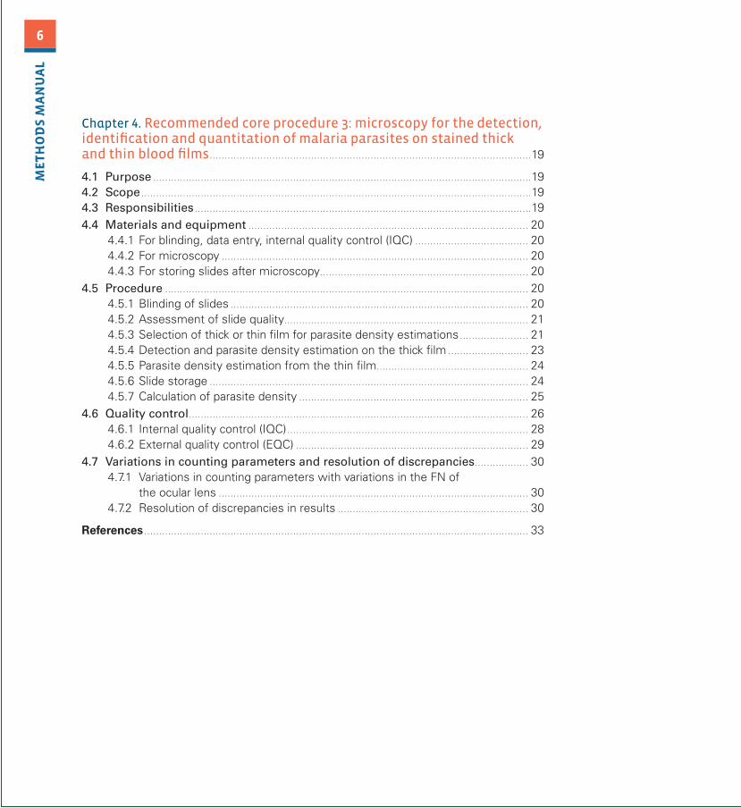
Chapter 4. Recommended core procedure 3: microscopy for the detection, identification and quantitation of malaria parasites on stained thick and thin blood films ............................................................................................................19
4.1 Purpose ...............................................................................................................................194.2 Scope ...................................................................................................................................194.3 Responsibilities .................................................................................................................194.4 Materials and equipment .............................................................................................. 20
4.4.1 For blinding, data entry, internal quality control (IQC) ...................................... 204.4.2 For microscopy ....................................................................................................... 204.4.3 For storing slides after microscopy ...................................................................... 20
4.5 Procedure .......................................................................................................................... 204.5.1 Blinding of slides .................................................................................................... 204.5.2 Assessment of slide quality .................................................................................. 214.5.3 Selection of thick or thin film for parasite density estimations ....................... 214.5.4 Detection and parasite density estimation on the thick film ........................... 234.5.5 Parasite density estimation from the thin film ................................................... 244.5.6 Slide storage ........................................................................................................... 244.5.7 Calculation of parasite density ............................................................................. 25
4.6 Quality control .................................................................................................................. 264.6.1 Internal quality control (IQC) ................................................................................. 284.6.2 External quality control (EQC) .............................................................................. 29
4.7 Variations in counting parameters and resolution of discrepancies .................. 304.7.1 Variations in counting parameters with variations in the FN of the ocular lens ........................................................................................................ 304.7.2 Resolution of discrepancies in results ................................................................ 30
References ................................................................................................................................. 33

7
ME
TH
OD
S M
AN
UA
L
Microscopy for the detection, identification and quantification of malaria parasites on stained thick and thin blood films in research settings
Mr El Hadji Ba, Research Institute for Development – Senegal, Dakar, Senegal; Professor J. Kevin Baird, Eijkman-Oxford Clinical Research Unit, Jakarta, Indonesia; Dr John Barnwell, United States Centers for Disease Control and Prevention (CDC), Atlanta, GA, USA; Dr David Bell, Global Good and Intellectual Ventures Laboratory Bellevue, WA, USA; Dr Jane Carter, African Medical and Research Foundation (Amref) Health Africa, Nairobi, Kenya; Dr Mehul Dhorda, WorldWide Antimalarial Resistance Network, Bangkok, Thailand; Professor Arjen Dondorp, Mahidol-Oxford Tropical Medicine Research Unit, Bangkok, Thailand; Ms Lenny Ekawati, Eijkman-Oxford Clinical Research Unit, Jakarta, Indonesia; Dr Michelle Gatton, Queensland University of Technology, Brisbane, QLD, Australia; Dr Iveth González, FIND, Geneva, Switzerland; Professor Philippe J. Guerin, WorldWide Antimalarial Resistance Network University of Oxford, Oxford, United Kingdom; Dr Sandra Incardona, FIND, Geneva, Switzerland; Dr Ken Lilley, Army Malaria Institute (AMI), Enoggera, QLD, Australia; Dr Didier Menard, Institut Pasteur du Cambodge, Phnom Penh, Cambodia; Professor Francois Nosten, Shoklo Malaria Research Unit, Mae Sot, Thailand; Dr Peter Obare, Kenya Medical Research Institute Kisumu, Kenya; Dr Bernhards Ogutu, Kenya Medical Research Institute Kisumu, Kenya; Dr Piero L. Olliaro, Special Programme for Research and Training in Tropical Diseases (TDR), a co-sponsored programme of UNICEF, UNDP, the World Bank and WHO, Geneva, Switzerland; Professor Ric N. Price, WorldWide Antimalarial Resistance Network, Darwin, Australia; Mr Stephane Proux, Shoklo Malaria Research Unit, Mae Sot, Thailand; Dr Andrew R. Ramsay, Special Programme for Research and Training in Tropical Diseases (TDR), a co-sponsored programme of UNICEF, UNDP, the World Bank and WHO, Geneva, Switzerland; Dr John C. Reeder, Special Programme for Research and Training in Tropical Diseases (TDR), a co-sponsored programme of UNICEF, UNDP, the World Bank and WHO, Geneva, Switzerland; Dr Kamolrat Silamut, Mahidol-Oxford Tropical Medicine Research Unit, Bangkok, Thailand; Dr Cheikh Sokhna, Research Institute for Development – Senegal, Dakar, Senegal.
Acknowledgements

8d
ef
init
ion
of
te
rm
s
Abbreviations
CDC Centers for Disease Control and Prevention, USA
EOCRU Eijkman-Oxford Clinical Research Unit, Indonesia
EQC External quality control
FN Field number
GG-IVL Global Good and Intellectual Ventures Laboratory
HPF High-power field
ID Identification
IPC Institut Pasteur du Cambodge, Cambodia
IQC Internal quality control
IRD Institut de Recherche pour le Développement, Senegal
KEMRI Kenya Medical Research Institute
MORU Mahidol-Oxford Tropical Medicine Research Unit, Thailand
QC Quality control
QUT Queensland University of Technology, Australia
RBC Red blood cell
ReMMS Research Malaria Microscopy Standards
SMRU Shoklo Malaria Research Unit, Thailand
SOP Standard operating procedure
TDR UNICEF/UNDP/World Bank/WHO Special Programme for Research and Training in Tropical Diseases
Vs Versus
WBC White blood cell
WHO World Health Organization
WWARN WorldWide Antimalarial Resistance Network, Oxford, United Kingdom

9
ME
TH
OD
S M
AN
UA
L
Microscopy for the detection, identification and quantification of malaria parasites on stained thick and thin blood films in research settings
Chapter 1 Introduction
Malaria microscopy remains a major reference standard for field trials of clinical interventions and other diagnostic plat-forms. The term ‘expert microscopy’ is commonly used to define a perceived higher level of competency than is normally seen among clinical microscopists, but this term is poorly defined. Microscopy standards have been developed by the World Health Organization (WHO) to standardize and benchmark competency for training and assessment of malaria microscopists in health services, national malaria programmes and national reference laboratories (WHO, 2009a). Mi-croscopy for malaria research has further specific requirements for expertise, often requiring particular rigour in parasite quantification and in diagnostic specificity of parasite detection. The participants of an informal consultation convened by TDR, the Special Programme for Research and Training in Tropical Diseases, jointly recognized a need to standardize research microscopy to improve the quality of clinical and diagnostic trials, enable comparisons of outcomes between clinical trials, and provide clarity in publication.
This manual was developed to guide a move towards common standards for undertaking and reporting research mi-croscopy for malaria parasite detection, identification and quantification.
It contains procedures based on agreed quality assurance standards for research malaria microscopy defined by the participants in the above consultation: TDR; the WorldWide Antimalarial Resistance Network (WWARN), United Kingdom; FIND, Switzerland; the Centers for Disease Control and Prevention (CDC), USA; the Kenya Medical Research Institute (KEMRI) and later expanded to include Amref Health Africa (Kenya); the Eijkman-Oxford Clinical Research Unit (EOCRU), Indonesia; Institut Pasteur du Cambodge (IPC); Institut de recherche pour le Développement (IRD), Senegal; the Global Good and Intellectual Ventures Laboratory (GG-IVL), USA; the Mahidol-Oxford Tropical Medicine Research Unit (MORU), Thailand; Queensland University of Technology (QUT), Australia, and the Shoklo Malaria Research Unit (SMRU), Thailand.
These ‘Research Malaria Microscopy Standards’ (ReMMS) constitute a suggested minimum standard for malaria microscopy in research. The collaborating institutions will aim to adhere to the ReMMS in published research studies. It is hoped that they will form a solid basis for the wider adoption of standardized reference microscopy protocols for malaria research.

10M
ET
HO
DS
MA
NU
AL

11
ME
TH
OD
S M
AN
UA
L
Microscopy for the detection, identification and quantification of malaria parasites on stained thick and thin blood films in research settings
Chapter 2 Recommended core procedure 1: preparation of blood films for malaria microscopy
2.1 PurposeThis section of the document describes the procedure for the preparation of blood films from peripheral blood collected by fingerprick or venepuncture for staining and microscopy to detect, identify and quantify malaria parasites.
2.2 ScopeThis procedure is intended for use in malaria clinical research studies such as those assessing drug or vaccine efficacy or diagnostic test performance. It is based on agreed quality assurance standards for research malaria microscopy de-fined during an informal consultation convened by TDR with representatives from the CDC, FIND, KEMRI, TDR and WWARN. It was later expanded to include Amref Health Africa, EOCRU, GG-IVL, IPC, IRD, MORU, QUT and SMRU.
The text of this procedure may need to be adapted or edited before implementation in different settings. However, cer-tain procedural details underlined in the text below must be retained in the standard operating procedure (SOP) and/or implemented as part of the procedure and its quality assurance in order to ensure compliance with ReMMS.
2.3 ResponsibilitiesThe tasks to be completed for this procedure are listed below. Each of these must be assigned to an individual(s) who has been trained to perform these tasks and in the use of relevant health and safety precautions.
• Labelling of slides
• Preparation of the blood film
2.4 Materials and equipmentFor preparation of thick and thin blood films.
• Glass slides: new, grease-free, with one frosted end
• Label with permanent marker

12M
ET
HO
DS
MA
NU
AL
• Micropipette with measuring range 2–20 µL
• Slide template
• ‘Spreader’ slide: bevelled corners, ground edges
• Methanol absolute, laboratory reagent grade or better.
2.5 Procedure
2.5.1 Labelling
At a minimum, slides must be labelled with study and subject identification (ID) or code, subject initials, date and time of blood collection. The study time-point should also be noted if study subjects are followed up.
Labels on which the writing area can be covered with a clear laminate (wrap-around labels) and/or barcoded labels should be used wherever possible. If these are not used, the labelling must be done so that the writing remains legible after staining, and after archiving of the slides for long periods.
i. Write the study and subject ID, subject initials, date and time of blood collection (and the time-point if appropriate) on a label using a permanent marker. Alternatively, if an appropriate validated system for slide tracking exists, the slides can be labelled only with a unique code.
ii. Attach the label at one end of the slide.
2.5.2 Thick and thin blood film preparation
The use of new, clean, grease-free, unscratched slides is essential. Well-made thick blood films are evenly spread, have 10–15 white blood cells (WBCs) per high-power field (HPF) on average (though this may vary depending on the WBC count and the field number – FN – of the ocular lens) at 1000× magnification, and are free of dust and other debris. Thick films of the correct thickness can be consistently prepared using measured volumes of blood spread over a fixed area. Though it is possible to make films of acceptable quality without using a micropipette and template, using these tools helps ensure reproducibility and facilitates the training of technicians. Good quality thin films have a feathered edge with a monolayer of red blood cells (RBCs). Thin films must be spread using the unchipped edge of a ground-edge slide. The preparation of standard films is aided by the use of slide templates that indicate the placement of the films and their size.
The volume of blood and the size of the thick film detailed in the procedure below have been calculated based on specifications of commonly available microscopes with 100× plan achromat oil immersion objective lens and a 10× ocular lens with FN 18 or 20 to give 10–15 WBC per HPF on average in a blood sample with a WBC count of 8000/µL.
The duration or method of drying the slides may be varied but the parameters of the method used should be validated with respect to the equipment used, typical humidity of the environment, and whether or not the blood is anti-coagulated to ensure that the thick films are neither heat-fixed nor washed off. The duration of drying may need to be prolonged if the blood has been collected with an anti-coagulant such as ethylenediaminetetraacetic acid (EDTA).
ChaPteR 2 Recommended core procedure 1

13
ME
TH
OD
S M
AN
UA
L
Microscopy for the detection, identification and quantification of malaria parasites on stained thick and thin blood films in research settings
i. Place the labelled slide on the template below (Figure 1).
ii. Using a micropipette, place 6 μl of blood for the thick film and 2–3 μl for the thin film as shown on the template in Figure 1.
Note: While transferring blood with a micropipette to a slide, it is advisable to use reverse-pipetting to prevent the formation of bubbles in the thick film, i.e. when the volume of liquid aspirated into the micropipette tip is greater than the volume delivered. Thick films can be made with lower or higher volumes of blood but the diameter of the film (and the template in Figure 1 below) will then need to be adjusted (see section 2.8).
iii. Using the ground edge of the spreader slide spread the blood for the thin film.
iv. Using the bevelled corner of the spreader slide spread the blood for the thick film until the entire circle of 12 mm diameter is covered evenly.
v. Dry the films on a flat surface, protected from dust and insects. Slides must be completely dry before staining by drying on a slide warmer at 37–40 ºC for 1 hour or overnight in a dehumidified chamber at ambient temperature.
Note: Hair driers may also be used to dry the slides but in all cases, slides must not be subjected to excessive and con-tinuous heat as this will prevent de-haemoglobinization of the thick film due to heat fixing.
vi. Fix the thin film by dipping it in absolute methanol for a few seconds and then letting the slide air dry. Dry the thin film at an acute angle, with the film-side of the slide facing up and the thin film downwards. This protects the thick film from being fixed by methanol fumes and run-off. The thick film must not be fixed.
Note: The slide can be stained immediately after drying or can be stored up to a month in a sealed box with ample desiccant in a cool, dry place for staining at a later date.
Figure 1. Template for preparation of slides with thick and thin blood films
Note: The dimensions of the thick film on printed versions of this template must be verified before use.

14M
ET
HO
DS
MA
NU
AL
2.6 Quality controlAll films should be checked for obvious signs of poor quality and repeated if any such signs are seen:
• dust or dirt on the films
• uneven or patchy thick films, ‘streaky’ thin films, often caused by greasy slides or by using a chipped spreader slide
• fixation of thick film.
2.7 Variations in the size of the thick filmTable 1. Variation in the size of the thick film with the volume of blood used
Volume of blood (µL)
Diameter of thick film (mm)Area of thick film
(mm2)
4 10 78.5
5 11 95.0
6 12 113.1
7 13 132.7
ChaPteR 2 Recommended core procedure 1

15
ME
TH
OD
S M
AN
UA
L
Microscopy for the detection, identification and quantification of malaria parasites on stained thick and thin blood films in research settings
Chapter 3 Recommended core procedure 2: staining of blood films for malaria microscopy
3.1 PurposeThis section of the Manual describes the procedure for staining blood films from peripheral blood collected by fingerprick or venepuncture for microscopy to detect, identify and quantify malaria parasites.
3.2 ScopeThis procedure is intended for use in malaria clinical research studies such as those assessing drug or vaccine efficacy or diagnostic test performance. It is based on agreed quality assurance standards for research malaria microscopy de-fined during an informal consultation convened by TDR with representatives from the CDC, FIND, KEMRI, TDR and WWARN. It was later expanded to include Amref Health Africa, EOCRU, GG-IVL, IPC, IRD, MORU, QUT and SMRU.
The text of this procedure may need to be adapted or edited before implementation in different settings. However, certain procedural details underlined in the text below must be retained in the SOP and/or implemented as part of the procedure and its quality assurance in order to ensure compliance with a common standard.
3.3 ResponsibilitiesThe tasks to be completed for this procedure are listed below. Each of these must be assigned to an individual(s) who has been trained to perform these tasks using the relevant health and safety precautions.
• Preparation of stock buffer solution and/or buffered water.
• Preparation of stock stain solution and/or working stain solution.
• Staining of blood films on slides.

16M
ET
HO
DS
MA
NU
AL
3.4 Materials and equipment
3.4.1 For buffered water:
• Volumetric flask or measuring cylinder, 1 L
• Distilled water
• Stock buffer solution or buffer tablets, pH 7.2
• 2% solutions of Na2HPO4 and KH2PO4
• pH meter.
3.4.2 For Giemsa stain solution:
• Funnel
• Filter paper
• Dark or opaque bottle with stopper or cap
• Stock Giemsa solution
• Measuring cylinder, 50–100 mL
• Micropipette (100–1000 µL) with tips or 1–2 mL pipette with pipetting aid
• Buffered water, pH 7.2.
3.4.3 For staining of blood films:
• Slide staining rack
• Timer
• Forceps
• Wash bottle
• Slide drying rack.
3.5 Procedure
3.5.1 Preparation of buffered water
Buffered water for dilution of stock Giemsa stain solution can be conveniently prepared using commercially manufactured pH 7.2 buffer tablets. If such tablets cannot be procured, stock phosphate buffer can be prepared using the recipe in WHO’s Basic Malaria Microscopy guide (WHO, 2010a). Water used for motor vehicle lead-acid batteries is distilled and can be used to prepare the stock and working solutions of the buffer. If distilled water is not available, water filtered using ceramic filters may be used. In all cases, the pH of the buffered water must be verified with a pH meter before use.
ChaPteR 3 Recommended core procedure 2

17
ME
TH
OD
S M
AN
UA
L
Microscopy for the detection, identification and quantification of malaria parasites on stained thick and thin blood films in research settings
i. Measure out roughly 900 mL of distilled water into the volumetric flask or measuring cylinder.
ii. Add 1 buffer tablet, pH 7.2 and swirl to mix.
iii. Using a calibrated pH meter, verify that the pH of the buffered water is within an acceptable range (pH 7.2 ± 0.1). Adjust the pH if needed using solutions of Na
2HPO
4 and KH
2PO
4 (2% w/v).
Note: If using commercially prepared buffer tablets, the pH is usually within acceptable ranges without adjustment. If needed, however, refer to the Basic Malaria Microscopy guide (WHO, 2010a) for recipes of the solutions and further details on how to adjust the pH.
iv. Complete the volume of the buffer solution up to 1 L and store at 2–8 ºC until further use.
Note: Even with storage at 2–8 ºC, the buffer solution may be contaminated by algal or other growth. Check the buffered water for turbidity (which indicates such growth) prior to use in staining of blood films.
3.5.2 Preparation of Giemsa working solution
Use of commercial stain preparations is recommended. The stain must be selected by testing 3% dilutions of various manufacturers’ products for different staining durations (e.g. 30, 40, 45, 50 minutes) for stain quality under the condi-tions in which the stain will be used routinely. If commercial stain preparations cannot be procured, stock Giemsa stain solution can be prepared using the recipe in the Basic Malaria Microscopy guide (WHO, 2010a). Working solutions must be freshly prepared just before use by diluting stock stain solution which has been filtered on the same day to reduce stain artefacts and deposits on the films. Any diluted stain left over after all slides have been stained must be discarded.
The procedure below outlines the steps for 3% v/v stain solution for use with the ‘slow’ staining method as it results in better staining and helps reduce parasite loss during staining. The ‘rapid’ staining method with 10% v/v diluted stain may be used for screening of patients for inclusion into studies and/or in cases where a quick diagnosis is needed. In such cases, however, a duplicate slide must also be made and stained with the slow staining method.
i. Using a funnel and filter paper, filter the stock Giemsa solution into a dry, dark or opaque bottle. Keep the bottle capped until the stain is used as moisture will cause the Giemsa stain to deteriorate.
Note: The stock stain solution can be filtered once daily. Any remainder may be poured back into the original container while taking care not to agitate the stain.
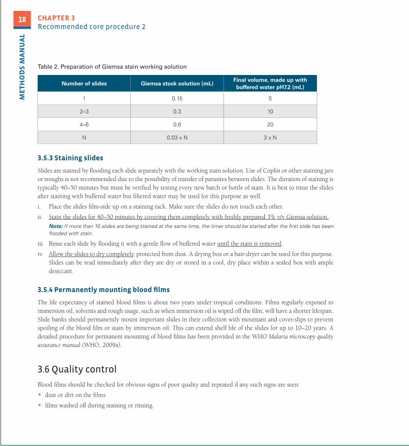
ii. Determine the volume of diluted stain (3% v/v) to be prepared from table 2.
iii. Transfer buffered water (about half of the final volume) to a measuring cylinder and add the appropriate volume of stock stain solution. Complete the volume up to the level determined from the table above.
Note: Ensure that only dry pipettes come in contact with the stock Giemsa stain, as any introduced moisture will hasten the deterioration of the stain. To ensure accurate dilutions, volumes of stock Giemsa stain should be measured using micropipettes or 1–2 mL serological pipettes with pipetting aids.
18M
ET
HO
DS
MA
NU
AL
Table 2. Preparation of Giemsa stain working solution
Number of slides Giemsa stock solution (mL)Final volume, made up with buffered water pH7.2 (mL)
1 0.15 5
2–3 0.3 10
4–6 0.6 20
N 0.03 x N 3 x N
3.5.3 Staining slides
Slides are stained by flooding each slide separately with the working stain solution. Use of Coplin or other staining jars or troughs is not recommended due to the possibility of transfer of parasites between slides. The duration of staining is typically 40–50 minutes but must be verified by testing every new batch or bottle of stain. It is best to rinse the slides after staining with buffered water but filtered water may be used for this purpose as well.
i. Place the slides film-side up on a staining rack. Make sure the slides do not touch each other.
ii. Stain the slides for 40–50 minutes by covering them completely with freshly prepared 3% v/v Giemsa solution.
Note: If more than 10 slides are being stained at the same time, the timer should be started after the first slide has been flooded with stain.
iii. Rinse each slide by flooding it with a gentle flow of buffered water until the stain is removed.
iv. Allow the slides to dry completely, protected from dust. A drying box or a hair-dryer can be used for this purpose. Slides can be read immediately after they are dry or stored in a cool, dry place within a sealed box with ample desiccant.
3.5.4 Permanently mounting blood films
The life expectancy of stained blood films is about two years under tropical conditions. Films regularly exposed to immersion oil, solvents and rough usage, such as when immersion oil is wiped off the film, will have a shorter lifespan. Slide banks should permanently mount important slides in their collection with mountant and cover-slips to prevent spoiling of the blood film or stain by immersion oil. This can extend shelf life of the slides for up to 10–20 years. A detailed procedure for permanent mounting of blood films has been provided in the WHO Malaria microscopy quality assurance manual (WHO, 2009a).
3.6 Quality control
Blood films should be checked for obvious signs of poor quality and repeated if any such signs are seen:
• dust or dirt on the films
• films washed off during staining or rinsing.
ChaPteR 3 Recommended core procedure 2

19
ME
TH
OD
S M
AN
UA
L
Microscopy for the detection, identification and quantification of malaria parasites on stained thick and thin blood films in research settings
Chapter 4 Recommended core procedure 3: microscopy for the detection, identification and quantitation of malaria parasites on stained thick and thin blood films
4.1 PurposeThis section of the document describes the procedure for microscopy of Giemsa-stained thick and thin blood films from peripheral blood to detect, identify and quantify malaria parasites.
4.2 ScopeThis procedure is intended for use in malaria clinical research studies such as those assessing drug or vaccine efficacy or diagnostic test performance. It is based on agreed quality assurance standards for research malaria microscopy de-fined during an informal consultation convened by TDR with representatives from the CDC, FIND, KEMRI, TDR and WWARN. It was later expanded to include Amref Health Africa, EOCRU, GG-IVL, IPC, IRD, MORU, QUT and SMRU.
The text of this procedure may need to be adapted or edited before implementation in different settings. However, certain procedural details underlined in the text below must be retained in the SOP and/or implemented as part of the procedure and its quality assurance in order to ensure compliance with a common standard.
4.3 ResponsibilitiesThe tasks to be completed for this procedure are listed below. Each must be assigned to an individual(s) who has been trained to perform these tasks using the relevant health and safety precautions.
• Blinding, data entry, coordination of duplicate/tie-breaker reading
• Assessment of slide quality
• Slide reading, detection and quantification of parasites.

20M
ET
HO
DS
MA
NU
AL
4.4 Materials and equipment
4.4.1 For blinding, data entry, internal quality control (IQC)
• Masking tape or barcode label with unique identifier
• Computer with Microsoft Access or Excel or similar database or spreadsheet software
• Microscopy results log books.
4.4.2 For microscopy
i. Microscope
- Objective: 100× oil-immersion, plan achromat, grid reticle
- Ocular: 10×, grid reticle (if no reticle on the objective)
- Electrical light source (LED or filament-type with blue filter).
ii. Synthetic immersion oil, refractive index ≥ 1.5.
iii. Single or multi-parameter tally counters.
4.4.3 For storing slides after microscopy
iv. Absorbent paper or Histoclear reagent.
v. Slide boxes.
vi. Adhesive tape.
vii. Desiccant (if storage is not in an air-conditioned room).
4.5 Procedure
4.5.1 Blinding of slides
Microscopy must be done on ‘blinded’ slides to prevent any bias in the microscopy, i.e. microscopists should neither be aware of patient and sample data nor of the result of any other diagnostic tests performed on the same sample. This can be done by assigning a unique identifier to each slide to track slides during microscopy, to log results, and to link laboratory results with case records.
i. Enter patient and sample data into the database or IQC Excel sheet and assign a unique identifier to the slide (can be a sequential number).
ii. Cover the slide label with opaque masking tape and write the unique identifier on it.
iii. Transfer the slide(s) to the microscopists for reading.
ChaPteR 4 Recommended core procedure 3

21
ME
TH
OD
S M
AN
UA
L
Microscopy for the detection, identification and quantification of malaria parasites on stained thick and thin blood films in research settings
4.5.2 Assessment of slide quality
Before slides are read to detect or to quantify malaria parasites, they must be assessed for quality with respect to the criteria below by scanning a few fields on the slide using immersion oil 100× objective. If the slide is not readable, it should be logged as such and the slide preparation must be repeated. Any deviations from these criteria should be noted as they may interfere with microscopy examination. The number of WBCs per HPF in a thick film with a fixed volume of blood and film diameter will vary with the FN (see sub-section 4.7.1) so the criteria defined below may need to be adapted to the specifications of the microscope being used. Slide quality assessment can be done while selecting the thick vs thin film for parasite density estimation.
Slide quality assessment criteria for thick films:
- 10–15 WBC per HPF on average (at 1000× total magnification with an assumed 8000 WBC/µL)
- pale blue-grey background (lysed RBC) free of Giemsa precipitates, dust and other artefacts
- WBC nuclei should be stained deep purple and platelets should be clearly visible and bright pink.
Slide quality assessment criteria for thin films:
- presence of a ‘tail’ or feathered edge with evenly distributed RBCs and no or very few overlapping cells
- RBCs stained grey-pink.
4.5.3 Selection of thick or thin film for parasite density estimations
Parasite density estimations can be performed from the thick or thin films with each method having its limitations. Par-asite density estimations from the thin film tend to be more accurate than those estimated from the thick film because there is little or no parasite loss during staining. However, the thin film method is mostly appropriate for higher parasite densities only. Parasite density estimation from the thick film may be somewhat less accurate due to parasite loss but can be adapted for use with a wide range of parasite densities at the medium to lower end of detectable parasitaemia (<~16000 parasite/µL). Thin films will be more accurate higher levels of parasitaemia (>~16000 parasites/µL), as it may be difficult to obtain precise parasite counts from thick films with these high parasite densities. In determinations of the parasite density from the thick film, the methods using the white cell count, the ‘per HPF’ method or the Earle-Perez method may be used (Earle & Perez, 1932). Accuracy with the Earle-Perez method may not differ greatly from counting against a known white cell count on a thick film (Bowers et al., 2009).
Thick films must be examined to check for the presence of gametocytes and/or for mixed infections irrespective of whether the asexual parasite density is estimated on the thick or thin film.
i. Place a small drop of immersion oil on the thick and thin films.
ii. Scan 10 fields on the thick film using the 100× immersion oil objective.
iii. Assess the slide quality according to the criteria outlined in section 4.6.2 above and reject the slide if the quality is poor (excessive artefacts or stain precipitate, non-standard stain or pH interfering with parasite detection and/or identification, fixed or patchy thick films, etc).
iv. Count the numbers of parasites and WBCs seen.

22M
ET
HO
DS
MA
NU
AL
v. If the number of asexual parasites is less than or equal to twice that of WBCs, continue with asexual parasite detec-tion or parasite density estimation on the thick film.
vi. If the number of parasites is greater than twice the number of WBCs, perform the asexual parasite density estimation on the thin film (WHO, 2009a).
4.5.4 Detection and parasite density estimation on the thick film
Detection of parasites (asexual or sexual forms) is performed against a standard number of WBCs or HPFs on the thick film. Identification of the species of the parasites is best done on the thin film but it may have to be done on the thick film in cases with low parasite density. A slide is declared negative if no parasites are seen after reading the set number of WBCs or HPFs. The area of the film in view in an HPF, and therefore the volume of blood examined per field, de-pends on the FN of the ocular lens. For example, reading 200 HPFs of a thick film (made with 6 µL blood, diameter 12 mm) on a microscope with an ocular of FN 20 corresponds to approximately 0.33 µL of blood examined but with a microscope of FN 18, the volume of blood examined is approximately 0.27 µL. This has obvious implications if a fixed volume of blood is to be examined on a thick film and/or for calculations of parasite density using the ‘per HPF’ method.
The estimation of parasite density on the thick film is most accurate when the method used to detect parasites is con-sistent with the method used to count parasites. In other words, if slides are to be declared negative against a standard number of WBCs, any parasites detected must also be reported per the number of WBCs counted OR if slides are to be declared negative after examining a standard number of HPFs, any parasites detected must be reported per number of HPFs examined. Accuracy is further improved by beginning WBC or HPF counts from the first good quality field seen whether or not there are parasites in the field of view, as this takes into account the entire volume of blood examined for the parasite density calculations. In cases where the slide is negative or the parasite density is <1/500 WBC this implies counting up to 2500 WBCs, which can significantly increase the time needed to read a slide. If this is not feasible, the “per HPF” method should be used.
Objective or ocular lenses with grid reticles may be used for parasite density estimations on the thick film from a well-defined volume of blood, as in the Earle-Perez method. The use of ocular lenses with grid reticles covering the centre of the field of view is advisable if the objective lens is not of the plan achromat type to assist with counting only in the part of the field that is in sharp focus. If reticles are used, only those parasites and WBCs that are within the grid on a given field must be counted.
The Earle-Perez (1932) method is a method for determining parasite density from the thick film, which can be used when actual WBC counts are not available (WHO-WPRO, 2010c; Laurens et al., 2012). However, results can be similar to calculation against a known white cell count, and either method is acceptable (Bowers et al., 2009). In the Earle-Perez method, a known volume of blood (5 µL) is spread evenly over a rectangle of 6 × 15 mm. The thick film is examined using an ocular fitted with a 6 × 6 grid reticule by moving across the width of the thick film, and the parasite density is estimated based on the number of parasites seen per ‘band’, i.e. calculated volume of blood, examined across the width of the film.
In cases of mixed infections, all infecting species must be reported. However, the asexual and/or sexual parasite density need not be reported separately for each species unless specifically required by the study’s objectives.
ChaPteR 4 Recommended core procedure 3

23
ME
TH
OD
S M
AN
UA
L
Microscopy for the detection, identification and quantification of malaria parasites on stained thick and thin blood films in research settings
i. Use tally counters set to zero to count the number of WBCs or fields and asexual/sexual parasites. Start counting from the first good quality field (i.e. fields with little or no debris/artefacts and 10–15 WBCs) – do not wait to find a parasite before starting to count WBCs/fields.
ii. If no asexual or sexual forms of the parasites are seen after 2500 WBCs or 200 HPFs, declare the slide negative.
iii. If parasites are seen before 500 WBCs or 40 fields have been counted, continue counting up to 500 WBCs or 40 fields before reporting the result. If parasites are seen after 500 WBCs or 40 fields have already been counted, stop the reading after all the parasites and/or WBCs on the field have been counted. Counting must be completed for the last field, i.e. all parasites and WBCs on the last field must be counted, and can be stopped if >490 WBCs have already been counted after a field.
Note: Parasites of different species need not be counted separately unless required by the study’s objectives. Gametocytes and malaria pigment should be reported if seen but need not be counted unless required by the study. Schizonts should be reported separately if required by the study’s objectives. Presence of P. falciparum schizonts, especially if present in large numbers, may indicate severe disease and clinicians must be informed accordingly.
iv. Report the result as raw figures, i.e. species of the parasites, the number of asexual or sexual forms of the parasite over the number of WBCs or fields counted.
Figure 2. Diagram of slide reading
Source: WHO (2009).

24M
ET
HO
DS
MA
NU
AL
4.5.5 Parasite density estimation from the thin film
Parasite detection is performed against a standard number of RBCs on the thin film in case of high parasite density (i.e. >~16 000 parasites/µL). This helps to reduce inaccuracy and imprecision caused by the loss of parasites during staining and by counting large numbers of parasites in a field. Counting of RBCs must begin from the first good quality field to improve accuracy of the parasite density estimate.
Gametocytes or schizonts may be seen on the thin film but they must not be counted from the thin film as this is likely to result in an overestimation of the gametocyte or schizont density.
i. Use tally counters set to zero to count the number of RBCs and asexual parasites. Start counting from the first good quality field (i.e. fields with little or no debris/artefacts and evenly spread, non-overlapping RBCs at the ‘tail’ or feathered edge of the thin film) – do not wait to find a parasite before starting to count RBCs.
ii. Count parasitized RBCs along with normal RBCs until a total of 2000 RBCs has been counted. Count RBCs with more than one parasite or with multinucleate forms as 1 infected RBC. Counting must be completed for the last field, i.e. all parasites and RBCs on the last field must be counted, and can be stopped if >1900 RBCs have already been counted after a field. Parasites of different species need not be counted separately. Note: The presence of P. falciparum schizonts, especially if present in large numbers, may indicate severe disease and clinicians must be informed accordingly. Parasites must be counted against 2000 RBCs at a minimum though this number may be increased if required by the study objectives.
iii. In the unlikely event that no asexual parasites are seen on the thin film after counting 2000 RBCs, continue exam-ining the slide on the thick film as described in section 4.6.4 above.
Note: If the selection of thin vs. thick film for parasite density estimation has been done correctly, it is unlikely that no asexual parasites will be seen on the thin film, unless the thick film is uneven, the subject has an abnormally high RBC count or an abnormally low WBC count.
iv. Count at least 500 WBC or 40 fields on the thick film to check for the presence of other species and/or sexual forms of the parasite.
v. Report the result as raw figures, i.e. the species of the parasite and the number of infected RBCs over the total number of RBCs counted.
4.5.6 Slide storage
Slides may need to be stored for extended periods after initial microscopy has been completed for re-checking and/or archiving. To prevent deterioration of the stain, the immersion oil must be removed from the slides and they must be protected from heat, humidity and light.
i. Remove the immersion oil by leaving the slides overnight face down on soft absorbent paper or by dipping in Histoclear reagent and swirling gently.
Note: Histoclear is a solvent and is flammable. If using permanent markers to label the slides, protect the label from exposure to Histoclear. Use Histoclear in an open, well-ventilated space away from sources of heat and/or open flames.
ii. Sort slides according to the date of collection, by patient, by study time-point, positive/negatives, etc., as convenient.
iii. Store the slides in slide boxes and log their positions. Protect the slides from humidity, dust and insects.
ChaPteR 4 Recommended core procedure 3

25
ME
TH
OD
S M
AN
UA
L
Microscopy for the detection, identification and quantification of malaria parasites on stained thick and thin blood films in research settings
4.5.7 Calculation of parasite density
Parasite density can be calculated by counting parasites against WBCs or per HPF on the thick film or against RBCs on the thin film. Parasite density estimations are most accurate when actual WBC or RBC counts per microlitre are used in the calculations. While assumed WBC (8000 WBC/µL) and/or RBC (5.0 × 10⁶/µL) counts can provide reasonably accurate estimations of the parasite density in most cases, they may also introduce errors. This can be obviated by using the per HPF method but the calculations for parasite density estimations must then take into account the volume of blood in the field: a function of the blood volume, the area of the thick film and the consistency of distribution, as well as the FN of the microscope used.
Microscopists must report the raw data, i.e. the number of parasites over WBCs/RBCs/HPFs. The final per microlitre density calculations should be done using computerized tools or a calculator. The calculations below are detailed for information only.
i. Parasite density estimation from parasites counted against WBCs on the thick film:
Parasite density per µL = Number of parasites counted × WBC count per µL ÷ Number of WBCs counted
The actual WBC count, either obtained concurrently with the blood film or at the time of diagnosis of the current infec-tion, must be used in this calculation. If this is not feasible, the ‘per HPF’ method for parasite density estimation from the thick film should be considered.
ii. Parasite density estimation from parasites counted against HPFs on the thick film
Parasite density per µL = Number of parasites counted ÷ (Number of HPFs × Volume of blood per HPF)
The volume of blood examined per HPF can be estimated from table 3 in sub-section 4.8.1.
iii. Parasite density estimation from parasites counted against RBCs on the thin film
Parasite density per µL = Number of parasites counted × RBC count per µL ÷ Number of RBCs counted
The actual RBC count, either obtained concurrently with the blood film or at the time of diagnosis of the current infec-tion, must be used in this calculation.

26M
ET
HO
DS
MA
NU
AL
4.6 Quality controlQuality assurance of malaria microscopy in research settings must include at a minimum confirmation of competency of technicians prior to the study (‘accreditation’ or ‘competency assessment’), IQC and external quality control (EQC), together with controls over the workload, workplace, and the quality of equipment and reagents.
IQC and EQC (i.e. re-checking of slides to assess film and stain quality and parasite detection and quantification) are critical components of a quality assurance system for malaria microscopy. Results from IQC should be used as an ‘early warning system’ to detect failings in film and stain quality as well as a tool for continuous competency assessment of the technicians and microscopists. EQC provides an independent assessment to verify the results and detect gross or systematic errors, which may be missed if all local microscopists are making the same mistakes. Depending on the competency of the microscopists, up to 100% of the slides for a given study should be read twice for IQC but fewer slides need to be rechecked for EQC.
The IQC and EQC scheme outlined below is the minimum that needs to be implemented for quality assurance of re-search malaria microscopy. It may need to be adapted or augmented with tests for additional parameters depending on the requirements of the study being undertaken. For example, in studies to assess parasite clearance rates, it may be necessary to recheck and/or compare results from all slides from a given subject. Implementation of IQC and EQC does not preclude the requirement for training and competency assessment of the microscopists before study initiation to establish their competency in following the procedures, and producing results according to the standards set out below.
Figure 3. Ensuring and demonstrating good performance in malaria microscopy
Source: WHO (2009)
ChaPteR 4 Recommended core procedure 3
Selection
Training Competency
Supervision
Equipment and reagents PERFORMANCE
Cross-checking of routinely taken slides
Workload and environment
Assessment

27
ME
TH
OD
S M
AN
UA
L
Microscopy for the detection, identification and quantification of malaria parasites on stained thick and thin blood films in research settings
The competency of the microscopists to detect, identify and quantify parasites must be verified before study initiation (‘accreditation’) by testing with a slide set and adhering to the minimum standards detailed below. Slide sets used for accreditation must be of good quality, very well characterized, with polymerase chain reaction (PCR) confirmation of species, and multiple-blinded readings for validation of the presence or absence of parasites, and of the estimated parasite density. SOPs to produce and validate such sets can be found in the WHO Malaria microscopy quality assurance manual (WHO, 2009a). Slides with low, intermediate and high parasite densities must be included in the set in order to assess the accuracy of the microscopists with respect to parasite density estimation. The possibility that some parasites/species may be missed purely by chance on thick films with low to very low parasite densities must be taken into account when scoring the performance of the microscopists in terms of sensitivity and kappa scores.
– Positive slides (minimum 50 slides):
•Criteriawithrespecttoparasitespecies:
• ~25 slides with Plasmodium falciparum monoinfection
• ~10 slides with mixed infection, all with P. falciparum and 1 other co-infecting species (selected according to local prevalence), each species’ parasite density must be ≥2–3 parasites per 500 WBC/40 HPFs
• ~15 slides of P. malariae, P. ovale, P. vivax;
•Criteriawithrespecttoparasitedensity:
• ~10 slides with 50–200 parasites/µL
• ~10 slides with 200–500 parasites/µL
• ~20 slides with 500–2000 parasites/µL
• ~10 slides with 10 000–250 000 parasites/µL.
– Negative slides (minimum 80 slides):
• include 10–12 slides with dust, debris, fungus and/or bacteria, Howell-Jolly bodies
• include at least 1 example of each of the following: under-stained; over-stained; made with stain diluted in buffer pH ≤6.8, in buffer pH ≥7.6.

28M
ET
HO
DS
MA
NU
AL
Table 3. Grades for accreditation of research malaria microscopists
Accreditation levelDetection of parasitaemia
Species identification
Parasite quantitation
(25% of true count)
False positive rate
Level 1 (Expert) ≥90% ≥90% ≥50% ≤2.5%
Level 2 80–<90% 80–<90% 40–<50% ≤5%
Level 3 70–<80% 70–<80% 30–<40% ≤10%
Level 4 <70% <70% <30% ≤20%
Source: Adapted from WHO levels of accreditation for malaria microscopy (WHO, 2009a).
Microscopy workloads must be managed to ensure that the performance of the microscopists is maintained at an ac-ceptable level. Depending on the proportion of positive slides read, a microscopist should be able to read approximately 25–40 slides per day. Additional details on workload estimation are available in the WHO Malaria microscopy quality assurance manual (WHO, 2009).
4.6.1 Internal quality control (IQC)
The proportion of slides that need to be read twice for IQC depends on the competency of the microscopists. For uncertified microscopists and/or microscopists with an accreditation level of >2, all slides should be double-read with a third read in case of discordant results. The second and third readings can be performed in a reference laboratory with certified microscopists. In cases where all the microscopists in a study have an accreditation level of 2 or better as determined in formal assessments, at least 20% of the slides must be double-read for IQC.
Third or ‘tie-breaker’ readings must be performed systematically in case of discordant results with respect to the detection of the species of parasites and to parasite density estimates. Different criteria for acceptable variation in parasite density estimates have been used previously, most often based on proportional ranges and/or absolute counted parasite numbers. A more sophisticated statistical method has also been published (Alexander et al., 2010) and another, based on the as-sessment of the probability that two readings are from the same theoretical distribution is proposed here (see additional details in sub-section 4.7.2). While the different approaches may not always flag the same set of paired readings thus requiring a third reading for parasite density estimations, they are likely to be adequate as aids to improving parasite density estimates, and as tools for quality assurance of microscopy in general. Additional details on the resolution of inconsistent results are in sub-section 4.7.2.
ChaPteR 4 Recommended core procedure 3

29
ME
TH
OD
S M
AN
UA
L
Microscopy for the detection, identification and quantification of malaria parasites on stained thick and thin blood films in research settings
i. Enter the raw data reported by the microscopists into an Excel sheet or database which calculates the parasite density and compares the results from each microscopist according to the following criteria:
• presence/absence of asexual forms of the parasite only;
• species of the parasites;
• parasite density if asexual parasites are detected – readings must be compared to assess concordance except if both estimates are ≤ 200/µL, in which case both parasite density estimates are considered to be within accepted ranges of variability as long as both detect parasites;
• sexual forms and schizonts can be reported separately but, depending on the study’s needs, may or may not need to be considered in assessments of discordance.
ii. In case of discordance between the first and second readings using these criteria, a third tie-breaker reading must be organized. A Microsoft® Excel-based calculator is available to determine the requirement for a third read.
iii. Microscopy results obtained by each microscopist should be compared periodically with the final reported result to verify their competency, monitor the effect of the workload on microscopy performance, and identify needs for refresher training, etc.
Note: Results are analysed to determine kappa scores for parasite detection, species identification, and/or in Bland-Altman plots to assess accuracy and precision of parasite density estimations. Other analyses may be necessary depending on the type of study being undertaken.
4.6.2 External quality control (EQC)
i. A minimum of 10 slides per microscopist per month during a trial (5 positive and 5 negative, each selected ran-domly), or 5% overall if the number of slides is above 200 per month, selected in the same way, and their results rechecked and analysed by external assessors using statistical tools described above.
Note: EQC may be performed at the end of the study but it is preferable to recheck slides periodically in batches over the duration of the study.
ii. External assessors should also have evidence of competency equivalent to Level 1 in the Competency Assessment.

30M
ET
HO
DS
MA
NU
AL
4.7 Variations in counting parameters and resolution of discrepancies
4.7.1 Variations in counting parameters with differences in the FN of the ocular lens
Table 4. Variations in counting parameters with differences in the FN of the ocular lens
FN 18 20 22 26.5
AREA OF HPF (mm2) 0.0255 0.0314 0.0380 0.0552
VOLUME PER HPF (µL) 0.00135 0.00167 0.00202 0.00293
VOLUME PER 200 HPFs (µL) 0.270 0.333 0.403 0.585
MEAN WBC/HPF (assumed 8000 WBC/µL)
11 13 16 23
4.7.2 Resolution of discrepancies in results
i. Initial microscopy
Films are read independently by two qualified microscopists, each blinded to the results of the other reader and to the results of any other diagnostic tests, for:
• presence or absence of malaria parasites (asexual forms, i.e. positive or negative);
• malaria species present;
• parasite density;
• presence or absence of gametocytaemia, multinucleate forms (optional).
ii. Definition of inconsistent results
Results are entered into a database and systematically compared according to the three criteria above. Results are considered discrepant in any of the following cases:
• presence/absence of asexual forms of the parasite only;
• species of the parasites;
• parasite density if asexual parasites are detected – readings must be compared to assess concordance except if both estimates are ≤ 200/µL, in which case both parasite density estimates are considered to be within accepted ranges of variability as long as both detect parasites. A third reading is recommended when at least one of the densities is >200 parasites/µL and there is a <10% chance of observing the two densities if both were random samples from the theoretical probability distribution with mean equal to the average of the reading 1 and 2 densities.
ChaPteR 4 Recommended core procedure 3

31
ME
TH
OD
S M
AN
UA
L
Microscopy for the detection, identification and quantification of malaria parasites on stained thick and thin blood films in research settings
iii. Resolution of discrepant results
A third qualified microscopist reading of each film for which the first two results are inconsistent. The third micros-copist is blinded to the results of the first two readers and to the results of any other diagnostic tests. The results of the third reading are entered into the computerized database; results are compared and resolved to ‘final’ values:
• for presence vs. absence of parasitaemia, the third reader is the tie-breaker;
• for species, if the third reader independently identifies the same species as identified by either one of the first two readers, this is the final result; but if the third reader does not agree with either of the first two, the three readers discuss and review until there is agreement between at least two of the readers;
• for parasite density, if the third reader’s parasite density is within an acceptable margin of one of the first two readers’ densities, the final density is the mean of two values: the third value and whichever initial reading was closer to the third. If the third reader’s parasite density is not within an acceptable margin of either the first or second reader’s densities, or if it is within an acceptable range of both the first and second reader’s densities, the final density is the mean of all three values. A Microsoft® Excel-based calculator is available to determine the requirement for a third read.
Note: An alternative could be to record as ‘failure to quantify’ and/or have all three microscopists re-read blindly.

32M
ET
HO
DS
MA
NU
AL

33
ME
TH
OD
S M
AN
UA
L
Microscopy for the detection, identification and quantification of malaria parasites on stained thick and thin blood films in research settings
References
Alexander N, Schellenberg D, Ngasala B, Petzold M, Drakeley C, Sutherland C (2010). Assessing agreement between malaria slide density readings. C.Malar J. 2010;9:4.
Bowers KM, Bell D, Chiodini PL, Barnwell J, Incardona S, Yen S et al. (2009). Inter-rater reliability of malaria parasite counts and comparison of methods. Malar J.8:267.
Earle WS, Perez MJ (1932). Enumeration of parasites in the blood of malarial patients. Lab Clin Med.17:1124.
Laurens MB, Duncan CJ, Epstein JE, Hill AV, Komisar JL, Lyke KE et al. (2012). A consultation on the optimization of controlled human malaria infection by mosquito bite for evaluation of candidate malaria vaccines. Consensus Group on Design of Clinical Trials of Controlled Human Malaria Infection. Vaccine.30:5302.
WHO (2009). Malaria microscopy quality assurance manual, version 1. Geneva: World Health Organization (http://www.who.int/malaria/publications/malaria_microscopy_QA_manual.pdf?ua=1, accessed 15 October 2014).
WHO (2010a). Basic malaria microscopy, Part 1. Learner’s guide second edition. Geneva: World Health Organization (http://whqlibdoc.who.int/publications/2010/9789241547826_eng.pdf, accessed 15 October 2014).
WHO-WPRO/FIND/UNDP/World Bank/WHO-TDR/FIND/GMP (2010b). Methods manual for laboratory quality control testing of malaria rapid diagnostic tests, version six. WHO Regional Office for the Western Pacific (WPRO), FIND, United Nations Development Programme (UNDP)/World Bank/WHO Special Programme for Research &Training in Tropical Diseases (TDR), WHO Global Malaria Programme (GMP). Geneva: World Health Organization (http://www.wpro.who.int/malaria/NR/rdonlyres/461C306D-D720-43CA-A476-1A250EC3C26A/0/rdt_laborato-ry_qc_testing_meth_man_v6.pdf, accessed 15 October 2014).

Notes

Notes

TDR/World Health Organization20, Avenue Appia1211 Geneva 27Switzerland
Fax: (+41) 22 [email protected]/tdr
The Special Programme for Research and Training in Tropical Diseases (TDR) is a global programme of scientific collaboration established in 1975. Its focus is research into neglected diseases of the poor, with the goal of improving existing approaches and developing new ways to prevent, diagnose, treat and control these diseases. TDR is sponsored by the following organizations:
World Bank
ISBN 978 92 4 154921 9