SPOTLIGHT VECTOR HAZARD REPORT
6
1 SPOTLIGHT VECTOR HAZARD REPORT First report of arbovirus vector Aedes (Fre.) vittatus in the Americas Photograph: Specimen 1 of 2 collected in Guantanamo Bay, July 2019. Summary: In July 2019, mosquito specimens were collected in a CO2-baited CDC light trap on the US Naval Base biosurveillance at Guantanamo Bay Naval Base, Cuba and sent to Public Health Command - Fort Meade as part of routine, on-going surveillance activities. There, Dr. Ben Pagac and Dr. Alex Spring noted two unusually marked females, and brought them to WRBU for further species verification. The specimens were morphologically and molecularly (using mtDNA COI gene & nuclear rDNA ITS2) confirmed as Aedes (Fredwardsius) vittatus (Bigot, 1861) by David Pecor, Jim Pecor and Dr. Yvonne Linton – a finding that represents the first record of this competent arboviral vector in the Americas. The mtDNA barcode signature of both specimens were identical, which may indicate a point-source introduction/interception. Further sampling is needed to determine whether this finding represents interception of this exotic taxa, or represents an established breeding population on the island of Cuba. There are no indications as to the source of the introduction at this time. Prepared by David Pecor & Yvonne Linton Walter Reed Biosystematics Unit; 26 August 2019
Transcript of SPOTLIGHT VECTOR HAZARD REPORT

1
SPOTLIGHT VECTOR HAZARD REPORT
First report of arbovirus vector Aedes (Fre.) vittatus in the Americas
Photograph: Specimen 1 of 2 collected in Guantanamo Bay, July 2019.
Summary: In July 2019, mosquito specimens were collected in a CO2-baited CDC light trap on the US Naval Base
biosurveillance at Guantanamo Bay Naval Base, Cuba and sent to Public Health Command - Fort Meade as part
of routine, on-going surveillance activities. There, Dr. Ben Pagac and Dr. Alex Spring noted two unusually marked
females, and brought them to WRBU for further species verification. The specimens were morphologically and
molecularly (using mtDNA COI gene & nuclear rDNA ITS2) confirmed as Aedes (Fredwardsius) vittatus (Bigot,
1861) by David Pecor, Jim Pecor and Dr. Yvonne Linton – a finding that represents the first record of this
competent arboviral vector in the Americas. The mtDNA barcode signature of both specimens were identical,
which may indicate a point-source introduction/interception. Further sampling is needed to determine whether
this finding represents interception of this exotic taxa, or represents an established breeding population on the
island of Cuba. There are no indications as to the source of the introduction at this time.
Prepared by David Pecor & Yvonne Linton
Walter Reed Biosystematics Unit; 26 August 2019

2
Aedes (Fredwardsius) vittatus (Bigot, 1861) Subfamily Culicinae; Tribe Aedini
Informal Name: “Banded French Pointy Mosquito” Originally described by Bigot in 1861 (as Culex vittatus) from collections made on the French Mediterranean island of Corsica, Aedes vittatus was later placed in the Aedes subgenus Stegomyia. Following taxonomic revision, and later backed by molecular phylogenies, Aedes vittatus is now regarded as the sole representative of Aedes subgenus Fredwardsius. There are two currently recognized synonyms: syn. albopunctata Theobald, 1907 and syn. brumpti Neveu-Lemaire, 1905.
Diagnostic Characters:
Head: Vertex (V) and occiput (Occ) with numerous erect forked scales, not restricted to occiput. Vertex with a
median stripe of narrow white scales and with a few narrow white scales scattered on occiput area and along
eye margin. Proboscis (P) dark scaled with pale yellowish scales occupying about middle 0.33–0.40. Clypeus with
a small patch of narrow white scales on each side. Thorax: Mesopostnotum bare; prespiracular area bare;
postspiracular setae present; lower mesepimeral setae present. Scutum: Acrostichal setae present; 3 pairs of
distinct, small, white spots of narrow scales on anterior 0.67 of scutum (on scuttle fossa, posterior scuttle fossa
and c. level with the wing root). Scutellum: Broad white scales on all three lobes; a few dark scales at apex of
mid-lobe. Wing: Scales mainly narrow and dark on all veins; scattered pale scales on costa. Abdomen: Segment
VIII completely retracted. Tergum I with a large median white spot; terga II––VII each with a basal white band
and with lateral white curved markings which do not connect with the basal bands. Legs: Tibia dark, each with
a sub-basal white spot and a white band c. level to the basal third of T-I, T-II, and at mid-point of T-III (anterior
view). T-III1-4 with basal white bands [ratio of white band to length of tarsomeres: T-III1=0.40, T-III2=0.40, T-
III3=0.50, T-III4=0.75]; T-III5 all white (Edwards 1941; Huang & Ward 1981).
Comparison of scutum of pristine NMNH Collection specimen (left) and the rubbed specimen (#1) of Ae. vittatus (right) from Cuba,
showing acrostichal setae and three distinctive pairs of white spots on scutum.
3
Head (Dorsal): Vertex with a median stripe of narrow white scales and with a few narrow white scales scattered on occiput area and
along eye margin. Proboscis (P) dark scaled with pale yellowish scales occupying about middle 0.33–0.40. Clypeus with a small patch
of narrow white scales on each side.
Abdomen: Abdominal segment VIII completely retracted. Tergum I with a large median white spot; terga II-VII each with a basal
white band and with lateral white curved markings which do not connect with the basal bands.
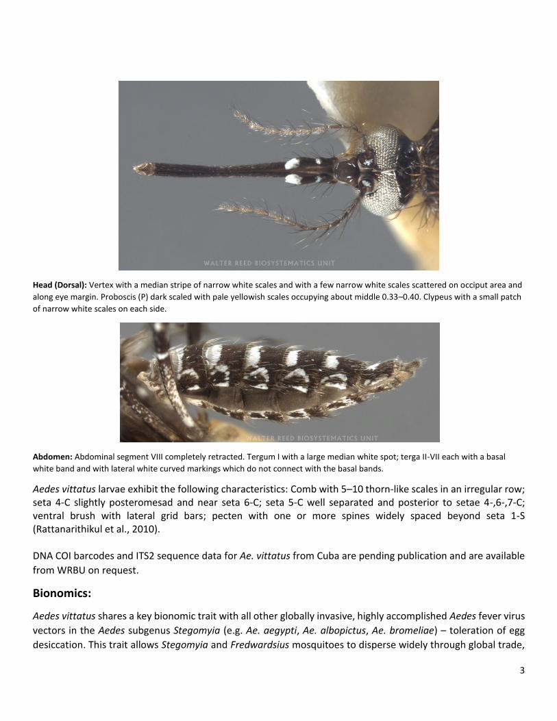
Aedes vittatus larvae exhibit the following characteristics: Comb with 5–10 thorn-like scales in an irregular row; seta 4-C slightly posteromesad and near seta 6-C; seta 5-C well separated and posterior to setae 4-,6-,7-C; ventral brush with lateral grid bars; pecten with one or more spines widely spaced beyond seta 1-S (Rattanarithikul et al., 2010). DNA COI barcodes and ITS2 sequence data for Ae. vittatus from Cuba are pending publication and are available
from WRBU on request.
Bionomics:
Aedes vittatus shares a key bionomic trait with all other globally invasive, highly accomplished Aedes fever virus
vectors in the Aedes subgenus Stegomyia (e.g. Ae. aegypti, Ae. albopictus, Ae. bromeliae) – toleration of egg
desiccation. This trait allows Stegomyia and Fredwardsius mosquitoes to disperse widely through global trade,

4
hiding in consignments as eggs, that hatch upon water stimulation in their new environment. Aedes vittatus is
a hardy species, capable of enduring harsh conditions. It is incredibly tolerant of hot, dry atmospheres, and has
been shown to survive the dry season (for at least 10 weeks) as eggs in dried rockpools in the northern African
deserts. In addition, studies have shown that Ae. vittatus eggs are stimulated to hatch not only by inundation,
but by vibrations of the aqueous environment, similar to that caused by rainfall. In Delhi, Ae. vitattus immatures
have adapted to occupy manmade containers including concrete drainage channels, stone fountains and garden
ornaments in the urban areas of Delhi, in addition to their natural habitats of rock holes and rock pools, as well
tree holes, bamboo pots, hoof prints and wells. Adults are highly anthropophilic, and commonly comprise the
most abundant taxa in human landing collections in Kedougou, Senegal (22.98%).
Biomedical importance: BBKV, CHIK, DEN, ONNV, NRIV, PGAV, SFV, YFV, ZIKA
Like Ae. aegypti & Ae. albopictus, Ae. vittatus is an effective vector of the top four Aedes fever viruses –
chikungunya, dengue, yellow fever and zika viruses. In Jaipur district (India), 20% of Ae. vittatus larvae test
showed evidence of vertically transmitted DEN virus, higher than Ae. albopictus (18.7%) and Ae. aegypti
(13.3%) from the same sites (Angel & Joshi, 2008). Aedes vittatus is involved in the sylvatic transmission
cycle of yellow fever to monkeys; YFV is also passes transovarially in Ae. vittatus.
CHIK has been repeatedly isolated from Ae. vittatus in Senegal and Cape Verde Islands, and lab infections of
these Ae. vittatus populations from Senegal and Cape Verde showed high susceptibility (50–100%) and early
dissemination and transmission following oral ingestion of CHIK isolates from mosquitoes (ArD30237), bats
(CS13-288), and humans (HD180738) (Diagne et al., 2014). ZIKV was isolated from Ae. vittatus from Cote d’Ivoire
(2 strains) and Senegal (>15 isolates) in West Africa (Akoua-Koffi et al., 2001; Diallo et al., 2011; Diallo et al.,
2012; Adam, 2013). Under lab conditions, Senegalese Ae. vittatus were shown to be potentially capable of
transmitting ZIKV after 15 dpi with 20% of mosquitoes delivering epidemic (HD 78788) and prototype (MR 766)
ZIKV strains in saliva (Diagne et al., 2015). The low reported competency of Ae. vittatus is discordant with the
high abundance of these species and their frequent association with ZIKV in the field in Senegal (Diallo et al.,
2012; Adam et al., 2013). Finding another competent vector of Aedes-borne arboviruses in the Caribbean,
especially in light of recent epidemics of non-native viral agents in the region, is of significant Public Health
concern.

5
Known distribution of Aedes vittatus:
Country-level distribution of Aedes vitattus (WRBU, 2019)
Prior to this current invasion into the Caribbean, Ae. vittatus has been reported from Europe, Africa, Middle
East, Indian subcontinent across Southeast Asia south as far as northern Borneo. Country records include:
Algeria, Angola, Bangladesh, Benin, Botswana, Burkina Faso, Cambodia, Cameroon, Central African Republic,
Comoros, Cote d'Ivoire, Cuba (herein), Democratic Republic of the Congo, Djibouti, Ethiopia, France (includes
Corsica), Gabon, Gambia, Ghana, Guinea, India, Iran, Italy (includes Sardinia), Kenya, Laos, Liberia, Madagascar
(includes Glorioso & Juan De Nova Is), Malawi, Malaysia, Mali, Mozambique, Namibia, Nepal, Niger, Nigeria,
Pakistan, People's Republic of China, Portugal, Republic of South Africa, Saudi Arabia, Senegal, Sierra Leone,
Somalia, Spain (includes Balearic Islands), Sri Lanka, Sudan & South Sudan, Tanzania, Thailand, Tunisia, Uganda,
Vietnam, Yemen, Zambia, Zimbabwe.
Location of (U.S. Naval Base, Guantanamo Bay, Cuba). Surveillance data available on VectorMap (vectormap.si.edu)

6
Select References:
Adam, F., Digoutte JP. Virus d’Afrique (Base de Données). Centre Collaborateur OMS de Référence et de Recherche pour
les Arbovirus et les Virus de Fièvres Hémorrhagiques. In: CRORA. 2013. www.pasteur.fr/recherche/banques/CRORA/.
Akoua-Koffi, C., et al. (2001) Investigation surrounding a fatal case of yellow fever in Cote d’Ivoire in 1999. Bull. Soc. Pathol.
Exot. 2001;94(3):227–30.
Diagne, C.T., et al. (2005) Potential of selected Senegalese Aedes spp. mosquitoes (Diptera: Culicidae) to transmit Zika
virus. BMC Infectious Diseases 15(1):1231. https://doi.org/10.1186/s12879-015-1231-2.
Diagne, C.T, et al. (2014) Competence of Aedes aegypti and Aedes vittatus (Diptera: Culicidae) from Senegal and Cape
Verde Archipelago for West African Lineages of Chikungunya Virus. Am. J. Trop. Med. Hyg.,91(3), 2014, pp. 635–641.
doi:10.4269/ajtmh.13-0627
Diallo D, et al. (2012) Landscape ecology of sylvatic chikungunya virus and mosquito vectors in southeastern Senegal. PLoS
Negl. Trop. Dis. 2012;6(6):e1649. doi:10.1371/journal.pntd.0001649.
Diallo D, et al. (2014) Zika Virus Emergence in Mosquitoes in Southeastern Senegal, 2011. PLoS One, 2014; 9(10): e109442.
Edwards, F. W. (1941). Mosquitoes of the Ethiopian Region. III.- Culicine adults and pupae. Mosquitoes of the Ethiopian
Region. III. - Culicine Adults and Pupae.
Huang, Y.-M. & Ward, R. (1981) A pictorial key to the identification of the mosquitoes associated with yellow fever in
Africa. Mosquito Systematics 13(2):138–149.
Rattanarithikul, R. et al. (2010). Illustrated keys to the mosquitoes if Thailand. VI. Tribe Aedini. Southeast Asian J Tropical
Medicine & Public Health, 41 (1):1-38.