Spontaneous rupture of a giant hepatic hemangioma...
4
Introduction The first reported use of transcatheter arterial em- bolization (TAE) prior to surgical resection of ruptured hepatic hemangioma was by Yamamoto et al. in 1991 (1). Since that time only 6 more cases have been reported in the literature, with no patient mortality (2-5). We pres- ent a case of hemorrhagic shock after severe bleeding of a ruptured hepatic hemangioma which was percuta- neously embolized in an emergency setting and surgi- cally ressected after hemodynamic stabilization. Case report A 50 year-old woman was admitted in our emergency room with sudden onset of upper abdominal and right thoracic pain, and nau- sea. There was no history of trauma in the last 24 hours neither sig- nificant medical conditions. She was submitted to the emergency room protocol for acute thoracic pain and addressed to surgery once car- diac conditions were excluded. At this moment the patient was con- scious, the blood pressure was 100/70 mmHg, pulse was 130 per min, and the initial blood test showed hemoglobin levels of 12,1 g/dL and high alanine amino-transferase levels (996 U/mL). After good response to volume resuscitation, she had an abdominal ultrasound which re- vealed a heterogeneous 13x10cm mass in the right liver lobe. Six hours after admission and initial adequate response to con- servative mesures the patient turned pale, the blood pressure dropped to 90/60 mmHg and pulse increased to 140 heart beats/minute. After initial resuscitation with 2000ml of saline and 2 packed red blood cells (RBC), patient underwent a contrast en- hanced computed tomography (CT) (Fig.1) that demonstrated a vas- cular lesion in right lobe of the liver with signs of rupture and acute bleeding. There were associated hemoperitoneum and mild right pleu- SUMMARY: Spontaneous rupture of a giant hepatic hemangioma. Se- quential treatment with preoperative transcatheter arterial embo- lization and conservative hepatectomy. R.M. LUPINACCI, D. SZEJNFELD, J.F.M. FARAH Hemangioma is the most common benign tumor of the liver and it is often asymptomatic. Spontaneous rupture of liver hemangiomas is a rare but potentially lethal complication. Emergent hepatic resection has been the treatment of choice but carries high operative morbidity and mortality. Recently, preoperative transcatheter arterial embolization (TAE) has been used successfully for the management of bleeding rup- tured liver tumors and non-operative treatment of symptomatic giant liver hemangiomas. We report a case of spontaneous rupture of a giant hepatic heman- gioma that presented with thoracic and abdominal pain and shock due to hemoperitoneum. Once proper diagnosis was made the patient was successfully managed by TAE, followed by conservative hepatic resection. RIASSUNTO: Rottura spontanea di emangioma epatico gigante. Trat- tamento sequenziale con embolizzazione arteriosa transcatetere preo- peratoria ed epatectomia conservativa. R.M. LUPINACCI, D. SZEJNFELD, J.F.M. FARAH L’emangioma è il più comune tumore benigno del fegato ed è spes- so asintomatico. La rottura spontanea di un emangioma del fegato è una complicanza rara ma potenzialmente letale. La resezione epatica è stata il trattamento di scelta, ma ha un’alta morbilità e mortalità ope- ratoria. Recentemente, l'embolizzazione arteriosa transcatetere (TAE) preoperatoria è stata utilizzata con successo per la gestione del sangui- namento da rottura di tumore epatico e per il trattamento conservati- vo degli emangiomi epatici giganti. Riportiamo un caso di rottura spontanea di emangioma epatico gigante sintomatici manifestatasi con dolore toracico e addominale e shock da emoperitoneo. Posta la diagnosi corretta la paziente è stata trattata con successo con TAE, seguita da resezione epatica. KEY WORDS: Giant hepatic hemangioma - Hemoperitoneum - Transcatheter arterial embolization - Liver resection. Emangioma epatico gigante - Emoperitoneo - Embolizzazione arteriosa transcatetere - Resezione epatica. Spontaneous rupture of a giant hepatic hemangioma. Sequential treatment with preoperative transcatheter arterial embolization and conservative hepatectomy R.M. LUPINACCI, D. SZEJNFELD 1 , J.F.M. FARAH G Chir Vol. 32 - n. 11/12 - pp. 469-472 November-December 2011 469 São Paulo Estate Employees Hospital, São Paulo, Brazil Department of General and Oncologic Surgery 1 Department of Radiology © Copyright 2011, CIC Edizioni Internazionali, Roma
Transcript of Spontaneous rupture of a giant hepatic hemangioma...
Introduction
The first reported use of transcatheter arterial em-bolization (TAE) prior to surgical resection of rupturedhepatic hemangioma was by Yamamoto et al. in 1991(1). Since that time only 6 more cases have been reportedin the literature, with no patient mortality (2-5). We pres-ent a case of hemorrhagic shock after severe bleeding ofa ruptured hepatic hemangioma which was percuta-neously embolized in an emergency setting and surgi-cally ressected after hemodynamic stabilization.
Case report
A 50 year-old woman was admitted in our emergency room withsudden onset of upper abdominal and right thoracic pain, and nau-sea. There was no history of trauma in the last 24 hours neither sig-nificant medical conditions. She was submitted to the emergency roomprotocol for acute thoracic pain and addressed to surgery once car-diac conditions were excluded. At this moment the patient was con-scious, the blood pressure was 100/70 mmHg, pulse was 130 per min,and the initial blood test showed hemoglobin levels of 12,1 g/dL andhigh alanine amino-transferase levels (996 U/mL). After good responseto volume resuscitation, she had an abdominal ultrasound which re-vealed a heterogeneous 13x10cm mass in the right liver lobe.
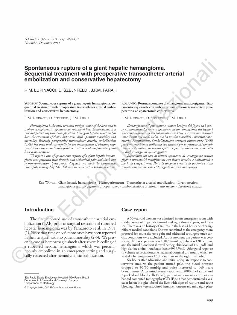
Six hours after admission and initial adequate response to con-servative mesures the patient turned pale, the blood pressuredropped to 90/60 mmHg and pulse increased to 140 heartbeats/minute. After initial resuscitation with 2000ml of saline and2 packed red blood cells (RBC), patient underwent a contrast en-hanced computed tomography (CT) (Fig.1) that demonstrated a vas-cular lesion in right lobe of the liver with signs of rupture and acutebleeding. There were associated hemoperitoneum and mild right pleu-
SUMMARY: Spontaneous rupture of a giant hepatic hemangioma. Se-quential treatment with preoperative transcatheter arterial embo-lization and conservative hepatectomy.
R.M. LUPINACCI, D. SZEJNFELD, J.F.M. FARAH
Hemangioma is the most common benign tumor of the liver and itis often asymptomatic. Spontaneous rupture of liver hemangiomas is arare but potentially lethal complication. Emergent hepatic resection hasbeen the treatment of choice but carries high operative morbidity andmortality. Recently, preoperative transcatheter arterial embolization(TAE) has been used successfully for the management of bleeding rup-tured liver tumors and non-operative treatment of symptomatic giantliver hemangiomas.
We report a case of spontaneous rupture of a giant hepatic heman-gioma that presented with thoracic and abdominal pain and shock dueto hemoperitoneum. Once proper diagnosis was made the patient wassuccessfully managed by TAE, followed by conservative hepatic resection.
RIASSUNTO: Rottura spontanea di emangioma epatico gigante. Trat-tamento sequenziale con embolizzazione arteriosa transcatetere preo-peratoria ed epatectomia conservativa.
R.M. LUPINACCI, D. SZEJNFELD, J.F.M. FARAH
L’emangioma è il più comune tumore benigno del fegato ed è spes-so asintomatico. La rottura spontanea di un emangioma del fegato èuna complicanza rara ma potenzialmente letale. La resezione epatica èstata il trattamento di scelta, ma ha un’alta morbilità e mortalità ope-ratoria. Recentemente, l'embolizzazione arteriosa transcatetere (TAE)preoperatoria è stata utilizzata con successo per la gestione del sangui-namento da rottura di tumore epatico e per il trattamento conservati-vo degli emangiomi epatici giganti.
Riportiamo un caso di rottura spontanea di emangioma epaticogigante sintomatici manifestatasi con dolore toracico e addominale eshock da emoperitoneo. Posta la diagnosi corretta la paziente è statatrattata con successo con TAE, seguita da resezione epatica.
KEY WORDS: Giant hepatic hemangioma - Hemoperitoneum - Transcatheter arterial embolization - Liver resection.Emangioma epatico gigante - Emoperitoneo - Embolizzazione arteriosa transcatetere - Resezione epatica.
Spontaneous rupture of a giant hepatic hemangioma. Sequential treatment with preoperative transcatheter arterialembolization and conservative hepatectomy
R.M. LUPINACCI, D. SZEJNFELD1, J.F.M. FARAH
G Chir Vol. 32 - n. 11/12 - pp. 469-472November-December 2011
469
São Paulo Estate Employees Hospital, São Paulo, BrazilDepartment of General and Oncologic Surgery1 Department of Radiology
© Copyright 2011, CIC Edizioni Internazionali, Roma
0521 5 Spontaneous_Lupinacci:- 23-11-2011 14:17 Pagina 469
470
R.M. Lupinacci et al.
ral effusion. Remained liver parenchyma was normal with no oth-er focal lesion. Digital Subtraction Angiography (DSA) revealed an-giographic findings of a liver hemangioma and arterial contrast ex-travasation was noted on the celiac angiogram, after which a selec-tive right hepatic arteriogram confirmed active contrast extravasa-tion from the lesion.
The hemangioma was embolized in a single session, via selectiveright hepatic artery embolisation using PVA (polyvinyl alcohol) par-ticles 500-750 µ (Cook Bloomington Inc., USA) delivered using aCobra catheter. At the end of the procedure the patient was hemo-dynamic stable, and a check angiogram showed successful emboli-sation with no contrast leak.
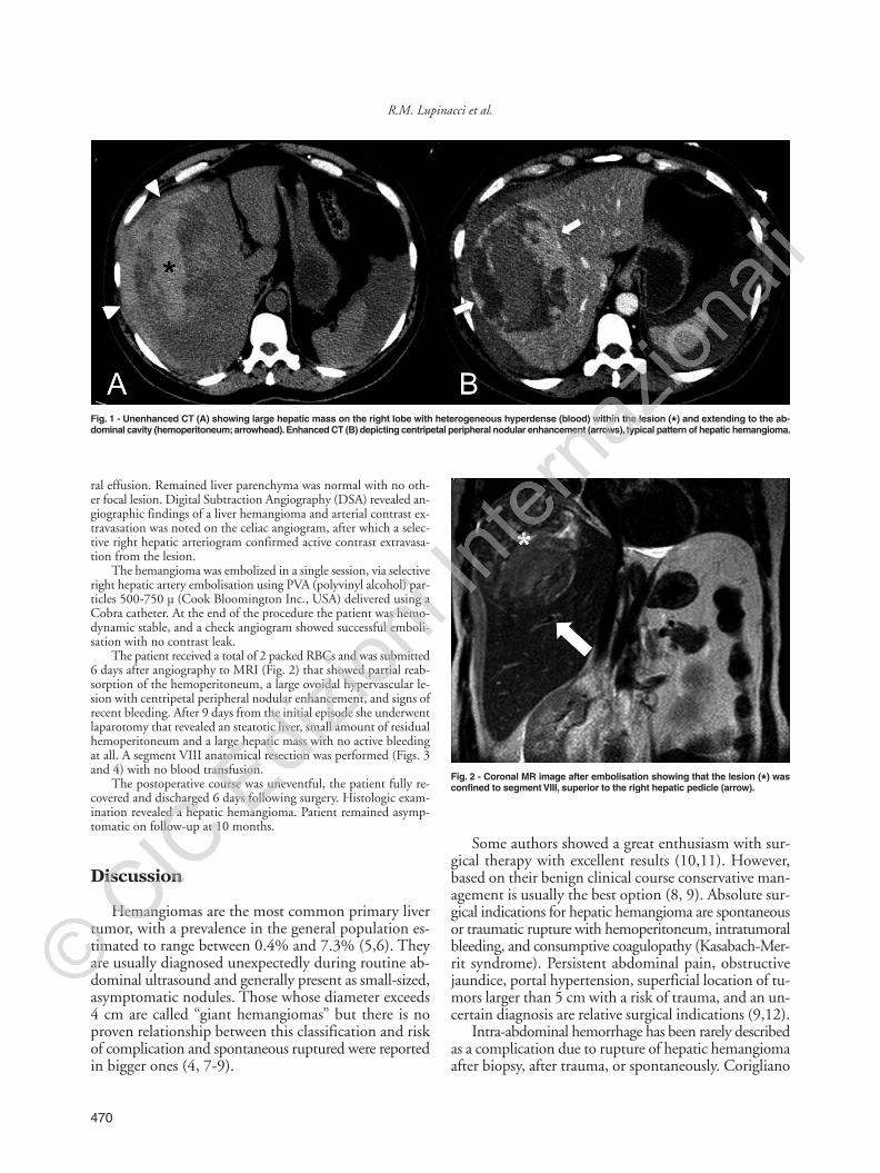
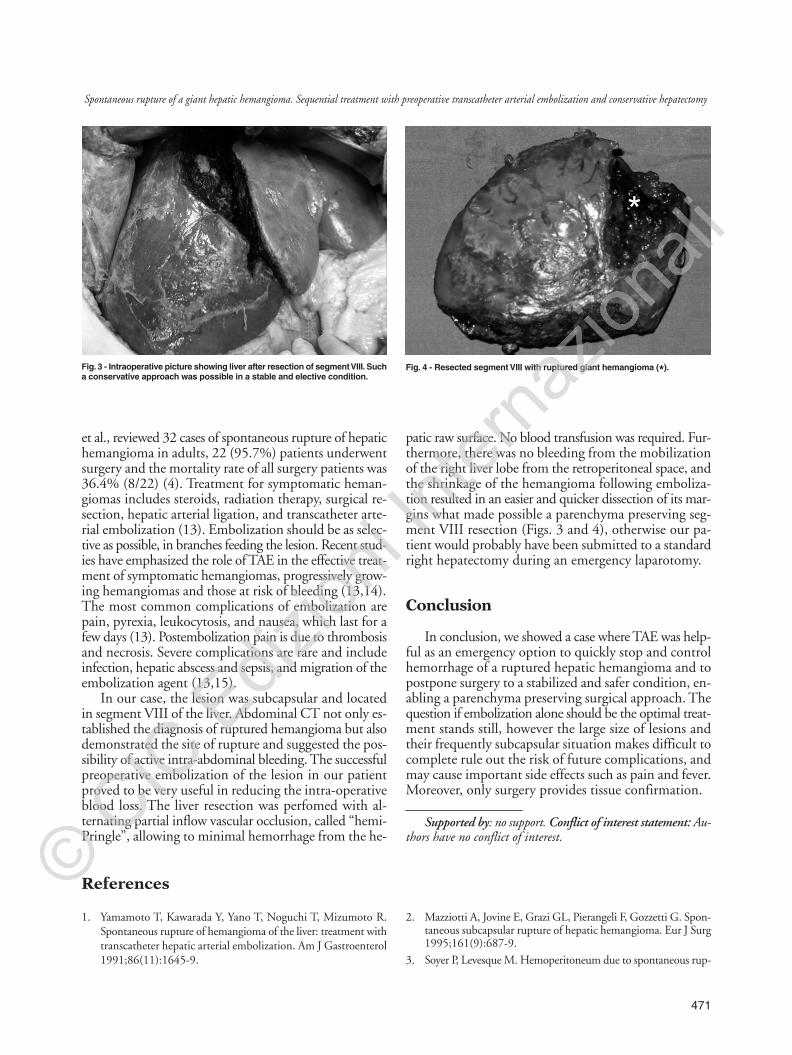
The patient received a total of 2 packed RBCs and was submitted6 days after angiography to MRI (Fig. 2) that showed partial reab-sorption of the hemoperitoneum, a large ovoidal hypervascular le-sion with centripetal peripheral nodular enhancement, and signs ofrecent bleeding. After 9 days from the initial episode she underwentlaparotomy that revealed an steatotic liver, small amount of residualhemoperitoneum and a large hepatic mass with no active bleedingat all. A segment VIII anatomical resection was performed (Figs. 3and 4) with no blood transfusion.
The postoperative course was uneventful, the patient fully re-covered and discharged 6 days following surgery. Histologic exam-ination revealed a hepatic hemangioma. Patient remained asymp-tomatic on follow-up at 10 months.
Discussion
Hemangiomas are the most common primary livertumor, with a prevalence in the general population es-timated to range between 0.4% and 7.3% (5,6). Theyare usually diagnosed unexpectedly during routine ab-dominal ultrasound and generally present as small-sized,asymptomatic nodules. Those whose diameter exceeds4 cm are called “giant hemangiomas” but there is noproven relationship between this classification and riskof complication and spontaneous ruptured were reportedin bigger ones (4, 7-9).
Some authors showed a great enthusiasm with sur-gical therapy with excellent results (10,11). However,based on their benign clinical course conservative man-agement is usually the best option (8, 9). Absolute sur-gical indications for hepatic hemangioma are spontaneousor traumatic rupture with hemoperitoneum, intratumoralbleeding, and consumptive coagulopathy (Kasabach-Mer-rit syndrome). Persistent abdominal pain, obstructivejaundice, portal hypertension, superficial location of tu-mors larger than 5 cm with a risk of trauma, and an un-certain diagnosis are relative surgical indications (9,12).
Intra-abdominal hemorrhage has been rarely describedas a complication due to rupture of hepatic hemangiomaafter biopsy, after trauma, or spontaneously. Corigliano
Fig. 2 - Coronal MR image after embolisation showing that the lesion (*) wasconfined to segment VIII, superior to the right hepatic pedicle (arrow).
Fig. 1 - Unenhanced CT (A) showing large hepatic mass on the right lobe with heterogeneous hyperdense (blood) within the lesion (*) and extending to the ab-dominal cavity (hemoperitoneum; arrowhead). Enhanced CT (B) depicting centripetal peripheral nodular enhancement (arrows), typical pattern of hepatic hemangioma.
0521 5 Spontaneous_Lupinacci:- 23-11-2011 14:17 Pagina 470
471
Spontaneous rupture of a giant hepatic hemangioma. Sequential treatment with preoperative transcatheter arterial embolization and conservative hepatectomy
et al., reviewed 32 cases of spontaneous rupture of hepatichemangioma in adults, 22 (95.7%) patients underwentsurgery and the mortality rate of all surgery patients was36.4% (8/22) (4). Treatment for symptomatic heman-giomas includes steroids, radiation therapy, surgical re-section, hepatic arterial ligation, and transcatheter arte-rial embolization (13). Embolization should be as selec-tive as possible, in branches feeding the lesion. Recent stud-ies have emphasized the role of TAE in the effective treat-ment of symptomatic hemangiomas, progressively grow-ing hemangiomas and those at risk of bleeding (13,14).The most common complications of embolization arepain, pyrexia, leukocytosis, and nausea, which last for afew days (13). Postembolization pain is due to thrombosisand necrosis. Severe complications are rare and includeinfection, hepatic abscess and sepsis, and migration of theembolization agent (13,15).
In our case, the lesion was subcapsular and locatedin segment VIII of the liver. Abdominal CT not only es-tablished the diagnosis of ruptured hemangioma but alsodemonstrated the site of rupture and suggested the pos-sibility of active intra-abdominal bleeding. The successfulpreoperative embolization of the lesion in our patientproved to be very useful in reducing the intra-operativeblood loss. The liver resection was perfomed with al-ternating partial inflow vascular occlusion, called “hemi-Pringle”, allowing to minimal hemorrhage from the he-
patic raw surface. No blood transfusion was required. Fur-thermore, there was no bleeding from the mobilizationof the right liver lobe from the retroperitoneal space, andthe shrinkage of the hemangioma following emboliza-tion resulted in an easier and quicker dissection of its mar-gins what made possible a parenchyma preserving seg-ment VIII resection (Figs. 3 and 4), otherwise our pa-tient would probably have been submitted to a standardright hepatectomy during an emergency laparotomy.
Conclusion
In conclusion, we showed a case where TAE was help-ful as an emergency option to quickly stop and controlhemorrhage of a ruptured hepatic hemangioma and topostpone surgery to a stabilized and safer condition, en-abling a parenchyma preserving surgical approach. Thequestion if embolization alone should be the optimal treat-ment stands still, however the large size of lesions andtheir frequently subcapsular situation makes difficult tocomplete rule out the risk of future complications, andmay cause important side effects such as pain and fever.Moreover, only surgery provides tissue confirmation.
Supported by: no support. Conflict of interest statement: Au-thors have no conflict of interest.
Fig. 3 - Intraoperative picture showing liver after resection of segment VIII. Sucha conservative approach was possible in a stable and elective condition.
Fig. 4 - Resected segment VIII with ruptured giant hemangioma (*).
1. Yamamoto T, Kawarada Y, Yano T, Noguchi T, Mizumoto R.Spontaneous rupture of hemangioma of the liver: treatment withtranscatheter hepatic arterial embolization. Am J Gastroenterol1991;86(11):1645-9.
2. Mazziotti A, Jovine E, Grazi GL, Pierangeli F, Gozzetti G. Spon-taneous subcapsular rupture of hepatic hemangioma. Eur J Surg1995;161(9):687-9.
3. Soyer P, Levesque M. Hemoperitoneum due to spontaneous rup-
References
0521 5 Spontaneous_Lupinacci:- 23-11-2011 14:17 Pagina 471

472
R.M. Lupinacci et al.
ture of hepatic hemangiomatosis: treatment by superselective ar-terial embolization and partial hepatectomy. Australas Radiol1995;39(1):90-2.
4. Corigliano N, Mercantini P, Amodio PM, Balducci G, Cateri-no S, Ramacciato G, Ziparo V. Hemoperitoneum from a spon-taneous rupture of a giant hemangioma of liver: report of a case.Surg Today 2003;33(6):459-63.
5. Ochsner JL, Halpert B. Cavernous hemangioma of the liver. Sur-gery 1958;43(4):577-82.
6. Ishak KG, Rabin L. Benign tumors of the liver. Med Clin NorthAm 1975;59(4): 995-1013.
7. Adam YG, Huvos AG, Fortner JG. Giant hemangiomas of theliver. Ann Surg 1970;172(2):239-45.
8. Terkivatan T, Vrijland WW, den Hoed PT, De Man RA, Hus-sain SM, Tilanus HW, Ijzermans JN. Size of lesion is not a cri-terion for resection during management of giant liver haeman-gioma. Br J Surg 2002;89(10):1240-4.
9. Herman P, Costa ML, Machado MA, Pugliese V, D'Albuquer-que LA, Machado MC, Gama-Rodrigues JJ, Saad WA. Mana-gement of hepatic hemangiomas: a 14-year experience. J Ga-
strointest Surg 2005;9(6):853-9.10. Brouwers MA, Peeters PM, De Jong KP, Haagsma EB, Klompmaker
IJ, Bijleveld CM, Zwaveling GH, Slooff MG. Surgical treatmentof giant hemangioma of the liver. Br J Surg 1997;84(3):314-6.
11. Ozden I, Emre A, Alper A, Tunaci M, Acarli K, Bilge O, TekantY, Ariogul O. Long-term results of surgery for liver hemangio-mas. Arch Surg 2000;135(8):978-81.
12. Moreno Egea A, Del Pozo Rodriguez M, Vicente Cantero M,Abellan Atenza J. Indications for surgery in the treatment of he-patic hemangioma. Hepatogastroenterology 1996;43(8):422-6.
13. Shrivastava DN, Gandhi D, Seith A, Pande GK, Sahni P. Tran-scatheter arterial embolization in the treatment of symptoma-tic cavernous hemangiomas of the liver: a prospective study. Ab-dom Imaging 2001;26(5):510-4.
14. Deutsch GS, Yeh KA, Bates WB, Tannehill WB. Embolizationfor management of hepatic hemangiomas. Am Surg2001;67(2):159-64.
15. Reading NG, Forbes A, Nunnerley HB, Williams R. Hepatic he-mangioma: a critical review of diagnosis and management. QJMed 1988;67(253):431-45.
0521 5 Spontaneous_Lupinacci:- 23-11-2011 14:17 Pagina 472