SPONTANEOUS INTRACEREBRAL HEMORRHAGE: MEDICAL MANAGEMENT · INTERACT: Mean 1.6 cc decreased in RCT...
56
SPONTANEOUS INTRACEREBRAL HEMORRHAGE: MEDICAL MANAGEMENT Neeraj Naval, MD Baptist Neurological Institute
Transcript of SPONTANEOUS INTRACEREBRAL HEMORRHAGE: MEDICAL MANAGEMENT · INTERACT: Mean 1.6 cc decreased in RCT...

SPONTANEOUS INTRACEREBRAL
HEMORRHAGE: MEDICAL MANAGEMENT
Neeraj Naval, MD
Baptist Neurological Institute

Disclosures/ COI
Nothing to disclose (what happens in Vegas stays in Vegas)
No Conflict of interest

Non-modifiable Impact Factors (ICH Score)
Age: < 80 : 0 > 80 : 1
GCS score 13–15 : 0
5–12 : 1
3–4 : 2
ICH location : Supratentorial : 0 Infratentorial : 1
ICH volume (AxBxC/2) < 30 mL : 0
> 30 mL : 1
IVH No : 0
Yes : 1 Total: 0 - 6

ICH Overview: (Outcome model)
ICHS Mortality (%)
0 0
1 13
2 26
3 72
4 97
5 100

Lecture Focus: Modifiable Impact Factors
Modifiable Risk factors for Outcomes aka Complications of ICH
Mass effect (Hematoma expansion- Hemostasis)
A) Blood pressure control
B) Hemostatic therapy
C) Reversal of bleeding diathesis
Mass effect (Cerebral edema/ Intracranial HTN)
Hydrocephalus (sec to IVH): CLEAR 3
Seizures: ppx?
Recurrent ICH

Lecture Focus: EXCLUDED
Reversal of NOAC (Dr. Rama/ Chen)
Role of Surgery in ICH: STITCH, STICH 2 (Dr. Lopes)
Role of Surgery in ICH: Incl. Minimally Invasive surgery (Dr. Lopes)

ICH Overview: Impact
Approximately 500,000 new strokes occur every year in the United States, 15% of them are hemorrhagic strokes.
These numbers are expected to double during next 50 years.
– Increased longevity of the population.
Overall population based mortality of ICH patients remains high:
– 6% die before reaching a hospital.
– 30 to 50% die within the first 30 days.
– GCS < 8 and ICH volume > 60 cc > 90% 30-day mortality Independent living after ICH:
–After 1 month: 10%,
–After 6 months: 20%.

ICH Overview: Location
Caudate Lobar Thalamic Putaminal Pontine

ICH: presentation
50% CAA
50% Other
Majority HTN
Qureshi; N Engl J Med 2001;344:1450-60
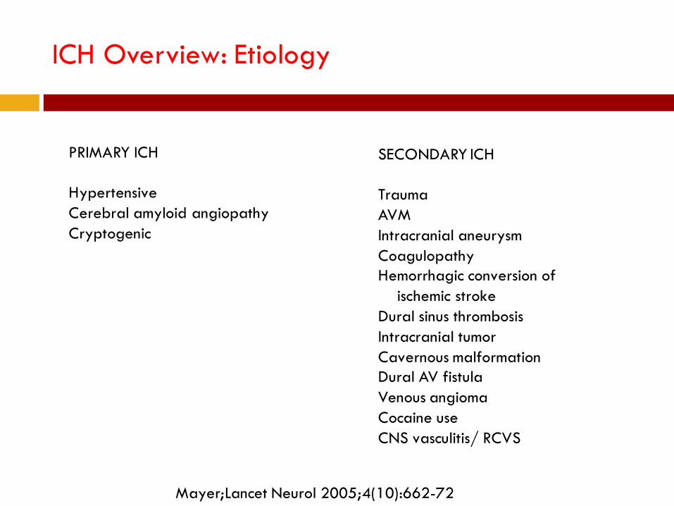
ICH Overview: Etiology
Mayer;Lancet Neurol 2005;4(10):662-72
PRIMARY ICH
Hypertensive
Cerebral amyloid angiopathy
Cryptogenic
SECONDARY ICH
Trauma
AVM
Intracranial aneurysm
Coagulopathy
Hemorrhagic conversion of
ischemic stroke
Dural sinus thrombosis
Intracranial tumor
Cavernous malformation
Dural AV fistula
Venous angioma
Cocaine use
CNS vasculitis/ RCVS

Mass Effect: Blood pressure/ Hematoma expansion
38%>33% growth over 24h
73% some growth over 24h
Independent predictor of bad outcome
Davis; Neurology 2006;66(8):1175-81
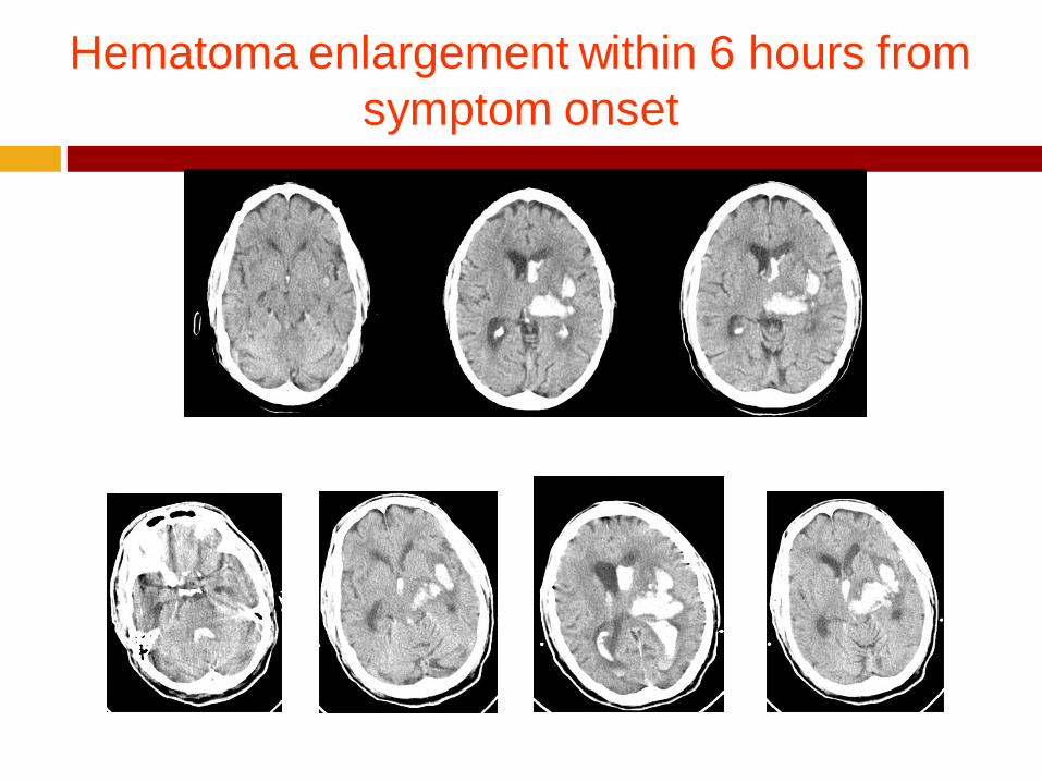
Hematoma enlargement within 6 hours from
symptom onset

ICH and IVH: Complications
Hematoma enlargement
– Early hematoma growth in ~ 30% of ICH patients within 3 hours of onset can cause significant neurologic deterioration.
– 1 cc larger volume ICH = 7% more likely to have worse clinical outcomes

Blood Pressure Targets – Association vs true evidence
Cause-effect
Does increased Blood pressure cause more bleeding?
Or is it a marker for more severe neurological injury (increased ICH volume
= Kocher Cushing’s reflex)
Treatment effect
Does better blood pressure control actually lead to hemostasis?
Or does it lead to worsening perihematomal ischemia?

Best available evidence in 90s
1. Increased BP is associated with larger ICH volumes: SBP goal < 160
(Retrospectively observed association)
2. Small-moderate ICH volumes (<45 cc) are NOT associated with
perihematomal ischemia (Powers et al)
ADC on MRI suggests inflammation (vasogenic edema) > ischemia (cytotoxic
injury)
15-20% acute decrease in BP in first 6 hours not associated with decrease in
critical perfusion.
MAP goal < 130 or SBP < 180 (1999 AHA guidelines)

Available evidence in 21st century
ATACH pilot: small Pilot study showing better hemostasis with SBP goal <
160 than historical controls.
ATACH study showed trends towards decreased hematoma expansion and
PHE in 110-140 compared to 140-170 compared to 170-200.
ATACH 2 ongoing
INTERACT: Mean 1.6 cc decreased in RCT with SBP goal < 140 compared
to SBP < 180; 36% reduced risk of SHE, no difference in clinical outcomes
(safe and non-inferior).
AHA revised guidelines: If ICP elevation an issue, Control ICP with SBP goal
< 180 until ICP controlled, otherwise SBP < 160. SBP < 140 is considered
SAFE
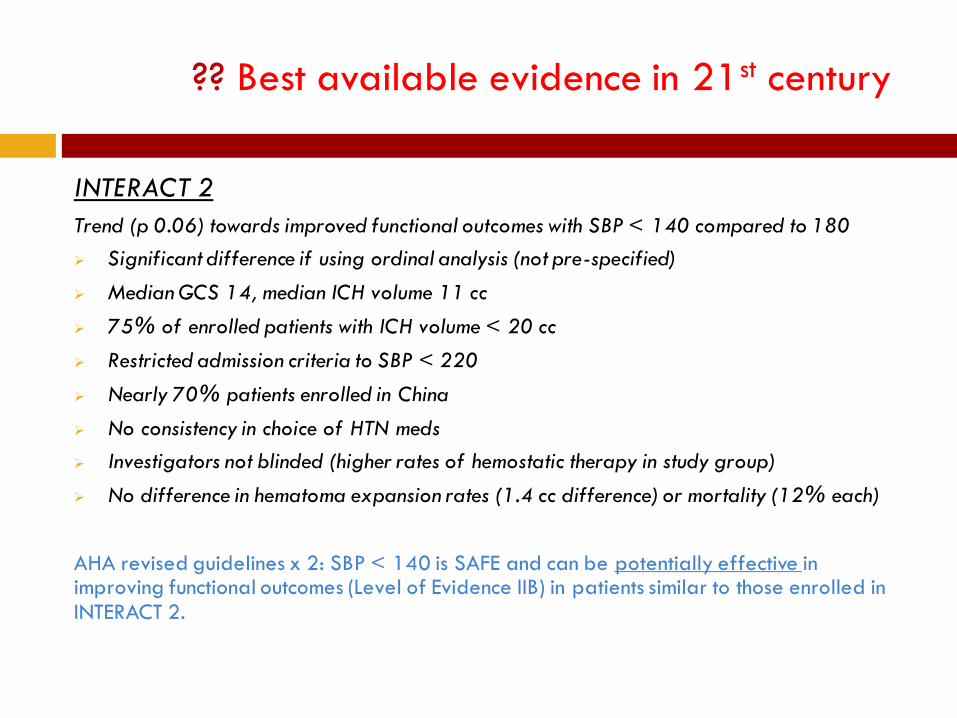
?? Best available evidence in 21st century
INTERACT 2
Trend (p 0.06) towards improved functional outcomes with SBP < 140 compared to 180
Significant difference if using ordinal analysis (not pre-specified)
Median GCS 14, median ICH volume 11 cc
75% of enrolled patients with ICH volume < 20 cc
Restricted admission criteria to SBP < 220
Nearly 70% patients enrolled in China
No consistency in choice of HTN meds
Investigators not blinded (higher rates of hemostatic therapy in study group)
No difference in hematoma expansion rates (1.4 cc difference) or mortality (12% each)
AHA revised guidelines x 2: SBP < 140 is SAFE and can be potentially effective in improving functional outcomes (Level of Evidence IIB) in patients similar to those enrolled in INTERACT 2.
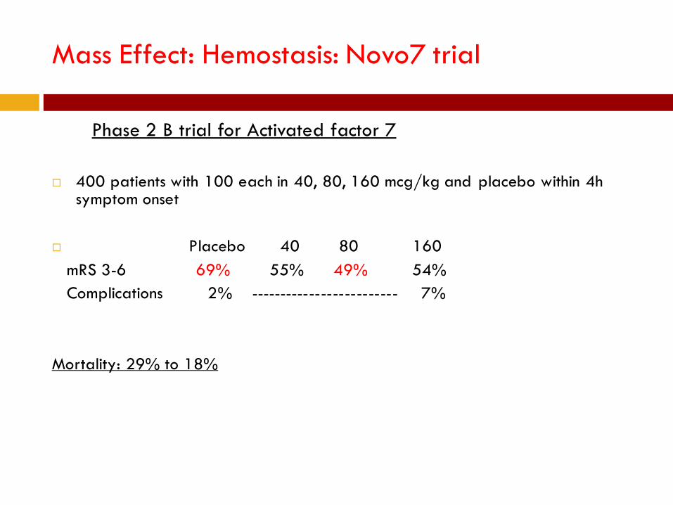
Mass Effect: Hemostasis: Novo7 trial
Phase 2 B trial for Activated factor 7
400 patients with 100 each in 40, 80, 160 mcg/kg and placebo within 4h symptom onset
Placebo 40 80 160
mRS 3-6 69% 55% 49% 54%
Complications 2% ------------------------- 7%
Mortality: 29% to 18%

Hematoma
expansion
(Not So) FAST trial
Mayer;N Engl J Med 2008;15;358(20):2127-37+Stroke 2009;40:833-40
FAST
Functional
outcome
Placebo vs. rFVIIa (20µg/kg or 80µg/kg) within 4hr
n=841
?benefit
No difference in good outcome, bad outcome or death
ICH volume<60mL; Age≤70; ≤2.5h to treatment; IVH<5mL

Why did FAST fail?
Mortality: Novo7: 26%, Placebo: 21%
Randomization: IVH in 29% Placebo, 41% study drug
Liberal inclusion criteria (age up to 80, GCS 6-8 OK, large ICH volumes ok)
Post-hoc analysis: Benefit in Novo-7 group
Age < 70
ICH Volume < 60 cc
IVH < 5cc
Drug within 2.5h
Too little, too late. Could we predict who would have expanded hematomas?

SPOT SIGN

Contrast extravasation/ Spot sign
Spot sign +ve: 77% likelihood of hematoma expansion (Wada et al)
Spot sign –ve: 4% likelihood of hematoma expansion (NPV 96%)
PREDICT TRIAL: > 33% increase or absolute increase of 6cc
PPV 60%
NPV 78%
Sensitivity: 51%
Specificity: 85%

SPOT SIGN
Delgado; Stroke 2009; Jun; EPUB ahead of print; Wada; Stroke 2007;38:1257-1262
CTA spot sign
NON-
CON
CTA POST-
CON
NON-
CON

Contrast extravasation/ Spot sign
What can we do about this data?
Hemostatic agents…… ongoing trials (STOP-IT, SPOTLIGHT study)
More aggressive BP control…….. SBP goal 140?
Reversal of platelet dysfunction…… PLT transfusion (PATCH trial)?
Reversal of coagulopathy……….high risk patients?
TRIAGE

Coagulopathies in a snapshot
Warfarin: PCC >> FFP (Sarode et al), + Vit K IV
NOAC: PCC (Xa), specific inhibition (DTI)
tPa: NS: Cryoprecipitate + platelets +/- FFP
S: Antifibrinolytic therapy (tranexamic acid/ Amicar)
Heparin gtt: Protamine (no more than 50mg; administration time based)
LMWH (lovenox): 0-8h: 1 mg protamine: 1 mg lovenox
8-12h: 0.5mg Protamine: 1 mg lovenox
12-24h: None unless surgery/ ongoing bleeding or ARF/ CRF
Fondaparinaux: ??

ASA/ Plavix Reversal
ASA: ? Platelets……PATCH trial
Plavix: Half life 8 hours…… ?PLT/ ddAVP (MOA- vWF)
DDAVP: Uremic platelet dysfunction, ? Anti-PLT (PFA correction)
Indication for PLT transfusion
A) Hematoma expansion/ Neurological deterioration
B) Spot Sign +
C) Surgical intervention (including EVD)
D) Abnormal PFA
Intracranial HTN: Clinical signs
NEUROLOGICAL
Blown pupils, anisocoria (new)
Altered mental status
Decerebrate/ decorticate posturing
Increase in ICP (if monitored)
NON-NEUROLOGICAL
Cushings response: Hypertension, reflex bradycardia, irregular respirations)
Nausea, vomiting

ABCs
Airway: GCS < 8
GCS > 8 with impending neurological deterioration
Cough/ gag/ increased secretions (Coplin et al)
Breathing: Ataxic/ cluster breathing patterns
Sat probe: Avoid hypoxia, sat goals > 94%
ABG/ ETCO2: Co2 goal 28-32
Circulation: MAP > 70
CPP > 60 (TBI)/ > 70 (comatose ICH/SAH), ICP < 20

Hyperventilation
Decrease in PCO2 from 40 to 30 mmHg --- Cerebral vasoconstriction --- 3% decrease in CBF/ 1 mmHg decrease in PCO2 (30% reduction in CBF) --- decreased ICP
Prolonged Cerebral vasoconstriction --- Cerebral Ischemia
CSF Ph normalizes --- Rebound Hyperemia --- Reperfusion Injury --- Rebound Increase in ICP
Caution: Avoid prophylactic use, avoid prolonged use

Hyperosmolar therapy
Options
Mannitol (1-1.5 gm/kg)
Hypertonic saline (3% bolus / 23.4% 30 cc ‘bullet’)

Mannitol
1-1.5 gm/kg 20% mannitol, acts within minutes, peaks at 1 h duration 4-6
hours.
Failure to respond: 2nd dose 1.5-2gm/kg
Can give through peripheral IV
Watch for Hypotension secondary to increased diuresis

Hypertonic saline
23.4% Saline 30 cc bolus, may repeat with second ‘bullet’
Requires Central venous access
Follow with maintenance 3% saline infusion with Na goal 145-155
Effective in ‘mannitol failures’
Watch for acute hypotension
© 2011 by the Society of Cri tical Care Medicine and Lippincott Williams & Wi lkins. Published by Lippincott Williams
& Wilkins, Inc. 2
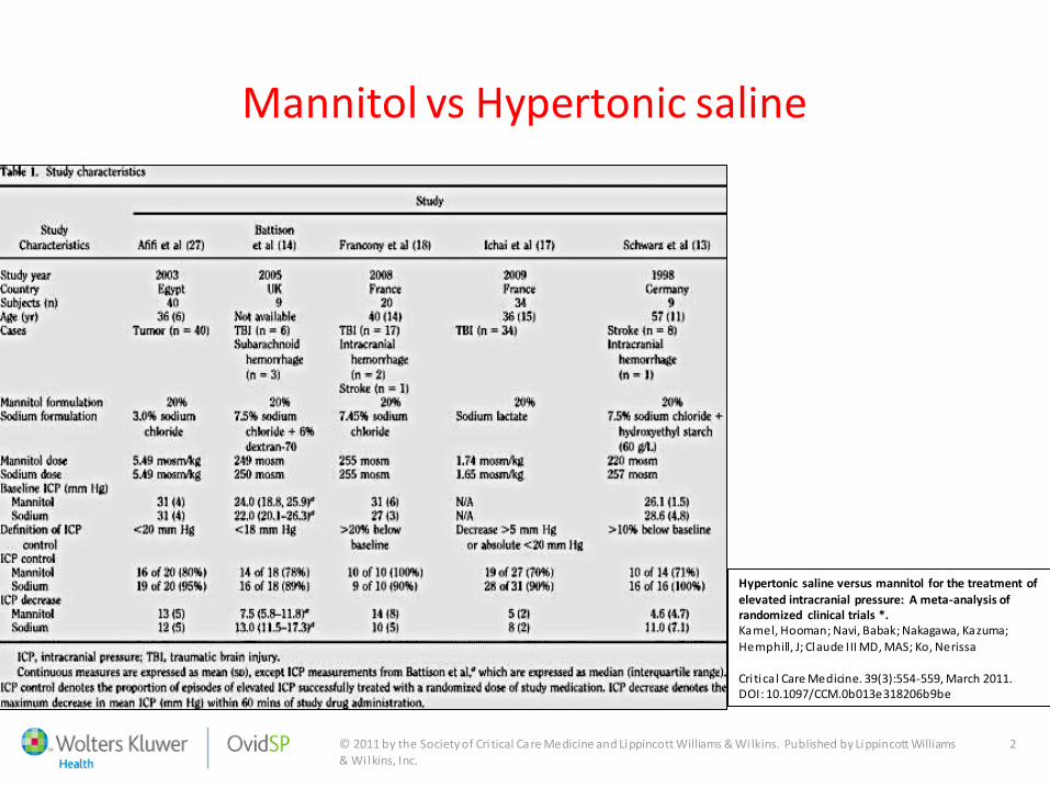
Mannitol vs Hypertonic saline
Hypertonic saline versus mannitol for the treatment of elevated intracranial pressure: A meta-analysis of randomized clinical trials *. Kamel, Hooman; Navi, Babak; Nakagawa, Kazuma; Hemphill, J; Claude III MD, MAS; Ko, Nerissa Cri tica l Care Medicine. 39(3):554-559, March 2011. DOI: 10.1097/CCM.0b013e318206b9be

© 2011 by the Society of Cri tical Care Medicine and Lippincott Williams & Wi lkins. Published by Lippincott Williams
& Wilkins, Inc. 2
Hypertonic saline versus mannitol for the treatment of
elevated intracranial pressure: A meta-analysis of randomized clinical trials *. Kamel, Hooman; Navi, Babak; Nakagawa, Kazuma;
Hemphill, J; Claude III MD, MAS; Ko, Nerissa
Cri tica l Care Medicine. 39(3):554-559, March 2011. DOI: 10.1097/CCM.0b013e318206b9be
Relative risk of successful control of elevated ICP

© 2011 by the Society of Cri tical Care Medicine and Lippincott Williams & Wi lkins. Published by Lippincott Williams
& Wilkins, Inc. 2
Hypertonic saline versus mannitol for the treatment of
elevated intracranial pressure: A meta-analysis of randomized clinical trials *. Kamel, Hooman; Navi, Babak; Nakagawa, Kazuma;
Hemphill, J; Claude III MD, MAS; Ko, Nerissa
Cri tica l Care Medicine. 39(3):554-559, March 2011. DOI: 10.1097/CCM.0b013e318206b9be
Difference in mean quantitative reduction of ICP

Advantages
Mannitol 23.4% saline
May be given through PIV
Higher reflection coefficient
Maintains intravascular volume status
May be followed by 3% saline as continuous infusion
?More robust action
?Longer duration of action
? Greater impact on brain oxygenation (Oddo et al)

ICP monitor / IVC placement
Intraventricular catheter (IVC) facilitates CSF drainage
ICP monitoring useful with unreliable clinical exam (GCS < 8)
Cerebral herniation possible without ICP elevation
Over-drainage especially in setting of cerebellar lesions can
cause upward herniation

Brain Code Supportive treatment
HOB > 30 degrees
Minimize neck compression
Sedation, minimize agitation
Maintain Volume status
Maintain Circulatory status (MAP goals)
Seizure prophylaxis
Glycemic control
Goal of normothermia

Seizures
Incidence of seizures with ICH: Lobar 14% seizures, deep 4% (Bladin et al)
Phenytoin associated with worse outcomes in ICH, CVA, SAH, TBI.
Naidech et al: 10-fold increase in poor outcomes with phenytoin
Fatal Flaws
ICH volume, IVH volume and location were NOT a predictor of outcomes
Difference in ICH volume in 2 groups > 20 cc, admission GCS 10 to 14

Seizure Prophylaxis: Counter-argument
Up to 26% of ICH patients could have only somnolence as manifestation of NCSE if monitored using cEEG monitoring (Vespa et al): 26%
Break up by location: 28% lobar, 21% deep.
Recommendation:
Routine prophylaxis for spontaneous ICH is NOT recommended.
Recommendation is relevant for SPONTANEOUS ICH.
If considering AED for any reason (temporal ICH, underlying structural lesion, craniotomy, cocaine use), consider alternative to PHT for prophylaxis for lobar
ICH
cEEG monitoring is highly advisable in patents with AMS following ICH.

Hydrocephalus/ IVH: Tuhrim et al

Hydrocephalus/ IVH


Subject
Post-rebleed
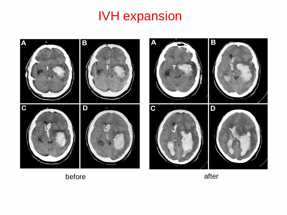
IVH expansion
before after

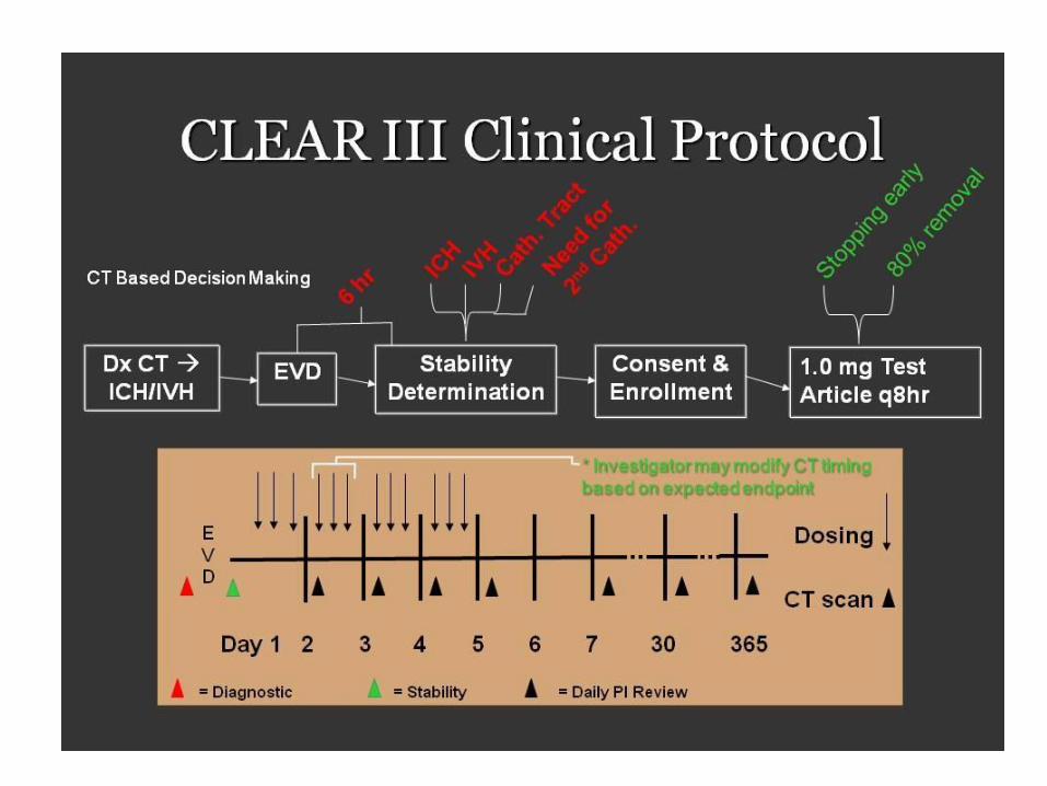
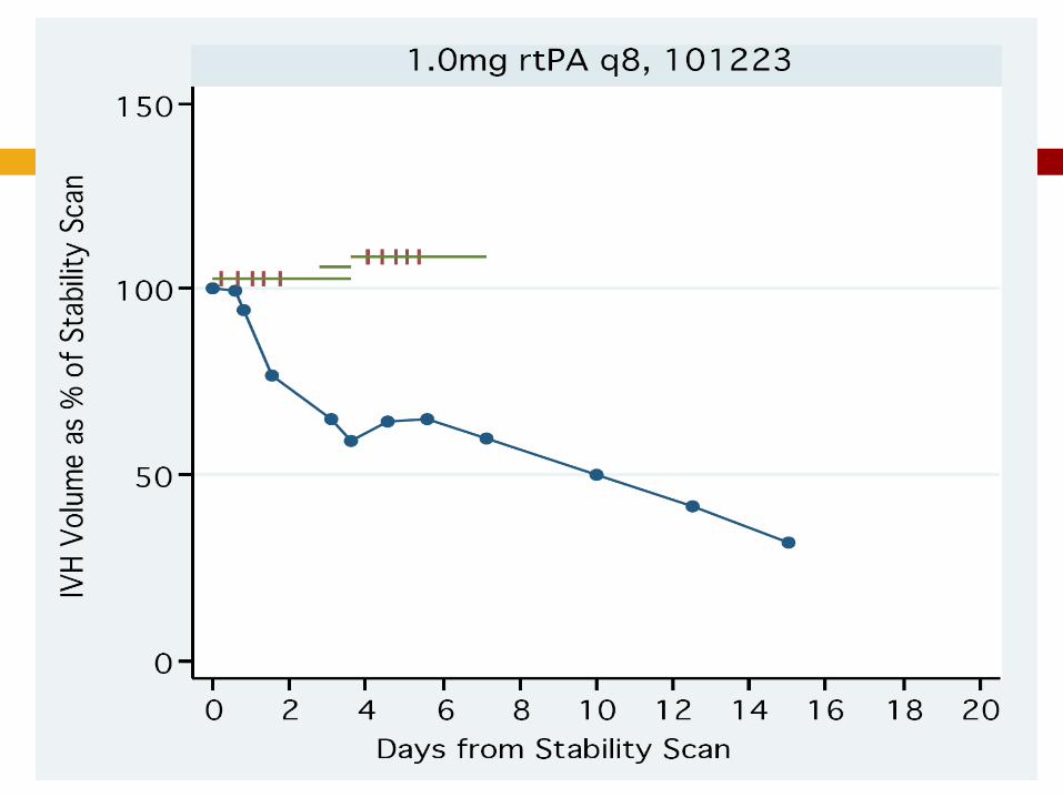
Pragmatic Phase 3 randomized control trial for tPa vs saline through EVD
Outcome assessment at 6 months:
Primary endpoint: mRS 0-3: 48% tPa vs 45% control (NS)
Secondary outcomes:
eGOS: ND
Mortality lower in tPa group: p 0.006, NNT 10
66% home/ rehab (tPa) vs 56% (control): p 0.06
Bleed: 2.6% vs 2% (safe)
Infection rates marginally lower (???)
CLEAR 3 Results

Subgroup analysis:
Clot lysis more effective in post hoc analysis in……..
A) IVH volume over 20 cc (10% higher rates of mRS 0- 3)
B) Time to treat < 48h
So what now……. CLEAR 4? 5? 6?
CLEAR 3 Results

ICH: MISTIE
Stereotactic thrombolysis/aspiration
MINIMALLY INVASIVE
CRANIOPUNCTURE
RCT minimally invasive surgery for basal
ganglia ICH
Cannula placed inside clot and 10,000U-
50,000U urokinase injected followed by
aspiration (dose based on hematoma
volume)
n=465
RCT minimally invasive surgery for basal
ganglia ICH
Cannula placed inside clot and tpa 1mg
q8h x max 72h injected and aspirated until:
1. ICH volume 10cc
2. 80% decrease
3. Dose limit reached 90d outcome
MRS 0-2 63% 41%
Dead 8% 9%
+tx -tx

Recurrent ICH: Vascular imaging
Cerebral angiography
–To make the etiological diagnosis in cases of:
– Aneurysms
– AVM’s
– Vasculitis
– Dural AV fistula

Vascular imaging
Cerebral angiography
–In young patients (< 45 y/o) w/o risk factors for ICH, the yield of angiography can reach 48% in putaminal, thalamic and posterior fossa ICH.
–In young patients with lobar ICH, yield of angiography can reach 65%.
–The yield of cerebral angiography in primary IVH is high regardless the age
of the patient (50 to 67%), commonest cause AVM > aneurysm.
–In this study of 206 ICH patients, any patient > 45 y/o with h/o hypertension, and hypertensive on presentation with ICH in “classic”
locations (BG/ thalamus), the yield of angiography was ZERO (Zhu et al)
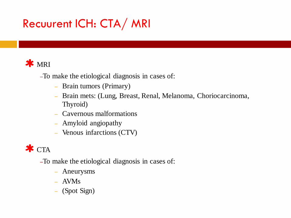
Recuurent ICH: CTA/ MRI
MRI
–To make the etiological diagnosis in cases of:
– Brain tumors (Primary)
– Brain mets: (Lung, Breast, Renal, Melanoma, Choriocarcinoma,
Thyroid)
– Cavernous malformations
– Amyloid angiopathy
– Venous infarctions (CTV)
CTA
–To make the etiological diagnosis in cases of:
– Aneurysms
– AVMs
– (Spot Sign)
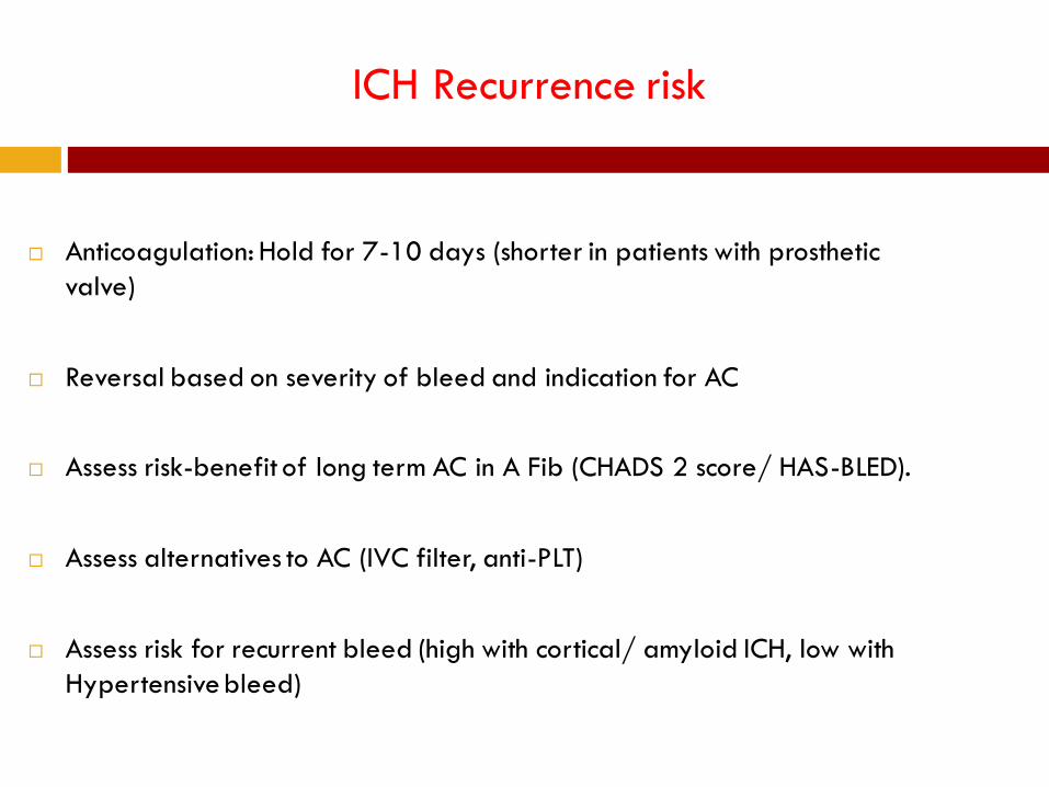
ICH Recurrence risk
Anticoagulation: Hold for 7-10 days (shorter in patients with prosthetic
valve)
Reversal based on severity of bleed and indication for AC
Assess risk-benefit of long term AC in A Fib (CHADS 2 score/ HAS-BLED).
Assess alternatives to AC (IVC filter, anti-PLT)
Assess risk for recurrent bleed (high with cortical/ amyloid ICH, low with
Hypertensive bleed)

ICH: Recurrence risk
Anti-platelets: Hold for 7 days; assess risk vs benefit of long term anti-
platelet therapy.
Statins: controversial (SPARCL vs met-analysis)
If lobar ICH and ‘weaker’ indication for statins (hypercholesterolemia)
or strong indication (CAD/ CVA) but low LDL, may be advisable to stop
statins
If deep ICH (hypertensive) and strong indication for statins (h/o MI,
CVA), may continue
Acutely may lower peri-hematomal edema

Thank you for allowing me to speak beyond my
allotted time