Specificity ofBoneMorphogenetic Protein-related …cgd.aacrjournals.org/cgi/reprint/5/6/585.pdf ·...
9
Vol. 5, 585-593, june 1994 Cell Growth & Differentiation 585 ing growth factor; cDNA, complementary DNA; UAS, upstream activation sequence. Specificity of Bone Morphogenetic Protein-related Factors: Cell Fate and Gene Expression Changes in Drosophila Embryos Induced by decapentaplegic but not 60A’ Karen Staehling-Hampton, P. David Jackson, Michael J. Clark, Andrea H. Brand, and F. Michael Hoffmann2 McArdle Laboratory for Cancer Research [K. S-H., P. D. J., M. J. C., F. M. H.] and Laboratory of Genetics [F. M. H.], University of Wisconsin, Madison, Wisconsin 53706, and Department of Genetics, Harvard Medical School, Boston, Massachusetts 021 15 [A. H. B.] Abstrad Reported assays of the bone morphogenetic proteins (BMPs) have not in general revealed specific fundions for the different proteins, belying the specificity implied by the evolutionary conservation and distind expression patterns of the genes encoding BMPs. We have used assays of developmental fundion to show that the two Drosophila homologues of the BMPs, decapentaplegic (dpp) and 60A, that both induce edopic bone formation in mammalian assay systems, have distind effects in Drosophila development. A binary expression system using the yeast transcriptional adivator GAL4 direded identical patterns of tissue and temporally specific dpp and 60A expression. When dpp enhancer elements drove GAL4 expression, GAL4-responsive dpp transgenes rescued dpp mutant phenotypes, but GAL4-responsive 60A transgenes did not. Edopic edodermal expression of dpp during gastrulation respecified the dorsal/ventral pattern of the embryo. In contrast, edopic 60A expression had no detedable effects on embryonic development but led to defeds in adult strudures or lethality during metamorphosis. Expression of 60A in cells expressing dpp did not interfere with dpp fundions, indicating that dysfundional heterodimers did not form at sufficient levels to inhibit dpp. These specific developmental responses in Drosophila indicate that in vivo fundions of BMP-like fadors can be more specific than indicated by the edopic bone formation assays and that the Drosophila embryo provides an assay system sensitive to the strudural differences that contribute to BMP specificity in vivo. Introdudion The DVR3 gene family (1), a subset of the transforming growth factor 13 superfamily, consists of the genes encoding Received 1/24/94; revised 3/1 6/94; accepted 3/23/94. 1 This work was supported by NIH Grant RR06610 (to F. M. H.), Cancer Center Core Support Grant CAO71 75, and Predoctoral Training Grant CAO91 35 (to K. S-H.). F. M. H. is the recipient of a Faculty Research Award from the American Cancer Society. 2 To whom requests for reprints should be addressed, at McArdle Laboratory for Cancer Research and Laboratory of Genetics, 1 400 University Ave., University of Wisconsin, Madison, WI 53706. 3 The abbreviations used are: DVR, decapentaplegic-vg-related; BMP, bone morphogenetic protein; dpp, Drosophila decapentaplegic; TGF, transform- mammalian BMPs 2-7 (2), the mouse OP2 and Nodal proteins (3, 4), chick dorsa!in-1 (5), Xenopus Vgl (6), and Drosophila decapentaplegic (dpp) and 60A (7-9). Like TGF-3s, DVR factors are synthesized as large precursor protei ns wh ich are secreted and processed proteolytically to release bioactive COOH-terminal homodimers (1 , 8, 10). Five of the mammalian BMP proteins can be subdivided into two classes, based on their similarity to each other and their Drosophila counterparts. BMP-2 and BMP-4 are 75% identical to dpp. BMP-5, -6, and -7 are 70% identical to 60A. In comparison, dpp and 60A are 55% identical to each other. It is therefore possible that dpp and 60A share com- mon evolutionary ancestral genes with BMP-2 and -4 and with BMP-5, -6, and -7, respectively (1). BMP factors are of clinical interest because oftheir osteo- inductive properties (2). The mammalian BMPs were first identified by their ability to stimulate bone growth when implanted ectopically in animals (11). Since then, other BMP-related proteins, including Drosophila dpp and 60A, have been shown to have osteo-inductive properties (2, 1 2-1 4). When implanted in rats, the two Drosophila pro- teins, dpp and 60A, both engender the same response of ectopic bone formation shown by all ofthe other BMPs (14). Conversely, expression of BMP-4 can rescue the mutant phenotypes of dpp mutations in Drosophila (1 5). Interest in the developmental functions of BMP proteins has been generated by the precise and dynamic expression patterns of the BMPs during mammalian development that implicate these factors in the normal development of bone, heart, skin, kidney, nervous system, and teeth (1 6-19). Gene disruption of BMP-5 led to only subtle defects and the suggestion that some of the factors may be functionally redundant (20). Clues to the potent developmental effects of these factors have come from studies in Xenopus oocytes where the TGF--related factors, activin and BMP-4, induce ectodermal cells to take on mesodermal cell fate (21 , 22). Different concentrations of activin induce different cell fates (23) or trigger cells with preexisting differences in develop- mental potential to express those differences (24). During vertebrate limb development, BMPs from the outer tip of the ectoderm, called the apical ectodermal ridge, are thought to influence the proliferation and differentiation of the under- lying mesoderm (1). Epithelial expression of BMP-4 induces expression of BMP-4 and several transcription factors in dental mesenchyme (19). Like their vertebrate counterparts, Drosophila dpp and 60A are expressed in specific patterns during development (8, 25, 26,). dpp expression in the dorsal blastoderm is required for proper dorsal/ventral polarity (27-30). Embryos that lack dpp do not form dorsal structures and secrete
-
Upload
truongxuyen -
Category
Documents
-
view
221 -
download
0
Transcript of Specificity ofBoneMorphogenetic Protein-related …cgd.aacrjournals.org/cgi/reprint/5/6/585.pdf ·...

Vol. 5, 585-593, june 1994 Cell Growth & Differentiation 585
ing growth factor; cDNA, complementary DNA; UAS, upstream activationsequence.
Specificity of Bone Morphogenetic Protein-relatedFactors: Cell Fate and Gene Expression Changesin Drosophila Embryos Induced bydecapentaplegic but not 60A’
Karen Staehling-Hampton, P. David Jackson,Michael J. Clark, Andrea H. Brand, andF. Michael Hoffmann2
McArdle Laboratory for Cancer Research [K. S-H., P. D. J., M. J. C.,F. M. H.] and Laboratory of Genetics [F. M. H.], University of Wisconsin,Madison, Wisconsin 53706, and Department of Genetics, Harvard
Medical School, Boston, Massachusetts 021 1 5 [A. H. B.]
Abstrad
Reported assays of the bone morphogenetic proteins(BMPs) have not in general revealed specific fundionsfor the different proteins, belying the specificity impliedby the evolutionary conservation and distind expressionpatterns of the genes encoding BMPs. We have usedassays of developmental fundion to show that the twoDrosophila homologues of the BMPs, decapentaplegic(dpp) and 60A, that both induce edopic bone formationin mammalian assay systems, have distind effects inDrosophila development. A binary expression systemusing the yeast transcriptional adivator GAL4 dirededidentical patterns of tissue and temporally specific dppand 60A expression. When dpp enhancer elementsdrove GAL4 expression, GAL4-responsive dpp transgenesrescued dpp mutant phenotypes, but GAL4-responsive60A transgenes did not. Edopic edodermal expressionof dpp during gastrulation respecified the dorsal/ventralpattern of the embryo. In contrast, edopic 60Aexpression had no detedable effects on embryonicdevelopment but led to defeds in adult strudures orlethality during metamorphosis. Expression of 60A incells expressing dpp did not interfere with dppfundions, indicating that dysfundional heterodimers didnot form at sufficient levels to inhibit dpp. These specificdevelopmental responses in Drosophila indicate that invivo fundions of BMP-like fadors can be more specificthan indicated by the edopic bone formation assays andthat the Drosophila embryo provides an assay systemsensitive to the strudural differences that contribute toBMP specificity in vivo.
Introdudion
The DVR3 gene family (1), a subset of the transforminggrowth factor 13superfamily, consists of the genes encoding
Received 1/24/94; revised 3/1 6/94; accepted 3/23/94.
1 This work was supported by NIH Grant RR06610 (to F. M. H.), Cancer
Center Core Support Grant CAO71 75, and Predoctoral Training GrantCAO91 35 (to K. S-H.). F. M. H. is the recipient of a Faculty Research Awardfrom the American Cancer Society.2 To whom requests for reprints should be addressed, at McArdle Laboratoryfor Cancer Research and Laboratory of Genetics, 1 400 University Ave.,
University of Wisconsin, Madison, WI 53706.3 The abbreviations used are: DVR, decapentaplegic-vg-related; BMP, bonemorphogenetic protein; dpp, Drosophila decapentaplegic; TGF, transform-
mammalian BMPs 2-7 (2), the mouse OP2 and Nodalproteins (3, 4), chick dorsa!in-1 (5), Xenopus Vgl (6), andDrosophila decapentaplegic (dpp) and 60A (7-9). LikeTGF-�3s, DVR factors are synthesized as large precursorprotei ns wh ich are secreted and processed proteolyticallyto release bioactive COOH-terminal homodimers (1 , 8, 10).Five of the mammalian BMP proteins can be subdividedinto two classes, based on their similarity to each other andtheir Drosophila counterparts. BMP-2 and BMP-4 are 75%identical to dpp. BMP-5, -6, and -7 are 70% identical to60A. In comparison, dpp and 60A are 55% identical to eachother. It is therefore possible that dpp and 60A share com-mon evolutionary ancestral genes with BMP-2 and -4 andwith BMP-5, -6, and -7, respectively (1).
BMP factors are of clinical interest because oftheir osteo-inductive properties (2). The mammalian BMPs were firstidentified by their ability to stimulate bone growth whenimplanted ectopically in animals (11). Since then, otherBMP-related proteins, including Drosophila dpp and 60A,have been shown to have osteo-inductive properties (2,1 2-1 4). When implanted in rats, the two Drosophila pro-teins, dpp and 60A, both engender the same response ofectopic bone formation shown by all ofthe other BMPs (14).Conversely, expression of BMP-4 can rescue the mutantphenotypes of dpp mutations in Drosophila (1 5).
Interest in the developmental functions of BMP proteinshas been generated by the precise and dynamic expressionpatterns of the BMPs during mammalian development thatimplicate these factors in the normal development of bone,heart, skin, kidney, nervous system, and teeth (1 6-19).Gene disruption of BMP-5 led to only subtle defects and thesuggestion that some of the factors may be functionallyredundant (20). Clues to the potent developmental effects of
these factors have come from studies in Xenopus oocyteswhere the TGF-�-related factors, activin and BMP-4, induceectodermal cells to take on mesodermal cell fate (21 , 22).Different concentrations of activin induce different cell fates(23) or trigger cells with preexisting differences in develop-mental potential to express those differences (24). Duringvertebrate limb development, BMPs from the outer tip of theectoderm, called the apical ectodermal ridge, are thought toinfluence the proliferation and differentiation of the under-lying mesoderm (1). Epithelial expression of BMP-4 inducesexpression of BMP-4 and several transcription factors indental mesenchyme (19).
Like their vertebrate counterparts, Drosophila dpp and60A are expressed in specific patterns during development(8, 25, 26,). dpp expression in the dorsal blastoderm isrequired for proper dorsal/ventral polarity (27-30). Embryosthat lack dpp do not form dorsal structures and secrete

586 Specificity of BMP-related Factors in Drosophila
cuticles that are completely ventralized. dpp protein is be-lieved to form an activity gradient in the dorsal half of theblastoderm that specificies dorsal or lateralcell fate, de-pending on the position of the cell along the activity gra-dient (29-31); high levels of dpp activity induce dorsalstructures, and low levels induce lateral structures. Laterduring embryogenesis, dpp is expressed in the visceral me-soderm in discrete domains along the developing gut tubewhere it is required for the development of the gastric caecaand the second constriction of the midgut (32, 33). Visceralmesoderm expression of dpp is required for maintaining thelocalized expression of the homeotic gene Ultrabithorax(Ubx) in the mesoderm (32, 34, 35, 36). Localized diffusionof dpp protein from the midgut mesoderm to the apposingendodermal cell layer enhances the expression of the ho-meotic gene labial in the endoderm (32, 34, 35, 36), anexample of a BMP-like factor mediating inductive interac-tions between tissues. In addition to the embryonic expres-sion patterns, dpp is expressed in Drosophila imagmnal disks(26, 37), epithelial sheets that give rise to the adult struc-tunes, where it is thought to play a role in stimulating cellproliferation, in proximal/distal axis formation, and in prop-agation of the morphogenetic furrow in the eye disk(26, 38-42).
Like dpp, 60A is expressed during embryogenesis; how-even, its pattern of expression is broader than that of dpp.60A protein can be found throughout the mesoderm but ismore concentrated in the foregut and hindgut (8). The func-tion of 60A is not known since mutations in the locus havenot been reported.
To determine if dpp and 60A, as representatives of theBMP-2/4 and the BMP-5/6/7 subgroups, respectively, areinterchangeable in contexts other than the ectopic boneformation assay, dpp and 60A transgenes were expressed inidentical temporal and spatial patterns during developmentof the Drosophila embryonic ectoderm or mesoderm. Wereport that the two proteins have different effects on alltissues examined including the eye imaginal disks, the em-bryonic ectoderm, and the embryonic mesoderm. As pre-dicted from the loss-of-function mutant phenotypes andinjections of dpp mRNA into blastoderm embryos, ectopicdpp can induce dorsal cell fate in the ventral/lateral ecto-derm. dpp was also capable of inducing changes inhomeotic gene expression in the embryonic midgut. Incontrast, ectopic 60A had no detectable effect on embryo-genesis when expressed either in the ectoderm or the vis-ceral mesoderm but did cause postembryonic lethality dun-ing the pupal stage, the first reported biological effect of60A in Drosophila. The inability of 60A to cause an em-bryonic phenotype also indicates that it did not interferewith the essential functions of the endogenous dpp expres-sion by the formation of dysfunctional heterodimers. Wepropose that these major differences in biological activitymay be due to one or more of nine amino acid differencesthat exist between the dpp/BMP2/BMP4 subgroup and the60A/BMP5/BMP6/BMP7 subgroup. The Drosophila embryoprovides an assay system in which to discover the aminoacids that are critically important to the specific in vivofunctions of these two related factors.
ResultsTargeted in Vivo Expression of dpp and 60A. To determineif dpp and 60A induce the same response, as suggested bythe ectopic bone assay (1 4), or different responses during
Drosophila development, the proteins were expressed inidentical tissue-specific patterns during embryogenesis. Tar-geted expression of dpp and 60A was accomplished usingthe GAL4/UAS system (43). dpp and 60A cDNAs wereplaced under the transcriptional control of the yeast GAL4UAS in P-element transposon vectors. The vectors alsocontained the white gene so that the presence of the trans-gene DNA in the Drosophila genome could be monitoredby wild-type red eye pigmentation. Transgenic animalswere generated using the Drosophila P-element transposonsequences (44). The two transgenes will be referred to asP[w�; UAS-dpp] and P[w�; UAS-6OAJ. We had demon-strated previously that the dpp and 60A cDNAs used inthese constructs generate processed, secreted and biologi-cally active dpp and 60A proteins in Drosophila cells (8, 10,1 4). Targeted expression of dpp or 60A was induced bymating P[w�; UAS-dppl or P[w�; UAS-60A] lines to fliesexpressing the yeast transcriptional activator GAL4 in tis-sue-specific patterns (43). The dpp and 60A proteins wereexpressed in identical GAL4-dniven patterns as assayed bystaining with antibodies to dpp and 60A (data not shown).
Rescue of dpp Phenotypes by UAS-dpp but not UAS-60A. dpp�Ik flies have a small eye phenotype resultingfrom low levels of dpp protein in the eye imaginal disks.This phenotype can be rescued when dpp transcription isdriven by a 4-kb DNA fragment within the 3’ disk regioncalled the blk enhancer fragment (26). This dpp enhancerfragment is sufficient to induce gene expression in imaginaldisks in patterns that are similar to those of the endogenousdpp gene. Transgenic flies that place a copy of the GAL4gene under the control of the b!k enhancer were made(P[w�; GAL4�dpp��k]) to drive GAL4 in the dpp disk-spe-cific pattern. P[w�; UAS-dppl lines were placed in dpp�#{176}�mutant backgrounds and mated to P[w�; GAL4�dppc�1s�(]flies that were also dpp��’. UAS-driven expression of dpprescued the mutant eye phenotype, restoring the number ofommatidia to approximately that of wild-type eyes (Fig. 1).UAS-dniven expression of dpp also rescued the eye andwing phenotypes of dpp�6, an intermediate disk allele (datanot shown). Rescue of 60A phenotypes using the P[w�;UAS-60A1 constructs could not be done since 60A mutantswere not yet available. P[w�; UAS-60A] lines were, how-ever, placed in dpp mutant backgrounds to determine if60A could substitute for dpp and rescue a dpp mutantphenotype. When GAL4�dppc�� was used to drive UAS-60A, the dpp mutant phenotypes were not rescued (data notshown), indicating that 60A could not substitute for dppfunction in the imaginal disks.
UAS-Transgene Expression of dpp and 60A in the Em-bryonic Edoderm or Mesoderm. To determine if the ec-topic expression of 60A or dpp affected ectoderm or me-soderm development, P[w�; UAS-dpp] and P[w�; UAS-60A] flies were crossed to the GAL4 69B and GAL4 24Blines. The patterns of GAL4-69B and GAL4-24B expressiondiffers from the endogenous expression patterns of dpp and60A (Fig. 2). The pattern of dpp expression is complexduring embryogenesis (for a full description of the expres-sion pattern, see Ref. 25). Early expression is limited to thedorsal surface of the blastoderm and, during gastrulation,ectodermal expression is limited to a stripe along the dorsaledge of the ectoderm and a segmented stripe through thelateral ectoderm. There are six specific sites of dpp expres-sion detectable along the embryonic alimentary canal. The60A expression pattern is less complex than dpp (for a fulldescription, see Ref. 8). Itis barely detectable at the bIas-

�-‘ ���Z;#{149}
�, .�
l�
Cell Growth & Differentiation 587
Fig. 1. UAS-driven expression of
dpp rescued the dpp mutant phe-
notype in the eye imaginal disk. Awild-type eye (A), a dppd.�( eye
(8), and a dpp’�’#{176}<; P[w; UAS-dppl/P]w�; GAL4�dppdi��] eye
(C). X 200. Introduction of thetransgenes into the dppd.� back-ground restored the number of
ommatidia to approximately thatof the wild-type eye (compare A
and C).
C ‘��2- D
. ,.
Fig. 2. Comparison of GAL4, dpp, and 60A expression patterns. Stage
14/1 5 wholemount embryos are shown. A, GAL4 69B/P]w�;UAS-lacZ1 em-
bryo stained with anti-)3-galactosidase. Note the anterior ectodermal stainingin each segment. The ectodermal stripes extended around the embryo, butthe staining close to the midline is not visible since it is above the focal plane
of the picture. B, GAL4 24B/P[w�;UAS-IacZ] embryo stained with anti-f3-galactosidase. Note the staining throughout the somatic musculature and
visceral mesoderm (arrowhead). C, wild-type embryo hybridized with dppRNA. Endogenous dpp mRNA was detected using a dpp RNA probe indiscrete domains of the developing gut tube in the pharynx ( ph), esophagus
(e), gastric caeca (c), midgut (mg), and hindgut (hg) and in the lateral
ectoderm in the posterior half of each segment. 0, wild-type embryo hy-bridized with 60A cDNA. 60A mRNA was present throughout the mesodermbut was more concentrated in the foregut and the hindgut. Left, the anteriorof each embryo. A, ventral view; 13, dorsal view; C and D, lateral views.
toderm stage and broadly expressed during gastrulation,with highest levels detected in the mesodenmal tissue layer(Fig. 2).
GAL4 69B-dniven ectodenmal expression of dpp alteredthe pattern of gene expression along the dorsal/ventral axisof the embryo (Fig. 3), consistent with previous reports thatdpp is involved in establishing embryonic dorsal/ventralpolarity (27-31). The embryos initially developed normaldorsal/ventral polarity as indicated by the localized expres-sion of tolloid (45) in the dorsal blastoderm cells and twist(46) in the ventral blastoderm cells (data not shown) and bynormal gastrulation as indicated by genmband elongation.However, by 6 h into embryogenesis when tolloid geneexpression marks the lateral ectoderm (Fig. 3A), donsaliza-tion of the ectodenm was evident by the shift of tolloidexpression toward the ventral midline (Fig. 3B). Furtherevidence that development along the dorsal/ventral axiswas altered came from examining the cuticles developed by
these embryos (data not shown). Wild-type cuticles have a
characteristic pattern of hairs on the dorsal surface anddenticle bands on the ventral surface, but in P[w�; UAS-dpp]/GAL4 69B embryos, the ventral denticle bands wereabsent and replaced by fine hairs characteristic of the dorsalsurface (data not shown). In contrast to the results withUAS-dpp, expression of UAS-60A in the same GAL4 69B
. � ectodenmal pattern had no detectable effect on the expnes-: � sion of tolloid on the pattern of the cuticle.
In the visceral mesoderm of the Drosophila embryonicmidgut, dpp is expressed in two specific locations where itis required for development of the gastric caeca and thesecond midgut constriction (32). Loss-of-function mutations
were used to establish that dpp expression is necessary fornormal expression levels of the homeotic gene labial in the
apposing endodermal cells (Fig. 3C; Refs. 32, 34, 35). Uni-form expression of dpp from the UAS tnansgene driven by
GAL4-24B in the visceral mesodenm led to uniform expres-sion of labial in the apposing endodenm (Fig. 3D). We have
also used the GAL4/UAS system to show that ectopic ex-pnession of dpp is sufficient to induce novel expression
patterns of Ultrabithorax and several other genes in themidgut (36).
To examine the effects of ectopic 60A expression in thevisceral mesoderm and to compare the results to thosereported for dpp (36), P[w�; UAS-60A] was expressedthroughout the embryonic mesodenm by the GAL4-24Btnansgene, and the embryos were stained for labial. In con-tnast to the effects with ectopic dpp, ectopic expression of60A in the same pattern did not affect the expression patternof labial on any other midgut homeotic gene expressionexamined (data not shown). However, misexpnession of60A in the GAL4-24B pattern did cause pupal lethality,presumably due to defects in the somatic on visceral meso-derm that were too subtle to detect morphologically in theembryo but sufficiently detrimental to the function of thesetissues to impair survival of the larvae.
The inability of 60A expression to alter gene expressionor onganogenesis ofthe midgut indicated that its expnession,unlike that of dpp, was not sufficient to induce any novelchanges. In addition, the expression of 60A in the samecells that express dpp and require dpp function for normalregulation of gene expression and monphogenesis did notinterfere with dpp function, e.g., UAS-dniven ectopic 60A inthe visceral mesodenm did not cause loss-of-function dppphenotypes in the midgut such as the absence of the secondconstriction, failure of gastric cacae formation, on loss oflabial or Ultrabithorax gene expression (32). We conclude

588 Specificity of BMP-related Factors in Drosophila
A B
C D
Fig. 3. Changes in ectoderm and mesoderm gene expression in response to dpp. A and B, detection of to/bid mRNA in a wild type embryo (A) and a P]w�;
UAS-dpp ]/GAL4 69B embryo (B). The lateral ectodermal patches of tolloidmRNA are closer to the midline in (B) than in (A). Cand D, expression ofthe labialhomeodomain protein in the midgut endoderm. Expression of labial is localized to cells adjacent to the cells expressing dpp in the central region of the midgutin the wildtype embryo (C). but in the P]w� ; UAS-dpp ]/GAL4 24B embryo, labial expression is spread through most of the endodermal cells. The localized
expression of labial in the wild-type embryo can be compared to the localized expression of dpp in the midgut (Fig. 2C). The broad expression of labial in theembryo with GAL4-24B-driven dpp expression can be compared to the uniform pattern of GAL4 in the visceral mesoderm (Fig. 28). In both cases (C and D),
dpp is expressed in the visceral mesoderm, and the labial response is induced in adjacent endodermal cells.
that at the levels of expression attainable with the UAStnansgenes, the formation of dysfunctional dpp/60A het-enodimers did not occur at a sufficent level to interfere withnormal dpp function. We attempted to increase the expres-sion levels of 60A in the visceral mesodenm by puttingmultiple copies of the 60A tnansgenes in the same fly, butthe increased copy number had no effect on embryonicdevelopment. This experiment does not rule out the fonma-tion of functional dpp/60A heterodimens.
lmaginal Disk Defects Caused by Edopic Expression of60A or dpp. Although expnession of 60A in the GAL4 69Bectodermal pattern did not disrupt embryogenesis, GAL4-69B-dniven expression of 60A was not without effect; itcaused postembnyonic lethality during the pupal stage. Be-cause the levels of expression of different tnansgenes differdue to position effects of their particular chromosomal in-sertion site, some lines of the UAS-60A gene were notstrong enough to be lethal. Animals from these lines thatescaped the 60A-induced pupal lethality all exhibited adistinctive inflated wing phenotype (Fig. 48; compare towild-type in Fig. 4A) similar to that observed for mutationsin a Drosophila integnin gene (47, 48). This result wasconsistent with the expression of GAL4 69B in the imaginaldisks (43) driving ectopic 60A expression during imaginaldisk development. When GAL4�dppd� tnansgene was usedto drive 60,4 expression, blistering of the wing blade wasobserved in the position consistent with the expressionpattern of the dpp”� enhancer (Fig. 4C; Ref. 26). The
severity of the blistering was increased when two copies ofthe UAS-60A transgene were present.
Discussion
60A and dpp are expressed in different temporal and spatialpatterns during normal development. This is also commonfor BMPs in mammalian development, e.g., the temporaland spatial differences of BMP2 and BMP4 during toothdevelopment (1 9). Therefore, it is difficult to know whetherthe effects of one factor are different from a second factorbecause they are acting at different times or on differenttarget tissues on because the two related factors have distincteffects on target cells. To evalute the question of in vivospecificity, we have used a system of regulated tnansgenesto express 60A and dpp in identical temporal and spatialpatterns. The results indicate that, in vivo, target cells candistingush between 60A and dpp.
Both dorsal and ventral ectoderm respond to dpp, mdi-cating that at least some of the receptor(s) and signal tnans-ducing molecules for this highly localized ligand arepresent throughout the embryo. Ectodenmal cells are not theonly type of cell that can change their fate in response todpp. dpp expression in the mesoderm in germ band ex-tended animals is sufficient to change homeotic gene ex-pression along the anterior/posterior axis, suggesting thatthe mesodermal cell fate as assayed by patterns of homeoticgene expression is also directed in part by dpp (36).

-� �
IB
7/�1
C ,/�. -
I 4
. . � 1:c� .v�!��I/
Cell Growth & Differentiation 589
Fig. 4. Expression of UAS-60A in the wing imaginal disk produces an inflated-wing phenotype. Scanning electron microscope images of a wild-type wing (A),
a wing from a P]w�; UAS-60A ]/GAL4 69B fly (B) and a P]w�; UAS-60A l/GAL4�dpp��k fly (C). The view in 8) is toward the posterior wing margin to show
thatthe dorsal and ventral surfaces ofthe wing blade were not apposed but separated to give an inflated, balloon-like appearance to the wing. In (C), the blisteringoccurs at the distal tip of the wing, centered on the central wing veins. Bars, 0.25 mss.
None of the changes in ectodenmal cell fate, embryonicmidgut monphology, on gene expression were observedwhen 60A expression was driven by GAL4 in the ectodenmon visceral mesoderm. Ectopic 60A expression was notwithout effect however, because it led to lethality during the
pupal stage. Surviving adults exhibited an inflated wingphenotype reminiscent of defects caused by mutations inDrosophila integnins. Studies with mammalian homologuesindicate a role for TGF-�3 family members in regulating celladhesion. TGF-�3s have been shown to alter the expressionof mRNAs for individual integnin subunits (49, 50). Further-more, mammalian OP-i (BMP7), which is closely related to60A, can induce expression of neural cell adhesion mole-cules and neural aggregation in a neunoblastoma-gliomahybrid cell line (51).
Ectopic 60A expression in the embryo also did not causeembryonic midgut phenotypes analogous to loss-of-func-tion dpp mutant phenotypes, indicating that the ectopic60A did not interfere with endogenous dpp function asmight have been expected if dysfunctional hetenodimenswere formed. Furthermore, when both UAS transgeneswere present in the same animal, GAL4-dniven 60A expnes-sion did not interfere with the phenotypic consequences ofGAL4-dniven dpp expression (data not shown). Het-erodimer formation has been shown to occur with theactivins/inhibins, other members of the TGF-� family. Theactivins/inhibins are dimenic proteins composed of variouscombinations of f3A, f3B, and a subunits. Different combi-nations of subunits have opposing functions, e.g., f3 ho-modimens, called activin, stimulate the production of folli-cle-stimulating hormone; whereas, ctf3 heterodimens, calledinhibin, inhibit follicle-stimulating hormone production(52, 53). By analogy, dpp and 60A could form both het-enodimers and homodimers, and each complex could havea distinct activity. The results of the studies presented hereindicate that 60A and dpp did not form dysfunctional het-enodimers sufficiently well to impair dpp function on that if
heterodimens were formed, they were biologically active inmimicking dpp homodimers.
The absence of embryonic phenotypes by ectopic 60Aexpression in either the ectoderm on the mesodenm are mostconsistent with a restricted embryonic distribution of thereceptor for 60A and/on saturation of 60A receptors by thebroad endogenous embryonic expression of 60A. Alterna-tively, tissue-specific differences in the processing of thedpp and 60A precursor proteins might exist. In Xenopus,Vgl proteins made from injected synthetic mRNAs were notprocessed and could not form dimers (54). When the loss-of-function 60A mutant phenotypes are known, they shouldprovide clues to molecular probes appropriate for detectingcellular responses to 60A and to the normal role of 60A inimaginal disk development and metamorphosis.
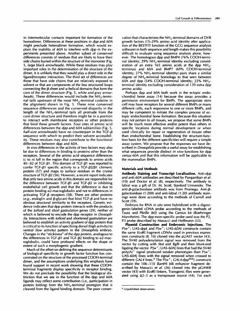
We can begin to speculate on the molecular basis for thebiological distinction between the function of the dpp and60A proteins by examining the conserved sequence differ-ences between these proteins relative to the crystallo-graphic structures for TGF-j31 and TGF-f32 (55, 56). Defi-nition of the conserved sequence differences begins withthe consideration that the carboxy terminal fragments ofdpp, BMP2, and BMP4 and of 60A, BMP5, BMP6, and OP-i(BMP7) belong to distinct homology subgroups that exhibitapproximately 70% sequence identity between the mem-bers within each subgroup and only about 50% identitybetween subgroups. Alignment ofthese sequences reveals anumber of sites at which amino acid identity is conservedwithin the subgroups but differs between subgroups (Fig. 5,sites labeled a-x). In addition to these sequence differences,a disparity in the length of the sequences upstream of theNH2-terminal-most cysteine has been conserved betweenthe members of the two subgroups (Fig. 5, top).
Interpretation of these sequence differences can be at-tempted by extrapolation from homologous residues in theTGF-�3 structures. Differences shown by arrowheads b, p. t,
and u in Fig. 5 are in positions that in TGF-f3 may participate

590 Specificity of BMP-related Factors in Drosophila
* * * * *
PTRRR NHDDT DPP
Q-K HKQ-KRLKS + 2K�
HKQ-KRLKS + 211
sPK.}I HSQ-ARKK-KNSPKQQ- - -K- -KI! 411
AANKRKNQN+N++SSHQDS+ -MSS VGDYNTS+Q KQA 5K�
-AS+RRRQQS+NR+TQSQD+A -VSS ASDYNSS+L KTA 6K�
-AS+RRRQQS+NR+TQSQD++ -GSG SSDYNGS+L XTA VQR1
-TG+KQRSQN+S+TPXNQEAL -M-N VAENSSSDQ +QA 7E�
-PGGKQRSQN+S+TPKNQEAL -M-S VAENSSSDQ +QA 7�
-TSNKHWNQE+A+TYKEQDNLPPANITDGI+ P-GK-RFL KQA 711
SA-HPRKRKKS VSPNNVPLLEPM ESTR S 60A
1 � *
CRBHSLYVDF LGYDAYY GKC FP H TNHAVVQTLV DPP
-K--P N P---H-F------+ L + 2K�
----P N P--H-F----+ L + 211
N P--Q-P----D L +
N P--Q-F----D L + 411
-KK-E---S-R-+--Q---+--+--A-F--D-+-S----++-+-+----+ 5K�
--K-E---S-Q-+--Q---#{247}--K--A-N--D-+-S---++-+-+-----+ 6H�
-KK--E---S-Q-+--Q-�--+--K---A-N--D-+--S---++-+-+----+ VOR1
-KK-E---S-R-+--Q---+--+--A-Y--E-+-A---+SY+-+----+ 7K�
-KX-E---S-R-+--Q---+--+--A-Y--E-+-A---+SY+-+----+ 7�
-KK-E-F-S--R-+--Q---+--+--A-Y---D-+--A---+SF+-+----+ 711
-Q1vIQT- -ID-K-L--H-- -I- �-E- -G-P---S-E-N---NA-M-A- ---I 60k
EPP
-sv- s-I E-S+IS--’--DEN+� D-V--E 2H�
-sv- TNI E-S+IS----DEN+K D-V-E 211
-sv- ssi E-S+IS-----DEYDI( V-E 4II�
-sv- ss]: E-S+IS- ---DEYDK V-E 411
++-F-DH---+--+--K-N+IS+--FD-S-+-I--+-++-V-R+---+ 5H�
++- --EY---+--+--K-N+IS+--FD-N-+-I--+-++-V-RA----+ 6K�
++---EY---+--+--X-N+IS+--FD-N-+-I--+-++-V-RA---+ V�3R1
+FI--ET---+--+-- --N+IS+--FD-S-+-I--+-++-V-PA---+ 7E�
+FI--DT---+--+----N+IS+--FD-S-+-I--+-++-V-PA---+ 7]�tl�
+FI--ET---+--+----NGIS+--FD-SA+-I--+-K+-V-QA---+ 711
HLLE-K--- -P- -A- -R-GALPV- -HLNDEN-N- -K-RN-I-KS- - -H 60k
Fig. 5. Conserved amino acid differences between the dpp subfamily and the 60A subfamily of BMPs may identify residues responsible for functional differencesbetween dpp and 60A. Sequences retrieved from the Genbank database include dpp, human BMP2 )2Hs), Xenopus BMP2 (2X1), human BMP4 (4Hs), Xenopus
BMP4 (4X1), human BMP5 (5Hs), human BMP6 (6Hs), mouse BMP6 (VGR1), human BMP7 (7Hs), mouse BMP7 (7 Mm), Xenopus BMP7 (7Xl), and Drosophila60A. Sequences begin after the predicted or actual proteolytic processing site. Top, the disparity in the length ofthe sequences upstream ofthe NH2-terminal-mostcysteine that has been conserved between the members of the two subgroups. Shaded regions include amino acids largely conserved throughout all sequences,
beginning with the first invariant cysteine and extending to the carboxy-terminus and including all seven invariant cysteines. A dash (-) indicates that the aminoacid is the same as shown in dpp. A plus (+) indicates thatthe amino acid is the same as shown in the 60A sequence. Sites at which members ofthe dpp subgroup
show amino acid conservation that differs in hydrophilicity or charge from members of the 60A subgroup are indicated by arrowheads labeled a-x. The following
structural properties of these residues were assigned based on the structures of TGF-f31 and TGF-)32 (53, 54): black arrowheads in positions where the amino acid
side chain is predicted to be buried by the monomer secondary structure (solvent accessible area = A<20 a2); grey arrowheads in positions where the amino
acid side chain is predicted to have intermediate solvent accessibility (2O�2 < A < 60�2); and open arrowheads in positions predicted to have side chains exposedto solvent )A>6O�2). The “ D” over five arrowheads indicates positions in the TGF-f32 monomer on the internal face of the homodimer. The half-size arrowheads
indicate amino acid differences between the BMP subfamilies in positions that are not in the TGF-j3 sequences, thus their structural significance could not be
extrapolated from the TGF-(3 structure.
Cell Growth & Differentiation 591
4 Unpublished observations.
in intermolecular contacts important for formation of thehomodimer. Differences at these positions in dpp and 60A
might preclude heterodimer formation, which would ex-plain the inability of 60A to interfere with dpp in the ex-peniments presented above. Another subset of conserveddifferences consists of residues that are likely to have theirside chains buried within the structure ofthe monomer (Fig.5, large black arrowheads). While these residues may playimportant roles in the determination of the structure of thedimer, it is unlikely that they would play a direct role in theligand/receptor interaction. The third set of differences arethose that have side chains that are relatively exposed tosolvent or that are components of the less structured loopsconnecting the f3-sheet and a-helical domains that form thecore of the dimer structure (Fig. 5, white and gray arrow-heads). These differences would include the NH2-termi-nal tails upstream of the most NH2-terminal cysteine inthe alignment shown in Fig. 5. These nine conservedsequence differences and the conserved disparity in thelength of the NH2-terminal tails all protrude from thecore dimer structure and therefore might be in a positionto interact with membrane receptors or other proteinsthat bind these growth factors. Four sites of amino aciddifferences between the dpp and 60A subfamilies (Fig. 5,half-size arrowheads) have no counterpart in the TGF-j3sequence with which to predict their solvent accessibil-ity. These residues may also contribute to the functionaldifferences between dpp and 60A.
In vivo differences in the activity of two factors may alsobe due to differences in binding to proteins other than thereceptors. Several of the amino acid sequence differences(C to n) fall in the region that corresponds to amino acids40-82 of TGF-f31 . This domain of TGF-f31 was reported toconfer TGF-f31 specific activity to a TGF-f32/f31 chimericprotein (57) and maps to surface residues in the crystalstructure ofTGF-j32 (56). However, a recent report indicatesthat only two amino acids in this domain are responsible forthe difference between TGF-�31 and TGF-j32 in inhibitingendothelial cell growth and that the difference is due toprotein binding a2-macroglobulin and not to differences inactivating TGF-f3 receptors (58). There are other proteins(e.g., endoglmn and �-glycan) that bind TGF-� and have noobvious structural similarity to the receptors. Genetic evi-dence indicates that dpp protein interacts with the productsof the to!Ioid and short gastru!ation genes (29), neither ofwhich is believed to encode the dpp receptor in Drosoph-ila. Interactions with tolloid and shortened gastrulation arebelieved to establish an activity gradient of dpp protein thatis critical to its function of specifying dorsal (high activity) toventral (low activity) pattern in the Drosophila embryo.Changes in the “stickiness” ofthe dpp protein, analogous tothe differences in TGF-�31 and TGF-g32 binding to a2-mac-roglobulmn, could have profound effects on the shape orextent of such a morphogenic gradient.
Much of the effort on defining the sequence determinantsof biological specificity in growth factor function has con-centrated on the structure of the processed COOH-terminaldimer, and the assumptions underlying this emphasis havefound support in recent work showing that these COOH-terminal fragments display specificity in receptor binding.We do not preclude the possibility that the biological dis-tinction that we see in the function of the dpp and 60Aligands may reflect some contribution (i.e., participation inprotein folding) from the NH2-terminal proregion that iscleaved from the ligand binding domain. The poor conser-
vation that characterizes the NH2-terminal domains of DVRgrowth factors (1 5-29% amino acid identity after applica-tion of the BESTFIT function of the GCG sequence analysissoftware) in both sequence and length makes this possibilitydifficult to evaluate using sequence analysis alone, how-ever. The homologous dpp and BMP4 (76% COOH-termi-nal identity; 29% NH2-terminal identity excluding consid-eration of an extra 165 amino acids at the dpp NH2-terminus) and 60A and BMP7 (69% COOH-terminalidentity; 27% NH2-terminal identity) pairs share a similardegree of NH2-terminal homology to that seen between60A and dpp (54% COOH-terminal identity; 22% NH2-terminal identity excluding consideration of 1 39 extra dppamino acids).
Perhaps dpp and 60A both work in the ectopic endo-chondral bone assay (1 4) because the assay provides apermissive environment for BMPs. The appropriate stemcell may have receptors for several different BMPs or manydifferent cells, each responsive to one or more BMPs, andmay be competent to initiate the complex process of ec-topic endochondral bone formation. Because this situationmay not pertain to all tissues, we propose that some BMPswill be much more effective and/or potent than others inspecific locations during normal development or whenused clinically for repair or regeneration of tissues otherthan endochondral bone. Establishing the structure-func-tion basis for the different specificities will require a simpleassay system. We propose that the responses we have de-scnibed in Drosophila provide a useful assay for establishingwhat sequences provide distinct in vivo functions for dppversus 60A and that this information will be applicable tothe mammalian BMPs.
Materials and Methods
Antibody Staining and Transcript Localization. Anti-dppand anti-60A antibodies are described by Panganiban et a!.(1 0) and Doctor et a!. (8), respectively. The antibody tolabial was a gift of Dr. M. Scott, Stanford University. Theanti-�-gaIactosidase antibody was from Promega. Anti-�3-galactosidase (1 :200) and anti-labial (1 :20) antibody stain-ings were done according to the methods of Carroll andScott (59).
Embryos for RNA in situ were hybridized with a digoxi-genin-labeled cDNA probe according to the methods ofTautz and Pfeifle (60) using the Genius kit (BoehringerMannheim). The dpp exon-specific probe used was the P2,P3 probe described by Masucci and Hoffmann (33).
Plasmid Construdion and Embryonic Injedions. TheP[w�; UAS-dpp] and P[w�; UAS-60A1 constructs containthe same EcoRI fragment cDNAs used in previous expres-sion constructs (8, 10) cloned into the pUAST vector (43).The SV4O polyadenylation signal was removed from thevector by cutting with Stul and Bglll and then blunt-endligating the vector. P[w�; UAS-60A1 lines that had the SV4Opo�y(��F signal produced weaker phenotypes than P[w�;UAS-60A1 lines with the signal removed when crossed todifferent GAL4 lines.4 The P[w�; GAL4-dpp�”�”J constructscontain the 106-1 10 BamHI blk enhancer fragment de-scribed by Masucci et a!. (26) cloned into the pGAWBvector (43) with EcoRl linkers. Transgenic flies were gener-ated using �2-3 as a transposase source (44). For each

592 Specificity of BMP-related Factors in Drosophila
experiment, three to five independent lines were tested. Allofthe lines produced the same qualitative result but differedin the strength of the phenotype due to position effect.
Scanning Eledron Microscopy. Specimens were dehy-drated in ethanol and were prepared for electron micros-copy using a Tousimis critical point dryer and a Bio-RadGold Sputter Coater. Scanning electron microscopy pic-tunes were taken on a Hitachi 5570 microscope.
Drosophila Stocks. Drosophila stocks were grown onstandard media at 25#{176}C.Canton S flies were used as awild-type control. P[w�; UAS-lacZJ, GAL4 69B, and GAL4
24B are described by Brand and Pernimon (43).
AcknowledgmentsWe thank Matthew Scott for sending antibodies and Nancy Lawrence forproviding expertise with the scanning electron microscope.
References1 . Lyons, K. M., Jones, C. M., and Hogan, B. L. M. The DVR gene family inembryonic development. Trends Genet., 7: 408-41 2, 1991.
2. Rosen, V., and Thies, R. S. The BMP proteins in bone formation andrepair. Trends Genet., 8: 97-102, 1992.
3. Ozkaynak, E., Schnegelsberg, P. N. J., Jin, D. F., Clifford, G. M., Warren,F. D., Drier, E. A., and Oppermann, H. Osteogenic protein-2: a new memberof the transforming growth factor-f3 superfamily expressed early in embryo-genesis. I. Biol. Chem., 267: 25220-25227, 1992.
4. zhou, x., Sasaki, H., Lowe, L., Hogan, B. L. M., and Kuehn, M. R. Nodalis a novel TGF-f3-like gene expressed in the mouse node during gastrulation.Nature (Lond.), 361: 543-547, 1993.
5. Basler, K., Edlund, T., Jessell, T., and Yamada, T. Control of cell patternin the neural tube: regulation of cell differentiation by Dorsalin-1, a novelTGF� family member. Cell, 73: 687-702, 1993.
6. Weeks, D. L., and Melton, D. A. A maternal mRNA localized to thevegetal hemisphere in Xenopus eggs codes for growth factor related toTGF-�. Cell, 51:861-867, 1987.
7. Padgett, R. W., St. Johnston, R. D., and Gelbart, W. M. A transcript froma Drosophila pattern gene predicts a protein homologous to the transforminggrowth factor-a family. Nature (Lond.), 325: 81-84, 1987.
8. Doctor, J. S., Jackson, P. D., Rashka, K. E., Visalli, M., and Hoffmann,F. M. Sequence, biochemical characterization, and developmental expres-sion of a new member of the TGF-� superfamily in Drosophila melangaster.Dev. Biol., 151:491-505, 1992.
9. Wharton, K. A., Thomsen, G. H., and Gelbart, W. M. Drosophila 60Agene, a new TGF-� family member is closely related to human bone mor-phogenetic proteins. Proc. NatI. acad. Sci. USA, 88: 9214-9218, 1991.
1 0. Panganiban, G. E. F., Rashka, K. E., Neitzel, M. D., and Hoffmann, F. M.
Biochemical characterization of the Drosophila dpp protein, a member ofthe transforming growth factor-a family. Mol. Cell. Biol., 10: 2669-2677,1990.
1 1 . Wozney, I. M., Rosen, V., Celeste, A. J., Mitsock, L. M., Whitters, M. J.,Kriz, R. W., Hewick, R. M., and Wang E. A. Novel regulators of boneformation: molecular clones and activities. Science (Washington DC), 242:1528-1534, 1988.
12. Wang, E. A., Rosen, V., D’Alessandro, J. S., Baudy, M., Cordes, P.,Harada, T., Israel, D. I., Hewick, R., Kerns, K. M., LaPan, P., Luxenberg, J. M.,McQuaid, D., Moutsatsos, I. K., Nove, J., and Wozney, J. M. Recombinanthuman bone morphogenetic protein induces bone formation. Proc. NatI.Acad. Sci. USA, 87: 2220-2224, 1990.
1 3. Sampath, T. K., Maliakal, J. C., Hauschka, P. V., Jones, W. K., Sasak, H.,Tucker, R. F., White, K. H., Coughlin, J. E., Tucker, M. M., Pang, R. H. L.,Corbett, C., Ozkaynak, E., Oppermann, H., and Reuger, D. C. Recombinanthuman Osteogenic Protein-i (hOP-i ) induces new bone formation in vivowith a specific activity comparable with natural bovine osteogenic protein
and stimulates osteoblast proliferation and differentiation in vitro. J. Biol.
Chem., 267:20352-20362, 1992.
14. Sampath, T. K., Rashka, K. E., Doctor, J. S., Tucker, R. F., and Hoffmann,F. M. Drosophila TGF-� superfamily proteins induce endochondrial boneformation in mammals. Proc. NatI. Acad. Sci. USA, 90: 6004-6008, 1993.
1 5. Padgett, R. W., Wozney, J. M., and Gelbart, W. M. Human BMP
sequences can confer normal dorsal-ventral patterning in the Drosophilaembryo. Proc. NatI. Acad. Sci. USA, 90: 2905-2909, 1993.
1 6. Lyons, K. M., Pelton, R. W., and Hogan, B. 1. M. Patterns of expressionof murine Vgr-1 and BMP-2a RNA suggest that the transforming growthfactor-a-like genes coordinately regulate aspects of embryonic development.Genes Dev., 3: 1 657-i 668, 1989.
1 7. Lyons, K. M., Pelton, R. W., and Hogan, B. L. M. Organogenesis and
pattern formation in the mouse: RNA distribution patterns suggest a role forBone Morphogenetic Protein-2A (BMP-2A). Development (Camb.), 109:
833-844, 1990.
1 8. Jones, C. M., Lyons, K. M., and Hogan, B. 1. M. Involvement of BoneMorphogenetic protein-4 (BMP-4) and vgr-1 in morphogenesis and neuro-genesis in the mouse. Development (Camb.), 1 1 1: 531-542, 1991.
19. Vainio, S., Karavanova, I., and Thesleff, I. Identification of BMP-4 as asignal mediating secondary induction between epithelial and mesenchymaltissues during early tooth development. Cell, 75: 45-58, 1993.
20. Kingsley, D. M., Bland, A. E., Grubber, J. M., Marker, P. C., Russell,1. B., Copeland, N. G., and Jenkins, N. A. The mouse short ear skeletalmorphogenesis locus is associated with defects in a bone morphogeneticmember of the TGF� superfamily. Cell, 71: 399-410, 1992.
21 . Smith, J. C., Price, B. M. J., Van Nimmen, K. V., and Huylebroeck, D.Identification of a potent Xenopus mesoderm-inducing factor as a homo-logue of activin A. Nature (Lond.), 345: 729-732, 1990.
22. Jones, M. C., Lyons, K. M., Lapan, P. M., Wright, C. V. E., and Hogan,B. L. M. DVR-4 (Bone Morphogenetic Protein-4) as a posterior-ventralizingfactor in Xenopus mesoderm induction. Development (Camb.), 1 15: 639-647, 1992.
23. Green, J. B. A., and Smith, J. C. Graded changes in the dose ofa XenopusActivin A homologue elicit stepwise transitions in embryonic cell fate.Nature (Lond.), 347: 391-394, 1990.
24. Sokol, S., and Melton, D. A. Preexistent pattern in Xenopus animal polecells revealed by induction with activin. Nature (Lond.), 351: 409-411,
1991.
25. Jackson, P. D., and Hoffmann, F. M. Embryonic expression patterns ofthe Drosophila decapentaplegic gene: separate regulatory elements controlblastoderm expression and lateral ectodermal expression. Dev. Dyn., 199:28-44, 1994.
26. Masucci, J. D., Miltenberger, R. J., and Hoffmann, F. M. Pattern-specificexpression of the Drosophila decapentaplegic gene in imaginal disks isregulated by 3’ cia-regulatory elements. Genes Dev., 4: 201 1-2023, 1990.
27. Irish, V. F., and Gelbart, W. M. The decapentaplegicgene is required for
dorsal-ventral patterning of the Drosophila embryo. Genes Dev., 1: 868-879, 1987.
28. Ray, R. P., Arora, K., NUsslein-Voihard, C., and Gelbart, W. M. Thecontrol of cell fate along the dorsal-ventral axis of the Drosophila embryo.Development (Camb.), 1 13: 35-54, 1991.
29. Ferguson, E. L., and Anderson, K. V. Localized enhancement and re-pression of the activity of the TGF-� family member, decapentaplegic, isnecessary for dorsal-ventral pattern formation in the Drosophila embryo.
Development (Camb.), 1 14: 583-597, 1992.
30. Ferguson, E. L., and Anderson, K. V. decapentaplegic acts as a morpho-gen to organize dorsal-ventral pattern in the Drosophila embryo. Cell, 71:
451-461, 1992.
31 . Wharton, K. A., Ray, R. P., and Gelbart, W. M. An activity gradient ofdecapentaplegic is necessary for the specification of dorsal pattern elements
in the Drosophila embryo. Development (Camb.), 117: 807-822, 1993.
32. Panganiban, G. E. F., Reuter, R., Scott, M. P., and Hoffmann, F. M. ADrosophila growth factor homolog, decapentaplegic, regulates homeoticgene expression within and across germ layers during midgut morphogen-esis. Development (Camb.), 1 10: 1041-1050, 1990.
33. Masucci, J. D., and Hoffmann, F. M. Identification of two regions fromthe Drosophila decapentaplegic gene required for embryonic midgut devel-opment and larval viability. Dev. Biol., 159: 276-287, 1993.
34. ImmerglUck, K., Lawrence, P. A., and Bienz, M. Induction across germlayers in Drosophila mediated by a genetic cascade. Cell, 62: 261-268,1990.
35. Reuter, R., Panganiban, G. E. F., Hoffmann, F. M., and Scott, M. P.Homeotic genes regulate the spatial expression of putative growth factors inthe visceral mesoderm of Drosophila embryos. Development (Camb.), 110:1031-1040, 1990.
36. Staehling-Hampton, K., and Hoffmann, F. M. Ectopic decapentaplegic inthe Drosophila midgut alters the expression of five homeotic genes, dpp andwingless, causing specific morphological defects. Dev. Biol., in press, 1994.
37. Blackman, R. K., Sanicola, M., Raftery, L. A., Gillevet, T., and Gelbart,W. M. An extensive 3’ cia-regulatory region directs the imaginal disk ex-pression of decapentaplegic, a member of the TGF-� family in Drosophila.Development (Camb.), 1 1 1: 657-665, 1991.

Cell Growth & Differentiation 593
38. Bryant, P. J. Localized cell death caused by mutations in a Drosophila
gene coding for a transforming growth factor-a homolog. Dev. Biol., 128:386-395, 1988.
39. Raftery, L. A., Sanicola, M., Blackman, R. K., and Gelbart, W. M. Therelationship of decapentaplegic and engrailed expression in Drosophila
imaginal disks: do these genes mark the anterior-posterior compartmentboundary? Development (Camb.), 1 13: 27-33, 1991.
40. Posakony, L. G., Raftery, L. A., and Gelbart, W. M. Wing formation inDrosophila melanogaster requires decapentaplegic gene function along theanterior-posterior compartment boundary. Mech. Dev., 33: 69-82, 1991.
41 . Campbell, G., Weaver, T., and Tomlinson, A. Axis specification in thedeveloping appendage: the role of wingless, decapentaplegic and the ho-meobox gene aristaless. Cell, 74: 1 1 1 3-1 1 23, 1993.
42. Heberlein, U., Wolff, T., and Rubin, G. M. The TGF-� homolog dpp andthe segment polarity gene hedgehog are required for propagation of amorphogenetic wave in the Drosophila retina. Cell, 75: 91 3-926, 1993.
43. Brand, A. H., and Perrimon, N. Targeted gene expression as a means ofaltering cell fates and generating dominant phenotypes. Development(Camb.), 1 18: 401 -41 5, 1993.
44. Robertson, H. M., Preston, C. R., Phillis, R. W., Johnson-Schlitz, D. M.,Benz, W. K., and Engels, W. R. A stable source of P-element transposase inDrosophila melangaster. Genetics, 1 18: 461-470, 1988.
45. Shimell, M. J., Ferguson, E. L., Childs, S. R. and O’Connor, M. B. TheDrosophila dorsal-ventral patterning gene tolloid is related to human bonemorphogenetic protein 1 . Cell 67: 469-481 , 1991.
46. Thisse, B., Stoetzel, C. El Messal, M., and Perrin-Schmitt, F. Genes of theDrosophila maternal dorsal group control the specific expression of thezygotic gene twist in presumptive mesodermal cells. Genes Dev., 1: 709-715, 1987.
47. Wilcox, M., DiAntonio, A., and Leptin, M. The function of PS integrinsin Drosophila wing morphogenesis. Development (Camb.), 107: 891-897,1989.
48. Brower, D. L., and Jaffe, S. M. Requirement for integrins during Dro-sophila wing development. Nature (Lond.), 342: 285-287, 1989.
49. lgnotz, R. A., and Massagu#{233},J. Cell adhesion receptors as targets fortransforming growth factor-f3 action. Cell, 51: 1 89-197, 1987.
50. Heino, J., and Massagu#{233},J. Transforming growth factor-a switches thepattern of integrins expressed in MG-63 human osteosarcoma cells and
causes a selective loss of cell adhesion to laminin. J. Biol. Chem., 264:
21806-21811, 1989.
51 . Perides, G., Safran, R. M., Rueger, D. C., and Charness, M. E. Inductionofthe neural cell adhesion molecule and neuronal aggregation by osteogenicprotein 1 . Proc. NatI. Acad. Sci. USA, 89: 10326-1 0330, 1992.
52. Mason, A. J., Hayflick, J. S., Ling, N., Esch, F., Ueno, N., Ying, S-Y.,Guillemin, R., Niall, H., and Seeburg, P. H. Complementary DNA sequences
of ovarian follicular fluid inhibin show precursor structure and homologywith transforming growth factor-�3. Nature (Lond.), 318: 659-663, 1985.
53. Ling, N., Ying, S-Y., Ueno, N., Shimasaki, S., Esch, F., Hotta, M., andGuillemin, R. Pituitary FSH is released by a heterodimer of the a-sub-units
from the two forms of inhibin. Nature (Lond.), 321: 779-782, 1986.
54. Dale, L., Matthews, G., and Colman, A. Secretion and mesoderm-inducing activity of the TGF-�3-reIated domain of Xenopus Ygi . EMBO J., 12:
4471-4480, 1993.
55. Schlunegger, M. P., and GrUtter, M. G. An unusual feature revealed by
the crystal structure at 2.2 a resolution of human transforming growth factor-�2. Nature (Lond.), 358: 430-434, 1992.
56. Daopin, S., Piez, K. A., Ogawa, Y., and Davies, D. R. Crystal structureof transforming growth factor-�2: an unusual fold for the superfamily.Science (Washington DC), 257: 369-373, 1992.
57. Qian, S. W., Burmester, J. K., Merwin, J. R., Madri, J. A., Sporn, M. B.,and Roberts, A. B. Identification of a structural domain that distinguishes theactions of the type 1 and type 2 isoforms of transforming growth factor � on
endothelial cells. Proc. NatI. Acad. Sci. USA, 89: 6290-6294, 1992.
58. Burmester, J. K., Qian, S. W., Roberts, A. B., Huang, A., Amatayakul-Chantler, S., Suardet, L., Odartchenko, N., Madri, J. A., and Sporn, M. B.Characterization of distinct functional domains of transforming growth factorf3. Proc. NatI. Acad. Sci. USA, 90: 8628-8632, 1993.
59. Carroll, S. B., and Scott, M. P. Localization of the fushi tarazu proteinduring Drosophila embryogenesis. Cell, 43: 47-57, 1985.
60. Tautz, D., and Pfeifle, C. A non-radioactive in situ hybridization method
for the localization of specific RNAS in Drosophila reveals translationalcontrol of the segmentation gene hunchback. Chromosoma (Berl.), 98: 81-85, 1989.