Species‑specificity of temporal processing in the auditory ...
19
1 3 J Comp Physiol A DOI 10.1007/s00359-015-0997-4 ORIGINAL PAPER Species‑specificity of temporal processing in the auditory midbrain of gray treefrogs: interval‑counting neurons Gary J. Rose · Jessica L. Hanson · Christopher J. Leary · Jalina A. Graham · Rishi K. Alluri · Gustavo A. Vasquez‑Opazo Received: 18 December 2014 / Revised: 26 February 2015 / Accepted: 27 February 2015 © Springer-Verlag Berlin Heidelberg 2015 Keywords Whole-cell recording · Anurans · Rise-time selectivity · Amplitude modulation · Inferior colliculus Abbreviations ICN Interval-counting neuron IC anuran Anuran inferior colliculus AM Amplitude modulation SAM Sinusoidal amplitude modulation SPL Sound pressure level in decibels (dB), re 2 × 10 −5 N/m 2 EPSP Excitatory postsynaptic potential IPSP Inhibitory postsynaptic potential BEF Best excitatory frequency Introduction North American gray treefrogs comprise two cryptic spe- cies, Hyla versicolor and H. chrysoscelis. H. versicolor (tetraploid) is believed to have evolved at least three times from hybridization (i.e., allopolyploidization events) between diploid species, which include an ancestral rep- resentative of extant H. chrysoscelis and two genetically distinct chrysoscelis-like lineages that now appear to be extinct (Holloway et al. 2006). Because heterospecific mat- ings between H. versicolor and H. chrysoscelis result in sterile triploid progeny (Johnson 1963), premating isola- tion mechanisms are of paramount importance. Advertise- ment calls of H. versicolor and H. chrysoscelis, which are the primary premating isolation mechanisms, consist of a series of pulses that differ in shape, duration and repeti- tion rate; in H. versicolor, intrapulse amplitude rises more slowly, pulse duration is greater and pulse rate is slower (Fig. 1). Behavioral studies have shown that pulse rise time and duration are particularly salient temporal features used Abstract Interval-counting neurons (ICNs) respond after a threshold number of sound pulses have occurred with specific intervals; a single aberrant interval can reset the counting process. Female gray treefrogs, Hyla chrysoscelis and H. versicolor, discriminate against syn- thetic ‘calls’ possessing a single interpulse interval 2–3 three times the optimal value, suggesting that ICNs are important for call recognition. The calls of H. versicolor consist of pulses that are longer in duration, rise more slowly in amplitude and are repeated at a slower rate than those of H. chrysoscelis. Results of recordings from mid- brain auditory neurons in these species include: (1) ICNs were found in both species and their temporal selectiv- ity appeared to result from interplay between excitation and inhibition; (2) band-pass cells in H. versicolor were tuned to slower pulse rates than those in H. chrysosce‑ lis; (3) ICNs that were selective for slow-rise pulse shape were found almost exclusively in H. versicolor, but fast- rise-selective neurons were found in both species, and (4) band-suppression ICNs in H. versicolor showed response minima at higher pulse rates than those in H. chrysos‑ celis. Selectivity of midbrain ICNs for pulse rise time and repetition rate thus correlate well with discrimina- tory abilities of these species that promote reproductive isolation. G. J. Rose (*) · J. L. Hanson · C. J. Leary · J. A. Graham · R. K. Alluri · G. A. Vasquez-Opazo Department of Biology, University of Utah, 257 South 1400 East Rm 204, Salt Lake City, UT 84112, USA e-mail: [email protected] C. J. Leary University of Mississippi, Oxford, MS 38677, USA
Transcript of Species‑specificity of temporal processing in the auditory ...

1 3
J Comp Physiol ADOI 10.1007/s00359-015-0997-4
ORIGINAL PAPER
Species‑specificity of temporal processing in the auditory midbrain of gray treefrogs: interval‑counting neurons
Gary J. Rose · Jessica L. Hanson · Christopher J. Leary · Jalina A. Graham · Rishi K. Alluri · Gustavo A. Vasquez‑Opazo
Received: 18 December 2014 / Revised: 26 February 2015 / Accepted: 27 February 2015 © Springer-Verlag Berlin Heidelberg 2015
Keywords Whole-cell recording · Anurans · Rise-time selectivity · Amplitude modulation · Inferior colliculus
AbbreviationsICN Interval-counting neuronICanuran Anuran inferior colliculusAM Amplitude modulationSAM Sinusoidal amplitude modulationSPL Sound pressure level in decibels (dB),
re 2 × 10⁻⁵ N/m2
EPSP Excitatory postsynaptic potentialIPSP Inhibitory postsynaptic potentialBEF Best excitatory frequency
Introduction
North American gray treefrogs comprise two cryptic spe-cies, Hyla versicolor and H. chrysoscelis. H. versicolor (tetraploid) is believed to have evolved at least three times from hybridization (i.e., allopolyploidization events) between diploid species, which include an ancestral rep-resentative of extant H. chrysoscelis and two genetically distinct chrysoscelis-like lineages that now appear to be extinct (Holloway et al. 2006). Because heterospecific mat-ings between H. versicolor and H. chrysoscelis result in sterile triploid progeny (Johnson 1963), premating isola-tion mechanisms are of paramount importance. Advertise-ment calls of H. versicolor and H. chrysoscelis, which are the primary premating isolation mechanisms, consist of a series of pulses that differ in shape, duration and repeti-tion rate; in H. versicolor, intrapulse amplitude rises more slowly, pulse duration is greater and pulse rate is slower (Fig. 1). Behavioral studies have shown that pulse rise time and duration are particularly salient temporal features used
Abstract Interval-counting neurons (ICNs) respond after a threshold number of sound pulses have occurred with specific intervals; a single aberrant interval can reset the counting process. Female gray treefrogs, Hyla chrysoscelis and H. versicolor, discriminate against syn-thetic ‘calls’ possessing a single interpulse interval 2–3 three times the optimal value, suggesting that ICNs are important for call recognition. The calls of H. versicolor consist of pulses that are longer in duration, rise more slowly in amplitude and are repeated at a slower rate than those of H. chrysoscelis. Results of recordings from mid-brain auditory neurons in these species include: (1) ICNs were found in both species and their temporal selectiv-ity appeared to result from interplay between excitation and inhibition; (2) band-pass cells in H. versicolor were tuned to slower pulse rates than those in H. chrysosce‑lis; (3) ICNs that were selective for slow-rise pulse shape were found almost exclusively in H. versicolor, but fast-rise-selective neurons were found in both species, and (4) band-suppression ICNs in H. versicolor showed response minima at higher pulse rates than those in H. chrysos‑celis. Selectivity of midbrain ICNs for pulse rise time and repetition rate thus correlate well with discrimina-tory abilities of these species that promote reproductive isolation.
G. J. Rose (*) · J. L. Hanson · C. J. Leary · J. A. Graham · R. K. Alluri · G. A. Vasquez-Opazo Department of Biology, University of Utah, 257 South 1400 East Rm 204, Salt Lake City, UT 84112, USAe-mail: [email protected]
C. J. Leary University of Mississippi, Oxford, MS 38677, USA
J Comp Physiol A
1 3
in call recognition by H. versicolor (Diekamp and Gerhardt 1995; Gerhardt and Schul 1999; Schul and Bush 2002). For example, females of this species strongly prefer (in dual speaker playback tests) pulses that are long in duration and have slow-rise shape; preferences for pulse rates of approx-imately 20 pulses/s (representing the average pulse rate for advertisement calls of this species) appear to be a byprod-uct of preferences for long pulse duration (Schul and Bush 2002; Gerhardt 2005). In contrast, call recognition in H. chrysoscelis is based primarily on pulse rate (Gerhardt
2005). Gray treefrogs thus provide an excellent opportu-nity to investigate the role of acoustic communication in speciation. An important step towards this goal is to under-stand how changes in sensory processing contributed to the reproductive isolation of tetraploids (i.e., H. versicolor) from their diploid counterparts (i.e., H. chrysoscelis).
Initial single-unit, extracellular recordings, aimed at identifying neural correlates of behavioral preferences in H. versicolor and H. chrysoscelis, used mainly sinusoi-dal amplitude modulation (SAM) to investigate temporal
H.chrysoscelis
H.versicolor
Advertisement Call
100 ms 10 ms 25 ms
Encounter Call
b
5
5 pps 20 pps 40 pps
200 ms
a
c
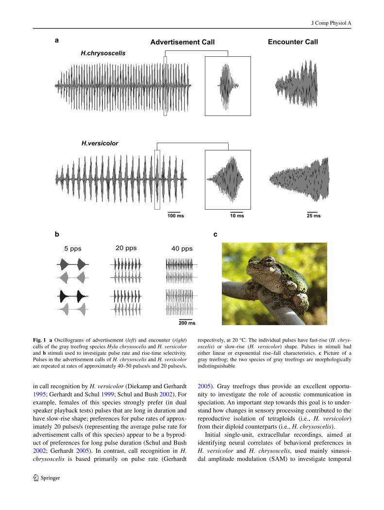
Fig. 1 a Oscillograms of advertisement (left) and encounter (right) calls of the gray treefrog species Hyla chrysoscelis and H. versicolor and b stimuli used to investigate pulse rate and rise-time selectivity. Pulses in the advertisement calls of H. chrysoscelis and H. versicolor are repeated at rates of approximately 40–50 pulses/s and 20 pulses/s,
respectively, at 20 °C. The individual pulses have fast-rise (H. chrys‑oscelis) or slow-rise (H. versicolor) shape. Pulses in stimuli had either linear or exponential rise–fall characteristics. c Picture of a gray treefrog; the two species of gray treefrogs are morphologically indistinguishable

J Comp Physiol A
1 3
processing in the anuran inferior colliculus (ICanuran) (Rose et al. 1985). Results revealed the presence of the same four classes of AM selective neurons (low-pass, band-pass, high-pass and band-suppression) found in other anuran species (Rose and Capranica 1983, 1984, 1985), and that band-pass neurons in H. versicolor were on average tuned to slower AM rates than those in H. chrysoscelis (Rose et al. 1985). Results also indicated that pulses with natu-ral shape (i.e., amplitude rise time characteristics) were more effective in eliciting spikes than were SAM signals; pulse amplitude rises more slowly and duration is greater in the natural calls of H. versicolor (Rose et al. 1985). Sub-sequently, Diekamp and Gerhardt (1995) reported that a higher proportion of units in the ICanuran of H. versicolor showed AM band-pass properties (i.e., tuning to AM, for stimuli that had the natural slow-rise versus fast-rise pulse shape).
More recently, studies in other anuran species have characterized ICanuran neurons using stimuli in which pulse duration and number were held constant and pulse rate varied (Alder and Rose 2000). Results suggest that most neurons can be classified as either ‘long-interval’ or ‘interval-counting’ types; duration-selective cells con-stitute a third class, which will not be considered further here (reviewed by Rose 2014). Long-interval cells, which can be either band-pass or low-pass to SAM, respond well to pulses that are presented at slow rates, but respond phasically or not at all to fast pulse rates (Edwards et al. 2008). Interval-counting cells, the focus of the current paper, respond only after a threshold number of pulses are presented with intervals that fall within a particular range, and a single aberrant interval can reset this inte-gration process (Edwards et al. 2007). Behavioral stud-ies, using calls in which a long interval has been inserted in various locations (Schwartz et al. 2010) or that differ in sequential pulse number (Velez and Bee 2011), sug-gest that interval-counting cells play an important role in call recognition.
To further characterize the differences in temporal pro-cessing between H. versicolor and H. chrysoscelis, we made single-unit extracellular and whole-cell recordings from ICanuran neurons in these species. With whole-cell recordings, mechanisms that underlie selectivity can be interpreted from subthreshold events associated with par-ticular stimulus features. In the current paper, we compare the response properties of interval-counting neurons in H. versicolor and H. chrysoscelis. Specifically we inves-tigated: (1) whether these neurons show tuning to slower pulse rates in H. versicolor; (2) whether interval tuning mechanisms are similar and ‘scalable’ between the two spe-cies, and (3) if slow-rise selectivity is exclusive to H. versi‑color, and, if so, whether it appears to be a mechanistically derived (evolutionarily) trait.
Materials and methods
Animals
Animals were prepared for recording according to previ-ously published methods (see Alder and Rose 2000), except that animals were immobilized with intramuscular injec-tion (1 µl/g) pancuronium Br− (4 mg/ml). All procedures were approved by the University of Utah Institutional Ani-mal Care and Use Committee. Recordings were made from 86 interval-counting neurons (ICNs) in 46 gray treefrogs. Frogs were from south and south-central Missouri, except for seven H. versicolor, which were from the northeastern lineage (Holloway et al. 2006). Either H. chrysoscelis or H. versicolor (not both species) were present at each collection site, and call characteristics were used to further establish the species identity of males. Recordings were made pre-dominately from males, with the exception of three female H. chrysoscelis. Recordings from 72 neurons from H. ver‑sicolor and H. chrysoscelis were of sufficient duration to determine both pulse-rate tuning and rise-time selectivity; these neurons were used in statistical comparisons.
Electrode construction
Extracellular electrodes were constructed from borosilicate capillary glass (1 mm outer diameter, 0.58 mm inner diam-eter; #5960; A-M Systems, Everett, WA) using a Flaming-Brown-type puller (model P-97; Sutter Instruments, Novato, CA) and were pulled to resistances between 0.8 and 1.2 MΩ. These electrodes were filled with a solution of 2 M NaCl.
Whole-cell patch intracellular recordings were made, in vivo, according to methods described in detail by Rose and Fortune (1996) and Edwards et al. (2007). Patch pipettes were constructed from borosilicate capillary glass (1 mm outer diameter, 0.58 mm inner diameter; #5960; A-M Sys-tems, Everett, WA) using a Flaming-Brown-type puller (model P-97; Sutter Instruments, Novato, CA). These elec-trodes had outside tip diameters of ∼1–1.5 μm and had resistances between 10 and 20 MΩ. The electrode tips were back-filled with a solution (pH 7.4) consisting of (in mM) 100 potassium gluconate, 2 KCl, 1 MgCl2, 5 EGTA, 10 HEPES, 20 KOH, and biocytin (20 or 43 mM); the lower concentration of biocytin minimized unintended labeling of neurons. Biocytin was replaced by mannitol in the solution used to fill pipette shanks.
The pipette was advanced into the brain using an “inch-worm” microdrive (Burleigh Instruments Model No. 6000, Fishers, NY) or a 3-axis Microdrive (Scientifica, model IVM-3000, East Sussex, England) while applying posi-tive pressure via a 30 cc syringe to prevent clogging the tip. After reaching the recording location, the pipette was advanced in 1.5-μm increments while maintaining slight

J Comp Physiol A
1 3
positive pressure and passing −0.1 nA square-wave pulses (500 ms) to monitor resistance. Contact with a cell was indicated by a small increase (10 %) in the voltage change that resulted from this current injection. Negative pressure was then applied to the pipette to increase the seal resist-ance to gigaohm levels. After a seal was formed, negative current (usually less than −0.5 nA) was manually applied to rupture the patch and achieve an intracellular recording. To label neurons, biocytin was iontophoresed (approxi-mately +0.02–0.05 nA) and tissue was processed accord-ing to methods described previously (Alder and Rose 2000).
Stimulus generation and delivery
Acoustic stimuli were constructed using Tucker Davis Technologies (Alachua, FL) System III hardware and cus-tom-made software (Alder and Rose 2000). Stimuli were presented free field in an audiometric room maintained at 20 °C, the temperature at which behavioral experiments have been performed in these species (Gerhardt 2008; Gerhardt and Schul 1999). The speaker was situated 0.5 m from the animal and contralateral to the recording site. Search stimulus carrier frequencies were systematically varied from 150 to 2200 Hz with modulation frequencies (sinusoidal AM, SAM) ranging from 10 to 100 Hz. The best excitatory carrier frequency (BEF) and threshold of each neuron were determined as in Alder and Rose (2000); threshold was defined as the sound level at which a neuron produced at least one spike in response to at least 50 % of the presentations of the preferred stimulus. For neurons in the present study, the range of thresholds was 37–84 dB SPL with a median of 62 dB SPL. The BEF of each neuron was used in generating pulse stimuli. The details regarding how the different pulse shapes were generated (for SAM, natural AM, and variable duty-cycle stimuli) are described in Alder and Rose (2000). Stimuli used to characterize pulse-rate selectivity were 400–500 ms in duration and consisted of pulses of constant duty-cycle (50 or 100 %) at rates that varied from 5–80 pulses/s. Stimuli used to characterize rise-time selectivity consisted of pulses with rise times that approximated those of the H. chrysoscelis and H. versicolor advertisement calls. Pulses were gener-ated by increasing the amplitude so that the maximum was reached at either 20 % (fast-rise) or 80 % (slow-rise) of the duration of the pulse. Both linear and exponential ampli-tude rise-time/fall-time functions were generated (Fig. 1b) and produced similar responses in the neurons tested. Stimulus amplitude was held at approximately 10–20 dB above threshold (Alder and Rose 2000). When possible, interval-counting characteristics were further assessed with additional stimuli presented at the optimal rise-time and pulse-rate for that neuron. To determine the threshold
pulse-number of interval-counting neurons, stimuli of the preferred pulse-rate were generated beginning with one pulse and then the number of pulses per stimulus was increased until firing was consistently induced. To assess the duration of the interval necessary to reset interval-counting, a pulse-train containing double the sub-thresh-old number of pulses for each neuron was generated. The middle interval of this pulse-train was then gradually increased until a decrease in the response to the stimulus was observed.
Neurophysiological measurements and statistics
Recordings were digitized at 10 kHz (power 1401, Cam-bridge Electronic Design, Cambridge, UK) and stored as data files using Spike-2 software, also from the same sup-plier. Analyses were performed using acquired and custom Spike-2 programs. Band-pass ICNs generally did not spike in response to pulse rates well below their best rate. The decline in response at pulse rates above the best rate varied across cells, and a decrease of at least 25 % was used as a criterion for band-pass classification. ICNs were classi-fied as high-pass if they showed increasing spike counts for pulse rates >60 pulses/s (the high end of the biologically relevant range). Neurons were classified as band-suppres-sion if there was 50 % (or greater) reduction in the response level for mid-pulse rates relative to lower or higher pulse rates.
To make population-level comparisons between ICNs in H. chrysoscelis and H. versicolor, we averaged the number of spikes produced in response to at least three presenta-tions of the same stimulus per neuron. For each of 72 ICNs from which we collected sufficient pulse rate and rise-time selectivity data, rise-time and ‘call’ selectivity indices were calculated. Rise-time selectivity was calculated using the following formula:
where SR and FR were the average number of spikes in response to slow-rise and fast-rise pulses of the preferred pulse rate. Thus, negative values indicate fast-rise selec-tivity and positive values indicate slow-rise selectivity. The ‘call’ selectivity index was calculated based on the responses to stimuli that had pulse rate and pulse rise-time characteristics of the advertisement calls of H. versicolor and H. chrysoscelis using the following formula:
where RHv was the highest spike average for slow-rise pulses between 10 and 20 pulses/s (characteristic of H. versicolor, Hv), and RHch
was the highest spike average for fast-rise pulses between 40 and 60 pulses/s (characteristic of H. chrysoscelis, Hch).
(1)Rise-time selectivity = (SR − FR)/(SR + FR),
(2)Call selectivity index = (RHv − RHch)/(RHv + RHch
),
J Comp Physiol A
1 3
A t test was used to analyze the pulse rate minima of band-suppression neurons for the two species; Kolmogo-rov–Smirnov (best pulse rate selectivity) and Mann–Whit-ney U tests (rise-time and ‘call’ selectivity) were used for comparisons between data sets that did not meet the assumption of normality. A signed-rank test was used on the ‘call’ selectivity indices to test for positive or negative biases in neural responses, which correspond to selectivity for ‘versicolor-like’ versus ‘chrysoscelis-like’ stimuli (see Eq. 2 above).
Results
Extracellular (n = 28) and whole-cell patch (n = 58) recordings were made from ICNs in the auditory midbrain of 26 Hyla versicolor (44 cells) and 20 H. chrysoscelis (42 cells). Labeled neurons were predominantly in the prin-cipal and magnocellular nuclei; the proportionality was approximately 2:1. We first present representative extracel-lular recordings to show general features of ICNs and how they differ between these species. We then present whole-cell recordings from neurons that represent the range of response characteristics observed. These recordings show stimulus-related, subthreshold changes in membrane potential that were used to make mechanistic comparisons between these neurons and ICNs recorded from other anu-ran species. Finally, we combined data from extracellular and whole-cell recordings to analyze species differences in the temporal selectivity of midbrain neurons. We show that ICNs in H. versicolor are generally tuned to slower pulse rates than those in H. chrysoscelis and that slow-rise selec-tivity is exclusive to H. versicolor. Although pulse rate tun-ing differed between the two species, in both cases selec-tivity appeared to result from interplay between excitation and inhibition, i.e., the shift in tuning seen in H. versi‑color appears to result from differential scaling of existing mechanisms.
General features of interval-counting neurons in H. versicolor and H. chrysoscelis
Extracellular recordings
Single-unit recordings from the ICanuran of H. chrysoscelis and H. versicolor revealed interval-counting neurons simi-lar to those recorded previously in Rana pipiens and Pseu‑dacris (formerly Hyla) regilla (Alder and Rose 1998, 2000; Edwards et al. 2007). Based on responses to stimuli shown in Fig. 1b, interval-counting neurons in gray treefrogs could be classified as band-pass/high-pass (n = 18) (Figs. 2, 3) or band-suppression (n = 7) (Fig. 4). Band-pass neurons (n = 9) recorded from H. chrysoscelis showed best pulse
rates (pulse rate that elicited maximal response) that ranged from 30 to 60 pulses/s (median 50 pulses/s). The repre-sentative band-pass/high-pass cell found in H. chrysosce‑lis (Fig. 2) showed little or no spikes to pulse rates below approximately 30 pulses/s, and the response level peaked at 50–60 pulses/s. This steep response level vs. pulse rate relation is characteristic of this neuron type. Response latency decreased markedly for 50 vs. 40 pulses/s but, in both cases, spiking stopped approximately 50–60 ms after the stimuli ended (Fig. 2b). This differential response onset time is consistent with an integration process, i.e., inter-val counting contributing to response latency, not sim-ply conduction time. Consistent with this interpretation, this neuron did not respond to stimuli that had five pulses (Fig. 2c, upper trace), or less, delivered with optimal inter-vals (20 ms between onsets of successive pulses). At least 10 pulses were required for the cell to respond on 50 % or more of the stimulus presentations (Fig. 2c). Response levels over the biologically important 5–60 pulses/s range were highly similar for fast-rise and slow-rise pulses; only a slight preference for fast-rise pulses was observed (Fig. 2a).
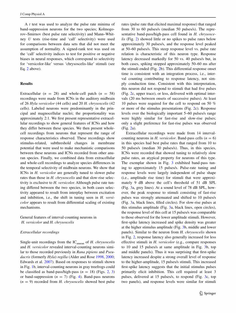
Extracellular recordings were made from 14 interval-counting neurons in H. versicolor. Band-pass cells (n = 6) in this species had best pulse rates that ranged from 10 to 50 pulses/s (median 30 pulses/s). Thus, in this species, ICNs were recorded that showed tuning to relatively slow pulse rates, an atypical property for neurons of this type. The exemplar shown in Fig. 3 exhibited band-pass tun-ing to approximately 15 pulses/s. Pulse-rate tuning and response levels were largely independent of pulse shape (i.e., amplitude rise time) for stimuli that were approxi-mately 9 dB above the cell’s threshold of 51 dB SPL (Fig. 3a, grey lines). At a sound level of 78 dB SPL, how-ever, the peak response to stimuli consisting of fast-rise pulses was strongly attenuated and shifted to 10 pulses/s (Fig. 3a, black lines, filled circles). For slow-rise pulses at this stimulus amplitude (Fig. 3a, black lines, open circles), the response level of this cell at 15 pulses/s was comparable to those observed for the lower amplitude stimuli. However, first-spike latency increased and spike density was greater at the higher stimulus amplitude (Fig. 3b, middle and lower panels). Similar to the neuron from H. chrysoscelis shown in Fig. 2, response latency also generally increased for less effective stimuli in H. versicolor (e.g., compare responses to 10 and 15 pulses/s at same amplitude in Fig. 3b, top and middle panels). Thus it was surprising that first-spike latency increased despite a strong overall level of response to the higher-amplitude, 15 pulses/s stimuli. This increased first-spike latency suggests that the initial stimulus pulses primarily elicit inhibition. This cell required at least 3 pulses, delivered at 15 pulses/s, to respond (Fig. 3c, top two panels), and response levels were similar for stimuli

J Comp Physiol A
1 3
in which pulses had slow-rise characteristics or fast-rise (not shown) characteristics (1.0 and 1.3 spikes/repetition, respectively). A sequence of four pulses, presented at inter-onset intervals of 75 ms, elicited strong responses on each stimulus presentation (Fig. 3c, 3rd panel). Increasing the middle interval to 125 ms resulted in a reduction in the strength of the response and a middle interval of 150 ms
almost completely reset this interval-counting process (Fig. 3c, bottom panel, grey and black traces).
Examples of interval-counting neurons that show band-suppression pulse-rate selectivity are shown in Fig. 4. Band-suppression neurons respond to slow pulse rate stimuli, provided that individual pulses are of sufficient duration (Edwards and Rose 2003; Leary et al. 2008). For stimuli such
50 pulses/s
0 0.05 0.10 0.15 0.20 0.25 0.30 0.35 0.40 0.45 0.50
4
2
02
1
0
Ras
ter
swee
pS
pike
co
unt
Ras
ter
swee
pS
pike
co
unt
40 pulses/s
0 0.05 0.10 0.15 0.20 0.25 0.30 0.35 0.40 0.45 0.50Time (s)
3
2
1
02
1
0
Pulse rate (pulses/s)0 20 40 60 80 100
Spi
kes
/ rep
.
0
2
4
6
8
0 0.05 0.10 0.15 0.20 0.25 0.30 0.35
6
4
2
02
1
0
0 0.05 0.10 0.15 0.20 0.25 0.30 0.35
4321
2
1
0
0 0.05 0.10 0.15 0.20 0.25 0.30 0.35
108642
2
1
0
Time (s)
0 0.05 0.10 0.15 0.20 0.25 0.30 0.35
2
1
2
1
0
Time (s)
a c
b
Ras
ter
swee
pS
pike
co
unt
5 pulses
12 pulses
10 pulses
8 pulses
Time (s)
Time (s)
Time (s)
Fig. 2 Extracellularly recorded responses of an interval-counting neuron in H. chrysoscelis. a Average spike count per presentation of stimuli that differed in pulse rate and pulse shape (20 % rise:80 % fall, closed circles, or 80 % rise:20 % fall, open circles). b Peri-stim-ulus-time histograms (PSTHs) and raster plots of spikes in response
to stimuli in which pulses had fast (20 %) rise and were presented at 40 or 50 pulses/s, with 50 % duty cycle (on and off periods are equal duration). c PSTHs and raster plots of responses to stimuli that consisted of 5, 8, 10 or 12 pulses. Carrier frequency (BEF) = 425 Hz, 62 dB SPL
J Comp Physiol A
1 3
as those depicted in Fig. 1b, pulse duty cycle (the proportion of the pulse interval that sound is present) is held constant, and, thus, pulse duration increases as pulse rate decreases. The band-suppression neuron recorded in H. versicolor (Fig. 4a)
showed a broad response minimum over the 40–60 pulses/s range, which encompasses the pulse rates of advertisement calls for western populations of H. chrysoscelis (Gerhardt 2005). Over most of the range in pulse rate tested, this cell
0 10 20 30 40 50 60
Spik
es /
rep.
0
2
4
6
8
0 0.05 0.10 0.15 0.20 0.25 0.30 0.35 0.40 0.45
1210
8642
2
1
0
0 0.05 0.10 0.15 0.20 0.25 0.30 0.35 0.40 0.45
6
4
2
01
0
0 0.1 0.2 0.3 0.4 0.5 0.6 0.7 0.8 0.9
3
2
1
2
1
0
10 pulses/s60 dB SPL
0 0.1 0.2 0.3 0.4 0.5 0.6 0.7 0.8 0.9
3
2
1
2
1
0
15 pulses/s60 dB SPL
Ras
ter
swee
pS
pike
co
unt
Ras
ter
swee
pS
pike
co
unt
0 0.1 0.2 0.3 0.4 0.5 0.6 0.7 0.8 0.9
15
10
5
02
1
0
0 0.1 0.2 0.3 0.4 0.5 0.6 0.7 0.8 0.9
6
4
2
02
1
0
0 0.1 0.2 0.3 0.4 0.5 0.6 0.7 0.8 0.9
4
2
02
1
0
15 pulses/s72 dB SPL
Time (s)
Time (s)
Pulse rate (pulses/s)
a
b
c
Fig. 3 Extracellularly recorded responses of an interval-counting neuron in H. versicolor. a Average spike count per presentation of stimuli that differed in pulse rate and pulse shape (20 % rise:80 % fall, closed circles, or 80 % rise:20 % fall, open circles); stimulus amplitude was either 60 dB SPL (grey lines) or 78 dB SPL (black lines). b Peri-stimulus-time histograms (PSTHs) and raster plots of spikes in response to stimuli that consisted of fast-rise pulses (20 %) and were presented at 10 or 15 pulses/s, with 50 % duty cycle; stim-
ulus amplitude was either 60 dB SPL (upper two panels) or 72 dB SPL (lower panel). c PSTHs and raster plots of responses to stimuli that consisted of 2, 3 or 4 pulses, repeated with intervals of 75 ms (15 pulses/s), upper 3 panels; with 75 ms intervals, 3 pulses were required to elicit spikes on at least 50 % of the stimulus presentations. Bottom panel shows the effects of increasing the duration of the mid-dle interval to 125 ms (grey) or 150 ms (black). Carrier frequency (BEF) = 280 Hz
J Comp Physiol A
1 3
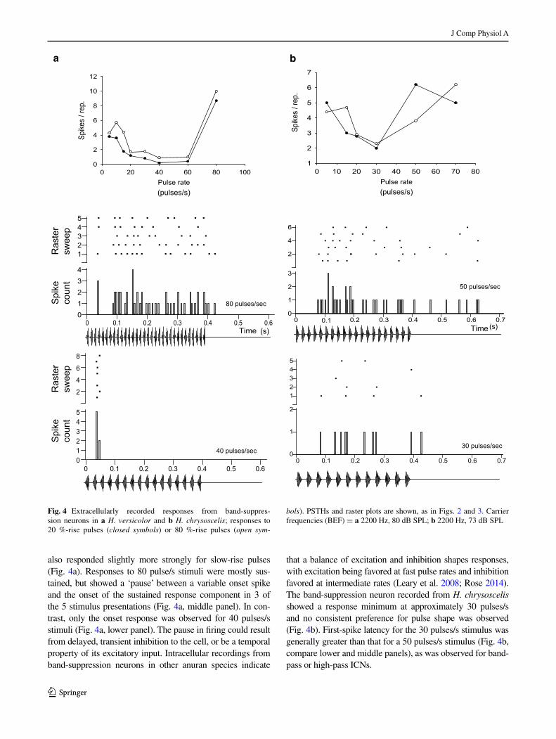
also responded slightly more strongly for slow-rise pulses (Fig. 4a). Responses to 80 pulse/s stimuli were mostly sus-tained, but showed a ‘pause’ between a variable onset spike and the onset of the sustained response component in 3 of the 5 stimulus presentations (Fig. 4a, middle panel). In con-trast, only the onset response was observed for 40 pulses/s stimuli (Fig. 4a, lower panel). The pause in firing could result from delayed, transient inhibition to the cell, or be a temporal property of its excitatory input. Intracellular recordings from band-suppression neurons in other anuran species indicate
that a balance of excitation and inhibition shapes responses, with excitation being favored at fast pulse rates and inhibition favored at intermediate rates (Leary et al. 2008; Rose 2014). The band-suppression neuron recorded from H. chrysoscelis showed a response minimum at approximately 30 pulses/s and no consistent preference for pulse shape was observed (Fig. 4b). First-spike latency for the 30 pulses/s stimulus was generally greater than that for a 50 pulses/s stimulus (Fig. 4b, compare lower and middle panels), as was observed for band-pass or high-pass ICNs.
Pulse rate0 20 40 60 80 100
Spike
s / r
ep.
0
2
4
6
8
10
12
0 0.1 0.2 0.3 0.4 0.5 0.6Time
54321
43210
80 pulses/sec
0 0.1 0.2 0.3 0.4 0.5 0.6
8
6
4
2
543210
40 pulses/sec0 0.1 0.2 0.3 0.4 0.5 0.6 0.7
54321
2
1
0
0 0.1 0.2 0.3 0.4 0.5 0.6 0.7
6
4
2
3
2
1
0
0 10 20 30 40 50 60 70 801
2
3
4
5
6
7
Pulse rate
Spike
s / r
ep.
Ras
ter
swee
pS
pike
co
unt
Ras
ter
swee
pS
pike
co
unt
(s)
50 pulses/sec
30 pulses/sec
a b
Time(s)
(pulses/s) (pulses/s)
Fig. 4 Extracellularly recorded responses from band-suppres-sion neurons in a H. versicolor and b H. chrysoscelis; responses to 20 %-rise pulses (closed symbols) or 80 %-rise pulses (open sym‑
bols). PSTHs and raster plots are shown, as in Figs. 2 and 3. Carrier frequencies (BEF) = a 2200 Hz, 80 dB SPL; b 2200 Hz, 73 dB SPL

J Comp Physiol A
1 3
Thus, extracellular recordings demonstrate that interval-counting neurons exist in the ICanuran of gray treefrogs, and some are highly similar in their response properties to those that have been recorded in other anuran species. In H. versicolor, however, interval-counting neurons were recorded that showed tuning to comparatively slow pulse rates and selectivity for slow-rise pulses. In other species, interval-counting neurons typically show band-pass or high-pass selectivity for mid- to fast-pulse rates, and this selectivity emerges from interplay between time-depend-ent excitation and inhibition (reviewed in Rose 2014). To determine whether interval-counting and interval selectiv-ity in gray treefrogs is mechanistically similar to that in other species, and to provide some insight into how selec-tivity for slow-rise pulses arises, we made whole-cell patch recordings from midbrain neurons in H. versicolor and H. chrysoscelis.
Whole‑cell recordings
For interval-counting neurons in H. chrysoscelis that showed temporal selectivity similar to that displayed in
Fig. 2 (i.e., band-pass or high-pass selectivity for pulse rate, with maximum responses in the 40–60 pulses/s range), whole-cell recordings revealed interplay between excitation and inhibition much like that found for interval-counting neurons in other species that have been studied (Edwards et al. 2007; Rose et al. 2011). For the exemplar shown in Fig. 5a, pulses presented at a rate of 5 pulses/s elicited primarily inhibition (hyperpolarizations). At pulse rates greater than approximately 20 pulses/s, however, stimuli elicited an initial hyperpolarization which then was progressively reversed, leading to depolarization and spikes. This rate-dependent shift in the balance of exci-tation and inhibition with successive pulses is one of the hallmarks of interval-counting neurons (Rose 2014). The best pulse rate for this neuron was 50 pulses/s, and slow-rise and fast-rise pulses were equally effective (Fig. 5a). Response characteristics of this general type were observed in just one of the 31 whole-cell recordings made from H. versicolor (Fig. 5b). This cell showed mainly inhibition for 10 pulses/s stimuli. At pulse rates of 30–60 pulses/s, however, strong depolarization followed initial hyperpo-larization, with maximal spike rate occurring at 40 pulses/s
20 pulses/s 40 pulses/s30 pulses/s10 pulses/s
9/4
10/3
60 pulses/s
27/4
28/4
0/4
1/4
22/4
17/4
0/4
0/4
a
b H. versicolor
14/323/6
11/413/4
8/56/3
1/20/3
0/50/4
H. chrysoscelis
200 ms10 mv
20 pulses/s 30 pulses/s10 pulses/s 50 pulses/s5 pulses/s
Fig. 5 Whole-cell recordings from interval-counting neurons from H. chrysoscelis (a) and H. versicolor (b) that showed selectivity for pulse rates typical of H. chrysoscelis advertisement calls. Top traces responses to single presentations of the stimuli shown; middle and lower traces in each panel are averaged responses, hence spike height
is smaller. Responses to 80 %-rise pulses (grey) and 20 %-rise pulses, along with spike counts over number of presentations of each stimu-lus are shown. Carrier frequencies (BEF) = a 270 Hz, 71 dB SPL; b 250 Hz; 58 dB SPL

J Comp Physiol A
1 3
(Fig. 5b). Importantly, this neuron did not show selectivity for slow-rise pulses; such selectivity would preclude strong responses to pulse rates in the 40–60 pulses/s range because the short-duration pulses in such stimuli have shorter rise times.
We now turn to whole-cell recordings from ICNs that are more representative of those recorded from H. versi‑color but unique in many respects (e.g., slow-rise selec-tivity, tuned to slow pulse rates) from ICNs found in other anuran species. Figure 6 shows whole-cell recordings from three interval-counting neurons in H. versicolor that showed tuning to pulse rates characteristic of the adver-tisement call (15–20 pulses/s) of this species. These cells represent the range of selectivity for slow-rise pulses that
was observed. The neuron that showed greatest slow-rise selectivity (Fig. 6a) failed to respond with spikes to fast-rise pulses at any pulse rate tested. Hyperpolariza-tions were observed at the slowest (5 pulses/s) and fast-est (40–50 pulses/s) pulse rates tested. At pulse rates of 10–20 pulses/s, slow-rise pulses elicited EPSPs that tem-porally summated to produce supra-threshold depolariza-tions. Successive fast-rise pulses, however, elicited depo-larizations that, at 10 pulses/s, decreased in amplitude. Further, these EPSPs occurred on a background of slow hyperpolarization, suggesting a build-up of inhibition. Fast-rise pulses that were presented at 20 pulses/s resulted in a maximal depolarization of approximately 4 mV, compared to at least 25 mV for slow-rise pulses. The neuron shown
0/70/6
4/31/6
7/50/4
0/60/2
0/30/3
0/50/4
2/80/7
5/50/11
0/300/4
1/70/12
200 ms10 mv
a
b
c
50 pulses/s5 pulses/s 10 pulses/s 15 pulses/s 20 pulses/s 40 pulses/s
5 pulses/s 10 pulses/s 15 pulses/s 20 pulses/s 40 pulses/s
0/40/5
1/60/5
4/34/4
3/137/11
5 pulses/s 10 pulses/s 15 pulses/s 20 pulses/s 40 pulses/s
0/120/3
Fig. 6 Whole-cell recordings from interval-counting neurons in H. versicolor that show tuning to pulse rates characteristic of the advertisement call of this species. As in Fig. 5, traces are membrane-potential fluctuations in response to single presentations of the stimuli shown, or averaged responses; the number of spikes and stimulus
presentations are shown. The recordings (a–c) represent the range of selectivity for slow-rise pulses, from strong (a) to nonselective (c). Carrier frequencies (BEFs) = a 1200 Hz, 81 dB SPL; b 160 Hz, 69 dB SPL; c 225 Hz (BEF), 61 dB SPL
J Comp Physiol A
1 3
in Fig. 6b also responded selectively to slow-rise pulses; across the range of pulse rates tested, only one spike was triggered with a fast rise time stimulus (at 10 pulses/s). In this case, the large differential in spiking for slow- vs. fast-rise pulses at 15 pulses/s resulted mainly from ‘thresh-olding processes’; the threshold for spike production was slightly below the depolarization reached in response to slow-rise pulses, and above (less negative membrane potential) the maximum depolarization for fast-rise pulses. The case shown in Fig. 6c showed band-pass selectivity for pulse rates in the 15–20 pulses/s range, but exhibited similar levels of response for slow-rise and fast-rise pulses. Thus, sharp band-pass tuning to slow pulse rates could be observed in H. versicolor even for interval-counting neu-rons that showed no slow-rise selectivity.
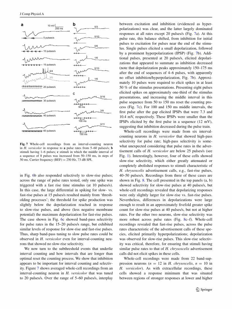
We now turn to the subthreshold events that underlie interval counting and how intervals that are longer than optimal reset the counting process. We show that inhibition appears to be important for interval counting and selectiv-ity. Figure 7 shows averaged whole-cell recordings from an interval-counting neuron in H. versicolor that was tuned to 20 pulses/s. Over the range of 5–60 pulses/s, interplay
between excitation and inhibition (evidenced as hyper-polarizations) was clear, and the latter largely dominated responses at all rates except 20 pulses/s (Fig. 7a). At this pulse rate, this balance shifted, from inhibition for initial pulses to excitation for pulses near the end of the stimu-lus. Single pulses elicited a small depolarization, followed by a prominent hyperpolarization (IPSP) (Fig. 7b). Addi-tional pulses, presented at 20 pulses/s, elicited depolari-zations that appeared to summate as inhibition decreased (note that depolarization peaks approximately 150–175 ms after the end of sequences of 4–6 pulses, with apparently no offset inhibition/hyperpolarization, Fig. 7b). Approxi-mately 10 pulses were required to elicit spikes in at least 50 % of the stimulus presentations. Presenting eight pulses elicited spikes on approximately one-third of the stimulus presentations, and increasing the middle interval in this pulse sequence from 50 to 150 ms reset the counting pro-cess (Fig. 7c). For 100 and 150 ms middle intervals, the first pulse after the gap elicited IPSPs that were 7.3 and 10.4 mV, respectively. These IPSPs were smaller than the IPSPs elicited by the first pulse in a sequence (12 mV), suggesting that inhibition decreased during the pulse train.
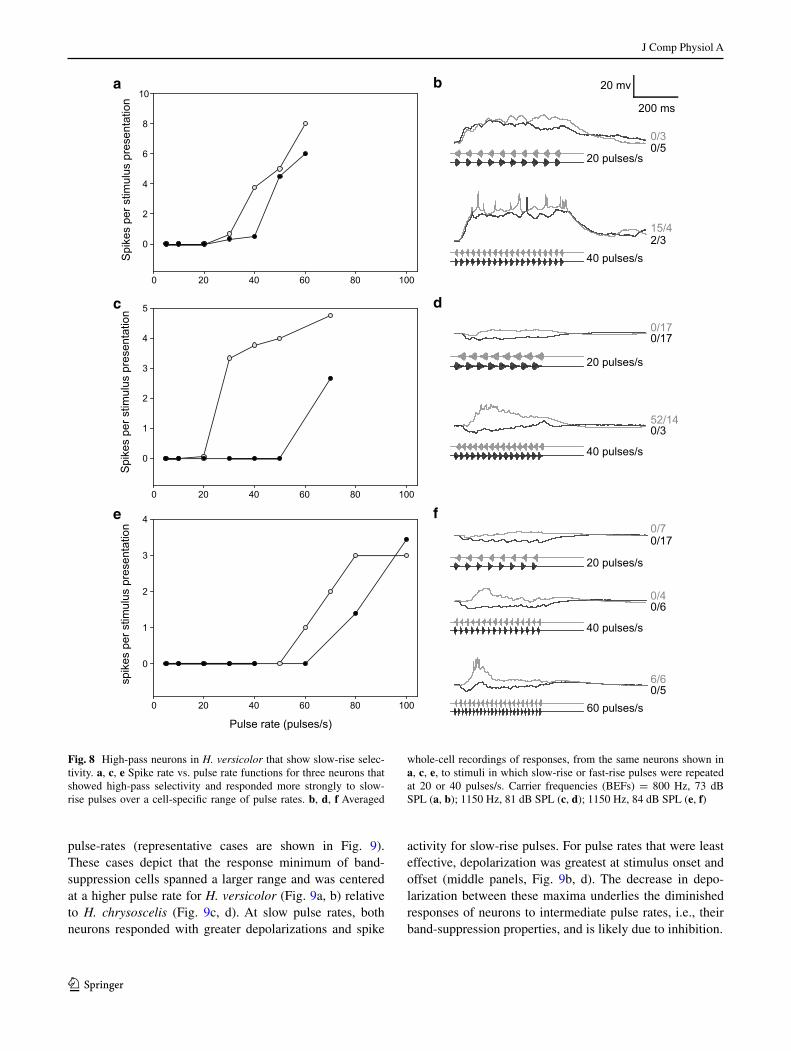
Whole-cell recordings were made from six interval-counting neurons in H. versicolor that showed high-pass selectivity for pulse rate; high-pass selectivity is some-what unexpected considering that pulse rates in the adver-tisement calls of H. versicolor are below 25 pulses/s (see Fig. 1). Interestingly, however, four of these cells showed slow-rise selectivity, which either greatly attenuated or completely abolished responses to stimuli characteristic of H. chrysoscelis advertisement calls, e.g., fast-rise pulses, 40–50 pulses/s. Recordings from three of these cases are shown in Fig. 8. The cell presented in the top panels (a, b) showed selectivity for slow-rise pulses at 40 pulses/s, but whole-cell recordings revealed that depolarizing responses were only slightly larger for slow-rise vs. fast-rise pulses. Nevertheless, differences in depolarizations were large enough to result in an approximately fivefold greater spike count for slow-rise pulses at 40 pulses/s, but not at higher rates. For the other two neurons, slow-rise selectivity was more robust across pulse rates (Fig. 8c–f). Whole-cell recordings revealed that fast-rise pulses, across the pulse rates characteristic of the advertisement calls of these spe-cies, elicited primarily hyperpolarizations; depolarization was observed for slow-rise pulses. This slow-rise selectiv-ity was critical, therefore, for ensuring that stimuli having similar pulse rates to that of H. chrysoscelis advertisement calls did not elicit spikes in these cells.
Whole-cell recordings were made from 22 band-sup-pression neurons (n = 12 in H. chrysoscelis, n = 10 in H. versicolor). As with extracellular recordings, these cells showed a response minimum that was situated between regions of stronger responses at lower and higher
10 pulses/s
20 pulses/s
10 mV200 ms
30 pulses/s
50 ms
100 ms
150 ms
a b
c
40 pulses/s
60 pulses/s
15 pulses/s
5 pulses/s
1/6
0/10
1/6
5/16
2/9
3/5
4/4
0/11
0/6
0/3
0/10
Fig. 7 Whole-cell recordings from an interval-counting neuron in H. versicolor in response to a pulse rates from 5–60 pulses/s; b stimuli having 1–6 pulses; c stimuli in which the middle interval of a sequence of 8 pulses was increased from 50–150 ms, in steps of 50 ms. Carrier frequency (BEF) = 250 Hz, 73 dB SPL
J Comp Physiol A
1 3
pulse-rates (representative cases are shown in Fig. 9). These cases depict that the response minimum of band-suppression cells spanned a larger range and was centered at a higher pulse rate for H. versicolor (Fig. 9a, b) relative to H. chrysoscelis (Fig. 9c, d). At slow pulse rates, both neurons responded with greater depolarizations and spike
activity for slow-rise pulses. For pulse rates that were least effective, depolarization was greatest at stimulus onset and offset (middle panels, Fig. 9b, d). The decrease in depo-larization between these maxima underlies the diminished responses of neurons to intermediate pulse rates, i.e., their band-suppression properties, and is likely due to inhibition.
a
c
e
Pulse rate (pulses/s)
0 20 40 60 80 100
spik
es p
er s
timul
us p
rese
ntat
ion
0
1
2
3
4
0 20 40 60 80 100
Spi
kes
per s
timul
us p
rese
ntat
ion
0
2
4
6
8
10
0 20 40 60 80 100
Spi
kes
per s
timul
us p
rese
ntat
ion
0
1
2
3
4
5
40 pulses/s
20 pulses/s
20 pulses/s
20 pulses/s
40 pulses/s
b
d
f
15/42/3
0/60/4
0/17
0/3
0/17
0/70/17
52/140/3
0/5
40 pulses/s
200 ms
20 mv
60 pulses/s0/56/6
Fig. 8 High-pass neurons in H. versicolor that show slow-rise selec-tivity. a, c, e Spike rate vs. pulse rate functions for three neurons that showed high-pass selectivity and responded more strongly to slow-rise pulses over a cell-specific range of pulse rates. b, d, f Averaged
whole-cell recordings of responses, from the same neurons shown in a, c, e, to stimuli in which slow-rise or fast-rise pulses were repeated at 20 or 40 pulses/s. Carrier frequencies (BEFs) = 800 Hz, 73 dB SPL (a, b); 1150 Hz, 81 dB SPL (c, d); 1150 Hz, 84 dB SPL (e, f)

J Comp Physiol A
1 3
Population analyses
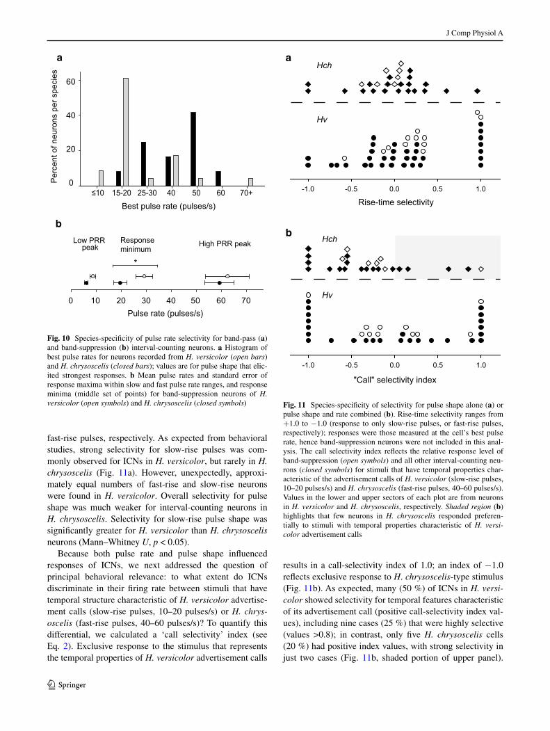
Spike rate data from extracellular and whole-cell record-ings were combined to further examine species differences in selectivity for pulse rate, pulse shape and their interac-tion. In general, analyses of these data show clear evidence of species-specific temporal selectivity. Band-pass interval-counting neurons in H. versicolor showed tuning to slower pulse rates than those recorded from H. chrysoscelis (K–S test, p < 0.05, Fig. 10a); cells that responded in a high-pass fashion were not included in this analysis. Peaks in this histogram occurred at pulse-rate regions that encompass pulse rates typical of advertisement calls of these species (i.e., pulse rates of advertisement calls are approximately 20 pulses/s in H. versicolor and 40–50 pulses/s in H. chrys‑oscelis at approximately 20 °C). In this analysis, the tuning value for each cell is the pulse rate, with either fast-rise or slow-rise pulse shape, that elicited the greatest number of spikes per stimulus presentation. Band-suppression neurons also showed species-specificity in their pulse rate selec-tivity (Fig. 10b). As shown in Figs. 4 and 9, when tested with stimulus regimes in which pulse duty cycle was held constant (pulse duration changes with pulse rate, Fig. 1), these neurons exhibited a prominent response minimum at
intermediate pulse rates. This response ‘suppression’ point (the mid-value in the band of lowest response) was lower in H. chrysoscelis (20 pulses/s) vs. H. versicolor (29 pulses/s) (t test, p < 0.01, Fig. 10b). Pulse-number thresholds were highly similar between neurons in the two species (mean 3.8 pulses/s for H. chrysoscelis, mean 4.2 pulses/s for H. versicolor).
In seven of the recordings made from H. versicolor, we tested whether increasing the middle interval of a series of optimally spaced pulses would decrease spiking, and the duration that is sufficient for resetting the interval-counting process. In six of the seven H. versicolor neurons increas-ing the middle interval by an average of 185 % decreased the average spiking rate to below 50 % of the response to a uniform-interval stimulus; one H. versicolor neuron did not reset.
Selectivity of ICNs for pulse shape is shown in Fig. 11a. Pulse-shape selectivity was quantified as the difference in spike rate for slow-rise pulses (80 % rise:20 % fall) vs. fast-rise pulses (20 % rise:80 % fall), normalized by divid-ing the difference by the total spike count for both (see Eq. 1). The maximum selectivity measured across the pulse rates tested was then plotted. Values of 1.0 and −1.0 indi-cate that the neuron responded exclusively to slow-rise or
5 pulses/s 40 pulses/s 100 pulses/s
20 pulses/s 40 pulses/s5 pulses/s
a b
d
2/9
10/22
1/7
1/2
3/7
29/13
0/6
1/4
17/9
16/6
0/3
0/2
slow-risefast-rise
0 20 40 60 80 100
Spi
kes
per
stim
ulus
pre
sent
atio
n
0.00.10.20.30.40.5
0 10 20 30 40
0.00.51.01.52.02.53.03.5
10 mV200 ms
Spi
kes
per
stim
ulus
pre
sent
atio
n
c
Pulse rate (pulses/s)
Pulse rate (pulses/s)
Fig. 9 Pulse-rate selectivity functions (a, c) and whole-cell record-ings (b, d) from band-suppression interval-counting neurons from H. versicolor and H. chrysoscelis, respectively. As in previous figures, upper traces in panels b and d are recordings of membrane potential responses to single presentations of the stimulus shown; other traces
are averaged recordings. Responses to pulses that have slow-rise (80 %) or fast-rise (20 %) shape are shown in grey and black, respec-tively. Carrier frequencies (BEFs) = a 225 Hz, 81 dB SPL; c 275 Hz, 73 dB SPL
J Comp Physiol A
1 3
fast-rise pulses, respectively. As expected from behavioral studies, strong selectivity for slow-rise pulses was com-monly observed for ICNs in H. versicolor, but rarely in H. chrysoscelis (Fig. 11a). However, unexpectedly, approxi-mately equal numbers of fast-rise and slow-rise neurons were found in H. versicolor. Overall selectivity for pulse shape was much weaker for interval-counting neurons in H. chrysoscelis. Selectivity for slow-rise pulse shape was significantly greater for H. versicolor than H. chrysoscelis neurons (Mann–Whitney U, p < 0.05).
Because both pulse rate and pulse shape influenced responses of ICNs, we next addressed the question of principal behavioral relevance: to what extent do ICNs discriminate in their firing rate between stimuli that have temporal structure characteristic of H. versicolor advertise-ment calls (slow-rise pulses, 10–20 pulses/s) or H. chrys‑oscelis (fast-rise pulses, 40–60 pulses/s)? To quantify this differential, we calculated a ‘call selectivity’ index (see Eq. 2). Exclusive response to the stimulus that represents the temporal properties of H. versicolor advertisement calls
results in a call-selectivity index of 1.0; an index of −1.0 reflects exclusive response to H. chrysoscelis-type stimulus (Fig. 11b). As expected, many (50 %) of ICNs in H. versi‑color showed selectivity for temporal features characteristic of its advertisement call (positive call-selectivity index val-ues), including nine cases (25 %) that were highly selective (values >0.8); in contrast, only five H. chrysoscelis cells (20 %) had positive index values, with strong selectivity in just two cases (Fig. 11b, shaded portion of upper panel).
a
b
*
High PRR peakLow PRR peak
Responseminimum
Best pulse rate (pulses/s)≤10 15-20 25-30 40 50 60 70+
Perc
ent o
f neu
rons
per
spe
cies
0
20
40
60
0 10 20 30 40 50 60 70Pulse rate (pulses/s)
Fig. 10 Species-specificity of pulse rate selectivity for band-pass (a) and band-suppression (b) interval-counting neurons. a Histogram of best pulse rates for neurons recorded from H. versicolor (open bars) and H. chrysoscelis (closed bars); values are for pulse shape that elic-ited strongest responses. b Mean pulse rates and standard error of response maxima within slow and fast pulse rate ranges, and response minima (middle set of points) for band-suppression neurons of H. versicolor (open symbols) and H. chrysoscelis (closed symbols)
"Call" selectivity index
Rise-time selectivity
a
bHch
Hv
Hch
Hv
-1.0 -0.5 0.0 0.5 1.0
-1.0 -0.5 0.0 0.5 1.0
Fig. 11 Species-specificity of selectivity for pulse shape alone (a) or pulse shape and rate combined (b). Rise-time selectivity ranges from +1.0 to −1.0 (response to only slow-rise pulses, or fast-rise pulses, respectively); responses were those measured at the cell’s best pulse rate, hence band-suppression neurons were not included in this anal-ysis. The call selectivity index reflects the relative response level of band-suppression (open symbols) and all other interval-counting neu-rons (closed symbols) for stimuli that have temporal properties char-acteristic of the advertisement calls of H. versicolor (slow-rise pulses, 10–20 pulses/s) and H. chrysoscelis (fast-rise pulses, 40–60 pulses/s). Values in the lower and upper sectors of each plot are from neurons in H. versicolor and H. chrysoscelis, respectively. Shaded region (b) highlights that few neurons in H. chrysoscelis responded preferen-tially to stimuli with temporal properties characteristic of H. versi‑color advertisement calls
J Comp Physiol A
1 3
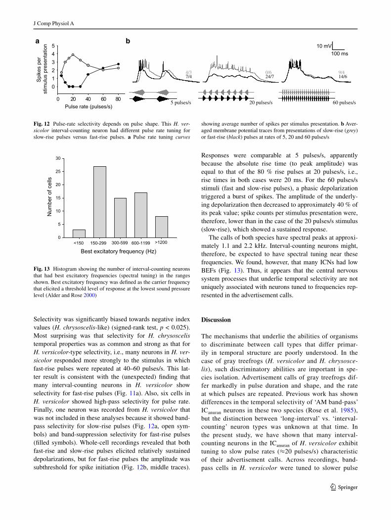
Selectivity was significantly biased towards negative index values (H. chrysoscelis‑like) (signed-rank test, p < 0.025). Most surprising was that selectivity for H. chrysoscelis temporal properties was as common and strong as that for H. versicolor‑type selectivity, i.e., many neurons in H. ver‑sicolor responded more strongly to the stimulus in which fast-rise pulses were repeated at 40–60 pulses/s. This lat-ter result is consistent with the (unexpected) finding that many interval-counting neurons in H. versicolor show selectivity for fast-rise pulses (Fig. 11a). Also, six cells in H. versicolor showed high-pass selectivity for pulse rate. Finally, one neuron was recorded from H. versicolor that was not included in these analyses because it showed band-pass selectivity for slow-rise pulses (Fig. 12a, open sym-bols) and band-suppression selectivity for fast-rise pulses (filled symbols). Whole-cell recordings revealed that both fast-rise and slow-rise pulses elicited relatively sustained depolarizations, but for fast-rise pulses the amplitude was subthreshold for spike initiation (Fig. 12b, middle traces).
Responses were comparable at 5 pulses/s, apparently because the absolute rise time (to peak amplitude) was equal to that of the 80 % rise pulses at 20 pulses/s, i.e., rise times in both cases were 20 ms. For the 60 pulses/s stimuli (fast and slow-rise pulses), a phasic depolarization triggered a burst of spikes. The amplitude of the underly-ing depolarization then decreased to approximately 40 % of its peak value; spike counts per stimulus presentation were, therefore, lower than in the case of the 20 pulses/s stimulus (slow-rise), which showed a sustained response.
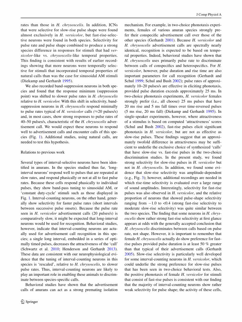
The calls of both species have spectral peaks at approxi-mately 1.1 and 2.2 kHz. Interval-counting neurons might, therefore, be expected to have spectral tuning near these frequencies. We found, however, that many ICNs had low BEFs (Fig. 13). Thus, it appears that the central nervous system processes that underlie temporal selectivity are not uniquely associated with neurons tuned to frequencies rep-resented in the advertisement calls.
Discussion
The mechanisms that underlie the abilities of organisms to discriminate between call types that differ primar-ily in temporal structure are poorly understood. In the case of gray treefrogs (H. versicolor and H. chrysosce‑lis), such discriminatory abilities are important in spe-cies isolation. Advertisement calls of gray treefrogs dif-fer markedly in pulse duration and shape, and the rate at which pulses are repeated. Previous work has shown differences in the temporal selectivity of ‘AM band-pass’ ICanuran neurons in these two species (Rose et al. 1985), but the distinction between ‘long-interval’ vs. ‘interval-counting’ neuron types was unknown at that time. In the present study, we have shown that many interval-counting neurons in the ICanuran of H. versicolor exhibit tuning to slow pulse rates (≈20 pulses/s) characteristic of their advertisement calls. Across recordings, band-pass cells in H. versicolor were tuned to slower pulse
0 20 40 60 80
Spi
kes
per
stim
ulus
pre
sent
atio
n
012345
5 pulses/s 20 pulses/s 60 pulses/s
10 mV100 ms
4/37/4
0/624/7
9/414/6
Pulse rate (pulses/s)
a b
Fig. 12 Pulse-rate selectivity depends on pulse shape. This H. ver‑sicolor interval-counting neuron had different pulse rate tuning for slow-rise pulses versus fast-rise pulses. a Pulse rate tuning curves
showing average number of spikes per stimulus presentation. b Aver-aged membrane potential traces from presentations of slow-rise (grey) or fast-rise (black) pulses at rates of 5, 20 and 60 pulses/s
Best excitatory frequency (Hz)
Num
ber o
f cel
ls
0
5
10
15
20
25
30
150-299 300-599 600-1199 >1200<150
Fig. 13 Histogram showing the number of interval-counting neurons that had best excitatory frequencies (spectral tuning) in the ranges shown. Best excitatory frequency was defined as the carrier frequency that elicited a threshold level of response at the lowest sound pressure level (Alder and Rose 2000)
J Comp Physiol A
1 3
rates than those in H. chrysoscelis. In addition, ICNs that were selective for slow-rise pulse shape were found almost exclusively in H. versicolor, but fast-rise-selec-tive neurons were found in both species. Selectivity for pulse rate and pulse shape combined to produce a strong species difference in responses for stimuli that had ver‑sicolor-like vs. chrysoscelis-like temporal properties. This finding is consistent with results of earlier record-ings showing that more neurons were temporally selec-tive for stimuli that incorporated temporal properties of natural calls than was the case for sinusoidal AM stimuli (Diekamp and Gerhardt 1995).
We also recorded band-suppression neurons in both spe-cies and found that the response minimum (suppression point) was shifted to slower pulse rates in H. chrysoscelis relative to H. versicolor. With this shift in selectivity, band-suppression neurons in H. chrysoscelis respond minimally to pulse rates typical of H. versicolor calls (≈20 pulses/s) and, in most cases, show strong responses to pulse rates of 40–50 pulses/s, characteristic of the H. chrysoscelis adver-tisement call. We would expect these neurons to respond well to advertisement calls and encounter calls of this spe-cies (Fig. 1). Additional studies, using natural calls, are needed to test this hypothesis.
Relations to previous work
Several types of interval-selective neurons have been iden-tified in anurans. In the species studied thus far, ‘long-interval neurons’ respond well to pulses that are repeated at slow rates, and respond phasically or not at all to fast pulse rates. Because these cells respond phasically to individual pulses, they show band-pass tuning to sinusoidal AM, or ‘constant duty-cycle’ stimuli such as those displayed in Fig. 1. Interval-counting neurons, on the other hand, gener-ally show selectivity for faster pulse rates (short intervals between successive pulse onsets). Because the pulse rate seen in H. versicolor advertisement calls (20 pulses/s) is comparatively slow, it might be expected that long-interval neurons would be used for recognition. Behavioral studies, however, indicate that interval-counting neurons are actu-ally used for advertisement call recognition in this spe-cies; a single long interval, embedded in a series of opti-mally timed pulses, decreases the attractiveness of the ‘call’ (Schwartz et al. 2010; Henderson and Gerhardt 2013). These data are consistent with our neurophysiological evi-dence that the tuning of interval-counting neurons in this species is ‘rescaled’, relative to H. chrysoscelis, to slower pulse rates. Thus, interval-counting neurons are likely to play an important role in enabling these animals to discrim-inate between species-specific calls.
Behavioral studies have shown that the advertisement calls of anurans can act as a strong premating isolation
mechanism. For example, in two-choice phonotaxis experi-ments, females of various anuran species strongly pre-fer their conspecific advertisement call over those of the other species (Gerhardt 2001). Because H. versicolor and H. chrysoscelis advertisement calls are spectrally nearly identical, recognition is expected to be based on tempo-ral properties. Indeed, behavioral studies have shown that H. chrysoscelis uses primarily pulse rate to discriminate between calls of conspecifics and heterospecifics. For H. versicolor, however, pulse duration and rise time are also important parameters for call recognition (Gerhardt and Schul 1999; Schul and Bush 2002); pulse rates of approxi-mately 10–20 pulses/s are effective in eliciting phonotaxis, provided pulse duration exceeds approximately 25 ms. In two-choice phonotaxis experiments, H. versicolor females strongly prefer (i.e., all choose) 25 ms pulses that have 20 ms rise and 5 ms fall times over time-reversed pulses (5 ms rise, 20 ms fall) (Diekamp and Gerhardt 1995). In single-speaker experiments, however, where attractiveness of a stimulus is based on computed ‘attractiveness’ scores (Schul and Bush 2002), fast-rise pulses elicit significant phonotaxis in H. versicolor, but are not as effective as slow-rise pulses. These findings suggest that an approxi-mately twofold difference in attractiveness may be suffi-cient to underlie the exclusive choice of synthesized ‘calls’ that have slow-rise vs. fast-rise pulses in the two-choice discrimination studies. In the present study, we found strong selectivity for slow-rise pulses in H. versicolor but not in H. chrysoscelis. In addition, we found some evi-dence that slow-rise selectivity was amplitude-dependent (e.g., Fig. 3); however, additional recordings are needed in which rise-time selectivity is evaluated over a large range of sound amplitudes. Interestingly, selectivity for fast-rise pulses was also observed in H. versicolor, and the relative proportion of neurons that showed pulse-shape selectivity ranging from −1.0 to <0.4 (strong fast-rise selectivity to moderate slow-rise selectivity) was quite similar between the two species. The finding that some neurons in H. chrys‑oscelis show rather strong fast-rise selectivity at first glance appears at odds with the generally accepted conclusion that H. chrysoscelis discriminates between calls based on pulse rate, not shape. However, it is important to remember that female H. chrysoscelis actually do show preference for fast-rise pulses provided pulse duration is at least 50 % greater than that typical of their advertisement calls (Gerhardt 2005). Slow-rise selectivity is particularly well developed for some interval-counting neurons in H. versicolor, which could underlie the strong preference for slow-rise pulses that has been seen in two-choice behavioral tests. Also, the positive phonotaxis of female H. versicolor for stimuli that consist of fast-rise pulses is consistent with our finding that the majority of interval-counting neurons show rather weak selectivity for pulse shape; the activity of these cells,
J Comp Physiol A
1 3
along with those that show appreciable fast-rise selectivity, could account for the single-speaker behavioral results. A second, but not mutually exclusive, hypothesis is that fast-rise selectivity might be a necessary component in generat-ing selectivity for slow-rise pulses. For example, these cells might inhibit the slow-rise selective interval-counting neu-rons. The latter hypothesis would be supported if neurons that are fast-rise selective are GABAergic; physiologically characterized neurons could be immunolabeled for GABA and biocytin to test this possibility.
When both pulse rate and pulse rise time are considered in the selective responses of interval-counting neurons, very few H. chrysoscelis neurons respond well to versi‑color-like stimuli, which is likely due to their pulse-rate tuning. In contrast, a large proportion of H. versicolor neu-rons were highly selective, but split evenly between chrys‑oscelis- and versicolor-like stimuli. While highly positive ‘call’ selectivity can be explained by strong slow-rise selec-tivity, the H. versicolor neurons with highly negative ‘call’ selectivity values (see Eq. 2) are likely a consequence of sharp tuning to chrysoscelis-typical rates (as in Fig. 5b). The presence, in H. versicolor of neurons that prefer chrys‑oscelis-like stimuli is surprising. One possible explanation is to permit recognition and avoidance of heterospecifics, thereby preventing mismatings. However, this scenario is qualitatively inconsistent with the behavioral finding that some H. versicolor females will approach H. chrysoscelis advertisement calls, given no other choice (Gerhardt and Doherty 1988; Schul and Bush 2002). Clearly our find-ing that the selectivity of ICNs was split evenly between chrysoscelis- and versicolor-like stimuli contrasts with the strong preferences of female H. versicolor for their conspe-cific call. Another possible resolution of this paradox is that ICNs tuned to fast pulse rates inhibit those tuned to slow pulse rates, thereby functioning to sharpen selectivity for the advertisement call. Finally, behavioral decisions may not be based on the activity of ICNs alone; other classes of temporally selective cells also play roles in behavioral decisions. Specifically, ‘long-interval’ neurons (Alder and Rose 2000; Edwards et al. 2008), which are also found in H. versicolor (unpublished observations), respond pref-erentially to slow pulse rates, including that characteristic of the advertisement calls. It is highly unlikely that our H. versicolor males were misclassified, as they were collected in areas that were devoid of H. chrysoscelis and identified from the calls that they produced.
How do neural mechanisms of pulse-rate selectivity for ICNs in these species relate to mechanistic information gleaned from behavioral studies? Phonotaxis studies have shown that H. chrysoscelis approach calls only if they have at least 6–9 sequential pulses, presented at fast rates (Velez and Bee 2011). This result is consistent with our finding that ICNs in this species can have pulse-number thresholds
in this range; for example, the neuron shown in Fig. 2 required at least 9–10 pulses to respond reliably. Similarly, H. versicolor females approach experimental calls that have six or more pulses (Bush et al. 2002). For ICNs, an average of approximately four pulses was required to elicit spikes on most stimulus presentations. Behavioral pulse-number thresholds appear, therefore, to be slightly greater than mean values for ICNs. This disparity might indicate that a critical level of activity within the population of ICNs must be reached before behavioral responses are elicited. Consistent with this hypothesis, stronger phonotaxis has been observed in these studies for calls in which the num-ber of pulses exceeds the threshold value.
From phonotaxis experiments, Schul and Bush (2002) postulated that the shift in preference to relatively slow pulse rates (H. versicolor) could result from a mechanisti-cally novel selectivity for pulses that rise slowly in ampli-tude and have long duration, as opposed to altering the parameters of an existing mechanism. They concluded that the evolution of signal structure preference in gray treefrogs does not exclusively fit either ‘classical sexual selection’ or ‘receiver bias’, e.g., sensory exploitation (Ryan and Rand 1993), models of signal and preference evolution. Our recordings provide direct evidence for novel neural selectiv-ity for slow-rise pulse shape in H. versicolor. Some ICNs in H. versicolor showed strong responses and tuning to approximately 20 pulses/s, provided that pulses had slow-rise characteristics; pulses with the same duration that had fast-rise properties were much less effective. This subset of strongly slow-rise selective neurons in H. versicolor were not found in H. chrysoscelis, and therefore do not seem to be a result of a shift in selectivity, but a novel feature in H. versicolor. However, we also found evidence in H. versicolor for quantitative changes in an existing mecha-nism, i.e., tuning for pulse rate per se. We recorded ICNs that showed little or no rise-time selectivity, yet were also tuned to slow pulse rates. Further, the cell in Fig. 3, which was selective for slow-rise pulses at high, but not at low, stimulus amplitudes, showed tuning to 15 pulses/s in both cases. In whole-cell recordings, neurons showed minimal depolarization, and no spikes, for pulse rates of 5 pulses/s, which for constant-duty-cycle stimuli, consisted of long-duration pulses. Thus, tuning to approximately 20 pulses/s cannot be attributed exclusively to selectivity for pulse dura-tion. Interplay between rate-dependent inhibition and exci-tation appears to also underlie this tuning, as has been seen in other anuran species (Edwards et al. 2007; Rose et al. 2011) and for pulse-rate selective neurons in the midbrain of electric fish (Carlson 2009; George et al. 2011; Baker and Carlson 2014). This tuning appears, therefore, to be mecha-nistically similar for both H. chrysoscelis and H. versicolor but ‘rescaled’ (shifted) in the allopolyploid H. versicolor. In addition, we found some H. versicolor neurons that were

J Comp Physiol A
1 3
tuned to 40–50 pulses/s, which may represent ‘remnants of the ancestral condition’ (‘ghosts of biases past’ Ryan and Rand 1993) that, as mentioned earlier, possibly function in a ‘lateral inhibitory’ manner to sharpen tuning of ICNs for pulse rates characteristic of H. versicolor advertisement calls (~20 pulses/s). Thus, both conserved mechanisms (found in the diploid H. chrysoscelis ancestor) and novel mechanisms (found in the tetraploid H. versicolor descend-ent) contribute to selectivity for slow pulse rates.
As mentioned earlier, a single long interval, embedded in a series of optimal intervals, can reset the interval-counting process, and decrease the attractiveness of calls to females. Recent behavioral studies of H. versicolor have shown that adding a trailing, but not preceding, appendage (e.g., tone burst) to a call that is interrupted in this manner can rescue its attractiveness (Henderson and Gerhardt 2013). For inter-val-counting neurons in H. versicolor that are selective for slow pulse rates, we found that appreciable depolarization can be seen even 200 ms after stimulus offset (e.g., Fig. 6). In contrast, interval-counting neurons that respond best at fast pulse rates generally show hyperpolarization over this time frame (Edwards et al. 2007; Rose et al. 2011). The onset of a trailing appendage would be expected to elicit additional excitation, but also inhibition that would increase in strength as the time between stimulus offset and append-age onset increases (e.g., Fig. 7). Thus, an appendage that occurs just 50 ms after the offset of a series of pulses (as was used in the aforementioned behavioral experiments) should elicit predominantly excitation, which could then summate with depolarization resulting from the pulse train. It is interesting that a leading appendage is relatively inef-fective in rescuing the behavioral response to a ‘gap call’. A long-duration tone burst is an effective stimulus for band-suppression neurons (Leary et al. 2008). Thus, if these neu-rons contributed to phonotactic behavior, gap calls that are preceded with a long-duration appendage should be attrac-tive to females. These results imply that band-suppression neurons may play little role in advertisement call detection in H. versicolor; preliminary recordings indicate, however, that aggressive (‘encounter’) calls (Fig. 1) are effective stimuli for these neurons. Activation of band-suppression neurons may, however, underlie the enhancement of call attractiveness by long-duration appendages in H. chrysosce‑lis (Seeba et al. 2010).
Evolutionary implications
Tetraploid H. versicolor is believed to have evolved via allopolyploidy events on at least three occasions from hybridization between ancestral diploids, including repre-sentatives of extant H. chrysoscelis and two genetically dis-tinct H. chrysoscelis-like lineages that appear to be extinct (Holloway et al. 2006). Matings between diploids and
tetraploids result in triploids that are largely sterile, thus mechanisms to avoid mismating are expected. Behavioral studies have shown that the advertisement calls serve as a strong premating isolation mechanism; in two-choice pho-notaxis experiments, females strongly prefer their conspe-cific advertisement call over that of the other species (Ger-hardt and Doherty 1988). Further behavioral analyses have shown that discrimination is based on analysis of temporal properties of these calls, consistent with the property that H. versicolor and H. chrysoscelis advertisement calls are spectrally nearly identical (Gerhardt 2001, 2005).
The question arises, therefore, as to how tetraploid ancestors of H. versicolor avoided mating with dip-loid parental species. One hypothesis is that polyploidy directly caused changes in the neural networks that con-trol vocal motor pattern generation and sensory process-ing of temporal information. In this scenario, polyploids produced calls in which pulses are longer in duration and rise-time, and repeated at slower rates; on the sensory side, the temporal tuning of interval-selective neurons may have been shifted to slower rates as a result of poly-ploidization. If substantial sensory–motor changes accom-panied polyploidy, the initial tetraploids may have been immediately able to differentiate between their calls and those of diploids. In support of this hypothesis, behavio-ral selectivity for highly conserved temporal features such as slow-rise pulses is highly similar across H. versicolor lineages (HC Gerhardt, personal communication). Recent studies of lab-generated autotriploids, created by cold-shocking eggs of (diploid) H. chrysoscelis (Keller and Gerhardt 2001; Tucker and Gerhardt 2012) provide addi-tional support for this hypothesis. Male autotriploids pro-duced calls in which pulse rates were approximately 13 % slower than characteristic for calls of diploid H. chrys‑oscelis (Keller and Gerhardt 2001), and female autotrip-loids showed preference for some synthetic stimuli rep-resentative of the calls of autotriploid males (Tucker and Gerhardt 2012). Future neurophysiological studies of autopolyploids should provide insight into the changes in neural processing of temporal information associated with polyploidy per se. Of particular interest is whether pronounced shifts in selectivity for rise time and pulse rate, as seen in H. versicolor, are observed in autopoly-ploids. This result would support the hypothesis that both an evolutionary novelty (slow-rise selectivity) and quan-titative changes in existing mechanisms (pulse rate tun-ing) resulted from polyploidy. Further differentiation in call parameters could then result from classical selection (against hybridization with diploids).
Acknowledgments We thank H.C. Gerhardt, J. Schwartz and their colleagues for providing the animals used in this study. We also thank Stephen Odom and Caleb Herrick for assisting in experiments and data processing. This work was supported by a grant from NIDCD.

J Comp Physiol A
1 3
References
Alder TB, Rose GJ (1998) Long-term temporal integration in the anu-ran auditory system. Nat Neurosci 1:519–523
Alder TB, Rose GJ (2000) Integration and recovery processes con-tribute to the temporal selectivity of neurons in the midbrain of the northern leopard frog, Rana pipiens. J Comp Physiol A 186:923–937
Baker CA, Carlson BA (2014) Short-term depression, temporal sum-mation, and onset inhibition shape interval tuning in midbrain neurons. J Neurosci 34:14272–14287
Bush SL, Gerhardt HC, Schul J (2002) Pattern recognition and call preferences in treefrogs (Anura: Hylidae): a quantitative analysis using a no-choice paradigm. Anim Behav 63:7–14
Carlson BA (2009) Temporal-pattern recognition by single neurons in a sensory pathway devoted to social communication behavior. J Neurosci 29:9417–9428
Diekamp B, Gerhardt HC (1995) Selective phonotaxis to advertise-ment calls in the grey treefrog Hyla versicolor: behavioral experi-ments and neurophysiological correlates. J Comp Physiol A 177:173–190
Edwards CJ, Rose GJ (2003) Interval-integration underlies amplitude modulation band-suppression selectivity in the anuran midbrain. J Comp Physiol A 189:907–914
Edwards CJ, Leary CJ, Rose GJ (2007) Counting on inhibition and rate-dependent excitation in the auditory system. J Neurosci 27:13384–13392
Edwards CJ, Leary CJ, Rose GJ (2008) Mechanisms of long-interval selectivity in midbrain auditory neurons: roles of excitation, inhi-bition, and plasticity. J Neurophysiol 100:3407–3416
George AA, Lyons-Warren AM, Ma X, Carlson BA (2011) A diversity of synaptic filters are created by temporal summation of excita-tion and inhibition. J Neurosci 31:14721–14734
Gerhardt HC (2001) Acoustic communication in two groups of closely related treefrogs. In: Slater PJB, Rosenblatt JS, Snowdon CT, Roper TJ (eds) Advances in the study of behavior, vol 30. Academic Press, New York, pp 99–167
Gerhardt HC (2005) Advertisement-call preferences in diploid-tetra-ploid treefrogs (Hyla chrysoscelis and Hyla versicolor): implica-tions for mate choice and the evolution of communication sys-tems. Evolution 59:395–408
Gerhardt HC (2008) Phonotactic selectivity in two cryptic species of gray treefrogs: effects of differences in pulse rate, carrier fre-quency and playback level. J Exp Biol 211:2609–2616
Gerhardt HC, Doherty JA (1988) Acoustic communication in the gray treefrog Hyla versicolor: evolutionary and neurobiological impli-cations. J Comp Physiol A 162:261–278
Gerhardt HC, Schul J (1999) A quantitative analysis of behavioral selectivity for pulse rise-time in the gray treefrog, Hyla versi‑color. J Comp Physiol A 185:33–40
Henderson JJ, Gerhardt HC (2013) Restoration of call attractiveness by novel acoustic appendages in grey treefrogs. Anim Behav 86:537–543
Holloway AK, Cannatella DC, Gerhardt HC, Hillis DM (2006) Poly-ploids with different origins and ancestors form a single sexual polyploid species. Am Nat 167:E88–E101
Johnson C (1963) Additional evidence of sterility between call-types in the Hyla versicolor complex. Copeia 1963:139–143
Keller MJ, Gerhardt HC (2001) Polyploidy alters advertisement call structure in gray treefrogs. Proc R Soc B 268:341–345
Leary CJ, Edwards CJ, Rose GJ (2008) Midbrain auditory neurons integrate excitation and inhibition to generate duration selec-tivity: an in vivo whole-cell patch study in anurans. J Neurosci 28:5481–5493
Rose GJ (2014) Time computations in anuran auditory systems. Front Physiol 5:206
Rose G, Capranica RR (1983) Temporal selectivity in the central auditory system of the leopard frog. Science 219:1087–1089
Rose G, Capranica RR (1984) Processing amplitude-modulated sounds by the auditory midbrain of two species of toads: matched temporal filters. J Comp Physiol A 154:211–219
Rose GJ, Capranica RR (1985) Sensitivity to amplitude modulated sounds in the anuran auditory nervous system. J Neurophysiol 53:446–465
Rose GJ, Fortune ES (1996) New techniques for making whole-cell recordings from CNS neurons in vivo. Neurosci Res 26:89–94
Rose GJ, Brenowitz EA, Capranica RR (1985) Species specific-ity and temperature dependency of temporal processing by the auditory midbrain of two species of treefrogs. J Comp Physiol A 157:763–769
Rose GJ, Leary CJ, Edwards CJ (2011) Interval-counting neurons in the anuran auditory midbrain: factors underlying diversity of interval tuning. J Comp Physiol A 197:97–108
Ryan MJ, Rand AS (1993) Sexual selection and signal evolution: the ghost of biases past. Proc R Soc B 340:187–195
Schul J, Bush SL (2002) Non-parallel coevolution of sender and receiver in the acoustic communication system of treefrogs. Proc R Soc B 269:1847–1852
Schwartz JJ, Huth K, Hunce R, Lentine B (2010) Effect of anoma-lous pulse timing on call discrimination by females of the gray treefrog (Hyla versicolor): behavioral correlates of neurobiology. J Exp Biol 213:2066–2072
Seeba F, Schwartz JJ, Bee MA (2010) Testing an auditory illusion in frogs: perceptual restoration or sensory bias? Anim Behav 79:1317–1328
Tucker MA, Gerhardt HC (2012) Parallel changes in mate-attracting calls and female preferences in autotriploid tree frogs. Proc R Soc B 279:1583–1587
Velez A, Bee MA (2011) Dip listening and the cocktail party prob-lem in grey treefrogs: signal recognition in temporally fluctuating noise. Anim Behav 82:1319–1327