Smartphone-based imaging systems for medical applications ...
22
Smartphone-based imaging systems for medical applications: a critical review Brady Hunt ,* Alberto J. Ruiz , and Brian W. Pogue Dartmouth College, Thayer School of Engineering, Hanover, New Hampshire, United States Abstract Significance: Smartphones come with an enormous array of functionality and are being more widely utilized with specialized attachments in a range of healthcare applications. A review of key developments and uses, with an assessment of strengths/limitations in various clinical work- flows, was completed. Aim: Our review studies how smartphone-based imaging (SBI) systems are designed and tested for specialized applications in medicine and healthcare. An evaluation of current research studies is used to provide guidelines for improving the impact of these research advances. Approach: First, the established and emerging smartphone capabilities that can be leveraged for biomedical imaging are detailed. Then, methods and materials for fabrication of optical, mechanical, and electrical interface components are summarized. Recent systems were catego- rized into four groups based on their intended application and clinical workflow: ex vivo diag- nostic, in vivo diagnostic, monitoring, and treatment guidance. Lastly, strengths and limitations of current SBI systems within these various applications are discussed. Results: The native smartphone capabilities for biomedical imaging applications include cameras, touchscreens, networking, computation, 3D sensing, audio, and motion, in addition to commercial wearable peripheral devices. Through user-centered design of custom hardware and software interfaces, these capabilities have the potential to enable portable, easy-to-use, point-of- care biomedical imaging systems. However, due to barriers in programming of custom software and on-board image analysis pipelines, many research prototypes fail to achieve a prospective clinical evaluation as intended. Effective clinical use cases appear to be those in which handheld, noninvasive image guidance is needed and accommodated by the clinical workflow. Handheld systems for in vivo, multispectral, and quantitative fluorescence imaging are a promising devel- opment for diagnostic and treatment guidance applications. Conclusions: A holistic assessment of SBI systems must include interpretation of their value for intended clinical settings and how their implementations enable better workflow. A set of six guidelines are proposed to evaluate appropriateness of smartphone utilization in terms of clinical context, completeness, compactness, connectivity, cost, and claims. Ongoing work should prioritize realistic clinical assessments with quantitative and qualitative comparison to non- smartphone systems to clearly demonstrate the value of smartphone-based systems. Improved hardware design to accommodate the rapidly changing smartphone ecosystem, creation of open- source image acquisition and analysis pipelines, and adoption of robust calibration techniques to address phone-to-phone variability are three high priority areas to move SBI research forward. © The Authors. Published by SPIE under a Creative Commons Attribution 4.0 Unported License. Distribution or reproduction of this work in whole or in part requires full attribution of the original pub- lication, including its DOI. [DOI: 10.1117/1.JBO.26.4.040902] Keywords: smartphone; smartphone imaging; smartphone systems; point-of-care; handheld; mobile. Paper 200421VR received Dec. 31, 2020; accepted for publication Mar. 29, 2021; published online Apr. 15, 2021. *Address all correspondence to Brady Hunt, [email protected] REVIEW Journal of Biomedical Optics 040902-1 April 2021 • Vol. 26(4) Downloaded From: https://www.spiedigitallibrary.org/journals/Journal-of-Biomedical-Optics on 29 Jan 2022 Terms of Use: https://www.spiedigitallibrary.org/terms-of-use
Transcript of Smartphone-based imaging systems for medical applications ...

Smartphone-based imaging systemsfor medical applications: a critical review
Brady Hunt ,* Alberto J. Ruiz , and Brian W. PogueDartmouth College, Thayer School of Engineering, Hanover, New Hampshire, United States
Abstract
Significance: Smartphones come with an enormous array of functionality and are being morewidely utilized with specialized attachments in a range of healthcare applications. A review ofkey developments and uses, with an assessment of strengths/limitations in various clinical work-flows, was completed.
Aim: Our review studies how smartphone-based imaging (SBI) systems are designed and testedfor specialized applications in medicine and healthcare. An evaluation of current research studiesis used to provide guidelines for improving the impact of these research advances.
Approach: First, the established and emerging smartphone capabilities that can be leveragedfor biomedical imaging are detailed. Then, methods and materials for fabrication of optical,mechanical, and electrical interface components are summarized. Recent systems were catego-rized into four groups based on their intended application and clinical workflow: ex vivo diag-nostic, in vivo diagnostic, monitoring, and treatment guidance. Lastly, strengths and limitationsof current SBI systems within these various applications are discussed.
Results: The native smartphone capabilities for biomedical imaging applications includecameras, touchscreens, networking, computation, 3D sensing, audio, and motion, in addition tocommercial wearable peripheral devices. Through user-centered design of custom hardware andsoftware interfaces, these capabilities have the potential to enable portable, easy-to-use, point-of-care biomedical imaging systems. However, due to barriers in programming of custom softwareand on-board image analysis pipelines, many research prototypes fail to achieve a prospectiveclinical evaluation as intended. Effective clinical use cases appear to be those in which handheld,noninvasive image guidance is needed and accommodated by the clinical workflow. Handheldsystems for in vivo, multispectral, and quantitative fluorescence imaging are a promising devel-opment for diagnostic and treatment guidance applications.
Conclusions: A holistic assessment of SBI systems must include interpretation of their valuefor intended clinical settings and how their implementations enable better workflow. A set ofsix guidelines are proposed to evaluate appropriateness of smartphone utilization in terms ofclinical context, completeness, compactness, connectivity, cost, and claims. Ongoing work shouldprioritize realistic clinical assessments with quantitative and qualitative comparison to non-smartphone systems to clearly demonstrate the value of smartphone-based systems. Improvedhardware design to accommodate the rapidly changing smartphone ecosystem, creation of open-source image acquisition and analysis pipelines, and adoption of robust calibration techniques toaddress phone-to-phone variability are three high priority areas to move SBI research forward.
© The Authors. Published by SPIE under a Creative Commons Attribution 4.0 Unported License.Distribution or reproduction of this work in whole or in part requires full attribution of the original pub-lication, including its DOI. [DOI: 10.1117/1.JBO.26.4.040902]
Keywords: smartphone; smartphone imaging; smartphone systems; point-of-care; handheld;mobile.
Paper 200421VR received Dec. 31, 2020; accepted for publication Mar. 29, 2021; publishedonline Apr. 15, 2021.
*Address all correspondence to Brady Hunt, [email protected]
REVIEW
Journal of Biomedical Optics 040902-1 April 2021 • Vol. 26(4)
Downloaded From: https://www.spiedigitallibrary.org/journals/Journal-of-Biomedical-Optics on 29 Jan 2022Terms of Use: https://www.spiedigitallibrary.org/terms-of-use

1 Introduction
Smartphone-based imaging (SBI) has been proposed for numerous biomedical applications,many of which use an optical attachment to augment or extend the native device capabilities(Fig. 1). In the past decade, the most common application for SBI has been diagnostic analysisof ex vivo specimens (i.e., point-of-care testing), which has utilized smartphones in a variety ofmicroscopy and microfluidic detection schemes.1–4 SBI is also frequently proposed for noninva-sive monitoring and diagnosis of externally accessible tissues, particularly in dermatologicalapplications.5 More recently, SBI for minimally invasive procedures and treatment guidance hasalso been reported, including photodynamic therapy (PDT),6–9 endoscopy,10–13 in vivo micros-copy,14–17 and surgery.18–22 As ex vivo diagnostic applications have been extensively reviewedelsewhere,1–4 this review focuses on SBI systems for real-time tissue imaging applications (i.e.,in vivomonitoring, diagnosis, and treatment guidance). However, recent developments in ex vivodiagnostic system designs, which may have relevance in tissue imaging applications are alsodiscussed for comparison and contrast.
The review is structured as follows. First, the established and emerging smartphone capa-bilities as well as methods and materials for SBI system interface design are reviewed, with aneye toward classifying them as to their optical, mechanical, and electrical components. Each ofthese can be passive in their functionality to simply extend what the phone camera itself is doing,or they can be active, in terms of adding function that the SBI itself could not achieve. Then,the emerging applications of SBI systems are presented within the three aforementioned rolesof tissue imaging (monitoring, diagnosis, and treatment guidance). Finally, the pros and cons ofsmartphone utilization in emerging applications are discussed alongside recommendations toimprove clinical translation and uptake of research advances in SBI.
2 System Interface Design
SBI systems leverage built-in sensors of modern smartphones in addition to various optical,mechanical, electrical, and software components to augment native device capabilities.When developing smartphone-based optical instrumentation, a fundamental design choice ishow custom hardware and software will interface with the smartphone. Current SBI system inter-faces vary greatly at both the hardware and software levels, ranging from basic utilization of theunmodified smartphone camera with built-in or third party software to application-specific opti-cal attachments being actively controlled with custom software. The terms “smartphone-based”and “using a smartphone” appear frequently in biomedical optics research abstracts but do notadequately capture the diversity of the underlying interface designs and degree of smartphone
Fig. 1 SBI for various biomedical imaging applications grouped into four clinical workflows.
Hunt, Ruiz, and Pogue: Smartphone-based imaging systems for medical applications. . .
Journal of Biomedical Optics 040902-2 April 2021 • Vol. 26(4)
Downloaded From: https://www.spiedigitallibrary.org/journals/Journal-of-Biomedical-Optics on 29 Jan 2022Terms of Use: https://www.spiedigitallibrary.org/terms-of-use

utilization. In this section, the first discussion is on the built-in capabilities of modern smart-phones that can be leveraged for biomedical imaging applications. Then, characterization ofsystems from the literature is done in terms of the hardware and software componentry utilizedto augment built-in capabilities, highlighting commonly used materials and methods for devel-oping SBI systems.
2.1 Built-in Capabilities
Driven by global demand for mobile computing and telecommunications, smartphones havebeen at the forefront of consumer electronics innovation for nearly two decades. As a result,built-in sensor capabilities continue to evolve at a rapid pace, making smartphones the“Swiss Army knife” of mobile computing. Using an internet database,23 we compared smart-phone specifications for several iOS and Android smartphones released over the past decade andcreated a summary of eight established and emerging capabilities which are relevant for bio-medical imaging applications (Fig. 2). We defined established capabilities as specificationswhich have been available for over 5 years and are common for current entry-level smartphones,whereas emerging capabilities are those available only on current high-end smartphones andmay become more widely available in the future.
2.1.1 Cameras
Camera technology continues to be an area of fierce competition and innovation among smart-phone manufacturers. Modern smartphones are equipped with several compact camera modulesfor front and rear photoacquisition at various magnifications. These preassembled modules aresmall form factor optical systems typically consisting of a multielement lenses, apertures, opticalfilters, CMOS sensors, and motors for autofocus and image stabilization.
Fig. 2 Established and emerging smartphone capabilities for biomedical imaging applications.
Hunt, Ruiz, and Pogue: Smartphone-based imaging systems for medical applications. . .
Journal of Biomedical Optics 040902-3 April 2021 • Vol. 26(4)
Downloaded From: https://www.spiedigitallibrary.org/journals/Journal-of-Biomedical-Optics on 29 Jan 2022Terms of Use: https://www.spiedigitallibrary.org/terms-of-use

The primary engineering constraint is the small form factor of the sensor and lens elements.Most smartphones utilize ∼1∕3 format sensors (active pixel area ∼17 mm2) with between 5and 12 MP (∼1.1 to 1.8 μm pixels). Newer models are moving toward larger (∼1∕1.3 00,∼65 mm2 active pixel area) and ultrahigh-resolution sensors (50þ MP with ∼2.4 μm effectivepixels after processing). These high-resolution CMOS sensors now support “4K,” or 2160 p,video acquisition at up to 60 fps, as well as ultrafast acquisitions at lower resolution (1080 pat 240 fps and 720 p at 960 fps).
Another major trend for smartphone cameras in the past few years has been a shift from asingle to multiple rear cameras with additional lenses for ultrawide, macro, and telephotoacqui-sition. Periscope lenses are becoming more common and use folding mirror geometries toachieve longer focal lengths and as high as 10× optical zoom. Equivalent focal lengths listedby manufacturers currently range from around 18 mm for ultrawide lenses all the way up to240 mm for the longest periscope lenses, with most primary widefield lenses being in the25 to 30 mm range. Adjusting for a ∼3- to 10-fold crop factor based on the 1∕4 00 to 1∕1.3 00
sensor format range, actual focal lengths for current smartphones range from around 3 to30 mm. Having a variety of lenses with the possibility for multicamera acquisition has not beenextensively utilized in SBI systems but could prove useful for biomedical imaging applications.
In conjunction with larger sensors and multilens systems, there has been movement towardmore sophisticated integrated signal processing for image denoising and reconstruction usingmultiple acquisitions. While these advances in computational photography may be advantageousfor some applications, the lack of fine-grained control of image acquisition and processing pipe-lines is not ideal for medical and scientific imaging. Over the last few years smartphones havegained the ability to fix imaging parameters and access the unprocessed (RAW) imaging data,which is essential for quantitative imaging applications. Section 2.3 provides a detailed discus-sion on these advances of imaging acquisition controls.
2.1.2 Other optical sensors
Current smartphones are also equipped with other built-in and/or peripheral optical technologieswhich can be utilized for biomedical imaging applications including: ambient light, proximity/depth, thermal, and wearable sensors. Recent advances and references to additional topicalreviews of these sensors are summarized in this section.
In contrast with cameras, ambient light sensors are simple photodetectors which only mea-sure the intensity of incident light. The primary purpose of ambient light sensing (ALS) is toautomatically adjust the user screen brightness based on lighting conditions; however, severalreports have demonstrated use of this sensor to measure intensity changes due to light attenu-ation24,25 as well as light emission26 from chemical assays. A recent review on the use of ALS inpoint-of-care testing stated that it can provide “resolution as low as 0.01 lux over a wide range ofwavelengths from 350 to 1050 nm.”27
Low-resolution proximity sensors to detect when a phone is being held close to the face/earhave been on smartphones for over a decade but have had little utility for biomedical applica-tions. However, on-board depth sensing technologies and associated software development kitsto support augmented reality are becoming more capable. Depth estimation using dual-camerastereoscopic images has been utilized to segment background from foreground objects and tocreate depth of field maps for synthetic “bokeh.” Although stereo depth estimations have goodspatial resolution, they are relatively low-precision and susceptible to variability in lighting/imaging conditions.28 Light-field imaging is an emerging alternative to stereoscopic imagingbut has been evaluated in a relatively small number of studies and has not yet to be integrated incommercial systems.29–31 Most recently, 3D time-of-flight cameras are an emerging capabilitywhich can potentially provide depth information at sufficiently quantitative spatial andtemporal resolutions for dynamically measuring distances and volumes.32,33 Ulrich et al.34
provided a comprehensive review of these emerging methods and other RGB-D sensingtechnologies.
In addition to depth sensing, compact IR cameras have also enabled thermal imaging onsmartphones. Two commercially available attachments are the FLIR One Pro and SEEKCompactPRO. Beyond clip-on thermal attachments, some phone models, such as the Caterpillar
Hunt, Ruiz, and Pogue: Smartphone-based imaging systems for medical applications. . .
Journal of Biomedical Optics 040902-4 April 2021 • Vol. 26(4)
Downloaded From: https://www.spiedigitallibrary.org/journals/Journal-of-Biomedical-Optics on 29 Jan 2022Terms of Use: https://www.spiedigitallibrary.org/terms-of-use

CAT S61, have now incorporated on-board thermal imaging sensors.35 Kirimtat et al.36 recentlyconducted a head-to-head comparison of the FLIR and SEEK attachments and broadly summa-rized related works in biomedicine, which use both smartphone-based and nonsmartphone-basedhandheld thermography. This review includes applications using the FLIR One smartphone sen-sor in both diagnostic and treatment guidance applications.22,37–41
Wearable optical sensors that wirelessly interface with smartphones hold great potential formore consistent and noninvasive health monitoring.42 Established capabilities for wrist-basedsensors are activity and heart rate monitoring, which use motion and optical sensing, respec-tively. In recent years, newer devices have added sensing capability for electrocardiography,skin temperature, blood oxygenation, and blood pressure monitoring. Wearable systems havebeen extensively reviewed elsewhere.43–47 This review focuses on approaches which utilizenative smartphone optical sensors or otherwise use the smartphone to actively control anexternal optical system, as opposed to approaches that passively acquire point-based opticalmeasurements.
2.1.3 Ancillary capabilities (touchscreen, networking, motion, and computation)
Other built-in capabilities that distinguish smartphones from traditional computing platformsinclude touchscreen displays, networking, motion/audio interface control, and computationalpower. Here, we briefly summarize advances in these areas which may play a role in SBI systemsmoving forward.
Modern smartphones provide high-performance displays which can be versatile interfaces formany applications. Displays with 10-bit color could provide advantages compared to established8-bit displays for applications involving high dynamic range imaging. Newer displays can alsoachieve higher display and touch refresh rates (up to 120 and 240 Hz, respectively), which couldbe helpful in applications where image data need to be played back and/or annotated with hightemporal fidelity.
Wireless networking is another great strength of smartphones, which can facilitate untetheredhandheld imaging. Mature communications protocols are well supported on smartphones includ-ing Wi-Fi 802.11.ac, 4G cellular networks, and Bluetooth low-energy. In applications wheredevice-to-device communication is needed, Bluetooth can support relatively low-latency com-munication (<100 ms) at up to 2 Mbps and over large distances (100 to 400 m).48 In cases whereeven lower latency and higher bandwidth device-to-device communication is needed, wired USBconnections can be utilized for up to 10 Gbps and submillisecond latency.
Motion and audio sensors are well established on smartphones but have not yet beenwidely utilized for biomedical imaging applications. Motion sensors will continue to playa role in photography image stabilization as well as for 3D depth sensing. One area for con-sideration in medical imaging is the use of motion and audio for contactless user interfacesthrough gesture or voice commands. For example, millimeter wave radar is an emergingsensing technology which could enable enhanced, 360-deg hand gesture recognition for userinterface control.49,50
Embedded computing architectures continue to improve and enable more data intensiveapplications on smartphones, including image processing pipelines. However, improvementsin mobile computing performance do not readily translate for biomedical imaging applicationswithout appropriate software frameworks to support full utilization of the hardware. Thisremains a barrier in the research community as computationally efficient image analysis onsmartphones requires significant programming expertise. However, machine learning-basedimage analysis could provide a relatively versatile, easy-to-use, and computationally efficientstrategy compared to traditional image processing toolkits, which are not well supported onmobile devices. Dedicated processors to support accelerated machine learning inference arelikely to become a standard moving forward.
2.2 Hardware
Hardware design of SBI systems ranges along a design spectrum from passive interfaces withminimal adaptation of built-in optics to active interfaces with battery-powered, smartphone-
Hunt, Ruiz, and Pogue: Smartphone-based imaging systems for medical applications. . .
Journal of Biomedical Optics 040902-5 April 2021 • Vol. 26(4)
Downloaded From: https://www.spiedigitallibrary.org/journals/Journal-of-Biomedical-Optics on 29 Jan 2022Terms of Use: https://www.spiedigitallibrary.org/terms-of-use
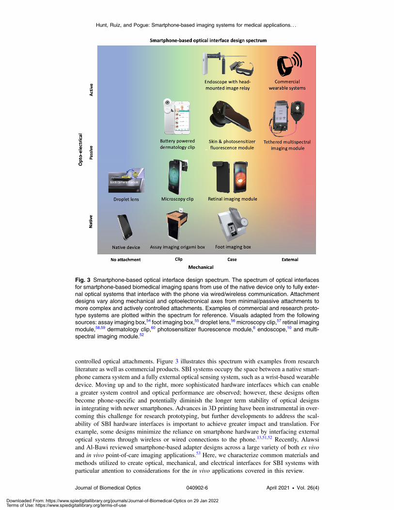
controlled optical attachments. Figure 3 illustrates this spectrum with examples from researchliterature as well as commercial products. SBI systems occupy the space between a native smart-phone camera system and a fully external optical sensing system, such as a wrist-based wearabledevice. Moving up and to the right, more sophisticated hardware interfaces which can enablea greater system control and optical performance are observed; however, these designs oftenbecome phone-specific and potentially diminish the longer term stability of optical designsin integrating with newer smartphones. Advances in 3D printing have been instrumental in over-coming this challenge for research prototyping, but further developments to address the scal-ability of SBI hardware interfaces is important to achieve greater impact and translation. Forexample, some designs minimize the reliance on smartphone hardware by interfacing externaloptical systems through wireless or wired connections to the phone.13,51,52 Recently, Alawsiand Al‐Bawi reviewed smartphone-based adapter designs across a large variety of both ex vivoand in vivo point-of-care imaging applications.53 Here, we characterize common materials andmethods utilized to create optical, mechanical, and electrical interfaces for SBI systems withparticular attention to considerations for the in vivo applications covered in this review.
Fig. 3 Smartphone-based optical interface design spectrum. The spectrum of optical interfacesfor smartphone-based biomedical imaging spans from use of the native device only to fully exter-nal optical systems that interface with the phone via wired/wireless communication. Attachmentdesigns vary along mechanical and optoelectronical axes from minimal/passive attachments tomore complex and actively controlled attachments. Examples of commercial and research proto-type systems are plotted within the spectrum for reference. Visuals adapted from the followingsources: assay imaging box,54 foot imaging box,55 droplet lens,56 microscopy clip,57 retinal imagingmodule,58,59 dermatology clip,60 photosensitizer fluorescence module,6 endoscope,10 and multi-spectral imaging module.52
Hunt, Ruiz, and Pogue: Smartphone-based imaging systems for medical applications. . .
Journal of Biomedical Optics 040902-6 April 2021 • Vol. 26(4)
Downloaded From: https://www.spiedigitallibrary.org/journals/Journal-of-Biomedical-Optics on 29 Jan 2022Terms of Use: https://www.spiedigitallibrary.org/terms-of-use

2.2.1 Optical
The primary optical interface for SBI systems is naturally the built-in camera. Smartphone cam-eras have been modified using off-the-shelf optical components including lenses (spherical,aspheric, achromats, infinity corrected objectives, Fresnel lenses, and reversed smartphonelenses from disassembled camera modules), filters (bandpass, longpass, neutral density, polar-izers), apertures, mirrors (folding, scanning, and dichroic), optical fibers for light relay of flashLED, diffusers, and diffraction gratings. Integration of these passive optics in front of the smart-phone camera is a fairly straightforward; however, it does impose some design limitations. Onemajor challenge toward computer-aided design of SBI systems is variability and/or unspecifiedoptical properties of built-in lenses, filters, and LEDs of preassembled smartphone camera mod-ules. Bae et al.10 approximated the smartphone lens of their system for ray tracing simulationusing the crop factor of the CMOS sensor, the fixed aperture specified by the vendor, and assum-ing the lens is set to infinite-focus. In both in vivo and ex vivo microscopy applications, othershave utilized an reversed smartphone lens to match the light collection angle of the built-in lensphone and relay distortion-free conjugate images to the CMOS sensor that fill the entiresensor.16,57,61,62 For reproducible calibration of spectral response across different smartphonemodels, several reports have utilized commercially available reference color targets (X-riteColorChecker for example) to apply phone-by-phone calibration factors.63–69 Quantitative meth-ods for calibration of SBI systems have also been proposed.70
One area that is not as well appreciated is control of stray light in SBI systems. Systems forlow light level applications require exclusion of ambient room lighting to preserve the signalspecificity or purity. This is especially important in spectroscopic, chromatic, and filtered lightapplications for external tissue measurements.6,17,52,64,68,71–73 Additionally, light emission fromtissue comes at high numerical aperture, and so control over access to these signals requirescareful lens design and external light control. Filtering of signals is always challenging giventhe range of choices and the typically short camera–tissue distances, and so evaluation of thecontaminating signals is important, as are choices about use of potentially multiple filters.6
Future developments in fabrication of customized optics could enable greater flexibility insmartphone optical interface design.74,75 Miniaturized polymer “droplet” lenses for both lightcollection76,77 and filtering56,78 are a promising development as they can potentially be fabricatedat very low cost and assembled with minimal adaptation of smartphone optics. Rapid advancesin computational imaging and optical design also hold great promise to be utilized in conjunc-tion with existing smartphone optical sensors or to be added through additional dedicatedsensors.79–84
2.2.2 Mechanical
Mechanical interfaces for SBI systems serve several functions depending on the use case includ-ing optical alignment and coupling of custom optics to the smartphone, background light rejec-tion in fluorescence applications, and ergonomic setup and clinical use. Custom enclosures foroptical attachments are typically fabricated using 3D printing to accommodate unique smart-phone geometries. Fused filament printing is most common and typically provides alignmentprecision within a few hundred microns. Stereolithography 3D printing is beginning to be morewidely used and can fabricate enclosures with sub-100 μm precision. This is often adequate forpositioning most optical components but may not be ideal for lens alignment, particularly highnumerical aperture lenses. For applications with custom lenses needing more precise alignment,cage rod assemblies mounted inside a 3D-printed enclosure have been used.15,16
In terms of attachment mechanisms, SBI systems can be broadly categorized into one of threecategories: (1) no attachment, (2) clip attachment, and (3) case attachment. SBI systems with noattachment utilize only the native device or otherwise may communicate wirelessly with externaloptics. For example, He and Wang demonstrated Weiner estimation to reconstruct “pseudo-hyperspectral” images in an attachment-free manner,68 whereas Cai et al.51 demonstrated asmartphone-interfaced wireless spectrometer for in vivo measurement of biosamples. Bothapproaches can, in principle, work for various smartphone models without requirement forhardware customization.
Hunt, Ruiz, and Pogue: Smartphone-based imaging systems for medical applications. . .
Journal of Biomedical Optics 040902-7 April 2021 • Vol. 26(4)
Downloaded From: https://www.spiedigitallibrary.org/journals/Journal-of-Biomedical-Optics on 29 Jan 2022Terms of Use: https://www.spiedigitallibrary.org/terms-of-use

Clip attachments designs intended be lower profile, less dependent on a specific phone geom-etry, and easily attached/detached from the smartphone. Clip attachments for the top of the phonenear the cameras and the base of the phone using the charging port have both beendemonstrated.57,85,86 Spigulis et al.87 demonstrated a sticky platform-like attachment that wasnot as low profile as other clip designs but enabled easy attachment/detachment of various smart-phones. Such designs are appealing in the sense that they could work with multiple phones;however, clip attachments that include custom lens assemblies are often impractical as manualalignment of lenses with the phone camera is cumbersome and error prone.
A more common design for SBI systems with custom optics is a case attachment. Caseattachments accommodate larger optics/electronics systems and more precisely couple thesystem to the camera. As smartphone manufacturers often published detailed specificationsfor third party manufacturing of phone cases, 3D design of case attachments is straight-forward. Their primary drawback is that they often require design modifications for eachsmartphone model, and with the rapid succession of models produced today, there wouldbe frequent changes needed for matching the updates. This change in phone shape with suc-cessive models on an annual basis is one of the most difficult challenges in these attachmentdevice approaches.
Sterilizability or sanitation is another important and potentially overlooked mechanicaldesign constraint. Attachments that are easily assembled/disassembled are preferable for fre-quent sterilization or cleaning. For applications where SBI systems are used by contact, useof biocompatible materials should be encouraged. Most 3D-printed prototypes do not complywith these needs and so merely serve as a rapid prototype that needs to be implemented in amedical grade material production. In sterile clinical environments, such as the surgical suite,clear sterile plastic sleeves are commonly used for camera and microscope systems.18,20
2.2.3 Electrical
Some SBI systems also use embedded electronics to facilitate controlled light delivery, activeoptical components (scanning mirrors, tunable filters, and motorized mounts), wireless/wireddata relay, and for microcontroller logic. Such active attachment designs are more common forfluorescence and multispectral imaging applications for more controlled light delivery.52,66,88–90
For example, Cavalcanti et al.88 used a multiplexed system of fiber optics to deliver colored lightfrom eight different LED sources across the visible spectrum to the tip of an otoscope. Wiredconnections have also been used to interface smartphones with USB cameras for tethered capsuleendoscopy as well as multispectral dermal imaging.13,52 Cai et al.51 developed a pencil-like spec-trometer based on a compact WiFi-enabled camera. Currently, the use of wired/wireless com-munication to embedded electronics seems somewhat underutilized and is one promising avenueto increase control and customization of SBI optical systems.
2.3 Software
In terms of software interfaces, there is a great deal of variety in the level of functionality sup-ported in research prototypes, with many systems relying on third party camera software andmanual postprocessing of images. Figure 4 highlights approaches and core functionalities ofsoftware supported by SBI systems in unrealistic and realistic deployments with increasingdegrees of control and customization. Clinical deployment of SBI systems for real-time use intoclinical settings necessitates custom software to facilitate both data acquisition and analysis.State-of-the-art SBI software interfaces should accomplish both of these functions in additionto facilitating easy operation for the end user.
Acquisition is one area where smartphones have great potential to streamline and enhance theusability for biomedical imaging. The core functionalities of good acquisition software are five-fold: (1) provide a video-rate preview of the camera sensor data, (2) control relevant acquisitionparameters (focus, exposure, gain, white balance, RAW pixel data, etc.), (3) trigger start and stopof camera acquisition, (4) facilitate storage and organization of acquired data (by patient or sam-ple, for example), and (5) interface with downstream image analysis pipelines (either through on-board or wireless communication). Despite smartphones having perfected these functionalities
Hunt, Ruiz, and Pogue: Smartphone-based imaging systems for medical applications. . .
Journal of Biomedical Optics 040902-8 April 2021 • Vol. 26(4)
Downloaded From: https://www.spiedigitallibrary.org/journals/Journal-of-Biomedical-Optics on 29 Jan 2022Terms of Use: https://www.spiedigitallibrary.org/terms-of-use

for everyday photography, these advances may not readily transfer to SBI systems with custom-ized optics and/or acquisition pipelines.
As medical providers are often multitasking while performing clinical examinations, real-time visualization, and easy triggering of acquisition is of particular importance for diagnosticand treatment guidance systems. For example, Bae et al.10 developed a custom app for theirendoscopy system, which facilitates image relay to a head-mounted display. Other SBI systemsthat facilitate improved ease of use and contactless acquisition through hand-gesture or voice-commands.91–93 The highly networked capabilities of smartphones seem largely underutilized,given the potential ability to interface with many peripherals at the same time, via multiple com-munication protocols (i.e., Bluetooth, Wi-Fi, cellular, or direct cable connection).
Another crucial function for acquisition software in scientific and medical applications isthe capability to control acquisition and postprocessing parameters, retain adequate bit depth,and interface with downstream image analysis pipelines. The ability to acquire reproducibleimages has been an ongoing challenge for smartphone-based systems due to their “point-and-shoot” design which automates imaging acquisition and postprocessing.94 Autofocusing/exposure is generally beneficial for improved usability of imaging systems; however, devel-opers should be careful of relying on high-level native and third party libraries to provide thisfunctionality as it may inhibit quantitative reproducibility. In recent years, the ability to fiximaging parameters and access the unprocessed RAW pixel data has become more accessibleon both iOS and Android platforms. This has been leveraged in more recently reported systemsfor improved quantitative accuracy in low-light applications, although incorporating RAWimage processing capability in downstream on-board analysis on phones has not yet beendemonstrated.6,16,64,73
In terms of analysis, common functions for SBI systems include image review/consulta-tion,91,95 region of interest selection,19,96 colorimetric quantification,73,97 intensity measure-ments,6,98,99 diagnostic classification,65,71,90,100 and segmentation of tissue structures.14,15,55,101
However, implementing custom image analysis routines on smartphone operating systemsrequires substantial programming expertise, leading researchers to continue to rely on manualimage processing workflows. The problem is further exacerbated by phone-to-phone variability,requiring additional work to ensure smartphone-based analysis routines function properly formultiple phone cameras and operating systems. Both one-time calibration of phone cameras
Fig. 4 Smartphone-based software design spectrum. Approaches and core functionalities sup-ported by SBI systems in unrealistic and realistic deployments with increasing degrees of custom-ization are highlighted. Unfortunately, the vast majority of SBI systems reported in the currentliterature use unrealistic acquisition and analysis pipelines.
Hunt, Ruiz, and Pogue: Smartphone-based imaging systems for medical applications. . .
Journal of Biomedical Optics 040902-9 April 2021 • Vol. 26(4)
Downloaded From: https://www.spiedigitallibrary.org/journals/Journal-of-Biomedical-Optics on 29 Jan 2022Terms of Use: https://www.spiedigitallibrary.org/terms-of-use

using optical targets as well as per-measurement calibration by measuring ambient lighting con-ditions have been proposed to improve quantitative reproducibility.68,73,86,94,102 An alternativesolution to phone-by-phone calibration for image analysis pipelines could be to leveragecloud-based computing with deep learning. Such an approach has been demonstrated bySong et al.100 for oral imaging. Centralizing image data acquired from multiple users/phonesto an appropriate privacy-compliant server enables collection of larger, more diverse datasetswhich can in turn be used to develop more robust image analysis pipelines and deploy themwithout having to continually update smartphone software.
3 Context and Applications
As noted in Sec. 1, SBI systems have been proposed for a variety of diagnostic applications andare increasingly being proposed for noninvasive monitoring of disease conditions. There are alsoemerging reports proposing the use of SBI to guide treatment procedures (surgical or PDT) or toconduct more invasive diagnostic imaging of deep tissues (endoscopy). This section contains astructured summary of recent reports using SBI tissue imaging for three types of applications:monitoring, diagnosis, and treatment guidance. While there are some commonalities in how SBIcould be advantageous in all three of these categories, understanding of the clinical context (i.e.,where is the measurement being taking and who is taking it) is important in assessing thestrengths and weaknesses of smartphone utilization. As there are some SBI systems and appli-cations, which may well fit into more than one of the three aforementioned categories, note thatthis categorization was not intended to be a rigid one, but rather a broad grouping for purposes ofdiscussion.
3.1 Monitoring
Monitoring applications often require repeated sampling over a sustained period (from hours upto months) to assess appreciable differences in disease states, and therefore benefit from beinglow-cost and noninvasive. In recent years, SBI systems have been proposed for monitoring ofvital signs,103–109 blood glucose,110–113 blood pressure,114,115 blood oxygenation,68,116 hemoglo-bin concentration,72 atrial fibrilation,117,118 jaundice,73,97,119,120 skin cancer,121 and diabetic footulcers.55,122 All of these applications propose utilizing either contact-based or contactless opticalmeasurements using the smartphone camera, most often by individuals on themselves (i.e., self-monitoring). With the exception of blood glucose monitoring, all are proposed for use throughinstallation of a software app onto native devices and do not require external optical attachments.
Vital sign monitoring using contact-based video recording of fingers (i.e., photoplethysmog-raphy) was an early SBI application proposed for smartphones,103 and there are continued reportsof novel ways to utilize this approach to extract additional hemodynamic metrics (blood pres-sure, oxygenation, cardiac arrhythmia, etc.).115,116,118 However, this is clearly one area wherepractical considerations of the context have been overlooked and smartphone utilization isincreasingly questionable, as to the use case. Although contact-based vital sign monitoringis achievable using smartphone cameras, continuous monitoring is infeasible without dedicatingthe smartphone for that purpose. Low-cost, dedicated wearable sensors which can relay data tothe smartphone are clearly a more practical and reliable long-term solution for obtaining contact-based vital sign measurements.
Noncontact-based methods for extracting hemodynamics using video recording are a morerecent development which more fully utilizes the spatial information provided by smartphoneimaging.68,72,104,123,124 Two recent works, by Park et al.72 and He andWang,68 used computationaltechniques to infer higher resolution spectral responses and predict hemoglobin content in tissueusing only RGB smartphone image data. Park et al. evaluated their method on a dataset acquiredfrom 153 participants referred for a blood count at an academic health center in Kenya, whereasHe and Wang performed their assessments on a few volunteers in a “dark” lab environment. Parket al. also performed a more rigorous quantitative comparison of their noninvasive hemoglobinconcentration prediction compared to the gold-standard obtained by venous blood draw, observ-ing good quantitative agreement of their noninvasive hemoglobin predictions (R2 ¼ 0.91 for
Hunt, Ruiz, and Pogue: Smartphone-based imaging systems for medical applications. . .
Journal of Biomedical Optics 040902-10 April 2021 • Vol. 26(4)
Downloaded From: https://www.spiedigitallibrary.org/journals/Journal-of-Biomedical-Optics on 29 Jan 2022Terms of Use: https://www.spiedigitallibrary.org/terms-of-use

15 test patients).72 The work by Park et al. showed a rigorous and realistic assessment of theirmethod in a clinical setting. However, both methods required careful calibration of smartphonecamera spectral response, used post-hoc image analysis routines, and were only conducted byresearch personnel, so it remains to be seen if they can be effectively deployed at scale onsmartphones.
Other monitoring application for SBI involves photographic surveillance of dermal condi-tions, skin cancer, or diabetic foot ulcers for example. In these applications, there is less need foroptical instrumentation, but rather software design to facilitate standardized acquisition, auto-mated analysis, and data relay to clinical providers as needed. For example, in the case ofdiabetic foot ulcers, Yap et al.125 developed an app that provides a “ghost outline” of the user’sfoot based on prior measurements to ensure that repeated measurements were well coregistered.And Ploderer et al.93 proposed the use of the front facing camera and voice guidance to enableusers to acquire photos of the bottom of their feet more conveniently. While less novel in theirextraction of optical tissue properties, these approaches demonstrate a focus on real-worldusability in SBI monitoring applications.
3.2 Diagnosis
Diagnostic measurements of tissue are typically performed to examine a suspicious area of tissuemore closely to assess the underlying cause of abnormality and/or severity of disease. As diag-nostics are typically used for triage to treatment interventions, such applications more likely to beperformed by medical personnel and in clinical settings rather than by individuals at home. Someof the primary in vivo diagnostic modalities proposed for SBI systems include white light im-aging,52,65,66,91,100–102,126–133 autofluorescence imaging,17,65,66,71,100 multispectral/hyperspectralimaging,52,64,88 endoscopy,10–13 in vivo spectroscopy,14,51,85 and in vivo microscopy.14–16,62,134
For these modalities, the most frequent imaging sites are external tissues (dermis, facial, andretinal), externally accessible tissues (oral cavity, cervix, and ear), as well as some deeper tissuesin the case of endoscopy (bladder, larynx, and esophagus).
In past years, a number of novel SBI systems have been developed for the application of skincancer diagnosis and surveillance through smartphone-based dermascopy. In 2016, Kim et al.published one of the first smartphone-based multispectral imaging systems and explored itspotential use for dermal lesion assessment.89 Their system consisted of a motor-controlled wheelof optical filters placed in front of the LED flash with embedded electronics and custom app tosynchronize acquisition of a spectral image cube from images acquired at nine different wave-lengths from 440 to 690 nm. They validated their spectral measurements against a non-SBIliquid-tunable-crystal-filter system using a colored optical target and performed exploratory nor-mal volunteer imaging at imaging two dermal sites (one acne and nevus region). Acquisitionspeed was not reported, but image cube processing speed was reportedly 30 s on the phone andcould be sped up to 3 s per image cube with cloud-based processing. More recently, Uthoff et al.reported a multispectral system that performs sequential illumination using eight different col-ored LEDs across the visible to near-infrared regime (450 to 940 nm) and was actively controlledusing a custom smartphone app.52 Their system implemented two different camera acquisitionmethods: one using the built-in camera and one using a tethered USB camera. The authors per-formed measurements using both camera setups and semiquantitatively assessed skin chromo-phores (hemoglobin, oxygenated hemoglobin, and melanin) in two clinical cases (one benignand one malignant). Acquisition reportedly required 20 s per image cube, and it was not specifiedwhether on-board image cube analysis was achieved. While both systems are state-of-the-artimplementations in terms of integration of custom hardware and software into novel, compactimaging systems, their primary shortcoming is a lack of integration into a practical clinical work-flow to more concretely demonstrate the advantages of having these modalities on an SBIsystem. This can be done but requires more extensive testing than was reported.
Endoscopic procedures using both rigid and flexible optical probes have incorporatedinto SBI systems for otolaryngological, esophageal, and cervical examination of epithelialtissues.10–15,135 In the context of endoscopy applications, handheld systems with rigid opticalprobes seem more synergistic with smartphone utilization as the probe and the device are com-pact enough to simultaneously manipulate and position.10,135 By contrast, other microendoscope
Hunt, Ruiz, and Pogue: Smartphone-based imaging systems for medical applications. . .
Journal of Biomedical Optics 040902-11 April 2021 • Vol. 26(4)
Downloaded From: https://www.spiedigitallibrary.org/journals/Journal-of-Biomedical-Optics on 29 Jan 2022Terms of Use: https://www.spiedigitallibrary.org/terms-of-use

systems utilize thin flexible coherent fiber bundles for image relay.14,15 These systems benefitless from smartphone utilization, as the optical probe is typically manipulated independently ofthe monitor screen and the optical probes can relay images over longer distances (often severalfeet), reducing the need for an ultracompact optical enclosure. Similarly, tethered capsule endo-scopes benefit minimally from smartphone utilization as the tethered connection enables relayover long distances and to any computer monitor.
3.3 Treatment Guidance
Treatment guidance imaging systems may have many overlapping requirements with diagnosticapplications but primarily differ in that they have more stringent requirements for real-timevisualization and analysis to provide active procedural guidance and support more rapid clini-cal decision making. Recent reports for SBI systems and applications that involve treatmentguidance include image-guided surgery,18–20,22,37,38,41 management of severe burn injuries,40,95
PDT,6–9 and venipuncture.63,136
The starkest example of SBI for treatment guidance is surgery. Because surgical proceduresare very costly in terms of medical infrastructure and human resource requirements, the typicaljustification of smartphone utilization based on low-cost becomes irrelevant, and usability meritsof the form factor, interactive interface design, and networking capabilities of the smartphoneplatform must clearly take precedence. In 2014, Teichman et al.19 proposed the use of a smart-phone app to take images and subsequent spatial measurements using software to postopera-tively verify placement of toric intraocular lenses during cataract surgery. While feasibilityof SBI in this application was demonstrated, no substantive assessment of clinical outcomeswas undertaken and the approach does not appear to have been widely adopted. In 2018, a teamof neurosurgical clinicians reported a retrospective analysis on the use of a smartphone-basedrigid endoscope to visualize the surgical field in over 42 neurosurgical procedures over a span offive years. The study was a nonrandomized retrospective analysis and was limited to a qualitativecase report of the usability of the smartphone in this application. The authors noted that theplacement of the smartphone screen in front of the endoscope made it “more intuitive” and“enhanced 3D perception” during operation. The attachment utilized with the rigid endoscopeappears to have since been discontinued.137 Overall, these clinical assessments of SBI systems insurgery were quite small scale and qualitative in nature, perhaps indicating a lack of confidencein providers and ethics committees to evaluate these techniques in a more substantive manner.
In two of the aforementioned applications (severe burn management and PDT), SBI systemshave been proposed to provide noninvasive, quantitative quality assurance of treatment proce-dures which, in current clinical workflows, require less expertise to deliver but can be moresubjective in nature. These applications are much better suited to leverage SBI systems. In photo-dynamic therapy, for example, Ruiz et al.6 developed a quantitative, handheld fluorescenceimaging system for PpIX-PDT dosimetry before, during, and after phototherapy. The systemconsists of a battery powered case attachment which includes an embedded LED ring (405-nm illumination) and 600-nm longpass emission filter, and the cylindrical enclosure enablescontact-based measurement at a fixed working distance and focal length, which helps ensureeasy operation and reproducibility of measurements. The system was assessed using intralipidphantoms with known PpIX concentrations as well as in an animal model for PDT treatment andshowed excellent quantitative precision in both applications. A clinical evaluation of another SBIsystem for photodynamic therapy of oral lesions at a medical center was recently reported byKhan et al.9 to assess low-cost technological treatments for this disease. Twenty-nine patientswith confirmed oral squamous cell carcinoma lesions and who were undergoing PDT wereimaged using white light, ultrasound, autofluorescence, and PpIX fluorescence imaging pre-and post-treatment. Fluorescence imaging guidance was demonstrated to provide visual guid-ance to demarcate lesion boundaries and quantitatively confirm the treatment region pre- andpost-treatment due to photobleaching. The authors noted some practical limitations of their cur-rent instrumentation (offline analysis routines as well as the handheld device being too bulky foreasy access to all regions of the oral cavity), but it is more evident that the clinical assessmentswere rigorously performed in the intended clinical setting and that future developments willcontinue to leverage SBI in appropriate and meaningful ways.
Hunt, Ruiz, and Pogue: Smartphone-based imaging systems for medical applications. . .
Journal of Biomedical Optics 040902-12 April 2021 • Vol. 26(4)
Downloaded From: https://www.spiedigitallibrary.org/journals/Journal-of-Biomedical-Optics on 29 Jan 2022Terms of Use: https://www.spiedigitallibrary.org/terms-of-use

4 Discussion
The pervasive rationale for smartphone utilization in biomedical imaging has emphasized amultitude of factors including cost, portability, connectivity, ease-of-use, and scalability.While these factors are clearly desirable features of biomedical imaging systems, rigorous jus-tification for how SBI systems outperform non-SBI systems is often lacking. The ubiquity ofsmartphones is frequently cited in literature as a justification that SBI systems are inherently low-cost, easy-to-use, and scalable biomedical imaging solutions. However, undermining this claimis the reality that the majority of original research for SBI systems is limited to a single phonemodel and utilizes manual, often fragmented image acquisition and analysis pipelines. While thisis partly the nature of research prototyping, it is important to openly discuss these limitations andcontinually move toward practices that will enable greater reproducibility and translation ofresearch advances in SBI.
4.1 Guidelines for Appropriate Use of Smartphone for Biomedical Optics
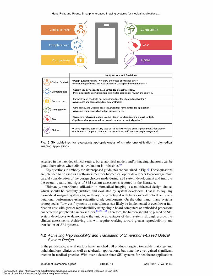
As discussed in the prior sections, smartphone-based hardware and software interface designvaries greatly, with some designs more fully/appropriately utilizing the capabilities of the smart-phone platform and others less so. Here, we provide six keys (the six C’s) as recommendedguidelines to assess the appropriateness of smartphone use in biomedical imaging systems.
Clinical context represents an understanding of the clinical need and the intended user. Thesefactors should always provide the overarching context for device design and development.Smartphone utilization should primarily be justified within this context and if the workflowneeds of the clinic match the capabilities of a SBI system, or if other more dedicated systemswould be superior. The ideal scenario is to assess new devices in their intended setting (at home,diagnostic lab, health clinic, central hospital, etc.) and in the hands of the intended operator(patient, lab technician, nurse, physician). When that is not feasible, device developers shouldbe careful not to overstate the impact of their systems and acknowledge this limitation.
Completeness represents achieving a complete implementation of the intended clinical work-flow, including custom app development and use and testing by nonresearch personnel. Manualimage processing steps using desktop software or evaluations performed only in lab settings areoften signs that a complete workflow has not been achieved. Achieving a complete implemen-tation is clearly the best way to test the value of the approach.
Compactness relates to the importance of portability and small size for the intended appli-cation. For diagnostic and treatment guidance systems, if handheld use and/or easy transportbetween exam rooms will enhance usability, smartphone use is more appropriate. Alternatively,if the use case is in healthcare outside the medical center, or in remote setting, does thecompact nature of the phone and the addition match the economics and portability needs ofthe situation?
Connectivity refers to the importance of wireless communication for the intended applica-tion. Applications that use wireless communication, through either Bluetooth or Wi-Fi, andfeatures such as cloud computing for centralized data are ideal for SBI systems. Scalability andmultiuser deployment are appropriate choices for these as well.
Cost is often the most emphasized reason for development of SBI systems, but it should benoted that this is rarely a true argument. Low-cost optoelectronic systems are now widespreadand the cost of the most advanced smartphones has grown. The cost issue should be carefullyevaluated, particularly if the application requires a hardware attachment and is intended for diag-nostic use or treatment guidance. In these contexts, costs associated with regulatory approval andmarketing will far outweigh material costs. Designs that prioritize the aforementioned C’s asopposed to minimizing prototyping costs are more likely to make an impact in medicine.
Claims refers to general statements regarding ease-of-use, cost, or scalability of SBI systemsby virtue of smartphone utilization alone. Such claims should not be promulgated in researchliterature but rather appropriately justified through quantitative assessments. Some studies havequantified usability improvements by measuring time for task completion, performing surveys,or conducting blinded image reviews.11,129,133 Broader use of such assessments to substantiateimproved usability of SBI systems should be encouraged. When possible, usability should be
Hunt, Ruiz, and Pogue: Smartphone-based imaging systems for medical applications. . .
Journal of Biomedical Optics 040902-13 April 2021 • Vol. 26(4)
Downloaded From: https://www.spiedigitallibrary.org/journals/Journal-of-Biomedical-Optics on 29 Jan 2022Terms of Use: https://www.spiedigitallibrary.org/terms-of-use
assessed in the intended clinical setting, but anatomical models and/or imaging phantoms can begood alternatives when clinical evaluation is infeasible.138
Key questions to embody the six proposed guidelines are contained in Fig. 5. These questionsare intended to be used as a self-assessment for biomedical optics developers to encourage morecareful consideration of the design choices made during SBI system development and improvethe overall quality and rigor of SBI system assessments reported in the literature.
Ultimately, smartphone utilization in biomedical imaging is a multifaceted design choice,which should be carefully justified and evaluated by system developers. That is to say, anybiomedical imaging system can, in theory, be prototyped with better overall optical and com-putational performance using scientific-grade components. On the other hand, many systemsprototyped as “low-cost” systems on smartphones can likely be implemented at even lower fab-rication cost with greater reproducibility using single board computers or embedded processorsconnected to peripheral camera sensors.80,139–143 Therefore, the burden should be placed on SBIsystem developers to demonstrate the unique advantages of their systems through prospectiveclinical assessments. Achieving this will require working toward greater reproducibility andtranslation of SBI systems.
4.2 Achieving Reproducibility and Translation of Smartphone-Based OpticalSystem Design
In the past decade, several startups have launched SBI products targeted toward dermatology andophthalmology clinics as well as telehealth applications, but none have yet gained significanttraction in medical practice. With over a decade since SBI systems for healthcare applications
Fig. 5 Six guidelines for evaluating appropriateness of smartphone utilization in biomedicalimaging applications.
Hunt, Ruiz, and Pogue: Smartphone-based imaging systems for medical applications. . .
Journal of Biomedical Optics 040902-14 April 2021 • Vol. 26(4)
Downloaded From: https://www.spiedigitallibrary.org/journals/Journal-of-Biomedical-Optics on 29 Jan 2022Terms of Use: https://www.spiedigitallibrary.org/terms-of-use

have been under development, the lack of commercial success for SBI systems should raiseconcern. While it is challenging to comprehensively identify barriers to translation of SBI sys-tems, we postulate that the speed at which smartphone technology evolves and the short lifecycleof these products is not readily conducive to medical device manufacturing standards. For hard-ware interfaces, new form factors and camera modules are launched each year, necessitatingredesign of optical attachments. The lack of standardization in software development and repro-ducibility of results for SBI are a barrier for research progress. Here, we propose the followingthree items to move toward wide adoption of SBI systems: (1) focusing on hardware design thatfacilitates adoption of varying phone models, (2) creation of open-source software for SBI sys-tem development, and (3) adoption of robust calibration methods to best facilitate quantitativereproducibility.
Hardware design that focuses on attachments that are adaptable to different placements of thecamera will be imperative for this field to gain long-term traction. Alternatively, the cost ofattachment development could be sufficiently low as to allow ease of development for multipleplatforms, similar to the smartphone case marketplace today. Today most devices are made for aspecific phone model and customized around it, but further thought into adaptive design forconstant changes in camera placement and phone sizes will be important. Hardware is moredifficult to standardize as people will likely elect to use different smartphones for development.As a starting point, sharing of CAD files for optical attachments, custom enclosures, and elec-tronic schematics with publication should be encouraged. Many research groups and startupsfocus on 3D printing of the hardware containers which is now a reliable and reasonable way toprototype. The shift from 3D printing technology to automated production of attachment hard-ware via machining, injection molding, or thermosetting will likely be important. The attach-ments with optical components can take advantage of highly developed optomechanicalengineering that has already revolutionized the smartphone camera industry. The major benefitsof spectral, polarization, or gated sensing and imaging remain to be fully exploited with customattachments.
Open-source software toolkits and starter applications for biomedical imaging are a goodplace to start addressing existing development and reproducibility problems in SBI. Effectivesmartphone app development and maintenance requires significant programming expertise andis currently a barrier for many researchers who might be developing their software from scratch.In order for research prototypes to achieve clinical translation, standardized methods for SBIsoftware development are needed. Cho et al.144 proposed a concept for a “retargetable applicationdevelopment platform for healthcare mobile applications.” Such a project is a worthy goal. Inanother recent review on smartphone point-of-care adapters, Alawsi and Al‐Bawi proposed thatcross platform app development using Ionic or Xamarin as a possible solution.53 Although cross-platform app development could help in principle, it would likely be limited to only the subsetof functionality which is common to all operating systems and would utilize the phone’s builtin compression algorithms. This would not be ideal for quantitative fluorescence imaging forexample. An alternative starting point is to create and maintain platform-specific templates thatsupport core functionality needed for biomedical imaging which would include support forRAW image acquisition and standardized processing routines for common biomedical imageanalysis tasks.
Robust calibration of SBI systems is essential for addressing reproducibility problems andachieving clinical translation. Two major factors in this regard are: (1) lack of characterization ofsensor performance (dynamic range, SNR, absorption spectra of built-in filters, demosaicing,and data acquisition rates) and (2) “black-box” processing that phones perform on the CMOSimaging data to generate traditional 8-bit RGB images. The use of RAW pixel data to confirmsuitable processing pipelines is a straightforward way to circumvent this issue for all applica-tions, including colorimetric and quantitative techniques. Given the increased complexity ofaccessing and analyzing RAW pixel data, suitable alternatives include color and gray-scalecalibration targets (X-rite ColorChecker, for example).67,69 Moving toward full system charac-terization using radiometric calibration methods to understand results presented in studiesshould also be encouraged. Relative radiometric calibration methods for smartphones have beenproposed.70 Absolute radiometric calibration methods that are optimized for SBI systems shouldbe developed to aid in the development of quantitative applications such as fluorescence
Hunt, Ruiz, and Pogue: Smartphone-based imaging systems for medical applications. . .
Journal of Biomedical Optics 040902-15 April 2021 • Vol. 26(4)
Downloaded From: https://www.spiedigitallibrary.org/journals/Journal-of-Biomedical-Optics on 29 Jan 2022Terms of Use: https://www.spiedigitallibrary.org/terms-of-use

imaging. Additionally, public or app-specific sharing of image measurements from commerciallyavailable optical phantoms/targets can help ensure reproducibility of optical measurements.Development of easily networked access to file spaces will enable platforms that take advantageof off-phone computing resources such as deep learning algorithms that interpret the image data.At a minimum, these steps would enable relative calibration and comparison across hardwaresystems in the literature.
5 Conclusions
SBI systems have demonstrated a large array of applications and exhibit great potential to facili-tate compact, easy-to-use biomedical imaging systems. However, for SBI systems to achieve thatpotential, more holistic assessments of SBI systems are needed to enable greater reproducibilityand demonstrate value within their intended clinical settings. Evaluation of SBI systems shouldtake into account clinical context, completeness, compactness, connectivity, cost, and claimsassociated with novel systems. Claims regarding the scalability and low-cost of SBI systemsbased on the ubiquity of smartphones should not be sufficient to justify their novelty and impact.Ongoing work in SBI for medical applications should prioritize realistic clinical assessmentswith quantitative and qualitative comparisons to other non-SBI systems in order to more clearlydemonstrate the value of SBI systems within their intended applications. Improved hardwaredesign to accommodate the rapidly changing smartphone ecosystem, creation open-source soft-ware and starter applications for SBI system development, and adoption of robust calibrationtechniques to address phone-to-phone variability are three high priority areas to move SBIresearch in biomedical imaging forward.
Disclosures
Two of the authors (BWP and AJR) are cofounders and employed part time by QUEL Imaging,developing tools for fluorescence imaging, including smartphone applications.
Acknowledgments
This work has been partially supported by the National Institutes of Health (Grant No.P01CA084203).
References
1. A. Roda et al., “Smartphone-based biosensors: a critical review and perspectives,” TRACTrends Anal. Chem. 79, 317–325 (2016).
2. S. Kanchi et al., “Smartphone based bioanalytical and diagnosis applications: a review,”Biosens. Bioelectron. 102, 136–149 (2018).
3. J. Liu et al., “Point-of-care testing based on smartphone: the current state-of-the-art(2017–2018),” Biosens. Bioelectron. 132, 17–37 (2019).
4. T. R. Kozel and A. R. Burnham-Marusich, “Point-of-care testing for infectious diseases:past, present, and future,” J. Clin. Microbiol. 55, 2313–2320 (2017).
5. B. P. Hibler, Q. Qi, and A. M. Rossi, “Current state of imaging in dermatology,” Semin.Cutan. Med. Surg. 35, 2–8 (2016).
6. A. J. Ruiz et al., “Smartphone fluorescence imager for quantitative dosimetry of proto-porphyrin-IX-based photodynamic therapy in skin,” J. Biomed. Opt. 25, 056003 (2020).
7. J. Hempstead et al., “Low-cost photodynamic therapy devices for global health settings:characterization of battery-powered LED performance and smartphone imaging in 3Dtumor models,” Sci. Rep. 5, 10093 (2015).
8. H. Liu et al., “Development and evaluation of a low-cost, portable, LED-based device forPDT treatment of early-stage oral cancer in resource-limited settings,” Lasers Surg. Med.51, 345–351 (2019).
Hunt, Ruiz, and Pogue: Smartphone-based imaging systems for medical applications. . .
Journal of Biomedical Optics 040902-16 April 2021 • Vol. 26(4)
Downloaded From: https://www.spiedigitallibrary.org/journals/Journal-of-Biomedical-Optics on 29 Jan 2022Terms of Use: https://www.spiedigitallibrary.org/terms-of-use

9. S. Khan et al., “Clinical evaluation of smartphone-based fluorescence imaging for guid-ance and monitoring of ALA-PDT treatment of early oral cancer,” J. Biomed. Opt. 25,063813 (2020).
10. J. K. Bae et al., “Smartphone-based endoscope system for advanced point-of-care diag-nostics: feasibility study,” JMIR Mhealth Uhealth 5, e99 (2017).
11. S. Lu et al., “Endockscope: a disruptive endoscopic technology,” J. Endourol. 33, 960–965(2019).
12. S. E. Maurrasse, T. W. Schwanke, and A. Tabaee, “Smartphone capture of flexiblelaryngoscopy: optics, subsite visualization, and patient satisfaction,” Laryngoscope 129,2147–2152 (2019).
13. G. Sharma et al., “Smartphone-based multimodal tethered capsule endoscopic platform forwhite-light, narrow-band, and fluorescence/autofluorescence imaging,” J. Biophotonics14, e202000324 (2020).
14. X. Hong et al., “A dual-modality smartphone microendoscope for quantifying the physio-logical and morphological properties of epithelial tissues,” Sci. Rep. 9, 15713 (2019).
15. B. D. Grant et al., “A mobile-phone based high-resolution microendoscope to imagecervical precancer,” PLoS One 14, e0211045 (2019).
16. E. E. Freeman et al., “Smartphone confocal microscopy for imaging cellular structures inhuman skin in vivo,” Biomed. Opt. Express 9, 1906–1915 (2018).
17. Q. He, T. Liu, and R. K. Wang, “Handheld swept-source optical coherence tomographyguided by smartphone-enabled wide-field autofluorescence photography for imagingfacial sebaceous glands,” Opt. Lett. 45, 5704 (2020).
18. M. Mandel et al., “Smartphone-assisted minimally invasive neurosurgery,” J. Neurosurg.130, 90–98 (2019).
19. J. C. Teichman, K. Baig, and I. I. K. Ahmed, “Simple technique to measure toric intra-ocular lens alignment and stability using a smartphone,” J. Cataract Refract. Surg. 40,1949–1952 (2014).
20. O. Aly, “Assisting vascular surgery with smartphone augmented reality,” Cureus 12, e8020(2020).
21. M. M. Çelikoyar, O. Topsakal, and S. Gürbüz, “Mobile technology for recording surgicalprocedures,” J. Visual Commun. Med. 42, 120–125 (2019).
22. T. Han et al., “Indocyanine green angiography predicts tissue necrosis more accurately thanthermal imaging and near-infrared spectroscopy in a rat perforator flap model,” Plast.Reconstr. Surg. 146, 1044–1054 (2020).
23. GSMArena, “GSMArena compare specs tool,” GSMArena.com https://www.gsmarena.com/compare.php3.
24. Y. M. Park et al., “Ambient light-based optical biosensing platform with smartphone-embedded illumination sensor,” Biosens. Bioelectron. 93, 205–211 (2017).
25. Y. Zhao et al., “A nanozyme- and ambient light-based smartphone platform for simulta-neous detection of dual biomarkers from exposure to organophosphorus pesticides,” Anal.Chem. 90, 7391–7398 (2018).
26. I. Hussain et al., “Design of a smartphone platform compact optical system operationalboth in visible and near infrared spectral regime,” IEEE Sens. J. 18, 4933–4939(2018).
27. S. Dutta, “Point of care sensing and biosensing using ambient light sensor of smartphone:critical review,” TRAC Trends Anal. Chem. 110, 393–400 (2019).
28. A. Breitbarth et al., “Measurement accuracy and dependence on external influences of theiPhone X TrueDepth sensor,” Proc. SPIE 11144, 1114407 (2019).
29. B. Krolla, M. Diebold, and D. Stricker, “Light field from smartphone-based dual video,”Lect. Notes Comput. Sci. 8926, 600–610 (2015).
30. D. W. Palmer et al., “Glare-free retinal imaging using a portable light field fundus camera,”Biomed. Opt. Express 9, 3178–3192 (2018).
31. H. M. Kim et al., “Miniaturized 3D depth sensing-based smartphone light field camera,”Sensors 20, 2129 (2020).
32. S. Lee et al., “A time-of-flight range sensor using four-tap lock-in pixels with high nearinfrared sensitivity for LiDAR applications,” Sensors 20, 116 (2020).
Hunt, Ruiz, and Pogue: Smartphone-based imaging systems for medical applications. . .
Journal of Biomedical Optics 040902-17 April 2021 • Vol. 26(4)
Downloaded From: https://www.spiedigitallibrary.org/journals/Journal-of-Biomedical-Optics on 29 Jan 2022Terms of Use: https://www.spiedigitallibrary.org/terms-of-use

33. “ISOCELL Vizion 33D | ToF Sensor | Samsung ISOCELL,” Samsung Semiconductor,www.samsung.com/semiconductor/minisite/isocell/vision-sensor/isocell-vizion-33d/.
34. L. Ulrich et al., “Analysis of RGB-D camera technologies for supporting different facialusage scenarios,” Multimedia Tools Appl. 79, 29375–29398 (2020).
35. “Cat S61—full phone specifications,” https://www.gsmarena.com/cat_s61-9076.php.36. A. Kirimtat et al., “FLIR vs SEEK thermal cameras in biomedicine: comparative diagnosis
through infrared thermography,” BMC Bioinf. 21, 88 (2020).37. J. T. Hardwicke, O. Osmani, and J. M. Skillman, “Detection of perforators using smart-
phone thermal imaging,” Plast. Reconstr. Surg. 137, 39–41 (2016).38. E. Y. Xue et al., “Use of FLIRONE smartphone thermography in burn wound assessment,”
Ann. Plast. Surg. 80, S236–S238 (2018).39. R. F. M. van Doremalen et al., “Validation of low-cost smartphone-based thermal camera
for diabetic foot assessment,” Diabetes Res. Clin. Pract. 149, 132–139 (2019).40. J. Goel et al., “A prospective study comparing the FLIR ONE with laser Doppler imaging
in the assessment of burn depth by a tertiary burns unit in the United Kingdom,” ScarsBurn Heal 6, 2059513120974261 (2020).
41. M. P. B. Obinah, M. Nielsen, and L. R. Hölmich, “High-end versus low-end thermalimaging for detection of arterial perforators,” Plast. Reconstr. Surg. Glob. Open 8, e3175(2020).
42. D. M. Roblyer, “Perspective on the increasing role of optical wearables and remote patientmonitoring in the COVID-19 era and beyond,” J. Biomed. Opt. 25, 102703 (2020).
43. S. C. Mukhopadhyay, “Wearable sensors for human activity monitoring: a review,” IEEESens. J. 15, 1321–1330 (2014).
44. A. Nag, S. C. Mukhopadhyay, and J. Kosel, “Wearable flexible sensors: a review,” IEEESens. J. 17, 3949–3960 (2017).
45. Z. S. Ballard and A. Ozcan, “Wearable optical sensors,” in Mobile Health, J. Rehg,S. Murphy, and S. Kumar, Eds., pp. 313–342, Springer, Cham, Switzerland (2017).
46. J. Heikenfeld et al., “Wearable sensors: modalities, challenges, and prospects,” Lab Chip18, 217–248 (2018).
47. A. Kamišalic et al., “Sensors and functionalities of non-invasive wrist-wearable devices:a review,” Sensors 18, 1714 (2018).
48. “Understanding reliability in Bluetooth® technology,” Bluetooth® Technology Website,2020, https://www.bluetooth.com/bluetooth-resources/understanding-reliability-in-bluetooth-technology/.
49. “Project Soli—Google ATAP,” https://atap.google.com/soli/technology/.50. C. Gu and J. Lien, “A two-tone radar sensor for concurrent detection of absolute distance
and relative movement for gesture sensing,” IEEE Sens. Lett. 1, 1–4 (2017).51. F. Cai et al., “Pencil-like imaging spectrometer for bio-samples sensing,” Biomed. Opt.
Express 8, 5427–5436 (2017).52. R. D. Uthoff et al., “Point-of-care, multispectral, smartphone-based dermascopes for
dermal lesion screening and erythema monitoring,” J. Biomed. Opt. 25, 066004 (2020).53. T. Alawsi and Z. Al‐Bawi, “A review of smartphone point‐of‐care adapter design,” Eng.
Rep. 1, e12039 (2019).54. J.-S. Yang et al., “Smartphone diagnostics unit (SDU) for the assessment of human stress
and inflammation level assisted by biomarker ink, fountain pen, and origami holder forstrip biosensor,” Sens. Actuators B 241, 80–84 (2017).
55. L. Wang et al., “Smartphone-based wound assessment system for patients with diabetes,”IEEE Trans. Biomed. Eng. 62, 477–488 (2015).
56. B. Dai et al., “Colour compound lenses for a portable fluorescence microscope,” Light Sci.Appl. 8, 75 (2019).
57. A. Orth et al., “A dual-mode mobile phone microscope using the onboard camera flash andambient light,” Sci. Rep. 8, 3298 (2018).
58. A. Russo et al., “A novel device to exploit the smartphone camera for fundus photography,”J. Ophthalmol. 2015, 823139 (2015).
59. “The Portable Ophthalmoscope for Your iPhone | Hand held fundus camera price| D-EYEfor Humans | D-EYE,” https://www.d-eyecare.com/en_US/product.
Hunt, Ruiz, and Pogue: Smartphone-based imaging systems for medical applications. . .
Journal of Biomedical Optics 040902-18 April 2021 • Vol. 26(4)
Downloaded From: https://www.spiedigitallibrary.org/journals/Journal-of-Biomedical-Optics on 29 Jan 2022Terms of Use: https://www.spiedigitallibrary.org/terms-of-use

60. “Tech specs of MoleScope II smart dermoscopy,” https://www.dermengine.com/tech-specs-of-molescope-ii.
61. N. A. Switz, M. V. D’Ambrosio, and D. A. Fletcher, “Low-cost mobile phone microscopywith a reversed mobile phone camera lens,” PLoS One 9, e95330 (2014).
62. G. N. McKay et al., “Visualization of blood cell contrast in nailfold capillaries with high-speed reverse lens mobile phone microscopy,” Biomed. Opt. Express 11, 2268–2276(2020).
63. J. H. Song, C. Kim, and Y. Yoo, “Vein visualization using a smart phone with multispectralWiener estimation for point-of-care applications,” IEEE J. Biomed. Health Inf. 19,773–778 (2015).
64. I. Kuzmina et al., “Study of smartphone suitability for mapping of skin chromophores,”J. Biomed. Opt. 20, 090503 (2015).
65. R. D. Uthoff et al., “Point-of-care, smartphone-based, dual-modality, dual-view, oralcancer screening device with neural network classification for low-resource communities,”PLoS One 13, e0207493 (2018).
66. R. D. Uthoff et al., “Small form factor, flexible, dual-modality handheld probe for smart-phone-based, point-of-care oral and oropharyngeal cancer screening,” J. Biomed. Opt. 24,106003 (2019).
67. M. Nixon, F. Outlaw, and T. S. Leung, “Accurate device-independent colorimetric mea-surements using smartphones,” PLoS One 15, e0230561 (2020).
68. Q. He and R. Wang, “Hyperspectral imaging enabled by an unmodified smartphone foranalyzing skin morphological features and monitoring hemodynamics,” Biomed. Opt.Express 11, 895–910 (2020).
69. B. Cugmas and E. Štruc, “Accuracy of an affordable smartphone-based teledermoscopysystem for color measurements in canine skin,” Sensors 20, 6234 (2020).
70. O. Burggraaff et al., “Standardized spectral and radiometric calibration of consumercameras,” Opt. Express 27, 19075–19101 (2019).
71. A. Lihachev et al., “Differentiation of seborrheic keratosis from basal cell carcinoma, nevi andmelanoma by RGB autofluorescence imaging,” Biomed. Opt. Express 9, 1852–1858 (2018).
72. S. M. Park et al., “mHealth spectroscopy of blood hemoglobin with spectral super-resolution,” Optica 7, 563–573 (2020).
73. F. Outlaw et al., “Smartphone screening for neonatal jaundice via ambient-subtracted sclerachromaticity,” PLoS One 15, e0216970 (2020).
74. N. Vaidya and O. Solgaard, “3D printed optics with nanometer scale surface roughness,”Microsyst. Nanoeng. 4, 18 (2018).
75. T. Gissibl et al., “Two-photon direct laser writing of ultracompact multi-lens objectives,”Nat. Photonics 10, 554–560 (2016).
76. W. M. Lee et al., “Fabricating low cost and high performance elastomer lenses using hang-ing droplets,” Biomed. Opt. Express 5, 1626–1635 (2014).
77. Y. L. Sung et al., “Fabricating optical lenses by inkjet printing and heat-assisted in situcuring of polydimethylsiloxane for smartphone microscopy,” J. Biomed. Opt. 20, 047005(2015).
78. J. Long et al., “Frugal filtering optical lenses for point-of-care diagnostics,” Biomed. Opt.Express 11, 1864–1875 (2020).
79. J. K. Adams et al., “In vivo fluorescence imaging with a flat, lensless microscope,” bioRxiv(2020).
80. L. Belcastro et al., “Handheld multispectral imager for quantitative skin assessment inlow-resource settings,” J. Biomed. Opt. 25, 082702 (2020).
81. X. Li et al., “Fast confocal microscopy imaging based on deep learning,” in IEEE Int. Conf.Comput. Photogr., IEEE, pp. 1–12 (2020).
82. Y. Li et al., “Diffuser-based computational imaging funduscope,” Opt. Express 28,19641–19654 (2020).
83. C. Liu et al., “High resolution diffuse optical tomography using short range indirectsubsurface imaging,” in IEEE Int. Conf. Comput. Photogr., IEEE, pp. 1–12 (2020).
84. W. Chen et al., “Optical design and fabrication of a smartphone fundus camera,” Appl. Opt.60, 1420 (2021).
Hunt, Ruiz, and Pogue: Smartphone-based imaging systems for medical applications. . .
Journal of Biomedical Optics 040902-19 April 2021 • Vol. 26(4)
Downloaded From: https://www.spiedigitallibrary.org/journals/Journal-of-Biomedical-Optics on 29 Jan 2022Terms of Use: https://www.spiedigitallibrary.org/terms-of-use
85. P. Edwards et al., “Smartphone based optical spectrometer for diffusive reflectance spectro-scopic measurement of hemoglobin,” Sci. Rep. 7, 12224 (2017).
86. K. G. Shah et al., “Mobile phone ratiometric imaging enables highly sensitive fluorescencelateral flow immunoassays without external optical filters,” Anal. Chem. 90, 6967–6974(2018).
87. J. Spigulis et al., “Smartphone snapshot mapping of skin chromophores under triple-wavelength laser illumination,” J. Biomed. Opt. 22, 091508 (2017).
88. T. C. Cavalcanti et al., “Smartphone-based spectral imaging otoscope: system develop-ment and preliminary study for evaluation of its potential as a mobile diagnostic tool,”J. Biophotonics 13, e2452 (2020).
89. S. Kim et al., “Smartphone-based multispectral imaging: system development and poten-tial for mobile skin diagnosis,” Biomed. Opt. Express 7, 5294–5307 (2016).
90. S. Kim et al., “Smartphone-based multispectral imaging and machine-learning basedanalysis for discrimination between seborrheic dermatitis and psoriasis on the scalp,”Biomed. Opt. Express 10, 879–891 (2019).
91. J. Mink and C. Peterson, “MobileODT: a case study of a novel approach to an mHealth-based model of sustainable impact,” Mhealth 2(4), 9832 (2016).
92. R. Gross, “How to get better images with a colposcope,” MobileODT, 2019, https://www.mobileodt.com/blog/get-better-images-with-a-colposcope/.
93. B. Ploderer et al., “Promoting self-care of diabetic foot ulcers through a mobile phone app:user-centered design and evaluation,” JMIR Diabetes 3, e10105 (2018).
94. A. Skandarajah et al., “Quantitative imaging with a mobile phone microscope,” PLoS One9, e96906 (2014).
95. L. A. Wallis et al., “A smartphone app and cloud-based consultation system for burn injuryemergency care,” PLoS One 11, e0147253 (2016).
96. C. Liu et al., “Wound area measurement with 3D transformation and smartphone images,”BMC Bioinf. 20, 724 (2019).
97. A. T. Sufian et al., “Chromatic techniques for in vivo monitoring jaundice in neonatetissues,” Physiol. Meas. 39, 095004 (2018).
98. J. L. D. Nelis et al., “A randomized combined channel approach for the quantificationof color- and intensity-based assays with smartphones,” Anal. Chem. 92, 7852–7860(2020).
99. M. K. Hasan et al., “Smartphone-based human hemoglobin level measurement analyzingpixel intensity of a fingertip video on different color spaces,” Smart Health 5–6, 26–39(2018).
100. B. Song et al., “Automatic classification of dual-modalilty, smartphone-based oral dys-plasia and malignancy images using deep learning,” Biomed. Opt. Express 9, 5318–5329(2018).
101. B. Askarian, S. C. Yoo, and J. W. Chong, “Novel image processing method for detectingstrep throat (Streptococcal pharyngitis) using smartphone,” Sensors 19, 3307 (2019).
102. C. Otero et al., “Comparison of different smartphone cameras to evaluate conjunctivalhyperaemia in normal subjects,” Sci. Rep. 9, 1339 (2019).
103. E. Jonathan and M. Leahy, “Investigating a smartphone imaging unit for photoplethysmog-raphy,” Physiol. Meas. 31, N79–N83 (2010).
104. M. Kumar, A. Veeraraghavan, and A. Sabharwal, “DistancePPG: robust non-contact vitalsigns monitoring using a camera,” Biomed. Opt. Express 6, 1565–1588 (2015).
105. N. Koenig et al., “Validation of a new heart rate measurement algorithm for fingertiprecording of video signals with smartphones,” Telemed. e-Health 22, 631–636 (2016).
106. Y. Nam et al., “Monitoring of heart and breathing rates using dual cameras on a smart-phone,” PLoS One 11, e0151013 (2016).
107. M. Alafeef, “Smartphone-based photoplethysmographic imaging for heart rate monitor-ing,” J. Med. Eng. Technol. 41, 387–395 (2017).
108. J. R. Maestre-Rendon et al., “A non-contact photoplethysmography technique for theestimation of heart rate via smartphone,” Appl. Sci. 10, 154 (2020).
109. A. Aitkulov and D. Tosi, “Optical fiber sensor based on plastic optical fiber and smart-phone for measurement of the breathing rate,” IEEE Sens. J. 19, 3282–3287 (2019).
Hunt, Ruiz, and Pogue: Smartphone-based imaging systems for medical applications. . .
Journal of Biomedical Optics 040902-20 April 2021 • Vol. 26(4)
Downloaded From: https://www.spiedigitallibrary.org/journals/Journal-of-Biomedical-Optics on 29 Jan 2022Terms of Use: https://www.spiedigitallibrary.org/terms-of-use
110. V. Dantu, J. Vempati, and S. Srivilliputhur, “Non-invasive blood glucose monitor based onspectroscopy using a smartphone,” in 36th Annu. Int. Conf. IEEE Eng. Med. and Biol. Soc.,IEEE, pp. 3695–3698 (2014).
111. K. Sun et al., “Ultrabright polymer-dot transducer enabled wireless glucose monitoring viaa smartphone,” ACS Nano 12, 5176–5184 (2018).
112. H. C. Wang et al., “Design, fabrication, and feasibility analysis of a colorimetric detectionsystem with a smartphone for self-monitoring blood glucose,” J. Biomed. Opt. 24, 027002(2019).
113. H. C. Wang et al., “Development and clinical trial of a smartphone-based colorimetricdetection system for self-monitoring of blood glucose,” Biomed. Opt. Express 11,2166–2177 (2020).
114. H. Luo et al., “Smartphone-based blood pressure measurement using transdermal opticalimaging technology,” Circ. Cardiovasc. Imaging 12, e008857 (2019).
115. P. Schoettker et al., “Blood pressure measurements with the OptiBP smartphone appvalidated against reference auscultatory measurements,” Sci. Rep. 10, 17827 (2020).
116. X. Ding, D. Nassehi, and E. C. Larson, “Measuring oxygen saturation with smart-phone cameras using convolutional neural networks,” IEEE J. Biomed. Health. Inf. 23,2603–2610 (2019).
117. K. Lee et al., “A comparative evaluation of atrial fibrillation detection methods in Koreansbased on optical recordings using a smartphone,” IEEE Access 5, 11437–11443 (2017).
118. G. Rozen et al., “Diagnostic accuracy of a novel mobile phone application for the detectionand monitoring of atrial fibrillation,” Am. J. Cardiol. 121, 1187–1191 (2018).
119. A. Mariakakis et al., “BiliScreen: smartphone-based scleral jaundice monitoring for liverand pancreatic disorders,” in Proc. ACM Interact. Mob. Wearable Ubiquitous Technol.,Vol. 1, pp. 1–26 (2017).
120. J. A. Taylor et al., “Use of a smartphone app to assess neonatal jaundice,” Pediatrics140, e20170312 (2017).
121. E. Chao, C. K. Meenan, and L. K. Ferris, “Smartphone-based applications for skinmonitoring and melanoma detection,” Dermatol. Clin. 35, 551–557 (2017).
122. R. B. Kim et al., “Utilization of smartphone and tablet camera photographs to predicthealing of diabetes-related foot ulcers,” Comput. Biol. Med. 126, 104042 (2020).
123. M. Kumar et al., “PulseCam: a camera-based, motion-robust and highly sensitive bloodperfusion imaging modality,” Sci. Rep. 10, 4825 (2020).
124. A. Pai, A. Veeraraghavan, and A. Sabharwal, “HRVCam: robust camera-based measure-ment of heart rate variability,” J. Biomed. Opt. 26, 022707 (2021).
125. M. H. Yap et al., “A new mobile application for standardizing diabetic foot images,”J. Diabetes Sci. Technol. 12, 169–173 (2018).
126. A. Goldstein et al., “Assessing the feasibility of a rapid, high-volume cervical cancerscreening programme using HPV self-sampling and digital colposcopy in rural regionsof Yunnan, China,” BMJ Open 10, e035153 (2020).
127. R. N. Maamari et al., “A mobile phone-based retinal camera for portable wide fieldimaging,” Br. J. Ophthalmol. 98, 438–441 (2014).
128. D. Toslak et al., “Wide-field smartphone fundus video camera based on miniaturizedindirect ophthalmoscopy,” Retina 38, 438–441 (2018).
129. P. Li et al., “Usability testing of a smartphone-based retinal camera among first-time usersin the primary care setting,” BMJ Innov. 5, 120–126 (2019).
130. T. P. Patel et al., “Smartphone-based, rapid, wide-field fundus photography for diagnosis ofpediatric retinal diseases,” Transl. Vision Sci. Technol. 8, 29 (2019).
131. M. W. M. Wintergerst et al., “Non-contact smartphone-based fundus imaging compared toconventional fundus imaging: a low-cost alternative for retinopathy of prematurity screen-ing and documentation,” Sci. Rep. 9, 19711 (2019).
132. A. D. Desai et al., “Open-source, machine and deep learning-based automated algorithmfor gestational age estimation through smartphone lens imaging,” Biomed. Opt. Express9, 6038–6052 (2018).
133. T. N. Kim et al., “A smartphone-based tool for rapid, portable, and automated wide-fieldretinal imaging,” Transl. Vision Sci. Technol. 7, 21 (2018).
Hunt, Ruiz, and Pogue: Smartphone-based imaging systems for medical applications. . .
Journal of Biomedical Optics 040902-21 April 2021 • Vol. 26(4)
Downloaded From: https://www.spiedigitallibrary.org/journals/Journal-of-Biomedical-Optics on 29 Jan 2022Terms of Use: https://www.spiedigitallibrary.org/terms-of-use

134. M. J. Fliotsos et al., “Qualitative and quantitative analysis of the corneal endothelium withsmartphone specular microscopy,” Cornea 39, 924–929 (2020).
135. J. K. Bae et al., “Quantitative screening of cervical cancers for low-resource settings: pilotstudy of smartphone-based endoscopic visual inspection after acetic acid using machinelearning techniques,” JMIR Mhealth Uhealth 8, e16467 (2020).
136. W. Lewis and W. Franco, “Smartphone imaging of subcutaneous veins,” Lasers Surg. Med.50, 1034–1039 (2018).
137. “ClearSCOPE 2.0 endoscope adaptor for smartphones announced; endoscopic video cap-ture reinvented,” PRWeb, https://www.prweb.com/releases/2014/09/prweb12187302.htm.
138. A. Langley and G. M. Fan, “Comparison of the glidescope®, flexible fibreoptic intubatingbronchoscope, iPhone modified bronchoscope, and the Macintosh laryngoscope in normaland difficult airways: a manikin study,” BMC Anesthesiol. 14, 10 (2014).
139. M. Pagnutti et al., “Laying the foundation to use Raspberry Pi 3 V2 camera moduleimagery for scientific and engineering purposes,” J. Electron. Imaging 26, 013014 (2017).
140. Y. Kim et al., “Real-time localization of the parathyroid gland in surgical field usingRaspberry Pi during thyroidectomy: a preliminary report,” Biomed. Opt. Express 9,3391 (2018).
141. P. Gordon et al., “Portable bright-field, fluorescence, and cross-polarized microscopetoward point-of-care imaging diagnostics,” J. Biomed. Opt. 24, 096502 (2019).
142. C. Li et al., “Handheld projective imaging device for near-infrared fluorescence imagingand intraoperative guidance of sentinel lymph node resection,” J. Biomed. Opt. 24, 080503(2019).
143. S. Parra et al., “Development of low-cost point-of-care technologies for cervical cancerprevention based on a single-board computer,” IEEE J. Transl. Eng. Health Med. 8,1–10 (2020).
144. C. H. Cho et al., “A novel re-targetable application development platform for healthcaremobile applications,” Int. J. Comput. Sci. Softw. Eng. 6(9), 196–201 (2019).
Brady Hunt is a research scientist at the Thayer School of Engineering at Dartmouth, where heis developing optical systems for treatment guidance in dermatology and radiotherapy applica-tions. He completed his doctorate under the mentorship of Rebecca Richards-Kortum at RiceUniversity, contributing significant clinical assessments of point-of-care imaging technologies inmedically underserved populations in Brazil. Prior to that, he graduated magna cum laude withhis bachelor’s degree in biophysics from Brigham Young University.
Alberto J. Ruiz is a PhD candidate and Innovation fellow working in the Optics in MedicineLab at Dartmouth College. He received his BS degree in applied physics from Harvey MuddCollege in 2014. He has worked as an applications engineer at Thorlabs Inc., and as an appli-cations engineering manager at Cree Inc. He is passionate about translational technologies,education, and social responsibility. His current research interests include photodynamic therapy,fluorescence-guided surgery, and low-cost optical system design.
Brian Pogue is the editor in chief of the Journal of Biomedical Optics, and endowed as theMacLean Professor of Engineering at Dartmouth. His work is in the role of optics in medicine,within biomedical engineering and medical physics, radiation therapy dosimetry, molecularguided surgery, photodynamic therapy and optically activated therapies. He has published over400 peer-reviewed publications at the intersection of optics, imaging, therapy and cancer.
Hunt, Ruiz, and Pogue: Smartphone-based imaging systems for medical applications. . .
Journal of Biomedical Optics 040902-22 April 2021 • Vol. 26(4)
Downloaded From: https://www.spiedigitallibrary.org/journals/Journal-of-Biomedical-Optics on 29 Jan 2022Terms of Use: https://www.spiedigitallibrary.org/terms-of-use