MEDICAL IMAGING SPIE MEDICAL IMAGING 2015 • MEDICAL IMAGING 2015 Physics of Medical Imaging...
20
MEDICAL IMAGING • CONNECTING MINDS. ADVANCING LIGHT. Call for Papers Submit Abstracts by 4 August 2014 www.spie.org/micall2 NEW LOCATION IN 2015 Renaissance Orlando at Sea World Orlando, Florida, USA Conferences & Courses 21–26 February 2015 C 2015 Call for Papers
-
Upload
phungquynh -
Category
Documents
-
view
232 -
download
0
Transcript of MEDICAL IMAGING SPIE MEDICAL IMAGING 2015 • MEDICAL IMAGING 2015 Physics of Medical Imaging...
MEDICALIMAGING•
CONNECTING MINDS. ADVANCING LIGHT.
Call for Papers Submit Abstracts by 4 August 2014 www.spie.org/micall2
NEW LOCATION IN 2015 Renaissance Orlando at Sea World Orlando, Florida, USA
Conferences & Courses 21–26 February 2015 C2015
Call for Papers
2015Medical Imaging.Where the physics of medical imaging are explored and presented.
CONTENTSMI101 Physics of Medical Imaging . . . . 2
MI102 Image Processing . . . . . . . . . . . . 4
MI103 Computer-Aided Diagnosis . . . . 5
MI104 Image-Guided Procedures, Robotic Interventions, and Modeling . . . . . . . . . . . . . . . . . . . . 6
MI105 Image Perception, Observer Performance, and Technology Assessment . . . . . . . . . . . . . . . . . 7
MI106 Biomedical Applications in Molecular, Structural, and Functional Imaging . . . . . . . . . . . 8
C.Call for Papers.
DATESConferences & Courses: 21-26 February 2015
NEW LOCATIONRenaissance Orlando at Sea World Orlando, Florida, USA
MI107 PACS and Imaging Informatics: Next Generation and Innovations . . . . . . . . . . . . . . . . . . 9
MI108 Ultrasonic Imaging and Tomography . . . . . . . . . . . . . . . . . 11
MI109 Digital Pathology . . . . . . . . . . . . 12
General Information . . . . . . . . . . . . . . . . . . . 13Awards . . . . . . . . . . . . . . . . . . . . . . . . . . . . . . 13Submission of Abstracts . . . . . . . . . . . . . . .16
+1 360 676 3290 • [email protected] • twitter (#MedicalImaging) 1
2015 SYMPOSIUM CHAIRS:
Plan to Participate.A targeted program for medical scientists and practitioners in the field of imaging.
David Manning Lancaster Univ. (United Kingdom)
Steven C. Horii The Univ. of Pennsylvania Health System (USA)
The SPIE Medical Imaging meeting is the internationally recognized premier forum for reporting state-of-the-art research and development in medical imaging. We invite contributions that address topics ranging from underlying fundamental scientific principles, to technology developments, scientific evaluation, and clinical application. The symposium covers the full range of medical imaging modalities including medical image acquisition, display, processing, analysis, perception, decision support, and informatics. Broad topics of interest include the following:
- imaging physics, systems analysis and modeling
- X-ray imaging and computed tomography
- ultrasonic acquisition and processing
- magnetic resonance imaging (MRI)
- molecular imaging
- digital pathology
- emerging image acquisition technologies
- tomographic image reconstruction
- quantitative imaging
- image processing and analysis
- computer-aided detection and diagnosis
- computational models
- image-guided therapies
- visual rendering of complex datasets
- visual perception and observer performance
- physiological and functional interpretation of image data
- clinical evaluations of new technologies
- image data management (storage, retrieval, transmission)
- medical informatics.
We encourage your contributions to Medical Imaging, where your work will be heard and read by colleagues from around the world. For those authors wishing to publish their work after the conference in a journal, SPIE copyright policy grants authors the right to include material from their Medical Imaging Proceedings papers in a peer-reviewed journal of their choice. Submissions to the SPIE Journal of Medical Imaging would be particularly welcome.
COOPERATING ORGANIZATIONS AAPM—American Association of Physicists
in MedicineAPS—American Physiological SocietyCARS—Computer Assisted Radiology and
SurgeryMIPS—Medical Image Perception Society
RSNA—Radiological Society of North America
SIIM—Society for Imaging Informatics in Medicine
WMIS—World Molecular Imaging SocietyThe DICOM Standards Committee
2 SPIE MEDICAL IMAGING 2015 • www.spie.org/micall2
MEDICAL IMAGING 2015
Physics of Medical Imaging (MI101)
Original papers are especially requested in the fol-lowing areas:
IMAGING SCIENCE• Physics of signal detection, image formation and
signal degradation • Object characterization and contrast
mechanisms • Characterization of detector and system
performance (MTF, NPS, DQE, task- and observer-based)
TECHNOLOGY• Novel medical imaging systems and methods
including contrast media / nanoparticles • Properties of scintillating, photoconductive, or
other sensor materials • Novel sources of radiation • Image reconstruction methods (e.g., for CT,
tomosynthesis, SPECT and PET, optical imaging, MRI, etc.)
• Multi-energy (spectral) x-ray and CT imaging • Computer simulation of imaging systems
including models for radiation sources, imaged objects, physical interactions, and detectors
• Phantoms (physical and numerical) • Photon counting• Proton based imaging• Radiation (e.g., optical) and signal transport • Radiation dose, dosimetry, and dose effects
(risk), as well as possible stratification
DEVICES• Advanced multi-slice or cone beam CT systems • Advanced radiographic, fluoroscopic, or
angiographic systems (including phase contrast and diffraction)
• Non-ionizing radiation systems (ultrasound, MRI, optical, thermal, magnetic particle imaging)
• Small animal imaging systems • Nuclear medical imaging methods • Multi-modality imaging devices • Low-cost imaging devices with global health
applications
APPLICATIONS• Cardiovascular imaging• Neuroimaging• Mammographic imaging• Interventional imaging• Imaging applications in therapy (e.g., radiation
therapy, surgery, in-vivo verification) • Advanced applications (clinical, translational,
preclinical, basic science, biomarkers)
Conference Chairs: Christoph Hoeschen, Helmholtz Zentrum München GmbH (Germany); Despina Kontos, The Univ. of Pennsylvania Health System (USA)
Conference Co-Chairs: Thomas G. Flohr, Siemens AG (Germany)
Program Committee: Andreu Badal, U.S. Food and Drug Administration (USA); Kirsten Boedeker, Toshiba Medical Research Institute USA (USA); Hilde Bosmans, Katholieke Univ. Leuven (Belgium); Guang-Hong Chen, Univ. of Wisconsin-Madison (USA); Mini Das, Univ. of Houston (USA); Mats E. Danielsson, KTH Royal Institute of Technology (Sweden); Maria Drangova, Robarts Research Institute (Canada); Rebecca Fahrig, Stanford Univ. School of Medicine (USA); Taly Gilat-Schmidt, Marquette Univ. (USA); Stephen J. Glick, Univ. of Massachusetts Medical School (USA); Michael Grass, Philips Research (Germany); Marc Kachelriess, Deutsches Krebsforschungszentrum (Germany); Karim S. Karim, Univ. of Waterloo (Canada); Hee-Joung Kim, Yonsei Univ. (Korea, Republic of); Joseph Y. Lo, Duke Univ. Medical Ctr. (USA); Robert M. Nishikawa, Univ. of Pittsburgh (USA); Jinyi Qi, Univ. of California, Davis (USA); Magdalena Rafecas, Instituto de Física Corpuscular (Spain); John A. Rowlands, Thunder Bay Regional Research Institute (Canada); John M. Sabol, GE Healthcare (USA); Anders Tingberg, Lund Univ. (Sweden); Bruce R. Whiting, Univ. of Pittsburgh (USA); John Yorkston, Carestream Health, Inc. (USA); Wei Zhao, Stony Brook Medicine (USA)
This conference will cover all aspects of image formation in medical imaging, including systems using ionizing radiation (x-rays, gamma rays) or non-ionizing techniques (ultrasound, optical, thermal, magnetic resonance, or magnetic particle imaging). Systems of interest include those producing pro-jection, tomographic, volumetric, dynamic, or time resolved studies, along with systems using specialized approaches for depth or tissue discrimination. Pa-pers of a theoretical nature or papers reporting new experimental results are invited. Topics of particular interest include experimental methods and results regarding image performance, image reconstruction, detector materials and electronic design, analytical and computer modeling of imaging systems, and nov-el methods for image formation including the physics of contrast media. The conference will cover predicted and measured system performance, including image noise and contrast, spatial and temporal resolution, and inherent artifacts. Work directed toward the imaging of human subjects, small animals, or tissue specimens are welcome. The conference will also cover dedicated approaches for various imaging tasks resulting from the above mentioned general imaging framework, like cardiovascular or neuroimaging tasks.
ABSTRACT DUE DATE: 4 AUGUST 2014
All submissions must include: a 250-word abstract and a two to four page supplemental file (see Submission Guidelines)
+13606763290•[email protected]•twitter(#MedicalImaging) 3
Call for PaPers
TOPIC AREAS: FOR ThIS COnFEREnCE OnlyDuringthesubmissionprocess,youwillbeaskedtochoosethreedifferentcategoriestoassistinthereviewprocess:Onewillbeforthemethodology(e.g.imagequalitymetrics,detectordevelopmentetc.),onewillbeforthesystem(phasecontrast,CT,CBCT,ultrasoundMRIetc.)andthelastonefor theapplication (cardiovascular,biomarkers,neuroimagingetc.).Pleaseselectoneoutofeachcategory.
Category 1 (methodology):• ALG-Algorithmicdevelopments,simulations,calibration,classification,etc.(forreconstructionusededicatedcategories)
• CLIN-Clinicalevaluation• CON-Physicsofcontrastenhancementusingcontrastmedia/nanoparticles
• DET-Detectortechnology;scintillators,photoconductors,diodes,TFT
• DOSE-Radiationdose,dosimetry,anddoseeffects
• METR-Measurementmethods(MTF,NPS,DQE,eDQE,gDQE,Spectra,...)
• PER-Observerorperception-basedperformanceevaluationsofsystems
• PHT-Workinvolvingdevelopmentofphantomsoranatomicalsimulationmodels
• RECON-ImagereconstructionincludingCT,SPECT,PET,OCTandtomosynthesis
• XIM-X-rayimaging,x-raysources,scatterreductiontechniques
• XME-Multi-energyradiographyormammography
• OTHER-Othermethodology
Category 2 (systems):• CT-Allconventionalandmulti-energyCTtopics(forconebeamusededicatedcategory)
• CTCB-ConebeamCT• IMG-Imagingmethodsincludingoptical,MR,ultrasound,etc.(forx-rayornuclearbasedmethodsusededicatedcategories)
• NUC-Nuclearmedicalimaginginnovations(forreconstructionusededicatedcategory)
• PCI-Photoncountingimaging• PHS-Phasecontrastimaging• PRI-Protonbasedimaging• TSY-Tomosynthesis• OSY-Othercompletesystems
Category 3 (applications):• CARD-Cardiovascularimaging• DIAG-Diagnosticimaging• IGI-Imageguidedinterventions• MAM-Imagingofthebreast(anydevice)• NEURO-Neuroimaging• ONC-Oncology• SMAX-Smallanimalormicroscopicimaging
• VER-In-vivoverification• OAPPS-Otherapplications(includingtranslationalpreclinicalimaging)
Published by SPIE
Aims And scopeThe Journal of Medical Imaging covers fundamental and translational research and applications focused on photonics in medical imaging, which continue to yield physical and biomedical advancements in the early detection, diagnostics, and therapy of disease, as well as in the understanding of normal.
JMI provides a home for the peer-reviewed communication and archiving of scientific developments, translational and clinical applications, reviews, and recommendations for the field.
Maryellen Giger, Editor-in-Chief, is the A. N. Pritzker Professor of Radiology/Medical Physics at The University of Chicago. She received her PhD in medical physics at The University of Chicago.
www.spie.org/JMI
4 SPIE MEDICAL IMAGING 2015 • www.spie.org/micall2
Image Processing (MI102)
Original papers are invited on all aspects of the processing and analysis of medical, small animal, or cellular images, with applications in medicine, biological, and pharmaceutical research. Of interest are algorithms applied to all imaging modalities, including x-ray, DSA, CT, MRI, nuclear medicine, optical, ultrasound, macroscopic, and microscopic imaging. Papers dealing with the challenges of bring-ing advances in research laboratories into clinical application are particularly welcomed.
Papers typically involve research that includes one or more of the following categories (in alphabetical order).
CATEGORIES• Classification • Compressive sensing/sparse reconstruction
methods • Computational anatomy and atlases • Deformable geometry • Diffusion MRI analysis• Functional imaging (e.g. fMRI) and connectivity
analysis• Image representation and compression • Image restoration and enhancement • Mathematical morphology • Machine Learning• Model-based image analysis• Motion/time series analysis • Multiresolution and wavelets • Open software for medical image processing
and translational research • Pattern detection and recognition • Population/clinical studies • Quantitative image analysis• Registration methodologies• Segmentation methodologies• Shape representation and analysis• Statistical methodology• Stereoscopic x-ray processing and visualization • Texture representation and analysis• Validation, including creation of ‘ground truth’
image repositories • Voxel/deformation/tensor-based morphometry
TOPIC AREAS: FOR THIS CONFERENCE ONLYDuring the submission process, you will be asked to choose no more than three topics from the list above to assist in the review process.
Conference Chairs: Sébastien Ourselin, Univ. College London (United Kingdom); Martin A. Styner, The Univ. of North Carolina at Chapel Hill (USA)
Program Committee: Rafeef Abugharbieh, The Univ. of British Columbia (Canada); Paul Aljabar, King’s College London (United Kingdom); Mostafa Analoui, The Livingston Group (USA); Elsa D. Angelini, Télécom ParisTech (France); Brian B. Avants, Univ. of Pennsylvania (USA); Meritxell Bach Cuadra, Univ. de Lausanne (Switzerland); Kyongtae Ty Bae, Univ. of Pittsburgh Medical Ctr. (USA); Christian Barillot, IRISA / INRIA Rennes (France); Benoit M. Dawant, Vanderbilt Univ. (USA); Marleen de Bruijne, Erasmus MC (Netherlands); Baowei Fei, Emory Univ. (USA); Aaron Fenster, Robarts Research Institute (Canada); Alejandro F. Frangi, The Univ. of Sheffield (United Kingdom); Mona K. Garvin, The Univ. of Iowa (USA); James C. Gee, Univ. of Pennsylvania (USA); Benjamin Glocker, Technische Univ. München (Germany); Guido Gerig, The Univ. of Utah (USA); Ghassan Hamarneh, Simon Fraser Univ. (Canada); David R. Haynor, Univ. of Washington (USA); Tobias Heimann, Siemens AG (Germany); Ivana Išgum, Univ. Medical Ctr. Utrecht (Netherlands); Stefan Klein, Erasmus MC (Netherlands); Bennett A. Landman, Vanderbilt Univ. (USA); Tianhu Lei, Univ. of Pittsburgh Medical Ctr. (USA); Boudewijn P. F. Lelieveldt, Leiden Univ. Medical Ctr. (Netherlands); Murray H. Loew, The George Washington Univ. (USA); Cristian Lorenz, Philips Research (Germany); Frederik Maes, Katholieke Univ. Leuven (Belgium); Vincent A. Magnotta, The Univ. of Iowa Hospitals and Clinics (USA); Sunanda D. Mitra, Texas Tech Univ. (USA); Kensaku Mori, Nagoya Univ. (Japan); Nassir Navab, Technische Univ. München (Germany); Mads Nielsen, Niels Bohr Institute (Denmark); Wiro J. Niessen, Erasmus MC (Netherlands); Brian Nutter, Texas Tech Univ. (USA); Dzung L. Pham, National Institutes of Health (USA); Josien P. W. Pluim, Univ. Medical Ctr. Utrecht (Netherlands); Jerry L. Prince, Johns Hopkins Univ. (USA); Sonia Pujol, Brigham and Women’s Hospital (USA); Punam K. Saha, The Univ. of Iowa (USA); Olivier Salvado, Commonwealth Scientific and Industrial Research Organisation (Australia); Philippe Thevenaz, Ecole Polytechnique Fédérale de Lausanne (Switzerland); Jayaram K. Udupa, Univ. of Pennsylvania (USA); Koen Van Leemput, Harvard Medical School (USA), Massachusetts General Hospital {United States); Tom K. Vercauteren, Univ. College London (United Kingdom); Tomaž Vrtovec, Univ. of Ljubljana (Slovenia); Andreas Wahle, The Univ. of Iowa (USA); Wolfgang Wein, Technical Univ. Muenchen (Germany)
MEDICAL IMAGING 2015
ABSTRACT DUE DATE: 4 AUGUST 2014
All submissions must include: a 250-word abstract and a two to four page supplemental file (see Submission Guidelines)
+1 360 676 3290 • [email protected] • twitter (#MedicalImaging) 5
CALL FOR PAPERS
Computer-Aided Diagnosis (MI103)
TOPIC AREAS: FOR THIS CONFERENCE ONLYDuring the submission process, you will be asked to choose no more than three topics (one Appli-cations, and up to two others) from the following list to assist in the review process.
Choose one or more applications topic from the following list:• Applications: Breast • Applications: (Cardio-)Vascular • Applications: Colon and other Gastrointestinal
Tract • Applications: Eye (including retina) • Applications: Head and Neck • Applications: Liver • Applications: Lung • Applications: Microscopy and Histopathology • Applications: Multiple Organ Systems • Applications: Musculoskeletal • Applications: Oncology • Applications: Prostate • Applications: Novel Applications • Applications: Other Organ Systems
Choose up to two topics from the following list:• Detection • Characterization and staging • Classification and/or machine learning• CAD system quality and/or risk assessment• Segmentation • False positive reduction • Feature extraction • Data management and/or reference libraries• Content-based image retrieval • Visualization and interaction • Validation and/or quantitative analysis• Observer studies• Human factors in CAD• Decision support systems• Comparative evaluation of different CAD
systems • Combining or fusing different CAD systems • Other (please specify)
Conference Chairs: Lubomir M. Hadjiiski, Univ. of Michigan Health System (USA); Georgia D. Tourassi, Oak Ridge National Lab. (USA)
Program Committee: Samuel G. Armato III, The Univ. of Chicago (USA); Susan M. Astley, The Univ. of Manchester (United Kingdom); Kyongtae Ty Bae, Univ. of Pittsburgh Medical Ctr. (USA); Matthew S. Brown, Univ. of California, Los Angeles (USA); Heang-Ping Chan, Univ. of Michigan Health System (USA); Marleen de Bruijne, Erasmus MC (Netherlands); Thomas M. Deserno, RWTH Aachen (Germany); Catalin Fetita, Télécom SudParis (France); Hiroshi Fujita, Gifu Univ. School of Medicine (Japan); Maryellen L. Giger, The Univ. of Chicago (USA); Hayit Greenspan, Tel Aviv Univ. (Israel); Horst Karl Hahn, Fraunhofer MEVIS (Germany); Khan M. Iftekharuddin, Old Dominion Univ. (USA); Nico Karssemeijer, Radboud Univ. Nijmegen Medical Ctr. (Netherlands); JongHyo Kim, Seoul National Univ. Hospital (Korea, Republic of); Joseph Y. Lo, Duke Univ. Medical Ctr. (USA); Marius George Linguraru, Children’s National Medical Ctr. (USA); Kensaku Mori, Nagoya Univ. (Japan); Janne J. Näppi, Massachusetts General Hospital (USA); Meindert Niemeijer, IDx, LLC (USA); Noboru Niki, Univ. of Tokushima (Japan); Carol L. Novak, Siemens Corp., Corporate Technology (USA); Nicholas A. Petrick, U.S. Food and Drug Administration (USA); Clarisa I. Sánchez, Radboud Univ. Nijmegen Medical Ctr. (Netherlands); Ronald M. Summers, National Institutes of Health (USA); Kenji Suzuki, The Univ. of Chicago (USA); Georgia D. Tourassi, Oak Ridge National Lab. (USA); Bram van Ginneken, Radboud Univ. Nijmegen Medical Ctr. (Netherlands); Eva M. van Rikxoort, Radboud Univ. Nijmegen Medical Ctr. (Netherlands); Rafael Wiemker, Philips Research (Germany); Axel Wismüller, Univ. of Rochester Medical Ctr. (USA); Xiaofeng Yang, Emory Univ. (USA); Hiro Yoshida, Massachusetts General Hospital (USA)
This conference will provide a forum for research-ers involved in development and application of computer-aided diagnosis and detection systems. Original papers are requested on all aspects of CAD, including segmentation, pattern recognition, feature extraction, classifier design, workstation design, human interaction, database construction, and evaluation. CAD methods involving any medical imaging modality are welcome, including x-ray, CT, MRI, nuclear medicine, molecular imaging, optical, ultrasound, endoscopy, macroscopic and microscopic imaging, and multi-modality technologies.
LIVE DEMONSTRATIONS WORKSHOPA workshop featuring real-time demonstrations of algorithms and systems will be held during the conference. This workshop is intended to be a forum for developers to exhibit their creations, find new collaborators, and inspire the attendees. All participants of the SPIE Medical Imaging Meeting are invited to submit a proposal for a demonstra-tion. More information will be provided in October.
ABSTRACT DUE DATE: 4 AUGUST 2014
All submissions must include: a 250-word abstract and a two to four page supplemental file (see Submission Guidelines)
6 SPIE MEDICAL IMAGING 2015 • www.spie.org/micall2
MEDICAL IMAGING 2015
Image-Guided Procedures, Robotic Interventions, and Modeling (MI104)
Submissions that cross over between this confer-ence and others at SPIE Medical Imaging, and which would be appropriate for combined sessions, are also welcomed.
AWARDS: Papers from student authors are particu-larly encouraged; there is a competition for the best student paper and limited student travel awards are also available. In addition, there is a conference specific competition, the young scientist award. This is a prize awarded to first authors of high quality papers where the applicant is the first author of a paper and an early career scientist (students or postdoctoral fellow).
TOPIC AREAS: FOR THIS CONFERENCE ONLYDuring the submission process, you will be asked to choose no more than three topics from the following list to assist in the review process.
• Abdominal Procedures • Calibration • Cardiac Procedures • Pelvic Procedures • Diagnosis • Disease Characterization • Localization and Tracking Technologies • Endoscopic Procedures • Enhanced Reality • Human Factors • Image-Guided Therapy • Data Integration for the Clinic/OR • Intraoperative Imaging • Medical Robotics • Modeling • Monitoring and Feedback • Multimodality Display • Neurosurgical Procedures • Registration • Segmentation • Stereoscopic Display • Surgical Simulation • Therapy Planning • Treatment Planning • Ultrasound Guidance • Validation/Evaluation • Visualization
Conference Chairs: Ziv R. Yaniv, Children’s National Medical Ctr. (USA); Robert J. Webster III, Vanderbilt Univ. (USA)
Program Committee: Purang Abolmaesumi, The Univ. of British Columbia (Canada); Wolfgang Birkfellner, Medizinische Univ. Wien (Austria); Alexandre X. Falcão, Univ. Estadual de Campinas (Brazil); Baowei Fei, Emory Univ. (USA); Gabor Fichtinger, Queen’s Univ. (Canada); George J. Grevera, Saint Joseph’s Univ. (USA); David R. Haynor, Univ. of Washington (USA); William E. Higgins, The Pennsylvania State Univ. (USA); David R. Holmes III, Mayo Clinic (USA); Pierre Jannin, Univ. de Rennes 1 (France); David M. Kwartowitz, Clemson Univ. (USA); Cristian A. Linte, Rochester Institute of Technology (USA); Lena Maier-Hein, Deutsches Krebsforschungszentrum (Germany); Michael I. Miga, Vanderbilt Univ. (USA); Kensaku Mori, Nagoya Univ. (Japan); Parvin Mousavi, Queen’s Univ. (Canada); Maryam E. Rettmann, Mayo Clinic (USA); Frank Sauer, Siemens Corp., Corporate Technology (USA); Guy Shechter, Philips Healthcare (USA); Eric J. Seibel, Univ. of Washington (USA); Andrew D. Wiles, Northern Digital Inc. (Canada); Ivo Wolf, Hochschule Mannheim (Germany); Kenneth H. Wong, Virginia Polytechnic Institute and State Univ. (USA)
This conference is primarily concerned with applica-tions of medical imaging data in the engineering of therapeutic systems. Original papers are requested in the following topic areas:• Image-guided procedures • Minimally invasive surgery • Computer-assisted therapy and therapy
planning • Robotic interventions and surgical tools • Localization technologies and navigation
systems • Tracking and calibration • Intraoperative imaging • Intraoperative patient-to-image/-model
registration • Mathematical modeling to guide and understand
therapy • Modeling of intraprocedural changes • Modeling and analysis of procedures and
procedure workflows• Techniques in population-specific and patient-
specific model generation • Image-based models for characterization of
tissue and disease properties • Medical image-based simulation and training• Validation/evaluation • 3D visualization • Novel interfaces for therapy and visualization of
data • Augmented, virtual, and enhanced reality • Clinical applications and technology integration • High performance computing for real-time
modeling and/or large dataset visualization • Safety and standards for image-guided and
robotic procedures • Other related areas.
ABSTRACT DUE DATE: 4 AUGUST 2014
All submissions must include: a 250-word abstract and a two to four page supplemental file (see Submission Guidelines)
+1 360 676 3290 • [email protected] • twitter (#MedicalImaging) 7
CALL FOR PAPERS
Image Perception, Observer Performance, and Technology Assessment (MI105)
Conference Chairs: Claudia R. Mello-Thoms, The Univ. of Sydney (Australia); Matthew A. Kupinski, College of Optical Sciences, The Univ. of Arizona (USA)
Program Committee: Craig K. Abbey, Univ. of California, Santa Barbara (USA); François O. Bochud, Ctr. Hospitalier Univ. Vaudois (Switzerland); Alastair G. Gale, Loughborough Univ. (United Kingdom); Howard C. Gifford, Univ. of Houston (USA); Stephen L. Hillis, The Univ. of Iowa (USA); Elizabeth A. Krupinski, The Univ. of Arizona (USA); Maciej A. Mazurowski, Duke Univ. (USA); Anthony J. Maeder, The Univ. of Western Australia (Australia); Mark F. McEntee, The Univ. of Sydney (Australia); Subok Park, U.S. Food and Drug Administration (USA); David L. Wilson, Case Western Reserve Univ. (USA); Federica Zanca, Katholieke Univ. Leuven (Belgium)
This conference focuses on a broad understanding of medical image perception, observer-performance assessment, and the application of these methods to evaluation of medical technology. Areas of traditional interest include, but are not limited to, optimizing image acquisition, display and workstations; psy-chophysical and vision-science based models of human observer performance; factors that affect the diagnostic process; eye-movement studies; observer performance methodologies; human-computer inter-action; medical decision-making strategies; statistical models for evaluation of observer performance; and observer variability assessment. The conference welcomes new areas of research as well.
Original papers and posters are requested in the following areas:• Technology assessment • Diagnostic-performance evaluation
methodologies (ROC, FROC and alternatives)
• Observer performance evaluation of new technologies (acquisition devices, CAD, display devices etc.)
• Cognitive aspects of image interpretation• Visual search of medical images• Perceptual and performance factors in
diagnostic workstation and environmental design
• Perceptual and performance factors in new modalities (e.g., digital pathology and telemedicine)
• Models of detection, discrimination, and localization
• The nature of reader expertise • Sources of observer variance
TOPIC AREAS: FOR THIS CONFERENCE ONLYTo assist the reviewers, choose up to three key-words in order of relevance from the following list.• Image Display • Image Perception • Observer Performance Evaluation • ROC Methodology • Model Observers • Technology Assessment• Technology Impact
The paper you present will live far beyond the conference roomAll proceedings from this event will be published in the SPIE Digital Library, promoting breakthrough results, ideas, and organizations to millions of key researchers from around the world.
www.SPIEDigitalLibrary.org
Helping engineers and scientists stay current and competitive
ABSTRACT DUE DATE: 4 AUGUST 2014
All submissions must include: a 250-word abstract and a two to four page supplemental file (see Submission Guidelines)
8 SPIE MEDICAL IMAGING 2015 • www.spie.org/micall2
MEDICAL IMAGING 2015
Biomedical Applications in Molecular, Structural, and Functional Imaging (MI106)
• Nuclear medicine: PET, SPECT, molecular breast imaging (MBI), scintigraphy
• Novel physiological imaging agents/probes: quantum dots, nanoparticles, radiopharmaceuticals
• Physiologic modeling: metabolism, receptor-ligand binding
• Pharmacokinetic models
TOPIC AREAS: FOR THIS CONFERENCE ONLYDuring the submission process, you will be asked to choose no more than three topics from the following list to assist in the review process.• Physiological modeling / computational
physiology • Novel imaging methods • Neuro-imaging, brain mapping, fMRI• Optical imaging • Vascular imaging• Breast imaging• Bone and skeletal imaging, biomechanics• Cardiac imaging and cardiomechanical
modeling • Imaging agents/molecular probes: receptor-
ligand binding / pharmacokinetic models • Pulmonary structure and function: perfusion,
ventilation, mechanics, and modeling• Image processing, detection, segmentation,
registration, perception, analysis• Magnetic particle imaging (MPI) • Nanoparticle imaging: sensing/therapy
Conference Chairs: Barjor Gimi, Geisel School of Medicine at Dartmouth (USA); Robert C. Molthen, Medical College of Wisconsin (USA)
Program Committee: Amir A. Amini, Univ. of Louisville (USA); Thorsten M. Buzug, Univ. zu Lübeck (Germany); Juan R. Cebral, George Mason Univ. (USA); Yu Chen, Univ. of Maryland, College Park (USA); Anne V. Clough, Marquette Univ. (USA); Alejandro F. Frangi, The Univ. of Sheffield (United Kingdom); Andreas H. Hielscher, Columbia Univ. (USA); Xiaoping P. Hu, Emory Univ. (USA); Xavier Intes, Rensselaer Polytechnic Institute (USA); Andrzej Krol, SUNY Upstate Medical Univ. (USA); John F. LaDisa, Marquette Univ. (USA); Armando Manduca, Mayo Clinic College of Medicine (USA); Erik Leo Ritman, Mayo Clinic College of Medicine (USA); Merryn Tawhai, The Univ. of Auckland (New Zealand); Nicholas J. Tustison, Univ. of Virginia (USA); John B. Weaver, Dartmouth Hitchcock Medical Ctr. (USA); Axel Wismüller, Univ. of Rochester Medical Ctr. (USA); Baohong Yuan, The Univ. of Texas at Arlington (USA)
This conference will cover all aspects of observing, measuring and quantifying molecular, structural and functional parameters from biomedical images. Descriptions of work based on any imaging technol-ogy, including multidimensional and multimodality, are invited. Techniques, methods, and systems for evaluation and interpretation of structure-function relationships and interrelationships from images of intact, living tissues, are of particular interest. Work in emerging areas such as novel contrast agents, small animal imaging, optical or electrical impedance tomography, and dual-modality imaging is also of specific interest.
Original papers are requested in, but not limited to, the following areas:• Imaging methods, processing, analysis,
registration• Preclinical imaging, small animal imaging,
molecular imaging • Multimodality imaging, hybrid imaging • Nanoparticle, biosensors and magnetic particle
imaging (MPI) • Optical, electrical impedance, terahertz or
microwave imaging • Pulmonary structure and function: perfusion,
ventilation, mechanics, and modeling • Vessel and airway imaging: detection, modeling,
trees, reactivity, blood flow, perfusion • Cardiac structure and function: perfusion,
modeling, electrophysiology • Functional neuro-imaging and brain mapping,
fMRI• Magnetic resonance imaging (MRI)• MRI quantitation of fat, diffusion and CEST• Soft tissue imaging: deformation, quantification,
analysis• Breast imaging• Bone and skeletal imaging: micro-structure,
orthopedic, finite-element models • Biomechanical imaging and modeling
ABSTRACT DUE DATE: 4 AUGUST 2014
All submissions must include: a 250-word abstract and a two to four page supplemental file (see Submission Guidelines)
+1 360 676 3290 • [email protected] • twitter (#MedicalImaging) 9
CALL FOR PAPERS
PACS and Imaging Informatics: Next Generation and Innovations (MI107)
neutral archive (VNA) for both DICOM and non-DI-COM data, as well as archival of research data for both human and animal studies. New and innovative concepts and technologies that integrate PACS into the electronic medical record will also be discussed.
IMAGING AND INFORMATION EXCHANGE WITH MOBILE DEVICESThis session includes the application of cloud com-puting technology in imaging informatics related to healthcare and the use of mobile devices in radiology. Suggested topics include distributed image process-ing, design, implementation, and challenges in cloud storage architecture as well as mobile application development, security and networking challenges, mobile display performance, and imaging workflow performance utilizing mobile devices.
INFORMATION MANAGEMENT, SYSTEMS INTEGRATION AND STANDARDSIntegration of radiology-based imaging with the electronic medical record and multimedia informa-tion from other specialties can positively impact diagnosis and treatment but must meet demands for enterprise-wide access and distribution of im-age-intensive data. In addition, enterprise-level PACS design and implementation, extending to all clinical areas and patient care settings, can be achieved through the utilization of standards such as DICOM and HL-7 along with a number of IHE initiatives. This section will also cover topics including fault tolerance, data security and data integrity. Research developed utilizing DICOM-SR and other imaging informatics standards (e.g., XML, HTML, XDS, XDS-I, etc.) will also be covered.
QUANTITATIVE ANALYSIS, DATA MINING AND IMAGE-BASED PATIENT-SPECIFIC DATA MODELINGLarge collections of reference images with reliable ground truth meta-information are required for comprehensive evaluation of image processing algo-rithms as well as for generation of reference models. Novel research aims at building patient-specific models, where individual morphology or function is integrated into the model. Although such data collections are used already in contests and applica-tions, research is required to automatically generate and maintain such collections from PACS. Research in database development, database aggregation and knowledge base development will be covered along with data mining tools, such as content-based image retrieval (CBIR) methodologies and the development of other tools to mine data. Medical imaging search related topics including web-, local-, and image-based search engines, indexing, ranking algorithms and natural language processing (NLP) integration will be included.
IMAGING INFORMATICS FOR DIAGNOSTIC AND THERAPEUTIC APPLICATIONSDICOM provides a data-rich standard for image data that can be used for various diagnostic, therapeutic and rehabilitation applications. DICOM integration within radiation oncology, optical imaging and pa-thology in addition to research advancement in the utilization of DICOM-RT objects will be included.
Conference Chairs: Tessa S. Cook, The Univ. of Pennsylvania Health System (USA); Jianguo Zhang, Shanghai Institute of Technical Physics (China)
Program Committee: William W. Boonn, The Univ. of Pennsylvania Health System (USA); Thomas M. Deserno, RWTH Aachen (Germany); Steven C. Horii, The Univ. of Pennsylvania Health System (USA); Maria Y. Law, Hong Kong Sanatorium and Hospital (Hong Kong, China); Heinz U. Lemke, Computer Assisted Radiology and Surgery (Germany); Brent J. Liu, The Univ. of Southern California (USA); Eliot L. Siegel, Univ. of Maryland Medical Ctr. (USA); Wyatt Tellis, Univ. of California, San Francisco (USA)
There are rapid developments and implementations of picture archiving and communication systems (PACS) that are related to imaging and healthcare information management systems. The continued emphasis on systems integration, workflow and globalization of information management has led to a need for more sophisticated imaging informatics solutions, including innovative techniques and con-cepts. In addition, the role of imaging informatics is bridging gaps between the diagnostic and ther-apeutic realms. The focus is on a new generation of PACS that accommodates other imaging-rich clinical specialties beyond radiology. The conference will include, but is not necessarily limited to, the following general session topics:
ADVANCED PACS-BASED RADIOLOGY WORKFLOW AND IMAGE SHARINGWith the advent of thin-slice volumetric imaging, the need for research into more efficient methods to analyze and navigate through large volumes of crucial clinical data becomes more apparent. Clinical experiences, workflow issues, systems performance, multimodality image display and navigation, new intelligent display technologies as well as sharing of images within a regional area will be discussed in this session.
PACS: CLINICAL APPLICATIONS BEYOND RADIOLOGYImages are used in multiple clinical specialties in addition to radiology. This topic covers information management in the digital operating room (OR) as well as imaging in other clinical specialties such as pathology, dermatology and cardiology, to name a few. Suggested topics include: surgical workflow, digital operating room ergonomics, therapy imaging and model management systems, PACS, DICOM and IHE in surgery and other imaging-rich specialties, management and assessment of OR systems, inte-gration and architecture of ORs and imaging suites.Surgical PACS and the Digital Operating Room will be the focus of a special workshop in 2015.
NEW GENERATION OF PACSPACS is often designed for image storage and trans-fer within radiology or a hospital. With its extended utilization to support enterprise imaging as well as bioimaging and bioinformatics research, a new gen-eration of dedicated PACS is emerging. Applications include development and integration of the vendor
(MI107) (continued next page)
10 SPIE MEDICAL IMAGING 2015 • www.spie.org/micall2
Topics relating to research work performed based on DICOM WG26 are encouraged as well. Image-in-tensive diagnostic and therapeutic applications (e.g., surgery, radiation therapy, chemotherapy, and rehabilitation) will also be discussed.
IMAGING INFORMATICS FOR TRANSLATIONAL RESEARCH AND BIG DATAThis session will discuss the extension of imaging informatics to translational research. Research ad-vancements toward personalized medicine will be investigated from genomics-related imaging infor-matics to small animal imaging to functional and/or whole body imaging and any imaging informatics tools developed to link various fields of research. Imaging both for clinical services and clinical trials generates big data which can be mined to discover more valuable information for healthcare and related research. Any topic related to imaging big data is welcome.
MANAGING IMAGING BIOMARKERS AND IMAGE-BASED SURROGATES IN CLINICAL TRIALSImaging has become a key issue in controlled clinical trials. Endpoints of studies are defined on quanti-tative measurements extracted from biomedical images. However, PACS and electronic data capture (EDC) systems still appear disconnected, in particular providing insufficient support of multi-center trials. Based on existing standards for communication, ap-plied research is required to implement transparent interconnection for data and information exchange between such systems, which must be conformant with data privacy and security requirements.
QUALITY AND PATIENT SAFETY ISSUES IN IMAGING INFORMATICSResearch on business intelligence and applications for quality and patient safety within imaging infor-matics, including radiation dose monitoring and tracking, productivity and efficiency, and other per-formance metrics and integration of CAD and PACS, will be discussed in this session.
PACS and Imaging Informatics: Next Generation and Innovations (MI107) (continued)
MEDICAL IMAGING 2015
TOPIC AREAS: FOR THIS CONFERENCE ONLYDuring the submission process, you will be asked to choose no more than three topics from the following list to assist in the review process.• Advanced PACS-Based Radiology Workflow
and Image Sharing• PACS Clinical Applications Beyond Radiology• New Generation of PACS• Imaging and Information Exchange with
Mobile Devices• Information Management, Systems Integration
and Standards• Quantitative Analysis, Data Mining and Image-
Based Patient-Specific Data Modeling• Imaging Informatics for Diagnostic and
Therapeutic Applications• Imaging Informatics for Translational Research
and Big Data• Managing Imaging Biomarkers and Image-
Based Surrogates in Clinical Trials• Quality and Patient Safety Issues in Imaging
Informatics.
+1 360 676 3290 • [email protected] • twitter (#MedicalImaging) 11
CALL FOR PAPERS
Ultrasonic Imaging and Tomography (MI108)
Conference Chairs: Johan G. Bosch, Erasmus Univ. Rotterdam (Netherlands); Neb Duric, Delphinus Medical Technologies (USA), Barabara Ann Karmanos Cancer Institute {United States)
Program Committee: Jeffrey C. Bamber, The Royal Marsden NHS Foundation Trust (United Kingdom); Jan D’Hooge, Katholieke Univ. Leuven (Belgium); Marvin M. Doyley, Univ. of Rochester (USA); Stanislav Y. Emelianov, The Univ. of Texas at Austin (USA); Mostafa Fatemi, Mayo Clinic College of Medicine (USA); Aaron Fenster, Robarts Research Institute (Canada); Jérémie Fromageau, The Institute of Cancer Research (United Kingdom); James F. Greenleaf, Mayo Clinic (USA); Emma J. Harris, Institute for Cancer Research (United Kingdom); Martin C. Hemmsen, Technical Univ. of Denmark (Denmark); Brecht Heyde, Katholieke Univ. Leuven (Belgium); Jørgen Arendt Jensen, Technical Univ. of Denmark (Denmark); Hyung Ham Kim, Univ. of Southern California (USA); Roman G. Maev, Univ. of Windsor (Canada); Stephen A. McAleavey, Univ. of Rochester (USA); Svetoslav I. Nikolov, BK Medical (Denmark); Serge Mensah, Aix-Marseille Univ. (France); Olivier Roy, Karmanos Cancer Institute (USA); Nicole V. Ruiter, Karlsruher Institut für Technologie (Germany); Kai E. Thomenius, General Electric Co. (USA); William F. Walker, Univ. of Virginia (USA)
This conference provides a forum for in-depth dis-cussion of all aspects related to medical ultrasound imaging: physics of ultrasound wave propagation, image reconstruction strategies, hardware and system design, new imaging modalities, contrast agents, biological and biomedical applications of new ultrasound modalities.
In addition, the 2015 conference will put special emphasis on 3D ultrasound procedure guidance (methodology, technology, application).
A joint session with the Image-Guided Procedures, Robotic Interventions, and Modeling conference will be held in order to have a high-level discussion on the state-of-the-art in ultrasound guidance of surgical interventions.
TOPIC AREAS: FOR THIS CONFERENCE ONLYDuring the submission process, you will be asked to choose no more than three topics from the following list to assist in the review process.• Physics and computer simulation • Transducers and beam forming • Novel imaging approaches • Ultrasound tomography • Acoustic microscopy • Ultrafast imaging• Ultrasound image analysis• Ultrasound functional imaging • Motion and deformation estimation• Elastography • Contrast imaging • Tissue characterization • Photoacoustic imaging • High frequency imaging• Ultrasound procedure guidance• New applications of ultrasound in medicine
and biology
IMPORTANT DATESAbstracts Due: 4 AUGUST 2014Author Notification: 24 OCTOBER 2014Manuscripts Due: 26 JANUARY 2015Please Note: Submissions imply the intent of at least one author to register, attend the conference, present the paper as scheduled, and submit a full-length manuscript for publication in the conference proceedings.
ABSTRACT DUE DATE: 4 AUGUST 2014
All submissions must include: a 250-word abstract and a two to four page supplemental file (see Submission Guidelines)
12 SPIE MEDICAL IMAGING 2015 • www.spie.org/micall2
MEDICAL IMAGING 2015
Digital Pathology (MI109)
TOPIC AREAS: FOR THIS CONFERENCE ONLYDuring the submission process, you will be asked to choose no more than three topics from the following list to assist in the review process.
IMAGE ACQUISITION• Acquisition, storage, and processing of
microscopy images • Image mosaicking of nontraditional near-
real-time microscopy (OCT, confocal)• Multispectral imaging• Multi-focus volume imaging
QUANTITATIVE IMAGE ANALYSIS• Computer-aided diagnosis and prognosis• Automated quantification of tissue
biomarkers• Grading and classification of pathology
images• Segmentation of cellular and tissue
structures • Shape analysis and morphology in
pathology imaging• Architectural feature extraction and
quantification• Multispectral- and volume-based
segmentation• Content-based image retrieval• High-performance computing for whole-
slide tissue image analysis
INFORMATION FUSION• Radiology-pathology registration and fusion• Registration of multiple stained tissue
microscopy images• Integration of digital image features with
‘omics’ data for fused diagnostics
OTHERS• Remote consultation• Metrics, variability and standardization
issues unique to digital pathology• Methodologies for the objective technical
assessment of digital pathology systems• Observer performance, human factors and
diagnostic interpretation issues• Optical probe tracking and visualization
tools• PACS and new DICOM standards for
histopathology• Color calibration
Conference Chairs: Metin N. Gurcan, The Ohio State Univ. Wexner Medical Ctr. (USA); Anant Madabhushi, Case Western Reserve Univ. (USA)
Program Committee: Selim Aksoy, Bilkent Univ. (Turkey); Ulysses J. Balis, Univ. of Michigan Health System (USA); Andrew Beck, Beth Israel Deaconess Medical Ctr. (USA); Rohit Bhargava, Univ. of Illinois at Urbana-Champaign (USA); Ulf-Dietrich Braumann, Hochschule für Technik, Wirtschaft und Kultur Leipzig (Germany); Eric Cosatto, NEC Labs. America, Inc. (USA); Andinet Enquobahrie, Kitware, Inc. (USA); Michael D. Feldman, The Univ. of Pennsylvania Health System (USA); David J. Foran, Rutgers Cancer Institute of New Jersey (USA); Brandon D. Gallas; Marios A. Gavrielides, U.S. Food and Drug Administration (USA); Stephen M. Hewitt, National Cancer Institute (USA); Jason Hipp, National Cancer Institute (USA); Elizabeth A. Krupinski, The Univ. of Arizona (USA); Richard M. Levenson, Univ. of California, Davis (USA); Olivier Lezoray, Univ. de Caen Basse-Normandie (France); Derek R. Magee, Univ. of Leeds (United Kingdom); Anne L. Martel, Sunnybrook Research Institute (Canada); Erik Meijering, Erasmus MC (Netherlands); James P. Monaco, VuCOMP, Inc. (USA); Tim W. Nattkemper, Univ. Bielefeld (Germany); Nasir M. Rajpoot, Qatar Univ. (Qatar); Badrinath Roysam, Univ. of Houston (USA); Berkman Sahiner, U.S. Food and Drug Administration (USA); John E. Tomaszewski, Univ. at Buffalo (USA); Darren Treanor, Univ. of Leeds (United Kingdom); Martin J. Yaffe, Sunnybrook Research Institute (Canada); Bülent Yener, Rensselaer Polytechnic Institute (USA)
This conference will address digital pathology, from acquisition of pathology data to its management, analysis, and interpretation by observers. The use of digital pathology data, by both the human and computer, is growing in importance with the recent advent of whole slide scanners and novel instru-mentation for multispectral, multiparametric tissue imaging. There is potential for digital pathology to improve diagnosis and grading of cancer and other pathology tasks, but there are still limitations and challenges that must be addressed before it can be incorporated in the clinical workflow.
Although there has been great progress in the de-velopment and application of digital pathology over recent years, there are a number of significant com-putational challenges specific to pathology imaging that distinguish it from its radiological counterpart. There are also unique challenges in terms of how dig-itized pathology specimens and correlated data are presented to, modified and interpreted by clinicians.
We invite submissions that address specific problems related to image acquisition, computer-aided diag-nosis, and quantitative image analysis of pathology specimens. We particularly welcome contributions that identify and address challenges encountered in digital pathology imaging as well as in new approach-es for image capture and analysis.
ABSTRACT DUE DATE: 4 AUGUST 2014
All submissions must include: a 250-word abstract and a two to four page supplemental file (see Submission Guidelines)
+1 360 676 3290 • [email protected] • twitter (#MedicalImaging) 13
GENERAL INFORMATION
VENUERenaissance Orlando Hotel at SeaWorld 6677 Sea Harbor Dr., Orlando, FL 32821While known as the epicenter of tourism fun and excite-ment, Orlando is also becoming known as the home to scores of medical imaging, radiology and diagnotic hos-pitals, labs, and research centers. Lake Nona’s medical city is emerging not only as the areas fastest-growing, most innovative master-planned medical community encompassing 7,000 acres, but also includes the 600-acre Science & Technology Park.
HOTEL INFORMATIONOpening of the hotel reservation process for SPIE Medical Imaging 2014 is scheduled for the beginning of June 2014. SPIE will arrange special discounted hotel rates for SPIE conference attendees.
The website will be kept current with any updates.
STUDENT TRAVEL GRANTSA limited number of SPIE student travel grants will be awarded based on need. Applications must be received no later than 25 November 2014. Eligible applicants must present an accepted paper at this meeting. Offer applies to undergraduate/graduate students who are enrolled full-time and have not yet received their PhD.
REGISTRATIONSPIE Medical Imaging registration will be available October 2014All participants, including invited speakers, con-tributed speakers, session chairs, co-chairs, and committee members, must pay a registration fee. Authors, coauthors, program committee members, and session chairs are accorded a reduced sympo-sium registration fee.
Fee information for conferences, courses, a registra-tion form, and technical and general information will be available on the SPIE website in October 2014.
SPIE SCHOLARSHIP PROGRAMInformation is available online at: www.spie.org/scholarships
CLEARANCE INFORMATIONIf government and/or company clearance is required to present and publish your presenta tion, start the process now to ensure that you receive clearance if your paper is accepted.
IMPORTANT NEWS FOR ALL VISITORS FROM OUTSIDE THE UNITED STATESFind important requirements for visiting the United States on the SPIE Medical Imaging website. There are new steps that ALL visitors to the United States need to follow. Online at: www.spie.org/visa
INTERESTED IN SPONSORING AN EVENT, ADVERTISING WITH SPIE OR TO LEARN MORE•Contact Al Ragan, +1 360 685 5539 · Fax: +1 360 647 1445 or [email protected]/mi15sponsor
S.SPONSORSHIPS
This year’s SPIE Medical Imaging conference sponsorships provide a unique opportunity to interact with the leading professionals in the field and gain high-profile visibility before, during, and after the event.
Conference Sponsorship OpportunitiesSPONSOR PACKAGES: Includes table onsite in high-traffic areaGold Sponsor Level – $5000 (2 Available)Silver Sponsor Level – $2000
EVENT SPECIFIC SPONSORSHIPS: Includes your company logo and acknowledgement in printed programs, Medical Imaging website, and at the event.Poster Sessions $1,250 (Exclusive each day)Conference Sponsor $1,250 (Exclusive, one sponsor per conference)Student networking Luncheon $2,500 (Exclusive) Workshop $1,250 (Exclusive)Conference Bags $500 (Exclusive)Conference Lanyards $500 (Exclusive)
ADVERTISING OPPORTUNITIESPrint and Web Ads
Each conference review committee recognizes a selected poster at the cum laude level for best poster presentation in their conference.
Congratulations to the 2014 Poster Award Winners
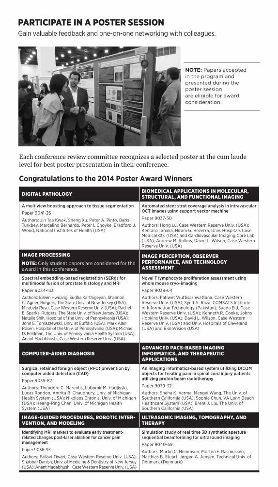
PARTICIPATE IN A POSTER SESSIONGain valuable feedback and one-on-one networking with colleagues.
DIGITAL PATHOLOGY BIOMEDICAL APPLICATIONS IN MOLECULAR, STRUCTURAL, AND FUNCTIONAL IMAGING
A multiview boosting approach to tissue segmentation
Paper 9041-26
Authors: Jin Tae Kwak, Sheng Xu, Peter A. Pinto, Baris Turkbey, Marcelino Bernardo, Peter L Choyke, Bradford J. Wood, National Institutes of Health (USA)
Automated stent strut coverage analysis in intravascular OCT images using support vector machine
Paper 9037-50
Authors: Hong Lu, Case Western Reserve Univ. (USA); Kentaro Tanaka, Hiram G. Bezerra, Univ. Hospitals Case Medical Ctr. (USA) and Cardiovascular Imaging Core Lab. (USA); Andrew M. Rollins, David L. Wilson, Case Western Reserve Univ. (USA)
IMAGE PROCESSING
NOTE: Only student papers are considered for the award in this conference.
IMAGE PERCEPTION, OBSERVER PERFORMANCE, AND TECHNOLOGY ASSESSMENT
Spectral embedding-based registration (SERg) for multimodal fusion of prostate histology and MRI
Paper 9034-133
Authors: Eileen Hwuang, Sudha Karthigeyan, Shannon C. Agner, Rutgers, The State Univ. of New Jersey (USA); Mirabela Rusu, Case Western Reserve Univ. (USA); Rachel E. Sparks, Rutgers, The State Univ. of New Jersey (USA); Natalie Shih, Hospital of the Univ. of Pennsylvania (USA); John E. Tomaszewski, Univ. at Buffalo (USA); Mark Alan Rosen, Hospital of the Univ. of Pennsylvania (USA); Michael D. Feldman, The Univ. of Pennsylvania Health System (USA); Anant Madabhushi, Case Western Reserve Univ. (USA)
Novel T lymphocyte proliferation assessment using whole mouse cryo-imaging
Paper 9038-64
Authors: Patiwet Wuttisarnwattana, Case Western Reserve Univ. (USA); Syed A. Raza, COMSATS Institute of Information Technology (Pakistan); Saada Eid, Case Western Reserve Univ. (USA); Kenneth R. Cooke, Johns Hopkins Univ. (USA); David L. Wilson, Case Western Reserve Univ. (USA) and Univ. Hospitals of Cleveland (USA) and BioInVision (USA)
COMPUTER-AIDED DIAGNOSISADVANCED PACS-BASED IMAGING INFORMATICS, AND THERAPEUTIC APPLICATIONS
Surgical retained foreign object (RFO) prevention by computer aided detection (CAD)
Paper 9035-82
Authors: Theodore C. Marentis, Lubomir M. Hadjiyski, Lucas Rondon, Amrita R. Chaudhury, Univ. of Michigan Health System (USA); Nikolaos Chronis, Univ. of Michigan (USA); Heang-Ping Chan, Univ. of Michigan Health System (USA)
An imaging informatics-based system utilizing DICOM objects for treating pain in spinal cord injury patients utilizing proton beam radiotherapy
Paper 9039-32
Authors: Sneha K. Verma, Mengyi Wang, The Univ. of Southern California (USA); Sophia Chun, VA Long Beach Healthcare System (USA); Brent J. Liu, The Univ. of Southern California (USA)
IMAGE-GUIDED PROCEDURES, ROBOTIC INTER-VENTION, AND MODELING
ULTRASONIC IMAGING, TOMOGRAPHY, AND THERAPY
Identifying MRI markers to evaluate early treatment-related changes post-laser ablation for cancer pain management
Paper 9036-93
Authors: Pallavi Tiwari, Case Western Reserve Univ. (USA); Shabbar Danish, Univ. of Medicine & Dentistry of New Jersey (USA); Anant Madabhushi, Case Western Reserve Univ. (USA)
Simulation study of real time 3D synthetic aperture sequential beamforming for ultrasound imaging
Paper 9040-59
Authors: Martin C. Hemmsen, Morten F. Rasmussen, Matthias B. Stuart, Jørgen A. Jensen, Technical Univ. of Denmark (Denmark)
NOTE: Papers accepted in the program and presented during the poster session are eligible for award consideration.
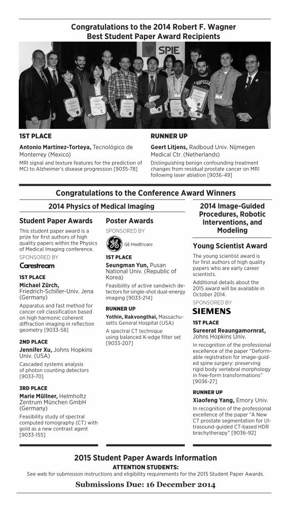
Congratulations to the 2014 Robert F. Wagner Best Student Paper Award Recipients
Congratulations to the Conference Award Winners
2015 Student Paper Awards InformationATTENTION STUDENTS:
See web for submission instructions and eligibility requirements for the 2015 Student Paper Awards.
Submissions Due: 16 December 2014
1ST PLACEAntonio Martínez-Torteya, Tecnológico de Monterrey (Mexico)MRI signal and texture features for the prediction of MCI to Alzheimer’s disease progression [9035-78]
RUNNER UPGeert Litjens, Radboud Univ. Nijmegen Medical Ctr. (Netherlands)Distinguishing benign confounding treatment changes from residual prostate cancer on MRI following laser ablation [9036-49]
Student Paper AwardsThis student paper award is a prize for first authors of high quality papers within the Physics of Medical Imaging conference.
SPONSORED BY
1ST PLACEMichael Zürch, Friedrich-Schiller-Univ. Jena (Germany)Apparatus and fast method for cancer cell classification based on high harmonic coherent diffraction imaging in reflection geometry [9033-58]
2ND PLACEJennifer Xu, Johns Hopkins Univ. (USA)Cascaded systems analysis of photon counting detectors [9033-70]
3RD PLACEMarie Müllner, Helmholtz Zentrum München GmbH (Germany)Feasibility study of spectral computed tomography (CT) with gold as a new contrast agent [9033-155]
Young Scientist AwardThe young scientist award is for first authors of high quality papers who are early career scientists.
Additional details about the 2015 award will be available in October 2014.
SPONSORED BY
1ST PLACESureerat Reaungamornrat, Johns Hopkins Univ.In recognition of the professional excellence of the paper “Deform-able registration for image-guid-ed spine surgery: preserving rigid body vertebral morphology in free-form transformations” [9036-27]
RUNNER UPXiaofeng Yang, Emory Univ.In recognition of the professional excellence of the paper “A New CT prostate segmentation for Ul-trasound-guided CT-based HDR brachytherapy” [9036-92]
Poster AwardsSPONSORED BY
1ST PLACESeungman Yun, Pusan National Univ. (Republic of Korea) Feasibility of active sandwich de-tectors for single-shot dual-energy imaging [9033-214]
RUNNER UPYothin, Rakvongthai, Massachu-setts General Hospital (USA)
A spectral CT technique using balanced K-edge filter set [9033-207]
2014 Physics of Medical Imaging 2014 Image-Guided Procedures, Robotic
Interventions, and Modeling
16 SPIE MEDICAL IMAGING 2015 • www.spie.org/micall2
By submitting an abstract, I agree to the following conditions:
ABSTRACT SUBMISSION
AN AUTHOR OR COAUTHOR (INCLUDING INVITED, ORAL, AND POSTER PRESENTERS) WILL:• Register at the reduced author registration rate
(current SPIE Members receive an additional discount on the registration fee).
• Attend the meeting.• Make the presentation as scheduled in the pro-
gram.• Submit a full-length manuscript (6 pages mini-
mum) for publication in the SPIE Digital Library, Proceedings of SPIE, and CD-ROM compilations.
• Obtain funding for their registration fees, travel, and accommodations, independent of SPIE, through their sponsoring organizations.
• Ensure that all clearances, including government and company clearance, have been obtained to present and publish. If you are a DoD contractor in the USA, allow at least 60 days for clearance.
Submit an abstract and summary online at: www.spie.org/micall2• Please submit a 250-word text abstract for techni-
cal review purposes that is suitable for publication. SPIE is authorized to circulate your abstract to conference committee members for review and selection purposes.
• Please also submit a 100-word text summary suitable for early release. If accepted, this sum-mary text will be published prior to the meeting in the online or printed programs promoting the conference.
• Identify the topics appropriate to the specific con-ference. During the submission process you will be asked to choose no more than three topics from a predefined list and/or add a topic not included on the list. (See individual conference Call for Papers for topic categories.)
• Prepare your 2-4 page supplemental MS Word or PostScript file. Supplemental file instructions can be found online. For full consideration this file must include the paper title, authors, 250-word abstract text, and the following supplemental information: - Description of purpose - Method(s) - Results - New or breakthrough work to be presented - Conclusions - Whether the work is being, or has been, submit-
ted for publication or presentation elsewhere, and, if so, indicate how the submissions differ.
- This file may contain supporting images/tables/figures
- Failure to follow these guidelines may disqualify your submission.
• Only original material should be submitted.• Abstracts should contain enough detail to clearly
convey the approach and the results of the re-search.
• Commercial papers, papers with no new research/development content, and papers where support-ing data or a technical description cannot be given for proprietary reasons will not be accepted for presentation in this conference.
• Please do not submit the same, or similar, ab-stracts to multiple conferences.
REVIEW, NOTIFICATION, AND PROGRAM PLACEMENT INFORMATION• To ensure a high-quality conference, all submis-
sions will be assessed by the Conference Chair/Editor for technical merit and suitability of con-tent.
• Conference Chair/Editors reserve the right to reject for presentation any paper that does not meet content or presentation expectations.
• The contact author will receive notification of acceptance and presentation details by e-mail no later than 24 October 2014.
• Final placement in an oral or poster session is subject to the Chairs’ discretion.
PROCEEDINGS OF SPIE AND SPIE DIGITAL LIBRARY INFORMATION• Manuscript instructions are available from the
“For Authors/Presenters” link on the conference website.
• Conference Chair/Editors may require manuscript revision before approving publication and reserve the right to reject for publication any paper that does not meet acceptable standards for a scientif-ic publication. Conference Chair/Editors’ decisions on whether to allow publication of a manuscript is final.
• Authors must be authorized to transfer copyright of the manuscript to SPIE, or provide a suitable publication license.
• Only papers presented at the conference and received according to publication guidelines and timelines will be published in the conference Proceedings of SPIE and SPIE Digital Library.
• Published papers are indexed in leading scientific databases including Astrophysical Data System (ADS), Chemical Abstracts (relevant content), Compendex, CrossRef, Current Contents, Deep-Dyve, Google Scholar, Inspec, Portico, Scopus, SPIN, and Web of Science Conference Proceed-ings Citation Index, and are searchable in the SPIE Digital Library. Full manuscripts are available to SPIE Digital Library subscribers worldwide.
2014 SYMPOSIUM CHAIRS:
DATESConferences & Courses: 21-26 February 2015
NEW LOCATIONRenaissance Orlando at Sea World Orlando, Florida, USA
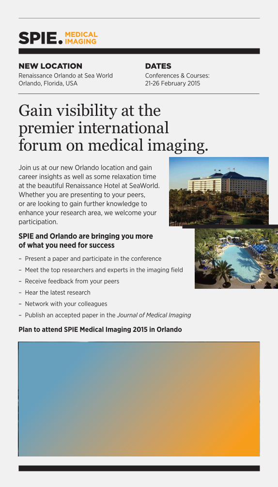
Gain visibility at the premier international forum on medical imaging.Join us at our new Orlando location and gain career insights as well as some relaxation time at the beautiful Renaissance Hotel at SeaWorld . Whether you are presenting to your peers, or are looking to gain further knowledge to enhance your research area, we welcome your participation .
SPIE and Orlando are bringing you more of what you need for success
– Present a paper and participate in the conference
– Meet the top researchers and experts in the imaging field
– Receive feedback from your peers
– Hear the latest research
– Network with your colleagues
– Publish an accepted paper in the Journal of Medical Imaging
Plan to attend SPIE Medical Imaging 2015 in Orlando
Non-
Profi
t Org
.U.
S. P
osta
ge
Paid
SPIE
21-2
6
FEB
RU
ARY
20
15
SUB
MIT
YO
UR
A
BST
RA
CT
TOD
AY
•
NEW
LO
CA
TIO
N IN
20
15R
enai
ssan
ce O
rlan
do a
t Se
a W
orld
O
rlan
do, F
lori
da, U
SA
ww
w.s
pie
.org
/mic
all2
Pla
n to
par
tici
pate
in th
e co
nfe
ren
ce w
her
e th
e la
test
info
rmat
ion
is p
rese
nte
d in
dig
ital
pa
thol
ogy,
tom
ogra
phy,
imag
e pr
oces
sin
g,
obse
rver
per
form
ance
, im
age
regi
stra
tion
, in
form
atic
s, im
age
segm
enta
tion
, com
pute
r-ai
ded
diag
nos
is, a
nd
ult
raso
un
d.
P.O
. Box
10
Bel
lingh
am, W
A 9
8227
-001
0 U
SA