Small compounds for 'targeted radiotherapy
112
Dissertation for the degree of Doctor Scientiarum Ellen Mengshoel Brevik Small compounds for 'targeted radiotherapy DEPARTMENT OF CHEMISTRY FACULTY OF MATHEMATICS AND NATURAL SCIENCES UNIVERSITY OF OSLO 03/2007
Transcript of Small compounds for 'targeted radiotherapy

Dissertation for the degreeof Doctor Scientiarum
Ellen Mengshoel Brevik
Small compounds for'targeted radiotherapy
DEPARTMENT OF CHEMISTRYFACULTY OF MATHEMATICSAND NATURAL SCIENCESUNIVERSITY OF OSLO 03/2007

© Ellen Mengshoel Brevik, 2007
Series of dissertations submitted to the Faculty of Mathematics and Natural Sciences, University of Oslo.No. 634
ISSN 1501-7710
All rights reserved. No part of this publication may be reproduced or transmitted, in any form or by any means, without permission.
Cover: Inger Sandved Anfinsen. Printed in Norway: AiT e-dit AS, Oslo, 2007.
Produced in co-operation with Unipub AS. The thesis is produced by Unipub AS merely in connection with the thesis defence. Kindly direct all inquiries regarding the thesis to the copyright holder or the unit which grants the doctorate.
Unipub AS is owned by The University Foundation for Student Life (SiO)

Summary
This thesis describes efforts in synthesising and investigating new tumour-seeking agents fortargeted radiotherapy of malignant melanomas and bone-related cancers. The concept oftargeted radiotherapy is presented, and selection criteria for therapeutic radionuclides arediscussed. Emphasis has been given to the heavier radiohalogens (radioiodine isotopes and211At) and their labelling chemistry.
Three classes of melanoma-seeking agents have been treated; phenothiazines, cyste-aminylphenols and benzamides. Stannylated derivatives of the two latter groups were success-fully synthesised and radiohalogenated, resulting in six potential melanoma-seeking agentslabelled with radioiodine or 211At. Melanoma affinity and in vivo stability were investi-gated in mice bearing the human melanoma xenografts HHMSX (pigmented) or SESX (non-pigmented). Four derivatives showed high stability to enzymatic dehalogenation in vivo,but only the radioiodinated benzamide [125I]IMBA demonstrated promising tumour accu-mulation and retention in the melanoma models. The results supports previous suggestionsof using [∗I]IMBA in scintigraphic imaging of pigmented and non-pigmented melanomas.[∗I]IMBA may also have a future in scintigraphic imaging of other cancers expressing a highdensity of sigma-receptors.
The bone-seeking iodobisphosphonate [∗I]HIPEBA was labelled with 123I, and its po-tential for clinical applications in humans was investigated in young pigs. Initial phar-macokinetic properties were studied using dynamic and static gamma-camera imaging,showing superior bone uptake and negligible renal excretion of [123I]HIPEBA compared to99mTc-MDP. Estimated radiation doses in humans indicated similar radiotherapeutic prop-erties for [131I]HIPEBA and 153Sm-EDTMP. Hence, [∗I]HIPEBA is a promising candidate fordiagnostic imaging and targeted radiotherapy of osseous lesions, and further evaluations inhumans are justified.
The thesis gives an extended overview of the work, presenting additional experiments andresults to those described in the papers.
i

ii

List of papers
I E. M. Brevik, E. Arstad, P. Hoff.An improved synthesis of an 125I- and 211At-labelled benzamide for melanoma imaging.Proceedings of the 14th Radiochemical Conference, Marianske Lazne 14–19 April 2002,Czech Republic. Czech. J. Phys. 53 (2003), Suppl.A A725–A729
II E. M. Brevik, E. Arstad, P. Hoff.Synthesis and biodistribution of N -(2-diethylaminoethyl)-3-[211At]astato-4-methoxy-benzamide for targeted radiotherapy of malignant melanoma.Manuscript. To be submitted to Melanoma Research.
III E. M. Brevik, E. Arstad, F. O. Levy, P. Hoff.Evaluation of 125I- and 211At-labelled benzamides for targeting of σ1-receptors in anamelanotic melanoma xenograft.Manuscript. To be submitted to Melanoma Research.
IV E. M. Brevik, K. H. Holm, D. S. Wilbur, D. K. Hamlin, P. Hoff.Syntheses and preliminary biodistribution studies of radioiodinated cysteaminylphenolderivatives for malignant melanoma.Manuscript. To be submitted to Nuclear Medicine and Biology.
V E. M. Brevik, A. Skretting, S. Bruheim, T. Bach-Gansmo, E. Arstad, P. Hoff.Pharmacokinetic properties and bone surface uptake of 1-hydroxy-(m-[123I]iodo-phenyl)ethylidene-1,1-bisphosphonic acid (123I-HIPEBA) in pigs: implications for tar-geted radiotherapy.Manuscript. To be submitted to Nuclear Medicine Communications.
iii

iv

Acknowledgements
The present work was carried out at the Nuclear Chemistry group, Department of Chemistry,University of Oslo, in collaboration with scientists at the Norwegian Radium Hospital andDepartment of Radiation Oncology, University of Washington, Seattle, USA. The study wasinitiated as part of a master degree. Post-graduate work was made possible through a grantfrom the Norwegian Research Council which is greatfully acknowledged.
I would like to express my gratitude and thanks to my supervisors Professor Per Hoff, forinitiating this doctoral thesis and allowing me to pursue my diverse ideas (whether fruitfulor not), and Chief scientist Kjetil H. Holm, for your chemical expertise, enthusiasm anddedication in solving the obstacles which emerged during organic syntheses.
I deeply appreciate the contributions from Professor Arne Skretting, for your expertiseand guidance in medical physics and nuclear medicine, and Professor D. Scott Wilbur, foryour great hospitality, interesting discussions and inspiring enthusiasm for this project.
Thanks are also due to:Don K. Hamlin, for your friendship and helpfulness in every way. Erik Arstad, for includingme in the bisphosphonate project and for accurate and quick guidance. Skjalg Bruheimand staff at the Norwegian Radium Hospital, for all help and assistance with the animalexperiments. Kurt Allan Krobert, my right-hand and instructor in pharmacology. EivindAtle Olsen, for production of 211At at the OCL cyclotron.
I am greatful to collegues and fellow students in the Nuclear Chemistry group, in particularHavar, Liv and Jorolf, for your friendship and support over many years.
Thanks to my family and friends, for reminding me of life outside Blindern and for backingme up in doing this ”cancer stuff”.
Finally, my profound gratitude goes to Havar and Eirik, for your endless love, supportand patience.
Ellen Mengshoel Brevik
v

vi

Contents
Summary . . . . . . . . . . . . . . . . . . . . . . . . . . . . . . . . . . . . . . . . . iList of papers . . . . . . . . . . . . . . . . . . . . . . . . . . . . . . . . . . . . . . . iiiAcknowledgements . . . . . . . . . . . . . . . . . . . . . . . . . . . . . . . . . . . . vContents . . . . . . . . . . . . . . . . . . . . . . . . . . . . . . . . . . . . . . . . . . vii
I INTRODUCTION 1
1 Introduction 3
2 Background 5
2.1 Targeted radiotherapy of cancer . . . . . . . . . . . . . . . . . . . . . . . . . . 52.2 Therapeutic radionuclides and radiobiology . . . . . . . . . . . . . . . . . . . 5
2.2.1 Radionuclides emitting β-particles . . . . . . . . . . . . . . . . . . . . 62.2.2 Radionuclides emitting α-particles . . . . . . . . . . . . . . . . . . . . 62.2.3 Radionuclides emitting low-energy electrons . . . . . . . . . . . . . . . 82.2.4 Radionuclide pairs for imaging and radiotherapy . . . . . . . . . . . . 8
2.3 Radiohalogens . . . . . . . . . . . . . . . . . . . . . . . . . . . . . . . . . . . . 82.3.1 Important radioiodine isotopes . . . . . . . . . . . . . . . . . . . . . . 92.3.2 Astatine-211 . . . . . . . . . . . . . . . . . . . . . . . . . . . . . . . . 9
2.4 Radiohalogen labelling chemistry . . . . . . . . . . . . . . . . . . . . . . . . . 102.4.1 Direct radiolabelling of small aromatic compounds . . . . . . . . . . . 112.4.2 Indirect radiolabelling of small aromatic compounds . . . . . . . . . . 11
2.5 Structure determination of radioiodinated and astatinated compounds . . . . 13
II MALIGNANT MELANOMA 15
3 Malignant melanoma 17
3.1 Origin and subtypes of malignant melanoma . . . . . . . . . . . . . . . . . . . 17
vii

viii CONTENTS
3.2 Melanogenesis . . . . . . . . . . . . . . . . . . . . . . . . . . . . . . . . . . . . 183.3 Sigma-receptors . . . . . . . . . . . . . . . . . . . . . . . . . . . . . . . . . . . 183.4 Radiolabelled melanoma-seeking agents . . . . . . . . . . . . . . . . . . . . . 20
3.4.1 Phenothiazine derivatives . . . . . . . . . . . . . . . . . . . . . . . . . 203.4.2 Cysteaminylphenol derivatives . . . . . . . . . . . . . . . . . . . . . . 223.4.3 Benzamide derivatives . . . . . . . . . . . . . . . . . . . . . . . . . . . 23
3.5 Aims of study . . . . . . . . . . . . . . . . . . . . . . . . . . . . . . . . . . . . 24
4 General methods 25
4.1 Radioactivity measurements . . . . . . . . . . . . . . . . . . . . . . . . . . . . 254.2 Production and distillation of 211At . . . . . . . . . . . . . . . . . . . . . . . . 254.3 Chromatography . . . . . . . . . . . . . . . . . . . . . . . . . . . . . . . . . . 264.4 Animals . . . . . . . . . . . . . . . . . . . . . . . . . . . . . . . . . . . . . . . 274.5 Tumour models . . . . . . . . . . . . . . . . . . . . . . . . . . . . . . . . . . . 274.6 Statistics . . . . . . . . . . . . . . . . . . . . . . . . . . . . . . . . . . . . . . 27
5 Phenothiazine derivatives 29
5.1 Introduction . . . . . . . . . . . . . . . . . . . . . . . . . . . . . . . . . . . . . 295.2 Results and discussion . . . . . . . . . . . . . . . . . . . . . . . . . . . . . . . 30
6 Cysteaminylphenol derivatives 33
6.1 Introduction . . . . . . . . . . . . . . . . . . . . . . . . . . . . . . . . . . . . . 336.2 Chemistry . . . . . . . . . . . . . . . . . . . . . . . . . . . . . . . . . . . . . . 34
6.2.1 Synthesis . . . . . . . . . . . . . . . . . . . . . . . . . . . . . . . . . . 346.2.2 Radiolabelling and purification . . . . . . . . . . . . . . . . . . . . . . 41
6.3 Biology . . . . . . . . . . . . . . . . . . . . . . . . . . . . . . . . . . . . . . . 416.3.1 Preliminary biodistribution studies . . . . . . . . . . . . . . . . . . . . 416.3.2 Biodistribution studies in the HHMSX model . . . . . . . . . . . . . . 42
6.4 Results and discussion . . . . . . . . . . . . . . . . . . . . . . . . . . . . . . . 42
7 Benzamide derivatives 47
7.1 Introduction . . . . . . . . . . . . . . . . . . . . . . . . . . . . . . . . . . . . . 477.2 Chemistry . . . . . . . . . . . . . . . . . . . . . . . . . . . . . . . . . . . . . . 47
7.2.1 Synthesis . . . . . . . . . . . . . . . . . . . . . . . . . . . . . . . . . . 477.2.2 Radiolabelling and purification . . . . . . . . . . . . . . . . . . . . . . 48
7.3 Biology . . . . . . . . . . . . . . . . . . . . . . . . . . . . . . . . . . . . . . . 497.3.1 Biodistribution studies in the HHMSX model . . . . . . . . . . . . . . 497.3.2 Biodistribution studies in the SESX model . . . . . . . . . . . . . . . 49
CONTENTS ix
7.4 Pharmacology . . . . . . . . . . . . . . . . . . . . . . . . . . . . . . . . . . . . 49
7.5 Results and discussion . . . . . . . . . . . . . . . . . . . . . . . . . . . . . . . 50
8 Malignant melanoma: Future challenges 55
III BONE-RELATED CANCER 57
9 Bone-related cancer 59
9.1 Bone, osteosarcoma and bone metastasis . . . . . . . . . . . . . . . . . . . . . 59
9.2 Bone-seeking radiopharmaceuticals . . . . . . . . . . . . . . . . . . . . . . . . 60
9.2.1 Bisphosphonates . . . . . . . . . . . . . . . . . . . . . . . . . . . . . . 61
9.3 Aim of study . . . . . . . . . . . . . . . . . . . . . . . . . . . . . . . . . . . . 62
10 Methods 63
10.1 Radioactivity measurements . . . . . . . . . . . . . . . . . . . . . . . . . . . . 63
10.2 Chromatography . . . . . . . . . . . . . . . . . . . . . . . . . . . . . . . . . . 63
10.3 Animals . . . . . . . . . . . . . . . . . . . . . . . . . . . . . . . . . . . . . . . 63
10.4 The FNOMIP method . . . . . . . . . . . . . . . . . . . . . . . . . . . . . . . 64
10.5 The OLINDA/EXM code . . . . . . . . . . . . . . . . . . . . . . . . . . . . . 64
11 Bisphosphonate derivatives 65
11.1 Introduction . . . . . . . . . . . . . . . . . . . . . . . . . . . . . . . . . . . . . 65
11.2 Chemistry . . . . . . . . . . . . . . . . . . . . . . . . . . . . . . . . . . . . . . 65
11.3 Biology . . . . . . . . . . . . . . . . . . . . . . . . . . . . . . . . . . . . . . . 66
11.4 Imaging and analysis . . . . . . . . . . . . . . . . . . . . . . . . . . . . . . . . 66
11.5 Radiation dose estimates in humans . . . . . . . . . . . . . . . . . . . . . . . 66
11.6 Results and discussion . . . . . . . . . . . . . . . . . . . . . . . . . . . . . . . 67
12 Bone-related cancer: Future prospects 69
REFERENCES 71
IV APPENDIX 83
A Chemical abbreviations 85
B Glossary of radiochemical and medical terms 86

x CONTENTS
C Additional spectroscopic data 87
D Biodistribution data 88
List of Figures 97
List of Tables 99

Part I
INTRODUCTION
1

2

Chapter 1
Introduction
The prognosis of surviving cancer has improved over the years. However, only 50 % of Nor-wegian patients are alive five years post-diagnosis [1]. Hence, development of more efficientcancer treatments is a major challenge in cancer research.
Successful curative therapy requires complete removal or destruction of malignant cells.However, for many malignancies there are no effective treatment for patients with exten-sive metastatic spread of the disease. Advances and research in alternative and improvedtreatment methods for metastatic cancer disease are highly required.
Systemic targeted radiotherapy is a promising modality for treatment of disseminated can-cers. The concept is based on selective irradiation of malignant cells by means of radionuclidesattached to ”tumour-seeking” molecules. Interest in this treatment modality has increasedover the years due to new developments of available radionuclides and carrier molecules.
In this thesis focus has been on developing new radiopharmaceuticals for targeted radio-therapy of two different forms of cancer where standard treatment modalities have failedor have had limited success. Part I presents therapeutic radionuclides for targeted radio-therapy, with emphasis on radioiodine isotopes, the α-emitter astatine-211 and their labellingchemistry. Part II presents the efforts in developing new radiohalogenated melanoma-seekingagents for diagnosis and targeted radiotherapy of metastatic malignant melanomas. Part IIIconcentrates on the investigation of a radioiodinated bisphosphonate with potential in pallia-tive treatment and targeted radiotherapy of osteosarcoma and bone metastases.
3

4 Introduction

Chapter 2
Background
2.1 Targeted radiotherapy of cancer
When tumours or metastases cannot be removed by surgery, the conventional therapeuticapproach is external beam radiotherapy and chemotherapy. However, these treatments showlow selectivity for cancer cells and tumour tissues, and might be damaging or cytotoxic tohealthy tissues when applied at curative levels.
An attractive alternative is to exploit a biological or chemical characteristic of the specificcancer or the affected tissue. In favourable cases, molecules with affinity for a specialisedbiological feature may function as specific, ”tumour-seeking” agents (e.g. antibodies, anti-body fragments, peptides, small molecules with tumour affinity). Targeted radiotherapy in-volves such agents incorporating a suitable therapeutic radionuclide, ideally giving site-specificradiation treatment of the primary tumour and its metastases while producing minimal ra-diation damage to surrounding normal tissues [2]. Thus, the choice of carrier molecule andradionuclide must be adjusted to the type of tumour or metastasis with regard to biologicalproperties, size, geometry and localisation relative to dose-limiting organs.
2.2 Therapeutic radionuclides and radiobiology
Important criteria in the choice of suitable therapeutic radionuclides include physical, chemi-cal, biological and economical aspects. Methods for rapid and specific labelling chemistryproducing radioactive agents with a chemically stable label are crucial. The half-life of theradionuclide must allow time for chemistry, purification, logistics and maximal (intra)cellularretention when distributed in vivo. The energy deposition of the emitted radiation mustbe considered, and the daughter nuclide(s) should have properties minimising the radiationburden to healthy tissues. The selection of therapeutic radionuclides is furthermore limited
5

6 Background
by cost and availability [3].After considering these restrictions, a suitable therapeutic radionuclide is essentially cho-
sen due to the quality of the emitted radiation expressed by the linear energy transfer (LET).In terms of causing a biological effect (irreparable damages to DNA, cell death), the focusis set on ionising radiation from radionuclides emitting charged particles [4]. Particles withhigh ionisation densities (high LET-values) have a direct interaction with tissue, inducing irre-pairable double strand breaks in DNA. This effect is optimal for particles having a LET-valueabout 100 keV/μm, i.e. when the distance between ionising events along the particle trackcoincides with the distance between the strings of the DNA double helix [5]. Particles withlow LET-values predominantly have an indirect interaction with tissue caused by radiation-induced radicals. Thus, the probability of inducing irreparable double strand breaks in DNAis strongly reduced. However, the effect of low-LET radiation depends not only on the appliedradiation dose, but also on dose rate, number of dose fractions, the cell cycle and the use ofradioprotectors and radiosensitisers [5].
Clinical radionuclide therapy has so far been limited to a few β−-emitting radionuclidesand a few types of tumours [2, 6].
2.2.1 Radionuclides emitting β-particles
The majority of current clinical applications in targeted radiotherapy involve the β−-emitters131I, 32P, 90Y and 89Sr [7, 8]. The β-particles have low LET-values (range 0.2–1.0 keV/μm)and a modest relative biological effectiveness (RBE). However, β-emitters are readily availableat low cost and offer a wide choice of candidates in terms of particle energies and chemicalproperties. The emitted β-particles have a tissue range of several millimeters (Table 2.1)and are well suited for treatment of larger tumour masses and tumours with a heterogenousuptake (through crossfire irradiation). The use of highly energetic β-particles is restricted bythe radiation burden and cell damages to surrounding healthy tissues, and especially by thesuppression of activity in red bone marrow.
2.2.2 Radionuclides emitting α-particles
The use of α-emitting radionuclides has a major advantage in radiotherapy of cancer. Theα-particles have mean LET-values of 80–100 keV/μm, close to maximal RBE and a cell-killingefficiency independent of biological and chemical factors [5]. The high cytotoxicity and shorttissue range (30–100 μm) restrict the cell-killing to a few cells surrounding the α-emittingradionuclide [9]. Thus, a carrier molecule incorporating a suitable α-emitter might induce aradiotherapeutic effect on tiny clusters of cancer cells, micrometastases and single-cell diseases,minimising the radiation doses to red bone marrow and healthy tissues.

2.2 Therapeutic radionuclides and radiobiology 7
Table 2.1: Data on some potential radionuclides for targeted radiotherapy.Nuclide Half- Decay Eβ max Eα max γ-energy Mean particle
life modea (MeV) (MeV) (keV)b tissue range32P 14.26 d β− 1.7 - - ∼ 3.0 mm89Sr 50.5 d β− 1.5 - (909) ∼ 2.4 mm90Y 64.1 h β− 2.3 - (2186) ∼ 3.6 mm117mSn 13.6 d e− - - 159 0.3 mmc
125I 59.41 d ECd, e− - - 35 ∼ 1 μm131I 8.02 d β− 0.6 - 364 ∼ 0.7 mm149Tb 4.1 h EC, α, β+ 1.8 3.97 352 ∼ 28 μme
153Sm 46.27 h β− 0.7 - 103 ∼ 0.8 mm166Ho 26.8 h β−, e− 1.9 - 81 ∼ 3.2 mm177Lu 6.71 d β− 0.5 - 208 ∼ 0.5 mm186Re 89.25 h β−, EC 1.1 - 137 ∼ 1.1 mm188Re 16.98 h β− 2.1 - 155 ∼ 3.4 mm211At 7.22 h EC, α - 5.87 (687) 55–80 μm212Bi 60.6 min β−, α 2.3 6.05 727 50–90 μme
213Bi 45.59 min β−, α 1.4 5.87 440 50–90 μme
223Ra 11.43 d α - 5.72 269 < 100 μm224Ra 3.66 d α - 5.69 241 < 100 μm225Ac 10.0 d α, e− - 5.83 100 < 100 μme
227Th 18.7 d α, e− - 6.04 236 < 100 μme
255Fm 20.1 h α, sf, e− - 7.02 (81) ∼ 100 μme
a EC = electron capture, e− = conversion electrons, β− = electron emission,
β+ = positron emission, α = alpha emission, sf = spontaneous fission.
b Intensities <1 % are given in brackets.
c Conversion electrons emitted by 117mSn are monoenergetic (0.13 MeV).
d Abundance of Auger-electrons >100 %.
e The α-particle range.

8 Background
Only a few α-emitters are suitable for targeted radiotherapy of cancer, e.g. 211At, 212Bi,213Bi, 223Ra, 224Ra, 225Ac, 227Th, 255Fm and 149Tb (Table 2.1) [9, 10]. Among these, only211At, 212Bi, 213Bi and 223Ra have been extensively evaluated in vivo for use in cancer therapy.The bismuth-isotopes are bound to chelating agents by complexation [11, 12], while 223Rasimply is used as 223RaCl2 [13]. Astatine is chemically quite similar to iodine and can beincorporated into an aromatic compound by covalent bonding [14–16]. The isotope 211At hasbeen regarded as the most promising and versatile α-emitter studied for radiotherapy of cancerso far [3, 15].
2.2.3 Radionuclides emitting low-energy electrons
Other therapeutical alternatives are radionuclides emitting Auger- or low-energy conversionelectrons (Table 2.1). These particles are characterised by a high ionisation density overa very short range in tissue; ∼0.3 mm for conversion electrons and subcellular (∼1 μm) forAuger-electrons [2, 17–19]. Thus, a maximal radiobiological effect is obtained when theAuger-electron emitters are located within the nuclei of cancer cells.
A future possibility in targeted radiotherapy is to combine the efficient single-cell killingproperties of α-particles and DNA-associated Auger-emitters, with β-particles eliminatinglarger malignancies mainly by crossfire irradiation.
2.2.4 Radionuclide pairs for imaging and radiotherapy
The dosimetry of radionuclide therapy depends on quantitative in vivo uptake measurementsof the therapeutic agents. Pre-therapy imaging, particularly positron emission tomography(PET), can improve the accuracy of such estimates provided that two radionuclides of thesame element are used successively for imaging and radiotherapy. Examples of such radio-nuclide pairs for PET-imaging/radiotherapy are 83Sr/89Sr, 86Y/90Y and 124I/131I. The radio-nuclide 64Cu is also very interesting as it combines β+-, β−- and Auger-electron emissions inits decay [7].
2.3 Radiohalogens
Several radiohalogens are important in the development of new radiopharmaceuticals aimingat diagnostic imaging or radiotherapy [16]. Radiohalogens emitting high yields of photonswith an energy of 100–400 keV (e.g. 123I) may be used in single photon emission computedtomography (SPECT). The positron-emitting radiohalogens 18F, 75Br, 76Br and 124I areinteresting for PET-imaging [20], while radiohalogens emitting α-particles, β−-particles or

2.3 Radiohalogens 9
Auger-electrons have properties suitable for radiotherapeutic applications. In this thesis, theradionuclides 123I, 124I, 125I, 131I and 211At deserve special attention.
2.3.1 Important radioiodine isotopes
The 123I (t1/2 13.2 h) decays through electron capture with emission of 159 keV γ-rays, result-ing in almost optimal gamma-camera performance in SPECT. This radionuclide also emitsAuger-electrons, thus it may be an attractive candidate for radiotherapeutic applications.
The 124I (t1/2 4.15 d) disintegrates by electron capture and emission of up to 2.13 MeV β+-particles. The radionuclide benefits from a longer half-life than most standard PET-isotopes,but high-energy positrons might result in loss of spatial resolution in PET-images compared to18F. Emission of high-energy γ (>603 keV) may be a limiting factor for clinical applications.
The 125I (t1/2 59.41 d) disintegrates by electron capture, emitting 35 keV γ and conversionelectrons. The emission of Auger-electrons makes 125I an interesting radionuclide for micro-tumour therapy, but the physical half-life is too long for clinical applications. Hence, thisradionuclide is mainly used preclinically due to its easy handling and storage.
The 131I (t1/2 8.02 d) disintegrates by β−-decay (0.6 MeV) with a main accompanyingγ-energy of 364 keV. The radionuclide is considered suitable for therapeutic applications, butit is not ideal for imaging purposes due to the radiation dose caused by the emitted β−-particles. However, 131I is the most commonly used radionuclide in radiotherapy, particularlyin the treatment of thyroid cancers and non-malignant thyroid disorders.
2.3.2 Astatine-211
The α-emitting radionuclide 211At (t1/2 7.22 h) disintegrates following a branched decayscheme as illustrated in Figure 2.1. In the first branch, the 211At decays by emission of5.87 MeV α-particles to 207Bi (t1/2 31.55 y), which disintegrates by electron capture to stable
211Po207Bi
211At
207Pb
0.52 s
7.22 h
31.55 y
stable
58% EC
100% alphaEC (beta+)
42% alpha
Ealpha = 7.45 MeV
Ealpha = 5.87 MeV
Figure 2.1: Decay scheme for 211At.

10 Background
207Pb. The second branch involves decay by electron capture to 211Po (t1/2 0.52 s), followedby an emission of 7.45 MeV α-particles giving stable 207Pb. Hence, the mean energy of theemitted α-particles is 6.7 MeV, resulting in a mean LET value of 97 keV/μm and a tissuerange corresponding to 55–80 μm from the point of decay. This high LET-value and shorttissue range make 211At particularly attractive for radiotherapy of micrometastatic cancers orin applications where single cells are targeted. However, diffusion of the 211Po daughter mightreduce the total tumour radiation dose [21]. Decay of 211Po results in polonium K X-rays(77–92 keV) which enable sample-detection as well as external imaging using SPECT. Thegeneral use of astatine is limited by the lack of long-lived astatine-isotopes and few productionfacilities for 211At.
2.4 Radiohalogen labelling chemistry
Several requirements have to be fulfilled when labelling a compound with a radionuclide. Ingeneral, the labelling chemistry should be fast, regiospecific, reproducible and give a stable,labelled product in high radiochemical yield. With short-lived radionuclides, labelling in thelast reaction step is often advantageous, reducing the loss of radioactivity, time for synthesisand purification as well as exposure. The specific radioactivity of the product should normallybe as high as possible (starting with no-carrier-added radionuclides), and the compoundshould retain its original biological properties after radiolabelling. Radiolysis has to be takeninto account in the labelling procedure, particularly when using high-LET radionuclides like211At [22, 23]. The in vivo stability of the radiolabelled product is of paramount importanceif the substance is to be used as a radiopharmaceutical. Thus, the choice of labelling methodis crucial for an optimal production of a radioactive preparation.
Most radiohalogenations (except for fluorinations) are conducted as electrophilic substitu-tion reactions. Electrophilic reactions require formation of an electrophilic radiohalogenationreagent through oxidation of halide ions:
X− � Xδ+
Electrophilic halogen species can be obtained in situ by using various oxidising agents,e.g. chloramine-T, tert -butyl-hydroperoxide (TBHP), N -chlorosuccinimide (NCS) andN -iodosuccinimide (NIS) [24].

2.4 Radiohalogen labelling chemistry 11
2.4.1 Direct radiolabelling of small aromatic compounds
Strongly activated aryl compounds (e.g. phenols, anilines and N -alkyl-anilines) are readilyradiohalogenated via a direct electrophilic halogenation using an appropriate oxidising agent:
Ar–H + ∗Xδ+–Xδ− → Ar–∗X + H–X where X = Cl, Br, I or
Ar–H + ∗X+–A− → Ar–∗X + H–A
where ∗X is the radiohalogen and A is a more electronegative group than ∗X. Theradiolabelling reaction will primarily give monohalogenated products with the radiohalogenincorporated ortho or para to the activating substituent. Direct halogenation may alsobe used for moderately activated aryl compounds, but the radiochemical yields might besignificantly reduced due to side reactions.
Direct electrophilic radioiodinations are very common, giving aryliodides with high chemi-cal stability [24, 25]. Direct electrophilic fluorinations, chlorinations and brominations requireharsh oxidising conditions, while astatinations give products with poor stability due to weakcarbon–astatine bonds [26]. For example, the phenolic ring is too activated to produce sta-ble compounds with astatine [25]. A limitation of this labelling method is the formation ofisomeric product mixtures which may be difficult to separate.
2.4.2 Indirect radiolabelling of small aromatic compounds
Radiohalogens can be labelled regiospecifically into moderately or non-activated aromaticrings using indirect labelling methods. The two most common methods include incorporationof radiohalogens via decomposition of aryl diazonium salts or via organometallic intermedi-ates [24, 25]. For labelling reactions with radioiodine and 211At, the use of organometallicprecursors or compounds incorporating an organoboron cage are preferred [27].
Halodemetallations are used for regiospecific labelling of a substrate molecule at theorganometallic site, giving radiohalogenated products in high radiochemical yields using mildlabelling conditions and short reaction times. Different organometallic intermediates havebeen studied, e.g. organothallium compounds, organomercury compounds, organostannanes,organosilanes and organogermanes. The outcome of a halodemetallation is affected by themetal, chemical labelling conditions as well as steric factors [14, 24].

12 Background
Halodemetallation with group 14 metals
Organometallic precursors containing a group 14 metal (Si, Ge, Sn) are advantageous whenlabelling with radiohalogens [14, 24, 28]. Aryltrialkylmetal precursors facilitate the no-carrier-added syntheses of radiolabelled arylhalides, described by the reaction mechanism in Figure2.2:
X
A
A R3MA
XMR3X
MR3
+ ++
-
- 1
-12
Figure 2.2: Proposed reaction mechanism of the halodemetallation using aryltrialkyl group14 organometallic precursors, from Refs. [14, 24].
where X+–A− is a halonium-anion species, and M is silicon, germanium or tin. The alkylsubstituent R introduces a +I inductive effect1 facilitating formation of the σ-complex inter-mediate, while electron release by the -MR3 group decreases the possibility of competitivehalodeprotonations. An alternative and competing reaction mechanism goes via a radical-ion-pair intermediate which collapses to the same σ-complex as shown in Figure 2.2 [29].
The carbon–metal bond dissociation energy and the covalent radius of the metal decreasein the order Si < Ge < Sn, resulting in increased reaction rates and increased radiochemicalyields of the labelled arylhalides. While the radiochemical yield of a halodestannylationis more or less unaffected by the labelling conditions, the efficiency of a halodesilylationis influenced by the choice of solvent, oxidising agent, radionuclide, alkyl substituent R,substituent(s) on the aromatic ring and pH [14, 30]. However, high radiochemical yieldsmay also be obtained for organosilicon compounds by optimising the labelling conditions. Ingeneral, the use of silylated compounds have an advantage due to low toxicity and greaterchemical inertness during multi-step syntheses.
The required aryltrialkylmetal precursors can often be synthesised from lithiated organiccompounds. Radiohalogenated derivatives are produced when treating this precursor with aradiohalogen using a mild oxidising agent.
1The term +I is used for functional groups being electron-donating relative to hydrogen.

2.5 Structure determination of radioiodinated and astatinated compounds 13
2.5 Structure determination of radioiodinated and astatinated
compounds
Structural assignment of a radioiodinated or astatinated compound can be predicted witha good degree of certainty from the expected product of the corresponding non-radioactiveiodination [24, 28]. If the starting compound contains an activated aromatic ring (e.g. phenolor aniline) and is treated with electrophilic radioiodine, the location of the radioactive label inthe product is a function of the +I and +M effects2 of the aromatic substituent(s) [31, 32]. Ifthe compound is labelled by a substitution reaction, the label should appear in the place of theleaving group (e.g. in iodine for bromine exchange, iododediazonisation or iododemetallation).
These structural assignments are made on the basis of analogy with non-radioactive re-actions. However, reactions with no-carrier-added radionuclides may proceed very differentlydue to the drastic change in ratios between reagents and the higher possibility of side effectsproduced by minor impurities in the reaction mixture. To analyse a radioiodinated or astati-nated compound, its behaviour in various forms of chromatography is measured and comparedto the non-radiolabelled, iodinated compound. Chromatographic investigations are normallyperformed by radio-HPLC [14, 28].
2The terms +I and -I describe field effects operating through space, solvent molecules or σ-bonds. The
terms +M and -M describe resonance effects operating through π-electrons.

14 Background

Part II
MALIGNANT MELANOMA
15

16

Chapter 3
Malignant melanoma
Malignant melanoma is the least common but most serious type of skin cancer. The incidenceis increasing rapidly, and the rates are doubled every 10–20 years in countries with whitepopulations [33]. Malignant melanoma is particularly common in Australia, New Zealandand Scandinavia, and it is the most frequent cancer type for men (age 30–54) and youngwomen (age 15–29) in Norway [1, 34]. It is assumed that genetic susceptibility and occasionalexposure to high levels of ultraviolet radiation are the most important risk factors.
Human malignant melanoma represents a difficult diagnostic and therapeutic challenge.Survival is good provided early diagnosis and surgical resection of the primary tumor whenit is thin (<1 mm) and has no nodal spread [33]. Surgery is often accompanied by radiationtherapy, chemotherapy or immunotherapy. However, malignant melanoma is characterisedby an early stage metastasis for which there is no effective therapy; cancer chemotherapyhas failed, immunotherapy is virtually ineffective, and the majority of malignant melanomasare resistant to radiation [35, 36]. Hence, targeted α-particle radiotherapy is an appealingtreatment modality for malignant melanoma.
The development of new melanoma-seeking agents for targeted radiotherapy warrantsan understanding of the biological and chemical properties characteristic for malignantmelanomas.
3.1 Origin and subtypes of malignant melanoma
Malignant melanoma is a cancer predominantly originating from pigment-producing cells(nevi) in the skin. Less frequently, primary melanomas are also found in other pigmentedorgans, e.g. eyes (uveal melanoma). The most common types of melanoma in the skin aresuperficial spreading melanoma, nodular melanoma, acral lentiginous melanoma and lentigomaligna melanoma. The different types of melanoma may or may not produce the pigment
17

18 Malignant melanoma
melanin. Amelanotic (non-pigmented) melanomas are rare and normally form cutaneousnodules, while melanotic (pigmented) melanomas are more common and very malignant typesof cancer.
3.2 Melanogenesis
A biological uniqueness of most melanocytes and malignant melanomas is the biosynthesisof melanin. Melanin is a dark pigment of complex polymer structure which is produced inhighly specialised intracellular organelles (melanosomes) in the melanocytes. Microgranulesof melanin are transferred to epithelial cells and form the pigment found in hair, epidermis,eyes and in substantia nigra in the brain [37, 38].
The melanogenesis begins with the oxidation of tyrosine (1) by the enzyme tyrosinaseto give the activated compound dopaquinone (2). Dopaquinone can react via two differentpathways to give the pigment polymers eumelanin and pheomelanin (Figure 3.1). The highreactivity of ortho-quinone 2 chemically controls the early process of melanogenesis, and theavailability of the protein cysteine determines the ratio of eumelanin to pheomelanin [39–41]. However, the melanogenesis is a complex process which is highly regulated by enzymes,receptors and hormones under genetic control, and where the different steps of the melaninsynthesis occur spontaneously dependent on the available concentrations of H+, metal ions,reducing agents, thiols and oxygen [42].
Melanin and many melanin-related metabolites subserve several different protective func-tions, e.g. photoprotection, metal binding, scavenging of toxic oxygen radical species as well asinflammatory and immune reactions [35, 37, 43, 44]. The process of melanogenesis, however,represents a potential cellular hazard due to the formation of toxic ortho-quinone metabolites.The ortho-quinones are highly reactive chemical species which undergo addition reactions withglutathione (GSH), sulphydryl (SH) enzymes such as DNA polymerase and other proteins andnucleic acids, causing cell inactivation or irreversible damages and cell death [44].
Most malignant melanomas show an enhanced biosynthesis of melanin and an increasedexpression of tyrosinase. Thus, the chemistry of the melanogenesis may be exploited in thedevelopment of new melanoma-seeking agents for diagnosis and therapy of this malignancy.
3.3 Sigma-receptors
Another feature of many malignant melanomas is the high expression of sigma-receptors. Thesigma (σ) receptor system consists of σ1- and σ2-receptors, and it is unique and different fromother neurotransmitter and hormone receptor families [45]. Sigma-receptors are membrane-bound proteins found in the central nervous system, liver, kidneys, lungs, gonads and ovaries,

3.3 Sigma-receptors 19
OH
H2N COOH H2N COOH
OH
H2N COOH
OH
O
O
NH
HO
HO
COOH
NH
HO
HO
NH
HO
HO
COOH
NHO
COOH
O
OH
H2N COOH
SG
OH
OH
H2N COOH
S
OH
COOH
NH2 H2N COOH
OH
S
N (COOH)
M P
P
TT
R
P
+ Cys
DOPA
DHI
DHICA
GTP
GSSG GSH
EUMELANINS
PHEO-MELANINS
Benzothiazinylalanines
Tyrosine Dopaquinone
Leucodopachrome Dopachrome
CysdopasGlutathionyldopas
TRP-1TRP-2
1 2
CO2- Cys
Figure 3.1: The synthesis of the melanin polymers eumelanin and pheomelanin, starting withthe tyrosinase oxidation of tyrosine (1) to give dopaquinone (2). Cys = cysteine, GSH =glutathione, T = tyrosinase, M = metal ions, GTP = glutamyltranspeptidase, R = glutathionereductase, P = peroxidase, TRP-1 = DHICA oxidase, TRP-2 = dopachrome tautomerase(from Refs. [41, 43]).

20 Malignant melanoma
as well as in certain tumour tissues [46]. Thus, sigma-receptors have been proposed as targetsfor selective binding of radiopharmaceuticals to cancers with an overexpression of sigma-receptors, e.g. malignant melanoma, breast cancer and prostate cancer cells [46–52]. Thesigma-receptor system also provides a means to target amelanotic melanomas.
3.4 Radiolabelled melanoma-seeking agents
Random metastatic dissemination of melanoma requires a systemic treatment reaching allmelanoma cells. This may be achieved using melanoma-seeking compounds carrying radio-nuclides with therapeutic properties (Sections 2.2–2.3).
Several melanoma-seeking radiopharmaceuticals have been developed (Table 3.1), butnone has so far achieved general clinical application in treatment of malignant melanomas[53]. The main obstacles have been low selectivity for melanoma tissue and low sensitivityin scintigraphic imaging detection of metastases. Thus, development of new radiolabelledmelanoma-seeking agents showing a selective uptake by the melanoma cells is critical.
There are a number of strategies for developing new melanoma-seeking agents. Oneapproach is based on the use of tyrosinase analogues designed to maximise the generationof reactive ortho-quinones [44]. A selective method currently under development is based onthe release of a cytotoxic agent from a prodrug during melanogenesis (melanocyte-directedenzyme-activated prodrug therapy, MDEPT) [44, 55–57]. The approaches most commonlyused for developing melanoma-seeking radiopharmaceuticals are based on the incorporation ofradiolabelled false precursors of melanin into the pigment polymers, or the use of radioactiveagents with a general affinity for melanin [44]. Compounds that bind to melanin are numerous,while compounds incorporated in pigment polymers during melanogenesis are examplified byderivatives of phenylalanine [58, 59], cysteaminylphenol [36, 60–63] and thiouracil [64–66].
In this work focus has been on three different classes of melanoma-seeking agents, havingdifferent uptake-mechanisms and, thus, different abilities to target melanotic and amelanoticmelanomas. The background for selecting phenothiazine, cysteaminylphenol and benzamidederivatives is presented in Sections 3.4.1–3.4.3.
3.4.1 Phenothiazine derivatives
In the 1950s it was found that the antipsychotic drug chlorpromazine (3) and sev-eral other derivatives of phenothiazine (4) exhibited high affinity for uveal pigment andbonded intracellulary to melanin (Figure 3.2) [67]. Thus, several derivatives with thephenothiazine structure were investigated as melanoma-seeking agents. The derivative3,7-(dimethylamino)phenazathionium chloride (methylene blue, MTB 5) was particularly

3.4 Radiolabelled melanoma-seeking agents 21
Table 3.1: Melanoma-seeking radiopharmaceuticals (from Refs. [53, 54]).Uptake tests 32P-phosphate
3H-DOPALabelled drugs 131I-chloroquine
131I-chlorpromazineAspecific tracers 67Ga-citrate
57Co-bleomycin111In-porphyrin201Tl-chloride99mTc-sestamibi
Metabolic tracers 123I-methyltyrosine123I/131I-iodoquinoline derivatives123I/131I-5-iodo-2-thiouracil derivatives123I-iodobenzamide derivatives123I-N -isopropyl-p-iodoamphetamine (IMP)123I/131I-metaiodobenzylguanidine (MIBG)18F-fluorodeoxyglucose (FDG)11C-methionine (MET)
Peptides 111In-pentetreotide123I-vasoactive intestinal peptide (VIP)111In-alpha-melanocyte stimulating hormone (MSH)212Pb-DOTA-Re(Arg11)CCMSH
Monoclonal antibodies e.g. 131I-anti-p97 Mab/F(ab)2 fragments99mTc-225.28 S F(ab)2 fragments99mTc-NR-ML-05111In-ZME-018
Immunoreactive agents Radiolabelled immunoreactive cells99mTc-interleukin-299mTc-J001X

22 Malignant melanoma
interesting, showing very high affinity for melanin and a strong charge transfer complex withthe melanin polymers resulting in a selective accumulation in pigmented melanomas [68, 69].
Link et al. have synthesised and evaluated radioiodinated and astatinated MTB-derivatives (7 and 8, Figure 3.2) as potential radiopharmaceuticals for diagnosis and radio-therapy of metastasised melanomas [68–75]. The reported results showed 95 % inhibition oftumour growth with 211At-MTB in pigmented melanoma models in vivo [68, 72] and goodsensitivity using 123I-MTB or 131I-MTB in scintigraphic detection of melanoma metastases inpatients [73, 75]. Hence, 211At-labelled MTB 8 and related thionin derivatives are promisingagents for selective and efficient radiotherapy of pigmented melanoma and its metastases.
S
N
S
HN
S
N
NNR1
R2 Cl R4
N
ClR3
3 4 5: R1=R2=R3=R4=CH3, Z = H
6: R1=R2=R3=R4=CH3, Z = NO2
7: R1=R2=R3=R4=CH3, Z = 123I, 125I, 127I, 131I
8: R1=R2=R3=R4=CH3, Z = 211At
Z
Figure 3.2: Chemical structures of chlorpromazine (3) and phenothiazine (4), and a generalthionin structure representing MTB 5, methylene green 6 and the radiohalogenated MTB-derivatives 7 and 8 previously investigated as melanoma-seeking agents.
3.4.2 Cysteaminylphenol derivatives
The enzyme tyrosinase is capable of oxidising a variety of natural and synthetic phenols,giving rise to highly reactive and cytotoxic ortho-quinones which may be incorporated intothe melanin polymers [37, 76]. Thus, several phenolic compounds have been evaluated aschemotherapeutic agents for malignant melanoma [77], of which the 4-S -substituted cyste-aminylphenols 9 (Figure 3.3) have been particularly promising [36, 60, 62, 63, 78–83].
Jimbow et al. have been leading the development of new cysteaminylphenol derivatives,evaluating their biochemistry, melanocytotoxicity and antimelanoma effects in vitro and invivo [36, 60, 61, 78–80, 84–96]. Experiments have shown that melanin-incorporated 4-S -cysteaminylphenol (4-S -CAP 10) inhibited tumour growth and increased the life span of micebearing the murine B16 melanoma model [36, 78, 84, 88, 90]. The N -acetylated derivativeN -acetyl-4-S -cysteaminylphenol (N -acetyl-4-S -CAP 11) has demonstrated similar or betterproperties in vivo. Results with 11 have shown high melanocytotoxicity through depigmen-tation of black hair follicles, promising antimelanoma effects in murine B16F10 melanoma

3.4 Radiolabelled melanoma-seeking agents 23
OH
S
OH
SNH
O
OH
SNH2
10 119: R1 = H, CH3, CH2CH3
R2 = H, CH3
R3 = H, CH3, COCH3, COCH2CH3
NR3
R2
R1
Figure 3.3: Examples of cysteaminylphenols investigated as melanoma-seeking agents.
colonies in mouse lungs and selective accumulation into murine B16 melanomas and pig-mented human melanoma xenografts [36, 78, 84, 85, 87, 90]. Thus, it has been suggestedthat N -acetyl-4-S -CAP 11 may be a valuable model for developing radiohalogenated agentsfor early detection of metastasis, staging, follow-up and targeted radiotherapy of pigmentedmalignant melanoma [86, 93].
3.4.3 Benzamide derivatives
Radioiodinated benzamide derivatives were first studied as central nervous system D-2dopamine receptor imaging agents [97]. By coincidence, their affinity for melanocytes wasfound by uveal uptake in mice [98]. The exact mechanism of benzamide accumulation inmelanocytes is unknown, however, uptake has been directly linked to the biosynthesis ofmelanin [53, 99]. Radioiodinated benzamides also exhibit moderate to high affinity for sigma-receptors found in melanoma cell membranes [47, 49, 100, 101], but the significance of thiscontribution is debated [48, 99].
The melanin affinity of numerous radioiodinated benzamides have been evaluated [48, 49,98, 100, 102–106], and the N -(2-dialkylaminoalkyl)-4-[∗I]iodobenzamide derivatives 12 (Fig-ure 3.4) have gained particular interest [98, 103, 107, 108]. Several derivatives of 12 have beenexplored in detection of melanoma metastases using SPECT, and a phase II scintigraphic clin-ical trial evaluating N -(2-diethylaminoethyl)-4-[123I]iodobenzamide ([123I]BZA 13) resulted ina diagnostic sensitivity of 81 % on a lesion basis and a specificity of 100 % [107].
The introduction of an electrondonating phenyl substituent resulted in a series ofnew benzamides, of which the radioiodinated derivative N -(2-diethylaminoethyl)-3-[∗I]iodo-4-methoxybenzamide ([∗I]IMBA 14) showed superior melanoma/non-target tissue dose ratiosand improved contrast in scintigraphic images of pigmented and non-pigmented metastasesin patients [104, 109]. It has been suggested that uptake of 14 in non-pigmented melanomaswas due to the σ1-receptor affinity of this compound (K i = 249 ± 17 nM) [48, 109]. Thus,radiohalogenated derivatives of IMBA 14 may have a future in targeted radiotherapy of both

24 Malignant melanoma
NH
O
NH
N
O
NR1
R2
NH
N
MeO
O
n
141312
*I
*I *I
Figure 3.4: Examples of benzamides investigated as melanoma-seeking agents.
melanotic and amelanotic malignant melanomas.
3.5 Aims of study
The objective of this study was to synthesise radioiodinated and astatinated melanoma-seeking agents and evaluate their prospective use in diagnosis and targeted radiotherapyof malignant melanoma and its metastases. The specific aims:
• Synthesise derivatives of MTB 5, N -acetyl-4-S -CAP 11 andIMBA 14 labelled with 125I, 131I or 211At.
• Characterise in vivo stability and melanoma affinity of the astatinatedand radioiodinated derivatives in melanoma-bearing mice.

Chapter 4
General methods
4.1 Radioactivity measurements
University of Oslo, Norway: Radioactivity measurements of radioiodinated and astatinatedcompounds were carried out using the radioisotope dose calibrators CRC-127R (CapintecInc.) or VDC-304 (Veenstra Instrumenten bv.). The radioactivity content in excised organswas measured with an 1480 WIZARD automatic gamma counter (Wallac) or using a 20 %Ge-detector measuring the polonium K X-rays of the 211At-daughter 211Po.University of Washington, Seattle, USA: Radioactivity measurements of the radioiodinatedcompounds were performed on a Capintec CRC-15R radioisotope calibrator (Capintec Inc.),and radioactivity content in tissue samples was measured in a LKB 1282 automatic gammacounter (Wallac).
4.2 Production and distillation of 211At
The 211At was produced at the Scanditronix MC-35 cyclotron, Oslo Cyclotron Laboratory,University of Oslo, through bombardment of stable bismuth with 29 MeV helium ions via the209Bi(α, 2n)211At reaction. Targets were prepared by melting 209Bi metal into a circular cave(diameter 25.4 mm, depth 0.50 mm) in the aluminium target backings (42 x 40 x 3mm), andthe bismuth was polished to a uniform 0.25 mm thick layer prior to use. The target back-side was water-cooled during irradiation (1–2 h), and the production efficiency was ∼35MBq211At/μAh using a beam intensity of 5-10 μA [110].
The dry-distillation method was adopted from Lindegren et al. [111]. The still consistedof a quartz tube inserted into a tubular oven. A custom-made teflon fitting connected theoutlet of the quartz tube to the condensing unit which consisted of a PEEK-capillary loopimmersed into a bath of dry-ice and ethanol (-77 ◦C). Any volatile astatine escaping the
25

26 General methods
condensation trap was forced through a gas wash-bottle containing aqueous Na2S2O5 andtraps with activated carbon.
The quartz tube was preheated for 1–2 h at 700–750 ◦C while flushing with nitrogen. Theirradiated target was placed in a smaller quartz tube which rapidly was pushed into the centerof the still. Astatine vapourised in the still for 1–10 min and condensed in the PEEK trapwhen evacuating the system. The PEEK tubing was dismounted, and astatine was recoveredin 0.5 mL methanol.
The 211At-distillation was conducted in a fume hood in a certificated type B laboratoryusing two pairs of gloves, a fresh-air mask with carbon filters (RACAL Safety Limited, UK)and a body dosimeter to monitor the radiation dose recieved from x-rays and γ-radiation.The laboratory was continuously monitored for airborne α-particles by a high sensitivityion-chamber (LASK radon counter, Norway).
4.3 Chromatography
Analytical high-performance liquid chromatography (HPLC) is a very convenient method forrapid purification and isolation of small radiolabelled compounds. It is also an efficient toolfor making preparations of radiolabelled agents for biological studies in vivo. If the mobilephase consists of solvents which are non-toxic or easily removed, an isolated product is simplydissolved in physiological phosphate buffered saline (20 mM, pH 7.4) and sterile filtered priorto use.
One of the radio-HPLC systems applied in this work used a general method which wasapplicable on a number of different compounds (Chapter 6 and Paper IV). It was basedon separation by reversed phase radio-HPLC using a nonpolar C-18 surface modified silicacolumn (stationary phase) and a polar, linear gradient elution system consisting of aqueousacidic acid and an increasing amount of methanol (mobile phase) [27]. The componentsin the mixture were separated as a function of polarity; the more hydrophilic componentswere eluted first, while the more lipophilic components eluted under relatively high methanolconcentrations.
The other radio-HPLC system was a result of systematic studies involving ionic strength,inorganic additives and pH, and was highly optimised for separation of the radiolabelledbenzamides [125I]IMBA 14b and [211At]AMBA 54 (Papers I–III) [112]. The system wasbased on a PLRP-S 100 A styrene-divinylbenzene column (stationary phase) and a polarmobile phase run under isocratic conditions, giving a similar elution of molecules as in reversedphase HPLC. However, the PLRP-S 100 A column has the advantage of a broader pH stabilityrange (pH 1–14) and a higher surface area than a C-18 column, making it particularly wellsuited for rapid separation of small molecules.

4.4 Animals 27
Detailed specifications of radio-HPLC systems, instruments and detectors are found inthe respective papers (Papers I–IV).
4.4 Animals
Male and female athymic Balb/c nude mice (Balb/c nu/nu) were bred in the nude rodentfacility at the Norwegian Radium Hospital (Chapter 6 and Papers II–III) or the Universityof Washington, Seattle, USA (Paper IV). The mice were kept under specific pathogen-free conditions at constant temperature (24–26 ◦C) and humidity (30–50 %). Sterilised foodand tap water were supplied ad libitum. Housing and all procedures involving animals wereperformed according to protocols approved by the animal care and use committee at the Nor-wegian Radium Hospital in compliance with the National Committee for Animal Experiment’sguidelines on animal welfare [113, 114], or by the University of Washington’s InstitutionalAnimal Care and Use Committee in compliance with the NIH guidelines [115]. The animalswere anaesthetised as described in the respective papers (Papers II–IV).
4.5 Tumour models
The malignant melanoma xenograft lines used in this work were established in athymic micefrom metastases of patients admitted to the Norwegian Radium Hospital [116, 117]. Themelanoma line HHMSX is highly metastatic and melanotic (Chapter 6 and Paper II), whilethe SESX line is an amelanotic melanoma model (Paper III).
4.6 Statistics
Data were expressed as mean values with standard deviations (SD) where n=3 or n=4.Probability calculations were made by the two-tailed Student’s t-test with 5 % significancelimits.

28 General methods

Chapter 5
Phenothiazine derivatives
5.1 Introduction
In this part of the study, the aim was to synthesise new radioiodinated and astatinated deriv-atives of the promising phenothiazine derivative MTB 5. Previous studies have evaluated ∗I-and 211At-labelled MTB-derivatives with the radiohalogen located on the thionin structure.In this study, the idea was to connect the thionin structure to a chemical moiety carry-ing the radioactive halogen. It was assumed that this strategy would result in derivativeswith the same melanoma-seeking behaviour as 5, and perhaps increase the melanoma up-take due to an increased lipophilicity. Introduction of an appropriate leaving group shouldfacilitate radiolabelling reactions with ∗I and 211At. The MTB-derivative 3-dimethylamino-7-methylaminophenothiazin-5-ylium chloride (Azure B 15, Figure 5.1) was chosen as startingagent, facilitating regiospecific reactions at the secondary amine function.
S
N
NN
Cl
S
N
NN
Cl
S
N
NHN
Cl
SnR3
SnR3
15
Linker
Linker =
= aryl, vinyl
alkyl, ether
Figure 5.1: Chemical structure of Azure B 15 and general structures of the proposed targetmolecules with and without a linker to the trialkylstannyl moiety, respectively.
29

30 Phenothiazine derivatives
5.2 Results and discussion
In the work with Azure B 15 the main challenge was to synthesise a derivative incorporatinga trialkylstannyl function. The chosen strategy was to connect 15 to a chemical moietyincorporating a trialkylstannyl leaving group, either directly or by using a linker. Generalstructures of the proposed target molecules are depicted in Figure 5.1.
Wilbur et al. have described and exploited the general applicability of the stannylatedaromatic activated ester Bu3SnAr-OTFP 16 for introduction of a chemical moiety with atrialkylstannyl group through the formation of an amide bond [27, 118]. Thus, the stannylatedester 16 was synthesised following the procedures depicted in Figure 5.2. The 4-iodobenzoicacid (17) reacted with 2,3,5,6-tetrafluorophenol using EDC as a coupling agent to give iodide18 (83 %). The iodinated ester was converted into 16 (33 %) using Bu6Sn2 in a palladium(0)-catalysed stannylation. Reaction procedures are described in Ref. [118], and spectroscopicdata are summarised in Appendix C.
O
II
OH
OOF
F
F O
Bu3Sn
OF
F
Fa b
17 18 16
FF
Figure 5.2: Reaction scheme for synthesis of the iodinated activated ester 18 and the stan-nylated derivative 16. Reagents and conditions: a) 2,3,5,6-tetrafluorophenol, EDC, EtOAc,rt; b) Bu6Sn2, [Ph3P]4Pd, toluene, reflux.
Although reported in low yield, N -alkylation on the secondary amine function inAzure B was assessed to be the shortest route to a derivatised analogue [119]. Thus, thefirst approach involved the introduction of a linker between 15 and the stannylated acti-vated ester 16. Feigenbaum et al. have reported the reaction of N -tert -butoxycarbonyl-2-chloroethylamine (19) and Azure B 15, giving the intermediate 20 which decomposed toN -(2-aminoethyl)-Azure B 21 on heating [119]. Based on this, a synthetic approach aimingat the stannylated Azure B derivative 22, and subsequently the radiohalogenated derivatives23 and 24, was suggested as indicated in Figure 5.3.

5.2 Results and discussion 31
S
N
NNNH2
S
N
NN
Cl
HN
O
SnBu3
S
N
NN
HN
O
*X
S
N
NN
HN O
O+
+-+
+
23: *X = *I
24: *X = 211At
15HN O
O
Cl +
192021
22
Cl -Cl -
Cl -
Figure 5.3: Retrosynthetic analysis of the 125I- and 211At-labelled Azure B derivatives 23 and24 and the stannylated precursor 22.
The linker reagent 19 was synthesised in 84 % yield from (BOC)2O and 2-chloroethylaminehydrochloride following the procedures of Feigenbaum et al. [119]. However, the subsequentreaction between 15 and 19 was in our hands not reproducible. Neither the reported productN -(2-aminoethyl)-Azure B 21 nor the intermediate 20 were possible to identify.
Several experiments were conducted trying to connect potential linkers or other reactivecompounds to the secondary amine function of Azure B 15 (summarised in Figure 5.4), butall product mixtures obtained were complex, and proper purification and identification of thedifferent components were not achieved.
The second approach was based on direct incorporation of a trialkylstannyl moiety toAzure B. It was suggested that 15 might react with the activated ester 16 via its secondaryamine function, giving the stannylated amide 25 and the radiohalogenated derivatives 26 and27 (Figure 5.5). However, once more the product mixture obtained was very complex, andthe stannylated Azure B derivative 25 was not identified (Figure 5.6).
Later, another group working with Azure B experienced the same kind of problems asthose mentioned above [120]. Hence, the Azure B project was abandoned, and time did notallow to pursue alternative routes to MTB-derivatives [121–123].

32 Phenothiazine derivatives
S
N
NN
S
N
NN
S
N
NN
S
N
NN
Cl+ -
+
Cl+
-
Cl+
-
NH215
b
e
f
g
OH
O
O
O
OH
O
O
Cl-
a
S
N
NN
Cl+-
K+
-
15a or d
S
N
NN
Cl+-
K+
-
S
N
NN+
c
Cl
Cl-
15
15
Figure 5.4: Summary of the different reactions performed in the efforts of synthesising anAzure B derivative with a linker. Reagents and conditions: a) (CH3)3COK, DMSO, rt; b)N -tert-butoxycarbonyl-2-chloroethylamine (19), Δ; c) 1-chlorodecane, rt; d) (CH3)3COK,4-DMAP, DMSO, rt; e) Succinic anhydride, rt; f) Acetic anhydride, 4-DMAP, pyridine, rt;g) Succinic acid, Ph3P, NCS, CH2Cl2, 0 ◦C.
S
N
NN
Cl SnBu3
O
S
N
NN
Cl
O
+ -+
-
26: *X = *I
27: *X = 211At
25
16+15
*X
Figure 5.5: Retrosynthetic analysis of the 125I- and 211At-labelled Azure B derivatives 26 and27 and the stannylated precursor 25.
S
N
NN
Cl SnBu3
O
+-
25
O
Bu3Sn
O
FF
16
a
FF
Figure 5.6: Reaction scheme for synthesis of the stannylated precursor 25. Reagents andconditions: a) i) Et3N, DMF, rt; ii) Azure B 15, rt.

Chapter 6
Cysteaminylphenol derivatives
6.1 Introduction
In this part of the study, focus has been on synthesising new radioiodinated and astatinatedderivatives of the promising cysteaminylphenol N -acetyl-4-S -CAP 11. The chosen strategywas to synthesise halogenated derivatives of 11, and derivatives with the same biologicallyactive structure as 11 having the halogen label located on a more distant chemical moiety.It was assumed that all these derivatives would show similar melanoma-seeking behaviour as11. The latter group would probably show higher in vivo stability, as well as higher tumouraccumulation due to a higher lipophilicity. However, rapid renal excretion and clearanceshould be expected for all cysteaminylphenol derivatives [124, 125]. The radiohalogenatedderivatives 28b, 29, 30b and 31 were chosen as target molecules (Figure 6.1).
It has been postulated that phenols incorporating a sulphur atom are better substrates fortyrosinase, giving rise to an increased accumulation in pigmented tissues [60, 84]. Hence, theradioiodinated tyramine homologues 32b and 33b,c (Figure 6.1) were pursued for comparisonwith the corresponding cysteaminylphenol derivatives in a pigmented melanoma model in vivo.
OH
SNH
O
OH
HN
O
*X *X
28b: *X = 125I
29: *X = 211At
30b: *X = 125I
31: *X = 211At
32b: *X = 131I 33b: *X =125I
33c: *X = 131I
HN
OH
*X
O
SNH
*X
OH
O
Figure 6.1: Chemical structures of the proposed target molecules 28b–33c.
33

34 Cysteaminylphenol derivatives
6.2 Chemistry
6.2.1 Synthesis
Synthetic efforts toward N -[2-(4-hydroxy-3-tributylstannyl-phenylsulphanyl)-
ethyl]acetamide (34) and its iodo derivative 28a
In the study of melanoma-seeking cysteaminylphenol derivatives, the first target moleculechosen was the cysteaminylphenol N -[2-(4-hydroxy-3-iodo-phenylsulphanyl)ethyl]acetamide(28a) and its radiohalogenated analogues 28b and 29 (Figure 6.2). A synthetic approachto 28b and 29 was suggested to proceed via the stannylated precursor N -[2-(4-hydroxy-3-tributylstannyl-phenylsulphanyl)ethyl]acetamide (34) as depicted in Figure 6.2. The stan-nylated precursor might be synthesised by standard stannylation reactions from the corre-sponding ortho-halogenated cysteaminylphenols 28a or 35, which should be obtained bydirect halogenation of 11. Both cysteaminylphenols N -acetyl-4-S -CAP 11 and 4-S -CAP 10would be synthesised following literature procedures [60, 93].
OH*X
SNH
O
OH
SNH
O
OH
SnBu3
SNH
O
X
OH
SNH
O
OH
SNH2
OH
28a: X = I35: X = Br
28b: *X = *I
29: *X = 211At
34
11 10
Figure 6.2: First retrosynthetic analysis of the stannylated precursor 34.
Miura et al. have reported the synthesis of 10 by refluxing phenol and cystamine di-hydrochloride in 47 % HBr [60]. Thus, cysteaminylphenol 10 was synthesised accordingly,followed by N,O-acetylation and subsequent saponification of the phenolic acetyl group togive 11 (Figure 6.3) [93]. However, the chemical yields of 10 and 11 were low (∼10 %), andsubsequent direct bromination or iodination of these compounds were unsuccessful.
The next strategy suggested direct halogenation of 4-mercaptophenol (36) producing thedihalogenated disulphides 37 and 38, which may be reduced to two thiols and converted into28a or 35 via the Wehrmeister reaction [126, 127]. However, analysis of the reaction product

6.2 Chemistry 35
OH
SNH
O
OH
SNH2
OH
1110
a b
Figure 6.3: Literature procedure for synthesising the cysteaminylphenols 10 and 11 (Refs.[60, 93]). Reagents and conditions: a) cystamine dihydrochloride, 47 % HBr, reflux; b) i)Ac2O, pyridine, rt; ii) NH3, MeOH, rt.
revealed close to quantitative yields of the disulphide 39 (Figure 6.4). All attempts in intro-ducing halogen in the aromatic ring gave very complex product mixtures and were thereforerejected. It was assumed that the halogenations did not work out as expected due to thesulphur atom interfering with the halogenation reactions. Sulphur is easily oxidised and mostsulphur ethers are unsuitable as protecting groups if exposed to iodine [128]. Consequently,the synthetic approach involving direct halogenation of sulphur compounds was abandoned.
OH
S
OH
SH
OH
S
X X
2 a or b
39: X = H37: X = I38: X = Br
36
ba
Figure 6.4: Halogenation of 4-mercaptophenol (36) gave the disulphide 39 in close to quan-titative yields. The halogenated derivatives 37 and 38 were not obtained. Reagents andconditions: a) I2, Et2O, H2O, rt; b) Br2, Et2O, H2O, rt.
A new strategy was outlined using an ortho-halogenated phenol which could react witha suitable sulphur derivative in the para-position (Figure 6.5). The method of Miura et al.should give the ortho-halogenated derivative of 10, but the prospects of low yields and amixture of isomers made this approach less appealing [60, 86]. However, Kita et al. havereported the para-selective thiocyanation of various types of phenols in good to excellentyields using PhICl2 and Pb(SCN)2 [129, 130]. Thus, PhICl2 was synthesised [131, 132], andthe two 2-halophenols 40 and 41 were subjected to thiocyanation using the in situ generated[PhI(SCN)2] reagent (Figure 6.6) giving the corresponding thiocyanates 42 and 43 in lowchemical yields (7–27 %). Increased chemical yields of 42 and 43 (>50 %) were obtained inthiocyanations of the respective halogenated phenols 40 and 41 using NH4SCN and Br2 inglacial acetic acid [133].

36 Cysteaminylphenol derivatives
OH*X
SNH
O
OH
SNH
O
OH
SnBu3
SNH
O
X
OH
SCN
OH
X X
OH
SR
X
28a: X = I35: X = Br
28b: *X = *I
29: *X = 211At
34
44: R = H, X = I45: R = H, X = Br
42: X = I43: X = Br
40: X = I41: X = Br
Figure 6.5: Second retrosynthetic analysis of the stannylated precursor 34.
OH
SCN
dry CH2Cl2,
0 oC, 6 h
X
OH
X
PhICl2 + Pb(SCN)2
dry CH2Cl20 oC, 20 min
[PhI(SCN)2]
40: X = I 41: X = Br
42: X = I 43: X = Br
Figure 6.6: Reaction scheme for thiocyanation of 40 and 41 using PhICl2 and Pb(SCN)2yielding 42 and 43, respectively (from Refs. [129, 131].)
It was assumed that a subsequent alkaline hydrolysis of the halogenated thiocyanates 42and 43 would give the corresponding thiols 44 and 45 [134, 135]. Thus, thiocyanates 42
and 43 were allowed to react with aqueous NaOH in MeOH, but under these conditions thecorresponding methylthiolethers 2-halo-4-methylsulphanylphenol emerged. However, chang-ing the solvent to aqueous Na2CO3 resulted in the dihalogenated disulphides 37 and 38 inhigh chemical yields (80–95 %). Subsequent NaBH4 reductions of 37 and 38 aiming at thecorresponding thiols 44 and 45 were inefficient, however treating 37 and 38 with Ph3P inthe presence of water rapidly gave 44 and 45 in high yields [32, 134]. To avoid rapid oxida-tion of the thiols in air, the crude product containing 44 or 45 was immediately refluxed in2-methyl-2-oxazoline and xylene according to the Wehrmeister reaction [126, 127] to give thecorresponding halogenated cysteaminylphenols 28a or 35 in >56 % yield.
Great efforts were made in stannylating the halogenated cysteaminylphenols 28a and

6.2 Chemistry 37
35, however, both palladium(0)-catalysed stannylations with R6Sn2/(Ph3P)4Pd (R =Me, Bu)and reactions using n-BuLi/Bu3SnCl were discouraging (Figure 6.7). The stannylated cyste-aminylphenol 34 was not identified in the palladium(0)-catalysed reactions. Similar resultswere obtained with the O-acetylated analogue 46 (Figure 6.7). These results were proba-bly due to inhibition by the sulphur atom [136]. In the n-BuLi/Bu3SnCl reactions a rapidhydrolysis was observed, mainly yielding N -acetyl-4-S -CAP 11 and minimal amounts of 34
(∼6 %). Hydrogen for iodine substitution was also observed when trying to obtain a precursorincorporating the less reactive trimethylsilyl leaving group (47, Figure 6.7). Hydrolysis wasverified by 1H-NMR and 13C-NMR when quenching the reaction with D2O gave no incor-poration of deuterium in the ortho-position. Consequently, it was assumed that hydrolysiswas caused by the slightly acidic amide N -H hydrogen reacting with the 3-lithio intermedi-ate giving N -acetyl-4-S -CAP 11. Thus, protection of the amide function using CH3MgBror (BOC)2O followed by lithiation, stannylation and deprotection steps might improve the
OH
SNH
X
O
OH
SNH
O
OH
SNH
I
O
O
SNH
SnR3
O
O
SNH
I
O
R = Me, Bu
OH
SNH
MR3
O
OH
SNH
X
O
OH
SNH
SnR3
O
R = Me, Bu
34: M = Sn, R = Bu47: M = Si, R = Me
+
11
MAJOR MINOR
a or b
d or e
c
46
28a: X = I35: X = Br
28a
28a: X = I35: X = Br
OO
a or b
Figure 6.7: Summary of different reactions performed in the efforts of synthesising the stannyl-ated precursor 34 or an analogue. Reagents and conditions: a) (Ph3P)4Pd, Bu6Sn2, toluene,115 ◦C; b) (Ph3P)4Pd, Me6Sn2, toluene, 115 ◦C; c) Ac2O, 4-DMAP, pyridine, rt; d) i) n-BuLi,THF, -78 ◦C; ii) Bu3SnCl, THF, -78 ◦C; iii) sat. NH4Cl(aq), rt; e) i) n-BuLi, THF, -78 ◦C;ii) Me3SiCl, THF, -78 ◦C; iii) sat. NH4Cl(aq), rt.

38 Cysteaminylphenol derivatives
OH
SnBu3
SNH
O
OH
S
OH
X
SCN
OH
X
OH
X
SNH
O
OH
X
S
Xa b c
e
40: X = I
41: X = Br
42: X = I
43: X = Br
37: X = I
38: X = Br
28b: *X = 125I
29: *X = 211At
34
OH*X
SNH
O
f
28a: X = I
35: X = Br
44: X = I
45: X = Br
d
OH
X
SH
Figure 6.8: Reaction scheme for synthesis of the stannylated precursor 34 and the 125I- and211At-labelled cysteaminylphenols 28b and 29, respectively. Reagents and conditions: a)NH4SCN, Br2, glacial acetic acid, 10 ◦C; b) NaOH(aq), Na2CO3(aq), 35 ◦C; c) Ph3P, dioxane,H2O, rt; d) 2-methyl-2-oxazoline, xylene, 160 ◦C; e) i) n-BuLi, THF, -78 ◦C; ii) Bu3SnCl,THF, -78 ◦C; iii) sat. NH4Cl(aq), rt; f) Na125I or 211At, NCS, AcOH, MeOH, rt.
yield of 34. Protection of the phenolic hydroxyl group may also be considered. However,it was decided to proceed with the available amount of 34 to evaluate in vivo stability andbiological properties of 28b and 29, as such experiments would be decisive for the need ofmore precursor 34.
The final synthetic route to precursor 34 and its radiohalogenated derivatives 28b and 29is depicted in Figure 6.8. All reactions were first established using the brominated derivatives(spectroscopic data are summarised in Appendix C). In general, the brominated derivativeswere obtained in higher chemical yields compared to their iodinated analogues. However,the non-radioactive iodide 28a was required as a reference compound in the radio-HPLCcharacterisations of 28b and 29. Detailed descriptions of reactions and spectroscopic data ofthe iodinated derivatives leading to precursor 34 are described in Paper IV.
Synthesis of N -[2-(4-hydroxy-phenylsulphanyl)ethyl]-4-tributylstannylbenzamide
(48) and its iodo derivative 30a
The synthesis of N -[2-(4-hydroxy-phenylsulphanyl)ethyl]-4-tributylstannylbenzamide (48)and its iodo derivative 30a is depicted in Figure 6.9. The cysteaminylphenol 11 was syn-thesised via the Wehrmeister reaction. The 4-mercaptophenol 36 was refluxed in 2-methyl-2-oxazoline to give acetamide 11, followed by a reduction to the corresponding amine 10

6.2 Chemistry 39
OH
SNH2
OH
SNH
O
X
OH
SNH
O
SnBu3
OH
SH
OH
SNH
O
HCl
36 11 10
48 30a: X = I
30b: X = 125I
31: X = 211At
b
c d
e
a
Figure 6.9: Reaction scheme for synthesis of the stannylated precursor 48 and the 125I- and211At-labelled cysteaminylphenols 30b and 31, respectively. Reagents and conditions: a) 2-methyl-2-oxazoline, 130–160 ◦C; b) conc. HCl(aq), reflux; c) Bu3SnAr-OTFP 16, Et3N, DMF,rt; d) IAr-OTFP 18, Et3N, DMF, rt; e) Na125I or 211At, NCS, AcOH, MeOH rt.
according to the procedures of Padgette et al. and Prezioso et al. [137, 138]. The aromaticactivated esters 16 and 18 reacted with the amine 10 to give the corresponding benzamides48 and 30a in 34 % and 51 % total yields, respectively. Detailed descriptions of reactions andspectroscopic data of the compounds are found in Paper IV.
Synthesis of N -[2-(4-hydroxyphenyl)ethyl]acetamide (49) and its iodo derivative
32a
The synthesis of N -[2-(4-hydroxyphenyl)ethyl]acetamide (49) and the iodinated derivative32a is outlined in Figure 6.10. Tyramine 50 was converted into the ester amide 51 througha N,O-acetylation, followed by a saponification of the phenolic acetyl group to give amide49. The amide 49 underwent direct electrophilic iodination yielding 32a using a generallabelling procedure. The non-radioactive iodide 32a was identified by LC-MS analysis of thereaction mixture, and the HPLC retention time was used to identify the peak of the radio-iodinated derivative 32b on radio-HPLC. Reactions and spectroscopic data of the compoundsare described in detail in Paper IV.

40 Cysteaminylphenol derivatives
OH
NH2
O
HN
O
OH
HN
O
OH
HN
O
O
Xa b c
50 51 49 32a: X = I
32b: X = 131I
Figure 6.10: Reaction scheme for synthesis of the 131I-labelled tyramine derivative 32b.Reagents and conditions: a) Ac2O, 4-DMAP, pyridine, rt; b) NaHCO3(aq), MeOH, rt; c)KI or Na131I, NCS, AcOH, MeOH, rt.
Synthesis of N -[2-(4-hydroxyphenyl)ethyl]-4-tributylstannylbenzamide (52) and
its iodo derivative 33a
The synthesis of N -[2-(4-hydroxyphenyl)ethyl]-4-tributylstannylbenzamide (52) and its iododerivative 33a is outlined in Figure 6.11. Precursor 52 and the iodinated derivative 33a weresynthesised using the methods of Wilbur et al. [118]. Tyramine 50 reacted with the aromaticactivated esters 16 and 18 to give the corresponding benzamides 52 and 33a in 63 % and58 % yields, respectively. Detailed descriptions of the reactions and spectroscopic data of thecompounds are found in Paper IV.
OH
NH2
OH
HN
O
X
OH
HN
O
SnBu3
a
b
c
50
52
33a: X = I
33b: X = 125I
33c: X = 131I
53: X = 211At
Figure 6.11: Reaction scheme for synthesis of the stannylated precursor 52 and the 125I-,131I- and 211At-labelled tyramine derivatives 33b, 33c and 53. Reagents and conditions: a)Bu3SnAr-OTFP 16, Et3N, DMF, rt; b) IAr-OTFP 18, Et3N, DMF, rt; c) Na125I or Na131I or211At, NCS, AcOH, MeOH, rt.

6.3 Biology 41
6.2.2 Radiolabelling and purification
Precursors 34, 48, 49 and 52 were radiohalogenated using the general labelling proceduredescribed in Paper IV (with minor modifications for 211At). Radiohalogenations with ∗X =125I, 131I or 211At were performed under acidic conditions using NCS as oxidising agent in asolution of MeOH/AcOH (9:1) for 5–30 min (Figures 6.8–6.11). The radioiodinated derivatives28b, 30b, 32b and 33b,c were obtained in 80–95 % integrated radiochemical yields, whilethe yields of the astatinated derivatives 29, 31 and 53 were in the range 50–90 % (Table 6.1).Compound 49 was radiolabelled via a direct electrophilic iodination to give 32b. Hence, anastatinated derivative of compound 49 was not obtained (Section 2.4.1).
The radiohalogenated products were identified on radio-HPLC by co-injection with thecorresponding non-radioactive iodinated derivative. Radio-HPLC purification and isola-tion gave the radiohalogenated products in 30–85 % isolated radiochemical yields. Isolatedproducts were concentrated in vacuo, transferred into PBS, pH adjusted and sterile filteredprior to use (Paper IV). Preparations were made by combining two or three compoundslabelled with different radionuclides in each biodistribution study.
Table 6.1: Integrated radiochemical yields estimated by radio-HPLC.Radiohalogenated compound Integrated radio-
chemical yield
N -[2-(4-hydroxy-3-[125I]iodo-phenylsulphanyl)ethyl]acetamide 28b 80 %
N -[2-(4-hydroxy-3-[211At]astato-phenylsulphanyl)ethyl]acetamide 29 50 %
N -[2-(4-hydroxy-phenylsulphanyl)ethyl]-4-[125I]iodobenzamide 30b 80 %
N -[2-(4-hydroxy-phenylsulphanyl)ethyl]-4-[211At]astatobenzamide 31 70 %
N -[2-(4-hydroxy-3-[131I]iodophenyl)ethyl]acetamide 32b 90 %
N -[2-(4-hydroxyphenyl)ethyl]-4-[125I]iodobenzamide 33b 95 %
N -[2-(4-hydroxyphenyl)ethyl]-4-[131I]iodobenzamide 33c 95 %
N -[2-(4-hydroxyphenyl)ethyl]-4-[211At]astatobenzamide 53 90 %
6.3 Biology
6.3.1 Preliminary biodistribution studies
Preliminary biodistribution studies were were conducted at the University of Washington,Seattle, USA (Paper IV). One group of normal Balb/c nude mice (male) were injected theradioiodinated acetamides 28b and 32b, and another group were injected the radioiodinatedbenzamides 30b and 33c. The injection volume contained approximately 370 kBq of eachradioiodinated compound. Animals were sacrificed 0.5 h, 2 h and 6 h post-injection whileunder xylazine/ketamine anesthesia.

42 Cysteaminylphenol derivatives
6.3.2 Biodistribution studies in the HHMSX model
Biodistribution studies in a melanoma model were conducted at the Norwegian Radium Hos-pital using experimental procedures identical to those described in Paper II. Animal studieswere carried out in Balb/c nude mice (male) bearing the malignant melanoma xenograftHHMSX (melanotic). The mice (weight 28–37 g) were injected 100 μL of preparation I) 28b
and 29, II) 30b and 31 or III) 32b, 33b and 53 into a lateral tail vein (Table 6.2). At0.5 h, 2 h, 6 h and 16 h post-injection, four animals were sacrificed by cardiectomy while underhypnorm/dormicum anesthesia.
Table 6.2: Properties of radiolabelled cysteaminylphenols and tyramine derivatives studiedin Balb/c nude mice bearing the HHMSX melanoma model.Preparation Radiohalogenated Activity injected/ tR
compound 100 μL
I 28b 130 kBq 11.5 min
I 29 110 kBq 12.9 min
II 30b 100 kBq 14.3 min
II 31 160 kBq 16.6 min
III 32b 80 kBq 9.4 min
III 33b 80 kBq 13.9 min
III 53 230 kBq 14.6 min
6.4 Results and discussion
Preliminary biodistribution studies
Preliminary biodistribution studies were performed with the 125I- or 131I-labelled derivatives28b, 30b, 32b and 33c in normal Balb/c nude mice. The studies were conducted to evaluatethe in vivo stability of the radioiodinated compounds, as well as the distribution and clearancefrom non-target tissues (Paper IV). The results indicated that all the radioiodinated deriva-tives showed high stability to enzymatic deiodination in vivo. Both the cysteaminylphenols28b and 30b and the corresponding tyramine derivatives 32b and 33c seemed to undergoa very fast metabolism and renal excretion, resulting in low concentrations of radioactivityin non-target tissues 6 h post-injection. Biodistribution data are presented in Tables D.1 andD.2 (Appendix D), and the results are summarised in Paper IV.
Biodistribution studies in the melanotic melanoma model HHMSX
The prospects of high tumour to non-target tissue dose ratios for the radioiodinated derivatives28b, 30b, 32b and 33c encouraged a new series of biodistribution studies in mice bearing the

6.4 Results and discussion 43
human malignant melanoma xenograft HHMSX. The astatinated derivatives 29, 31 and 53were included in the studies to evaluate their potential in targeted radiotherapy of malignantmelanoma. Biodistribution data are presented in Tables D.3–D.6 (Appendix D), and theresults are summarised in Figures 6.12 and 6.13.
Figure 6.12: Results of the biodistribution studies of 28b, 30b, 32b and 33b in Balb/cnude mice bearing the HHMSX melanoma model. Gastric and intestinal values include theircontents.
Generally, the concentration of radioiodine (% ID/g) measured in all organs and tissueswere significantly lower than the values reported for 28b, 30b and 32b in the preliminarybiodistribution studies. This observation was not expected since the use of internal standardsshould have eliminated differences in the detection instruments, as well as in the differentamounts of radioactivity injected in the mice. However, the results revealed the same trendsin the distribution of the radioiodinated compounds between different organs, producing

44 Cysteaminylphenol derivatives
proportionally similar organ-to-organ uptake ratios as reported in Paper IV. Consequently,the new results were compared and evaluated strictly in terms of uptake ratios.
Free radioiodide is primarily localised in thyroid and stomach, and preferentially in thy-roid, yielding thyroid-to-stomach uptake ratios above unity [27]. In these studies the uptakein thyroid was significantly lower than in stomach (P<0.05), verifying the in vivo stabilitytowards dehalogenation of 28b, 30b, 32b and 33b,c reported in Paper IV. Similarly, thetrends of very rapid renal excretion and clearance were confirmed (Tables D.3–D.6).
The results did not indicate any specific accumulation of 28b, 30b, 32b or 33b in themelanotic melanoma model HHMSX, giving approximately the same amount of detectedradioiodine in tumour as in blood and healthy skin (Table 6.3). The only compounds show-ing steadily increasing tumour-to-blood and tumour-to-muscle dose ratios were 30b and 32b,however, the values were low and the potential tumour-accumulation over time was not con-vincing. Given these results, the postulated difference in melanocyte incorporation betweencysteaminylphenols and tyramine derivatives could neither be demonstrated nor evaluated inthis study using the HHMSX melanoma model.
Table 6.3: Melanoma/non-target tissue dose ratios of 28b, 30b, 32b and 33b in Balb/c nudemice bearing the melanotic melanoma xenograft HHMSX.
28b 30b 32b 33b 29 31 53
Tumour/Blood
0.5 h 0.6 0.7 1.0 0.8 1.2 0.7 1.4
2 h 0.6 0.9 1.0 0.5 1.9 3.1 1.7
6 h 0.9 1.0 1.7 0.8 2.3 2.8 2.2
16 h 1.3 2.5 3.5 0.7 2.0 NA 1.8
Tumour/Muscle
0.5 h 1.5 2.4 1.9 2.9 2.8 2.5 3.3
2 h 2.3 2.8 4.1 3.0 3.6 5.3 3.6
6 h 2.0 3.8 NA 2.4 3.9 5.3 4.2
16 h 2.0 5.0 NA 1.7 4.6 NA 2.8
Tumour/Skin
0.5 h 0.8 0.7 1.2 1.0 0.6 0.5 0.7
2 h 0.6 1.1 1.1 0.6 0.8 1.1 0.8
6 h 0.3 0.8 0.6 0.5 0.8 0.6 0.7
16 h 0.4 0.6 1.2 0.5 0.6 NA 0.4
NA = not applicable (% ID/g muscle ∼0.)
The best method for evaluating in vivo stability of astatinated compounds is by performingco-injected dual label experiments with radioiodine [27]. Radioiodinated and astatinatedcompounds that are stable to dehalogenation may show very similar distribution in vivo.However, free astatide will primarily localise in lungs, spleen, thyroid and stomach whilefree radioiodide will accumulate in thyroid and stomach. Thus, large differences between theconcentrations of the radiohalogens in these tissues are good indicators of in vivo instability of

6.4 Results and discussion 45
astatinated compounds. In the HHMSX biodistribution studies of the astatinated derivatives29, 31 and 53, high amounts of 211At were detected in lungs, spleen, thyroid and stomach(Figure 6.13). This result indicated that most, if not all, of the detected 211At was present asfree astatide. Hence, it was concluded that the astatinated derivatives 29, 31 and 53 wereunstable in vivo.
Figure 6.13: Results of the biodistribution studies of 29, 31 and 53 in Balb/c nude micebearing the HHMSX melanoma model. Gastric and intestinal values include their contents.
Given these results, the amounts of intact 29, 31 and 53 undergoing tumour-accumulationwould be low, and it is impossible to distinguish between tumour uptake of the astatinatedcysteaminylphenol derivatives and of the free astatide. Consequently, the HHMSX tumouruptake ratios of 29, 31 and 53 could not be further evaluated. However, the tumour/non-target tissue dose ratios have been included in Table 6.3 for comparison.
The carbon–astatine bond is weaker than the carbon–iodine bond. Thus, a situation

46 Cysteaminylphenol derivatives
showing high in vivo stability of radioiodinated compounds and extensive deastatination ofthe corresponding astatinated analogues often occur. A different chemical approach yieldingmore stable astatine-compounds might involve the use of carborane-cages giving astatinatedboron cage molecules [16].
Conclusion
Biodistribution data of 28b, 29, 30b, 31, 32b, 33b and 53 were analysed using a coarsemethod based on organ uptake ratios. Nevertheless, it was revealed that these radiohalo-genated cysteaminylphenol and tyramine derivatives did not possess the biological propertiesrequired for diagnostic and therapeutic radiopharmaceuticals for malignant melanoma. Theastatinated compounds 29, 31 and 53 were unstable to deiodinase enzymes in vivo. Theradioiodinated compounds 28b, 30b, 32b and 33b showed high in vivo stability, but biodis-tribution studies did not indicate tumour accumulation in the HHMSX melanoma model.Thus, a new series of HHMSX biodistribution studies with these compounds to obtain a moredetailed analysis of the organ uptake values (% ID/g), were not considered justified.

Chapter 7
Benzamide derivatives
7.1 Introduction
In the work with benzamide derivatives the aim was to synthesise an astatinated analogue ofthe promising benzamide [∗I]IMBA 14. It was suggested that the astatinated analogue wouldshow a melanoma-seeking behaviour and a sigma-receptor affinity similar to 14, facilitatingtargeted α-particle radiotherapy of malignant melanoma and its metastases.
In the literature, [∗I]IMBA 14 was synthesised using the Tl(TFA)3/I− method [104, 109,139]. Generally, electrophilic aromatic thallations with Tl(TFA)3 in TFA are rarely used insynthetic organic chemistry due to limited versatility and harsh reaction conditions. Thereaction mechanism is complex [32], and it is difficult to control yields and isomeric productsof the in situ thallation [14, 24]. It is also a matter of controversy whether the halodethallationmay give the radioiodinated or astatinated products without the addition of a carrier [14, 24,104, 109, 112, 140, 141].
Thus, an improved method for synthesising no-carrier-added radioiodinated and astati-nated derivatives of 14 was required. The chosen strategy was to synthesise a stannylatedprecursor of 14, facilitating regiospecific astatination to give the target molecule N -(2-diethyl-aminoethyl)-3-[211At]astato-4-methoxybenzamide ([211At]AMBA 54, Figure 7.1).
7.2 Chemistry
7.2.1 Synthesis of N -(2-diethylaminoethyl)-3-(tri-n-butylstannyl)-
4-methoxybenzamide (55) and its iodo derivative 14a
The synthesis of the stannylated precursor N -(2-diethylaminoethyl)-3-(tri-n-butylstannyl)-4-methoxybenzamide (55) and its iodo derivative 14a is depicted in Figure 7.1. The
47

48 Benzamide derivatives
method was based on the ATE-synthesis described by Zalutsky et al. [142]. The3-bromo-4-methoxybenzoic acid (56) was converted into its stannylated derivative 57 usingn-BuLi/Bu3SnCl. The benzoic acid 57 reacted with N -hydroxysuccinimide using N,N’ -dicyclohexylcarbodiimide as a coupling reagent. The resulting activated ester 58 reactedwith 2-(diethylamino)ethylamine to give the stannylated benzamide 55 in 65 % total chemi-cal yield [112]. Detailed descriptions of reactions and spectroscopic data are found in PapersI–II.
NH
O
NX
H3CO
OH
O
H3CO
Br
NH
O
N
H3CO
Bu3Sn
OH
O
H3CO
Bu3SnO
O
H3CO
Bu3Sn N
O
O
56 57 58
14a: X = I
14b: X = 125I
54: X = 211At
55
a b c
d
Figure 7.1: Reaction scheme for synthesis of the stannylated precursor 55 and the radio-labelled products [125I]IMBA 14b and [211At]AMBA 54. Reagents and conditions: a) i)n-BuLi, THF, -78 ◦C; ii) Bu3SnCl, THF, -78 ◦C; iii) sat. NH4Cl(aq), rt; b) NHS, DCC, THF,rt; c) 2-(diethylamino)ethylamine, CHCl3, rt; d) KI or Na125I or 211At, NCS, AcOH, MeOH,rt.
7.2.2 Radiolabelling and purification
Precursor 55 was radiohalogenated with ∗X = 125I or 211At using the labelling procedure de-scribed in Papers I–II. Radiohalogenations were performed under acidic conditions using NCSas oxidising agent in a solution of MeOH/AcOH (9:1) for 5–15 min (Figure 7.1). Radio-HPLCanalysis showed [125I]IMBA 14b and [211At]AMBA 54 in 70–90 % integrated radiochemicalyields. The products 14b and 54 were obtained in 55–60 % isolated yields and ≥99 % radio-chemical purities [112]. The radiohalogenated benzamides were identified on radio-HPLCby co-elution with the non-radioactive IMBA 14a. The isolated products were concentratedin vacuo, dissolved in PBS, pH adjusted and sterile filtered prior to use. Preparations forbiodistribution studies were made by combining solutions of 14b and 54 (Papers II–III).

7.3 Biology 49
7.3 Biology
7.3.1 Biodistribution studies in the HHMSX model
Biodistribution studies were conducted at the Norwegian Radium Hospital using Balb/c nudemice (male) bearing the melanotic melanoma xenograft HHMSX (Paper II). The animalswere injected 100 μL of a preparation of 14b (120 kBq) and 54 (430 kBq) in PBS. At 0.5 h,2 h, 6 h and 16 h post-injection, three or four animals were sacrificed by cardiectomy whileunder hypnorm/dormicum anesthesia.
7.3.2 Biodistribution studies in the SESX model
Biodistribution studies were conducted at the Norwegian Radium Hospital using Balb/c nudemice (female) bearing the amelanotic melanoma xenograft SESX (Paper III). The animalswere injected 100 μL of a preparation of 14b (110 kBq) and 54 (450 kBq) in PBS. At 1 h, 4 hand 16 h post-injection, three animals were killed by cervical dislocation [112].
7.4 Pharmacology
Pharmacology experiments were performed at the Department of Pharmacology, Universityof Oslo. Preparations of SESX cell membranes for radioligand binding assays were made fromfrozen tumour nodules as described in Paper III. To avoid complications with necrotic tissue,cells were harvested from small tumours weighing 0.1–0.25 g. Radioligand binding assayswere performed with the selective σ1-receptor ligand [3H]-(+)-pentazocine in the absence(total binding) or presence (non-specific binding) of haloperidol. The corresponding K d andBmax values were determined by analysis of specific binding data (Paper III). Pharmacologyexperiments with HHMSX cell membranes were conducted for comparison, and the resultsare summarised in Table 7.1.
Table 7.1: Results of pharmacological binding studies of the SESX and HHMSX melanomamodels. Bmax is the total number of σ1-receptors and K d is the equilibrium dissociationconstant.Melanoma model Bmax (mol/mg protein) K d (nM)
SESX (1.5 ± 0.3) × 10−12 6.8 ± 0.9
HHMSX (242 ± 12) × 10−15 16.3 ± 0.3

50 Benzamide derivatives
Figure 7.2: Results of the biodistribution studies of 14b and 54 in Balb/c nude mice bearingthe melanoma xenografts HHMSX (left) and SESX (right). Gastric and intestinal valuesinclude their contents.
7.5 Results and discussion
Paired label biodistribution studies of the benzamides [125I]IMBA 14b and [211At]AMBA 54
were conducted in Balb/c nude mice bearing the melanotic melanoma xenograft HHMSX(Paper II) or the amelanotic melanoma xenograft SESX (Paper III). The studies wereperformed to evaluate in vivo stability of the astatinated benzamide 54, distribution andclearance from non-target tissues, as well as uptake of 14b and 54 in pigmented and non-pigmented melanomas. Biodistribution data are presented in Tables D.7 and D.8 (AppendixD), and the results are summarised in Figure 7.2.
Both studies showed low uptake values of 211At in the neck and other tissues with affinityfor free astatide. In the HHMSX study the thyroid gland was excised and the accumu-lated radioactivity was measured, yielding moderate and steadily decreasing uptake valuesof 211At (mean values from 4.9 % ID/g to 1.1 % ID/g). In contrast, enzymatic deastatin-ation should give increasing uptake values in thyroid and other non-target tissues during thefirst 4 h post-injection [27]. Consequently, it was assumed that the 211At detected in lungs,spleen, stomach and thyroid primarily was due to uptake of unmetabolised 54 and other211At-labelled metabolites rather than free astatide. Although some decomposition of 54
might have taken place, no extensive enzymatic deastatination in vivo was observed.

7.5 Results and discussion 51
Table 7.2: Melanoma/non-target tissue dose ratios of 14b and 54 in Balb/c nude mice bearingthe melanotic melanoma xenograft HHMSX and the amelanotic melanoma xenograft SESX.
HHMSX SESX
0.5 h 2 h 6 h 16 h 1 h 4 h 16 h
Tumour/Blood
14b 1.6 3.6 10.7 12.0 2.0 9.4 8.6
54 0.9 1.5 0.8 1.4 2.6 2.8 1.5
Tumour/Muscle
14b 2.3 10.0 50.0 30.0 5.0 23.5 NA
54 1.5 5.0 4.4 4.0 3.3 3.3 3.8
Tumour/Skin
14b 1.1 2.5 5.4 8.6 0.9 5.2 4.3
54 0.4 0.5 0.3 0.4 0.8 0.9 0.5
NA = not applicable (% ID/g muscle ∼0.)
Tumour accumulation in the melanotic melanoma model HHMSX
The tumour uptake mechanism of the benzamides 14b and 54 in the pigmented melanomamodel HHMSX should go via the melanin synthesis and the σ1-receptors expressed on theHHMSX cell membranes. However, pharmacology experiments revealed a very low density ofσ1-receptors on these cell membranes (Table 7.1), indicating that the melanin synthesis willbe the primary uptake mechanism of 14b and 54 in this melanoma model.
Results from the biodistribution studies in mice bearing the melanotic melanoma modelHHMSX showed slightly higher tumour uptake values for 54 than 14b (0.9 % ID/g and0.5 % ID/g after 2 h, respectively). However, the iodinated benzamide 14b demonstratedfaster clearance from non-target tissues which gave rise to higher melanoma/non-target tissuedose ratios (Table 7.2). Thus, the proposed diagnostic value of the 123I-labelled IMBA inscintigraphic imaging of malignant melanomas was confirmed in this study [48, 104, 109].The melanoma/non-target tissue dose ratios also indicated a small tumour accumulation of54, but the low tumour uptake combined with a slow clearance from most non-target tissuessuggested limited prospects of using 54 in targeted radiotherapy of malignant melanomas(Paper II).
However, it is difficult to evaluate the melanoma-seeking properties of 14b and 54 usingthe results obtained with the HHMSX melanoma xenograft. While Mohammed et al. reporteda maximum tumour uptake of 6.7 % ID/g with [131I]IMBA in the murine melanoma model B16[104], the tumour uptake values were significantly lower for both 14b and 54 in the HHMSXmelanoma model. It is assumed that the different tumour uptake values of [∗I]IMBA mainlyreflect different physiological properties of the murine B16 and the human HHMSX melanomamodels. Consequently, a final conclusion considering the melanoma-seeking properties of 54
could not be made.

52 Benzamide derivatives
A standard reference melanoma model for biodistribution studies in mice is highly war-ranted, facilitating comparison of tumour uptake values and comparison of the potential ofdifferent melanoma-seeking agents. Widespread use of the murine B16 melanoma model sug-gests that this model should be used when investigating new melanoma-seeking agents, butthis melanoma model was not available for this work.
Tumour accumulation in the amelanotic melanoma model SESX
Amelanotic melanomas are characterised by melanocytes with no melanin synthesis. Hence,it has been proposed that uptake of iodobenzamides goes via sigma-receptors expressed onthe melanoma cell membranes [109]. It has been shown that [131I]IMBA has moderate affinityfor the σ1-receptor subgroup (K i = 249 ± 17 nM) [48], but the compound has not previouslybeen used for specific targeting of σ1-receptors in vivo. It is not known whether astatinatedbenzamides have a similar affinity for σ1-receptors as their iodinated analogues. On the otherhand, it is generally assumed that iodinated and astatinated analogues should have similarin vivo behaviour, implying that 54 should have affinity for σ1-receptor binding sites.
Pharmacology experiments showed a high density of σ1-receptors and a high-affinity bind-ing on the SESX cell membranes. Biodistributions studies in mice bearing the amelanoticmelanoma model SESX showed a small uptake in tumour for both 54 and 14b 1 h post-injection (1.3 % ID/g and 1.0 % ID/g, respectively) [112]. However, only 14b showed highmelanoma/non-target tissue dose ratios, indicating tumour accumulation and tumour reten-tion at the σ1-receptors (Table 7.2). Thus, the promising prospects of using 14b in specifictargeting of σ1-receptors were demonstrated (Paper III).
In contrast, the slow non-target tissue clearance of 54 hampered a corresponding evalu-ation of the σ1-receptor affinity of this benzamide (Table 7.2). The results did not show apreferential accumulation of the astatinated analogue in amelanotic melanomas compared tonormal tissues, indicating that 54 can not be used as a radioligand in specific σ1-receptortargeted radiotherapy (Paper III).
Comparison of the HHMSX and SESX studies
Figure 7.3 shows that the tumour uptake values of 14b and 54 in the pigmented HHMSX andthe non-pigmented SESX melanoma models were almost the same. This result indicates thattargeting of σ1-receptors may be of greater significance in melanoma uptake of benzamidesthan previously assumed.

7.5 Results and discussion 53
Figure 7.3: Tumour uptake values of [125I]IMBA 14b (left) and [211At]AMBA 54 (right) inBalb/c nude mice bearing the melanoma xenografts HHMSX and SESX.
Conclusion
The astatinated benzamide 54 was considered stable to enzymatic deastatination in vivo,but inefficient as a melanoma-seeking agent for targeted radiotherapy of the melanotic andamelanotic melanoma models HHMSX and SESX. The reported high contrast for scinti-graphic imaging of malignant melanoma was confirmed with 14b in both melanoma models.The results indicated that 14b may be used for simple and efficient targeting of canceroustissues expressing a high density of σ1-receptors. Consequently, targeting of σ1-receptors onmelanoma cell membranes may supplement the more complex uptake mechanism related tomelanin biosynthesis.

54 Benzamide derivatives

Chapter 8
Malignant melanoma: Future
challenges
The main challenge in this field is to develop an efficient and specific melanoma-seeking agent.In order to achieve this, it is imperative to identify the biologically active part(s) of chemicalstructures responsible for melanoma uptake.
There is also a need for a better general understanding of the mechanisms by whichmelanoma-seeking agents bind to melanotic tissue. The use of a standard reference model forpigmented melanomas would enable proper comparison of the melanoma-seeking properties ofdifferent agents. Thus, characterisation and development of such a standard system is highlyrequired.
However, it is questionable whether the results obtained with melanoma-seeking agents ina standard melanoma model will give results generally valid for clinical use, or whether theresults only will be representative for that specific melanoma model. For future investigationsit is important to find the pigmented melanoma model which gives the most representativeresults compared to the clinical situation in humans.
55

56 Malignant melanoma: Future challenges

Part III
BONE-RELATED CANCER
57

58

Chapter 9
Bone-related cancer
Primary bone cancers are rare (less than 1% of all malignant tumours), although quite fre-quent among children under age 15 [1, 34]. However, almost 50 % of all soft tissue tumourseventually spread to bone, making bone metastases a main problem in clinical oncology[143, 144].
Generally, bone-related cancers are associated with skeletal pain, pathological fractures,spinal cord compression, suppression of bone marrow function, hypercalcemia and finallydeath. Besides palliative therapy to reduce pain, current treatment modalities includessurgery, external beam irradiation, hormone therapy and chemotherapy. The therapeuticefficiency is often limited due to severe side-effects, and the number of patients with bonemetastases achieving complete curation is negligible. Alternative therapeutic methods fortumour control and pain palliation are therefore highly required.
The development of new bone-seeking radiopharmaceuticals for targeted radiotherapy isbased on the well-known mechanisms of bone physiology and pathology.
9.1 Bone, osteosarcoma and bone metastasis
Bone tissue consists essentially of mineral (mainly hydroxyapatite Ca10(PO4)6(OH)2), organicmatrix, cells and water. The bone tissue is organised in an outer part consisting of tightlypacked bone tissue (cortical or compact bone) and an inner part consisting of porous bonestructures (trabecular or spongy bone) containing the red bone marrow [145, 146]. Bonehomeostasis is controlled by osteoblasts (bone-forming cells), osteoclasts (bone-resorbing cells)and osteocytes (mature bone cells). Normally, there is an equilibrium between the osteoblastsand osteoclasts maintaining the bone tissue. Generally, the remodulation of trabecular boneis faster than that of cortical bone tissue [147].
Osteosarcoma is a primary malignant bone cancer originating from osseous tissue cells.
59

60 Bone-related cancer
It is usually of osteoblastic nature, forming a primitive tumoural bone matrix (osteoid).Treatment is often problematic due to location (e.g. spine, skull, pelvis) and micrometastaticspread to other organs, e.g. lungs.
Bone metastases are dominated by skeletal metastases arising from breast, prostate orlung cancer [148]. Tumours metastasised to osseous tissue stimulate the osteoblastic andosteoclastic activities, resulting in increased bone-turnover [147].
9.2 Bone-seeking radiopharmaceuticals
Bone-seeking radiopharmaceuticals with therapeutic properties are attractive alternatives toexternal beam irradiation. The concept is based on the enhanced and selective uptake of bone-seeking agents in tissues of high osteoblastic activity (e.g. in sclerotic lesions), combined withradionuclides emitting short range radiation with high cytotoxicity. Hence, bone-seekingradiopharmaceuticals may deliver high radiation doses to tumours with relative sparing ofhealthy tissues, particularly the red bone marrow.
Therapeutic radiopharmaceuticals for bone pain palliation have been used successfullyfor more than 30 years [149]. Nevertheless, only three bone-seeking radiopharmaceuticalsare currently approved by the FDA1 for routine clinical application in humans (Figure 9.1).The 32P-sodium orthophosphate (59) was extensively used in the 1970s, but has been widelyreplaced by 89SrCl2 (Metastron 60) and 153Sm-EDTMP (Quadramet 61). The latter
Na332PO4
89SrCl2 153Sm3+
117mSn4+186Re4+
N NPO3H2
PO3H2
H2O3P
H2O3P
NHO2C
HO2CNN
CO2H
CO2HCO2H
H2O3POH
Me
PO3H2
223RaCl2177Lu3+
N NPO3H2
PO3H2
H2O3P
H2O3P
65
616059
6362
64
Figure 9.1: Bone-seeking radiopharmaceuticals, of which agents 59–61 are approved by theFDA.
1U.S. Food and Drug Administration.

9.2 Bone-seeking radiopharmaceuticals 61
agents are equally efficient, reporting significant pain relief in 70–80 % of patients, but lessradiotoxic to the red bone marrow than Na3
32PO4 [2, 149, 150].Radiation-induced bone marrow suppression is the limiting factor when administrating
high radiation doses of bone-seeking radiopharmaceuticals, especially of agents emitting high-energetic β−-particles (e.g. Na3
32PO4, 89SrCl2) [151, 152]. Consequently, new bone-seekingagents have been labelled with radionuclides emitting medium- to low-energy β−-particles andradionuclides decaying by electron capture (e.g. 153Sm-EDTMP 61, 186Re-HEDP 62, 117mSn-DTPA 63, 177Lu-EDTMP 64) [17, 153–161]. Dense irradiation of bone tissues and sparingof red bone marrow may also be achieved using α-emitting radionuclides (e.g. 223RaCl2 65)[149, 162–165].
9.2.1 Bisphosphonates
Bisphosphonates is a group of bone-seeking agents extensively used in diagnosis and treat-ment of different bone-related diseases [166–168]. They have specific affinity for bone matrix,and accumulate in recently formed bone tissues in regions of high bone turnover [147]. Bis-phosphonates are also characterised by a high in vivo stability, favourable kinetics and lowtoxicity.
All bisphosphonates contain a metabolically stable P–C–P structure 66 (Figure 9.2). Thephosphonate groups and the R1 side chain are essential for the chemisorption to hydroxy-apatite, while R2 determines the pharmacological properties of the compound. Thus, R2 isthe key for making radiolabelled bisphosphonates for radiotherapeutic applications as thisside chain does not interfer with the bone-seeking properties [147].
OPO3H2
Y
PO3H2
131I
Z
Y
X
NH
PO3H2
OH
PO3H2
O
70c
PO3H2
OH
PO3H2
131I
R2PO3H2
R1
PO3H2
Y = H, OH or NH2Z = H, OH or NH2
Y = C or N
X = 125I, 131I or 211At
69:
67:66 68
PO
99mTc
OO
P99mTc
O
HO
HO O
OO
O
O
O
Figure 9.2: Examples of bisphosphonates investigated as bone-seeking agents. The structureof the 99mTc-MDP complex 68 is indicated (from Refs. [169, 170]).
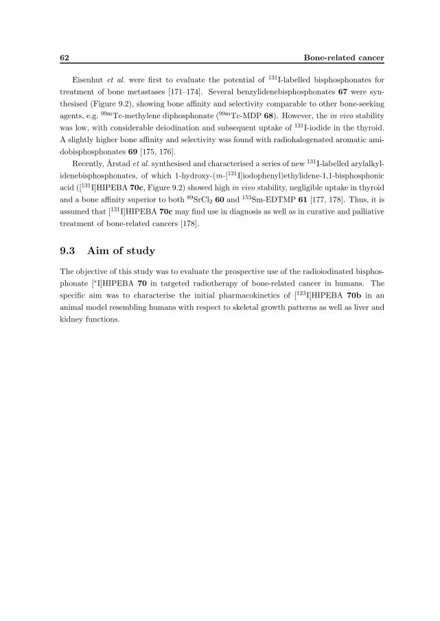
62 Bone-related cancer
Eisenhut et al. were first to evaluate the potential of 131I-labelled bisphosphonates fortreatment of bone metastases [171–174]. Several benzylidenebisphosphonates 67 were syn-thesised (Figure 9.2), showing bone affinity and selectivity comparable to other bone-seekingagents, e.g. 99mTc-methylene diphosphonate (99mTc-MDP 68). However, the in vivo stabilitywas low, with considerable deiodination and subsequent uptake of 131I-iodide in the thyroid.A slightly higher bone affinity and selectivity was found with radiohalogenated aromatic ami-dobisphosphonates 69 [175, 176].
Recently, Arstad et al. synthesised and characterised a series of new 131I-labelled arylalkyl-idenebisphosphonates, of which 1-hydroxy-(m-[131I]iodophenyl)ethylidene-1,1-bisphosphonicacid ([131I]HIPEBA 70c, Figure 9.2) showed high in vivo stability, negligible uptake in thyroidand a bone affinity superior to both 89SrCl2 60 and 153Sm-EDTMP 61 [177, 178]. Thus, it isassumed that [131I]HIPEBA 70c may find use in diagnosis as well as in curative and palliativetreatment of bone-related cancers [178].
9.3 Aim of study
The objective of this study was to evaluate the prospective use of the radioiodinated bisphos-phonate [∗I]HIPEBA 70 in targeted radiotherapy of bone-related cancer in humans. Thespecific aim was to characterise the initial pharmacokinetics of [123I]HIPEBA 70b in ananimal model resembling humans with respect to skeletal growth patterns as well as liver andkidney functions.

Chapter 10
Methods
10.1 Radioactivity measurements
Radioactivity measurements of radioiodine and preparations of [∗I]HIPEBA 70 were carriedout with a VDC-304 dose calibrator (Veenstra Instrumenten bv). Scintigraphic imaging of[123I]HIPEBA 70b in pigs was performed using a mobile Picker CX 250 compact gamma-camera equipped with a general purpose collimator (Paper V).
10.2 Chromatography
The radio-HPLC system for purification and isolation of [∗I]HIPEBA 70 was developed byArstad [177]. The system was based on a PLRP-S 100 A styrene-divinylbenzene column(stationary phase) and a polar mobile phase run under isocratic conditions. A proper samplepreparation prior to radio-HPLC was critical to avoid clogging of the column. The best resultswere obtained when the reaction mixture was added aqueous phosphoric acid and mixed inan ultrasound bath for ∼10 min before injection on the column [177]. Detailed specificationsof the radio-HPLC system, instruments and detectors are found in Paper V.
10.3 Animals
Healthy young pigs (3 months) of both genders were transported to the large animal facilityat the Center for Comparative Medicine, University of Oslo, one day prior to the experi-ment (Paper V). Housing and all procedures involving animals were performed according toprotocols approved by the animal care and use committee, in compliance with the NationalCommittee for Animal Experiment’s guidelines on animal welfare [113, 114]. The animalswere anaesthetised as described in Paper V.
63

64 Methods
10.4 The FNOMIP method
The new scintigraphic image analysis method FNOMIP (”fixed number of most intensepixels”) was developed by Skretting in connection with the experiment described in PaperV [179, 180]. The FNOMIP method ensures that a selective region of interest (SROI) hasa constant size and covers the same anatomical structure at all time points during a study,thus correcting for small movements and for different animal positions in static studies. Themethod is illustrated in Figure 10.1. This method enables generation of dynamic uptakecurves using the total number of counts in a SROI covering a specific anatomical structure(Paper V).
Figure 10.1: Illustration of the FNOMIP method. Upper frame shows the original images,while the lower frame shows the automatically generated selective regions of interest (blackregions) of the knee of a pig administrated [123I]HIPEBA 70b. Note the movement of theknee between image 6–8.
10.5 The OLINDA/EXM code
The OLINDA/EXM personal computer code performs dose calculations and kinetic modelingof radiopharmaceuticals [181]. The estimated radiation dose to different organs of the humanbody can be calculated from user-supplied biokinetic data obtained from animal or humanstudies (Paper V).

Chapter 11
Bisphosphonate derivatives
11.1 Introduction
Previous biodistribution studies with [∗I]HIPEBA 70 in healthy and tumour-bearing rodentshave demonstrated a potential for this radioiodinated bisphosphonate as a bone-seeking agentin diagnosis and targeted radiotherapy of bone-related cancer [177, 178]. To provide a basisfor future clinical trials in humans, [∗I]HIPEBA 70 should be investigated in an animal modelmore closely resembling humans with respect to skeletal growth patterns, liver and kidneyfunctions.
In this study, the 123I-labelled HIPEBA was synthesised and its initial pharmacokineticproperties investigated in young pigs using gamma-camera imaging.
11.2 Chemistry
The silylated bisphosphonate 1-hydroxy-(m-trimethylsilylphenyl)ethylidene-1,1-bisphos-phonic acid (71) was provided by Arstad, and was radiolabelled and isolated according toliterature procedures (Figure 11.1) [177].
Precursor 71 was radioiodinated under acidic conditions using NCS as an oxidis-
SiMe3
HO PO3H2
PO3H2
123I
HO PO3H2
PO3H2
a, b
71 70b
Figure 11.1: Radioiodination of the silylated precursor 71 yielding [123I]HIPEBA 70b.Reagents and conditions: a) Na123I, NCS, AcOH, TFA, rt; b) H3PO4(aq), ultrasound, rt.
65

66 Bisphosphonate derivatives
ing agent, yielding the radioiodinated product 1-hydroxy-(m-[123I]iodophenyl)ethylidene-1,1-bisphosphonic acid ([123I]HIPEBA 70b). Purification by radio-HPLC yielded the radio-iodinated bisphosphonate 70b in >95 % integrated radiochemical yield. However, theyield of isolated material was only 35–40 %. The final preparation consisted of 1.3 GBq[123I]HIPEBA 70b in 15.7 mL of sterile filtered phosphate buffer (50 mM, pH 7.1, added2 % EtOH). The radioiodination is depicted in Figure 11.1, and detailed descriptions of theprocedure are found in Paper V.
11.3 Biology
The following experiment in pigs was approved by the Committee of Ethics for the use ofAnimals in Research in Norway.
Biodistribution studies were conducted at the Center for Comparative Medicine, Univer-sity of Oslo, using four 3 months old pigs (two of each gender) weighing 17–21 kg (Paper V).The animals were anaesthetised using Stresnil vet/Ketalar/Pentotal, then two of the pigs (oneof each gender) were administered [123I]HIPEBA 70b (204 MBq and 234 MBq, respectively)while the other two pigs were administered the reference compound 99mTc-MDP 68 (126 MBqand 107 MBq, respectively). The pigs were sacrified after 8 h by injection of Pentotal.
11.4 Imaging and analysis
Biodistribution and biodynamics of [123I]HIPEBA 70b (t1/2 13.2 h, 159 keV γ in 83.3 %) and99mTc-MDP 68 (t1/2 6.02 h, 141 keV γ in 87.2 %) were investigated using a mobile gamma-camera. Dynamic imaging studies were carried out during the first 30 min post-injection,followed by static imaging studies for 5min at specific time intervals up to 7.5 h (Paper V).
The analysis of bone deposition patterns was based on a visual comparison of staticimages obtained with 70b and 68. Dynamic curves for uptake in knee were obtained usingthe FNOMIP method [179, 180].
11.5 Radiation dose estimates in humans
Estimated absorbed radiation doses for administration of the 131I-labelled HIPEBA 70c (t1/2
192.48 h, mean β−-energy 0.182 MeV) and 153Sm-EDTMP 61 (t1/2 46.27 h, mean β−-energy0.225 MeV) in humans were obtained using the OLINDA/EXM code (adult male model) [181].The input values for the surfaces of trabecular and cortical bone in humans were obtainedfrom data on ”Reference man” [182], and the calculations were made under the assumptionthat accumulated 70c was not released from bone surfaces. It was assumed that 100 % of 70c

11.6 Results and discussion 67
Figure 11.2: Static images showing the uptake of [123I]HIPEBA 70b (left) and 99mTc-MDP 68
(right) in head and neck, thorax and pelvis of the pigs 4 h post-injection.
and 80 % of 61 were adsorbed on bone surfaces. To facilitate a direct comparison between70c and 61, the estimated dose values of 61 were converted to correct for the difference inhalf-lives (Paper V).
11.6 Results and discussion
Biodistribution studies were conducted in healthy young pigs to compare the initial pharmaco-kinetic properties of [123I]HIPEBA 70b with those of the standard bone scintigraphic agent99mTc-MDP 68 (Paper V). The results are summarised in Figure 11.2.
Visual inspection of static gamma-camera images of the neck 4 h post-injection showedno accumulation of 70b in the thyroid (Figure 11.2). This result verified the in vivo stabilityreported for this radioiodinated bisphosphonate in previous studies [177, 178]. However,thyroid blocking agents may be used as a precaution in clinical studies with [∗I]HIPEBA 70
in humans.
Dynamic studies of the knee revealed that the bone uptake patterns of 70b and 68 wereindistinguishable during the first 0.5 h post-injection. However, 70b showed a higher boneaccumulation than 68 at later time points, and the uptake increased throughout the experi-ment. The lower bone accumulation of 68 was due to a rapid renal excretion (Figure 11.2)which reduced the amount of 68 available for uptake in bone. In contrast, a negligible renalexcretion of 70b indicated that almost all the injected activity would end up in bone tissue.These observations were also consistent with the different biological half-lives calculated for

68 Bisphosphonate derivatives
Table 11.1: Estimated absorbed radiation doses for injection of [131I]HIPEBA 70c and 153Sm-EDTMP 61 in humans. The values were obtained using the OLINDA/EXM code (adult malemodel). The dose values for 61 have been converteda to enable direct comparison with 70c.
[131I]HIPEBA 153Sm-EDTMPconverted
Organ Dose (mSv/MBq) Dose (mSv/MBq)a
Red bone marrow 4.51 4.66
Osteogenic cells 16.7 24.34
Ovaries 0.24 0.025
Testes 0.13 0.012
Thyroid 0.24 0.029
Effective whole body dose 0.86 0.82a Converted 153Sm-EDTMP dose = Estimated 153Sm-EDTMP dose * T1/2(131I) / T1/2(153Sm)
the blood/extravascular compartments (Paper V).The results indicated that [∗I]HIPEBA 70 may have favourable properties as a bone-
seeking agent in targeted radiotherapy of osseous bone metastases and osteosarcomas inhumans. Thus, the radiotherapeutic prospects of the 131I-labelled HIPEBA 70c wereestimated and compared to the bone-seeking radiopharmaceutical 153Sm-EDTMP 61 usingthe OLINDA/EXM code. The estimated radiation doses for 70c and 61 were very similar forred bone marrow, osteogenic cells and for the effective whole body dose (Table 11.1). How-ever, the estimated doses to more distant critical organs were larger for 70c due to emissionof photons with higher energies and yields.
Conclusion
The results obtained with [123I]HIPEBA 70b in pigs indicated high in vivo stability anda bone uptake comparable to 99mTc-MDP 68. Estimated radiotherapeutic properties of[131I]HIPEBA 70c were comparable to the FDA-approved 153Sm-EDTMP 61. Negligiblerenal uptake and excretion of [∗I]HIPEBA 70 facilitate diagnostic scintigraphic imaging ofthe pelvis and is particularly advantageous for targeted radiotherapy, minimising the radia-tion doses to kidneys and bladder walls. Hence, future investigations of the biodynamics of[∗I]HIPEBA 70 in humans are justified.

Chapter 12
Bone-related cancer: Future
prospects
High accumulation of HIPEBA in bone combined with an almost negligible renal excretionindicate that [123I]HIPEBA and [124I]HIPEBA may be used in diagnosis of bone-formingmalignancies in SPECT and PET, respectively. The compounds are particularly advantageousfor investigations of the pelvis, and may thus be used as a supplement to current bone-seekingagents with considerable renal excretion (e.g. 99mTc-MDP, 153Sm-EDTMP).
The HIPEBA may also have a future in palliative and curative treatment of bone meta-stases using therapeutic radionuclides like 131I and 211At. The [131I]HIPEBA has a much morefavourable half-life than 89SrCl2 and 153Sm-EDTMP, and is more easily available at low costprovided that a kit for in situ 131I-labelling is developed. A more strongly focused dose tomicrometastases containing bone tissue may be obtained using the 211At-labelled analogue ofHIPEBA. The rapid distribution and bone accumulation of HIPEBA are advantageous withregard to the relatively short half-life of 211At. Thus, the 211At-labelled analogue may havepotential in treatment of osseous micrometastases, while [131I]HIPEBA would be more effi-cient for metastatic lesions where pathologically enhanced bone formation is inhomogenouslydistributed. Treatment of bone-related cancers may also benefit from using [131I]HIPEBA incombination with other bone-seeking radiopharmaceuticals emitting high-LET radiation, e.g.223RaCl2. The radiation dose to red bone marrow will be the limiting factor in humans, unlessstem cell support is offered.
69

70 Bone-related cancer: Future prospects

References
[1] Cancer Registry of Norway. Institute of Population-based Cancer Research. Cancer in Norway 2004.
http://www.kreftregisteret.no/forekomst−og−overlevelse−2004/kreft−i−norge−2004web.pdf.
[2] W. A. Volkert and T. J. Hoffman. Therapeutic Radiopharmaceuticals. Chem. Rev., 99:2269–2292, 1999.
[3] H. Lundqvist V. Tolmachev, J. Carlsson. A Limiting Factor for the Progress of Radionuclide-based Cancer
Diagnostics and Therapy. Acta Oncol., 43:264–275, 2004.
[4] T. E. Wheldon and J. A. O’Donoghue. The radiobiology of targeted radiotherapy. Int. J. Radiat. Biol., 58:1–21,
1990.
[5] E. J. Hall. Radiobiology for the Radiologist. ISBN 0-397-51248-1. J.B. Lippincott Company, 4. edition, 1994.
[6] A. C. Perkins. In vivo molecular targeted radiotherapy. Biomed. Imaging Interv. J., 1:1–7, 2005.
[7] J. Sweit. Radionuclides and carrier molecules for therapy. Phys. Med. Biol., 41:1905–1914, 1996.
[8] C. A. Hoefnagel, S. E. M. Clarke, M. Fischer, J. F. Chatal, V. J. Lewington, S. Nilsson, L. Troncone, and M. R.
Vieira. Radionuclide therapy practice and facilities in Europe. Eur. J. Nucl. Med., 26:277–282, 1999.
[9] S. K. Imam. Advancements in cancer therapy with alpha-emitters: a review. Int. J. Radiation Oncology Biol.
Phys., 51:271–278, 2001.
[10] M. R. McDevitt, G. Sgouros, R. D. Finn, J. L. Humm, J. G. Jurcic, S. M. Larson, and D. A. Scheinberg.
Radioimmunotherapy with alpha-emitting nuclides. Eur. J. Nucl. Med., 25:1341–1351, 1998.
[11] P. M. Pathare, D. K. Hamlin, D. S. Wilbur, M. W. Brechbiel, and L. A. Bray. Synthesis and radiolabeling of a
biotin-CHX-B chelate for Bi-213. J. Labelled Compd. Radiopharm., 41:595–603, 1998.
[12] S. G. Gouin, J.-F. Gestin, L. Monrandeau, F. Segat-Dioury, J. C. Meslin, and D. Deniaud. Synthesis and
metal complexation properties of Ph-DTPA and Ph-TTHA: novel radionuclide chelating agents for use in nuclear
medicine. Org. Biomol. Chem., 3:454–461, 2005.
[13] G. Henriksen, P. Hoff, J. Alstad, and R. H. Larsen. 223Ra for endoradiotherapeutic applications prepared from
an immobilized 227Ac/227Th source. Radiochim. Acta, 89:661–666, 2001.
[14] H. H. Coenen, S. M. Moerlein, and G. Stocklin. No-Carrier-Added Radiohalogenation Methods with Heavy
Halogens. Radiochim. Acta, 34:47–68, 1983.
[15] M. R. Zalutsky and G. Vaidyanathan. Astatine-211-Labeled Radiotherapeutics: An Emerging Approach to Tar-
geted Alpha-Particle Radiotherapy. Curr. Pharm. Design, 6:1433–1455, 2000.
[16] M. J. Adam and D. S. Wilbur. Radiohalogens for imaging and therapy. Chem. Soc. Rev., 34:153–163, 2005.
71

72 REFERENCES
[17] S. C. Srivastava, H. L. Atkins, G. T. Krishnamurthy, I. Zanzi, E. B. Silberstein, G. Meinken, L. F. Mausner,
F. Swailem, T. D’Alessandro, C. J. Cabahug, Y. Lau, T. Park, and S. Madajewicz. Treatment of Metastatic Bone
Pain with Tin-117m Stannic Diethylenetriaminepentaacetic Acid: A Phase I/II Clinical Study. Clin. Cancer Res.,
4:61–68, 1998.
[18] S. J. Adelstein, A. I. Kassis, L. Bodei, and G. Mariani. Radiotoxicity of Iodine-125 and Other Auger-Electron-
Emitting Radionuclides: Background to Therapy. Cancer Biother. Radiopharm., 18:301–316, 2003.
[19] T. C. Karagiannis. Targeted cancer therapy with ultra-short range Auger electron emitting isotopes (Electronic
letter). http://caonline.amcancersoc.org/cgi/eletters/56/4/226, 2006.
[20] M. Glaser, S. K. Luthra, and F. Brady. Applications of positron-emitting halogens in PET oncology (Review).
Int. J. Oncol., 22:253–267, 2003.
[21] S. Palm, J. L. Humm, R. Rundqvist, and L. Jacobsson. Microdosimetry of astatine-211 single-cell irradiation:
Role of daughter polonium-211 diffusion. Med. Phys., 31:218–225, 2004.
[22] O. R. Pozzi and M. R. Zalutsky. Radiopharmaceutical Chemistry of Targeted Radiotherapeutics, Part 1: Effects
of Solvent on the Degradation of Radiohalogenation Precursors by 211At α-Particles. J. Nucl. Med., 46:700–706,
2005.
[23] O. R. Pozzi and M. R. Zalutsky. Radiopharmaceutical Chemistry of Targeted Radiotherapeutics, Part 2: Ra-
diolytic Effects of 211At α-Particles Influence N -Succinimidyl-3-211At-Astatobenzoate Synthesis. J. Nucl. Med.,
46:1393–1400, 2005.
[24] H. H. Coenen, J. Mertens, and B. Maziere. Radioiodination reactions for pharmaceuticals. ISBN 1-4020-4560-3.
Springer, 2006.
[25] D. S. Wilbur. Radiohalogenation of Proteins: An Overview of Radionuclides, Labeling Methods, and Reagents
for Conjugate Labeling. Bioconjugate Chem., 3:433–470, 1992.
[26] K. Berei and L. Vasaros. Organic chemistry of astatine. In S. Patai and Z. Rappoport, editors, The Chemistry of
Functional Groups, Supplement D, chapter 10, pages 405–440. John Wiley & Sons Ltd., 1983.
[27] D. S. Wilbur, M.-K. Chyan, D. K. Hamlin, B. B. Kegley, R. Risler, P. M. Pathare, J. Quinn, R. L. Vessella,
C. Foulon, M. Zalutsky, T. J. Wedge, and M. F. Hawthorne. Reagents for Astatination of Biomolecules: Com-
parison of the in Vivo Distribution and Stability of Some Radioiodinated/Astatinated Benzamidyl and nido-
Carboranyl Compounds. Bioconjugate Chem., 15:203–223, 2004.
[28] R. H. Seevers and R. E. Counsell. Radioiodination Techniques for Small Organic Molecules. Chem. Rev., 82:575–
590, 1982.
[29] C. Galli. Aromatic Iodination: Evidence of Reaction Intermediate and of the σ-Complex Character of the Tran-
sition State. J. Org. Chem., 56:3238–3245, 1991.
[30] S. M. Moerlein and H. H. Coenen. Regiospecific No-Carrier-Added Radiobromination and Radioiodination of
Aryltrimethyl Group IVb Organometallics. J. Chem. Soc., Perkin Trans. I, pages 1941–1947, 1985.
[31] M. Argentini. Labelling with iodine. A review of the literature. Federal Institute for Reactor Research, Wueren-
lingen, Switzerland., 1982.
[32] J. March. Advanced Organic Chemistry; Reactions, Mechanisms, and Structure. ISBN 0-471-60180-2. John Wiley
& Sons, Inc., 4. edition, 1992.
[33] Cancer Research UK. UK Malignant Melanoma statistics. http://info.cancerresearchuk.org/cancerstats/types/melanoma.

REFERENCES 73
[34] The Norwegian Radium Hospital. http://www.radiumhospitalet.no/.
[35] B. S. Larsson. Interaction Between Chemicals and Melanin. Pigment Cell Res., 6:127–133, 1993.
[36] K. Jimbow, T. Iwashina, F. Alena, K. Yamada, J. Pankovich, and T. Umemura. Exploitation of Pigment
Biosynthesis Pathway as a Selective Chemotherapeutic Approach for Malignant Melanoma. J. Invest. Dermatol.,
100:231S–238S, 1993.
[37] P. A. Riley. Melanin. Int. J. Biochem. Cell Biol., 29:1235–1239, 1997.
[38] S. I. Fox. Human Physiology. ISBN 0-07-111584-6. McGraw-Hill Companies, Inc., 9. edition, 2005.
[39] E. J. Land and P. A. Riley. Spontaneous Redox Reactions of Dopaquinone and the Balance between the Eumelanic
and Pheomelanic Pathways. Pigment Cell Res., 13:273–277, 2000.
[40] E. J. Land, S. Ito, K. Wakamatsu, and P. A. Riley. Rate Constants for the First Two Chemical Steps of Eume-
lanogenesis. Pigment Cell Res., 16:487–493, 2003.
[41] S. Ito. A Chemist’s View of Melanogenesis. Pigment Cell Res., 16:230–236, 2003.
[42] A. Slominski, D. J. Tobin, S. Shibahara, and J. Wortsman. Melanin Pigmentation in Mammalian Skin and Its
Hormonal Regulation. Physiol. Rev., 84:1155–1228, 2004.
[43] G. Prota. Pigment Cell Research: What Directions? Pigment Cell Res., 10:5–11, 1997.
[44] P. A. Riley. Melanogenesis and melanoma. Pigment Cell Res., 16:548–552, 2003.
[45] G. Ronsisvalle, A. Marrazzo, O. Prezzavento, A. Cagnotto, T. Mennini, C. Parenti, and G. M. Scoto. Opioid and
sigma receptor studies. New developments in the design of selective sigma ligands. Pure Appl. Chem., 73:1499–
1509, 2001.
[46] C. S. John, B. J. Vilner, B. C. Geyer, T. Moody, and W. D. Bowen. Targeting Sigma Receptor-binding Benzamides
as in Vivo Diagnostic and Therapeutic Agents for Human Prostate Tumors. Cancer Res., 59:4578–4583, 1999.
[47] C. S. John, W. D. Bowen, T. Saga, S. Kinuya, B. J. Vilner, J. Baumgold, C. H. Paik, R. C. Reba, R. D.
Neumann, V. M. Varma, and J. G. McAfee. A Malignant Melanoma Imaging Agent: Synthesis, Characterization,
In Vitro Binding and Biodistribution of Iodine-125-(2-Piperidinylaminoethyl)-4-Iodobenzamide. J. Nucl. Med.,
34:2169–2175, 1993.
[48] M. Eisenhut, W. E. Hull, A. Mohammed, W. Mier, D. Lay, W. Just, K. Gorgas, W. D. Lehmann, and U. Haberkorn.
Radioiodinated N -(2-Diethylaminoethyl)benzamide Derivatives with High Melanoma Uptake: Structure-Affinity
Relationships, Metabolic Fate, and Intracellular Localization. J. Med. Chem., 43:3913–3922, 2000.
[49] C. S. John, W. D. Bowen, S. J. Fisher, B. B. Lim, B. C. Geyer, B. J. Vilner, and R. L. Wahl. Synthe-
sis, in Vitro Pharmacologic Characterization, and Preclinical Evaluation of N-[2-(1’-Piperidinyl)Ethyl]-3-Iodo-
4-Methoxybenzamide (P[125I]MBA) for Imaging Breast Cancer. Nucl. Med. Biol., 26:377–382, 1999.
[50] V. Cavaliers, H. Everaert, C. S. John, T. Lahoutte, and A. Bossuyt. Sigma Receptor Scintigraphy with N -[2-
(1’-Piperidinyl)Ethyl]-3-123I-Iodo-4-Methoxybenzamide of Patients with Suspected Primary Breast Cancer: First
Clinical Results. J. Nucl. Med., 43:1647–1649, 2002.
[51] K. T. Wheeler, L.-M. Wang, C. A. Wallen, S. R. Childers, J. M. Cline, P. C. Keng, and R. H. Mach. Sigma-2
receptors as a biomarker of proliferation in solid tumours. Br. J. Cancer, 82:1223–1232, 2000.
[52] M. Friebe, A. Mahmood, C. Bolzati, A. Drews, B. Johannsen, M. Eisenhut, D. Kraemer, A. Davison, and
A. G. Jones. [99mTc]Oxotechnetium(V) Complexes of Amine-Amide-Dithiol Chelates with Dialkylaminoalkyl
Substituents as Potential Diagnostic Probes for Malignant Melanoma. J. Med. Chem., 44:3132–3140, 2001.

74 REFERENCES
[53] C. A. Hoefnagel. Role of nuclear medicine in melanoma. Eur. J. Nucl. Med., 25:1567–1574, 1998.
[54] Y. Miao, M. Hylarides, D. R. Fisher, T. Shelton, H. Moore, D. W. Wester, A. R. Fritzberg, C. T. Winkelmann,
T. Hoffman, and T. P. Quinn. Melanoma Therapy via Peptide-Targeted α-Radiation. Clin. Cancer Res., 11:5616–
5621, 2005.
[55] P. A. Riley, C. J. Cooksey, C. I. Johnson, E. J. Land, A. M. Latter, and C. A. Ramsden. Melanogenesis-targeted
anti-melanoma pro-drug development: Effect of side-chain variations on the cytotoxicity of tyrosinase-generated
ortho-quinones in a model screening system. Eur. J. Cancer, 33:135–143, 1997.
[56] A. M. Jordan, T. H. Khan, H. M. I. Osborn, A. Photiou, and P. A. Riley. Melanocyte-Directed Enzyme Prodrug
Therapy (MDEPT): Development of a Targeted Treatment for Malignant Melanoma. Bioorg. Med. Chem.,
7:1775–1780, 1999.
[57] A. M. Jordan, T. H. Khan, H. Malkin, H. M. I. Osborn, A. Photiou, and P. A. Riley. Melanocyte-Directed Enzyme
Prodrug Therapy (MDEPT): Development of Second Generation Prodrugs for Targeted Treatment of Malignant
Melanoma. Bioorg. Med. Chem., 9:1549–1558, 2001.
[58] K. Ishiwata, K. Kubota, R. Kubota, R. Iwata, T. Takahashi, and T. Ido. Selective 2-[18F]fluorodopa uptake for
melanogenesis in murine metastatic melanomas. J. Nucl. Med., 32:95–101, 1991.
[59] K. Ishiwata, M. Shiono, K. Kubota, K. Yoshino, J. Hatazawa, T. Ido, C. Honda, M. Ichihashi, and Y. Mishima. A
unique in vivo assessment of 4-[10B]borono-L-phenylalanine in tumor tissues for boron neutron capture therapy of
malignant melanomas using positron emission tomography and 4-borono-2-[18F]fluoro-L-phenylalanine. Melanoma
Res., 2:171–179, 1992.
[60] S. Miura, T. Ueda, K. Jimbow, S. Ito, and K. Fujita. Synthesis of cysteinylphenol, cysteaminylphenol, and related
compounds, and in vivo evaluation of antimelanoma effect. Arch. Dermatol. Res., 279:219–225, 1987.
[61] S. Ito, T. Kato, K. Ishikawa, T. Kasuga, and K. Jimbow. Mechanism of selective toxicity of 4-S-cysteinylphenol
and 4-S-cysteaminylphenol to melanocytes. Biochem. Pharmacol., 36:2007–2011, 1987.
[62] N. J. Lant, P. McKeown, L. R. Kelland, P. M. Rogers, and D. J. Robins. Synthesis and antimelanoma activity of
analogues of N-acetyl-4-S-cysteaminylphenol. Anticancer Drug Des., 15:295–302, 2000.
[63] J. Yukitake, H. Otake, S. Inoue, K. Wakamatsu, and S. Ito. Comparison of in vivo anti-melanoma effect of
enantiomeric α-methyl- and α-ethyl-4-S -cysteaminylphenol. Melanoma Res., 14:115–120, 2004.
[64] B. S. Larsson, B. Larsson, and A. Roberto. Boron Neutron Capture Therapy for Malignant Melanoma: An
Experimental Approach. Pigment Cell Res., 2:356–360, 1989.
[65] U. Mars, V. Tolmachev, and A. Sundin. Positron Emission Tomography of Experimental Melanoma with [76Br]5-
Bromo-2-Thiouracil. Nucl. Med. Biol., 27:845–849, 2000.
[66] V. Tolmachev, A. Orlova, and A. Sundin. Preparation of [76Br]5-bromo-2-thiouracil, a positron-emitting melanoma
localizing agent. J. Radioanal. Nucl. Chem., 251:406–412, 2002.
[67] A. M. Potts. Uveal Pigment and Phenothiazine Compounds. Trans. Am. Ophthalmol. Soc., 60:517–552, 1962.
[68] E. M. Link, I. Brown, R. N. Carpenter, and J. S. Mitchell. Uptake and Therapeutic Effectiveness of 125I- and211At-Methylene Blue for Pigmented Melanoma in an Animal Model System. Cancer Res., 49:4332–4337, 1989.
[69] E. M. Link, A. S. Michalowski, and F. Rosch. 211At-Methylene Blue in Targeted Radiotherapy of Disseminated
Melanoma. Pigment Cell Res., 7:358–362, 1994.

REFERENCES 75
[70] I. Brown, R. N. Carpenter, E. M. Link, and J. S. Mitchell. Potential diagnostic and therapeutic agents for
malignant melanoma: Synthesis of heavy radiohalogenated derivatives of methylene blue by electrophilic and
nucleophilic methods. J. Radioanal. Nucl. Chem. Letters, 107:337–351, 1986.
[71] P. J. Blower and N. J. Carter. Rapid preparation of iodine-123-labelled methylene blue and toluidine blue:
potential new agents for parathyroid scintigraphy. Nucl. Med. Comm., 11:413–420, 1990.
[72] E. M. Link and R. N. Carpenter. 211At-methylene blue for targeted radiotherapy of human melanoma xenografts:
treatments of cutaneous tumours and lymph node metastases. Cancer Res., 52:4385–4390, 1992.
[73] E. M. Link, D. C. Costa, D. Lane, P. J. Blower, and M. F. Spittle. Radioiodinated methylene blue for diagnosing
early melanoma metastases. The Lancet, 348:753, 1996.
[74] P. J. Blower, K. Clark, and E. M. Link. Radioiodinated Methylene Blue for Melanoma Targeting: Chemical
Characterisation and Tumour Selectivity of Labelled Components. Nucl. Med. Biol., 24:305–310, 1997.
[75] E. M. Link, P. J. Blower, D. C. Costa, D. M. Lane, D. Lui, R. S. D. Brown, P. J. Ell, and M. F. Spittle. Early
detection of melanoma metastases with radioiodinated methylene blue. Eur. J. Nucl. Med., 25:1322–1329, 1998.
[76] K. Hasegawa, S. Ito, S. Inoue, K. Wakamatsu, H. Ozeki, and I. Ishiguro. Dihydro-1,4-benzothiazine-6,7-dione,
the Ultimate Toxic Metabolite of 4-S -Cysteaminylphenol and 4-S -Cysteaminylcatechol. Biochem. Pharmacol.,
53:1435–1444, 1997.
[77] G. Prota, M. d’Ischia, and D. Mascagna. Melanogenesis as a targeting strategy against metastatic melanoma: a
reassessment. Melanoma Res., 4:351–358, 1994.
[78] F. Alena, K. Jimbow, and S. Ito. Melanocytotoxicity and Antimelanoma Effects of Phenolic Amine Compounds
in Mice in Vivo. Cancer Res., 50:3743–3747, 1990.
[79] S. Inoue, S. Ito, K. Wakamatsu, K. Jimbow, and K. Fujita. Mechanism of growth inhibition of melanoma cells by
4-S -cysteaminylphenol and its analogues. Biochem. Pharmacol., 39:1077–1083, 1990.
[80] M. Tandon, P. D. Thomas, M. Shokravi, S. Singh, S. Samra, D. Chang, and K. Jimbow. Synthesis and Anti-
tumour Effect of the Melanogenesis-based Antimelanoma Agent N -Propionyl-4-S -cysteaminylphenol. Biochem.
Pharmacol., 55:2023–2029, 1998.
[81] S. Inoue, K. Hasegawa, K. Wakamatsu, and S. Ito. Comparison of antimelanoma effects of 4-S -cysteaminylphenol
and its homologues. Melanoma Res., 8:105–112, 1998.
[82] N. J. Lant, P. McKeown, M. C. Timoney, L. R. Kelland, P. M. Rogers, and D. J. Robins. Synthesis and anti-
melanoma activity of analogues of N-acetyl-4-S-cysteaminylphenol substituted with two methyl groups alpha to
the nitrogen. Anticancer Drug Des., 16:49–55, 2001.
[83] J. Yukitake, H. Otake, S. Inoue, K. Wakamatsu, C. Olivares, F. Solano, K. Hasegawa, and S. Ito. Synthesis and
selective in vitro anti-melanoma effect of enantiomeric α-methyl- and α-ethyl-4-S -cysteaminylphenol. Melanoma
Res., 13:603–609, 2003.
[84] T. Miura, K. Jimbow, and S. Ito. The in vivo antimelanoma effect of 4-S-cysteaminylphenol and its N-acetyl
derivative. Int. J. Cancer, 46:931–934, 1990.
[85] F. Alena, T. Iwashina, A. Gili, and K. Jimbow. Selective in Vivo Accumulation of N -Acetyl-4-S -cysteaminylphenol
in B16F10 Murine Melanoma and Enhancement of Its in Vitro and in Vivo Antimelanoma Effect by Combination
of Buthionine Sulfoximine. Cancer Res., 54:2661–2666, 1994.
[86] S. Singh, K. Jimbow, P. Kumar, A. J. McEwan, and L. I. Wiebe. Synthesis and radioiodination of 3-(E)-(2-
iodovinyl)-N -acetyl-4-cysteaminylphenol, a putative tyrosinase substrate for imaging neural crest tumours. J.
Labelled Cpd. Radiopharm., 41:355–361, 1998.

76 REFERENCES
[87] P. D. Thomas, H. Kishi, H. Cao, M. Ota, T. Yamashita, S. Singh, and K. Jimbow. Selective Incorporation and
the Specific Cytocidal Effect as the Cellular Basis for the Antimelanoma Action of Sulphur Containing Tyrosine
Analogs. J. Invest. Dermatol., 113:928–934, 1999.
[88] K. Jimbow, T. Miura, S. Ito, and K. Ishikawa. Phenolic Melanin Precursors Provide a Rational Approach to the
Design of Antitumor Agents for Melanoma. Pigment Cell Res., 2:34–39, 1989.
[89] J. M. Pankowich, K. Jimbow, and S. Ito. 4-S-Cysteaminylphenol and its Analogues as Substrates for Tyrosinase
and Monoamine Oxidase. Pigment Cell Res., 3:146–149, 1990.
[90] P. G. Parsons, D. Favier, M. McEwan, H. Takahashi, K. Jimbow, and S. Ito. Action of cysteaminylphenols on
human melanoma cells in vivo and in vitro: 4-S-cysteaminylphenol binds protein disulphide isomerase. Melanoma
Res., 1:97–104, 1991.
[91] K. Yamada and K. Jimbow. Selective in vivo and in vitro incorporation and accumulation of phenolic thioether
amine into malignant melanoma and identification of a (58 kD) binding glycoprotein. Melanoma Res., 2:225–233,
1992.
[92] C. J. Cooksey, K. Jimbow, E. J. Land, and P. A. Riley. Reactivity of orthoquinones involved in tyrosinase-
dependent cytotoxicity: differences between alkylthio- and alkoxy-substituents. Melanoma Res., 2:283–293, 1992.
[93] T. Iwashina, K. Jimbow, and L. I. Wiebe. The Synthesis of N -Acetyl-4-S -Cysteaminyl [U-14C]Phenol as a Basis
for the Development of an Antimelanoma and Melanoma-radioimaging Agent. Appl. Radiat. Isot., 45:703–705,
1994.
[94] M. Jimbow, H. Marusyk, and K. Jimbow. The in vivo melanocytotoxicity and depigmenting potency of N -2,4-
acetoxyphenyl thioethyl acetamide in the skin and hair. Br. J. Dermatol., 133:526–536, 1995.
[95] Y. Minamitsuji, K. Toyofuku, S. Sugiyama, K. Yamada, and K. Jimbow. Sulphur Containing Tyrosine Analogs Can
Cause Selective Melanocytotoxicity Involving Tyrosinase-Mediated Apoptosis. J. Invest. Dermatol. Symposium
Proceedings, 4:130–136, 1999.
[96] A. Gili, P. D. Thomas, M. Ota, and K. Jimbow. Comparison of in vitro cytotoxicity of N -acetyl and N -propionyl
derivatives of phenolic thioether amines in melanoma and neuroblastoma cells and the relationship to tyrosinase
and tyrosine hydroxylase enzyme activity. Melanoma Res., 10:9–15, 2000.
[97] R. A. Murphy, H. F. Kung, M.-P. Kung, and J. Billings. Synthesis and Characterization of Iodobenzamide
Analogues: Potential D-2 Dopamine Receptor Imaging Agents. J. Med. Chem., 33:171–178, 1990.
[98] J. M. Michelot, M. F. Moreau, P. G. Labarre, J. C. Madelmont, A. J. Veyre, J. M. Papon, D. F. Parry, J. F.
Bonafous, J. Y. Boire, and G. G. Desplanches. Synthesis and evaluation of new iodine-125 radiopharmaceuticals
as potential tracers for malignant melanoma. J. Nucl. Med., 32:1573–1580, 1991.
[99] H. Dittmann, H. H. Coenen, F. Zolzer, K. Dutschka, W. Brandau, and C. Streffer. In Vitro Studies on the Cellular
Uptake of Melanoma Imaging Aminoalkyl-Iodobenzamide Derivatives (ABA). Nucl. Med. Biol., 26:51–56, 1999.
[100] S. M. N. Efange, R. H. Michelson, B. Knusel, F. Hefti, R. J. Boudreau, J. R. Thomas, and J. R. Tennison. Synthesis
and Biological Evaluation of Radioiodinated N -2-(4-Piperidyl)ethyl Benzamides. Nucl. Med. Biol., 20:527–538,
1993.
[101] B. J. Vilner, C. S. John, and W. D. Bowen. Sigma-1 and Sigma-2 Receptors Are Expressed in a Wide Variety of
Human and Rodent Tumor Cell Lines. Cancer Res., 55:408–413, 1995.
[102] L. Maffioli, L. Mascheroni, V. Mongioj, M. Gasparini, M. T. Baldini, E. Seregni, M. R. Castellani, N. Cascinelli,
and G.-L. Buraggi. Scintigraphic Detection of Melanoma Metastases with a Radiolabeled Benzamide ([123I]-(S)-
IBZM). J. Nucl. Med., 35:1741–1747, 1994.

REFERENCES 77
[103] M. F. Moreau, J. Michelot, J. Papon, M. Bayle, P. Labarre, J. C. Madelmont, D. Parry, J. Y. Boire, N. Moins,
H. Seguin, A. Veyre, and L. Mauclaire. Synthesis, Radiolabeling, and Preliminary Evaluation in Mice of Some
(N -Diethylaminoethyl)-4-Iodobenzamide Derivatives as Melanoma Imaging Agents. Nucl. Med. Biol., 22:737–747,
1995.
[104] A. Mohammed, C. Nicholl, U. Titsch, and M. Eisenhut. Radioiodinated N-(Alkylaminoalkyl)-Substituted 4-
Methoxy-, 4-Hydroxy-, and 4-Aminobenzamides: Biological Investigations for the Improvement of Melanoma-
Imaging Agents. Nucl. Med. Biol., 24:373–380, 1997.
[105] N. Moins, J. Papon, H. Seguin, D. Gardette, M.-F. Moreau, P. Labarre, M. Bayle, J. Michelot, J.-C. Gramain,
J.-C. Madelmont, and A. Veyre. Synthesis, characterization and comparative biodistribution study of a new series
of p-Iodine-125 benzamides as potential melanoma imaging agents. Nucl. Med. Biol., 28:799–808, 2001.
[106] M. Wolf, U. Bauder-Wust, A. Mohammed, F. Schonsiegel, W. Mier, U. Haberkorn, and M. Eisenhut. Alkylating
benzamides with melanoma cytotoxicity. Melanoma Res., 14:353–360, 2004.
[107] J. M. Michelot, M. F. Moreau, A. J. Veyre, J. F. Bonafous, F. J. Bacin, J. C. Madelmont, F. Bussiere, P. A.
Souteyrand, L. P. Mauclaire, and F. M. Chossat. Phase II scintigraphic clinical trial of malignant melanoma and
metastases with iodine-125-N-(2-diethylaminoethyl)-4-iodobenzamide. J. Nucl. Med., 33:1260–1266, 1993.
[108] C. S. John, T. Saga, S. Kinuya, N. Le, J. M. Jeong, C. H. Paik, R. C. Reba, V. M. Varma, and J. G. McAfee.
An Improved Synthesis of [125I]N -(diethylaminoethyl)-4-iodobenzamides: a Potent Ligand for Imaging Malignant
Melanoma. Nucl. Med. Biol., 20:75–79, 1993.
[109] C. Nicholl, A. Mohammed, W. E. Hull, B. Bubeck, and M. Eisenhut. Pharmacokinetics of Iodine-123-IMBA for
Melanoma Imaging. J. Nucl. Med., 38:127–133, 1997.
[110] G. Henriksen, S. Messelt, E. Olsen, and R. H. Larsen. Optimisation of cyclotron production parameters for the209Bi(α, 2n)211At reaction related to biomedical use of 211At. Appl. Radiat. Isot., 54:839–844, 2001.
[111] S. Lindegren, T. Back, and H. J. Jensen. Dry-distillation of astatine-211 from irradiated bismuth targets: a
time-saving procedure with high recovery yields. Appl. Radiat. Isot., 55:157–160, 2001.
[112] E. M. Brevik. Syntesemetoder for renfremstilling av 125I- og 211At-merkete benzamider. Master’s thesis, Depart-
ment of Chemistry, University of Oslo, 2000.
[113] Royal Society/Universities federation for animal welfare (UFAW). Guidelines on the care of laboratory animals
and their use for scientific purposes. 1. Housing and care. UFAW, London, UK, 1987.
[114] United Kingdom Coordinating Committee on Cancer Research (UKCCCR). UKCCCR guidelines for the welfare
of animals in experimental neoplasia. Br. J. Cancer, 58:156–160, 1988.
[115] The National Institutes of Health (NIH). Guide for the care and use of laboratory animals. NIH Publication No.
86-23. Bethesda, US, 1985.
[116] K. M. Hwang, Ø. Fodstad, R. K. Oldham, and A. C. Morgan Jr. Radiolocalization of xenografted human malignant
melanoma by monoclonal antibody (9.2.27) to a melanoma-associated antigen in nude mice. Cancer Res., 45:4150–
4155, 1985.
[117] V. N. Ivanov, Ø. Fodstad, and Z. Ronai. Expression of ring finger-deleted TRAF2 sensitizes metastatic melanoma
cells to apoptosis via up-regulation of p38, TNFα and suppression of NF-κB activities. Oncogene, 20:2243–2253,
2001.
[118] D. S. Wilbur, D. K. Hamlin, P. M. Pathare, and S. A. Weerawarna. Biotin Reagents for Antibody Pretargeting.
Synthesis, Radioiodination, and in Vitro Evaluation of Water Soluble, Biotinidase Resistant Biotin Derivatives.
Bioconjugate Chem., 8:572–584, 1997.

78 REFERENCES
[119] J. J. Feigenbaum, M. D. Choubal, K. Payza, J. R. Kanofsky, and D. S. Crumrine. Receptor Inactivation by
Dye-Neuropeptide Conjugates: 1. The Synthesis of Cys-Containing Dye-Neuropeptide Conjugates. Peptides,
17:991–994, 1996.
[120] K. H. Holm. Personal communication.
[121] H. Masuya, H. Shimadzu, T. Miyawaki, and M. A. Motsenbocker. Preparation of phenothiazines for use in
photodynamic therapy and immunoassays. Eur. Pat. Appl., 1992.
[122] N. Leventis, M. Chen, and S. Sotiriou-Leventis. Synthesis of Substituted Phenothiazines Analogous to Methylene
Blue by Electrophilic and Nucleophilic Aromatic Substitutions in Tandem. A Mechanistic Perspective. Tetrahe-
dron, 53:10083–10092, 1997.
[123] M. Wainwright and R. M. Giddens. Phenothiazinium photosensitisers: choises in synthesis and applications. Dyes
and Pigments, 57:245–257, 2003.
[124] R. Nishii, K. Kawai, L. G. Flores II, S. Jinnouchi, S. Nagamachi, and S. Tamura. Synthesis and preliminary
biodistribution of 3-iodo-4-hydroxyphenyl-L-cystein. J. Labelled Cpd. Radiopharm., 42, Suppl. 1:S795–S797, 1999.
[125] R. Nishii, K. Kawai, L. G. Flores II, H. Kataoka, S. Jinnouchi, S. Nagamachi, Y. Arano, and S. Tamura. A novel
radiopharmaceutical for detection of malignant melanoma, based on melanin formation: 3-iodo-4-hydroxyphenyl-
L-cysteine. Nucl. Med. Comm., 24:575–582, 2003.
[126] H. L. Wehrmeister. Reactions of Aromatic Thiols with Oxazolines. J. Org. Chem., 28:2587–2588, 1963.
[127] H. L. Wehrmeister. N-(Arylmercaptoalkyl) Amides by Reaction of Thiols with Amino Alcohols and Carboxylic
Acid. J. Org. Chem., 28:2589–2591, 1963.
[128] T. W. Greene and P. G. M. Wuts. Protective groups in organic synthesis. ISBN 0-471-16019-9. John Wiley &
Sons, Inc., 3. edition, 1999.
[129] Y. Kita, T. Okuno, M. Egi, K. Iio, Y. Takeda, and S. Akai. An Effective p-Thiocyanation of Phenols Using
Phenyliodine Dichloride–Lead(II) Thiocyanate. Synlett, pages 1039–1040, 1994.
[130] Y. Kita, Y. Takeda, T. Okuno, M. Egi, K. Iio, K. Kawaguchi, and S. Akai. An Efficient p-Thiocyanation of Phenols
and Naphthols Using a Reagent Combination of Phenyliodine Dichloride and Lead(II) Thiocyanate. Chem. Pharm.
Bull., 45:1887–1890, 1997.
[131] D. Koyuncu, A. McKillop, and L. McLaren. A Simple and Inexpensive Procedure for the Preparation of
(Dichloroiodo)arenes. J. Chem. Research (S), page 21, 1990.
[132] A. Varvoglis. Hypervalent Iodine In Organic Synthesis. In A. R. Katritzky, O. Meth-Cohn, and C. W. Rees,
editors, Best Synthetic Methods, ISBN 0-12-714975-9, chapter 2.2 and 6.4.5. Academic Press, Inc., San Diego,
1997.
[133] R. Q. Brewster and W. Schroeder. p-Thiocyanodimethylaniline. Org. Synth., II:574–575, 1943.
[134] J. L. Wardell. Preparation of thiols. In S. Patai, editor, The chemistry of functional groups; The chemistry of
the thiol group, chapter 4, pages 163–269. John Wiley & Sons Ltd., 1974.
[135] R. J. Laufer. 4-Mercaptophenol. In Chem. Abstr., volume 75, pages 88310–88311. 1971.
[136] T. Matsui, M. Harada, M. Toba, and Y. Yoshimura. Effect of the coexistence of nitrogen compounds on the sulfur
tolerance and catalytic activity of Pd and Pt monometallic catalysts supported on high-silica USY zeolite and
amorphous silica. Appl. Catal. A, 293:137–144, 2005.

REFERENCES 79
[137] S. R. Padgette, H. H. Herman, J. H. Han, S. H. Pollock, and S. W. May. Antihypertensive Activities of Phenyl
Aminoethyl Sulfides, a Class of Synthetic Substrates for Dopamine β-Hydroxylase. J. Med. Chem., 27:1354–1357,
1984.
[138] J. A. Prezioso, N. Wang, and W. D. Bloomer. Thymidylate synthase as a target enzyme for the melanoma-
specific toxicity of 4-S-cysteaminylphenol and N -acetyl-4-S-cysteaminylphenol. Cancer Chemother. Pharmacol.,
30:394–400, 1992.
[139] M. M. Edreira and O. R. Pozzi. Iodide benzamides for the in-vivo detection of melanoma and metastases.
Melanoma Res., 16:37–43, 2006.
[140] D. L. Gilliland, G. P. Basmadjian, A. P. Marchand, G. H. Hinkle, A. Earlywine, and R. D. Ice. Iodine Labelled
Radiopharmaceuticals from Arylthallium bis(trifluoroacetate) Intermediates. J. Radioanal. Chem., 65:107–113,
1981.
[141] G. W. M. Visser and E. L. Diemer. The Synthesis of Organic At-Compounds through Thallium Compounds. Int.
J. Appl. Radiat. Isot., 33:389–390, 1982.
[142] M. R. Zalutsky and A. S. Narula. A Method for the Radiohalogenation of Proteins Resulting in Decreased Thyroid
Uptake of Radioiodine. Appl. Radiat. Isot., 38:1051–1055, 1987.
[143] R. E. Coleman. The treatment of metastatic bone disease. In O. L. M. Bijvoet, H. A. Fleisch, R. E. Canfield, and
R. G. G. Russell, editors, Bisphosphonate on Bones, ISBN 0-444-89132-3, pages 349–363. Elsevier Science B.V.,
Amsterdam, 1995.
[144] A. Stuhaug. Nytt skyts mot benmetastaser. http://www.legemidlerogsamfunn.no/, 2002.
[145] E. Shane and J. P. Bilezikian. Normal bone and mineral homeostasis. In O. L. M. Bijvoet, H. A. Fleisch, R. E.
Canfield, and R. G. G. Russell, editors, Bisphosphonate on Bones, ISBN 0-444-89132-3, pages 3–21. Elsevier
Science B.V., Amsterdam, 1995.
[146] S. Ott. Osteoporosis and Bone Physiology. http://courses.washington.edu/bonephys/, 2007.
[147] H. Fleisch. From the Laboratory to the Patient. Bisphosphonates in bone disease. New York: The Parthenon
Publishing Group, 1995.
[148] R. D. Rubens. Bone involvement in solid tumours. In O. L. M. Bijvoet, H. A. Fleisch, R. E. Canfield, and
R. G. G. Russell, editors, Bisphosphonate on Bones, ISBN 0-444-89132-3, pages 337–347. Elsevier Science B.V.,
Amsterdam, 1995.
[149] V. J. Lewington. Bone-Seeking Radionuclides for Therapy. J. Nucl. Med., 46:38S–47S, 2005.
[150] V. J. Lewington. A practical guide to targeted therapy for bone pain palliation. Nucl. Med. Commun., 23:833–836,
2002.
[151] L. G. Bouchet, W. E. Bolch, S. M. Goddu, R. W. Howell, and D. V. Rao. Considerations in the Selection of
Radiopharmaceuticals for Palliation of Bone Pain from Metastatic Osseous Lesions. J. Nucl. Med., 41:682–687,
2000.
[152] C. K. Lee, D. M. Aeppli, J. Unger, R. J. Boudreau, and S. H. Levitt. Strontium-89 Chloride (Metastron) for
Palliative Treatment of Bony Metastases: The University of Minnesota Experience. Am. J. Clin. Oncol., 19:102–
107, 1996.
[153] C. Collins, J. F. Eary, G. Donaldson, C. Vernon, N. E. Bush, S. Petersdorf, R. B. Livingston, E. E. Gordon, C. R.
Chapman, and F. R. Appelbaum. Samarium-153-EDTMP in bone metastases of hormone refractory prostate
carcinoma: a phase I/II trial. J. Nucl. Med., 34:1839–1844, 1993.

80 REFERENCES
[154] Ø. S. Bruland, A. Skretting, Ø. P. Solheim, and M. Aas. Targeted radiotherapy of osteosarcoma using 153Sm-
EDTMP. A new promising approach. Acta. Oncol., 35:381–384, 1996.
[155] C. L. Maini, S. Bergomi, L. Romano, and R. Sciuto. 153Sm-EDTMP for bone pain palliation in skeletal metastases.
Eur. J. Nucl. Med. Mol. Imaging, 31 (Suppl. 1):S171–S178, 2004.
[156] W. Brenner, W. U. Kampen, C. Brummer, C. von Forstner, M. Zuhayra, N. Czech, C. Muhle, and E. Henze.
Bone Uptake Studies in Rabbits Before and After High-Dose Treatment with 153Sm-EDTMP or 186Re-HEDP. J.
Nucl. Med., 44:247–251, 2003.
[157] J. M. de Klerk, A. D. van het Schip, B. A. Zonnenberg, A. van Dijk, J. M. Quirijnen, G. H. Blijham, and P. P.
van Rijk. Phase 1 study of rhenium-186-HEDP in patients with bone metastases originating from breast cancer.
J. Nucl. Med., 37:244–249, 1996.
[158] M. G. E. H. Lam, J. M. H. de Klerk, and P. P. van Rijk. 186Re-HEDP for metastatic bone pain in breast cancer
patients. Eur. J. Nucl. Med. Mol. Imaging, 31 (Suppl. 1):S162–S170, 2004.
[159] A. Bishayee, D. V. Rao, S. C. Srivastava, L. G. Bouchet, W. E. Bolch, and R. W. Howell. Marrow-Sparing
Effects of 117mSn(4+)Diethylenetriaminepentaacetic Acid for Radionuclide Therapy of Bone Cancer. J. Nucl.
Med., 41:2043–2050, 2000.
[160] G. A. R. Sola, M. G. Arguelles, D. L. Bottazzini, J. C. Furnari, I. G. Parada, A. Rojo, and H. V. Ruiz. Lutetium-
177-EDTMP for bone pain palliation. Preparation, biodistribution and pre-clinical studies. Radiochim. Acta,
88:157–161, 2000.
[161] S. Chakraborty, T. Das, P. R. Unni, H. D. Sarma, G. Samuel, S. Banerjee, M. Venkatesh, N. Ramamoorthy, and
M. R. A. Pillai. 177Lu labelled polyaminophosphonates as potential agents for bone pain palliation. Nucl. Med.
Comm., 23:67–74, 2002.
[162] G. Henriksen, K. Breistøl, Ø. S. Bruland, Ø. Fodstad, and R. H. Larsen. Antitumor Effect from Bone-seeking, α-
Particle-emitting 223Ra Demonstrated in an Experimental Skeletal Metastases Model. Cancer Res., 62:3120–3125,
2002.
[163] G. Henriksen, D. R. Fischer, J. C. Roeske, Ø. S. Bruland, and R. H. Larsen. Targeting of Osseous Sites with
α-Emitting 223Ra: Comparison with the β-Emitter 89Sr in Mice. J. Nucl. Med., 44:252–259, 2003.
[164] S. Nilsson, R. H. Larsen, S. D. Fossa, L. Balteskard, K. W. Borch, J.-E. Westlin, G. Salberg, and Ø. S. Bruland.
First Clinical Experience with α-Emitting Radium-223 in the Treatment of Skeletal Metastases. Clin. Cancer
Res., 11:4451–4459, 2005.
[165] Ø. S. Bruland, S. Nilsson, D. R. Fisher, and R. H. Larsen. High-Linear Energy Transfer Irradiation Targeted to
Skeletal Metastases by the α-Emitter 223Radium: Adjuvant or Alternative to Conventional Modalities? Clin.
Cancer Res., 12:6250s–6257s, 2006.
[166] O. L. M. Bijvoet, H. A. Fleisch, R. E. Canfield, and R. G. G. Russell. Bisphosphonate on Bones. Elsevier Science
B.V., Amsterdam, 1995. ISBN 0-444-89132-3.
[167] L. Widler, K. A. Jaeggi, M. Glatt, K. Muller, R. Bachmann, M. Bisping, A.-R. Born, R. Cortesi, G. Guiglia,
H. Jeker, R. Klein, U. Ramseier, J. Schmid, G. Schreiber, Y. Seltenmeyer, and J. R. Green. Highly Potent
Geminal Bisphosphonates. From Pamidronate Disodium (Aredia) to Zoledronic Acid (Zometa). J. Med. Chem.,
45:3721–3738, 2002.
[168] M. D. Michaelson and M. R. Smith. Bisphosphonates for Treatment and Prevention of Bone Metastases. J. Clin.
Oncol., 23:8219–8224, 2005.

REFERENCES 81
[169] K. Libson, E. Deutsch, and B. L. Barnett. Structural Characterization of a 99Tc-Diphosphonate Complex. Impli-
cations for the Chemistry of 99mTc Skeletal Imaging Agents. J. Am. Chem. Soc., 102:2476–2478, 1980.
[170] J. R. Dilworth and S. J. Parrott. The biomedical chemistry of technetium and rhenium. Chem. Soc. Rev., 27:43–55,
1998.
[171] M. Eisenhut. Iodine-131-Labeled Diphosphonates for the Palliative Treatment of Bone Metastases: I. Organ
Distribution and Kinetics of I-131 BDP3 in Rats. J. Nucl. Med., 25:1356–1361, 1984.
[172] M. Eisenhut, R. Berberich, B. Kimmig, and E. Oberhausen. Iodine-131-Labeled Diphosphonates for Palliative
Treatment of Bone Metastases: II. Preliminary Clinical Results with Iodine-131 BDP3. J. Nucl. Med., 27:1255–
1261, 1986.
[173] M. Eisenhut, P. Fritz, B. Kimmig, F. Wingen, and B. Krempien. Iodine-131-Labeled Diphosphonates for the
Palliative Treatment of Bone Metastases–III. Considerations of Interaction, Binding and Absorbed Dose. Appl.
Radiat. Isot., 37:741–747, 1986.
[174] M. Eisenhut, J. Barber, and D. M. Taylor. 131I Labeled Diphosphonates for the Palliative Treatment of Bone
Metastases–IV. Syntheses of Benzylidenediphosphonates and their Organ Distribution in Rats. Appl. Radiat.
Isot., 38:535–540, 1987.
[175] K. M. Murud. Synthesis and preclinical evaluation of radiohalogenated aromatic amidobisphosphonates. PhD
thesis, Faculty of Mathematics and Natural Sciences, University of Oslo, Norway, 1999.
[176] R. H. Larsen, K. M. Murud, G. Akabani, P. Hoff, Ø. S. Bruland, and M. R. Zalutsky. 211At- and 131I-Labeled
Biphosphonates with High In Vivo Stability and Bone Accumulation. J. Nucl. Med., 40:1197–1203, 1999.
[177] E. Arstad. Novel Bisphosphonates in Targeted Radiotherapy. PhD thesis, Faculty of Mathematics and Natural
Sciences, University of Oslo, Norway, 2001.
[178] E. Arstad, P. Hoff, L. Skattebøl, A. Skretting, and K. Breistøl. Studies on the Synthesis and Biological Properties
of Non-Carrier-Added [125I and 131I]-Labeled Arylalkylidenebisphosphonates: Potent Bone-Seekers for Diagnosis
and Therapy of Malignant Osseous Lesions. J. Med. Chem., 46:3021–3032, 2003.
[179] A. Skretting, T. Bach-Gansmo, and P. Hoff. A method for movement correction based on a fixed optnumber of
descending sorted pixels region of interest definition (Abstract). Nuklearmedizin, 6:A206–A207, 2006.
[180] A. Skretting. To be published, 2007.
[181] M. G. Stabin, R. B. Sparks, and E. Crowe. OLINDA/EXM: The Second-Generation Personal Computer Software
for Internal Dose Assessment in Nuclear Medicine. J. Nucl. Med., 46:1023–1027, 2005.
[182] ICRP Publication 89. Basic anatomical and physiological data for use in radiological protection: reference values.
Ann. ICRP, 32:1–277, 2002.

82 REFERENCES

Part IV
APPENDIX
83

84

Appendix A
Chemical abbreviations
Table A.1: Chemical abbreviations
Azure B 3-dimethylamino-7-methylaminophenothiazin-5-ylium chloride
(BOC)2O di-tert-butyl dicarbonate
Bu3SnAr-OTFP 4-(tri-n-butylstannyl)benzoic acid-2,3,5,6-tetrafluorophenyl ester
Bu3SnCl tri-n-butyltin chloride
n-BuLi n-butyllitium
Bu6Sn2 hexa-n-butyldistannane
(CH3)3COK potassium tert-butoxide
DCC N,N’ -dicyclohexylcarbodiimide
4-DMAP 4-dimethylaminopyridine
DMF N,N -dimethylformamide
DMSO dimethylsulphoxide
DTPA diethylenetriamine pentaacetic acid
EDC N -(3-dimethylaminopropyl)-N’ -ethylcarbodiimide hydrochloride
EDTMP ethylene diamine tetramethylene phosphonate
HEDP hydroxyethylidene diphosphonate
IAr-OTFP 4-iodobenzoic acid-2,3,5,6-tetrafluorophenyl ester
NCS N -chlorosuccinimide
NHS N -hydroxysuccinimide
PBS phosphate buffered saline
Ph3P triphenylphosphine
[Ph3P]4Pd tetrakis(triphenylphosphine)palladium(0)
TFA trifluoroacetic acid
TFP-OH 2,3,5,6-tetrafluorophenol
THF tetrahydrofuran
Tl(TFA)3 thallium (III) trifluoroacetate
85

Appendix B
Glossary of radiochemical and
medical terms
Table B.1: Glossary of radiochemical and medical terms
α-particle a helium nucleus; 4He2+
ad libitum in accordance with desire
amelanotic non-pigmented; do not produce melanin
amelanotic melanoma a form of melanoma in which the malignant cells (melanocytes)
do not synthesise the pigment melanin
β−-particle an electron; e−
β+-particle a positron; e+
epidermis the outermost layer of the skin
in situ in position; in the natural or original position or place
in vivo in the living; in the living body of a plant or animal
in vitro in glass; outside the living body and in an artificial environment
LET linear energy transfer; a measure of energy deposition from a charged
particle to a medium per unit length
melanin pigments present in animal or plant structures (as skin or hair);
black, dark brown, reddish-brown or yellow
melanocyte an epidermal cell which produces melanin
melanoma a highly malignant tumour arising from melanocytes in normal
skin or moles
melanosome melanin-producing organelle found in the melanocytes
melanotic pigmented; melanin-producing
melanogenesis the formation of melanin; melanin synthesis
PET positron emission tomography
RBE relative biological effectiveness
SPECT single photon emission computed tomography
xenografts cells, tissues or organs which are transplanted from one species to another
86

Appendix C
Additional spectroscopic data
1H- and 13C-NMR spectra were recorded on a Bruker DPX 300 MHz instrument, and the chemical shifts were calibrated
using the CHCl3-signal. Electron ionisation mass spectrometry was performed on a Micromass Prospec Q instrument.
Aromatic activated tetrafluorophenyl esters
4-iodobenzoic acid-2,3,5,6-tetrafluorophenyl ester (18): 1H-NMR (CDCl3): δ 7.03 (m, 1 H, PhH4); 7.90 (s, 4
H, PhH2′ ,3′,5′,6′ ).13C-NMR: δ 103.0 103.4 126.6 131.9 138.3 162.2. MS (EI): m/z 396 (M+ not detected), 231
(100), 203 (20) and 104 (9).
4-tributylstannylbenzoic acid-2,3,5,6-tetrafluorophenyl ester (16): 1H-NMR (CDCl3): δ 0.8–1.6 (m, 27 H,
-Sn(C4H9)3); 7.01 (m, 1 H, PhH4); 7.62 (d, 2 H, J = 9 Hz, PhH3′,5′ ); 8.08 (d, 2 H, J = 9 Hz, PhH2′ ,6′ ).13C-NMR: δ 9.8 13.8 27.4 29.1 129.4 136.8. MS (EI): m/z 559 (M+ not detected), 503 (100), 447 (32), 391 (54),
196 (8), 161 (8), and 105 (18).
Brominated cysteaminylphenol derivatives
2-bromo-4-thiocyanatophenol (43): 1H-NMR (CDCl3): δ 5.8 (broad, 1 H, Ar-OH); 7.06 (d, 1 H, J = 9 Hz, PhH6);
7.43 (dd, 1 H, J = 2 and 9 Hz, PhH5); 7.71 (d, 1 H, J = 2 Hz, PhH3). 13C-NMR: δ 111.5 117.8 128.4 133.2
135.5 152.7. MS (EI): m/z 231 (M+ 92), 229 (92), 150 (100), 122 (67) and 95 (19).
2,2’-dibromo-4,4’-dimercaptodiphenol (38): 1H-NMR (CDCl3): 1H-NMR (CDCl3): δ 5.6 (broad, 2 H, Ar-OH);
6.93 (d, 2 H, J = 9 Hz, PhH6); 7.28 (dd, 2 H, J = 2 and 9 Hz, PhH5); 7.56 (d, 2 H, J = 2 Hz, PhH3).13C-NMR: δ 110.6 116.8 129.5 132.2 134.4 152.9. MS (EI): m/z 410 (M+ 57), 408 (100), 406 (50), 205 (99), 204
(62), 203 (92), 125 (21), 124 (31) and 95 (31).
N -[2-(4-hydroxy-3-bromo-phenylsulphanyl)ethyl]acetamide (35): 1H-NMR (CDCl3): δ 1.96 (s, 3 H,
-NH(CO)CH3); 2.94 (t, 2 H, J = 7 Hz, -SCH2CH2NH-); 3.40 (q, 2 H, J = 6 Hz, -SCH2CH2NH-); 5.80 (broad,
1 H, Ar-OH); 6.94 (d, 1 H, J = 9 Hz, PhH5); 7.27 (dd, 1 H, J = 2 and 9 Hz, PhH6); 7.54 (d, 1 H, J = 2 Hz,
PhH2). 13C-NMR: δ 23.2 35.5 38.6 111.0 116.8 127.5 133.0 135.3 152.3 170.4. MS (EI): m/z 291 (M+ 9), 289
(8), 232 (100), 230 (98), 151 (77), 138 (14) and 86 (76).
87

Appendix D
Biodistribution data
Table D.1: Biodistribution data of 28b and 32b in normal Balb/c nude mice.
Organ 0.5 ha 2 h 6 h
28b
Blood 2.0 ± 0.2 0.7 ± 0.4 0.38 ± 0.07
Eye 0.39 ± 0.03 -0.2 ± 0.8 0.04 ± 0.25
Muscle 0.6 ± 0.2 0.21 ± 0.08 0.2 ± 0.2
Lung 1.5 ± 0.2 1.3 ± 0.6 0.31 ± 0.06
Kidney 4.7 ± 0.9 0.6 ± 0.1 0.7 ± 0.7
Spleen 0.6 ± 0.1 0.4 ± 0.2 0.22 ± 0.05
Liver 8.6 ± 0.9 1.9 ± 1.0 0.32 ± 0.09
Stomachb 1.3 ± 0.9 4.9 ± 2.9 1.7 ± 0.3
Intestineb 4.7 ± 3.6 14.7 ± 0.4 4.7 ± 1.7
Urine 96.5 ± 56.6 43.2 ± 20.1 79.1 ± 23.1
Thyroid 1.2 ± 0.3 3.7 ± 2.0 3.3 ± 3.0
32b
Blood 2.6 ± 0.1 1.3 ± 0.4 0.4 ± 0.2
Eye 0.57 ± 0.05 0.5 ± 0.2 0.18 ± 0.04
Muscle 0.6 ± 0.2 0.31 ± 0.07 0.1 ± 0.1
Lung 1.8 ± 0.2 1.4 ± 0.4 0.3 ± 0.1
Kidney 6.5 ± 0.5 1.09 ± 0.06 0.4 ± 0.3
Spleen 1.2 ± 0.3 0.8 ± 0.2 0.2 ± 0.1
Liver 4.3 ± 0.4 1.4 ± 0.7 0.16 ± 0.05
Stomachb 2.8 ± 1.3 6.5 ± 2.9 2.0 ± 0.8
Intestineb 3.8 ± 2.3 7.8 ± 1.4 0.5 ± 0.1
Urine 124.1 ± 79.1 80.7 ± 27.9 42.8 ± 14.3
Thyroid 3.4 ± 0.9 7.9 ± 3.2 4.9 ± 4.7a Values are mean % ID/g ± SD (n = 4).b Gastric and intestinal values include their contents.
88

89
Table D.2: Biodistribution data of 30b and 33c in normal Balb/c nude mice.
Organ 0.5 ha 2 h 6 h
30b
Blood 1.8 ± 1.2 0.7 ± 0.2 0.2 ± 0.1
Eye 0.49 ± 0.08 -0.04 ± 0.54 -0.04 ± 0.19
Muscle 0.6 ± 0.1 0.22 ± 0.05 0.04 ± 0.02
Lung 4.8 ± 2.3 0.9 ± 0.4 0.27 ± 0.09
Kidney 3.4 ± 1.3 1.2 ± 0.3 0.9 ± 0.5
Spleen 0.8 ± 0.6 0.22 ± 0.05 0.07 ± 0.02
Liver 5.6 ± 1.3 7.0 ± 2.5 3.4 ± 0.6
Stomachb 8.5 ± 9.8 5.9 ± 3.6 0.5 ± 0.2
Intestineb 3.2 ± 0.9 6.0 ± 8.8 11.5 ± 4.7
Urine 168.2 ± 137.3 22.1 ± 26.9 49.6 ± 8.7
Thyroid 2.2 ± 0.3 1.1 ± 0.3 0.5 ± 0.4
33c
Blood 3.4 ± 0.9 0.9 ± 0.2 0.3 ± 0.1
Eye 1.0 ± 0.2 0.5 ± 0.2 0.10 ± 0.03
Muscle 1.0 ± 0.1 0.26 ± 0.05 0.05 ± 0.01
Lung 4.1 ± 1.5 1.1 ± 0.2 0.3 ± 0.1
Kidney 6.5 ± 2.3 1.6 ± 0.4 0.8 ± 0.5
Spleen 1.3 ± 0.5 0.52 ± 0.04 0.14 ± 0.05
Liver 4.9 ± 1.3 3.4 ± 1.6 1.0 ± 0.2
Stomachb 7.4 ± 6.0 8.3 ± 2.2 1.6 ± 0.5
Intestineb 3.2 ± 0.7 5.7 ± 7.6 7.4 ± 2.8
Urine 352.9 ± 316.6 24.9 ± 29.8 41.2 ± 7.4
Thyroid 4.5 ± 0.5 5.9 ± 2.3 2.5 ± 2.1a Values are mean % ID/g ± SD (n = 4).b Gastric and intestinal values include their contents.

90 Biodistribution data
Table D.3: Biodistribution of 28b and 29 in Balb/c nude mice bearing the melanoticmelanoma xenograft HHMSX.
Organ 0.5 ha 2 h 6 h 16 h
28b
Blood 0.103 ± 0.009 0.09 ± 0.03 0.016 ± 0.005 0.003 ± 0.001
Tumour 0.06 ± 0.03 0.06 ± 0.02 0.014 ± 0.006 0.004 ± 0.003
Muscle 0.04 ± 0.05 0.024 ± 0.008 0.007 ± 0.001 0.002 ± 0.003
Lung 0.16 ± 0.02 0.11 ± 0.03 0.017 ± 0.003 0.006 ± 0.002
Kidney 0.6 ± 0.2 0.2 ± 0.1 0.04 ± 0.01 0.009 ± 0.007
Spleen 0.07 ± 0.03 0.07 ± 0.03 0.009 ± 0.003 0.005 ± 0.003
Liver 0.7 ± 0.4 0.3 ± 0.2 0.029 ± 0.008 0.007 ± 0.002
Large intestineb 0.10 ± 0.02 1.2 ± 0.5 0.10 ± 0.07 0.009 ± 0.003
Urine 62.6 ± 41.9 15.2 ± 7.5 8.5 ± 5.0 0.6 ± 0.4
Thyroid 0.17 ± 0.03 0.3 ± 0.2 0.08 ± 0.04 0.004 ± 0.002
Stomachb 1.5 ± 0.7 1.2 ± 0.5 0.16 ± 0.08 0.015 ± 0.003
Skin 0.08 ± 0.01 0.10 ± 0.02 0.046 ± 0.007 0.010 ± 0.003
Tail 0.30 ± 0.06 0.17 ± 0.07 0.17 ± 0.05 0.05 ± 0.05
Eye 0.027 ± 0.009 0.04 ± 0.01 0.013 ± 0.004 0.011 ± 0.005
Heart 0.05 ± 0.02 0.05 ± 0.02 0.011 ± 0.002 0.002 ± 0.002
Femur 0.04 ± 0.02 0.04 ± 0.01 0.010 ± 0.009 0.000 ± 0.004
Brain 0.007 ± 0.001 0.005 ± 0.002 0.003 ± 0.002 0.001 ± 0.001
Small intestineb 4.4 ± 3.3 1.5 ± 1.2 0.2 ± 0.1 0.012 ± 0.003
29
Blood 1.39 ± 0.08 1.2 ± 0.2 0.6 ± 0.2 0.4 ± 0.1
Tumour 1.7 ± 0.4 2.3 ± 0.4 1.5 ± 0.4 0.7 ± 0.2
Muscle 0.59 ± 0.05 0.7 ± 0.1 0.37 ± 0.08 0.15 ± 0.05
Lung 9.5 ± 1.3 8.9 ± 1.7 5.0 ± 1.5 2.5 ± 0.8
Kidney 4.4 ± 0.2 4.1 ± 0.2 2.1 ± 0.5 1.0 ± 0.4
Spleen 7.5 ± 0.4 8.1 ± 2.1 3.7 ± 0.5 2.2 ± 0.6
Liver 1.5 ± 0.1 1.5 ± 0.3 0.8 ± 0.2 0.4 ± 0.1
Large intestineb 1.8 ± 0.4 2.5 ± 0.6 0.9 ± 0.2 0.5 ± 0.1
Urine 32.7 ± 25.0 7.8 ± 2.7 24.8 ± 5.9 5.2 ± 0.9
Thyroid 6.0 ± 0.4 8.9 ± 2.5 6.6 ± 1.6 2.0 ± 0.6
Stomachb 14.8 ± 7.4 14.9 ± 5.2 12.7 ± 3.4 5.6 ± 1.3
Skin 2.7 ± 0.2 2.8 ± 0.2 1.8 ± 0.8 1.2 ± 0.3
Tail 2.3 ± 0.1 2.0 ± 0.1 1.5 ± 0.5 0.7 ± 0.2
Eye 0.89 ± 0.05 0.9 ± 0.2 0.5 ± 0.2 0.2 ± 0.2
Heart 2.9 ± 0.4 2.9 ± 0.4 1.5 ± 0.4 0.7 ± 0.2
Femur 1.4 ± 0.1 1.4 ± 0.3 0.7 ± 0.2 0.5 ± 0.2
Brain 0.27 ± 0.04 0.22 ± 0.02 0.14 ± 0.07 0.04 ± 0.04
Small intestineb 2.7 ± 0.4 2.5 ± 0.6 1.1 ± 0.3 0.6 ± 0.2a Values are mean % ID/g ± SD (n = 4).b Gastric and intestinal values include their contents.
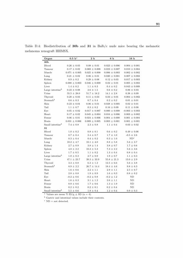
91
Table D.4: Biodistribution of 30b and 31 in Balb/c nude mice bearing the melanoticmelanoma xenograft HHMSX.
Organ 0.5 ha 2 h 6 h 16 h
30b
Blood 0.26 ± 0.03 0.08 ± 0.01 0.023 ± 0.006 0.004 ± 0.001
Tumour 0.17 ± 0.02 0.069 ± 0.008 0.023 ± 0.007 0.010 ± 0.004
Muscle 0.071 ± 0.005 0.025 ± 0.008 0.006 ± 0.003 0.002 ± 0.002
Lung 0.21 ± 0.02 0.06 ± 0.01 0.020 ± 0.001 0.007 ± 0.002
Kidney 0.9 ± 0.2 0.29 ± 0.08 0.12 ± 0.03 0.017 ± 0.003
Spleen 0.088 ± 0.003 0.046 ± 0.009 0.02 ± 0.01 0.005 ± 0.002
Liver 1.4 ± 0.2 1.1 ± 0.3 0.4 ± 0.2 0.043 ± 0.009
Large intestineb 0.22 ± 0.08 2.6 ± 1.5 0.6 ± 0.2 0.06 ± 0.03
Urine 55.5 ± 28.0 51.7 ± 18.2 14.1 ± 2.8 0.38 ± 0.09
Thyroid 0.20 ± 0.03 0.11 ± 0.03 0.03 ± 0.01 0.004 ± 0.002
Stomachb 0.6 ± 0.3 0.7 ± 0.4 0.2 ± 0.2 0.01 ± 0.01
Skin 0.23 ± 0.04 0.06 ± 0.01 0.028 ± 0.005 0.02 ± 0.01
Tail 1.1 ± 0.7 0.3 ± 0.2 0.16 ± 0.09 0.11 ± 0.08
Eye 0.05 ± 0.02 0.017 ± 0.007 0.000 ± 0.008 -0.003 ± 0.003
Heart 0.17 ± 0.02 0.045 ± 0.004 0.016 ± 0.006 0.003 ± 0.002
Femur 0.06 ± 0.01 0.024 ± 0.006 0.004 ± 0.008 0.001 ± 0.004
Brain 0.031 ± 0.006 0.009 ± 0.005 0.003 ± 0.001 0.001 ± 0.001
Small intestineb 7.4 ± 0.8 2.3 ± 0.8 1.1 ± 0.4 0.03 ± 0.02
31
Blood 1.0 ± 0.2 0.8 ± 0.1 0.6 ± 0.2 0.49 ± 0.06
Tumour 0.7 ± 0.4 2.4 ± 0.7 1.7 ± 1.0 -0.5 ± 2.6
Muscle 0.3 ± 0.4 0.4 ± 0.2 0.3 ± 1.0 NDc
Lung 10.2 ± 4.7 10.1 ± 4.0 8.8 ± 5.8 5.6 ± 3.7
Kidney 2.7 ± 0.9 3.8 ± 1.4 3.8 ± 0.7 1.7 ± 0.6
Spleen 4.8 ± 2.2 10.3 ± 5.3 7.3 ± 2.2 5.6 ± 3.6
Liver 1.7 ± 0.5 1.1 ± 0.2 1.3 ± 0.4 0.8 ± 0.4
Large intestineb 1.0 ± 0.3 2.7 ± 0.9 1.9 ± 0.7 1.1 ± 0.3
Urine 47.1 ± 23.7 39.3 ± 33.9 55.8 ± 21.3 13.0 ± 2.9
Thyroid 3.3 ± 0.8 6.3 ± 1.2 12.5 ± 6.6 5.6 ± 3.8
Stomachb 8.9 ± 2.2 26.7 ± 14.4 18.4 ± 4.6 9.8 ± 6.3
Skin 1.6 ± 0.6 2.2 ± 1.1 2.9 ± 1.1 2.5 ± 0.7
Tail 2.8 ± 0.8 1.9 ± 0.9 1.6 ± 0.3 0.8 ± 0.2
Eye -0.2 ± 0.6 -0.2 ± 0.8 -0.2 ± 1.2 ND
Heart 1.6 ± 0.3 3.1 ± 1.2 2.6 ± 1.1 ND
Femur 0.9 ± 0.6 1.7 ± 0.6 1.3 ± 1.9 ND
Brain 0.2 ± 0.2 0.2 ± 0.1 0.2 ± 0.4 ND
Small intestineb 5.5 ± 0.6 1.8 ± 0.4 1.2 ± 0.4 0.9 ± 0.3a Values are mean % ID/g ± SD (n = 4).b Gastric and intestinal values include their contents.c ND = not detected.

92 Biodistribution data
Table D.5: Biodistribution of 32b in Balb/c nude mice bearing the melanotic melanomaxenograft HHMSX.
Organ 0.5 ha 2 h 6 h 16 h
32b
Blood 0.04 ± 0.02 0.04 ± 0.02 0.003 ± 0.001 0.002 ± 0.008
Tumour 0.05 ± 0.02 0.04 ± 0.02 0.005 ± 0.004 0.007 ± 0.001
Muscle 0.02 ± 0.02 0.009 ± 0.004 -0.001 ± 0.009 -0.004 ± 0.017
Lung 0.03 ± 0.01 0.05 ± 0.02 -0.002 ± 0.004 -0.001 ± 0.012
Kidney 0.14 ± 0.04 0.11 ± 0.05 -0.001 ± 0.007 -0.004 ± 0.008
Spleen 0.03 ± 0.03 0.03 ± 0.02 0.003 ± 0.007 0.000 ± 0.003
Liver 0.15 ± 0.06 0.12 ± 0.05 0.001 ± 0.001 0.001 ± 0.003
Large intestineb 0.08 ± 0.06 0.10 ± 0.06 0.01 ± 0.02 -0.008 ± 0.011
Urine 70.9 ± 14.2 6.7 ± 4.2 0.3 ± 0.3 0.7 ± 0.8
Thyroid 0.06 ± 0.03 0.13 ± 0.09 0.005 ± 0.016 0.06 ± 0.06
Stomachb 0.5 ± 0.4 0.3 ± 0.2 0.02 ± 0.01 0.03 ± 0.03
Skin 0.04 ± 0.02 0.03 ± 0.02 0.009 ± 0.005 0.006 ± 0.008
Tail 0.10 ± 0.03 0.07 ± 0.03 0.008 ± 0.007 0.021 ± 0.008
Eye 0.02 ± 0.05 -0.005 ± 0.036 -0.003 ± 0.045 0.005 ± 0.037
Heart 0.02 ± 0.02 0.02 ± 0.02 0.002 ± 0.010 0.005 ± 0.010
Femur -0.004 ± 0.026 0.03 ± 0.02 0.006 ± 0.006 0.01 ± 0.03
Brain 0.002 ± 0.004 0.005 ± 0.007 0.000 ± 0.003 0.001 ± 0.002
Small intestineb 0.6 ± 0.5 0.2 ± 0.1 0.006 ± 0.007 0.001 ± 0.006a Values are mean % ID/g ± SD (n = 4).b Gastric and intestinal values include their contents.

93
Table D.6: Biodistribution of 33b and 53 in Balb/c nude mice bearing the melanoticmelanoma xenograft HHMSX.
Organ 0.5 ha 2 h 6 h 16 h
33b
Blood 0.8 ± 0.1 0.4 ± 0.1 0.04 ± 0.01 0.07 ± 0.04
Tumour 0.7 ± 0.4 0.24 ± 0.07 0.03 ± 0.01 0.05 ± 0.03
Muscle 0.23 ± 0.07 0.08 ± 0.02 0.013 ± 0.006 0.03 ± 0.05
Lung 0.7 ± 0.1 0.7 ± 0.4 0.08 ± 0.02 0.11 ± 0.02
Kidney 2.9 ± 1.3 1.5 ± 0.7 0.14 ± 0.02 0.3 ± 0.1
Spleen 0.6 ± 0.3 0.3 ± 0.1 0.02 ± 0.01 0.05 ± 0.03
Liver 3.5 ± 1.1 2.0 ± 0.6 0.16 ± 0.04 0.31 ± 0.06
Large intestineb 3.1 ± 2.6 11.2 ± 3.5 1.6 ± 0.9 3.2 ± 1.4
Urine 312.9 ± 51.1 33.6 ± 15.3 8.0 ± 1.1 11.5 ± 3.3
Thyroid 1.3 ± 0.3 1.2 ± 0.2 0.2 ± 0.1 0.6 ± 0.6
Stomachb 9.0 ± 5.6 4.4 ± 2.2 0.2 ± 0.1 0.5 ± 0.3
Skin 0.6 ± 0.1 0.4 ± 0.1 0.06 ± 0.02 0.10 ± 0.05
Tail 1.3 ± 0.3 0.6 ± 0.1 0.23 ± 0.08 0.21 ± 0.04
Eye 0.27 ± 0.07 0.16 ± 0.04 0.03 ± 0.06 0.02 ± 0.04
Heart 0.5 ± 0.1 0.21 ± 0.06 0.03 ± 0.01 0.03 ± 0.03
Femur 0.3 ± 0.1 0.2 ± 0.1 0.01 ± 0.01 0.02 ± 0.03
Brain 0.081 ± 0.007 0.04 ± 0.02 0.007 ± 0.004 0.008 ± 0.001
Small intestineb 12.2 ± 10.8 5.6 ± 3.9 0.4 ± 0.2 1.7 ± 0.3
53
Blood 1.1 ± 0.2 1.2 ± 0.3 0.56 ± 0.03 0.6 ± 0.2
Tumour 1.6 ± 0.7 2.0 ± 0.5 1.2 ± 0.2 1.0 ± 0.4
Muscle 0.5 ± 0.2 0.56 ± 0.05 0.29 ± 0.05 0.4 ± 0.3
Lung 6.9 ± 3.3 6.8 ± 3.3 4.2 ± 0.4 4.5 ± 0.5
Kidney 4.1 ± 0.9 4.8 ± 2.2 1.7 ± 0.2 2.1 ± 0.5
Spleen 5.3 ± 2.2 6.3 ± 1.5 3.4 ± 0.2 3.9 ± 1.1
Liver 3.1 ± 0.9 2.1 ± 0.4 0.81 ± 0.06 0.9 ± 0.2
Large intestineb 3.3 ± 3.3 6.1 ± 2.5 1.6 ± 1.3 1.8 ± 0.8
Urine 285.9 ± 102.0 28.9 ± 10.7 15.6 ± 4.6 16.9 ± 9.1
Thyroid 4.4 ± 1.5 6.9 ± 1.3 5.2 ± 2.0 12.9 ± 9.1
Stomachb 12.8 ± 5.5 13.9 ± 2.7 9.8 ± 2.9 10.8 ± 4.6
Skin 2.1 ± 0.7 2.7 ± 0.5 1.7 ± 0.3 2.3 ± 0.4
Tail 2.5 ± 0.8 2.2 ± 0.2 1.3 ± 0.2 1.6 ± 0.3
Eye 0.7 ± 0.2 0.7 ± 0.2 0.48 ± 0.06 0.5 ± 0.1
Heart 2.2 ± 0.7 2.7 ± 0.6 1.22 ± 0.05 1.3 ± 0.4
Femur 1.1 ± 0.3 1.4 ± 0.7 0.61 ± 0.03 0.8 ± 0.4
Brain 0.19 ± 0.06 0.21 ± 0.07 0.12 ± 0.03 0.08 ± 0.02
Small intestineb 8.3 ± 7.9 3.2 ± 0.9 1.0 ± 0.1 1.2 ± 0.3a Values are mean % ID/g ± SD (n = 4).b Gastric and intestinal values include their contents.

94 Biodistribution data
Table D.7: Biodistribution of 14b and 54 in Balb/c nude mice bearing the melanoticmelanoma xenograft HHMSX.
Organ 0.5 ha 2 ha 6 hb 16 hb
14b
Blood 0.5 ± 0.1 0.14 ± 0.03 0.014 ± 0.002 0.005 ± 0.001
Tumour 0.8 ± 0.2 0.5 ± 0.1 0.15 ± 0.09 0.06 ± 0.02
Muscle 0.35 ± 0.07 0.05 ± 0.03 0.003 ± 0.004 0.002 ± 0.002
Lung 1.2 ± 0.2 0.18 ± 0.03 0.022 ± 0.007 0.006 ± 0.001
Kidney 2.4 ± 0.5 0.60 ± 0.08 0.05 ± 0.03 0.013 ± 0.005
Spleen 0.8 ± 0.2 0.09 ± 0.02 0.011 ± 0.004 0.004 ± 0.003
Liver 2.3 ± 0.4 0.42 ± 0.04 0.08 ± 0.02 0.03 ± 0.01
Large intestinec 2.1 ± 1.1 3.4 ± 2.2 0.3 ± 0.2 0.008 ± 0.001
Urine 183.1 ± 109.1 107.6 ± 15.0 20.5 ± 18.4 0.10 ± 0.03
Thyroid 1.5 ± 0.3 0.4 ± 0.1 0.04 ± 0.02 0.004 ± 0.001
Stomachc 1.7 ± 1.5 0.6 ± 0.1 0.05 ± 0.02 0.006 ± 0.002
Skin 0.7 ± 0.1 0.20 ± 0.04 0.028 ± 0.007 0.007 ± 0.004
Tail 1.7 ± 0.5 0.5 ± 0.2 0.14 ± 0.08 0.2 ± 0.2
Eye 0.24 ± 0.04 0.05 ± 0.02 0.008 ± 0.003 0.003 ± 0.005
Heart 0.5 ± 0.1 0.098 ± 0.005 0.009 ± 0.002 0.004 ± 0.002
Femur 0.4 ± 0.1 0.07 ± 0.02 0.004 ± 0.004 0.003 ± 0.004
Brain 0.21 ± 0.06 0.023 ± 0.005 0.002 ± 0.001 0.001 ± 0.001
Small intestinec 3.1 ± 2.6 0.6 ± 0.4 0.05 ± 0.02 0.007 ± 0.002
54
Blood 1.04 ± 0.09 0.6 ± 0.2 0.48 ± 0.08 0.29 ± 0.09
Tumour 0.9 ± 0.7 0.9 ± 0.4 0.4 ± 0.3 0.4 ± 0.2
Muscle 0.6 ± 0.2 0.18 ± 0.09 0.2 ± 0.1 0.1 ± 0.1
Lung 5.5 ± 2.9 2.8 ± 0.9 3.8 ± 2.3 2.0 ± 0.9
Kidney 3.8 ± 0.6 2.1 ± 1.0 1.23 ± 0.8 0.9 ± 0.3
Spleen 3.4 ± 0.9 3.8 ± 1.5 2.5 ± 0.9 1.9 ± 0.9
Liver 2.2 ± 0.2 0.8 ± 0.3 0.6 ± 0.2 0.30 ± 0.03
Large intestinec 2.4 ± 1.6 2.5 ± 0.6 0.5 ± 0.1 0.5 ± 0.1
Urine 137.0 ± 70.4 149.1 ± 27.6 28.1 ± 17.2 5.0 ± 1.5
Thyroid 4.9 ± 3.4 3.4 ± 2.0 2.3 ± 0.8 1.1 ± 0.2
Stomachc 4.3 ± 4.0 11.2 ± 5.4 5.0 ± 1.3 4.4 ± 1.3
Skin 2.2 ± 1.0 2.0 ± 0.5 1.3 ± 0.5 1.1 ± 0.6
Tail 3.0 ± 1.7 1.2 ± 0.3 0.9 ± 0.2 0.6 ± 0.2
Eye 0.7 ± 0.4 0.5 ± 0.3 0.43 ± 0.05 0.3 ± 0.3
Heart 1.2 ± 0.3 1.9 ± 0.9 0.8 ± 0.1 0.9 ± 0.5
Femur 0.6 ± 0.3 0.5 ± 0.2 0.4 ± 0.1 0.4 ± 0.1
Brain 0.2 ± 0.2 0.16 ± 0.05 0.10 ± 0.06 0.10 ± 0.03
Small intestinec 2.5 ± 2.2 1.1 ± 0.2 0.71 ± 0.08 0.6 ± 0.2a Values are mean % ID/g ± SD (n = 4).b Values are mean % ID/g ± SD (n = 3).c Gastric and intestinal values include their contents.

95
Table D.8: Biodistribution of 14b and 54 in Balb/c nude mice bearing the amelanoticmelanoma xenograft SESX.
Organ 1 ha 4 h 16 h
14b
Blood 0.5 ± 0.1 0.05 ± 0.02 0.011 ± 0.001
Skin 1.1 ± 0.4 0.09 ± 0.02 0.022 ± 0.005
Heart 0.7 ± 0.7 0.03 ± 0.01 0.008 ± 0.003
Lung 0.6 ± 0.2 0.056 ± 0.005 0.018 ± 0.006
Liver 2.3 ± 0.5 0.26 ± 0.04 0.103 ± 0.004
Kidney 1.2 ± 0.9 0.15 ± 0.03 0.052 ± 0.004
Spleen 0.4 ± 0.1 0.032 ± 0.006 0.010 ± 0.003
Stomachb 6.4 ± 7.3 0.3 ± 0.1 0.02 ± 0.01
Small intestineb 7.5 ± 4.1 0.34 ± 0.02 0.03 ± 0.01
Eye 0.2 ± 0.1 0.001 ± 0.002 0.002 ± 0.004
Tumour 1.0 ± 0.3 0.47 ± 0.07 0.095 ± 0.006
Muscle 0.2 ± 0.1 0.02 ± 0.01 0.000 ± 0.000
Femur 0.2 ± 0.1 0.04 ± 0.04 0.001 ± 0.002
Urine – – 0.11 ± 0.09
Large intestineb 4.7 ± 4.5 1.2 ± 0.6 0.02 ± 0.02
54
Blood 0.5 ± 0.1 0.24 ± 0.08 0.10 ± 0.04
Skin 1.7 ± 0.4 0.7 ± 0.2 0.3 ± 0.1
Heart 1.2 ± 0.2 0.6 ± 0.1 0.23 ± 0.04
Lung 2.5 ± 1.9 1.1 ± 0.4 0.8 ± 0.1
Liver 1.6 ± 0.3 0.62 ± 0.02 0.210 ± 0.009
Kidney 2.0 ± 0.9 1.1 ± 0.3 0.42 ± 0.05
Spleen 3.7 ± 1.0 2.0 ± 0.7 0.8 ± 0.2
Stomachb 4.7 ± 3.1 3.2 ± 0.8 1.2 ± 0.4
Small intestineb 4.1 ± 2.1 0.48 ± 0.05 0.17 ± 0.05
Eye 0.3 ± 0.1 0.11 ± 0.02 0.05 ± 0.04
Tumour 1.3 ± 0.4 0.66 ± 0.07 0.15 ± 0.06
Muscle 0.4 ± 0.1 0.20 ± 0.09 0.04 ± 0.02
Femur 0.5 ± 0.2 0.29 ± 0.08 0.13 ± 0.03
Urine – – 1.3 ± 0.6
Large intestineb 3.5 ± 1.8 0.64 ± 0.08 0.2 ± 0.1a Values are mean % ID/g ± SD (n = 3).b Gastric and intestinal values include their contents.

96 Biodistribution data

List of Figures
2.1 Decay scheme for 211At. . . . . . . . . . . . . . . . . . . . . . . . . . . . . . . . . . . . . . . . . 9
2.2 Proposed reaction mechanism of the halodemetallation using aryltrialkyl group 14 organometal-
lic precursors. . . . . . . . . . . . . . . . . . . . . . . . . . . . . . . . . . . . . . . . . . . . . . . 12
3.1 The melanin synthesis. . . . . . . . . . . . . . . . . . . . . . . . . . . . . . . . . . . . . . . . . . 19
3.2 Chemical structures of chlorpromazine (3), phenothiazine (4), MTB 5, methylene green 6 and
the 125I- and 211At-labelled MTB-derivatives 7 and 8. . . . . . . . . . . . . . . . . . . . . . . . 22
3.3 Examples of cysteaminylphenols investigated as melanoma-seeking agents. . . . . . . . . . . . . 23
3.4 Examples of benzamides investigated as melanoma-seeking agents. . . . . . . . . . . . . . . . . 24
5.1 Chemical structure of Azure B 15 and general structures of the proposed target molecules. . . 29
5.2 Reaction scheme for synthesis of the activated esters 16 and 18. . . . . . . . . . . . . . . . . . 30
5.3 Retrosynthetic analysis of the stannylated precursor 22. . . . . . . . . . . . . . . . . . . . . . . 31
5.4 Summary of reactions between Azure B 15 and different linkers. . . . . . . . . . . . . . . . . . 32
5.5 Retrosynthetic analysis of the stannylated precursor 25. . . . . . . . . . . . . . . . . . . . . . . 32
5.6 Reaction scheme for synthesis of the stannylated precursor 25. . . . . . . . . . . . . . . . . . . 32
6.1 Chemical structures of the proposed target molecules 28b–33c. . . . . . . . . . . . . . . . . . 33
6.2 First retrosynthetic analysis of the stannylated precursor 34. . . . . . . . . . . . . . . . . . . . 34
6.3 Literature procedure for synthesising the cysteaminylphenols 10 and 11. . . . . . . . . . . . . . 35
6.4 Halogenation of 36 giving disulphide 39. . . . . . . . . . . . . . . . . . . . . . . . . . . . . . . . 35
6.5 Second retrosynthetic analysis of the stannylated precursor 34. . . . . . . . . . . . . . . . . . . 36
6.6 Reaction scheme for thiocyanation of 40 and 41 using PhICl2 and Pb(SCN)2 giving 42 and 43. 36
6.7 Summary of reactions performed aiming at precursor 11. . . . . . . . . . . . . . . . . . . . . . 37
6.8 Reaction scheme for precursor 34 and the 125I- and 211At-labelled cysteaminylphenols 28b and
29. . . . . . . . . . . . . . . . . . . . . . . . . . . . . . . . . . . . . . . . . . . . . . . . . . . . . 38
6.9 Reaction scheme for precursor 48 and the 125I- and 211At-labelled cysteaminylphenols 30b and
31. . . . . . . . . . . . . . . . . . . . . . . . . . . . . . . . . . . . . . . . . . . . . . . . . . . . . 39
6.10 Reaction scheme for the 131I-labelled tyramine derivative 32b. . . . . . . . . . . . . . . . . . . 40
6.11 Reaction scheme for precursor 52 and the ∗I- and 211At-labelled tyramine derivatives 33b,c and
53. . . . . . . . . . . . . . . . . . . . . . . . . . . . . . . . . . . . . . . . . . . . . . . . . . . . . 40
6.12 Results of the biodistribution of 28b, 30b, 32b and 33b in HHMSX-bearing Balb/c nude mice. 43
6.13 Results of the biodistribution of 29, 31 and 53 in HHMSX-bearing Balb/c nude mice. . . . . . 45
7.1 Reaction scheme for precursor 55 and the 125I- and 211At-labelled benzamides 14b and 54. . . 48
97

98 LIST OF FIGURES
7.2 Results of the biodistribution of 14b and 54 in HHMSX- and SESX-bearing Balb/c nude mice. 50
7.3 Tumour uptake values of 14b and 54 in HHMSX- and SESX-bearing Balb/c nude mice. . . . . 53
9.1 Bone-seeking radiopharmaceuticals. . . . . . . . . . . . . . . . . . . . . . . . . . . . . . . . . . . 60
9.2 Examples of bisphosphonates investigated as bone-seeking agents. . . . . . . . . . . . . . . . . . 61
10.1 Automatic generation of selective regions of interest using the FNOMIP method. . . . . . . . . 64
11.1 Reaction scheme for the 123I-labelled bisphosphonate 70b. . . . . . . . . . . . . . . . . . . . . . 65
11.2 Uptake of 68 and 70b in head, neck, thorax and pelvis 4 h post-injection. . . . . . . . . . . . . 67

List of Tables
2.1 Data on some potential radionuclides for targeted radiotherapy. . . . . . . . . . . . . . . . . . . 7
3.1 Melanoma-seeking radiopharmaceuticals. . . . . . . . . . . . . . . . . . . . . . . . . . . . . . . . 21
6.1 Integrated radiochemical yields estimated by radio-HPLC. . . . . . . . . . . . . . . . . . . . . . 41
6.2 Properties of radiolabelled cysteaminylphenols and tyramine derivatives studied in HHMSX-
bearing Balb/c nude mice. . . . . . . . . . . . . . . . . . . . . . . . . . . . . . . . . . . . . . . . 42
6.3 Melanoma/non-target tissue dose ratios of 28b, 30b, 32b and 33b in HHMSX-bearing Balb/c
nude mice. . . . . . . . . . . . . . . . . . . . . . . . . . . . . . . . . . . . . . . . . . . . . . . . . 44
7.1 Results of pharmacological binding studies of the SESX and HHMSX melanoma models. . . . . 49
7.2 Melanoma/non-target tissue dose ratios of 14b and 54 in HHMSX- and SESX-bearing Balb/c
nude mice. . . . . . . . . . . . . . . . . . . . . . . . . . . . . . . . . . . . . . . . . . . . . . . . . 51
11.1 Estimated radiation doses for injection of [131I]HIPEBA 70c and 153Sm-EDTMP 61 in humans. 68
A.1 Chemical abbreviations . . . . . . . . . . . . . . . . . . . . . . . . . . . . . . . . . . . . . . . . 85
B.1 Glossary of radiochemical and medical terms . . . . . . . . . . . . . . . . . . . . . . . . . . . . 86
D.1 Biodistribution data of 28b and 32b in normal Balb/c nude mice. . . . . . . . . . . . . . . . . 88
D.2 Biodistribution data of 30b and 33c in normal Balb/c nude mice. . . . . . . . . . . . . . . . . 89
D.3 Biodistribution data of 28b and 29 in HHMSX-bearing Balb/c nude mice. . . . . . . . . . . . 90
D.4 Biodistribution data of 30b and 31 in HHMSX-bearing Balb/c nude mice. . . . . . . . . . . . 91
D.5 Biodistribution data of 32b in HHMSX-bearing Balb/c nude mice. . . . . . . . . . . . . . . . . 92
D.6 Biodistribution data of 33b and 53 in HHMSX-bearing Balb/c nude mice. . . . . . . . . . . . 93
D.7 Biodistribution data of 14b and 54 in HHMSX-bearing Balb/c nude mice. . . . . . . . . . . . 94
D.8 Biodistribution data of 14b and 54 in SESX-bearing Balb/c nude mice. . . . . . . . . . . . . . 95
99