Labelling techniques of biomolecules for targeted radiotherapy
196
IAEA-TECDOC-1359 Labelling techniques of biomolecules for targeted radiotherapy Final report of a co-ordinated research project 1998–2002 July 2003
Transcript of Labelling techniques of biomolecules for targeted radiotherapy

IAEA-TECDOC-1359
Labelling techniques of biomolecules for
targeted radiotherapy Final report of a co-ordinated research project
1998–2002
July 2003

The originating Section of this publication in the IAEA was:
Industrial Applications and Chemistry Section International Atomic Energy Agency
Wagramer Strasse 5 P.O. Box 100
A-1400 Vienna, Austria
LABELLING TECHNIQUES OF BIOMOLECULES FOR TARGETED RADIOTHERAPYIAEA, VIENNA, 2003 IAEA-TECDOC-1359ISBN 92–0–107303–8
ISSN 1011–4289© IAEA, 2003
Printed by the IAEA in Austria July 2003

FOREWORD
Radiotherapy using open radioactive sources has become an important and promising mode of treatment of cancer patients, particularly in conjunction with other modalities like surgery and chemotherapy. For this purpose therapeutic radiopharmaceuticals are designed to deliver high doses of radiation to selected target organs or tissues with an aim of minimizing unwanted radiation to surrounding healthy tissue.
Several radionuclides have been developed and tagged into appropriate carriers for targeted radiotherapy. As radionuclide carriers, chelating chemical agents, monoclonal antibodies, peptides, biodegradable particles, colloids, etc., have been investigated as radiopharmaceuticals for therapeutic nuclear medicine.
In spite of the potential of targeted radiotherapy to treat a wide range of malignant conditions, the routine clinical use has been, hampered by the lack of suitable radionuclide carrier molecules which would adequately concentrate these radionuclides in target issues of interest. Much higher and specific uptake of the radiopharmaceutical in the target tissue is needed for effective therapeutic applications than is possible with most of the diagnostic agents currently available. Following the recommendations of a group of experts in 1997, and in view of the realization that higher levels of targeting for therapeutic agents would be possible using biomolecules such as monoclonal antibodies, peptides and other receptor avid molecules as carriers of the radionuclide, in 1998 the International Atomic Energy Agency (IAEA) launched a Co-ordinated Research Project (CRP) on Labelling Techniques of Biomolecules for Targeted Radiotherapy. The CRP aimed at developing and optimizing existing procedures for radiolabelling of monoclonal antibodies and small peptides with therapeutic radionuclides, such as 90Y, 188Re and others.
With the participation of 15 scientists from selected research institutes in Argentina, Austria, Brazil, Cuba, Greece, Finland, Hungary, India, Italy, Mexico, Pakistan, China, Romania, Thailand and Uruguay several laboratory procedures and protocols for the radiolabelling and quality control of monoclonal antibodies and peptides were optimized. Moreover, the CRP provided the participants an opportunity to study the feasibility of using bioactive molecules for developing radiopharmaceuticals and studying its biological efficacy in tumour models. The first part of the report includes standardized labelling and quality control protocols used and validated during the CRP; the second part includes reports of each individual research agreement and contract holder.
The IAEA wishes to thank all the participants in the CRP for their valuable work and scientific contributions. The IAEA officers responsible for this publication were H. Vera Ruiz and D.V.S. Narasimhan of the Physical and Chemical Sciences Division.

EDITORIAL NOTE
This publication has been prepared from the original material as submitted by the authors. The views expressed do not necessarily reflect those of the IAEA, the governments of the nominating Member States or the nominating organizations.
The use of particular designations of countries or territories does not imply any judgement by the publisher, the IAEA, as to the legal status of such countries or territories, of their authorities and institutions or of the delimitation of their boundaries.
The mention of names of specific companies or products (whether or not indicated as registered) does not imply any intention to infringe proprietary rights, nor should it be construed as an endorsement or recommendation on the part of the IAEA.
The authors are responsible for having obtained the necessary permission for the IAEA to reproduce, translate or use material from sources already protected by copyrights.

CONTENTS
INTRODUCTION...................................................................................................................... 1
ACHIEVEMENTS OF THE CRP ............................................................................................. 2
SCOPE OF THE CRP AND TECHNICAL OBSERVATIONS ............................................... 3
REMARKS ON METHODOLOGIES FOR RADIOLABELLING AND QUALITY CONTROL OF SELECTED BIOMOLECULES ........................................... 14
POTENTIAL AREAS OF FUTURE RESEARCH ................................................................. 16
COLLABORATIVE ACTIVITIES ......................................................................................... 17
TECHNICAL CONCLUSIONS OF THE CRP....................................................................... 17
PUBLICATIONS AND PRESENTATIONS ORIGINATING FROM THE CRP ................. 18
BIBLIOGRAPHY .................................................................................................................... 25
COUNTRY REPORTS
Optimization of biomolecules labelling with Rhenium-188using direct and indirect methods..................................................................................... 31 S.G. de Castiglia, J. Crudo, E. Obenaus, M. Edreira, E. D’orio
Labelling of peptides with 188Re/99Tcm and monoclonal antibodies with 90Y/111In ................. 45 B. Linkowski Faintuch, C.P.G. Silva, N.P.S. Pereira, E.B. Araujo
Labelling technique of biomolecules for target radiotherapy .................................................. 65 Hongsheng Bai, Jin Xiaohai, Cheng Zhen, Jia Bing Fan Hongqiang, Lu Weiwei
Evaluation of biomolecules for metabolic radiotherapy .......................................................... 73 G.J. Pimentel, M. Miranda, W. Quesada, H.E. Garay, O. Reyes, J. Vazquez, L.J. Cruz, I. Sanchez
Development of radioactively labelled cancer seeking biomolecules for targeted therapy ................................................................................................................ 85 A.D. Varvarigou, S.C. Archimandritis, M. Fani, P. Bouziotis, S. Vranjez,S. Xanthopoulos, A. Leonti, Th. Sourlingas, M. Sotiropoulou
Preparation, quality control and animal testing of Re-188 labelled compounds ..................... 99 G.A. Jánoki, L. Körösi, L. Balogh, G. Andócs, K. Bodó, J. Láng, A. Polyák, R. Király, Domokos
Development of radioactively labelled cancer seeking biomolecules for targeted therapy... 107 M.R.I. Pillai, M. Venkatesh, S. Banerjee, G. Samuel, K. Kothari, A. Dash, P.R. Unni, A. Korde, K. Bapat, U. Pandey, T. Dash, S. Chakraborty, A. Mukherjee, H.D. Sharma
Labelled peptides through the DOTA chelating agent for diagnosis and treatment of receptor positive tumours ............................................................................................... 123 M. Chinol
Labelled biomolecules for targeted radiotherapy: Biodistribution in mice with induced malignant tumours.......................................................................................................... 129 C. Arteaga de Murphy, G. Ferro-Flores, M. Pedraza-López, E.M. Molina-Trinidad, E. Murphy-Stack, S. Hernández-Gutiérrez

Labelled biomolecules with 153Sm, 188Re, and 90Y for targeted radiotherapy........................ 139 A. Mushtaq, S. Perverz, M. Khalid
Development of 188Re radiolabelling procedures of peptides and monoclonal antibodies.................................................................................................... 155 V.V. Lungu, G. Mihailescu, D. Niculae
Radiolabelling of monoclonal antibodies for radiotherapy.................................................... 165 J. Sangsuriyan, C. Wardwilai, W. Iamsam-Ang, T. Ngamprayad, P. Picha, M. Dangpraset
Labelling and control of biomolecules with 188Re and 153Sm ................................................ 183 H.S. Balter, E.S. Verdera, G. Rodríguez, P. Oliver, B. Souto, L. Mallo, A. Robles
LIST OF PARTICIPANTS .................................................................................................... 197

1
INTRODUCTION
Malignant tumour disease accounts for approximately one third of deaths worldwide. Gastrointestinal adenocarcinomas, prostate and breast cancers are among the most frequently appearing tumours. Radiotherapy is an essential mode of treatment of all cancer patients either alone or in conjunction with other modalities like surgery and chemotherapy. In most cases radiotherapy is given using external radiation sources. It is also possible to administer radiotherapy by specifically localizing radioisotopes emitting particulate radiation in the tumour tissue. This targeted therapy has proved to have several advantages over external beam therapy, notably the possibility of selectively delivering higher radiation doses to the targeted tumour cells and treating multiple metastases. Procedures for therapy of thyroid carcinoma and hyper-thyroidism using radioiodine (131I) introduced about five decades ago, have stood the test of time and are still widely used the world over. In addition to the therapeutic nuclides of the first generation 131I, 89Sr, 32P, 90Y, etc., which are still widely utilized and accepted by the medical community, many other beta emitting radionuclides with relatively short half-lives such as 153Sm, 186Re, 188Re, 166Ho, 165Dy, etc. have also been recently made available for therapy and used with promising good results.
In spite of the potential of targeted radiotherapy to treat a wide range of malignant conditions, routine clinical use is mostly confined to therapy of thyroid carcinoma, hyperthyroidism, metastatic bone pain and synovectomy. In most of the cases, the limitation is obviously not the availability of suitable radionuclides but rather the lack of suitable carrier molecules that would adequately concentrate these radionuclides in target tissues of interest. Much higher and specific uptake of the radiopharmaceutical in the target tissue is needed for effective therapeutic applications than is possible with most of the diagnostic agents currently available. It is expected that higher levels of targeting needed for therapeutic agents would be possible using biomolecules such as monoclonal antibodies, peptides and other receptor avid molecules as carriers of the radionuclide.
During the last decade considerable efforts were directed towards stable binding of many radionuclides to monoclonals using a variety of bifunctional chelating agents (BFCA). Many antibody based agents have now been cleared for human diagnostic use. Radiolabelled antibodies to treat successfully hematological malignancies have been reported. Attempts were also made to pre-target tumours with other substrates and accelerated clearance of circulating antibodies to achieve the necessary specific concentration. Based on the experience gained with diagnostic agents, it can be expected that monoclonal antibody based therapeutic radiopharma-ceuticals will soon be available for regular clinical use.
Moeover, the introduction of small peptides as carrier molecules for radionuclides has opened up new possibilities for development of diagnostic agents as evidenced by the use of 111In-octreotide. Efforts are underway to extend the use of peptides as carriers for other therapeutic radionuclides such as 188Re, 90Y, 153Sm, etc. The initial successful attempts to use Auger electrons of 111In for therapeutic effect by high dose administration of 111In octreotide has provided a strong impetus for the search, development and use of more accessible and perhaps more economical radionuclides like 153Sm, 90Y, 177Lu, etc. coupled with peptides and monoclonal antibody carriers. The incorporation of beta emitters into a variety of bifunctional chelates of bioactive peptides including octreotide and VIP analogues is vigorously pursued at many leading laboratories. These efforts are expected to result in the development of several new radiopharmaceuticals for the radiotherapy of a number of malignancies.

2
Based on the above considerations, the scope of the Co-ordinated Research Project (CRP) has focused on the synthesis of the required BFCAs for MoAbs and peptide labelling, development and optimization of radiolabelling techniques with beta emitting radioisotopes, including quality control procedures and evaluation of the labelled products in in vitro and in vivo models. Biomolecule carriers that were considered for investigation included somatostatin analogues like lanreotide, Tyr-octreotide, ior-P1394 and vasointestinal peptide (VIP), as well as anti-CEA monoclonal antibody. Using either the bifunctional chelate approach or direct reductive exposure of -SH groups, labelling of these biomolecules with therapeutic radionuclides was pursued. Through these attempts it was expected to identify one or two agents with high radiochemical purity and labelling efficiency for further evaluation. Promising agents would be selected for further investigation using biodistribution studies in animals and uptake in tumour bearing animals.
Fifteen selected research institutes from Argentina, Austria, Brazil, Cuba, Greece, Finland, Hungary, India, Italy, Mexico, Pakistan, Peoples Republic of China, Romania, Thailand and Uruguay with recognized expertise in the field of therapeutic radionuclides and radiopharmaceutical research were selected to participate in the CRP. The first research co-ordination meeting to plan the work and define the research protocols to be investigated was held in Milan, Italy in July 1998. At this meeting it was decided to concentrate the efforts on two radionuclides, 188Re and 90Y, which are produced by generator systems and result in carrier free products; lanreotide was selected as a model peptide. Synthesis of the bifunctional chelating agent DOTA was also recommended for the labelling of biomolecules with 90Y. The development of the corresponding coupling techniques, optimization of protocols for direct and indirect labelling of lanreotide and anti-carcino embrionic antigen (CEA) monoclonal antibody and in vitro evaluation in cell lines and membrane receptors were also included in the work plan for the following period. IgG labelled with 131I and 125I and 99Tcm was also included in the work plan for the initial investigation and method evaluation.
The second RCM to review the progress and discuss future work was held in Mumbai, India in February 2000. In view of the progress reported at this meeting, it was recommended that in the remaining period the CRP should concentrate on the testing of in vitro bioactivity of the labelled 188Re lanreotide, confirm the results reported on the direct labelling method for 188Re–lanreotide and continue the investigations to label DOTA conjugated peptides with 90Y.The third and final RCM was held in Budapest, Hungary in October 2001 to assess the progress and achievements of the CRP and draft the final report.
A list of general references to guide the reader into recent research work done in the field is given in Section VIII.
ACHIEVEMENTS OF THE CRP
– The CRP provided an opportunity for the participants to study the feasibility of using bioactive molecules for developing radiopharmaceuticals and studying its biological efficacy in tumour models.
– Most of the participants achieved the development of technology for the preparation of a generator, synthesis of bifunctional chelating agents and radiolabelling of the biomolecules with several radionuclides.

– Radiolabelling of lanreotide with 90Y through bifunctional chelating agent was also achieved. The procedure developed is suitable for making clinical doses of 90Y labelled lanreotide.
– Radiolabelling of peptides, in particular lanreotide, in microgram quantities with 188Re was done during the course of the CRP. Radiolabelling studies are significant in that they provided opportunities to develop radiotracers in nanomolar concentration of ligands/ peptides. The studies carried out during the CRP also culminated in the development of directly labelled 188Re-lanreotide. The labelling technique developed could be extended to several other biomolecules.
– The participants developed several quality control techniques for radionuclides and for labelled peptides, which will be useful for evaluating peptide-based radiopharmaceuticals.
– The participants during the course of the CRP developed in vitro and in vivo biological techniques. These techniques are essential and the knowledge accumulated is relevant for studying the biological efficacy of receptor-based radiopharmaceuticals.
– Most of the participants generated good quality scientific data, which were presented in meetings and published in journals.
– Interactive co-operation and collaboration was established between several participants. Both information and material exchange were established between participants. The collaborations established between the groups during the course of the CRP are expected to continue and benefit the participants.
SCOPE OF THE CRP AND TECHNICAL OBSERVATIONS
Radionuclides
Rhenium-188
188Re is an attractive radionuclide for use in targeted radiotherapy due to its favourable physical characteristics. Another advantage is its availability through a generator system. Production of the parent (188W) involves a double neutron capture reaction and requires a very high neutron flux in the order of 1015 n/s/cm2 to get good yield. The major problem with the production of 188W/188Re generator is therefore the availability of high specific activity 188W.
Two batches of 188W/188Re generator from MAP Medical Technologies Oy, Finland were made available by the IAEA to all participants for laboratory use and animal studies. Evaluation of generator parameters was done until six months after calibration to assure good quality of the eluates for their use in radiolabelling studies. The results indicated that elution yield was in the order of 70-80% and elution profile showed the maximum of activity (88% of total activity) in the first three mL. Thus the activity concentration could be at least 10 mCi/mL, which is sufficient for peptide labelling. The pH was according to specifications. Chromatography in saline and methylethylketone (MEK) as solvents on Whatman Nº 1 showed radiochemical purity higher than 99%. The level of radionuclidic impurities, as determined by gamma ray spectrometry, was in all cases below the limits allowed for this type of generators. The presence of 188W ranged from 5.2 to 9.6×10⁻³%. Chemical controls showed that levels of Al3+ were in all cases lower than 5 ppm. Moreover, no toxic effects were observed in the studies conducted in animals.
3

4
Also, some participants used 188W/188Re generators from Oak Ridge National Laboratory, USA. The elution volumes were generally higher thus requiring a further step of concentration.
90Sr-90Y generator
90Y was selected as the radionuclide of choice for labelling biomolecules through a chelating agent. Commercial source of 90Y is expensive and hence efforts are to be directed towards developing 90Sr-90Y generator for local production of 90Y. One generator concept was developed in India based on the supported liquid membrane technique. The generator consists of a glass cell comprising of two compartments, namely feed and receiver, of capacity 5 mL each, separated by a polytetrafluoroethylene membrane impregnated with 2-ethylhexyl-2-ethylhexyl phosphonic acid (KSM-17). A suitable aliquot of high level waste depleted of actinides-lanthanides and 137Cs in 0.5 M HNO3 (up to 100 mCi, 3.7 GBq) was diluted to 5 mL, adjusted to pH 1-2 and used as feed. 5 mL of 1 M HCl/HNO3 was used in the receiver compartment. Both the feed and receiver compartments were constantly stirred with small teflon coated magnetic stirrer to enable quick and effective transport of 90Y across the membrane. After 4 hours (h) the solution containing yttrium chloride/nitrate was drawn from the receiving compartment and used.
Solvent extraction/absorption of 90Y from 90Sr
An alternate method is the separation of 90Sr and 90Y by solvent extraction followed by ion exchange chromatography. This separation is based on an initial solvent extraction of 90Yfrom dilute nitric acid solution of the 90Sr-90Y mixture using D2EHPA/dodecane. Further purification of 90Y was carried out using ion exchange technique based on the absorption of the product on a column containing Dowex-50×12 and selective elution of 90Y in 0.6M sodium acetate buffer solution at pH 5.57. Based on the experimental data, a process was demonstrated in which the recovery of 90Y obtained is higher than 75%. The radionuclidic purity of the 90Y obtained proved to be satisfactory for medical application. The details of the procedure are available in the country report.
The most important criteria for a 90Sr-90Y generator are the quality control tests to ensure the absence of 90Sr in the eluate. Analytical methods developed for measuring 90Srbreakthrough are discussed in the quality control section.
Holmium-166
166Ho can be prepared in high specific activity by irradiating natural Ho in a medium to high flux reactor. Typically, 6 mg of natural Ho2O3 powder (100% 165Ho, 65 barns) was weighed and sealed in a quartz ampoule and irradiated at a thermal neutron flux of 3x10¹³
n/cm2/s for seven days and cooled for 6 h. Irradiated Ho2O3 powder was dissolved in 5 mL of 0.1 M HCl by gentle warming. The resultant solution was evaporated to near dryness and reconstituted in 10 mL of double distilled water.
The gamma ray spectrum of 166Ho did not show the presence of any radionuclidic impurity. Major gamma peak observed was at 81 keV (6%), photopeak of 166Ho. Minor peaks of 166Ho at 1379 keV (0.9%), 1581 keV (0.18%) and 1662 keV (0.12%) were also notable.

5
Lutetium-177
177Lu is one of the isotopes offering very good potential for targeted therapy due to the possibility of making the isotope in high specific activity (reaction cross-section 2100 barns). Lu also has a very favourable chemistry and could be used with chelating agents that are suitable for Y. A typical procedure was given: 1.2 mg of enriched Lu2O3 powder (60.7% 176Lu) was dissolved in 1M HCl on gentle warming. The resulting solution was evaporated to near dryness and reconstituted in 1.2 mL of double distilled water. 10 µg of the solution (10 µg of Lu203) was taken in a quartz ampoule and carefully evaporated to dryness. The target was irradiated in a medium to high flux reactor for a period of seven days.
Around 20-22 mCi of 177Lu activity was obtained at 6 h after EOB from 10 µg of enriched Lu2O3 powder irradiated for 7 days, corresponding to a specific activity of 2000-2200 Ci/g. Theoretical calculations show that 7 day irradiation at 1.8×10¹³ n/cm2/s flux would yield ~1800 Ci/g of 177Lu activity using enriched Lu2O3 target. On the other hand, by using natural Lu target the specific activity of 177Lu would be ~100 Ci/g for 7 day irradiation at the same flux. The radionuclidic purity of 177Lu was ~100% as obtained by analysing the γ ray spectrum. It is worthwhile to note that there is a possibility for the formation of 177mLu (T1/2 =160.5 d) on thermal neutron bombardment of Lu2O3 target. However, the γ ray spectrum did not show any significant peak (at 128 keV, 153 keV, 228 keV, 378 keV, 414 keV, 418 keV) corresponding to 177mLu. This is expected as radioactivity since 177mLu produced will not be insignificant after a 7 day irradiation owing to its comparatively low cross-section (σ=7 barns) and long half-life (t1/2=161 days) for its formation.
Gallium-67
67Ga is a useful cyclotron produced radioisotope in nuclear medicine. Its physical half-life of 3.26 days and various γ radiations following electron capture (EC) reactions are suitable for tracer studies. The important oxidation state is Ga(III). Its co-ordination chemistry is similar to that of Fe (III). Several chelates for 67Ga have been developed. Peptides conjugated to DOTA or DTPA chelators can be labelled with 67Ga with promising binding to receptors.
Technetium-99m
99Tcm is the most popular radionuclide for clinical imaging because it has ideal nuclear properties, i.e. a single photon energy of 140 Kev, a half-life of 6 h. It is readily available from a 99Mo/99Tcm generator. Some participants labelled biomolecules with 99Tcm in order to establish a comparison between this radionuclide and 188 Re. Because of the similar chemistry of these two radionuclides, participants used their experience to label peptides (lanreotide, Tyr-octreotide, ior-P1394, N4-Lys-biotin) and antibodies (IgG, iorCEA1, scFv).
Thallium-201
201Tl is an Auger electron emitting radioisotope. In this case, if the carrying biomolecule is labelled within the tumour cell, the labelled species can be useful for radiotherapeutic applications.
Thallium-201 was used by the participant from Greece to label human polyclonal immunoglobulin (IgG, sandoglobulin).

6
In this case, in a commercially available thallous chloride (201TlCl) solution of 0.1–0.5 mL, a solution of H2SO4 5M (1.0 mL) was added followed by a fresh solution of KBrO3 5% w/v (0.2 ml). The mixture was allowed to stand at room temperature for 15 minutes (min) before the addition of 50 µL 4M NaCl solution. The 201Tl+++ so formed, was extracted by adding 1.5 mL butyl acetate. The content was mixed for 20 min at room temperature. The organic phase was separated from the water phase and was evaporated in a water bath by adding a small volume of water. 201Tl+++ was received as an aqueous solution ready to use for labelling.
Other radioisotopes like Iodine-125 and Iodine-131 were used as non-carrier added preparations in high radioactive concentration. Receptor studies, molecule characterization, and method development experiments were conducted to gain experience which eventually can be used in replacing them with therapeutic radioisotopes.
Indium-111
111In is a radionuclide available from commercial sources and which shows chemical similarities with 90Y widely used in radiotherapy but lacks gamma photons. The same biomolecules could be labelled with this radionuclide to develope new radiotracers and follow the in vivo behaviour in view of replacement with 90Y. For example, in the development of new somatostatin analogues DOTATOC, the affinity for somatostatin receptors and the invivo stability of molecule was tested with 111In prior to the use with 90Y. Dosimetric studies are necessary in the planning of radiotherapeutic trials with new radioligands. With these 111In studies we can get information on the pharmacokinetics and biodistribution of the tracer and also to calculate the residence times in the various organs. These parameters are essential for planning the therapeutic doses to be administered.
Another application of this radionuclide could be the possibility to perform radiotherapy due of its Auger electrons if the 111In labelled molecule is internalized within the tumour cell.
In the frame of this CRP, four participants used this radionuclide for labelling somatostatin analogues and biotin derivatives.
Samarium-153
153Sm is a radiolanthanide, which possesses excellent physical characteristics for radioimmunotherapy. It is a beta-emitter with a half-life of 1.95 days. In addition, it emits 103 keV gamma rays (28%) that are suitable for gamma camera detection.
It can be produced in the reactor by neutron irradiation of enriched 152Sm. For that, Samarium oxide, isotopically enriched in 152Sm (152Sm2O3), can be irradiated. Under these conditions, the specific activity of the 153Sm obtained can be very high depending on the irradiation time and the neutron flux of the reactor. The activated oxide is then dissolved in a solution of 6M HCl, to produce Samarium chloride (153SmCl3). For the labelling, 153SmCl3 is diluted to the desired volume with ultra pure water and the stock solution is ready for further use.
The 153Sm produced under these conditions is suitable for the labelling of MoAbs, biotin-DTPA and biotin-EDTA molecules as well as DOTA-lanreotide.

7
For labelling biotin and Lanreotide good results could be obtained without reaching high specific activities. For DOTA-Lanreotide, extra purification steps are needed. The specific activity of the labelled product must be as high as possible. Specific activities lower than 100mCi/mg did not give so good results.
153Sm was used by eight of the participating countries, either by irradiating in their own reactors (Argentina, China, Greece, India, Mexico, Pakistan and Thailand), or within the frames of IAEA or the ARCAL XV programme, Peru provided 153Sm to Uruguay.
Chelating agents and biomolecules
Synthesis of bifunctional chelating agents
A) DOTA (1,4,7,10-tetraazacyclododecane-N,N’,N’’,N’’’ tetraacetic acid)
1,4,7,10-Tetraazacyclododecane-N, N’, N’’, N’’’ tetraacetic acid (DOTA) was synthesized in four steps. The synthetic route involved the use of inexpensive starting materials.
Step I: Triethylene tetramine (3.3g, 0.027 moles) was tosylated using p-toluene sulphonyl chloride (26.3 g, 0.14 moles) in acetonitrile (125 mL) in the presence of triethylamine (16 mL, 0.115 moles). The reaction involved a portion wise addition of p-toluene sulphonyl chloride to a magnetically stirred solution of triethylene tetramine and triethyl amine at room temperature under N2 circulation for 1 h. Stirring was continued for 5 h while monitoring the progress of the reaction on a silica gel TLC in methanol: chloroform: ammonium hydroxide (5:94.5:0.5 v/v/v) (Rf 0.8). The solvent was removed under vacuum to yield a white semisolid. The workup involves addition of DDW and extraction using 3×25 mL of chloroform after saturation with brine. The pooled organic extracts were dried over sodium sulphate and dried under vacuum to give the tetratosylated product as a white solid (19.8g, 97%).
Step II: Cyclization of the tetratosylate was effected by condensation with 1,2-dibromoethane in dimethyl formamide as solvent and K2CO3 as base, to yield the tetratosylated cyclen. In a typical reaction, a magnetically stirred mixture of tetratosylated triethylene tetramine (10 g, 0.013 moles) and anhydrous K2CO3 (4.5g, 0.032 moles) in freshly distilled dry DMF (70 mL), under N2 atmosphere, was stirred for 1.5 h when a white precipitate was observed. To this was added dropwise 2.4 g (0.013 moles) of 1,2-dibromoethane in 30 mL of distilled and dry DMF. The progress of the reaction was monitored by silica gel TLC in methanol: chloroform: ammonium hydroxide (3:96.5:0.5 v/v/v) (Rf 0.6). The reaction was continued for 24 h following which DMF was removed by vacuum distillation. The workup involves addition of DDW (50 mL) and extraction using 5×25 mL of chloroform, and subsequent removal of chloroform to give the product (8 g, 78%).
Step III: Following the cyclization reaction, detosylation is effected using conc. H2SO4to yield 1,4,7,10-tetrazacyclododecane (cyclen), the key synthon. 9.3 g (0.012 moles) of tetratosylated cyclen along with 35 mL of concentrated H2SO4 was heated to 110oC for 24-28 h. The reaction was monitored by TLC using chloroform: methanol: 3:97 (v/v) (Rf 0.2). The reaction mixture was allowed to cool to room temperature and the pH adjusted to ~ 12 under cooling (0-5oC) and extracted with 6×25 mL of dichloromethane after saturation with brine. The pooled organic extracts were washed with 3×25 mL brine and solvent removed to yield 0.69g (33%) of cyclen.

8
Step IV: A mixture of cyclen (0.24g, 0.0014 moles) and chloroacetic acid (0.6g, 0.006 moles), in 25 mL of DDW, is stirred for 10 min, resulting in a clear solution. The pH of the solution is then adjusted to 9.5-10 by dropwise addition of 5N NaOH. The resultant solution is heated to 50-60oC. The progress of the reaction is indicated by decrease in the pH of the solution, which is maintained at approximately 10 after attaining room temperature. After 30 h of reaction, silica gel TLC using ammonium hydroxide: methanol (40:60 v/v) shows completion of the reaction (Rf 0.8). On attaining room temperature, the pH of the solution is adjusted to 2 using concentrated HCl. The reaction mixture is concentrated to dryness under vacuum to yield the crude product. The purification of DOTA was carried out by dissolving the solid in 0.1% TFA in water and loaded on a silica gel column. The first solvent used was 150 mL of 0.1% TFA in H2O. The second solvent used was 150 mL of 0.1% TFA in acetonitrile. The solvent from the fractions collected was removed by vacuum distillation to give the pure product (500 mg).
In another procedure, cyclen 1,2-disulphate (2g, 0.005 moles) was converted to DOTA via a one step reaction involving the liberation of cyclen in alkaline medium and its subsequent conversion to DOTA with chloroacetic acid (2.3g, 0.023 moles), following the same procedure as above. After a similar workup the yield of DOTA was 90%.
All the reaction conditions were standardized and the intermediates characterized with the help of TLC in suitable solvents. The intermediates and final products were characterized by FT-IR and high resolution 1H-NMR spectroscopy.
1H-NMR, D2O, (δ ppm) 2.44, 2.67 (16 H, broad singlets, -CH2 of cyclen) 3.12 (8H, singlet, -CH2COOH)
B) 5-hydroxy-3,7-diazanonan-1,9-dithiol (DAHPES)
To a stirred solution of 2-hydroxy-1,3-diaminopropane (4g, 0.044moles) in 30mL dry toluene was added a mixture of ethylene sulphide (5.87g, 0.097 moles) in 20mL of dry toluene under reflux and nitrogen flushing, over a period of 3h. The refluxing was continued for a period of 40 h, following which silica gel TLC using NH4OH : CH3OH (6:94 v/v) indicated the completion of the reaction (product Rf ~ 0.6). Toluene was removed under vacuum distillation when a white residue results. The residue was washed with a mixture of methanol and dichloromethane (1:1) 5x10mL. The pooled organic extracts were concentrated to yield a viscous liquid, which subsequently solidified to a pale white solid (5.2g, 57%) on storage. The final product was characterized by FT-IR, high resolution 1H-NMR spectroscopy and mass spectroscopy.
IR (KBr, ν cm-1): 3364(-NH), 2960 (-SH); 1H-NMR (CD3OD, δ ppm): 2.64-2.71 [2H dd –NHCHAHB(CHOH)], 2.79-2.93 [6H m –NHCHAHB(CHOH)] and –NHCH2CH2SH, 3.16 (4H t , J = 5.4 Hz , -CH2SH), 3.65-3.80 (1H m ,-CHOH); Mass Spectra (EI) m/z:210(M+)
C) DOTA-lanreotide (Mauritius)
The synthesis of Mauritius was carried out in a three-step reaction using Lanreotide (Pichem) and locally synthesized DOTA.
Step I: The reaction in the first step involved the protection of the free amino residue of lysine in Lanreotide (LAN) with ditert butyl dicarbonate (BOC-anhydride) to yield BOC-LAN. BOC-anhydride (2.2 equivalents, 2.1 mg), at 0oC was added to a stirred solution of 5

9
mg lanreotide (0.0045mM) in 0.1mL dioxane, 0.05mL DDW. The pH of the solution was checked and found to be ~8. To ensure alkalinity, 0.01 mL 1M sodium hydroxide solution was added when pH was found to be 10. The resultant solution was stirred at room temperature for 30 min. Dioxane was removed under a slow stream of nitrogen when a white residue was observed.
Step II: In the second step, the peptide bond formation is effected via the formation of the N-hydroxy succinimide ester of DOTA in the presence of DCC as the condensing agent. The LANBOC-DOTA conjugate is formed at the β-naphthyl amino residue.
To the residue obtained in Step I, 1 mL DDW and 1 mL DMF, N-hydroxysuccinimide (5.1 mg), dicyclohexyl carbodiimide (7 mg) and DOTA (6 mg) were added. The pH of the solution was maintained at ~9 by the addition of 0.02 mL of 1M NaOH. The reaction was kept stirred for 16 h at room temperature following which turbidity was observed. The solvent was removed under a slow stream of nitrogen gas. The progress of the reaction at this stage was checked by silica gel TLC in NH4OH: methanol (40: 60 v/v) (Rf –0.8).
Step III: The final step involved de-blocking of the BOC protected NH2 group to yield the conjugated product LAN-DOTA. The deprotection was carried out by stirring the product obtained from Step II using trifluoroacetic acid (0.13 mL) in methylene chloride (1.5 mL) at room temperature for 30 min. The solvent was removed under nitrogen.
– Purification of lanreotide-DOTA (LANDOTA): Purification of the final product was effected on preparative TLC using NH4OH: methanol (40:60 v/v ) (Rf of LAN-DOTA – 0.7, Rf of lanreotide – 0.3). The desired zone was scrapped off and extracted in methanol to obtain the pure product.
– Characterization: It was observed that the UV spectra of the synthesized sample of Lanreotide coupled DOTA compared well with that of the authentic sample obtained from Pichem, Austria. Samples for recording NMR were prepared by dissolving 3 mg of the peptides in 540 µL H2O and 160 µL of D2O. 1H-NMR spectra was recorded in a Varian Unity Plus 600 MHz FT-NMR spectrometer operating at a 1H resonance frequency of 600 MHz. Preliminary information towards sequence specific 1Hresonance assignments were attempted using standard methodology reported by Wuthrich. The coupling of a carboxyl group in DOTA with the free amino group of the β-naphthyl alanine moiety is observable through the changes in the 1H-NMR signals at the site of the alanine moiety.
CHANGES OBSERVED IN THE NMR SPECTRA OF LANREOTIDE AND LAN-DOTA
Changes observed at DOTA Lanreotide Lanreotide-DOTA -NH2 – (Ala) - 8.26 d 8.37 d-NH – (Cys) - 8.37 d 8.47 d-Cα-H (Ala) - 4.4 t 5.0 broad t-Cβ-CH2-naphthyl (Ala) - 3.938 t 4.08-4.17 m-CH2COOH (DOTA) 3.12 s - 3.208 broad sLanNHCO-CH2DOTA 3.12 s - 4.012 tRing CH2-DOTA 2.44-2.67 broad s - 5 –3.852 m
D) ior-P1394 The labelling method through an intrinsic linear chelating amino acidic sequence seems
to be an attractive alternative for directly 188Re labelled peptide.

10
The ior-P1394 was synthesized adding at the N-terminal the 99Tcm-chelating sequence (AGGGβA). A group of four amino acids Ala-Gly-Gly-Gly was chosen as chelating moiety, in order to supply a chelating configuration type N4 through their NH2 groups. Furthermore, an additional amino acid β-Ala was inserted as a spacer between the chelating moiety and the primary peptide. This amino acid has the purpose of minimizing any possible steric hindrance resulting from the 99Tcm chelation.
The peptide was synthesized using the Boc/Bzl strategy and the “tea bag method” on 200 mg of MBHA resin (substitution level 1 mM/g) each. The Boc group was used for Nα-protection. The side chain protecting groups used were 4-methoxybenzyl (Mob) for Cys, benzyl (Bzl) for Asp; Formyl (For) for Trp, dichlorobenzyl (Cl2-Bzl) for Tyr, and chlorobenzyloxycarbonyl (Cl-Z) for Lys. Cleavage of the Boc group was carried out with 37.5% TFA in DCM during 30 min. The TFA salt was neutralized with 5% DIEA in DCM three times for 2 min each. The amino acids were coupled using DIPCDI and the completion of the reaction was monitored by ninhydrine test. Side chain deprotection and cleavage from the resin were performed following the “low-high” HF procedure with HF-DMS-p-cresol (25:65:10) during 2 h at 0° and with HF-DMS-anisole-thiocresol (79.8:10:10:0.2) during 1 h at 0°, respectively. The peptide was extracted with 30% Hac in water and lyophilized.
Oxidation of the l groups was carried out with 20% DMSO in water. The peptide was dissolved in Hac-H2O (1:19) at 0.2 mM and pH was adjusted to 6 with ammonium hydroxide solution (25% in water). DMSO was added to achieve the desirable concentration. The completion of the oxidation reaction was monitored by Ellman test.
Radiolabelling
(1) Gallium− 67 labelling with DOTA lanreotide (MAURITIUS)
Dissolve 5 mg of DOTA lanreotide in 10 µl of absolute ethanol and 10 µl of acetic acid. Dilute the solution with 580 µl dionized water (purified in a Chelex 100 column) and dispense 10 and 20 µl aliquots into 1 mL of plastic Eppendorf reaction vials. Store this solution in −20°C or −70 °C.
(2) Labelling
Add 10 µl of 67Ga chloride solution to 10 or 20 µl of 0.5 mM DOTA lanreotide into plastic vial. Add sufficient amount of 0.1M ammonium acetate buffer (pH 6.0) to the same Eppendorf vial to reach pH 5.0. With continuous stirring, incubate the solution in boiling water bath for 15 to 20 min.
On Gelman ITLC SG sheet with mobile phase of 20 mM EDTA pH (5−6) radiolabelled peptides remains at the origin (Rf = 0) and the free 67Ga-chloride moves with the solvent front as 67Ga-EDTA complex (Rf = 1).
Reverse phase C18 HPLC with 0.1% TFA and 0−90% acetonitrile gradient −shortly before injection dilute 1 µl of the reaction mixture with an EDTA solution −radiolabelled peptide has slightly altered elution time (cf UV trace) and free 67Ga is eluted with the solvent front as an EDTA complex.

11
Biomolecules
Different biomolecules – antibody, antibody fragments, biotin and peptides, were investigated in this CRP as carrier molecules for different radionuclides.
Argentina, Cuba, China, Greece, Mexico, Romania and Uruguay worked on labelling antibodies such as anti-CEA, human IgG, 83D4, 147ft and fragments with different radionuclides such as 153Sm, 188Re, 90Y, 201Tl+3, 111In, 125I, 99Tcm. Some of these carrier molecules were locally produced, i.e. ior-P1394, ior-CEA1 and fragments in Cuba, and 83D4MoAb and fragments in Uruguay.
The observation that somatostatin receptors are over expressed on most of tumours has made somatostatin analogues favourable target molecules. The focus was put on lanreotide, which was labelled with 153Sm, 188Re, 90Y and 99Tcm. For that, 10 mg of lanreotide from PiChem Co., Austria was supplied by the IAEA to each participant. Other somatostatin analogues (octreotide, tyr-octreotide) were also investigated, as well as an RGD peptide labelled with 111In, 188Re and/or 90Y. In the case of RGD peptide, the structure of the complex with oxorhenium was elucidated by NMR spectroscopy with the use of two dimensional techniques.
Labelling methodologies, either direct or indirect, were investigated, and the influence of pH, temperature, molar ratio of reactants, etc. were evaluated. Radiochemical quality control procedures were established and good labelling yields were achieved without purification steps in many cases. In vitro binding studies and biodistribution in normal and tumour bearing animals were also conducted.
Some radiolabelled analogues were evaluated in patients with tumours, expressing somatostatin receptors. In particular, tyr- octreotide through the HYNIC chelator was labelled with 99Tcm (Argentina) and administered in twenty five patients in comparison with 111In-tyr- octreotide.
Several participants investigated biotin derivatives. Biotin was conjugated with various chelating agents for labelling with different radionuclides. Some groups used biotin-DTPA and EB1(EDTA biotin) for labelling with 153Sm, and studied biodistribution alone in pre-targeting systems in animal models.
Other groups made their own conjugation with MAG3 and N4-Lys chelators for the labelling with the 188Re and 99Tcm. One patient was studied in Uruguay using the 3 steps pre-targeting approach. 99Tcm- N4-Lys-biotin was administered in the third step.
Quality control methods
Radionuclidic purity
Yttrium-90
As 90Sr is a bone seeker, the limit set for its level in 90Y preparations to be used in humans is <2 µCi. Hence, estimation of 90Sr contamination in 90Y is an important aspect. The 90Sr contamination in 90Y can be estimated by spiking the 90Sr solution feed solution with trace levels of 85/89Sr. The 90Y separated can be subjected to gamma ray spectroscopy to identify presence of the spiked isotopes.

12
Chromatographic methods for the separation of 90Sr and 90Y acetates could also be carried out. In paper electrophoresis, (30 cm Whatman No.1 paper, 0.03 M NaCl and 0.15 g/L sodium citrate, 500 V, 2 h) the 85/89/90Sr+2 moves towards the cathode while 90Y+3 moiety moved towards anode. Both, Sr and Y need to be in the form of acetate.
The radionuclidic purity of 90Y (in acetate form) can be estimated by paper chromatographic technique on Whatman No. 1 or ITLC-SG develop in 0.9% NaCl, 90Sr migrates to solvent front with Rf 0.8-1.0, while 90Y (as 90Y acetate) remains at origin with Rf 0.0-0.1. However, an accurate determination of Sr-90 breakthrough should be done by Doering’s method, which permits the determination of 90Sr-in 90Y.
Radiochemical purity
The estimation of the radiochemical purity of labelled biomolecules was carried out by the standard chromatography techniques like ITLC or paper chromatography, and by HPLC.
ITLC−SG sheet was developed mostly in 0.9% NaCl and methyl ethyl ketone to obtain the percentage of free perrhenate or pertechnetate. The labelled transchelating agent remained at the origin in MEK and migrates with the solvent front in NaCl 0.9%. For checking colloid formation in labelled peptides ethanol−chloride acid 0.01N 90:10 was used as solvent on Alugramá RP-18W/UV254 strips.
HPLC chromatography was done using reverse phase HPLC with appropriate gradient systems containing water/acetonitrile mixtures with TFA or acetic acid as ion pairing agent. Labelled IgG and MoAb with different radionuclides were checked by HPLC using a gel permeation column.
In vitro stability studies
In vitro stability of the labelled lanreotide was studied by means of quality control methods described below.
– Challenge with cysteine: in vitro stability of labelled biomolecules was checked by cysteine challenge (molar ratios cysteine: biomolecule 0.5 to 500) in 0.4M phosphate buffer, pH 7 at 37oC for 1 h. Most of the biomolecules showed good stability in respect to transchelation.
– Stability in serum: stability in the presence of human serum was done for selected biomolecules by incubating them in increasing serum concentration at 37oC during 24 h in 5% CO2 atmosphere as well as at atmospheric conditions. 90Y-DOTA-lanreotide showed good stability even after 24 h incubation in serum.
Evaluation of radiolabelled biomolecules
In vitro binding assays
Binding studies were done using cell lines (AR 42J and A 431), which express somatostatin receptors. The binding characteristics of the newly synthetized somatostatin analogues were investigated by competition binding assay using unlabelled somatostatin as well as somatostatin analog peptides as inhibiting agents. A typical protocol used for the binding studies is given below.

13
A431 (Epidermoid mammary carcinoma) cell line expressing somatostatin receptors was used for in vitro cell binding studies. The cells were maintained in DMEM with 10% Fetal Calf Serum in 5% CO2/95% air at 37°C. Adherent cells were passaged with trypsin-EDTA solution after confluency was reached. Before being used in binding experiments, the cells were washed with 50 mM Tris HCl buffer (pH 7.5) and resuspended in 50 mM Tris HCl buffer (pH 7.5) containing 5 mM MgCl2, 1 mM CaCl2 and 0.1 M NaCl. Binding was studied by incubating 105 cells with 0.3 µg/tube of 90Y-DOTA-lanreotide for 30 min at 37°C. In order to determine the specificity of binding of 90Y-DOTA-lanreotide to the receptors, cells were incubated with 0.3 µg of 90Y-DOTA-Lanreotide along with cold lanreotide of concentrations 25 µg and 100 µg, respectively. Reaction was also carried out using 90Y-DOTA. After incubation, reaction mixture was diluted 1:10 with assay buffer at 4°C and centrifuged at 4000 rpm for 10 min. The pellet was washed twice with buffer and the pellet as well as the supernatant were counted using NaI(Tl) scintillation counter. Cell binding was determined from the above data.
In the case of 90Y-DOTA-Lanreotide the binding challenging to A431 cell line expressing somatostatin receptors showed acceptable binding, which was inhibited when increasing amounts of cold lanreotide were added (India).
The binding of labelled antibodies was mainly analysed by challenging cells from different cancer types (Colo 205, A-431, LNCaP), as well as their specific antigens in vitro(CEA, PSA, Tn). The results show that the labelled antibodies maintain their capability of recognition to the antigen.
In vitro analysis included use of immunoradiometric or Elisa methods based on immobilization of antigen to the solid phase.
Receptor binding studies using rat brain cortex membrane
Binding characteristics of the newly synthesized somatostatin analogue(s) were investigated by a competition binding assay using rat brain cortex membrane as a source of somatostatin receptors (1) and [125I-Tyr3]-octreotide as a specific ligand.
Rat cortex membrane binding studies were also carried out as per the protocol recommended in the 2nd RCM held in Mumbai. Some modifications in the procedure were incorporated by Uruguay in order to overcome the use of ultracentrifuge and improve the separation step in the binding assays.
The protocol used for the preparation of cortex membrane from adult rat brains is, as follows:
The dissected cortex (2) are immediately placed in ice cold Hank’s balanced salt solution (HBSS) pH 7.5, rinsed twice with cold HBSS, cut into small pieces and minced with two surgical blades in 10 mL fresh HBSS on ice. The fine, uniform cell aggregate suspension is then transferred into two sterile 50 mL test tubes and diluted with 40 ml ice cold HBSS. The tubes are centrifuged at 500 g for 10 min at 4oC and the supernatant is removed and placed in ice bath. The pellet is re-suspended in 20 mL homogenization buffer (25 mM Tris-buffer pH 7.5), the homogenate is centrifuged as above and the pellet homogenized in the same way for three more times, saving the supernatant after each centrifugation. The combined supernatants are then centrifuged at 48,000 g for 45 min at 4oC. Supernatant is discarded and the pellet is washed twice with 50 mM Tris buffer (pH 7.5) containing 5 mM

14
MgCl2, 20 mg/L bacitracin, 0.25M PMSF, 100,000 KIU/L aprotinin and 1000 i.u./ml Rnase inhibitor.
The final pellet is resuspended in 5 mL of the washing buffer, separated into 50 µL aliquots (40 µg protein), frozen immediately and stored at –80oC.
A typical binding study is, as follows:
– 20000 cpm of [125I-Tyr3]-octreotide, 40 µg of cortex membranes and the new analogue(s) (1 µM to 1 pM) are incubated at room temperature for 30 min. The membranes are isolated by rapid filtration and the activity assayed in a well counter. For each data point, triplicates are performed, averaged and data analysed by a competition-curve analysis. Binding curves and IC50 for displacement of [125I-Tyr3]-octreotide binding by the unlabelled new analogue(s) are calculated using a computer program (MUNSON, P.J., RODBARD, D., Ligand: A Versatile Computerized Approach for Characterization of Ligand Binding Systems. Anal Biochem 107; 220-239, 1980.).
– Non-specific binding is defined as the amount of activity bound to the membranes in the presence of 1 µM unlabelled analogue(s).
In vivo biodistribution studies Candidate therapeutic radiopharmaceuticals should show high and long lasting uptake
in the targeted tumour tissue and low uptake and fast excretion from the normal organs/tissues. The above requirements can be determined in well planned experiments in animals bearing the relevant tumours containing cells carrying the target receptor or antigen. The biodistribution and in vivo stability of 188Re lanreotide and 90Y-DOTA–lanreotide were determined in normal mice and rats. Some of the data are presented in the individual reports of the participants.
The kinetic data collected during these experiments are useful to calculate radiation dose delivery to normal and target tissue.
Therapeutic efficacy study
Therapeutic efficacy of the compound can ultimately be determined in a direct way in the relevant animal tumour model. Depending upon the experimental model, efficacy of the therapeutic radiopharmaceutical can be determined by the direct measurement of the tumour volume or by the assessment of animal survival as compared to non-treated control group. Dose escalation and body weight measurements of experimental animal are also advised. These biodistribution results are promising for undertaking clinical studies.
REMARKS ON METHODOLOGIES FOR RADIOLABELLING AND QUALITY CONTROL OF SELECTED BIOMOLECULES
Labelling of lanreotide with rhenium−188 by direct method
The labelling of lanreotide with 188Re by direct method can be performed by two technical approaches: (1) pre−reduction of perrhenate with reducing agents and attachment to the lanreotide, and (2) reduction of lanreotide and 188Re are made simultaneously.

15
Lanreotide solution ( in water or in buffers)
– Reducing agents: stannous chloride, stannous fluoride or stannous tartrate – Transchelating agents: hydroxyethylene diphosphonate (HEDP), citric acid – Buffer: tartrate or phtalate pH 5.6, acetate buffer pH 5.0, bicarbonate pH 9.0. – Stabilizers: ascorbic acid, gentisic acid.
General procedure
Lanreotide solution and the reducing agent solution is added to a vial including the transchelating and stabilizer agents with a final pH (1.0−5.5). Freshly eluted 188Re from the 188W/188Re generator in 0.9% saline solution is added. The vial is heated in a water bath at 100°C for 1 to 2 h.
Examples of this procedure
1) 18 mg of HEDP is dissolved in 0.2 mL of 0.5 M bicarbonate buffer and the volume made up to 1 mL with saline. 20 µl of stannous chloride solution (2.1 mg in concentrated HCl) is added to the HEDP solution followed by addition of 20 µL (10 µg, 0.05 µM) of ReO4
−. The reaction mixture is purged with nitrogen for 10 min. The pH of the reaction mixture is adjusted to 2.0 and was reacted with 250 µg (0.025 µM) of lanreotide in a boiling water bath for 90 min.
2) 5.3 mg of SnCl2.2H2O in 1 mL of nitrogened 0.1M citrate buffer pH 5, 1.4 mg of gentisic acid, 2 mg of tartaric acid, pH 5.5 and 60 µg of Lanreotide in 16 µl of acetate buffer are added to 1 mL of a saline solution of perrhenate (10 mCi) fresh eluted in a nitrogen atmosphere. The vial is sealed and heated for 45 min in boiling water.
3) Add to a vial 350 µg of lanreotide in 0.35 mL, 2.5 mL of tartrate/phthalate buffer solution pH 5.6 and 3.5 mg of stannous chloride solution in 0.1N HCl. Allow the mixture to stand at room temperature for 4 h with agitation. Then the vial can be frozen at −30°C until use. For labelling add to the vial at room temperature 73 to 370 MBq of 188Re fresh eluted from 188W/188Re generator. Then the mixture is heated in a water bath for 1 h.
Observation: the whole procedure must be done in a nitrogen atmosphere to avoid reoxidation of perrhenate.
Quality control
Radiochemical purity of 188Re lanreotide can be performed by ITLC−SG (Gelman Science, Inc.) and HPLC using a reverse phase column in a gradient solvent.
ITLC chromatography
Solvent Rf peptide Rf188ReO4 Rf ReO2
Saline 0.9% 0.0 1.0 0.0 Ethanol 10% HCl 0.001N 1.0 1.0 0.0
HPLC chromatography
Reverse phase C18 column Solvent system System A: acetonitrile, solvent B: water TFA 0.1%

16
Gradient: 0−3 min 100% B, linear increase of eluent A to 50% from 3−13 min, 13−18min 50% A, 18−20 min linear increase of eluent A to 70%
Flow rate: 1mL/min.
Labelling conjugated peptide with 90Y
Reagents
– DOTA-peptide dissolved in ultra pure water at a concentration 1µg/µl– Buffer solution with gentisic acid made by mixing 0.4 M sodium acetate with 30 mg/ml
gentisic acid
– 90Y in 0.04 M HCl (usually 1mCi/µl concentration).
Place in an acid washed glass vial (conical better) 30-50 µg DOTA-peptide in an equal volume of solution 2. Then add to it the 90YCl3 (usually 1mCi/µg of peptide); if necessary, adjust the pH to 5.0-6.0 with sodium hydroxide. Mix well by vortexing and incubate for 30-60 min in boiling water in a thermostatic bath. Cool the contents to room temperature. DTPA can be added to remove uncomplexed 90Y. Then remove a small aliquot for quality control, placing it in an Eppendorf tube.
Quality control
Sep Pak: Add to the Eppendorf tube equal volume of 50 mM DOTA solution and then apply to a Sep Pak cartridge pre-conditioned with 2 mL methanol. Pass 2 mL of acetate buffer collecting the eluate (it contains free 90Y or 90Y-DTPA), and add 2 mL of methanol collecting again (90Y-DOTA-peptide). Count separately the two fractions and calculate the labelling yield.
ITLC: In 0.004 M EDTA, pH 4.0. Rf = 0 for 90Y-DOTA-peptide and 1.0 for free 90Y.
HPLC: RP C18, Mobile phase: Solvent A: 0.1% TFA in H2O, solvent B: H2O/CH3CN (20/80), linear gradient 0% to 100% over 20 min.
POTENTIAL AREAS OF FUTURE RESEARCH
– Continue with clinical studies using 188Re-lanreotide
– Continue clinical research with the biomolecules investigated under the project, radiolabelled with the radionuclides also used in the CRP
– Start work with new peptides like DOTA-Lys8-Vasotocin labelled with radionuclides using bifunctional chelators
– Study biomolecule labelling with radionuclides like 177Lu
– Work with in vivo generators like 166Dy-166Ho or 212Pb-212Br for labelling of several molecules
– Explore the possibility for regional research reactors to produce 188W of sufficient specific activity and further develop a gel type generator system to overcome the problem of the availability of economical 188Re for research and applications.

17
COLLABORATIVE ACTIVITIES
The general scope of the CRP focused on the optimization of labelling, quality control, as well as in vitro and in vivo evaluation of biomolecules based on therapeutic radionuclides.
In order to accomplish the overall work plan, as decided by the CRP participants, collaborative activities were agreed upon.
The expert from Italy provided important advice to some of the contract holders. A close collaboration between the groups in Argentina and Italy on the development of peptides and biotin derivatives radiolabelled with 188Re and 90Y was carried out. This exchange was implemented by the scientific staging in both institutes by investigators involved in the CRP. The result was presented in scientific meetings and included in this report.
There was also a close collaboration between Uruguay and Italy on the development of 99Tcm-N4-Lys-biotin compound. The clinical use of a 3 step pretargeting approach has increased the scientists’ and clinicians’ interest in developing new radiolabelled biomolecules and their clinical applications.
Co-operation between the participants enabled the transfer of materials, information and expertise, e.g. Argentina with Uruguay, Cuba with Mexico and Greece in providing the IOR-CEA antibody, Greece with Uruguay in the synthesis of chelating agents.
National interdisciplinary collaboration was also established or improved.
As a result closer relationships and future technical co-operation projects were agreed between them.
TECHNICAL CONCLUSIONS OF THE CRP
– Several isotopes were used during the course of the CRP. These included 90Y, 188Re, 201Tl, 166Ho, 153Sm and 177Lu for therapeutic applications. 131/125I, 111In, 67Ga and 99Tcm
were also used by some of the participants as diagnostic/control techniques.
– 188W-188Re generator was made available to all the participants. The generator was purchased by the IAEA from MAP Technologies and distributed to all the participants. Part of the 188W used was provided free of charge by Oak Ridge National Laboratory, USA. The elution efficiency, radionuclidic purity and radiochemical purity of the eluted 188ReO4
- were appropriate for labelling studies. 188Re obtained from the generator was used for radiolabelling lanreotide, monoclonal antibodies and other biomolecules such as IgG and ior-1394 (the peptide developed in Cuba). The participants got the opportunity to use the generator and get familiarized with labelling techniques using generator eluted 188Re.
– 90Y was used by several participants. The source of 90Y was either from commercial sources or from a locally produced generator, as in India and Thailand. A 90Sr-90Ygenerator based on supported liquid membrane was developed by India. This generator could be loaded with up to 100 mCi (3.7 GBq) of 90Sr. 60-70 mCi of 90Y could be eluted from this generator. Separation of 90Y from 90Sr-90Y mixture by solvent extraction and further purification by ion exchange chromatography on Dowex 50x12 was also developed. The method could be used in a centralized radiopharmacy for getting 90Y in acetate form for labelling studies.

18
– Quality control techniques to ensure absence of radionuclidic impurity, especially 90Sr, will have to be instituted for the eluted 90Y. Gamma ray spectroscopy using 85/89Sr spike, paper electrophoresis and paper chromatography are some of the quality control techniques, which can be availed for the above purpose. The above quality control techniques need to be validated before using 90Y in clinical studies.
– The participants tried several other isotopes. 201Tl as Tl(+3) was proposed as an Auger electron therapy radionuclide. 153Sm and 166Ho were used by some of the participants. However, the specific activity of these radionuclides will not be optimal for labelling peptides. Radiolabelling studies with 125/131I were also carried out by several participants who obtained high Radiolabelling yields. The isotope 177Lu could offer specific activity adequate for radiolabelling of peptides. The longer half-life of 177Lu might be advantageous in situations matching the biological half-life of the peptides.
– The selected peptide, lanreotide, was used by all the participants and labelled with 90Ythrough the DOTA chelator and 188Re. Use of other biomolecules such as monoclonal antibodies and other peptides such as ior-P1394 (the peptide developed by Cuba), oxytocin was also described by some of the participants.
– The peptide, lanreotide, was directly labelled with generator eluted 188ReO4-. High
radiolabelling yields could be obtained using the protocols developed by the participants. The influence of reducing agents and secondary ligands/stabilizing agents were thoroughly investigated by several participants. The biological efficacy of the labelled peptide needs to be established.
– Modification of lanreotide by conjugation with MAG3 was successfully developed. The MAG3-lanreotide gave high radiolabelling yields with 188Re.
– Radiolabelling of DOTA-lanreotide with 90YCl3 at pH 5-6 was carried out by several groups. The radiolabelling yield was generally high. However, purification by SepPak separation was performed whenever necessary.
– Cell binding studies using different cell lines expressing somatostatin receptors were performed by several participants to evaluate the biological efficacy of 90Y-DOTA-lanreotide. Cell binding and subsequent inhibition on addition of cold lanreotide were shown thereby indicating the biological activity of the labelled peptide. Binding studies with rat brain cortex membrane were also done on the newly synthetized somatostatin analogues. The results obtained for 188Re-lanreotide in these assays suggest that the labelled peptide by direct as well as indirect methods was able to bind the brain cortex membrane receptors of rats.
– Biodistribution studies in normal mice, tumour induced mice/nude mice were done by several participants. The studies demonstrated the preferential uptake of radiolabelled peptides in the tumour.
PUBLICATIONS AND PRESENTATIONS ORIGINATING FROM THE CRP International publications
ANDOCS, G., BALOGH, L., BODO, K., POLYAK, A., MATHE, D., et al., “Pharmacokinetic evaluation of 188-Re-HEDP in healthy beagle dogs and having spontaneous osteosarcoma”, European J. of Nucl. Med. 28(8), 1246 (2001).
BEHE, M., DU, J., BECKER, W., BEHR, T., ANGERSTEIN, C., et al., “Biodistribution, blood, half-life and receptor binding of a somatostatin-dextran conjugate”, Medical Oncology, 18 No.1 (2001) 59-64.

19
CHAKRABORTY, S., DAS, T., UNNI, P.R., SARMA, H.D., SAMUEL, G., et al., “177Lu labelled polyaminophosphonates as potential agents for bone pain palliation”, Nucl. Med. Commun. (2001).
CRUDO, J., EDREIRA, M., OBENAUS, E., CHINOL, M., PAGANELLI, G., CASTIGLIA, S.G., “Optimization of antibody labeling with rhenium-188 using a prelabeled MAG3 chelate”, Int. J. of Pharmaceutics, vol. 248, Issue 1–2, (2002) 173–182.
DAS, T., BANERJEE, S., SAMUEL, G., SARMA, H.D., RAMAMOORTHY, N., et al., “188Re-ethylene dicysteine: a novel agent for possible use in endovascular radiation therapy”, Nucl. Med. Commun. 21 (2000), 939-945.
DAS, T., CHAKRABORTY, S., UNNI, P.R., BANERJEE, S., SAMUEL, G., et al., “177Lu labelled cyclic polyamino-phosphonates as potential agents for bone pain palliation.”, Appl. Radiat. Isot. (2001).
DU, J., HILTUNEN, J., MARQUEZ, M., NILSSON, S., HOLMBERG, A.R., “Technetium-99m labelling of glycosylated somatostatin-14”, Applied Rad. and Iso. 55 (2001) 181-187.
DU, J., MARQUEZ, M., HILTUNEN, J., NILSSON, S., HOLMBERG, A.R., “Radiolabelling of dextran with rhenium-188” Appl. Rad. and Iso., 53 (2000) 443-448.
FERRO-FLORES, G., PIMENTEL-GONZÁLEZ, G., GONZÁLEZ-ZAVALA, M.A., ARTEAGA DE MURPHY, C., MELÉNDEZ-ALAFORT L., et al., “Radiolabelling of F(ab)` fragments with Re-188 and Sm-153 by combination of avidin-biotin strategy”, Nucl Med. Biol. 26 (1999) 57-62.
FERRO-FLORES, G., RAMIREZ, F.M., TENDILLA, J.I., PIMENTEL, G., ARTEAGA DE. MURPHY, C., et al., “Preparation and pharmacokinetics of Sm(III)-153 labelled DTPA-bisbiotin. Characterization and theoretical studies of the samarium(III)-152 conjugate”, Bioconjugate Chemistry; 10 (1999) 726-734.
KOTHARI, K., SAMUEL, G., BANERJEE, S., UNNI, P.R., SARMA, H.D., et al., “186Re-1,4,8,11-tetraaza cyclotetradecyl-1,4,8,11-tetra-methylene phosphonic acid : a novel agent for possible use in metastatic bone-pain palliation”, Nucl. Med. Biol. (2001).
MARQUEZ, M., DU, J., NILSSON, S., LENNARTSSON, L., HILTUNEN, J., et al., Cytotoxic effects of cationic dextran in human urinary bladder cancer cell lines, submitted to Cancer Res. (2001).
MUKHERJEE, A., PANDEY, U., SARMA, H.D., DAS, T., PILLAI, M.R.A., et al., “Evaluation of 90Y-DTPA and 90Y-DOTA for potential application in intravascular radionuclide therapy”, Nucl. Med. and Biol. (2001).
MUSHTAQ, S., PERVEZ, S., HAIDER, I., “Preparation of 188Re-lanreotide peptide and its quality control”, Radiochem. Acta 88 (2000) 495-498.
PANDEY, U., MUKHERJEE, A., SARMA, H.D., PILLAI, M.R.A., VENKATESH, M., “Preparation and evaluation of 90Y-skin patches for therapy of superficial tumours in mice”, Nucl. Med. Comm. (2001).
PANDEY, U., SHUKLA, A., CHAUDHARY, P.R., PILLAI, M.R.A., VENKATESH, M., “Preparation and studies with particles labelled with 90Y for use in radiation synovectomy”, Applied Radiat. Iso. 55 (2001) 471-475.

20
PERVEZ, S., MUSHTAQ, S., ARIF, M., “Technetium-99m direct radiolabelling of lanreotide: a somatostatin analog”, Appl. Radioiso. (2001).
PIMENTEL, G., QUESADA, W., GARAY, H.E., REYES, O., CRUZ, L.J., et al., “An intrinsic linear chelating amino acidic sequence as alternative for the direct labelling of vapreotide, Nucl. Med. Comm. (2001).
PIMENTEL, G.J., VÁZQUEZ, J.E., QUESADA, W., FELIPE, Y., CARDERÓN, C., et al., “ScFv hexa-histidine tag as a novel alternative for one step direct labelling of a single chain Fv antibody fragment with 99Tcm”, Nucl. Med. Comm. (2001).
SOUTO, B., BALTER, H., RODRÍGUEZ, G., LÓPEZ, A., GONCALVEZ, Z., BERBEJILLO, J., VERDERA, S., “153Sm-biotin as a potential radiotherapy radiopharmaceutical”, Nucl. Med. Comm. 20 (1999) 969 pp.
SZILVASI, I., JANOKI, G., ANGELBERGER, P., VIRGOLINI, I., “Biodistribution of Ga-67-dota-lanreotide in animals”, European J. of Nucl. Med. 28(8), 1230 (2001).
SZILVASI, I., JANOKI, G., BARTHA, E., VIRGOLINI, I., “Somatostatin receptors on lymphocytes of patients with graves's disease studied by Ga-67-labelled DOTA-lanreotide”, European J. of Nucl. Med. 28(8), 968 (2001).
VENKATESH, M., PANDEY, U., DHAMI, P.S., KANNAN, R., ACHUTHAN, P.V., et al., “90Y — A promising therapeutic radionuclide; complexation studies with 90Y from a novel 90Sr-90Y generator”, Radiochimica Acta 89 ( 2001) 413-417.
WULBRAND, U., FELDMAN, M., PFESTROFF, A., FEHMAN, H.C., DU, J., et al., A novel somatostatin conjugate with high affinity to all five somatostatin receptor subtypes, submitted to Cancer Res. (2001).
Presentations in national and international conferences
BANERJEE, S., SAMUEL, G., DAS, T., PANDEY, U., VENKATESH, M., PILLAI, M.R.A., “Preparation of 90Y-DOTA-lanreotide for use as a cancer seeking agent”, 32nd
Annual Conference of the Society of Nuclear Medicine, India, 2000.
CREMONESI, M., FERRARI, M., BODEI, L., LEONARDI, L., CHINOL, M., SECCO, E., STABIN, M., HOLMBERG, A., HILTUNEN, J., PAGANELLI, G., “Methods to reduce renal toxicity during 90Y-DOTATOC therapy”, PS 467 in EANM Meeting, Naples, Italy, 25-29 August 2001.
CRUDO, J., OBENAUS, E., EDREIRA, M., DE CASTIGLIA, S.G., “Obtención de biomoléculas marcadas con 188-Re utizando MAG-3 como quelante”, XVI Congreso de Asociación Latinoamericana de Sociedades de Biología y Medicina Nuclear, Buenos Aires, Argentina, 24-28 October 1999. — “Obtención de un radioinmunoconjugado de IgG policlonal usando 153-Sm”, XVI Congreso de Asociación Latinoamericana de Sociedades de Biología y Medicina Nuclear, Buenos Aires, Argentina, 24-28 October 1999. — “Labelling of the anti-melanoma 14F7 monoclonal antibody with Rhenium-188 using a prelabelled MAG3 chelate : optimization, in vitro and in vivo analysis”, Congreso Europeo de Medicina Nuclear, Napoli, Italy, 24-29 August 2001.

21
— “Optimization of the labelling of antibodies with Rhenium-188 using a prelabelled MAG3 chelate”, The Tenth European Symposium on Radiopharmacy and Radiopharmaceuticals, Granada, Spain, 5-8 May 2001. — “IgG policlonal humana marcada: comparación entre tres radionucleídos terapeúticos 153Sm, 177Lu y 188Re”, II Conferencia Argentina Multidisciplinaria sobre Cancer de la European School Of Oncology, Buenos Aires, Argentina, 17-19 April 2000.
CRUDO, J.L., EDREIRA, M., DE CASTIGLIA, S.G., "Anticuerpo monoclonal antimelanoma 14f7 marcado con188Re: ensayos in vitro e in vivo”, II Conferencia Argentina Multidisciplinaria Sobre Cancer De La European School Of Oncology. Buenos Aires, Argentina, 17-19 April 2000. — “Anticuerpo monoclonal antimelanoma 14F7 marcado con Re-188: ensayos in vivo e in vitro”, XVII Congreso de ALASBIMN, Porto, Portugal.17-21 October 2000.
DAS, T., CHAKRABORTY, S., UNNI, P.R., BANERJEE, S., SAMUEL, G., PILLAI, M.R.A., “177Lu labelled phosphonates: possible agents for bone pain palliation”, Nuclear and Radiochemistry Symposium, February 2001).
DASH, A., RAMURAM, GANDHI, S., PILLAI, M.R.A., "Large scale preparations of 90Ysuitable for nuclear medicine applications”, 32nd Annual Conference of the Society of Nuclear Medicine, India, 2000.
DE CASTIGLIA, S.G., “Biomoléculas marcadas para diagnóstico y terapia”, II Conferencia Argentina Multidisciplinaria sobre Cancer de la European School Of Oncology, Buenos Aires, Argentina, 17-19 April 2000.
FAINTUCH, B.L., BASSAN, R., PEREIRA, N.P.S., SILVA, C.P.G., “Initial studies on the labelling of lanreotide with Rhenium-188”, XVI Congress of the Latin-American Societies of Nuclear Biology and Medicine, Buenos Aires, Argentina, Congress, 24-28 October 1999. — “Labelling results of 188Re-Octreotide in different buffer solutions”, 9th International Society of Radiolabelled Blood Elements, Rio de Janeiro, Brazil, 20-23 October 1999.
FAINTUCH, B.L., PEREIRA, N.P.S., SILVA, C.P.G., “Optimization of labelling of lanreotide and octreotide with Rhenium-188 for radioimmunotherapy”, 47th Annual Meeting of the Society of Nuclear Medicine, 3-7 June 2000, St. Louis, Missouri, USA. — “99Tcm-lanreotide and 99Tcm-octreotide for imaging: Optimization of labelling, biodistribution evaluation, in vitro stability”, Peptide Radiopharmaceuticals in Diagnosis and Therapy, Castel Gandolfo, Rome, Italy, 25-28 May 2000.
FANI, M., ARCHIMANDRITIS, S.C., LEONTI, A., DATSERIS, J., SOURLINGA, TH., POTAMIANOS, S., XANTHOPOULOS, S., BOUZIOTIS, P., VARVARIGOY, A.D., “Radiolabelled monoclonal antibodies for therapy”, 7th Panhellenic Symposium of Nuclear Medicine, Heraklion, Kreta, Hellas, May 2000.
FANI, M., ARCHIMANDRITIS, S.C., XANTHOPOULOS, S., BOUZIOTIS, P., PELEKANOU, M. , VARVARIGOY, A.D., “Study of monoclonal antibodies labelled with Sm-153”, 9th Panhellenic Symposium of Pharmacochemistry, Athens, Hellas, February 2000.
FANI, M., ARCHIMANDRITIS, S.C., XANTHOPOULOS, S., BOUZIOTIS, P., POTAMIANOS, S., LOUDOS, G., VARVARIGOY, A.D. ”Sm-153 Labelled monoclonal antibodies for Radioimmunotherapy”, European Association of Nuclear Medicine, Annual Congress, Napoli, Italy, August 2001.

22
FANI, M., VRANJES, S.D., ARCHIMANDRITIS, S.C., XANTHOPOULOS, S., BOUZIOTIS, P., POTAMIANOS, S., LOUDOS, G., VARVARIGOY, A.D., “Monoclonal antibodies for application in Radioimmunotherapy”, 18th International Conference on Advances in the Application of Monoclonal Antibodies in Clinical Oncology, Athens, Hellas, June 2001.
FERRO, G., PIMENTEL, G., CROFT, B., “Labelling of monoclonal antibodies: radioimmunoconjugates of avidin-biotin with Sm-153 and Re-188”, International Seminar – First National Workshop on Use and Development of Health Related Industrial Isotope Products, La Habana, Cuba, January 2000.
LUNGU, V., MIHAILESCU, G., “186Re labelling biomolecules”, Conferinta ANEA, Bucuresti, 30 Septtember – 2 October 1998.
LUNGU, V., MIHAILESCU, G., DUMITRESCU, G., "Rhenium-186 direct labelled HIgG", International Seminar of Therapeutic Applications of Radiopharmaceuticals, IAEA-SR-209/49, Hyderabad, India, January (1999). — “186Re-anti CEA Mab preparation and biological evaluation”, 9th European Symposium on Radiopharmacy and Radiopharmaceuticals, Lillehammer, Norway, March 1999. — “Metoda de marcare cu 186Re a biomoleculelor”, Conferinta NUCINFO 99, Constanta, June 1999. — “The Sn(II)/186Re(VII) ratio evaluation in Na186ReO4 reducing process”,. Constanta, Romania, June 1999.
LUNGU, V., MIHAILESCU, G., NICULAE, D., MANEA, S., "Sn (II) / 186Re(VII) redox system study for achievment of radiotherapy radiopharmaceuticals", PS 2 - 201, Al XXVI-lea Simpozion National de Chimie, Calimanesti-Caciulata, Valcea, Romania, Oct. (2000)
LUNGU, V., MIHAILESCU, G., NICULAE, D., MANOLESCU, COMISEL, N.V., “Labelling of 8-mer lanreotide with rhenium–188”, Peptide Radiopharmaceuticals in Diagnosis and Therapy Symposium, Rome, 25-28 May 2000.
LUNGU, V., MIHAILESCU, G., NICULAE, D., PURICE, M., TURCU, I., "Preparation of 188Re-Lanreotide. In vitro studies", The 10th European Symposium on Radiopharmacy & Radiopharmaceuticals, Granada, Spain, May, (2001)
LUNGU, V., PURICE, M., TURCU, I., DIACONEASA, G., "8mer Lanreotide labelled with 188Re: in vitro and in vivo evaluation", Vol. PO-W060, 14th IFCC-FESCC European Congress of Clinical Chemistry and Laboratory Medicine , Prague, Czech Republic, May (2001)
LUNGU, V.V., MIHAILESCU, G., NICULAE D., “188Re-anti CEA-Mab, preparation and biological evaluation”, 3rd Balkan Congress of Oncology BUON, Poiana Brasov, Romania, September 2000.
PIMENTEL G. “Histidine tag, a novel alternative for the one-step direct labelling of scFv with Tc-99m”, International Seminar – First National Workshop on Use and Development of Health Related Industrial Isotope Products, La Habana, Cuba, January 2000. — “Análogos de somatostatina para la detección de tumoures neuroendocrinos”, II Curso Regional de Nuevas Técnicas en Medicina Nuclear, La Habana, Cuba, June 2001. — “Development of Cuban biomolecule for the target radioimmunotherapy”, ALASBIMN 99, Buenos Aires, Argentina, October 1999.

23
— “Development of ior-CEA1 as potential agent for radiotherapy of the colorectal cancer”, ALASBIMN 99, Buenos Aires, Argentina, October 1999. — “Radioisotope agents for the therapy with opened source. Development and Limitations”, Nuclear Oncology at the Beginning of a New Millennium, La Habana, Cuba, June 2000. — “Radioisótopos en la Radioimmunoterapia”, Curso Pre-congreso, Latin American Congress of Oncology, "Oncología 99", La Habana, Cuba, September 1999. — “Effect of the biomolecule structure in the pharmacokinetic parameters”, International Seminar – First National Workshop on Use and Development of Health Related Industrial Isotope Products, La Habana, Cuba, January 2000.
PIMENTEL, G., QUESADA, W., “Amino-acidic arm as novel alternative for the peptide labelling with Tc-99m and Re-188”, ALASBIMN 99, Buenos Aires, Argentina, October 1999.
QUESADA, W., PIMENTEL, G. “Evaluation of the NHS-MAG-3 for the indirect labelling of peptides with Tc-99m”, International Seminar – First National Workshop on Use and Development of Health Related Industrial Isotope Products, La Habana, Cuba, January 2000.
SANGSURIYAN, J., DANGPRASERT, M., WARDWILAI, C., IAMSAM-ANG, W., NGAMPRAYAD, T., “Production of yttrium-90 from 99Sr/99Y generator”, 8th National Conference on Nuclear Science and Technology, Bangkok, Thailand, June 2001.
SHUKLA, A., KORDE, A., BAPAT, K., SAMUEL, G., PILLAI, M.R.A., VENKATESH, M., “Cell binding studies of 90Y-DOTA lanreotide”, 32nd Annual Conference of the Society of Nuclear Medicine, India, 2000.
USHA, C., UNNI, P.R., VENKATESH, M., PILLAI, M.R.A. “Development of radiation synovectomy agents”, Nuclear & Radiochemistry Symp., NUCAR-99, Mumbai, India, 1999.
VENKATESH, M., USHA, C.,. PILLAI, M.R.A, “90Y and 105Rh labelled preparations — Potential therapeutic agents”, Seminar on Therapeutic Applications of Radiopharmaceuticals, IAEA-SR-209, Hyderabad, India, 1999.
VERDERA, S., GAUDIANO, J., BALTER, H., RODRIGUEZ, G., SOUTO, B., HERMIDA, J., CRUDO, J., CHINOL, M., “First clinical Latin American experience with 99Tcm-labelled biotin using a 3 step pretargeting approach”, XIII Congreso Argentino de Biologia y Medicina Nuclear, Buenos Aires, Argentina, 2-4 November 2001.
National publications
CRUDO, J.L., OBENAUS, E., EDREIRA, M., DE CASTIGLIA, S.G., “IgG policlonal humana marcada: comparación entre tres radionucleídos terapeúticos 153Sm, 177Lu y 188Re”, Nuevas Tendencias en Oncología, IX. Nº 1 (Suplemento) (2000) 99.
CRUDO, J.L., EDREIRA, M., DE CASTIGLIA, S.G., “Anticuerpo monoclonal antimelanoma 14f7 marcado con 188Re: ensayos in vitro e in vivo”, Nuevas Tendencias en Oncología, IX., Nº 1 (Suplemento) (2000) 100.
EDREIRA, M., COLOMBO, L., PEREZ, J., SAJAROFF, E., DE CASTIGLIA, S.G., “Captación por ganglio centinela de tres radiocoloides diferentes marcados con 99Tcm”,Nuevas Tendencias en Oncología, IX Nº 1 (Suplemento) (2000) 100-101.

24
LU, W., JIN, X., BAI, H., “Directly labelling of anti-CEA-hepatoma monoclonal antibody fragment with 188Re”, Isotope, 13 (2), (2000) 65-72.
JIN, X., PU, J., BAI, H., "Preparation of 188W/188Re generator for clinic use", China Nuclear Science and Technology Report, (2000).
MELÉNDEZ, A.L., ARTEAGA-MURPHY, C., FERRO, F.G., "Preparation of beta-naphthyl peptide for tumour targeting", XXXVI Congress Sociedad Mexicana de Medicina Nuclear, México, Memorias (1999).
PEDRAZA, L.M., ARTEAGA MURPHY, C., FERRO, F.G., VILLANUEVA, O., "Biodistribution of beta-naphthyl peptide in athymic mice", XXXVII Congress Sociedad Mexicana de Medicina Nuclear, México, Memorias (2000). — “Athymic mice as a model for induced tumours”, Abstracts, International Congress of the American Society of Animal Science, San Antonio, USA (2000).
BALTER, H., “Interacciones proteína carbohidrato: estudio de la estructura Tn”, Tesis de doctorado en Química, Montevideo, Uruguay (1999).
BALTER, H., FAIVRE-CHAUVERT, A., BABINO, A., ROBLES, A., VUSIO, P. et al., “IgM y fragmentos VH y scFv anti-Tn biotinilados para uso potencial en radioinmunoterapia”, ALASBIMN Journal, 3(9): 8.3. (2000).

25
BIBLIOGRAPHY
APARICIO, T., DUCREUX, M., BAUDIN, E., SABOURIN, J.C., DE BAERE, T., et al., “Anti-tumour activity of somatostatin analogues in progressive metastatic neuroendocrine tumours”, Eur. J. Cancer 37 (2001) 1014-1019.
BAUER, W., BRINER, U., DOEPFNER, W., HALLER, R., HUGUENIN, MAINA, T., et al., “Synthesis,19-387) labelled with technetium-99m”, Eur. J. Nucl. Med. 21 (1994) 437-444.
BERNHARDT, P., KOLBY, L., JOHANSON, V., BENJEGARD, S.A., NILSSON, O., et al., “Biokinetics of 111In-DTPA-D-Phe1-octreotide in nude mice transplanted with a human carcinoid tumour”, Nucl. Med. Biol. 28 (2001) 67-73.
BONIFACE, G.R., et al., “Labelling of monoclonal antibodies with Samarium-153 for combined radioimmunoscintigraphy and radioimmunotherapy”, J. Nucl. Med. 30 (1989) 683–691.
BONIFACE, G.R., IZARD, M.E., et al., “Labelling of monoclonal antibodies with samarium–153 for combined Radioimmunoscintigraphy and Radioimmunotherapy, J. ,Nucl. Med. 30 (1989) 683 – 691.
BUCKLEY, R.G., et al., “An efficient method for labelling antibodies with 111In”, FEBS Let. 166 1984) 202–204.
BUSSOLATI, G., et al., “ Immunolocalization and gene expression of oxytocin receptors in carcinomas and non-neoplastic tissues of the breast”, Am. J. Pathol. 148 (1996) 1895–1903.
CASSONI, P., et al., “Presence and significance of oxytocin receptors in human neuroblastomas and glial tumours”. Int. J. Cancer 77 (1998) 695–700.
CHINOL, M., HNATOWICH, D.J., “Generator–produced yttrium-90 for radioimmunotherapy”, J. Nucl. Med. 28 (1987) 1465–1470.
CHINOL, M., PAGANELLI, G., SUDATI, F., MEARES, C., FAZIO, F., “Bioditribution in tumour bearing mice of two 90Y labelled biotins using three step tumour targeting”, Nucl. Med. Commun. 18 (1997) 176–182.
DADACHOVA, E., MIRZADEH, S., “The role of tin in the direct labelling of proteins with rhenium-188”, Nucl. Med. Biol. 24(6) (1997) 605-60.
DECRISTOFORO, C., MATHER, S.J., “Technetium-99m somatostatin analogues: effect of labelling methods and peptide sequence”, Eur. J. Nucl. Med. 26 (1999) 869–876.
FERRO, F.G., HASHIMOTO, K., “Direct labelling of monoclonal antibodies and antibody fragments with 188Re using a weak competing ligand”, Radiochimica Acta 79 (1997) 63-70.
FERRO, F.G., PIMENTEL, G.G., GONZÁLEZ, Z.M.A., MURPHY, C.A., MELÉNDEZ, et al., “Preparation, biodistribution and dosimetry of 188Re-labelled MoAb ior cea1 and its F(ab’)2 fragments by avidin-biotin strategy”, Nucl. Med Biol. 26 (1999) 57-62.
FERRO, F.G., RAMÍREZ, F.M., TENDILLA, J.I., PIMENTEL, G.G., MURPHY, et al., “Preparation and pharmacokinetics of Sm(III)-153 labelled DTPA-bis-Biotin. Characterization and theoretical studies of the Sm(III)-152 conjugate”, Bioconjugate Chem. 10 (1999) 726-734.

26
GOODWIN, D.A., “Tumour pretargeting: almost the bottom line”, J. Nucl. Med. 36 (1995) 876-879.
GRANA, C., CHINOL, M., MAGNANI, P., SICCARDI, A.G., PAGANELLI, G., “In vivotargeting based on the avidin-biotin system”, Tumour Targeting 2 (1996) 230-239.
GRIFFITHS, G.L., GOLDENBERG, D.M., JONES, A.L., HANSEN, H.J., “Radiolabelling of monoclonal antibodies and fragments with technetium and rhenium”, Bioconjugate Chem. 3(1992) 160-168.
GRIFFITHS, G.L., GOLDENBERG, D.M., KNAPP JR., F.F., CALLAHAN, A.P., CHANG, C.H., et al., “Direct radiolabelling of monoclonal antibodies with generator-produced rhenium-188 for radioimmunotherapy: labelling and animal biodistribution studies”, Cancer Res. 51 (1991) 4594–4602.
HNATOWICH, D.J., “Recent developments in the radiolabelling of antibodies with iodine, indium, and technetium”, Sem. Nucl. Med. 20 (1990) 80-91.
HOFFLAND, L.J., VAN DER PLUIJM, M., VAN KOETSVELD, P.M., DE JONG, M., SETYONO-HAN, B., et al., “A new radiolabelled somatostatin analogue [111In-DTPA-D-Phel] RC-160 preparation, biological activity, receptor scintigraphy in rats and comparision with[111In DTPA-D-Phel]Octreotide”, Eur. J. Nucl. Med. 21 (1994) 328-335.
HOFLAND, L.J., VAN KOETSVELD, P.M., VERLEUN, T.M., et al., “Long term culture rat mamatrope and somatrope subpopulations separated on continuous Percoll density gradients: effects of dopamine, TRH, GHRH, and somatostatin”, Acta Endocrino 122 (1990) 127–136.
INTERNATIONAL ATOMIC ENERGY AGENCY, Labelling Techniques of Biomolecules for Targeted Radiotherapy (Report, Research Co- ordination Meeting, Mumbai, India) IAEA, Vienna, Austria (2000).
JOHN, E., THAKUR, M.L., et al., “Rhenium–186 labelled monoclonal antibodies for radioimmunotherapy: preparation and evaluation”, J. Nucl. Med. 34 (1993) 260–267.
KRAEBER-BODÉRÉ, F., et al., “Pharmacokinetics and biodistribution of samarium-153 labelled OC125 antibody coupled to CITCDTPA in a xenograft model of ovarian cancer”, Eur. J. Nucl. Med. 23(5) (1996) 560–567.
KWEKKEBOOM, D.J., KOOIJ, P.P., BAKKER, W.H., MACKE, H.R., KRENNING, E.P., “Comparison of 111In-DOTA-Tyr3-octreotide and 111In-DTPA-octreotide in the same patients: Biodistribution, kinetics, organ and tumour uptake”, J. Nucl. Med. 40 (1999) 762-767.
LINDMO, T., BUUN, P.A., Jr., “Determination of the true immunoreactive fraction of monoclonal antibodies after radiolabelling, Methods in Enzymology 121 (1986) 678–691.
McCARTY, K.E., et al., “In situ radiotherapy with 111In-pentetreotide”, Q. J. Nucl. Med. 44(2000) 88–95.
MELÉNDEZ-ALAFORT, L., FERRO-FORES, G., ARTEAGA-MURPHY, C., PEDRAZA-LÓPEZ M, GONZÁLEZ-ZAVALA, M., et al., “Labelling peptides with rhenium-188”, Int. J. Pharm. 182 (1999) 165-172.
MONTEMBAULT, et al., “Synthesis of dicarboxymethyl tetraazacyclododecane derivatives for polycondensation”, Synthetic Communication 29: 23 (1999) 4279–4294.

27
MUNSON, P.J., RODBARD, D., Ligand; a versatile computerized approach for characterization of ligand binding systems”, Anal. Biochem. 107 (1980) 220-239.
PAGANELLI, G., MAGNANI, P., SICCARDI, A.G., FAZIO. F., “Clinical application of the avidin-biotin system for tumour targeting”, Cancer Therapy with Radiolabelled Antibodies, (GOLDENBERG, D.M., Ed.), CRC Press, USA (1995) 239-254.
PAGANELLI, G., ZOBOLI, S., CREMONESI, M., BODEI, L., FERRARI, M., et al., “Receptor mediated radiotherapy with 90Y-DOTA-D-Phe1-Tyr3-octreotide”, Eur. J. Nucl. Med. 28 (2001) 426-434.
PAIK, C.H., PHAN, L., HONG, J.J., SAHAM, M.W., HEALD, S.C., et al., “The labelling of high affinity sites of antibodies with 99Tcm”, Int. J. Nucl. Med. Biol. 12 (1985) 3–8.
REUBI, J.C., KRENNING, E., LAMBERTS, S.W.J., KVOLS, L., “Somatostatin receptors in malignant tissues” (Proc. 2nd International EORTC Symposium on Hormonal Manipulation of Cancer: Peptides, Growth Factors and New (Anti) Steroidal Agents, Pergamon Press (1990) 1073-1077.
REUBI, J.C., PERRIN, M.N., RIVIER, J.E., VALE, W., “High affinity binding sites for somatostatin-28 analog in rat brain”, Life Sci. 28 (1981) 2191–2198.
RHODES, B.A., “Direct labelling of proteins with Tc-99m”, Nucl. Med. Biol. 18 (1991) 667-676.
ROSEBROUGH, S.F., “Pharmokinetics and biodistribution of radiolabelled avidin, strptavidin and biotin”, Nucl. Med. Biol. 20 (1993) 663-668.
SCHALLY, A.V., “Oncological applications of somatostatin analogues”, Cancer. Res. 48(1988) 6977-6985.
SERENGULAM, V.G., GOLDENBERG, D.M., HANSEN, H.J., GRIFFITHS, G.L., “Advances in the use of monoclonal antibodies in cancer radiotherapy”, Pharm. Sci. Tech. Today 3 (2000) 90-98.
SKRABA, W.J., ARINO, H., KRAMER, H.H., “A new 90Sr / 90Y radioisotope generator”, Int. J. Appl. Radiation and Isotopes 19 (1979) 91–96.
SMITH-JONES, P.M., BISCHOF, C., LEIMER, M., GLUDOVACZ, D., ANGELBERGER, P., et al., “DOTA-lanreotide: a novel somatostatin analog for tumour diagnosis and therapy”, Endocrinology 140 (1999) 5136-5148.
SMITH-JONES, P.M., BISHOP, C., LEIMER, M., GLUDOVACZ, D., ANGELBERGER, P., et al., “DOTA-lanreotide: a novel somatostatin analog for tumour diagnosis and therapy”, Endocrinology 140 (11) (1999) 5136–5148.
SYKES, T.R., SOMAYAJI, V.V., et al., “Radiolabelling of monoclonal antibody B 43.13 with rhenium–188 for immunoradiotherapy”, Appl. Radiat. Isot. 48 (1997) 899–906.
TAYLOR, J.E., BOGDEN, A.E., MOREAU, J.P., COY, D.H., “In vitro and in vivo inhibition of human small cell lung carcinoma (NCI-H69) growth by a somatostatin analogue”, Biochem. Biophys. Res. Commun. 153 (1988) 81-86.
THAKUR, M.L., PALLELA, V.R., “Radiolabelled peptides: experimental and clinical applications”, Modern Trends in Radiopharmaceuticals for Diagnosis and Therapy (Proc. Symp. Lisbon, 1998), IAEA-TECDOC-1029, IAEA, Vienna (1998) 63-77.

28
VAN GOG, F.B., VISSER, G.W.M., GOWRISING, R.W.A., SNOW, G.B., VAN DONGEN, G.A.M.S., “Synthesis and evaluation of 99Tcm/99Tc-MAG3-biotin conjugates for antibody pretargeting strategies”, Nucl. Med. Biol. 25 (1998) 611-619.
VIRGOLINI I., et al, “Somatostatin receptor subtype specificity and in vivo binding of a novel tumour tracer, 99Tcm-P829”, Cancer Res. 58 (1998) 1850–1859.
VIRGOLINI, I., SZILVASI, I., KURTARAN, A., ANGELBERGER P, RADERER M, et al. Indium-111-DOTA-lanreotide: biodistribution, safety and radiation absorbed dose in tumour patients. J. Nucl. Med. 39 (1998) 1928-1936.
VIRGOLINI, I., TRAUB, T., NOVOTNY, C., LEIMER, M., FÜGER, B., et al., “New trends in peptide receptor radioligands”, Q. J. Nucl. Med. 45 (2001) 153-159.
VIRZI, F., FRITZ, B., RUSCKOWSKI, M., GIONET, M., MISRA, H., et al., “New indium-111 labelled biotin derivatives for improved immunotargeting”, Nucl. Med. Biol. 18 (1991) 719–726.
VISSER, G.W.M., GERRESTSEN, M., et al., “Labelling of monoclonal antibodies with rhenium–186 using the MAG3 chelate for radioimmunotherapy of cancer: a technical protocol”, J. Nucl. Med. 34 (1993) 1953–1963.
ZAMORA, P., GULKHE, S., BENDER, H., DIEKMANN, D., RODEES, B.A., et al., “Experimental radiotherapy of receptor-positive human prostate adenocarcinoma with 188Re-RC-160: A directly labelled somatostatin analog”, Int. J. Cancer 65 (1996) 214-220.
ZAMORA, P.O., MAREK, M.J., KNAPP, F.F. Jr., “Preparation of 188Re-RC-160 somatosatin analog: a peptide for local/regional radiotherapy”, Appl. Radiat. Isot. 48 (1997) 305–309.

COUNTRY REPORTS

31
OPTIMIZATION OF BIOMOLECULES LABELLING WITH RHENIUM-188 USING DIRECT AND INDIRECT METHODS
S.G. DE CASTIGLIA, J. CRUDO, E. OBENAUS, M. EDREIRA, E. D’ORIOCentro Atómico Ezeiza Comisión, Nacional de Energía Atómica, Buenos Aires, Argentina
Abstract
Active tetrafluorophenol-MAG3-188Re ester, obtained from S-benzoyl-MAG3, is useful for the preconjugate radiolabelling of a variety of biomolecules. We report the optimization of polyclonal IgG labelling by 188Re using S-benzoyl-MAG3 as a model for labelling monoclonal antibodies. We examined the in vitrostability of the labelled protein and its localization and excretion in mice with induced focal inflammation. Stability in serum was greater than 85.5% after 24 h. Biodistribution and imaging studies following administration to mice showed mainly renal and hepatic excretion and high IT/NT ratios (4.5 and 4.6) at 24 and 48 h, respectively. Likewise, the monoclonal antibody 14f7 was labelled with 188Re using this technique and the same controls were carried out with the labelled protein but in mice bearing a tumour. Tumour uptake increased in 24 h from 3.9 to 8.8% ID/gr and stood constant since then. On the other hand, a direct labelling method was studied and lanreotide-188Re was obtained with almost 100% of radiochemical purity. Lanreotide was also labelled with 111 In and 90 Y through DOTA chelator, showing mainly renal excretion when administered to rats. Finally DOTA-TOC was labelled with 90 Y and data showed that a lower mass is needed in order to label it with the same amount of activity than DOTA-lanreotide.
1. INTRODUCTION
The utility of radiolabelled monoclonal antibodies (MoAb) and small peptides in various therapeutic applications has increased interest in radiolabelling procedures, in particular for the high energy beta emitters 90Y, 186Re and 188Re. 188Re has several advantageous properties. It can be obtained carrier-free, relatively cheaply and on demand from an in-house 188W/188Re generator (188W T1/2=69.4 h; 188Re T1/2=17 h) [1][2], and it decays by emission of high energy beta particles (Emax=2.11 MeV) suitable for radiotherapy, followed by emission of 155 keV gamma photons in 15.88% abundance.
As with 99Tcm [3] there are two methods of labelling antibodies with rhenium: the direct method via endogenous or generated thiol groups [4] and the indirect method via chelators such as N2S2 [5], PN2S [6], hydrazinonicotinyl (HYNIC) derivatives [7] and S-benzoyl-mercaptoacetyltriglycine (S-benzoyl-MAG3) [8,9]. Using the indirect method both preconjugate and postconjugate approaches may be employed [10].
S-benzoyl-MAG3 was used extensively in nuclear medicine because it is easy to synthesize, has a long shelf-life, and its rhenium complex is stable, both in vitro and in vivo.In the preconjugate approach, radiolabelled S-benzoyl-MAG3 is chemically activated by esterification (producing 188Re-MAG3-activated ester) and this is coupled to the free aminogroups of antibodies (polyclonal or monoclonal) [11][12], peptides or biotin derivatives, maintaining their biological activity.
The aim of the present study was to optimize procedures for labelling biomolecules with 188Re using direct and indirect methods and to label somatostatine analogues with 111 In and 90
Y through DOTA chelator. Quality assurance tests were performed on the products at each step and on the final radiolabelled biomolecules. Stability studies in saline and human serum

32
were also performed and the biodistribution of the labelled biomolecules was evaluated in animal models.
2. MATERIALS AND METHODS
2.1. Labelling of s-benzoyl-MAG 3 with 188Re
A saline solution of 188ReO4- (approximately 370 MBq) was freshly eluted from a
188W/188Re generator (MAP Medical Technologies, Finland) and evaporated to dryness. The influence of stannous chloride dihydrate (SnCl2.2H2O) (range 6-5000 µg) and S-benzoyl-MAG3 (kindly provided by CGM Nuclear, Chile) (range 100-2600 µg), on the efficiency with which S-benzoyl-MAG3 was labelled by 188Re was studied. For example in one experiment,1,5 mg of SnCl2.2H2O (250µl of a 6mg/ml soln. in 0.1M citrate buffer pH=5.5) and 750µg of S-benzoyl-MAG3 (250µl of a 3 mg/ml soln. in acetonitrile:water (6:4 v/v)) were added to the rhenium in a nitrogen atmosphere, the vial was sealed and heated for 30 min at 90 ºC. The 188Re-MAG3 was obtained.
HPLC was employed to check the labelling: a Waters 600 HPLC with radiometric and UV detectors and a Deltapak C18 column. The gradient was: solvent A: acetonitrile, solvent B: water TFA 0.1%, gradient: 0-3min 100% B at 1ml/min, linear increase of eluent A to 50% from 3-13 min at 1ml/min, 13-18 min 50% A at 1ml/min, 18-20 min linear increase of eluent A to 70% at 1ml/min.
2.2. Synthesis of 188Re- MAG 3-TFP ester
After optimization of the above reaction, the product (188Re-MAG3) was allowed to cool and molar ratios of 2,3,5,6-tetrafluorophenol (TFP) to MAG3 in the range 7:1 to 70:1 were tried and the optimal yield obtained. For example, 74.5 µmol of TFP (125µl of a 100 mg/ml soln. in CH3CN:H2O, 9:1) and 265 µmol (50 mg) of 1-ethyl-3-(3-dimethylaminopropyl)-carbodiimide (EDC) were added and the mixture agitated at room temperature for 40 min. The 188Re-MAG3-TFP ester was subsequently purified with Sep-Pack C-18 Waters cartridge. The cartridge was washed successively with 5 mL ethanol and 5 mL water. The reaction mixture was loaded and the cartridge washed with 5 mL deionized water and two fractions of 1 mL acetonitrile. The majority of the eluted ester was collected in the first acetonitrile fraction. The solvent was evaporated under nitrogen at room temperature.
2.3. Labelling of biomolecules using the labelled active ester
2.3.1. Preparation of 188Re- MAG 3-IgG and 188Re- MAG 3-14f7
The active ester and IgG were conjugated at molar ratios of 60:1, 90:1, 100:1 and 300:1. Thus different amounts of IgG (10 mg/ml in CO3HNa 0.1M pH=10) were added to the activeC18 purified ester and the reaction mixture incubated 15 min at room temperature. In the case of labelling the 14f7 the molar ratios active ester to MoAb were 37:1,100:1, 110:1, 145:1 and 300:1.
2.3.2. Purification and quality control of labelled IgG and 14f7
Quality control of the labelled IgG and monoclonal antibody was carried out by HPLC using a Protein Pack SW-300 (Waters) column and phosphate buffer 0.02M pH=7.2 as eluent at 1ml/min. The product was purified by HPLC using the same column and buffer as for

33
quality control. 1 ml fractions were collected and the fraction containing the labelled protein was used subsequently for the in vitro and in vivo studies.
2.4. Stability in saline and serum
Aliquots (100 µl) of 188Re-MAG3 and purified 188Re-MAG3-IgG or 188Re- MAG3-14f7were incubated in the saline solution and human serum (1:3 v/v, labelled product : human serum) for 24 h, respectively, at room temperature. The incubated mixture was analysed by HPLC using the same system as for quality control and the results were also checked with instant thin layer chromatography (ITLC SG, Gelman Instruments Company) run in sodium citrate 0.1M.
2.5. Experimental animals
2.5.1. Testing 188Re- MAG 3-IgG in NIH mice
Eight to twelve-week old inbred NIH mice from the National Atomic Energy Commission facility, with average weight of 25 g, were kept (5-10 animals/cage) with water and food ad libitum in accordance with the Guide for the Care and Use of Laboratory Animals. Focal inflammation was induced by injecting 40 µl of turpentine in the right thigh. The animals were left for 48 h, following which normal and inflamed mice were then injected intravenously (lateral tail vein) with 100 µCi of 188Re- MAG 3-IgG . Whole body images were obtained 4, 24 and 48 h after injection, using a gamma camera equipped with a medium-energy collimator. 500,000 counts were obtained in a 128 x 128 matrix.
The biodistribution in normal NIH mice, and those with induced focal inflammation, was determined at 4, 24, 48 and 120 h post i.v. injection of 10 µCi of labelled IgG. To do this, three animals per group were sacrificed by cervical dislocation at each time. Blood samples were taken and organs of interest [including inflammed thigh (IT) and normal thigh (NT)] were resected, rinsed, dried and placed into pre-weighed tubes. The activity of all samples was counted together with appropriate dilutions of the labelled IgG standard and results were expressed as percentage of the ID per tissue gram (%ID/g). The total injected dose (ID) was calculated by comparison with a standard.
2.5.2. Testing 188Re- MAG 3-14f7 in mice bearing a tumour
BC57 mice were injected i.v. with 10µCi of labelled MoAb and biodistributions were carried out at 2,24 and 48 h in the same manner as that described with labelled IgG.
2.6. Labelling and quality control of 188Re-Lanreotide via a direct method
2.6.1. Labelling of lanreotide with 188Re
Lanreotide was labelled with 188 Re via a direct method using SnCl2.2H2O in order to reduce both the radionuclide and peptide. The influence of stannous chloride dihydrate and peptide mass on the labelling efficiency was studied. As an example: 5.3 mg of SnCl2.2H2Oin 1 mL of nitrogened 0.1M citrate buffer pH=5.0, 1.4 mg of gentisic acid and 60 µg of lanreotide in 16µl of acetate buffer were added to 1 mL of a saline solution of perrhenate (10

34
mCi) freshly eluted and in a nitrogen atmosphere. The vial was sealed and heated during 45’ at 97ºC.
2.6.2. In vitro studies
A cysteine challenge was carried out with the labelled peptide and six cysteine solutions of different concentrations from 83 mM to 0.83 mM. 90 µl of the labelled peptide (0.2 mM) were added to each of the cysteine solutions. Samples were incubated 30’ at 37ºC. ITLC were perfomed in saline.
Serum stability was determined after 4 and 24 h incubation of the labelled peptide in human serum at 37ºC. After precipitation of proteins with acetonitrile the incubation mixture was analysed by HPLC.
2.6.3. Biodistributions
Biodistributions were performed in NIH normal mice at 1, 4 and 24 h after the injection of 10 µCi of labelled peptide.
2.7. Labelling and quality controls of peptides with 111In or 90Y
2.7.1. 111In-DOTA-lanreotide: in vitro and in vivo assays
2.7.1.1. Radiolabelling
Same volumes of 111In Chloride (100µCi) and sodium acetate 0.1M were mixed in order to obtain 111In acetate. A solution of lanreotide-DOTA (30 µg in 60 µl sodium acetate 0.4M) was added. The vial was sealed and heated at 95ºC during 30’.
2.7.1.2. Serum stability
Serum stability was determined after 4 and 24 h incubation of the labelled peptide in human serum at 37ºC. After precipitation of proteins with acetonitrile the incubation mixture was analysed by HPLC.
2.7.1.3. Biodistributions in normal rats
Normal Wistar rats were injected i.v. with 0.37 MBq of 111In labelled peptide. Animals were sacrified after 4h and samples of different organs obtained and counted and results were expressed as%ID/gr.
2.7.2. Labelling of DOTA-Lanreotide and DOTA-TOC with 90Y
90 Y used for radiolabelling was obtained from Nordion as a solution and under the advice of Dr. Marco Chinol from IEO (Milan, Italy). Solutions of gentisic acid in sodium acetate 0.4M pH=5 and DOTA-Lanreotide or DOTA-TOC were added to the 90Y chloride. The final solution was heated at 90ºC during 25’. We tried different amounts of the two peptide complexes per mCi of 90 Y and stability studies at 24h and room temperature were performed with the labelled peptides. Reverse phase HPLC was employed to check the labelling.

35
3. RESULTS
3.1. Labelling of MAG 3 with 188Re
This was a solid phase reduction of perrhenate with stannous ion, with citrate as transfer ligand. Yields of more than 95% were obtained when the ratio of SnCl2.2H2O to 188Re activity was greater than 125µg/mCi as shown in Fig. 1. In this case nearly quantitative yields were obtained when the ratio of S-benzoyl-MAG3 to 188Re activity exceeded 15µg/µCi as shown in Fig. 2. Specific activities up to 253 mCi/mg MAG3 were obtained.
Labeling yield vs µg SnCl2/ mCi 188Re
0
20
40
60
80
100
120
0.6 6 62 125 250 500
µg SnCl2/mCi 188Re
%La
belin
g yi
eld
FIG. 1. Influence of SnCl2 amount/mCi 188Re on labelling yield.
%Labeling Yield vs µg MAG3/ mCi de 188Re
0
20
40
60
80
100
120
0 2 4 6 8 10 12 14 16
µMAG3 / mCi 188Re
% L
Y M
AG
3
FIG. 2. Influence of MAG3 amount/mCi 188Re on labelling yield.

36
3.2. Esterification of 188Re- MAG 3 with TFP
The best esterification yield was obtained when the molar ratio of TFP to 188Re-MAG3was 35:1 in which case up to 75% of the activity was recovered after purification. The ester was stable in acetonitrile at room temperature for 24 h.
3.3. Conjugation of biomolecules with 188Re- MAG 3 –TFP
3.3.1. Conjugation of IgG with the labelled ester
Fig. 3 shows the various molar ratios of purified radiolabelled ester and IgG studied. The maximum efficiency (82.8%) was obtained when the molar ratio of ester to protein was300:1, in which case the specific activity of labelled IgG was 1.65 mCi/mg IgG. Protein aggregates were not detected on subsequent HPLC when purified 188Re-MAG3-TFP was used.
3.3.2. Conjugation of 14f7 with the labelled ester
The recovery of Sep-Pack C-18 purification for 188Re-MAG3-TFP was 75% of the total activity. The maximum efficiency for the conjugation (46.9%) was obtained when the molar ratio of ester to protein was 110:1 as shown in Fig. 4, in which case the specific activity of labelled 14f7 was 90.3 MBq/mg (2.44 mCi/mg MoAb).
188ReMAG3IgG : %LY vs MR
0102030405060708090
100
0 100 200 300 400
Molar Ratio ester:IgG
%La
belin
g yi
eld
%LY
FIG. 3. Effect of mole ratios on the complexation of active ester and IgG.
3.3.4. Quality controls
The HPLC retention times were 1.88, 11.55 and 15.59 min for 188ReO4-, 188Re-MAG3
and 188Re-MAG3-TFP respectively, using a Deltapack C18 reverse phase column. The HPLC chromatograms of the labelled IgG or 14f7 using a Protein Pak column had a retention time of 7.73 min for the labelled protein, 12.43 min for the labelled ester, and 13.2 min for 188ReO4
-.

37
188ReMAG34f7 : %LY vs MR
0
10
20
30
40
50
0 50 100 150 200 250 300 350
Molar Ratio active ester:14f7
%LY
%LY
FIG. 4. Effect of mole ratios on the complexation of active ester and MoAb.
3.4. Stability in saline and serum
The results of in vitro stability assays of 188Re-MAG3-IgG and 188Re-MAG3 are shown in Fig. 5. Over 96% of 188Re-MAG3 activity was recovered after incubation for 24 h in saline and serum at room temperature. Similarly 96.2% and 85.5% of the activity remained bound to IgG-MAG3 after incubation in saline and serum, respectively. ITLC data were consistent with these findings. HPLC analysis after incubation of labelled IgG in saline demonstrated that 3.8% of the radioactivity was associated with either 188Re or 188ReMAG3. When 188Re-MAG3-IgG was incubated with human serum, 14.3% of the radioactivity was associated with serum proteins. In the case of MoAb, 80.0% and 70.3% of the activity remained bound to 14f7MoAb-MAG3 after incubation in saline and serum, respectively.
Stability in saline and serum
0
20
40
60
80
100
120
0 5 10 15 20 25 30
Incubation time (h)
% o
f act
ivity
188Re-Mag3 (saline)188Re-Mag3 (serum)188Re-Mag3-IgG (saline)188Re-Mag3-IgG (serum)188Re-Mag3-14F7 (saline)188Re-Mag3-14F7 (serum)
FIG. 5. Stability in saline and serum at 24h of labelled MAG3 and labelled proteins.
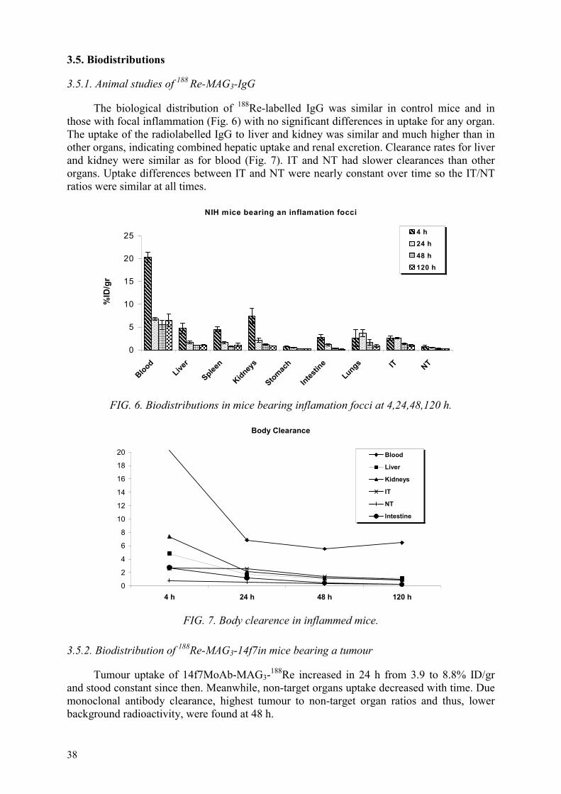
38
3.5. Biodistributions
3.5.1. Animal studies of 188 Re-MAG3-IgG
The biological distribution of 188Re-labelled IgG was similar in control mice and in those with focal inflammation (Fig. 6) with no significant differences in uptake for any organ. The uptake of the radiolabelled IgG to liver and kidney was similar and much higher than in other organs, indicating combined hepatic uptake and renal excretion. Clearance rates for liver and kidney were similar as for blood (Fig. 7). IT and NT had slower clearances than other organs. Uptake differences between IT and NT were nearly constant over time so the IT/NT ratios were similar at all times.
NIH mice bearing an inflamation focci
0
5
10
15
20
25
BloodLive
r
Spleen
Kidneys
Stomach
Intestin
eLungs IT NT
%ID
/gr
4 h24 h48 h120 h
FIG. 6. Biodistributions in mice bearing inflamation focci at 4,24,48,120 h.
Body Clearance
0
2
4
6
8
10
12
14
16
18
20
4 h 24 h 48 h 120 h
Blood
Liver
Kidneys
IT
NT
Intestine
FIG. 7. Body clearence in inflammed mice.
3.5.2. Biodistribution of 188Re-MAG3-14f7in mice bearing a tumour
Tumour uptake of 14f7MoAb-MAG3-188Re increased in 24 h from 3.9 to 8.8% ID/gr and stood constant since then. Meanwhile, non-target organs uptake decreased with time. Due monoclonal antibody clearance, highest tumour to non-target organ ratios and thus, lower background radioactivity, were found at 48 h.

39
Biodistribution 188Re-MAG3-14f7
0
5
10
15
20
25
30
35
40
Blood
Liver
splee
n
kidne
ys
stomac
h
intes
tine
lungs
tumor
%ID
/g22448
FIG. 8. Biodistribution in mice bearing a tumour at 2, 24 and 48 h.
Body clearance
0
5
10
15
20
25
30
35
40
0 10 20 30 40 50
Time (h)
%ID
/g
BloodLiverspleenkidneysstomachintestinelungstumor
FIG. 9. Body clearance of labelled MoAb in mice bearing a tumour.
3.6. Labelling and quality control of lanreotide with 188Re via a direct method
3.6.1. Radiolabelling
Different experiments were carried out in order to obtain the highest specific activity. Decreasing amounts of peptide per mCi of 188Re were used. The formation of a single derivative of labelled lanreotide was influenced by the experimental conditions applied, namely by the amount of peptide and reducing agent, the reaction time and the temperature. We found that 6µg of lanreotide and 0.53 mg of SnCl2 per mCi are needed in order to obtain one peak at the HPLC and 100% of labelling yield as shown in Fig. 10. In this case the specific activity rised 166µCi/µg. This product was instable at room temperature after 24h and all the rhenium was in the form of perrhenate.

40
Minutes0 2 4 6 8 10 12 14 16 18 20
Vol
ts
0.0025
0.0050
0.0075
0.0100
0.0125
Vol
ts
0.0025
0.0050
0.0075
0.0100
0.0125Detector BLanreotide - ReLanr -Re sin secado
FIG. 10. Reverse phase C18 HPLC radiochromatogram of 188Re-lanreotide.
An alternative approach was carried out using different reducing agents as secondary reducing agent, in order to improve the reduction properties of stannous chloride. Oxalate, tartrate and ascorbate at pH=5.0 were tried as secondary reducing agent in combination with gentisic acid. The best stability after 24 h at room temperature was obtain when 2.0 mg of tartrate (10 mg/mL ) pH=5 was used as shown in HPLC chromatograms (Fig. 11). Fig. 12 shows the HPLC profiles of the labelled peptide when ascorbate was used as a secondary reducing agent. ITLC was also performed in saline and acidified ethanol. Results showed 3% of colloid.
Minutes0 2 4 6 8 10 12 14 16 18 20
Vots
0.000
0.005
0.010
0.015
0.020
0.025
0.030
0.035
0.00
0.00
0.01
0.0
0.0
0.0
0.0
0.0
Detector BLanreotide - Remarc14,6nCi-7.738SnCl2-75gent-2mgrtart-18pept
Minutes0 2 4 6 8 10 12 14 16 18 20
Vots
0.00
0.01
0.02
0.03
0.04
0.05
0.00
0.01
0.02
0.03
0.04
0.05Detector BLanreotide - Reestab 24hs marc14,6nCi-7,738SnCl2-75gent-2mgrtart-18pept
FIG. 11. Reverse phase C18 HPLC radiochromatograms of 188 Re-Lanreotide at 0h (left) and 24 h (right) at room temperature using tartrate.

41
Minutes0 2 4 6 8 10 12 14 16 18 20
Volts
0.000
0.005
0.010
0.015
0.020
0.025
0.030
Volts
0.000
0.005
0.010
0.015
0.020
0.025
0.030Detector BLanreotide - Remarc14,6nCi-7.738SnCl2-75gent-2mgrasc-18pept
Minutes0 2 4 6 8 10 12 14 16 18 20
Volts
0.002
0.004
0.006
0.008
0.010
0.012
Volts
0.002
0.004
0.006
0.008
0.010
0.012
Detector BLanreotide - Reestab 24hs marc14,6nCi-7,738SnCl2-75gent-2mgrasc-18pept
FIG. 12. Reverse phase C18 HPLC radiochromatograms of 188Re-Lanreotide at 0 h (left) and 24 h (right) at room temperature using ascorbate.
3.6.2. In vitro studies
There was no displacement of 188 Re from the labelled peptide at the different cysteine concentrations studied. The 188 Re-lanreotide was stable after 4 h incubation in serum at 37ºC. After 24 h, 95% of 188 Re remained bound to the peptide.
3.6.3. Biodistribution of 188 Re-lanreotide in mice
Fig. 13 shows the results of in vivo evaluation of the labelled peptide. The biodistribution data showed a tendency towards hepatobiliary elimination of the labelled peptide.
Biodistribution 188Re-Lanreotide
0
1
2
3
Blood Liver Spleen Kidneys Stomach Intestine Lungs
% ID
/g 1 h
4 h
24 h
FIG. 13. Biodistribution in Balb/c mice of 188Re-Lanreotide at 1,4 and 24 h.

42
3.7. Labelling and quality control of peptides with 111In or 90Y
3.7.1. 111In-DOTA-Lanreotide
3.7.1.1. Radiolabelling
The radiochemical purity of labelled peptide was higher then 98% when using peptide amounts higher than 30µg. Fig. 14 shows the HPLC profile.
FIG. 14. Reverse phase HPLC radiochromatogram of 111In-DOTA-lanreotide.
3.7.1.2. Serum stability
HPLC analysis after 4h incubation in human serum showed no degradation of the radiolabelled peptide after precipitation with acetonitrile. After 24h incubation the chromatogram showed 95% of labelled peptide.
3.7.1.3. Biodistribution
Fig. 15 shows biodistribution results at 4 h with 111In-DOTA-lanreotide. The labelled peptide showed a tendency towards renal excretion and lower levels of activity in liver and intestine.
3.7.2. Labelling DOTA-TOC and DOTA-lanreotide with 90Y
A radiochemical purity of 100% was obtained when using higher amounts than 8 µg of DOTA-TOC per mCi of 90Y as it shown in Fig. 16.
111In-Lan (4 h)
0
5
10
15
20
25
30
35
Blood
Liver
Spleen
Kidney
Inteti
ne
Pancre
as
Adrena
lsLu
ng
%ID
/gr
FIG. 15. Biodistribution in normal rats of 111In-DOTA-Lanreotide at 4h.
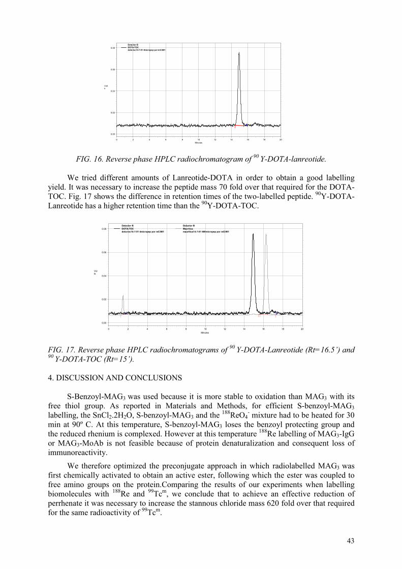
43
Minutes0 2 4 6 8 10 12 14 16 18 20
Volts
0.00
0.02
0.04
0.06
0.08
Volts
0.00
0.02
0.04
0.06
0.08Detector BDOTA-TOCdota-toc14-7-01 4micropep por mCi001
FIG. 16. Reverse phase HPLC radiochromatogram of 90 Y-DOTA-lanreotide.
We tried different amounts of Lanreotide-DOTA in order to obtain a good labelling yield. It was necessary to increase the peptide mass 70 fold over that required for the DOTA-TOC. Fig. 17 shows the difference in retention times of the two-labelled peptide. 90Y-DOTA-Lanreotide has a higher retention time than the 90Y-DOTA-TOC.
Minutes0 2 4 6 8 10 12 14 16 18 20
Volts
0.00
0.02
0.04
0.06
0.08
0.00
0.02
0.04
0.06
0.08Detector BDOTA-TOCdota-toc14-7-01 4micropep por mCi001
Detector BMauritiusmauritius14-7-01 600micropep por mCi001
FIG. 17. Reverse phase HPLC radiochromatograms of 90 Y-DOTA-Lanreotide (Rt=16.5’) and 90 Y-DOTA-TOC (Rt=15’).
4. DISCUSSION AND CONCLUSIONS
S-Benzoyl-MAG3 was used because it is more stable to oxidation than MAG3 with its free thiol group. As reported in Materials and Methods, for efficient S-benzoyl-MAG3labelling, the SnCl2.2H2O, S-benzoyl-MAG3 and the 188ReO4
- mixture had to be heated for 30 min at 90º C. At this temperature, S-benzoyl-MAG3 loses the benzoyl protecting group and the reduced rhenium is complexed. However at this temperature 188Re labelling of MAG3-IgG or MAG3-MoAb is not feasible because of protein denaturalization and consequent loss of immunoreactivity.
We therefore optimized the preconjugate approach in which radiolabelled MAG3 wasfirst chemically activated to obtain an active ester, following which the ester was coupled to free amino groups on the protein.Comparing the results of our experiments when labelling biomolecules with 188Re and 99Tcm, we conclude that to achieve an effective reduction of perrhenate it was necessary to increase the stannous chloride mass 620 fold over that required for the same radioactivity of 99Tcm.

44
The indirect method of labelling antibodies using a 188Re-labelled active ester of MAG3produced 188Re-MAG3-IgG of high in vitro stability and favourable uptake at sites of focal inflammation. Likewise, application of this optimized labelling procedure to anti-melanoma MoAb, produced labelled antibody with high specific activity (2.44 mCi/mg MoAb) showing that the procedure reported here can be extended to the 188Re labelling of other antibodies.
We efficiently labelled lanreotide with 188Re via a direct method and clinical studies in patients have started.
REFERENCES
[1] EHRHARDT, G., KETRING, A.R., TURPIN, T.A., RAZAVI, M.S., VANDERHEYDEN, J.L., et al., “An improved tungsten-188/rhenium-188 generator for radiotherapeutic applications”, J. Nucl. Med. 28 (1987) 656–657.
[2] EHRHARDT, G., KETRING, A.R., LIANG, Q. “Improved 188W/188Re zirconium tungstate gel radioisotope generator chemistry”, Radioactivity & Radiochemistry 3,num. 4 (1992) 38–41.
[3] VIAGGI, M., CRUDO, J.L., CASTIGLIA, S.G., “In vitro and in vivo studies of 99Tcm
IgG: A comparison between three labelling methods”, J. Rad. and Nucl. Chem. 241(1999) 173-177.
[4] GRIFFITHS, G., GOLDEMBERG, D., KNAPP, F., CALLAHAN, A., CHANG, C., HANSEN, H., “Direct radiolabelling of monoclonal antibodies with generator-produced rhenium-188 for radioimmunotherapy: labelling and animal biodistribution studies”, Cancer Research 51 (1991) 4594-4602.
[5] GLASER, M., HOWARD, M., HOWLAND, K., POWELL, A.K., RAE, M.T., et al., “Synthesis, structure and bioconjugation of an active ester containing oxorhenium (V) thioether complex”, Technetium, Rhenium and Other Metals in Chemistry and Nuclear Medicine, SGE Editoriali, Italy, vol. 5 (1999) 167-172.
[6] VISENTIN R., SANTIMARIA M., GIRON M.C., MAZZI U., NICOLINI, M., “A PN2S chelating system for acting as bifunctional ligand towards 188Re and 99Tcm”, Technetium, Rhenium and Other Metals in Chemistry and Nuclear Medicine, SGE Editoriali, Italy, vol. 5, (1999) 173-177.
[7] YOKOYAMA K., TEGA H., KINUYA S., HIRAMATSU K., KONISHI S., et al., “Stability and biodistribution of Re-186-HYNIC labelled A7 monoclonal antibody: comparison with Re-186-MAG3 labelled counterpart”, Eur. J. Nucl. Med. 26, num. 9 (1999) 1194.
[8] VAN GOG, F.B., VISER, G.W.M., KLOK, R., VAN DER SCHORS, R., SNOW, G. B., et al., “Monoclonal antibodies labelled with 186Re using the MAG3 chelate: relationship between the number of chelated groups and biodistribution characteristics”, J. Nucl. Med. 37 (1996) 352-362.
[9] GUHLKE, S., SCHAFFLAND, A., ZAMORA, P.O., BIERSACK, H.J., “188Re and 99Tcm-MAG3 as prosthetic groups for labelling amines and peptides: approaches with pre and post conjugate labelling”, Nucl. Med. Biol. 25 (1998) 621-631.
[10] CRUDO, J.L., EDREIRA, M., DE CASTIGLIA, S.G., “Anticuerpo monoclonal antimelanoma 14f7 marcado con 188Re: ensayos in vitro e in vivo”, Abstract, Nuevas Tendencias en Oncología, Revista Latinoamericana de la European School of Oncology, Vol. IX-Num. 1 (suplemento) (2000).
[11] CRUDO, J.L., OBENAUS, E., EDREIRA, M., DE CASTIGLIA, S.G., “IgG policlonal humana marcada: comparación entre tres radionucleídos terapeúticos 153Sm, 177Lu y 188Re”, Abstract, Nuevas Tendencias en Oncología, Revista Latinoamericana de la European School of Oncology, Vol. IX-Num. 1 (2000).

45
LABELLING OF PEPTIDES WITH 188Re/99Tcm AND MONOCLONAL ANTIBODIES WITH 90Y/111In
B. LINKOWSKI FAINTUCH, C.P.G. SILVA, N.P.S. PEREIRA, E.B. ARAUJO Instituto de Pesquisas, Energeticas e Nucleares, São Paulo, Brazil
Abstract
Radiopharmaceuticals derived from oncophilic small molecular weight peptides have attracted attention as vehicles to deliver radioactivity to tumour cells for external imaging and targeted radiotherapy [1,2]. The aim of the present work was the study of labelling of biomolecules, specifically peptides or antibodies with beta emitter radioisotopes such as 188Re. The direct labelling of two somatostatin analogues, Octreotide and Lanreotide, with 188Re and 99Tcm was done, including optimization of this labelling and biological studies. The radiochemical yield was greater than 95%. 188Re-lanreotide gave better radiochemical yields than 188Re-octreotide and was stable for 24 h. Similar results were obtained with 99Tcm-octreotide and 99Tcm-lanreotide. However, a difference in mass ratio (reducing agent/peptide) was observed, being much greater with 188Re. 99Tcm-labelled peptides were localized in the gastrointestinal tract or in the kidneys and blood clearance wasn’t observed during 6 h. Biodistribution of 188Re-peptides showed similar results as obtained with 99Tcm-peptides. The indirect method was used to label DOTATOC with 188Re and 99Tcm and competition binding assays were done with the labelled product. The results showed a radiochemical yield in the range of 92 to 94%. We observed better uptake of 99Tcm-DOTATOC in AR42J cell membranes than in brain cortex membranes. The results obtained with 188Re-DOTATOC were inconclusive. Labelling of monoclonal antibody for prostate cancer cells (J591) with 111In and 90Y, and immunoreactivity of the product was also studied. The radiolabelling yield for huJ591 with 111In and 90Y was higher than 90%. An average of five DOTA molecules could be randomly conjugated to J591, yielding specific activities of 280 MBq 111In/mg DOTA-J591 and 360 MBq 90Y/mg DOTA-J591, with little apparent loss of immunoreactivity.
1. INTRODUCTION
The major radiopharmaceuticals used in nuclear medicine are guided to diagnostic procedures. There are very few agents for cancer radiotherapy commercially available. It is necessary to obtain results that confirm that treatment with therapeutic radiopharmaceuticals had effect and cured disease over the period of many years.
Over the years, the development of radioimmunotherapeutics has generally been performed using radionuclides such as 111In and 90Y [3].
For cancer radiotherapy, beta emitters are used. Yttrium-90 and rhenium-188 have been determined to be among the best nuclides for radiation therapy [4]. Their half-lives are long enough to permit accumulation in target tissues, and they possess strong beta emission capable of delivering high radiation doses to tissues.
For radioimmunotherapy, rhenium-188 is a very good radionuclide with advantages over other therapeutic radionuclides, including the fact that it is carrier-free, and can be obtained cost-effectively and on demand from an in house 188W/188Re generator [5]. 188Re decays by emission of a relatively high-energy beta particle (2.27 MeV), followed by gamma emission (155 keV, 15%) that can be used to monitor the biodistribution by scintigraphy.
Biomolecules (antibodies, peptides) have generally been considered particularly attractive as selective carriers of therapeutic agents due to their unique in vitro specificity and high affinity for their antigen.

46
Peptides have shown advantages over antibodies with respect to tumour-to-nontumour ratios. Lower-molecular weight agents generally provide better target-to-nontarget ratios, due to their rapid background clearance [6].
Covell, et al. (1986) [7] pointed out that the body residence time for antibodies is much longer than that for fragments of antibodies, which may be an advantage when delivering a sufficient quantity of antibody to the tumour site in order to perform radioimmunotherapy, especially in the case of solid tumours.
Somatostatin is a peptide hormone consisting of 14 amino acids. It is present in the hypothalamus, the cerebral cortex, the brain stem, the gastrointestinal tract and the pancreas. Various tumours contain high numbers of somatostatin receptors, which enable in vivolocalization of the primary tumour and its metastases by scintigraphy with radiolabelled somatostatin analogue peptides [8,9].
Lanreotide [D-βNal-Cys-Tyr-D-Trp-Lys-Val-Cys-Thr.NH2] as octreotide [H2N-D-Phe-Cys-Phe-D-Trp-Lys-Thr-Cys-Thr(ol)] are synthetic octapeptide analogues of the native hormone somatostatin, produced by the hypothalamus and pancreas10. They are cyclized via cysteine bridge.
Many studies reported octreotide labelled with various radionuclides like 131I, 111In, 188Re and 99Tcm [8, 11, 12]. But with lanreotide we can find only some studies made with radioisotope 90Y by indirect method conjugated with the bifunctional chelator DOTA [10]. There are few studies reported about the labelling of lanreotide with 188Re and with 99Tcm.
There are two methods to label peptides with 188Re or 99Tcm, namely the direct and indirect method. There are advantages and disadvantages associated with each of these methods.
The direct method of labelling peptides containing a cyclized cysteine bridge gives some advantages, because it is simple, rapid and efficient and does not require commercially unavailable agents, such as the bifunctional chelating agents (BFCAs) [2].
But the reduction of cysteine bridge can inactivate the peptide. On the other side the indirect method is very complex, lengthy, with insufficient labelling yields [13].
Prostate cancer is the most frequently diagnosed cancer and the second most common cause of cancer mortality in United States males. Monoclonal antibodies for prostate specific membrane antigen (PMSA) were labelled with 111In and 90Y for the evaluation of their diagnostic and therapeutic potential [14].
Objectives
• In the first part of the research project, studies were developed for the labelling of peptides with 188Re by direct method, the optimization of this method by varying some parameters such as mass of ligand, reductor and peptide ratio, incubation time and stability of the labelling. We made the same studies for labelling peptide by direct method using as the radioisotope technetium-99m, and also biodistribution and stability in vitro studies of the labelled product (Results shown in the 2nd Co-ordination Meeting in Mumbai, India).
• As the second part, indirect labelling of conjugated peptide (HYNIC-TOC) with rhenium-188 and technetium-99m was made, followed by preparation of rat brain cortex cell

47
membranes and cell binding assays of the labelled products. These studies were done in St. Bartholomew’s Hospital, London with Dr. Stephen Mather.
As the third part, we studied the labelling of monoclonal antibody for prostate cancer (mAbs PCas) with 111In and 90Y. These studies were carried out in The New York Presbyterian Hospital, Cornell University with Dr. Shankar Vallabhajosula.
• As the final part, we completed the studies of direct labelling of peptides with 188Re, specifically on standardization of labelling protocols, cysteine challenge and biodistribution studies in animals.
2. MATERIALS AND METHODS
2.1. Studies of peptide labelling with 188Re and 99Tcm by direct method [11]
Materials: Lanreotide 8-mer (from PiChem-Austria) and 188W/188Re generator (MAP Medical Technologis Oy Finland) were provided by IAEA, Octreotide was provided by Novartis, Brazil and the 99Mo/99Tcm generator by the Radiopharmacy Center of IPEN/CNEN-SP, Brazil.
LabellingThe direct labelling of peptide (octreotide and lanreotide) with 188Re and 99Tcm isotopes
was achieved through three main steps: (1) Preparation of the peptide, (2) Labelling and quality control, (3) Optimization of the labelling as indicated in the diagram below.
Lanreotide (350 µg) or Octreotide (250 µg) for 188Re or Lanr/Octr (100µg) for 99Tcm Tartrate/phthalate buffer, pH 5.6 or acetic acid/acetate buffer pH 4.2 SnCl2.2H2O in HCl 0.1N or in acetic acid 10%
74 to 370 MBq 188Re/99Tcm
Nitrogen atmosphere, 4 h, room temp., agitation water bath
Frozen (-30°C) until use 100 ° C, 30 min/15 min
Radiochemical control
After labelling the 188Re-peptide passed through a 0.22 µm filter (Millipore) and was analysed for radiolabelled yield using thin layer chromatography (TLC) and reverse phase step-gradient separation on C18-SepPak columns (Millipore Inc.). For 99Tcm-peptide it was not necessary the step of filtration.
Optimization of radiolabelling
It was achieved by varying some parameters as mass of peptide (100 µg to 500 µg and 10,50,100 µg), stannous chloride/peptide mass ratio (3.0, 3.5 ,4.0 and 0.4, 0.6, 0.8, 1),

48
stability of radiochemical purity of the complex (24 h and 6 h), reaction time (1,2,3 h and 30, 60 min) for 188Re and 99Tcm studies, respectively.
Cysteine challenge studies with 99Tcm and 188Re-peptides
The bond strength of the radioisotope 99Tcm or 188Re peptide complexes was evaluated by displacement of bound radionuclide with cysteine [15]. Known amounts of 99Tcm or 188Re-lanreotide and 99Tcm- or 188Re-octreotide were challenged over a range of cysteine concentrations (0.1mM – 100mM) at 37 °C for 1 h. The percent 99Tcm displaced by cysteine was determined by TLC using 0.9% saline as a mobile phase, and the amount of displaced radiolabel found at the solvent front determined.
Biodistribution studies with octreotide and lanreotide labelled by direct method with 188Re and 99Tcm
The biodistribution studies were performed in groups of three animals by intravenous injection of 0.1 mL of radiolabelled peptides with 500 µCi of 99Tcm/188Re. The animals were sacrificed at determined times after injection and selected organs were removed, weighed and the radioactivity was determined with a well-type gamma counter. Radioactivity was also measured in a sample of blood. The results were expressed as% of injected dose per gram of tissue.
Dynamic imaging studies were performed at the first 10 min after the administration of the drug and static imaging after 15 min, 2, 4 and 6 h post injection for 99Tcm-peptides.
2.2. Studies of peptide labelling with 188Re and 99Tcm by indirect method (conducted at St. Bartholomew’s Hospital, London)
Materials: Tyr3-octreotide (TOC) was purchased from Bachem Ltd., Saffron Walden, UK. The hydrazinonicotinyl (HYNIC) was synthesized and protected (BOC-HYNIC) in the Imperial Cancer Research Foundation.
Preparation of HYNIC-TOC conjugate
Five micromoles 6-BOC-hydrazinopyridine-3-carboxylic acid (BOC-HYNIC), 6 µmol O-(7-azobenzotriazolyl)-1,1,2,2-tetramethyl-uronium hexafluorophosphate, and 20 µmoldiisopropylethylamine in 300 µL dimethylformamide were allowed to react for 15 min at room temperature. Sixty microliters of this solution were added to 1 µmol [Lys5-BOC]-protected TOC (Bachem) in a mixture of 20 µL dimethylformamide (DMF): 5 µL water and allowed to react for 1 h. The resulting solution was purified on a SepPak column, and the peptide was deprotected with trifluoroacetic acid containing 2% thianisole and purified on HPLC.
Purification in HPLC [16]
A solvent module 125 (Beckman Coulter, Inc., Fullerton, CA) with a 166 nm ultraviolet detector (Beckman) and radiometric detection was used for reverse-phase high-performance liquid chromatography (HPLC) analysis and preparation.
An Ultrasphere ODS 5-um system (Beckman), 4.6×250 mm column, 1 mL/min flow rate, and 220-nm ultraviolet detection, was used with the following solvent systems:

49
acetonitrile (ACN):0.1% trifluoroacetic acid:water (0-3 min, 0% ACN; 3-10 min, 0-40% ACN; 10-20 min, 40% ACN; 20-23 min, 40-70% ACN; 26-27 min, 70-100% ACN.
Radiolabelling
Labelling of HYNIC-TOC using (EDDA) as coligandTen micrograms HYNIC-TOC were incubated with 0.5 mL EDDA solution and 0.5 mL
was added of 99TcmO4- solution (500 MBq) or 188Re solution (238 MBq), and 5-10 uL or 80
µL tin(II) solution (10 mg SnCl2.2H2O/10 mL or 10 mg/1,6 mL) nitrogen-purged 0.1 N HCl for 60 min at room temperature or with heating for 30 min in water bath (70 °C) for 99Tcm and 188Re, respectively.
Radiochemical control The radiochemical control was done using HPLC as described by Decristofo & Mather,
1999 [16].
Somatostatin receptor binding assays done for DOTA-TOC labelled with technetium and rhenium
Receptor binding studies were performed in order to determine the receptor affinity of the radiolabelled peptides.
The binding affinity of peptide conjugates (188Re-HYNIC-TOC and 99Tcm-HYNIC-TOC) was tested in a competition assay using rat pancreatic tumour cell (AR42J) membranes and rat brain cortex membranes as a source for somatostatin receptors.
Preparation of cell membranes AR42J
AR42J cells growing in suspension were spun down at 2000xg (3000rpm) in a Beckman GS-6R centrifuge for 20 min, resuspended in 20 ml of cold HEPES buffer (20mM, pH 7.3) and homogenized for 10s using and Ultraturrax T8 Netzgerat homogenizer (IKA Labortechnik) on the highest setting. The sample was centrifuged (2000 xg (3000 rpm) for 20 min), the supernatat was removed, the pellet was resuspended in 20 mL of HEPES buffer solution and homegenized again. This procedure was repeated twice.
The final cell membrane pellet was resuspended in HEPES buffer and homogenized for 10 s. The protein concentration was determined using the Bio-Rad DC Protein Assay, and the membranes were then aliquotted into 2 ml Cryo Tubes each containing 2.5 mg of protein in 0.5 mL of sample, i.e. sufficient membranes for two binding assays. The tubes were frozen immediately in an acetone/dry ice bath and stored at –70 oC until required.
Preparation of rat brain cortex membraneTissue membranes were prepared using a modification of the method of Raynor &
Reisine, 1989 described by Kolan, et al., 1996 [17] and the IAEA, 2000 [18].
Competition binding assays Millipore Multiscreen Filtration System plates with glass fiber filters (Millipore MAFB
NOB 10) were prepared the day before the assay was performed using poliethyleneimine aqueous solution.
A stock solution of competitor peptide was prepared and sequential dilutions were made. The radioligand was also diluted so the radioactivity corresponded to approximately 50000 cpm per 50 µl of solution. The competitor solutions were added to the wells of the

50
assay plate prepared one day before, followed by the addition of radioligand followed by the cell membranes. The plate was incubated at room temperature using a Thermolyne Maxi-Mix Shakes at low speed for 2 h. Once the incubation was finished, the plate was filtered, washed and dried in the oven at 60-70 °C for about 20 min. The filters were punched out and placed into tubes and counted in a gamma counter.
Inhibitory concentration of 50% (IC50) values were calculated using non-linear regression with version 5.0 Origin software (Microcal, Northampton, MA). The specific binding of the 99Tcm-HYNIC-TOC and 188Re-HYNIC-TOC was determined by competition against unmodified TOC in a similar assay.
2.3. Studies of monoclonal antibodies for prostate cancer with 111In and 90Y
Murine monoclonal antibody mAbs J591 for prostate cancer cells was conjugated to the bifunctional chelate DOTA and then labelled with 111In or 90Y.
Antibody conjugation
HuJ591 antibody was modified with DOTA by a method analogous to that used by Lewis [19]. The main steps are:
a) Concentration of the antibody b) Washing with 1% DTPA solution c) Changing of the antibody buffer d) Development of an active ester of DOTA e) Conjugation of the ester of DOTA with J591 f) Purification and concentration of DOTA-J591.
111In and 90Y labelling of DOTA conjugate – Radiolabelling of DOTA-J591 with 111In was achieved by adding the radionuclide to the ammonium acetate-buffered DOTA-J591. The mixture was allowed to react at 37° C for 20 min and then washed in a column with 5 ml of 1% HAS-PBS before the main 111In-DOTA-J591 fraction was eluted with 3 ml of 1% HAS-PBS. A similar procedure was used for radiolabelling with 90Y, but an incubation time of 5 min was used, and the labelling mixture included 50mM ascorbic acid.
Quality control of radiolabelling – Free 111In in the radiolabelled DOTA-J591 preparations was determined using the ITLC method with a silica gel-impregnated glass fiber support and a mobile phase of 1% DTPA (pH 5.5).
Immunoreactivity studies – The immunoreactivity of 111In and 90Y-labelled Mab preparations was assessed by the method of Lindmo. et al., 1994 [20], which extrapolates the binding of the radiolabelled antibody at an infinite excess antigen. Briefly, six test solutions were prepared (in duplicate) and contained 20000 cpm of the radiolabelled antibody, and increasing amounts of membranes were prepared from LNCaP cells in a total test volume of 250 µl of PBS. The solutions were incubated at 37° C for 45 min prior to being filtered through a glass membrane filter and washed with ice-cold 10 mM Tris-0.9% buffer. Filters were counted in a gamma counter with standards representing the total radioactivity added. Data were then plotted as the reciprocal of the substrate concentration (X axis) against the reciprocal of the fraction bound (Y axis). The data were then analysed according to a least squares linear regression method using Origin software. The Y intercept gave the reciprocal of the immunoreactive fraction. A similar method using intact or permeated LNCaP cells and centrifugational isolation of the cells gave the same results.

51
3. EXPERIMENTAL RESULTS
Studies of peptide labelling with 188Re and 99Tcm by direct method
Labelling efficiency determined by thin-layer chromatography when using 0.9% saline as a mobile phase showed that the unbound radioactivity migrated with the solvent front, while the bound radioactivity remained at the origin (Fig. 1).
FIG .1 Radiochromatogram of 188Re-Lanreotide in saline 0.9%.
The amount of colloids formed in the peptide solution was determined using 85% ethanol as a mobile phase. The radiolabelled peptides moved to the solvent front and colloids remained at the origin (Fig. 2).
FIG. 2 Radiochromatogram of 188Re-Lanreotide in ethanol 85% as solvent.

52
During radiochemical analysis made by C18 SepPak the radioactivity eluting with 10% ethanol was assumed to be unbound 188Re, that eluting in 85% ethanol to be peptide bound, and that resident on the cartridge to be radiocolloid (Fig. 3).
Labelling efficiencies were generally greater than 95% as determined by TLC until 2 h after labelling. (Figs 4, 5 and 6). Reverse phase chromatography using C18 SepPak confirmed the high labelling efficiency (Fig. 3).
FIG. 3. Radiochemical analysis FIG. 4. Labelling efficacy of 188Re-C18 SepPak. octreotide in tartrate/phthalate buffer.
FIG. 5. Labelling efficacy of 188Re FIG. 6. Labelling efficacy of 188Re- octreotide in acid/acetate buffer. lanreotide.
FIG. 7. Amount of radiocolloid after FIG. 8. Analysis of 188Re-octreotide in 2h for 188Re-octreotide. different buffer solutions.
0
20
40
60
80
100
10%EtOH 85% EtOH C18 Cartridge
% T
otal
Rad
ioac
tiviry
2 hours4 hours
24 hours
3,50
4,000
20
40
60
80
100% R.P
SnCl2.2H2O/Octreotide
188ReO2 188ReO4188Re-
octreotide
no filtered
filtered0
20
40
60
80
100
% P.R.
188ReO2188ReO4
188Re-Octreotide
Acetate/Acetic AcidPhtalate/Tartrate0
20
40
60
80
100
% R.P.
3,003 ,50
4 ,00
24 hours
4 hours
2 ho urs
0
1 0
2 0
30
40
5 0
6 0
70
80
90
100% R.P.
SnCl2.2H2O/Octreotide Ratio
3,003,50
4,00
2 hours
4 hours
24 ho urs
0
20
40
60
80
100
% R.P.
SnCl2.2H20/Lanreotide Ratio

53
The amount of radiocolloid determined by ITLC with 85% acidified ethanol as the mobile phase was greater when we didn’t make a purification by using filter with membrane 0.22 µm . When we made the filtration much activity was retained in the filter (Fig. 7).
For 188Re-Lanreotide we obtained high labelling efficiency using 350 µg of the ligand lanreotide and for 188Re-Ocreotide a mass of 250 µg only was sufficient for good labelling in both buffers.
The reaction time was 2 h for both complexes and the best ratio between mass of stannous chloride and peptide was 3.5 for 188Re-Octreotide and 4.0 for 188Re-Lanreotide (Figs 4, 6). With the ratio of 3.0 we observed that the amount of stannous chloride was insufficient to reduce all the rhenium so we observed a larger amount of free perrhenate (Fig. 5).
The radiochemical purity of 188Re-Lanreotide was 98.76±0.87% and the product stayed stable for 24 h (Fig. 6).
188Re-Octreotide presented 97.14±1.2% purity in tartrate/phthalate buffer. In acetic acid/acetate buffer, findings were similarly close to 97% (Fig. 8).
188ReO2188ReO4
188Re-Octreotide
24 hours
4 hours
2 hours
0
10
20
30
40
50
60
70
80
90
100% R.P.
FIG. 9. Labelling stability of 188Re-octreotide in acetate/acetic acid buffer.
However, 188Re-octreotide stayed stable for 4 h and with 24 h, the stability fell by about 50% (Fig. 9).
When we tried to label a very small mass of ligand such as 10 µg and also 100 µg with a mass ratio of stannous chloride to peptide in the range of 10 to 50 to label with 188Re, very small radiolabelled yield was achieved.
When octreotide was labelled with 99Tcm it was observed that when the mass ratio between the reducing agent and the peptide was low, the yield obtained was low. The best finding was for a ratio of one (1.0) (Table I).

54
TABLE I. RADIOCHEMICAL PURITY OF 99TCm-OCTREOTIDE VARYING THE MASS RATIO OF SNCL2.H2O/OCTREOTIDE
m SnCl2.H2O/m octreotide
%99TcmO4 % 99TcmO2 % 99Tcm-octreotide
0.4 91.04 2.9 6.06 0.6 30.52 1.88 67.60 0.8 48.91 0.98 50.11 1.0 3.48 1.38 95.14
The study of the labelling of lanreotide with 99Tcm was based on the results obtained with 99Tcm-octreotide. So, we observed that the best ratio between mSnCl2.H2O/m lanreotide was 1.0 for a mass of lanreotide and a mass of stannous chloride above 50 µg (Table II).
TABLE II. RADIOCHEMICAL PURITY OF 99TCm-LANREOTIDE
lanreotide m (µg)
SnCl2.H2Om (µg)
ratio %99TcmO4 % 99TcmO2 % 99Tcm-lanreotide
10 30 3 93.65 1.05 5.30 20 20 1 44.67 1.30 53.96 50 50 1 0.60 1.51 97.89
100 100 1 0.18 3.09 96.73
Both, 99Tcm-octreotide and 99Tcm-lanreotide were radiochemically stable for 6 h (Table III). However, 99Tcm-octreotide had a loss in the radiochemical purity of 5.6%.
TABLE III. RADIOCHEMICAL STABILITY
Time (h) 99Tcm-octreotide 99Tcm-lanreotide t=0 97.59 96.25 t=2 92.10 97.86 t=4 91.89 98.35 t=6 91.24 96.73
Cysteine challenge
Analysis by TLC-aluminium saline of 99Tcm/188Re-labelled peptides incubated in solutions containing cysteine invariably showed migration. Since the aim of this measurement was to determine instability in face of cysteine transchelation and not oxidation to 99TcmO4
-
/188ReO4-, it was important to ensure that migrating activity was due to labelled cysteine and
not due to pertechnetate or perrhenate which also migrate in this system and with an Rf value close to that for labelled cysteine.
Transchelation to cysteine was studied at five molar ratios. We can observe that 99Tcm-octreotide was relatively more susceptible to Cys challenge than 99Tcm-lanreotide (Fig. 10).

55
FIG. 10. Percent dissociation of 99Tcm from lanreotide and octreotide vs. cysteine concentration during incubation for 1 h at 37°C.
The results of Cys challenge for 188Re-peptides revealed a relatively high 188Re-chelation strength for both the peptides (Fig. 11). The CD50 value (concentration of Cys required to displace 50% of peptide bound 188Re) for 188Re-octreotide was about 10 mM and for 188Re-lanreotide was about 60 mM.
FIG. 11. Percent dissociation of 188Re from lanreotide and octreotide vs. cysteine concentration during incubation for 1 h at 37°C.
Biodistribution with 99Tcm-labelled peptides
The biodistribution of 99Tcm-lanreotide and 99Tcm-octreotide was evaluated in normal Wistar rats by dynamic imaging techniques during the first 10 min after injection, and by static imaging at 15 min, 1, 2 and 4 h post injection, and also by invasive method (organ removal).
0
5
10
15
20
0.1 1 10 50 100
CYS (mM)
% D
isso
ciat
ion
99mTc-lanreotide 1h99mTc-octreotide 1h
00,20,40,60,8
11,21,41,61,8
kidne
ylive
rlun
ghe
art
stomac
h
splee
n
large
int.
small
int.
brain
blood
% D
/g15 min2 hours4 hours6 hours
01020304050607080
0 0.01 0.1 1 10 50 100
CYS (mM)
% D
isso
ciat
ion
188Re-octreotide 1h188Re-lanreotide 1h

56
00.20.40.60.8
11.21.41.61.8
kidne
ylive
rlun
ghe
art
stomac
hsp
leen
large
int.
small
int.
brain
blood
% D
/g15 min2 hours4 hours6 hours
FIG. 12. Biodistribution of 99Tcm-lanreotide in normal rats.
99Tcm-labelled peptides were localized in the gastrointestinal tract or in the kidney (Figs 12,13). For 99Tcm-octreotide the uptake by the kidneys increased until 4 h p.i. with decrease after this time (Fig. 13). The small intestine had also a great uptake. With 99Tcm-lanreotide the greater uptake of the drug was by the liver at the first 15 min p.i., and we did not observe blood clearance during 6 h (Fig. 12).
Other organs presenting uptake were kidney and stomach.
0
0.2
0.4
0.6
0.8
1
1.2
1.4
1.6
kidne
ylive
rlun
ghe
art
stomac
hsp
leen
large
int.
small
int.
blood
% D
/g
15 min2 hours4 hours6 hours
FIG. 13. Biodistribution of 99Tcm-octreotide in normal rats.

57
0
5
10
15
20
25
30
kidne
yLiv
erlun
g
stomac
hsp
leen
large
int.
small
int.
blood
%ID
/g15 m in1h2h4h24h
FIG. 14. Biodistribution of 188Re-octreotide in normal mice.
Biodistribution of 188Re-peptides was evaluated in swiss mice by invasive method at different times after injection. Results were similar to those obtained with 99Tcm. For 188Re-octreotide, the highest uptake observed was by the intestine followed by the liver, kidneys and stomach (Fig. 14). With 188Re-lanreotide the largest uptake observed was by the small intestine followed by the liver in the first 15 min, post-injection (Fig. 15).
0
1 0
2 0
3 0
4 0
5 0
kidne
ylive
rlun
g
stomac
hsp
leen
large
int.
small
int.
blood
%ID
/g
1 5 m in1 h2 h4 h2 4 h
FIG. 15. Biodistribution of 188Re-lanreotide in normal mice.
Studies of peptide labelling with 188Re and 99Tcm by indirect method
The radiochemical purity of the 188Re-HYNIC-TOC and 99Tcm-HYNIC-TOC was assessed by means of thin layer chromatography and high performance liquid chromatography in two differents solvents. The results showed a radiochemical yield in the range of 92 to 94%.
Receptor binding to the AR42J rat pancreatic tumour cell membranes and rat brain cortex cell membranes was determined in the absence and presence of saturation.

58
FIG. 16. Receptor binding of 99Tcm-HYNIC-TOC-EDDA on cell AR42J.
Concentration of somatostatin to determine the total and non-specific binding at increasing concentrations of 99Tcm-HYNIC-TOC. The specific binding was determined as the difference between total and non-specific binding.
In Figs 16 and 17 we have the results of competition assays of 99Tcm-HYNIC-TOC using rat pancreatic tumour cell (AR42J) and rat brain cortex membranes, respectively.
FIG. 17. Receptor binding of 99Tcm-HYNIC-TOC on rat brain cell membranes.
Binding 230500 Radioligand: batch 5 (Batch May) 99Tcm-HYNIC-TOC-EDDA Competitor: Octreotide Membranes: Cell AR42J Total counts 44670 NSB 149.0628 ± 11.92765 SB 499.44124 ± 27.45272 IC50 0.18255 ± 0.03672
Binding 23050 Radioligand: batch 5 (BatchMay) 99Tcm-HYNIC-TOC Competitor: Octreotide Membranes: Rat brain Total counts 44670 NSB 128.71926 ± 9.4889 SB 130.29752 ± 13.18011 IC50 1.44349 ± 0.63563

59
The receptor binding properties are summarized in Table IV.
TABLE IV. RECEPTOR BINDING PROPERTIES
Assay system IC50 NSB SB AR42J 0.18 149.06 499.44 Brain cortex cells 1.44 128.71 130.29
NSB (non-specific binding) SB (specific binding).
Studies of monoclonal antibodies for prostate cancer with 111In and 90Y
The radiolabelling yield for huJ591 with 111In and 90Y was higher than 90%. An average of five DOTA molecules could be randomly conjugated to J591. Specific activities of 280 MBq 111In/mg DOTA-J591 and 360 MBq 90Y/mg DOTA-J591 were achieved, with little apparent loss of immunoreactivity as can be observed in Figs 18 and 19.
FIG. 18. Lindmo immunoreactivity for 90Y-DOTA- hu-J591.

60
FIG. 19. Lindmo immunoreactivity for 111In-DOTA- hu-J591.
4. DISCUSSION
Small peptides radiolabelled with 99Tcm or 188Re are being currently investigated as potential radiopharmaceuticals, mainly for cancer diagnosis and therapy [5, 21].
Rhenium chemistry is very similar to technetium chemistry due to the periodic table relationship between the two elements [22] and as such, rhenium also forms a stable anionic species ReO4 (perrhenate) and will not bind to organic ligands without reduction to a lower oxidation state.
Direct labelling of peptide with rhenium isotope can be done using the same procedures as used for direct labelling with 99Tcm but the labelling conditions for rhenium must be more energetic because of a lower redox potential and therefore a greater tendency to reoxidize. So the reaction time is longer and the concentration of Sn (II) is higher.
The involvement of tin in protein labelling as a tin-protein intermediate was suggested by Rhodes [23], for a “pretinning” procedure, when the reduction of native disulfide bridges was carried out by incubating the protein with stannous ions. This procedure is, however, limited to labelling only those peptides, which contain cyclized cysteines in their structure. This procedure can also be a limitation because it uses very small mass of ligand, with the formation of the intermediate compound Sn-tartrate labelled with 188Re.

61
It is known that in direct protein labelling with 188Re the best labelling yields are achieved within a relatively narrow pH range: 4-5.5. A pH lower than 4 compromises the immunoreactivity of the peptides, and a higher pH contributes to reoxidation of 188Re(V) back to perrhenate [24]. A final pH value of 4 was here used for labelling in tartrate/phathalate buffer, but in acetate/acetic acid buffer for 188Re-Octreotide the final pH was 3.0. For 99Tcm-labelling peptides the final pH was 5.0.
The assay of cysteine-challenge in vitro ranks different 99Tcm/188Re labelling methods according to the stability of the label towards transchelation to cysteine and to determine whether it is possible to predict the relative radioactivity levels in organs and blood in normal animals.
The challenge assay is dependent on various factors, which could be broadly categorized as non-molecular and molecular factors. The non-molecular factors are pH, temperature, and other labelling conditions including buffers. Among molecular factors are the nature of amino acids involved in the formation of the complex [25]. Peptides labelled with 99Tcm show different responses to the cysteine challenge assay. Some 99Tcm-peptides appear to undergo rearrangement over time, presumably to thermodynamically stable states, which increases resistance to displacement with cysteine, while other peptides show no change over time [15]. Thakur [2] showed that 99Tcm-sandostatin and 99Tcm-RC-160 when submitted to cysteine challenge with a 100-fold molar excess of cysteine suffered less than 10% of loss of radioactivity by transchelation. In our experiments with 99Tcm-peptides, less than 20% was transchelated but for 188Re-peptides the transchelation reached nearly 70% in 1 h of incubation.
The maximum concentration of cysteine one expects the peptide to be exposed in vivo is 1 mM [26]. So the high molar ratio demonstrated to cause displacement also suggests that the binding strength of 99Tcm-peptides is very high, but not for 188Re-peptides.
In biodistribution studies of 99Tcm-labelled peptides, main organ uptake by 99Tcm-lanreotide was observed in the liver, being cleared to small intestines, and for 99Tcm-octreotide, main uptake was by small intestine, cleared to kidneys. The difference between both complexes is due the different structure (amino acid sequence) that influences the lipophilicity and biodistribution. In addition to lipophilicity, the stability of the complex also seems to be an important factor in influencing biodistribution. Some 99Tcm- labelled peptides have a potential ability to pass the blood-brain barrier16 and have been suggested as imaging agents for brain tumours. So, a significant uptake of 99Tcm-lanreotide by brain was observed but was insufficient for observing by imaging methods because the study was made in whole body.
The peptide lanreotide is very similar to another peptide fully studied that is Vapreotide (RC-160) with differences in the extremes of the molecule. As 188Re-RC-160 was studied in treatment of small cell lung carcinoma11 maybe this can explain some uptake of 99Tcm-lanreotide by the lung. 188Re-peptides showed similar biodistribution as 99Tcm-peptides.
Since the direct labelling technique can be applied only for cyclized peptides, many studies are performing the indirect method using a bifunctional chelating agent, so that it can be used to label any peptide of biomedical interest. Nevertheless, the method requires a long synthesis and so far has resulted in low labelling yields.

62
Somatostatin receptors are hyperexpressed on many malignant tumours. Somatostatin and related analogues have been demonstrated to bind to tumours that are well differentiated and/or have neuroendocrine features [27].
Receptor-binding, low molecular weight peptides provide an excellent basis for the design of specific-binding radiopharmaceuticals. A major challenge was to label such molecules with radioisotopes without loss of receptor binding affinity or specificity.
Octreotide binds with high affinity to SSTR2 and SSTR5, to a lesser extend to SSTR3, whereas it does not bind to SSTR1 and to SSTR4. Because the subtype 3 receptor is also expressed on most adenocarcinomas, lanreotide scintigraphy and/or radionuclide therapy would have a broader field of application [28]. However, comparative studies in SSTR2 receptor-positive tumour-bearing rats indicated that internalization, as well as tumour localization by radiolabelled lanreotide was lower than that for the octreotide analogs [29].
Hydrazinonicotinamide conjugate to Tyr3-octreotide was labelled with 99Tcm and 188Re with a high specific activity. EDDA was used as coligand and showed that it is a good coligand. The binding assay is a very important step to evaluate the specificity of the radiopharmaceutical. We observed better uptake of the product in AR42J cell membranes than in brain cortex membranes probably because there are fewer receptors in the latter. Binding to receptors with 188Re-HYNIC-TOC was inconclusive.
5. CONCLUSIONS
The direct labelling of peptides with rhenium-188 isotope can be done using the same general procedures as used for the direct labelling with 99Tcm. The labelling was simple and efficient, although the reaction time for 188Re- labelled peptides was much longer (2 h) than for 99Tcm-labelled peptides (30 min).
188Re-lanreotide gave a better radiochemical yield than 188Re-octreotide and remained stable for 24 h, but the mass of lanreotide had to be greater than that of octreotide, and the ratio between the mass of stannous chloride and lanreotide was also greater than that used for labelling octreotide.
As the products labelled with 188Re can be obtained with a high radiochemical purity, they may be used for in vivo studies to evaluate their efficacy in radioimmunotherapy similarly to 99Tcm agents in radiodiagnosis of cancer.
99Tcm-HYNIC-TOC had good in vitro and in vivo properties as a high affinity molecule for SSTR2 receptors.
J591 is a monoclonal antibody that may allow targeting of viable PMSA expressing tissue with diagnostic and therapeutic metallic radionuclides.
REFERENCES
[1] FISCHMAN, A.J., BABICH, J.W., STRAUSS, H.W., “A ticket to ride – peptide radiopharma-ceuticals”, J. Nucl. Med. 34 (1993) 2253-2263.
[2] THAKUR, M.L., Radiolabelled peptides: Now and future, Nucl. Med. Commun. 6 (1995), 724-732.

63
[3] DE JONG, M., BAKKER, W.H., BREEMAN, W.A., BERNARD, B.F., HOFLAND, D.J., et al., “Pre-clinical comparison of [DTPA]octreotide, [DTPA, tyr]octreotide and [DOTA, tyr]octreotide as carriers for somatostatin receptor-targeted scintigraphy and radionuclide therapy”, Int. J. Cancer 75 (1998) 406-411.
[4] WESSELS, B.W., ROGUS, R.D, “Radionuclide selection and model absorbed dose calculations for radiolabelled tumour associated antibodies”, Med. Phys. 11 (1984) 638-645.
[5] Griffiths, G.L., Goldenberg, D.M., KNAPP, JR., F.F., CALLAHAN, A.P., CHANG, C.H., et al., “Direct radiolabelling of monoclonal antibodies with generator-produced rhenium-188 for radioimmunotherapy: labelling and animal biodistribution studies”, Cancer Res. 51 (1991) 4594-4602.
[6] BEHR, T., BECKER, W., BAIR, H.J., KLEIN, M, STUHLER, C.M., CIDLINSKY, et al., “Comparison of complete versus fragmented 99Tcm-labelled anti-CEA monoclonal antibodies for immunoscintigraphy in colorectal cancer”, J. Nucl. Med. 36 (1995) 430-441.
[7] COVELL, D.G., BARBET, J., HOLTON, O.D., BLACK, C.D.V., PARKER, R.J., et al., “Pharmacokinetics of monoclonal immunoglobulin IgG1, F(ab’)2 and Fab’in mice”, Cancer Res. 46 (1986) 3969-3978.
[8] KRENNING, E.P., KWEKKEBOOM, D.J., BAKKER, W.H., “Somatostatin receptor scintigraphy with [111In-DTPA-D-Phe] and 123I-Tyr]-octreotide: the Rotterdam experience with more than 1000 patients”, Eur. J. Nucl. Med. 20 (1993), 716-731.
[9] OLSEN, J.O., POZDERAC, R.V., “Somatostatin receptor imaging of neuroendocrine tumours with indium-111 pentetreotide (octreoscan)”, Sem. Nucl. Med. 25 (1995) 251-261.
[10] SMITH-JONES, P.M., BISHOP, C., LEIMER, M., GLUDOVACZ, D., ANGELBERGER, P., PANGERL, T., “DOTA-Lanreotide: A novel somatostatin analog for tumour diagnosis and therapy”, Endocrinology 140 (11) (1999) 5136-5148.
[11] ZAMORA, P.O., BENDER, H., KNAPP, JR., F.F., BIERSACK, H.J., “Radiotherapy of intrathoracic carcinoma senografts with 188Re-RC-160, a somatostatin analogue”, Tumour Targeting 2 (1996) 49-59.
[12] THAKUR, M.L., KOLAN, H., LI, J., “Radiolabelled somatostatin analogues in prostate cancer”, Nucl. Med. Biol. 24 (1997) 105-113.
[13] OKARVI, S.M., Recent developments in 99Tcm labelled peptide-basedradiopharmaceuticals: An overview”, Nucl. Med. Comm. 20 (1999) 1093-1112.
[14] SMITH-JONES, P.M., VALLABHAJOSULA, S., GOLDSMITH, S.J., NAVARRO, V., HUNTER, J.H., BASTIDAS, D., BANDER, N.H., “In vitro characterization of radiolabelled monoclonal antibodies specific for the extracellular domain of prostate-specific membrane antigen”, Cancer Res. 60 (2000) 5237-5243.
[15] RHODES, B.A., ZAMORA, P.O, MAREK, M.J., SHARMA S.D., WALL, F.J., “Direct 99Tcm-labelling of proteins and peptides: factors that alter radiochemical yields and in vivo stability”, Technetium, Rhenium and others Metals in Chemistry and Nuclear Medicine, Mario Nicolini, Ulderico Mazzi, SG Editoriali, Padova, Italy (1995) 281-286.
[16] DECRISTOFORO, C., MATHER, S.J., “Technetium-99m somatostatin analogues: effect of labelling methods and peptide sequence”, Eur. J. Nucl. Med 26 (1999) 869-876.
[17] KOLAN, H., LI, J., THAKUR, M.L., “Sandostatin labelled with 99Tcm: In vitro stability, in vivo validity and comparison with 111In-DTPA-Octreotide”, PeptideResearch 9 (3)(1996).

64
[18] INTERNATIONAL ATOMIC ENERGY AGENCY, Report of the Research Co-ordination Meeting on Labelling Techniques of Biomolecules for Targeted Radiotherapy, Mumbai, India, IAEA, Vienna, Austria, 2000.
[19] LEWIS, M.R., RAUBITSCHEK, A., SHIVELY, J.E., “A facile, water-soluble method for modification of proteins with DOTA. Use of elevated temperature and optimized pH to achieve high specific activity and high chelate stability in radiolabelled immunoconjugates”, Bioconj. Chem. 5 (1994) 565-576.
[20] LINDMO, T., BOVEN, E., CUTTITTA, F., FEDORKO, J., BUNN, P.A., “Determination of the immunoreactive fraction of radiolabelled monoclonal antibodies by linear extrapolation to binding at infinite antigen excess”, J. Immunol. Methods 72 (1994) 77-89.
[21] HNATOWICH, D.J., WINNARD, Jr, P., VIRZI, F., FOGARASI, M., SANO, T., et al., “Technetium -99m labelling of DNA oligonucleotides”, J. Nucl. Med. 36 (1995) 2306-2314.
[22] COTTON, F.A., WILKINSON, G., “Technetium and rhenium”, Advanced Inorganic Chemistry, Wiley, New York, 4th ed. (1980) 883-901.
[23] RHODES, B.A., “Direct labelling or proteins with 99Tcm”, Nucl. Med. Biol. 18 (1991) 667-676.
[24] DADACHOVA, E., MIRZADEH, S., The role of tin in the direct labelling of proteins with rhenium-188, Nucl. Med. Biol. 24 (6) (1997) 605-60.
[25] SHARMA, S.D., RHODES, B.A., “Differential stability of Syn and Anti-Isomers in 99Tcm-complexes-evidence from cysteine challenge studies”, Technetium, Rhenium and Others - Metals in Chemistry and Nuclear Medicine, Mario Nicolini, Ulderico Mazzi, SG Editoriali, Padova, Italy, (1999) 697-670.
[26] COOPER, A.J.L., Biochemistry of sulfur-containing amino acids, A. Ver. Biochem. 52 (1983) 187-222.
[27] REUBI, J.C., KRENNING, E., LAMBERTS, S.W.J., KVOLS, L., “Somatostatin receptors in malignant tissues”, Proceedings of the 2nd international EORTC symposium on “Hormonal manipulation of cancer: Peptides, growth factors and new (anti) steroidal agents”, Pergamon Press, (1990) 1073-1077.
[28] VIRGOLINI, I., Mack Forster Award Lecture, “Receptor nuclear medicine: vasointestinal peptide and somatostatin receptor scintigraphy for diagnosis and treatment of tumour patients”, Eur. J. Clin. Invest. 27 (1997) 793-800.
[29] DE JONG, M., DE BRUIN, E., BERNARD, B., “Internalization of In-111 labelled somatostatin analogue for tumour scintigraph and radionuclide therapy”, J. Nucl. Med. 39 (1998) 261 pp.

65
LABELLING TECHNIQUE OF BIOMOLECULES FOR TARGET RADIOTHERAPY
HONGSHENG BAI, JIN XIAOHAI, CHENG ZHEN, JIA BING FAN HONGQIANG, LU WEIWEI Department of Isotope, China Institute of Atomic Energy, Beijing, China
Abstract
Labelling techniques were developed for the preparation of biomolecules (DOTA-IgG, DOTA-lanreotide, anti-hepatoma antibody fragment, lanreotide) with radionuclides such as 90Y, 153Sm and 188Re. The labelling yield and radiochemical purity of these labelling biomolecules were determined by PC, ITLC and Sep-Pak C18 cartridge. The stability in vitro and bio-behaviour in normal rats were also evaluated. The experimental results showed that labelling efficiency of biomolecules (DOTA-IgG and DOTA-lanreotide) with 90Y and 153Sm is more than 95% and had good stability in vitro, but the labelling efficiency of biomolecules (anti-hepatoma antibody fragment and lanroetide) with 188Re via directly labelling technique is at range of 88%~95% and stability in vitrowas less.
1. INTRODUCTION
Increased effort was made to label monoclonal antibody and peptide with radionuclides because of their potential role in the radioimmunotherapy and radiodiagnosis of tumour.
For some years monoclonal antibody to tumours’ antigenic sites were labelled with emit gamma rays, permitting detection of disease. Recently, people took efforts to investigate labelling monoclonal antibody with radionuclides emitting beta rays for therapy of tumour and obtained some promising results.
Somatostatin is a cyclic disulfide-containing peptide hormone of 14 amino acids It exists in the hypothalamus, the cerebral cortex, the brain stem, gastrointestinal tract and pancreas, and exerts an inhibitory effect on several cell functions such as secretion of peptide hormones and growth factors. The clinic value of Somatostatin is limited due to its very short half-life in vivo. In recent years much attention are drawn to the development radiolabelled somatostatin analogues (such as octreotide, RC-160 and lanreotide) with radionuclides (example for 188Re, 131/125/123I, 99Tcm, 111In, 90Y, etc.) for a variety of diagnostic applications as well as for therapy of malignant tumours. Lanreotide (D-β-Nal-Cys-Try-D-Trp-Lys-Val_Cys-Thr•NH2) is a new somatostatin analogue. It can bind to human somatostatin receptor (hSSTR) subtype 2 through 5 with high affinity and to hSSTR subtype 1 with low affinity. Virgolini, I., et al, investigated biodistribution, safety and radiation absorbed dose of 111In-DOTA-lanreotide in 1998 and these experimental results were promising.
2. MATERIALS AND METHODS
2.1. Preparation of DOTA-IgG with 90Y
Lanreotide and DOTA-lanreotide (provided by IAEA, HPLC grade). Stannous chloride (HPLC grade), acetate, sodium acetate, concentrated hydroxyl chloride, ethanol (analysis grade, Beijing Chemical Reagent Co.) and 0.9% sodium chloride solution (Sijiazhuang the Fourth Pharmacy Factory). 188WO4
- solution (USA, Oak Bridge); 188W/188Re generator (China Institute of Atomic Energy and Finland); 90Sr/90Y solution (Russia); 90Sr/90Y generator

66
(China Institute of Atomic Energy); enriched Sm2O3 (Russia, Sm-152 is more than 95%). Anti-hepatoma antibody fragment (provided by the 4th People Military Hospital).
2.1.1. Conjugation of DOTA with antibody (IgG)
The conjugation of DOTA with antibody went through two steps: the first step was activation of DOTA; and the second step was reaction of DOTA-NHS with antibody and purification of DOTA-IgG by Sephadex G50 column.
2.1.2. Preparation of DOTA-NHS
DOTA (0.435g) and NHS (0.115g) were dissolved with 5 mL of dry DMSO in the 20 mL of vial, 3 mL of solution containing DCC (0.213 g) was added to the vial , mixed and stirred. The mixture was continued at room temperature overnight. The precipitated dicyclohexylurea was removed by filtration and washed with hot DMSO, and DMSO in the filtered solution was removed by distillation.
2.1.3. Preparation and purification of DOTA-IgG
According to different mole ratio of DOTA/IgG, DOTA-NHS (solid) was added to IgG solution (13.6 mg/mL) in the vial, mixed by vortex and incubated for 2 h at room temperature and 37ºC, respectively. After conjugation, DOTA was removed from DOTA-IgG by the Sephadex G50 column and 0.85mol/l NaAc solution as elution agent. We did plenty of experiments to determine the optimal conjugation condition of DOTA with IgG.
2.1.4. Labelling DOTA-IgG with 90Y
DOTA-IgG is dissolved with metal free water to make 24mg/mL DOTA-IgG solution 40 uL of 90YCl3 solution (6-8mCi) was added to 100 uL of 0.3mol/L sodium acetate (pH~5.5) buffer solution in the vial, the mixture was mixed carefully by Vortex. Then we added 100 uL of DOTA- IgG into above the vial and mixed again by Vortex, and the reaction mixture incubated in the water bath (37ºC) for 2 h.
2.2. Preparation of lanreotide with 188Re
2.2.1. Preparation of 188Re- citrate /tartate (188Re-citrate/tartatic acid)
1 mL of tartate solution (0.15mol/l) and 1 mL of citrate solution (0.015mol/l) are added to 10 mL of vial, mixes with 0.2 mL SnCl2 solution (10 mg/mL), 1 mL of 188ReO4
- solution (20mCi/mL) is added. The mixture reacts for 45 min at 60ºC and pH 2-3. The pH value of mixture is adjusted to 5-7 with 0.5mol/L sodium acetate solution. The labelling efficiency is determined by paper chromatography using acetone and 0.9% sodium chloride solution as mobile phase.
2.2.2. Preparation of 188Re-lanreotide
0.02 mL of lanreotide solution (0.4mg/mL) and 0.05mL stannous chloride (10 mg/mL) are added to the vial together. The mixture incubates for 20 min at room temperature, then 0.2 mL of 188Re-CT/TT (37MBq) is added, mixes and reacts for 40 min at 60ºC. After cooling, pH value of the mixture is adjusted to 5-6 with 0.5 mol/L sodium acetate solution.

67
2.2.3. Preparation of DOTA-lanreotide with 153Sm
DOTA-lanreotide is dissolved with metal free water to make 4mg/mL DOTA-lanreotide solution. 40 µL of samarium chloride solution (6-8mCi, Sm about 10µg ) is added to 100 µLof 0.3 mol/L sodium acetate (pH~5.5) buffer solution in the vial, the mixture is mixed carefully by Vortex. Then, 100µL of DOTA-kanreotide is added into above vial, and mixed again by Vortex, the reaction mixture incubated in the water bath (90~100ºC) for 1 h.
2.3. Preparation of anti-hepatoma monoclonal antibody fragment with 188Re
2.3.1. Reduction of anti-hepatoma monoclonal antibody fragment
0.45 mg of anti-hepatoma monoclonal antibody fragment is dissolved in 0.02 mol/L phosphonic buffer solution (PBS) to make 8 g/L solution, then added NaHSO3 solution (mole ratio of NaHSO3/antibody=2500:1), reaction volume is 100 µL, the mixture is mixed by Vortex and incubated for 1.5 h at room temperature.
2.3.2. Reduction of 188Re
40 µL of tartate (0.15mol/l)-citrate solution (0.015mol/l) is added to 10 mL of vial, mixed with 12 µL SnCl2 solution (10 mg/mL) and 10 µL 188ReO4
- solution (37-148 GBq/L), reaction volume is 100 µL. The mixture reacts for 45 min at 60ºC and pH 2-3.
2.3.3. Labelling antibody
6 µl of ascorbic acid (6 mg/ml) is added to reduced antibody (mole ratio of ascorbic acid and antibody=750:1), then added reduced 188Re solution. The mixture is mixed carefully by Vortex and incubated for 2 h in 37ºC water bath.
2.3.4. Quality control
Thin layer chromatography (TLC) is used to monitor the quality of labelling yield. In the TLC studies, TLC-SG (Gelman Sciences Inc. Ann Arbor, Michigan) chromatography paper is cut into 1.5×15cm strip and activated by heating for 30 min at 110ºC according to manufacture’s instructions. After heating, the strips are stored dry at room temperature until use. 0.005 mL portion of sample is spotted at 2 cm from lower end of the TLC paper and dried in air. The strips are developed in the different developing systems until the solvent reaches up to 12 cm of strip. After drying, the strip is cut into 1 cm piece and radioactivity is measured in a NaI(Tl) well detector.
2.3.5. Animal test
Biodistribution of labelling biomolecules is performed in male Kunming white mice (weight:20±2g). 20µCi of labelling biomolecules in 0.1 ml volume is injected though tail vein and these mice are sacrificed at specific time intervals. The tissues and organs are excised, weighed and counted in a NaI(Tl) well detector. The uptake of activity in different organs is calculated as percentage injected dose per gram organ.

68
3. RESULTS AND DISCUSSION
3.1. Labelling DOTA-IgG with 90Y
20 mCi of 90Sr/90Y generator was prepared, 90Ysolution was obtained from the generator with 0.003 mol/L EDTA solution. The elution efficiency, elution curve and 90Sr breakthrough were elevated, the prepared 90Ysolution reached to the demand of medical use. 90Sr breakthrough is less than 10-5, and elution efficiency is more than 75%. Based on different distribution coefficients of metal element in the Dowex 50×8 cation exchange resin, 90Ysolution is purified with resin column using different concentration of H2SO4 and HCl solution as elution agent. The purified 90Y solution could promote labelling efficiency of labelling antibody with 90Y. Optimal conjugation of DOTA with IgG is listed in Table I. The conjugation efficiency was determined by paper chromatography and is 40%-50% under the optimal conjugation condition.
TABLE I. OPTIMAL CONJUGATION CONDITION OF ANTIBODY AND DOTA
Mole ratio of DOTA/IgG
Temperature Reaction time
H Concentration of antibody
5/1 37ºC 2 h 8.0-8.5 13.6 mg/ml
In order to obtain optimal labelling condition of DOTA-IgG with 90Y, the mole ratio of DOTA-IgG/90Y, effect of pH, reaction time, mole ratio and temperature on labelling efficiency were studied .Experimental results showed that labelling efficiency was more than 95% under optimal condition of mole ratio of DOTA-IgG/90Y: 50/1, pH: 7.5-8.5, reaction temperature: 3h and reaction temperature: 37ºC.
TABLE II. DEVELOPMENT SYSTEM AND RF VALUE OF 90Y-DOTA-IgG AND 90YCL3
Developing system 90Y-DOTA-IgG 90YCl390Y-DOTA
System 1 0.0 0.9-1.0 0.9-1.0 System 2 0.0 0.0 0.9-1.0
Developing system 1:NH4OH: H2O=2ml:40ml+2gNH4Cl+10ml 0.03mol/L EDTA solution Developing system 2: NH4OH: H2O=2ml: 40ml+2gNH4Cl.
The stability of 90Y-DOTA-IgG was investigated by adding 100 ul of 90Y-DOTA-IgG to 4 ml of 0.9% sodium chloride solution, 4 ml of 1.0% BSA solution and 4 ml of 0.03 mol/l DTPA solution, respectively, incubated for seven days. The radiochemical purity of 90Y-DOTA-IgG was determined daily with HPLC and paper chromatography. The experiment results showed that the radiochemical purity was more than 98% when 90Y-DOTA-IgG was incubated in three solutions for seven days. This showed that 90Y-DOTA-IgG was very stable in vitro.
Animal test results of 90Y-DOTA-IgG are given listed in Table III in normal rats at 1hr, 12hr, 24 hr , 24hr and 54hr post-injection. The experimental results showed that the uptake of 90Y-DOTA-IgG was 2.54% I.D./g in the bone at 48hr post-injection; the uptake of 90Y-DOTA-IgG in the liver was 3.40%I.D./g at 48hr post-injection, and uptake of 90Y-DOTA-IgG in the blood was 20.40%I.D/g, 7.6%I.D./g and 4.6%I.D./g at 1hr, 24hr and 48hr post-injection, respectively. This showed that 90Y-DOTA-IgG was very stable in vivo and the blood clearance of 90Y-DOTA-IgG was slow. The urine sample of 90Y-DOTA-IgG was

69
analysed by HPLC, the experimental result was shown that 90Y-DOTA-IgG in the urine exited in form of 90Y-DOTA.
3.2. Labelling lanreotide directly with 188Re
The optimal labelling condition is that 10µg of Lanreotide reacts with 0.4 mL of 188Re-CT/TT solution for 40min at pH 2-3 and 600C, the labelling yield is at range of 88%-94%. The determination of labelling yield and RP of 188Re-Lanreotide is performed with two developing systems respectively. The TLC method is used widely for measurement of labelling yield of radiolabelled peptide, there is three components (unbound 188Re, 188Re-Lanreotide and radiocolliod) in the labelling mixture. The Rf value of those components in different developing system is listed in the Table IV. The labelling yield is at range of 88%-94%.
TABLE III. ANIMAL TEST OF 90Y-DOTA-IgG IN NORMAL MICE AT DIFFERENT TIME POST-INJECTION (n=3); UPTAKE OF 90Y-DOTA-IgG(% I.D/g)
Organ 1 h 12 h 24 h 48 h Blood 20.40 15.80 7.6 4.6 Heart 3.0 1.8 1.0 0.6 Lung 2.8 1.5 1.3 1.1 Liver 13.4 10.34 8.65 3.4 Spleen 2.4 2.3 1.60 1.20 Kidney 7.3 7.80 5.40 2.0 Muscle 0.8 0.30 0.12 0.10 Bone 0.3 1.15 1.81 2.54
TABLE IV. RF VALUE OF COMPONENT IN TWO KINDS OF MOBILE PHASE
Componentmobile phase
188ReO4- 188ReO2188Re-
lanreotide 85% acidic ethanol (pH3.5) 0.8-1.0 0.0-0.1 0.8-1.0 0.9%sodium chloride 0.8-1.0 0.0-0.1 0.0-0.1
For Sep-Pak C18 cartridge method, Sep-Pak C18 cartridge is used for labelling yield and purification of 188Re-lanreotide. Each cartridge is washed with 10 ml of ethanol followed by 10 ml of 0.001 mol/L HCl solution. Aliquots of 0.1 ml sample is loaded onto the cartridge, unbound peptide (188ReO4- or 188Re-ct/tt) is eluted with 0.001 mol/L HCl solution, 188Re-lanreotide is eluted with 80% aqueous ethanol solution, but radio-colloid is kept on the cartridge. The labelling yield is at range of 88%-94%. Though labelling yield is 88%-94%, 188Re-lanreotide must be purified before stability study in order to get correct evaluation about stability in vitro. 0.1 mL of purified 188Re-lanreotide is added to one vial contained 1 mL of normal saline solution and other vial contained 1 mL mixed solution of normal saline and 1 mg ascorbic acid. Then two vials are incubated for 24 h at 37ºC and radiochemical purity (RP) is tested with TLC at specific time intervals. The results are given in Fig. 1. It is demonstrated that the RP of 188Re-lanreotide is more that 95% within 2.5 h without ascorbic acid, but RP keeps no change for 6 h with ascorbic acid. This shows that 188Re-lanreotide is unstable in vitro and ascorbic acid promotes stability of 188Re-lanreotide. The biological

70
behaviour of 188Re-lanreotide is given in Table V. Biodistribution studies with 188Re-lanreotide shows about 2% ID/g in the blood at 3 h post-injection, 188Re-lanreotide is eliminated rapidly from the blood and concentrates in the lung and intestine. The uptake of thyroid increases with time of post-injection increasing. This indicates that dissociation of 188Re-Lanreotide occurs. Uptake of adrenal gland is 3.05% ID/g at 3 h post-injection and uptake of muscle is very low.
TABLE V. BIODISTRIBUTION IN VIVO OF 188RE-LANREOTIDE IN RAT, n=3% ID/g
Time/h Tissue
0.5 1 3 6 24 Blood 3.52% 2.11% 1.65% 0.86% 0.28% Liver 10.12% 6.76% 3.48% 1.58% 0.98% Spleen 3.18% 4.50% 2.15% 1.25% 0.56% Lung 2.18% 3.58% 1.91% 1.03% 0.37% Kidney 1.54% 2.52% 1.19% 0.86% 0.46% Heart 1.45% 1.09% 0.86% 0.53% 0.13% Muscle 0.64% 0.76% 0.38% 0.15% 0.07% Intestine 2.42% 4.58% 5.69% 2.89% 1.32% Thyroid 0.87% 1.52% 4.95% 2.45% 0.65% Adrenal gland 0.68% 2.87% 3.02% 2.53% 1.87%
Labelling DOTA-lanreotide
The optimal labelling condition is pH 7~10, mole ratio of DOTA-lanreotide/Sm>50:1, reaction time: 1 h and reaction temperature: 80~100º C. The labelling efficiency is more than 97%. The labelling efficiency is determined with TLC, the development agent is NH3:EDTA(0.4mol/L) :NH4Cl =2ml: 30ml:0.5g, Rf value of 153Sm is 0.8~1.0, Rf value of Sm-DOTA-lanreotide is 0.0. 153Sm-DOTA-lanreotide is incubated in 0.9% sodium chloride solution for seven days, the radiochemical purity is determined every day. The radiochemical purity is more than 95% for seven days, this shows that 153Sm -DOTA-lanreotide is stable invitro. The biological behaviour of 153Sm -DOTA-lanreotide is given in Table VI. Biodistribution studies with Sm-153-DOTA-lanreotide show about 0.84%I.D/g in the blood at 3 h post injection, 153Sm-DOTA-lanreotide is eliminated rapidly from the blood and concentrates in the lungs and intestine. Uptake of adrenal gland is 2.05% ID/g at 3 h post injection. The uptake in the liver is very low; this shows that labelling compound is stable in vivo.
Labelling anti-hepatoma monoclonal antibody fragment with 188Re
188Re anti-hepatoma monoclonal antibody is stored for 8 h in 0.9% sodium chloride solution at 37ºC, the radiochemical purity is more than 95%. The animal test is listed in Table VIII. The results show that 188Re anti-hepatoma monoclonal antibody is eliminated slowly in blood, and has high uptake in liver, kidney and spleen.

71
TABLE VI. BIODISTRIBUTION IN VIVO OF 153SM DOTA-LANREOTIDE IN RAT, n=3%ID/g
Time/h Tissue 0.5 1 3 6 24
Blood 3.15% 1.67% 0.84% 0.53% 0.21% Liver 0.23% 0.35% 0.44% 0.20% 0.15% Lung 0.78% 1.56% 2.65% 2.78% 2.03% Spleen 0.24% 0.54% 0.34% 0.23% 0.15% Kidney 1.45% 3.45% 2.78% 0.89% 0.78% Heart 0.54% 0.24% 0.28% 0.25% 0.18% Muscle 0.21% 0.18% 0.15% 0.10% 0.11% Bone 0.34% 0.56% 0.87% 0.54% 0.34% Intestine 1.87% 2.78% 4.67% 3.45% 2.34% Adrenal gland 0.68% 1.26% 2.05% 1.89% 1.78%
TABLE VII. RF VALUE OF LABELLING COMPOUNDS IN DIFFERENT DEVELOPING SYSTEMS
Developing agent 188ReO2188ReO4
- 188Re-Tart/Cit 188Re-Mcab Acetone 0.0 0.9~1.0 0.9~1.0 0.0 0.9%NaCl 0.0 0.9~1.0 0.0 0.0 Water: ethanol: NH3=5:2:1 0.0 0.9~1.0 0.9~1.0 0.5~1.0
TABLE VIII. BIODISTRIBUTION IN VIVO OF 188RE-ANTI-HEPATOMA MONOCLONAL ANTIBODY FRAGMENT Y IN RAT, n=3%ID/g
Time/h Tissue 24 48 72
Blood 0.41 0.13 0.06 Liver 1.68 0.66 0.66 Spleen 1.30 0.33 0.23 Lung 0.59 0.29 0.30 Kidney 17.91 4.06 1.92 Heart 0.25 0.1 0.06 Muscle 0.08 0.04 0.02 Intestine 0.59 0.13 0.12 Thyroid 0.86 0.29 0.12

73
EVALUATION OF BIOMOLECULES FOR METABOLIC RADIOTHERAPY
G.J. PIMENTEL, M. MIRANDA, W. QUESADA, H.E. GARAY, O. REYES, J. VAZQUEZ, L.J. CRUZ, I. SANCHEZ National Institute of Oncology and Radiobiology, Havana, Cuba
Abstract
We evaluated several biomolecules as radiotherapeutical agents, such as the antibodies ior-CEA1 and an anti-CEA fragment scFv, as well as the peptides ior-P1394, lanreotide and DOTA-lanreotide (MAURITIUS). The first three agents were synthesized and obtained by our group and the others were supplied by the IAEA. The synthesis of ior-P1394, containing a peptide sequence AGGGβA as chelator for 188Re is described. Except MAURITIUS, all other agents were first evaluated with 99Tcm, attaining a yield of >90%. Likewise the 188Re evaluation exhibited yields of >90% labelling efficiency without major effects on in vitro stability for all agents, except the scFv, which was not evaluated with 188Re. For DOTA-lanreotide, it was only obtained up to 83% labelling efficiency with 90Y.
1. INTRODUCTION
The science of the bioconjugates is a research activity of increasing importance. This field has reached a fast development, in which are harmonized two important molecular functions: the chemistry and the biological functions [1].
The prospect of localizing or treating neoplasic diseases using specific antibodies labelled with radioactive isotopes was the goal of many researchers during the last three decades. Recent success in this area was largely due to genetic and molecular techniques that now permit production of a large number of suitable monoclonal antibodies directed against specific epitopes individually of specific tumour. Specificity of these monoclonal antibodies for an associated tumour antigen was employed in the technique of immunoscintigraphy, which is now a routine diagnostic procedure for the management of cancer patients [2, 3, 4]. For over 15 years labelled monoclonal antibodies (MAbs) have shown their efficiency in the diagnosis of neoplasic diseases through a relative high sensitivity and specificity. That is, the radioimmunoscintigraphy began to establish a new technique, opening the doors to radioimmunotherapy as immediate goal.
Recently there has been a significant growth of radiotherapy branch of clinical nuclear medicine with the introduction of a number of new radionuclides and radiopharmaceuticals for the treatment of metastatic bone pain, neuroendocrine and other tumours. Achieving the molecular recognizing units (peptides) is of considerable importance in this field [5, 6].
The advantage of the peptides over monoclonal antibodies is in their pharmacokinetic properties and biodistribution. The transport of these agents to the exact lesion focus contributes, in a crucial manner, to an efficient diagnosis and therapy, with significant reduction of secondary dose to the normal organs [7].
In the framework of the CRP we developed and evaluated several biomolecules as radiotherapeutical agents, such as the antibodies ior-CEA1 and an anti-CEA fragment scFv, as well as the peptides ior-P1394, lanreotide and DOTA-lanreotide (MAURITIUS). Our group developed the ior-CEA1, anti-CEA fragment scFv and ior-P1394. Lanreotide and DOTA-lanreotide were supplied by the IAEA.

74
2. MATERIALS AND METHODS
2.1. Generator
The Re-generator was supplied by the IAEA from MAP Medical Technologies Oy, Finland, with a total activity of 3,7 GBq.
2.2. Biomolecule
Monoclonal antibody (MAb) ior-CEA1: This is a murine IgG-1 antibody secreted by clone K3/J5. This IgG is highly specific against protein epitope on cell-bound CEA and it is included in the gold 1 group according to Hedin's classification [8].
Anti-CEA fragment scFv: The genes encoding for the heavy (VH) and light (VL) variable regions of the anti-CEA specific mouse MAb ior-CEA1 were individually amplified by reverse transcription-polymerase chain reaction (RT-PCR) starting from hybridoma RNA. Overlapping PCR [9] was used then to construct the VH-linker-VL scFv, using the linker structure suggested by Chaudhary, et al. [10]. Finally, the scFv gene was cloned in an appropriate vector and expressed in a recombinant strain of the methylotrophic yeast Pichiapastoris. In this model, the biologically active scFv containing a hexahistidine tag at its C-terminus is secreted to the culture supernatant at a concentration of approximately 1.2 g/l.
Synthesis of ior-P1394: The peptide was synthesized using the Boc/Bzl strategy and the “tea bag method” [11] on 200 mg of MBHA resin (substitution level 1 mM/g) each. The Boc group was used for Nα-protection. The side chain protecting groups used were 4-methoxybenzyl (Mob) for Cys; benzyl (Bzl) for Asp; Formyl (For) for Trp; dichlorobenzyl (Cl2-Bzl) for Tyr and chlorobenzyloxycarbonyl (Cl-Z) for Lys. Cleavage of the Boc group was carried out with 37.5% TFA in DCM during 30 min. The TFA salt was neutralized with 5% DIEA in DCM three times for 2 min each. The amino acids were coupled using DIPCDI and the completion of the reaction was monitored by ninhydrine test [12]. Side-chain deprotection and cleavage from the resin were performed following the “Low-High” HF procedure [13] with HF-DMS-p-cresol (25:65:10) during 2 h at 0° and with HF-DMS-anisole-thiocresol (79.8:10:10:0.2) during 1 h at 0°, respectively. The peptide was extracted with 30% HAc in water and lyophilized.
Oxidation of thiol groups was carried out with 20% DMSO in water [14]. The peptide was dissolved in HAc-H2O (1:19) at 0.2 mM and pH was adjusted to 6 with ammonium hydroxide solution (25% in water). DMSO was added to achieve the desirable concentration. The completion of the oxidation reaction was monitored by Ellman test [15].
Lanreotide: (D)βNaI-Cys-Try-D-Trp-Lys-Val-Cys-Thr-NH2. It is a somatostatin analogue. It can bind to the human somatostatin receptor subtypes with high affinity. It was obtained commercially from the firm piCHEM, Austria.
DOTA-Lanreotide (MAURITIUS): [α-DOTA-(D)βNaI,ε-Boc-Lys]-lanreotide is a somatostatin analogue, which was provided by the IAEA and supplied gently by Dr. Virgolini’s group from Austria.

75
2.3. Labelling with 99Tcm
By concept of work algorithm, we evaluated first each molecule with 99Tcm, except MAURITIUS, i.e. ior-CEA1 [16], scFv [17], ior-P1394 [18] and lanreotide.
Ior-CEA1: The MAb ior-CEA1 was labelled according to the Schwarz method modified as reported [19]. Briefly, this procedure was performed using 1 mg of 2-mercaptoethanol-reduced protein and 5 mCi of 99TcmO4, reported previously elsewhere [16].
ScFv: The recombinant protein has great potential for improved tumour penetration, better blood clearance, and reduced immunogenicity [20-21]. In this work we evaluated a novel one-step method for direct 99Tcm labelling of an anti carcinoembryonic antigen (CEA) recombinant scFv fragment, using a C-termini hexa-histidine tag as labelling site. Labelling: 99Tcm can directly bind to proteins via histidine residues [17]. Briefly, purified scFv (1 mg/mL in phosphate buffer) was purged with nitrogen for 10 min. The purged protein solution was mixed 1:1 with 20 mM tartrate, 80 mM phtalate buffer, pH 5.6, containing stannous chloride, to a final concentration of 0.01 mM stannous ions, and ca. 20 µM protein. The labelling reaction was accomplished by addition of 1-3 mCi of 99TcmO4
- in a volume of 20 µL, and incubation at room temperature for 20 min.
Ior-P1394: We have synthesized of a new analogue of somatostatin derived from the Vapreotide, containing an amino acidic sequence (AGGGβA) as 99Tcm-chelating moiety [18]. Labelling: Aliquots of 20 µL sodium tartrate of 50 mg/mL, dissolved in 0.5 M NaHCO3, 0.25 M NH4AcO and 0,175 M NH4OH pH 9.2, were added to 50 µg of peptide (1 mg/mL), followed by 10 µL of SnCl2.2H2O, containing the correspond amounts. All solutions were previously gassed under nitrogen atmosphere. After the pH adjustment, approximately 1 mCi (37 MBq) of 99TcmO4
- was added to the reaction mixture and allowed to incubate during 1 h at room temperature.
2.4. Labelling with Re-188
Re-labelling was performed with the biomolecules: ior-CEA1, ior-P1394 and lanreotide, testing different weak ligands, such as MDP, tartrate, gluconate, citrate, phtalate/tartrate. The influence of pH, amount of ligand, amount of Sn and reaction time on labelling yield were also investigated.
Once the weak ligand was selected, an experimental design was carried out, using as independent variables: amounts of SnF2 (2–8 mM); pH (1.5–5); citric acid (10–170 mM) and reaction time (1–3h). Labelling in case of protein: aliquots of 250 µg of previously reduced ior-CEA1 (1 mg/mL) were added to 160 µL of citric acid and 40 µL SnF2 followed by 20 µLof ReO4
-. After the pH adjustment, the reaction mixture was left to react for 3 h at room temperature. In case of peptides: aliquots of 50 µL citric acid were added to 50 µg of the peptide (1 mg/mL), followed by 20 µL of ReO4
- and 20 µL of SnF2, containing the corresponding concentrations. After the pH adjustment, the reaction mixture was left to react for 1-2 h at 90°C. No previous reduction of the disulphide bridge was necessary. All solutions were previously gassed under nitrogen atmosphere.
2.5. Labelling with 90Y
DOTA-lanreotide (MAURITIUS) was labelled with carrier-free 90Y obtained from CENTIS, in Havana.. Briefly, 90Y stock solution (in 0.04 M HCl) was adjusted to pH6.5. MAURITIUS was dissolved in 0.2 M sodium acetate buffer at a concentration of 1µg/µL, pH

76
6.5. Then, 1 mCi 90Y was added and allowed to react at 90°C for 0.5-1.5 h. The reaction was quenched by addition of 20 µL of 0.025M DTPA.
2.6. Quality control
After radiolabelling, the resulting product was tested by Instant Thin Layer Chromatography (ITLC) (Gelman Instruments, USA), Alugramá RP-18W/UV254 (Macherey-Nagel, Germany), Sep-pak RP-C18 cartridge (Waters & Assoc., Milford, Mass., USA).
ITLC was performed on 10×150 mm silica gel-impregnated glass using Methylethylketone (MEK) as moving phase for the determination of 188ReO4
-. It was allowed to migrate for 120 mm on the strips and air-dried and cut in 1.0 cm sections, which were then measured in a gamma scintillation counter (1272 Clinigamma, LKB Wallac, Sweden). Alugramá RP-18W/UV254 strips, using EtOH:saline 1:1 as moving phase, were also employed to determine radiocolloids or aggregates.
Sep-pak RP-C18 cartridge was used in order to purify the preparation. It was activated with 5 mL of absolute ethanol and 5 ml of 0.1 mM HCl. Samples were applied and eluted with 5 mL of 0.1 mM HCl, removing the hydrophilic species, such as 188ReO4
-, 188Re-citrate or tartrate, etc. A second elution with EtOH was used to remove the labelled peptide and the radiocoloides or aggregates remained in the column. The radioactivities in the eluted fractions and Sep-pak RP-C18 cartridge were measured in a well counter under similar geometric conditions.
In case of Y-90 labelling, quality controls were performed in paper chromatography (in Whatman No. 3), using saline as mobile phase. Aliquots were spotted on chromatographic paper and allowed to run 10-12 cm from origin. 90Y-labelled peptide showed Rf values ~ 0.0, while 90Y-DTPA, 90Y-DOTA exhibited 1.
2.7. Transchelation challenge test
The samples were submitted to two different challenge media, serum and cysteine solution, in order to test the “in vitro stability”. In serum: 10 µg of the labelled antibody is added to 1 mL of fresh human serum. After mild shaking the samples were incubated for 60 min at 37°C and analysed by paper and ITLC chromatography, as well as by electrophoresis when considered necessary. In cysteine: a set of standard solutions was prepared starting from a fresh 10 mg/mL cysteine (sigma) solution in 100 mM phosphate buffer pH 7.0, considering 5 different cysteine:biomolecule molar ratios (500:1, 50:1, 5:1, 0.5:1, and 0:1). These mixtures were incubated for 60 min at 37°C and analysed by Sep-pak RP-C18 cartridge and a paper chromatographic system [16], whose mobile phase was phosphate buffer pH = 7.2.
2.8. Biodistribution study Balb/C female mice weighing 18–25 g were obtained from CENPALAB (Havana).
About 100 µL of labelled molecule, corresponding to ~3.7 MBq (100 µCi), was injected intraperitoneally. The animals were sacrificed 4 and 24 h later administration was done by cervical dislocation. Radioactivity in each organ was counted in a gamma counter and recorded as percentage of injected dose/g tissue.

77
3. RESULTS
Table I summarizes the most relevant results of the 99Tcm-evaluation of each molecule.
TABLE I. RELEVANT RESULTS OF THE 99TCm EVALUATION FOR DIFFERENT BIOMOLECULES
99Tcm-ior-CEA1
99Tcm-scFv 99Tcm-ior-P1394 99Tcm-lanreotide
Labelling >95% >95% ∼ 92% >93%
Principal impurities 99TcmO4- 99TcmO4
- Colloid/aggregates 99TcmO4-
Stability in 100 mM Cysteine (% dissociation)
5% 12% 10% 7.3%
Biological reactivity +++ ++ n.d. +++ Biodistribution Normal
behaviour for protein
Uptake in bone narrow*
Higher uptake in somatostatin receptor-positive tissues
n.d.
n.d.= not determined *Note: In this work we observed that the scFv showed a high uptake in bone marrow, perhaps associated to a
cross-reaction with NCA-95 antigen expressed in granulocytes. Therefore our group decided to enhance the biological characteristic of the molecule, before applying it in the target therapy.
3.1. 188Re-ior-CEA1
The radiolabelled products obtained through MDP route showed great instability, as well as precipitation formation. The experimental design established that citric acid as well as pH played a significant role in antibody labelling. The best results for the ior-CEA1 were obtained with citric acid as weak ligand, 2.6 mM SnF2 and pH 3. Fig.1 shows the labelling yields, at different citric acid concentrations in the system. As it can be observed, the best results were attained in a concentration range of 10-32 mM citric acid and between 2-3 reaction h. Afterwards the labelling yield decreases until values <80% at 24 h.
Labelling kinetic using citric acid as weak ligand
0.020.040.060.080.0
100.0
0 5 10 15 20 25
time (h)
Labe
lling
yie
ld (%
)
10 mM cit ac32 mM cit ac85 mM cit ac170 mM cit ac
FIG. 1. Yield of 188Re-ior-CEA1 with 2.6 mM SnF2 and pH 3. (n= 3).

78
In order to enhance the label stability of 188Re-iorCEA1 at 24 h, 100 µL of 2% BSA and 10 µL of 3% ascorbic acid (AA) were added, which preserved the reoxidation and colloid formation of 188Re. These additives were added once the labelling reaction was completed, i.e. after 3h. From Fig. 2, the influence of the additives in the stability can be observed. If the pH is increased, stability is affected.
0
50
100
150
0 10 20 30
time (h)
% L
abel
ling
yiel
d
without additive
BSA + AA
pH 6 + BSA + AA
FIG. 2. Labelling yield of 188Re-iorCEA1 without and with additive of ascorbic acid and BSA, whose final concentration was 0.03% and 0.5% respectively.
In vitro stability studies of 188Re-ior-CEA1 in challenge media of cysteine at 37oC are shown in Fig. 3. The activity associated with protein decreases with the increment of cysteine concentration. Nevertheless, addition of AA and BSA guarantees a minimal transchelation.
0.0
5.0
10.0
15.0
20.0
25.0
% d
isso
ciat
ion
0:1 0.5:1 5:1 50:1 500:1 5000:1
m o lar r atio C ys :Ac
S ta bility in cyste ine cha lle nge m e dia
w ithout additive
w ith BSA + A A
FIG. 3. In vitro stability of 188Re-ior-CEA1 at 37oC in cysteine challenge media during 1 h.
The results obtained in the biodistribution of 188Re-iorCEA1 suggested that there is no significant accumulation in critical organs. The excreting organs behaved as normal (Fig. 4).

79
Biodistribution of 188Re-ior-CEA1 in Balb/C mice
0
2
4
6
8
10
12
Liver
Kidney
Spleen
Stomach
Small in
t.
Large
int .
HeartLu
ng
Muscle
Femur
Blood
% ID
/g ti
ssue 4h
24h
FIG. 4. Biodistribution of Re-188-ior CEA1 in BALB/C mice at 4 h and 24 h.
The radioactivity uptake in the different organs is relatively low and similar to those obtained with 99Tcm-iorCEA1[16], except the uptake in spleen, which is slightly higher.
3.2. 188Re-ior-P1394 and 188Re-lanreotide Fig. 5 shows the results obtained with different ligands, fixing the rest of the parameters
in the plane centre. The best results were attained with citric acid for both peptides: ior-P1394 and lanreotide.
Lanreotide
01020304050607080
tartrate gluconate citrate phtalateligand
% la
belli
ng y
ield
1 h2 h
ior-P1394
01020304050607080
tartrate gluconate citrate phtalateligand
% la
belli
ng y
ield
1 h2 h
FIG. 5. Labelling yield of 188Re-ior-P1394 and 188Re-Lanreotide using different weak ligands.
As a result of the experimental design, it was concluded that all independent variables influence positively the labelling yield. The variable that had highest influence in the system was the SnF2 amount, followed by pH. The influence of the weak ligand concentration in the system was minimal, in contrast to the antibody labelling. As optimal, the following parameters were fixed: 50 µg peptide, 4 mM SnF2, 80 mM citric acid, pH 3 and 1 h reaction
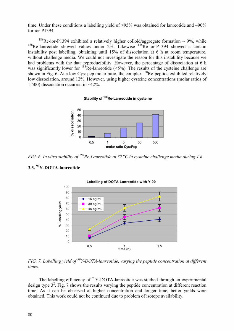
80
time. Under these conditions a labelling yield of >95% was obtained for lanreotide and ∼90% for ior-P1394.
188Re-ior-P1394 exhibited a relatively higher colloid/aggregate formation ∼ 9%, while 188Re-lanreotide showed values under 2%. Likewise 188Re-ior-P1394 showed a certain instability post labelling, obtaining until 15% of dissociation at 6 h at room temperature, without challenge media. We could not investigate the reason for this instability because we had problems with the data reproducibility. However, the percentage of dissociation at 6 h was significantly lower for 188Re-lanreotide (<5%). The results of the cysteine challenge are shown in Fig. 6. At a low Cys: pep molar ratio, the complex 188Re-peptide exhibited relatively low dissociation, around 12%. However, using higher cysteine concentrations (molar ratios of 1:500) dissociation occurred in ~42%.
Stability of 188Re-Lanreotide in cysteine
01020304050
0.5 1 5 50 500molar ratio Cys:Pep
% d
isso
ciat
ion
FIG. 6. In vitro stability of 188Re-Lanreotide at 37 oC in cysteine challenge media during 1 h.
3.3. 90Y-DOTA-lanreotide
Labelling of DOTA-Lanreotide with Y-90
0
10
20
30
40
50
60
70
80
90
100
0.5 1 1.5time (h)
% L
abel
ling
yiel
d
15 ng/mL30 ng/mL45 ng/mL
FIG. 7. Labelling yield of 90Y-DOTA-lanreotide, varying the peptide concentration at different times.
The labelling efficiency of 90Y-DOTA-lanreotide was studied through an experimental design type 32. Fig. 7 shows the results varying the peptide concentration at different reaction time. As it can be observed at higher concentration and longer time, better yields were obtained. This work could not be continued due to problem of isotope availability.

81
4. DISCUSSION
Radiolabelling of iorCEA1 is accomplished by direct method using 2-mercaptoethanol as reducing protein agent. Two different weak chelating agents were employed, which had been reported in other published works before [23, 24]. All experiments carried out with MDP showed low labelling yields and high precipitate formation, even using SnF2, which has better redox properties. The best labelling yields were attained with low citric acid concentration, as well as 2.6 mM SnF2 at pH 3.
As it can be observed from Fig 1, under the conditions specified earlier, relatively high labelling yields could be achieved, within 1 h incubation (>80%). Experiments in which a transfer ligand was formed “in situ” during the labelling reaction showed better results than those where Re weak chelate had been already obtained.
188Re-iorCEA1 was extremely stable when challenged with cysteine, if pre-protected with the addition of AA and BSA. At 5000:1 Cys: MAb molar ratio only a dissociation of 3,5% was observed. Inclusively, without adding such additive, the dissociation was <5% at low cysteine concentrations (Fig. 3). These results were better than those obtained by John, et al. [24] at similar or lower cysteine concentration, suggesting the formation of a more stable complex.
The biodistribution in BALB/C mice was similar as 99Tcm-iorCEA1, except for spleen, where uptake was relative high. This could be explained as due to the binding of the Fc portion of the labelled murine Ab with the citoplasmatic membrane of the red blood cells (RBCs), which could harm RBC, product of the radiolisis of the strong β emission from 188Re. Although this phenomenon could compromise its application in the radiotherapy, it could not be observed in humans for this antibody because it is a murine protein.
On the other hand, in order to enhance the pharmacokinetic parameters of ior-CEA1 to be used in the metabolic therapy, our group developed a single chain fragment of this protein through genetic engineering techniques. These fragments are particularly attractive due to their small size (ca. 25-27 kD).
The results obtained in the 99Tcm labelling indicate that the scFv was adequately labelled and its biological activity was conserved. The cysteine challenge demonstrated that dissociation by transchelation to cysteines at a very high concentration of competitor occurred in a similar fashion for the intact MAb and the scFv, even though in the latter the binding for 99Tcm was made through histidine nitrogen atoms and not by thiol group. Nevertheless, although the chemical characteristics of the molecule accomplished with the established requirements to be used in the radiotherapy, the “in vivo” experiments attained a high uptake in bone marrow, perhaps associated to a cross-reaction with NCA-95 antigen expressed in granulocytes. Therefore, our group decided to enhance the biological characteristic of the molecule before continuing its evaluation for the target therapy.
During the last years many peptides were investigated for their potential to improve the receptor images. Their smaller size conducts to expect a more rapid clearance from the circulation and to be less immunogenic than labelled proteins, maintaining in most of them similar affinity level for their receptors. So, given the physical 188Re characteristic, the 188Re -labelled peptides attained a great sympathy for target therapy.
The labelling method through an intrinsic linear chelating amino acidic sequence seems to be an attractive alternative for directly 188Re-labelled peptide. Thakur and collaborators showed recent developments in the use of these radioactive agents [25, 26].

82
We have synthesized a peptide adding at the N-terminal the 99Tcm (188Re) -chelating sequence (AGGGβA). A group of four amino acids Ala-Gly-Gly-Gly was chosen as chelating moiety, in order to supply a chelating configuration type N4, through their NH2 groups. The proposed structure of 188Re -ior-P1394 is given in Fig. 8. Furthermore, an additional amino acid β-Ala was inserted as a spacer between the chelating moiety and the primary peptide. This amino acid has the purpose of minimizing any possible steric hindrance resulting from the 99Tcm (188Re)-chelation. The cycle peptide was obtained with high yield using the DMSO method for disulphide bond formation. No olygomeric population was observed during the oxidation reaction. This fact was determinant for the high yield of the final product. The resulting peptide has an expected molecular weight of 1444.9, which was determined experimentally. It showed good agreement with the theoretical average masses calculated for each peptide, taking into account the modifications contained within their structures.
N N
N NCH 3 H
O
Re
O
O
ON
O O
(N) RC-160 (C)
FIG. 8. Proposed structure of 188Re-ior-P1394.
The radiolabelling of ior-P1394 with Re-188 is a simple and single-step procedure with a high efficiency (~90%). The best results were obtained at pH 3, in a concentration of 80 mM citric acid and 4 mM SnF2. Under these conditions, it can be also observed the presence of colloids/aggregates relatively high (~9%). We had not time to investigate the character of these aggregates, neither its cause. We mind that the formation of these aggregates could be related with the molecule polarity and its isoelectric point. However, although this work requires more investigation, the results are sufficiently encouraging to suggest that the Intrinsic Linear Chelating Amino Acidic Sequence was a good alternative for the peptide direct labelling with Re-188.
Likewise the 188Re-Lanreotide was obtained through direct method with high labelling yields (>95%). Using Ellman’s test [15] for the sulphidrile group determination, we could prove that once lanreotide is in contact with Sn ions, the sulphidrile bridge is broken without requiring heating. This suggests that the co-ordinate binding can occur through thiol groups.
The cysteine challenge demonstrated that dissociation by transchelation to cysteines at a low concentration of competitor occurred relatively low. However, at high concentrations the complex 188Re-peptide exhibited dissociation ~42%, suggesting the formation of a less stable complex than formed with 99Tcm, whose values were around 18%.
The highest labelling efficiency for 90Y-dOTA-lanreotide was around 83%. In this experiment both the peptide concentration and reaction time played a significant role, with a positive tendency for higher peptide concentrations and reaction times. This experiment could not be continued, because no isotope was available at the time.
5. CONCLUSIONS
From the evaluation of ior-CEA1, scFv, ior-P1394, lanreotide and DOTA-lanreotide, the following can be concluded:

83
– The chosen labelling method for ior-CEA1 with 188Re attained high radiochemical purity of >95%, exhibiting high stability for 24 h with the addition of 2% BSA and 3% AA. Its biodistribution showed relatively higher uptake in spleen, a phenomenon that could compromise its application in radiotherapy.
– Although the evaluation of scFv with 99Tcm attained high labelling yields and good stability, biodistribution studies showed a high uptake in bone marrow, perhaps related to a cross-reaction with NCA-95 antigen expressed in granulocytes. Therefore, it was decided to enhance the biological characteristics of the molecule before its application in the target therapy.
– Ior-P1394 and lanreotide were evaluated with 188Re, obtaining yields of ~90% and >95%, respectively. Although 188Re-labelling of ior-P1394 exhibited a relatively higher colloid formation, our results were sufficiently encouraging to suggest that the strategy of using an amino acidic sequence (AGGGβA) as isotope-chelating moiety for the direct labelling was correct. Likewise 188Re-Lanreotide exhibited good stability at least for 6 h.
– 90Y-labelling method with DOTA-lanreotide showed yields up to 83%. The parameters with a high influence in the system were peptide concentration and reaction time.
REFERENCES
[1] MEARES, C.F., “Introduction to bioconjugate chemistry”, Perspectives in Bioconjugate Chemistry, American Chemical Society, Washington, D.C. (1993) 1-8.
[2] OLIVA, J., PIMENTEL, G., CASCANTE, L., PERALTA, R., BORRÓN, M., et al., “ior-CEA1: Un nuevo anticuerpo monoclonal anti-CEA marcado con 99Tcm para la inmunogammagrafía de los tumoures colorectales. Resultados finales de un ensayo clínico fase I-II”, Rev. Esp. Med. Nucl. 14 (1995) 213-221.
[3] OLIVA, J., OSORIO, M., PIMENTEL, G., BORRÓN, M., DOPICO, et al., “Detection of an unknown cerebral metastasis using the radio-immunoscintigraphy with the monoclonal antibody ior-EGF-R3-Tc-99m”, Rev. Esp. Med. Nucl. 16 (1997) 321-323.
[4] OLIVA, J.P., PIMENTEL, G., BORRÓN, M., PERALTA, R., ORTIZ, R., et al., “Ensayo piloto con el anticuerpo monoclonal ior-c5 como agente potencial de la inmunocentelleografía del cáncer colorrectal”, Rev. Esp. Med. Nuclear 20 (2001) 282-288.
[5] WALDHERR, C., PLESS, M., MAECKE, H.R., HALDEMANN, A., MUELLER-BRAND, ”The clinical value of [90Y-DOTA]-D-Phe1-Tyr3-octreotide (90Y-DOTATOC) in the treatment of neuroendocrine tumours: a clinical phase II study”, Ann. Oncol. 12 (2001) 941-945.
[6] STEWART, P.M., JAMES, R.A., “The future of somatostatin analogue therapy”, Baillieres Best Pract. Res. Clin. Endocrinol. Metab. 13 (1999) 409-418.
[7] CREMONESI, M., FERRARI, M., CHINOL, M., BARTOLOMEI, M., STABIN, M.G., et al., “Dosimetry in radionuclide therapies with 90Y-conjugates: the IEO experience”, Q J Nucl Med 44 (2000) 325-332.
[8] TORMO, G., GAVILONDO, B.R., DOMINGUEZ, J.V, et al., “CEA in colonic adenocarcinomas and precancerous lesions. An immunohistochemical study with a novel monoclonal antibody”, APMIS 97 (1989) 1073-1080.
[9] DAVIS, G.T., BEDZYK, W.D., VOSS, E.W., JACOBS, T.W., “Single chain antibody (SCA) encoding genes: One step construction and expression in eucaryotic cells”, Biotechnology 9 (1991) 165-169.

84
[10] CHAUDHARY, V.K., BATRA, J.K., GALLO, M.G., et al., “A rapid method of cloning functional variable-region antibody genes in E. coli single chain immunotoxins”, Proc. Nat. Acad. Sci., USA 87 (1990) 1066-1070.
[11] HOUGHTEN, R.A. “General methods for the rapid solid-phase synthesis of large numbers of peptides: specificity of antigen-antibody interaction at the level of individual amino acids”, Proc. Nat. Acad. Sci., USA 82 (1985) 5131-5135.
[12] SARIN, V.K, KENT, S.B.H., TAM, J.P., MERRIFIELD, R.B., “Quantitative monitoring of solid-phase peptide synthesis by the ninhydrin reaction”, Analytical Biochemistry 117 (1981) 147-157.
[13] HOUGHTEN, R.A., BRAY, M.K., DEGRAW, S.T., KIRBY, C.J.. “Simplified procedure for carrying out simultaneous multiple hydrogen fluoride cleavages of protected peptide resins”, Int. J. Pept. Protein Res. 27 (1986) 673-678.
[14] ANDREU, D., ALBERICIO, F., SOLÉ, N.A., MUNSON, M. C., FERRER, M.. et al., Peptide Synthesis Protocols, Methods in Molecular Biology (PENNINGTON, M. W. AND DUNN, B. M., Eds.), Totowa, New Jersey (1994) 105-109.
[15] ELLMAN, G.L., “Tissue sulfhydryl groups”, Arch. Biochem. Biophys 82 (1959) 70-77. [16] PIMENTEL, G., OLIVA, J.P., SÁNCHEZ, I., “Anticuerpo monoclonal ior-CEA1:
Comparación de dos métodos de marcaje con 99Tcm”, Rev. Esp. Med. Nucl. 15 (1996)143-148.
[17] PIMENTEL, G.J., VÁZQUEZ, J.E., QUESADA, W., FELIPE, Y., CARDERÓN, C., et al., “ScFv Hexa-histidine tag as a novel alternative for one-step direct labelling of a single-chain Fv antibody fragment with 99Tcm”, Nucl. Med. Comm. (2001).
[18] PIMENTEL, G., QUESADA, W., GARAY H.E., REYES, O., CRUZ, L.J., et al., “An intrinsic linear chelating amino acidic sequence as alternative for the direct labelling of vapreotide, Nucl. Med. Comm. (2001).
[19] MATTER, J.S., ELLISON, D., Reduction-mediated technetium-99m labelling of monoclonal antibodies, J. Nucl. Med. 31 (1990) 692-697.
[20] PIETERSZ, G.A., MARK, R.P., CHESTER, K.A., “Preclinical characterization and in vivo imaging studies of an engineered recombinant technetium-99m labelled metallothionein containing anti-carcinoembryonic antigen single chain antibody”, J. Nucl. Med. 39 (1998) 47-56.
[21] LARSON, S.M., EL-SHIRBINY, A.M., DIVGI, C.R., SGOUROS, G., FINN, R.D., et al., “Single chain antigen binding protein (scFv CC49). First human studies in colorectal carcinoma metastatic to liver”, Cancer (Suppl) 80 (1997) 2458-2468.
[22] HNATOWICH, D.J., VIRZI, F., WINNARD, P. JR., FOGARASI, M., RUSCKOWSKI, M., “Investigations of ascorbate for direct labelling of antibodies with technetium-99m”, J. Nucl. Med. 35 (1994) 127-134.
[23] GRIFFITH, G.L., GOLDENBERG, D.M., HANSEN, H.J., et al., “Direct radiolabelling of monoclonal antibodies with generator produced Rheniun 188 for radioimmunotherapy: labelling and biodistribution studies”, Cancer Res. 51 (1991) 4594-4602.
[24] JOHN, E., THAKUR, J., DEFULVIO, M.R., MCDEVITT, DAMJANOV, “Rhenium-186 labelled monoclonal antibodies for radioimmunotherapy: preparation and evaluation”, J. Nucl. Med. 34 (1993) 260-267.
[25] THAKUR, M.L., MARCUS, C.S., SAEED, S., PALLELA, V., MINAMI, C., et al., “99Tcm-labelled vasoactive intestinal peptide analog for rapid localization of tumours in humans, J. Nucl, Med, 41 (2000) 107-110.
[26] THAKUR, M.L., PALLELA, V.R., CONSIGNY, P.M., RAO, P.S., VESSILEVA-BELNIKOLOVKA, D., et al., “Imaging vascular thrombosis with 99Tcm-labelled fibri α-chain peptide”, J. Nucl. Med. 41 (2000) 161-168.

85
DEVELOPMENT OF RADIOACTIVELY LABELLED CANCER SEEKING BIOMOLECULES FOR TARGETED RADIOTHERAPY
A.D. VARVARIGOU, S.C. ARCHIMANDRITIS, M. FANI,P. BOUZIOTIS, S. VRANJEZ, S. XANTHOPOULOS,A. LEONTI, TH. SOURLINGAS, M. SOTIROPOULOUNational Centre for Scientific Research "Demokritos", Athens, Greece
Abstract
In radioimmunotherapy (RIT) potentially therapeutic radionuclides are transported to the desired site of action by target-selective vehicles, such as monoclonal antibodies (MoAbs) and peptides. The labelling of MoAbs with radionuclides suitable to be used in the field of therapy was investigated. For this purpose we used 153Sm and 201Tl. For the labelling of the antibodies we used the dicyclic anhydride of diethylenetriaminepenta-acetic acid (cDTPAa) as intermediate chelating agent. Radiochemical purity was confirmed with radiochemical techniques (HPLC, ITLC-SG, paper electrophoresis, SDS-PAGE). The factors which were proven to affect labelling were mainly the pH and concentration of the antibody, both during labelling and formation of the immunoconjugate. Labelling with 153Sm was performed at a pH range of 5-7, for 60 min at room temperature. The labelling yield was higher than 90%. As far as labelling with 201Tl is concerned, low concentrations of antibody and immunoconjugate were used. Labelling is performed at pH 6-7, for 60 min at room temperature. The labelling yield reached a maximum of 40%. After purification the labelled antibodies were biologically evaluated in normal Swiss mice. For the 201Tl labelled antibody, high uptake in muscle and kidneys was observed. For 153Sm-antiCEA, slow blood clearance was observed, while the liver and muscles showed high uptake. Intravenous injection in nude mice transplanted with colon cancer cells afforded imaging of the experimental tumour on a camera. Work on peptide labelling was initially carried out on non-radioactive nuclides, so as to optimize labelling conditions and purification techniques. Thus, the peptide lanreotide was labelled with both “cold” 185/187Re and “hot”188Re, affording one major product. DOTA-lanreotide was labelled with “cold” 152Sm and “hot” 153Sm. HPLC data showed the formation of one major product, the yield of which depended on the Sm to peptide molar ratio. In all cases, the labelled peptides can easily be isolated and characterized. Experiments are still in progress.
1. INTRODUCTION
Monoclonal antibodies (MoAbs) have attracted considerable attention due to their potential to serve as selective carriers of radionuclides to specific in vivo antigens for the development of agents for tumour imaging (RIS: radioimmunoscintigraphy) and for tumour therapy (RIT: radioimmunotherapy) [1]. The majority of the studies of RIT used 131I as the radionuclide for antibody labelling. However, 131I is far from ideal because of its tendency to deiodinate in vivo and its high irradiation of normal tissues due to its long half-life and medium energy emission [2]. Other potential therapeutic radionuclides that were studied include short-lived emitters, like 212Bi and 211At and - emitters, like 67Cu, 109Pd, 47Sc and 90Y [3]. At this stage, therapy is largely experimental but progress is being made in developing useful clinical agents [4].
The over expression of peptide receptors on human tumour cells as compared to normal tissues and cells has provided the basis for the successful use of radiolabelled somatostatin (SST) analogues for tumour targeting [5, 6]. Recently, a novel human somatostatin receptor-recognizing ligand, DOTA-lanreotide, was developed. This compound binds to all known hSSTR as well as to a variety of primary tumour cells, which have been shown to express the one or the other hSSTR subtype [7].

86
Successful radiolabelling of MoAbs depends on a number of factors including the chemical characteristics of the nuclide used and the techniques employed for its incorporation into the protein. The most common method for labelling of antibodies with radiometals is by the use of strong chelating groups covalently attached to antibodies so that they may be radiolabelled with the metallic radionuclides in order to result in high stability in vivo [2]. As far as 153Sm is concerned, it is already know that the cation of 153Sm3+ has good chelation properties with polyaminopolycarboxylic acids, like ethylene-diamine-tetraacetic acid (EDTA) or diethylene-triamine-pentaacetic acid (DTPA) [8,9]. DOTA is also one of the more effective chelators for yttrium and the lanthanides, known to form thermodynamically stable chelates with most transition metals, lanthanides and even some of the alkaline earth metals.
Samarium-153 (153Sm) is a radiolanthanide which has not yet been widely used, but which possesses excellent physical characteristics for radioimmunotherapy [10]. It is a beta-emitter with a half-life (t1/2) of 1.95 days. In addition it emits a 130 keV gamma ray (28%) that is suitable for gamma camera detection. It can be produced by neutron activation of enriched 152Sm.
Rhenium-186 and 188Re have been identified as important radionuclides with therapeutic potential. 188Re is available in high specific activities as 188ReO4
- from 188W/188Re generators. 188Re, which has a higher beta-particle energy and short half-life (t1/2 = 17h) is generally more suitable for preparing radiopharmaceuticals that target larger tumours and have reasonably fast clearance from the blood and other non-target tissues.
Two general approaches are being employed for the synthesis of labelled site-specific radiopharmaceuticals:
1. direct labelling of biomolecules and 2. metalation of biomolecules via a covalently appended chelating framework.
For our study of monoclonal antibodies, we chose DTPA as the intermediate chelating agent, which is linked to the antibodies via the bicyclic anhydrite (cDTPAa) [11,12]. As far as we know, the studies of labelling antibodies with 153Sm are limited [13-15]. The main purpose of this study was to investigate the factors, which possibly affects labelling, so as to find those which lead to a labelled product obtained in high yield and with high radiochemical purity. The next step was the radiobiological evaluation of the labelled antibodies in normal mice and in nude mice transplanted with cancer cells. A second purpose was to use the method for labelling of antibodies with 153Sm for other radionuclides in the +3 oxidation state. For this purpose we worked with 201Tl, which is already known in nuclear medicine as an imaging agent for the myocardium. Tl-201 emits -rays suitable for imaging and has a half-life of 3.04 days. It is also emits Auger electrons (22.7–77 keV) and conversion electrons (1.6–167 keV). These properties make Tl-201 attractive for use as a radiotherapeutic agent, too. For the labelling of antibodies with 201Tl with the developed method, 201Tl must be oxidized from the +1 to the +3 oxidation state.
In our studies of peptides, we labelled lanreotide with 188Re directly, taking advantage of the sulphur and nitrogen atoms found within the peptide molecule, which act as donor atoms, complexing the radiometal and forming a stable radiometal chelate. We have also labelled the peptide with 153Sm via the bifunctional chelator DOTA, which holds the radiometal with high stability under physiological conditions. This stability is essential to avoid metal ion dissociation and excessive radiation damage to nontarget cells. The study of the complexation of DOTA-lanreotide was performed with 153Sm which is readily available,

87
and at a low cost, from the “Demokritos” Nuclear Reactor, so that a work protocol could be set up with respect to the radiolabelling of biomolecules with trivalent metal ions in general. Our work on the labelling of peptides with radionuclides was initiated with the labelling of the very same peptides with the non-radioactive nuclides 152Sm and 185/187Re, so as to facilitate the study of the complexation of the peptides with these isotopes.
2. 153Sm/ 201Tl – LABELLED MONOCLONAL ANTIBODIES
2.1. 153Sm–labelled monoclonal antibodies
2.1.1. 153Sm
Samarium-153 was produced in the “Demokritos” reactor by neutron irradiation of 152Sm.
Samarium oxide, isotopically enriched in Sm-152 (152Sm2O3), was irradiated for 5 to 6 h, by a neutron flux of 4.81 × 10¹³ neutrons/cm-2. Sec. Specific activity of the produced Sm-153 was 18 – 32 mCi/mg. The activated oxide was dissolved in a solution of HCl 6M, to produce Samarium chloride (153SmCl3). For the labelling 153SmCl3 was diluted to the appropriate volume with ultra pure water, to produce a stock solution.
2.1.2. Abs
The study was initiated on the human polyclonal immunoglobulin “Sandoglobuline”, commercially available. It was applied thereafter on the monoclonal antibody anti-CEA, kindly offered by Cuba, which selectively localizes in colon cancer.
2.1.3. 153Sm – DTPA
Labelling of DTPA with 153Sm was initially investigated for comparison. DTPA was dissolved in acetate buffer 0.1 , pH=5.6 in a concentration of 1.0 mg/ml. The pH of the final solution was 5.2. For the labelling 5–10 µl of the solution of 153SmCl3 (150–300 µCi) was added to the solution of DTPA. The final pH was adjusted in a pH range 5.0–5.5, if it is necessary, with a dilute solution of NaOH. The solution was stirred gently and was incubated at room temperature for 60 min. Labelling was studied by ITLC-SG.
Labelling of DTPA was evaluated by instant thin layer chromatography on silica gel (ITLC-SG) using NH4OH : H2O 1: 25, as solvent system. Rf values were 0.0 for 153SmCl3 and 0.9 – 1.0 for 153Sm – DTPA.
2.1.4. Immunoconjugate Ab – DTPA
For the preparation of the immunoconjugate, the bicyclic anhydride of DTPA was added in solid form to the solution which contains the antibody in phosphate buffer 0.1M, NaCl 0.1M, pH=8.0. The molar ratio of Ab to DTPA was 1:10. The mixture was allowed to stand at room temperature for 30 min. Separation of the Ab–DTPA from free DTPA was achieved by gel filtration in a Bio Gel P30 column, eluted with acetate buffer 0.1M, pH=5.6. Fractions were collected and their absorbance at 280 nm was measured for the determination and isolation of those which contain the immunoconjugate. The fractions were pooled together and aliquoted to 0.5 or 1.0 ml fractions and frozen.

88
Gel filtration led to the isolation of the immunoconjugate Ab–DTPA from any excess of the ligand, as presented in Fig. 1.
Ab - DTPA
00.5
11.5
1 3 5 7 9 11 13 15 17 19 21 23 25Fraction No.
Abs
orba
nce
(280
nm)
FIG. 1. Ab–DTPA elution profile.
2.1.5. 153Sm–DTPA – Ab
For the labelling of the antibody a small volume of 153SmCl3 (5 – 15 µl) was added to the solution which contained the Ab–DTPA in acetate buffer 0.1M, pH = 5.6. The mixture was allowed to stand at room temperature for 60 min. The labelled antibody was then purified by gel filtration, eluted with acetate buffer. During gel filtration fractions were collected and their absorbance at 280 nm was measured for the determination of those which contain the antibody, and then their radioactivity was measured in an isotope counter. The labelling yield can be calculated from the ratio of the radioactivity of fractions that contain Ab–DTPA to the total radioactivity. The results from a representative experiment are shown in the Fig. 2.
FIG. 2. AB-DTPA elution profile
The radiochemical purity was confirmed with a combination of radiochemical techniques as HPLC, ITLC-SG and SDS-PAGE.
The purified antibodies were analysed by size exclusion HPLC (column: TSK 2000 SW, solvent: phosphate buffer 0.1M, Na2SO4 0.1M, NaN3 0.05% w/v, pH=6.7, flow rate 1ml/min), compared with unlabelled antibodies. Retention time with the UV detector was 5.98 min while with the -detector was 6.33 min. Instant thin layer chromatography was performed on silica gel
0
0,1
0,2
0,3
0,4
1 3 5 7 9 11 13 15 17 19
Fraction No.
Abs
orba
nce
(280
nm
)
020406080
100
1 3 5 7 9 11 13 15 17 19
Fraction No.
Rad
ioac
tivity
(µ
Ci)

89
using acetate buffer 0.1M, pH=5.6 as mobile phase. The radiochromatographs were studied with electronic autoradiography. Rf values were 0.9 – 1.0 for 153SmCl3 and 0.0 for 153Sm – DTPA – Ab.
2.1.6. Factors affecting labelling
We studied many factors that may affect labelling. Table I shows the most important of these.
Antibody concentration effect: In order to see if antibody concentration affects labelling, we used antibody in two different concentrations, 3mg/ml and 15mg/ml, for the preparation of the immunoconjugate. As we can see from the results in all cases using the same quantity of immunoconjugate, higher labelling yields were obtained using Ab–DTPA produced from the antibody with a concentration of 15mg/ml, compared to those produced from the 3mg/ml preparation.
TABLE I. FACTORS AFFECTING LABELLING
Ab–DTPA (mg)
CAb (mg/ml)
153Sm (µCi)
Incubation time
% yield
Antibody concentration effect
0.5
0.5
1.0
1.0
3
15
3
15
155
140
145
175
60 min
60 min
60 min
60 min
20
35
45
91
Incubation time effect
1.0
1.0
1.0
15
15
15
180
175
195
30 min
60 min
18 h
75
91
87
Immunoconjugate concentration effect
0.3
0.5
1.0
2.0
15
15
15
15
250
140
175
280
60 min
60 min
60 min
60 min
30
35
91
93
Incubation time effect: The labelling was studied for incubation time periods of 30 min, 60 min and overnight at room temperature, for the same concentration of Ab and Ab – DTPA. At 30 min the labelling yield was 75%, while at 60 min it became 91% and remained practically the same up to 18 h later.
Immunoconjugate concentration effect: Finally we studied the effect of immunoconjugate concentration on the labelling yield. Using different quantities of Ab-DTPA, starting from the same concentration of antibody (15mg/ml) and using the same labelling conditions, we observed that the yield increases significantly by increasing the concentration of Ab–DTPA. The yield reached a maximum of 93% using 2 mg Ab–DTPA.
pH: Labelling can be achieved at a pH range from 5.0 to 7.0.
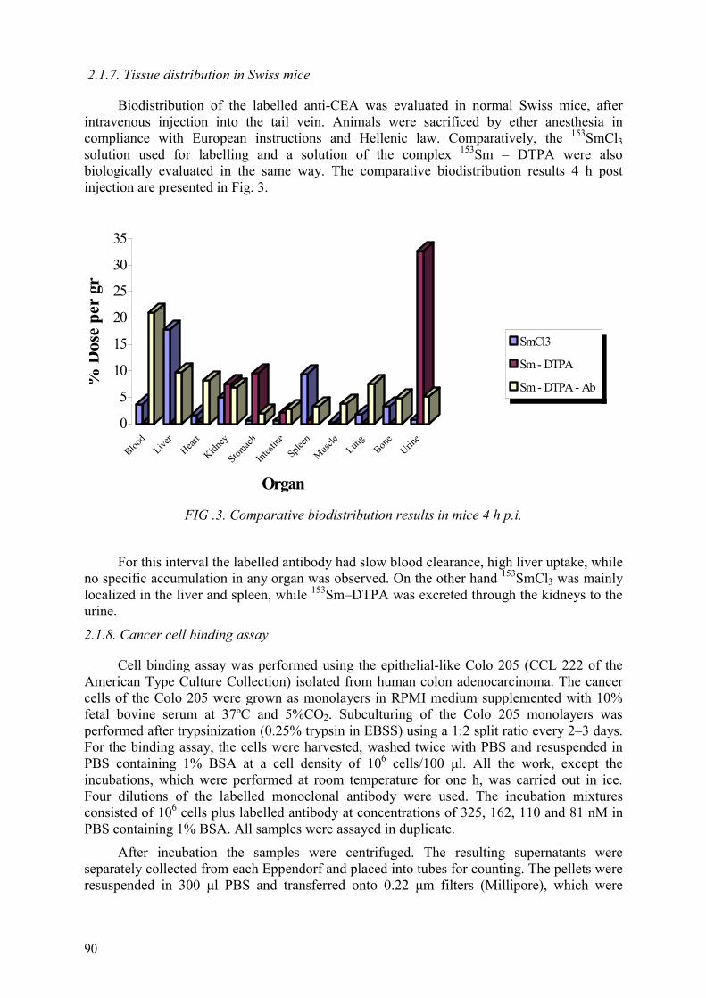
90
2.1.7. Tissue distribution in Swiss mice
Biodistribution of the labelled anti-CEA was evaluated in normal Swiss mice, after intravenous injection into the tail vein. Animals were sacrificed by ether anesthesia in compliance with European instructions and Hellenic law. Comparatively, the 153SmCl3solution used for labelling and a solution of the complex 153Sm – DTPA were also biologically evaluated in the same way. The comparative biodistribution results 4 h post injection are presented in Fig. 3.
0
5
10
15
20
25
30
35
%D
ose
per
gr
Blood
Liver
Heart
Kidney
Stomach
Intest
ineSple
en
Muscle
Lung
Bone
Urine
Organ
SmCl3
Sm - DTPA
Sm - DTPA - Ab
FIG .3. Comparative biodistribution results in mice 4 h p.i.
For this interval the labelled antibody had slow blood clearance, high liver uptake, while no specific accumulation in any organ was observed. On the other hand 153SmCl3 was mainly localized in the liver and spleen, while 153Sm–DTPA was excreted through the kidneys to the urine.
2.1.8. Cancer cell binding assay
Cell binding assay was performed using the epithelial-like Colo 205 (CCL 222 of the American Type Culture Collection) isolated from human colon adenocarcinoma. The cancer cells of the Colo 205 were grown as monolayers in RPMI medium supplemented with 10% fetal bovine serum at 37ºC and 5%CO2. Subculturing of the Colo 205 monolayers was performed after trypsinization (0.25% trypsin in EBSS) using a 1:2 split ratio every 2–3 days. For the binding assay, the cells were harvested, washed twice with PBS and resuspended in PBS containing 1% BSA at a cell density of 106 cells/100 µl. All the work, except the incubations, which were performed at room temperature for one h, was carried out in ice. Four dilutions of the labelled monoclonal antibody were used. The incubation mixtures consisted of 106 cells plus labelled antibody at concentrations of 325, 162, 110 and 81 nM in PBS containing 1% BSA. All samples were assayed in duplicate.
After incubation the samples were centrifuged. The resulting supernatants were separately collected from each Eppendorf and placed into tubes for counting. The pellets were resuspended in 300 µl PBS and transferred onto 0.22 µm filters (Millipore), which were

91
placed into the wells of a manifold filtration apparatus (12 wells/apparatus). The samples were vacuum dried onto the filters. The filters were washed four times with PBS and dried in the same manner as that described above. In continuation, the filters were placed into counting tubes and the radioactivity of the filters as well as that of the first supernatants were measured in a gamma counter (Minaxi autogamma 5000 series, Packard). The results were assessed using the Scatchard analysis. Scatchard analysis of the data obtained from the binding assay experiments of the 153Sm-DTPA-antiCEA to the binding sites of the Colo 205 human adenocarcinoma revealed an apparent Kd of 2.0 nM. (Fig. 4). This finding is indicative of a high affinity amongst the binding sites of the colon cancer cell line.
FIG. 4. Scatchard plot.
2.1.9. -camera imaging of nude mice
Nude mice were transplanted with colon cancer cells, subcutaneously. After 10 – 15 days the tumour had been developed, as verified by histochemical techniques. The labelled anti-CEA was intravenously injected into the tail vein. The animals were sacrificed at 4 h post injection. As we can see in the image from the -camera, Fig. 5, the experimental tumour can be clearly visualized.
FIG. 5. Scintigram of a nude mouse, 4 h p.i.

92
2.2. 201Tl – Labelled monoclonal antibodies
2.2.1. Preparation of 201Tl for labelling of antibodies
2.2.1.1. Preparation of 201Tl
Oxidation of 201Tl+ to 201Tl+++
To a solution of 201TlCl (0.1 – 0.5 ml) a solution of H2SO4 5M (1.0 ml) was added followed by a fresh solution of KBrO3 5% w/v (0.2 ml). The mixture was allowed to stand at room temperature for 15 min before the addition of a solution of NaCl 4M (50 µl).
Extraction of 201Tl+++
201Tl3+ was extracted with the addition of 1.5 ml butyl acetate. The mixture was mixed for 20 min at room temperature. The organic phase was separated from the water phase and was evaporated in a water bath, by adding a small volume of water. 201Tl was received as an aqueous solution ready to use for labelling.
2.2.1.2. Radiochemical control
201Tl+++ was checked by low voltage electrophoresis in nitrocellulose acetate strips (200V, 30min), in comparison with the solution of 201Tl1+. 201Tl3+ migrates to the negative electrode, while 201Tl1+ stays at the origin, as shown in Fig 6.
FIG. 6. Electrophoresis of 201Tl1+ and 201Tl3+.
2.2.2. 201Tl – DTPA – Ab
For the labelling of antibodies with 201Tl we used human polyclonal immunoglobulin (Sandoglobuline). The antibody was conjugated with DTPA by the bicyclic anhydrite of DTPA. The immunoconjugate was produced as is described for the labelling of antibodies with 153Sm.
Labelling was performed by the addition of the solution of 201Tl3+ to the solution, which contained the immunoconjugate Ab – DTPA in NaCl 0.9% w/v. The mixture was allowed to stand at room temperature for 60 min. The labelled antibody was then purified by gel filtration in a Bio Gel P30 column, eluted with saline.

93
The radiochemical purity was confirmed with low voltage electrophoresis in nitrocellulose acetate strips (200V, 30min), in the same way as in the case of the 201Tl3+
control. The 201Tl–DTPA–Ab remains at the origin, while 201Tl3+ migrates to the negative electrode.
2.2.3. Biodistribution studies
The labelled antibody was biologically evaluated in normal Swiss mice after intravenous injection into the tail vein. Comparatively, the 201Tl3+ solution was biologically evaluated in the same way. Table II shows the comparative biodistribution results 4 h post injection.
201Tl3+ is rapidly excreted through the kidneys and as expected there is a high accumulation in the heart and muscle. From the results of the biodistribution of 201Tl–IgG one can conclude that the radioimmunoconjugate shows good in vivo stability (lower stomach values), low accumulation in liver and lower renal excretion.
TABLE II. COMPARATIVE BIODISTRIBUTION RESULTS 4 H P.I
Organ Tl(III) Tl – DTPA - IgG
% dose per gram
Blood 0.56 ± 0.1 1.24 ± 0.1
Heart 6.24 ± 0.2 4.12 ± 0.3
Lungs 4.03 ± 0.4 2.89 ± 0.5
Liver 3.65 ± 0.1 2.73 ± 0.7
Kidneys 58.4 ± 3.2 38.56 ± 5.2
Spleen 6.93 ± 0.5 4.06 ± 0.7
Stomach 5.24 ± 0.3 3.12 ± 0.4
Intestine 7.07 ± 0.9 3.70 ± 0.8
Muscle 5.39 ± 0.7 2.47 ± 0.8
Urine (% Dose/organ)
4.36 ± 0.8 2.94 ± 1.2
3. 153Sm/ 188Re–LABELLED PEPTIDES
3.1. 185/187Re/188Re-lanreotide
3.1.1. Preparation of 185/187Re-lanreotide
To a suspension of ReOCl3[P(C6H5)3]2 (1.899 mg, 2.28x10⁻³mmol) and CH3COONa (1.869 mg) in MeOH (2 mL) a solution of Lanreotide (2,5 mg, 2.28x10⁻³ mmol) in MeOH (1 mL) was added under stirring. The mixture was stirred at R.T. overnight, while its initial yellowish color has disappeared, leaving a clear, colorless solution. HPLC analysis (C18column, acetonitrile-0.1% TFA gradient elution system: 0-60% acetonitrile over 20 min)

94
showed the formation of one main product, with a retention time of 18 min. The formation of another product, with a retention time of 15 min, was also noted.
3.1.2. Preparation of 188Re-lanreotide
The 188Re eluate is acquired from a 188W/188Re generator. 5.0 mL eluate was acquired. 2.0 mL of the eluate was added to a mixture of reducing agents (SnCl2: 1.6 mg, ascorbic acid: 2.46 mg, citric acid: 16 mg). The resulting mixture is left for 2 h at room temperature. The 2.0 mL mixture containing the reduced rhenium-188 is added to 3 mg of a mixture of SnCl2,sodium gluconate and sodium bicarbonate (this mixture is prepared by adding 1 g sodium gluconate, 2 g sodium bicarbonate and 15 mg SnCl2 to an empty vial, and shaking well). The resulting mixture is left for 15 min. A solution of the peptide is prepared (1 mg peptide/mL). 0.4 mL 188Re-gluconate solution was added to 0.5 mL peptide solution (containing 0.5 mg peptide). The resulting mixture is heated at 90ºC for 60 min. The volume of the mixture was brought to 2 mL with saline.
HPLC studies of 188Re-Lanreotide showed the formation of one major product, with a retention time of 17.5 min. The formation of this product was influenced by the reaction time and the temperature. Complex formation is accelerated by increasing the temperature. By heating the reaction mixture at 90ºC for 60 min, a yield of approximately 90% was obtained.
Biodistribution studies in normal Swiss mice showed that the radiolabelled biomolecule presented fast blood clearance and practically complete hepatobiliary elimination. Urine elimination was negligible.
In vitro cell binding studies were not satisfactory, and are currently being repeated. Tumour uptake was evaluated in female BALB/c nude mice, 5 weeks old. The experimentally induced gastrointestinal tumour is clearly delineated 2 h after intravenous injection of the radiolabelled peptide. PANC-1 cells are currently being grown and will soon be implanted into nude mice so as to see whether this Re-188 derivative shows specificity for this type of cancer.
3.2. 152Sm/153Sm-DOTA-lanreotide
3.2.1. Preparation of 152Sm-DOTA-lanreotide
A stock solution of samarium chloride was prepared by dissolving 366 mg SmCl3.6H2O(FW 366) in 1000 µL (1 mL) ultra pure water. The final concentration of the SmCl3 solution is 1000 nmol/ 1000 µL. The DOTA-lanreotide conjugate was dissolved in freshly prepared 0.2 M ammonium acetate buffer (metal-free: pH 7.00). The concentration of the final solution was 1000 µg/ 1000 µL. Reaction ratios of 0.5:1, 1:1 and 2:1 SmCl3:Peptide were used, and the complexing reaction was allowed to proceed for 60 min at 90ºC. An aliquot was then removed and its purity assessed by HPLC (C18 column, same as for Rhenium-lanreotide analysis). HPLC analysis of the DOTA-lanreotide conjugate afforded one main peak at 16.00 min (84%), with two shoulders on either side of this peak, at 15.10 min (0.89%) and 17.17 min (4.04%). DOTA-lanreotide, when labelled with “cold” SmCl3, gave practically the same elution profile in all three cases, as can be seen below.
The HPLC profile of the “neat” peptide conjugate differs from that of 152Sm-DOTA-lanreotide, possibly because of the formation of the samarium-peptide complex, via the

95
DOTA linker. The UV profile remains practically unchanged even when the molar ratio of SmCl3: peptide changes significantly (Table III).
TABLE III. HPLC DATA OF DOTA-LANREOTIDE AND 152SM-DOTA-LANREOTIDE
Product under evaluation tr(UV)(retention time)
%
DOTA-lanreotide 16.00 84.00
SmCl3: DOTA-lanreotide 0.5 : 1 15.77 41.88
SmCl3: DOTA-lanreotide 1 : 1
15.87 40.63
SmCl3: DOTA-lanreotide 2 : 1
15.82 41.00
3.2.2. Preparation of 153Sm-DOTA-lanreotide
A stock solution of 153Sm was prepared as described above (Part A). DOTA-Lanreotide was dissolved in ammonium acetate buffer (see: Synthesis of 152Sm-DOTA-Lanreotide). The pH of the final solution was 6.9. For the labelling with 153Sm, a variety of volumes of 153SmCl3 solutions were added to the DOTA-Lanreotide solution (radioactivity range 25 – 200 µCi). The solutions were incubated at 90 °C for 60 min. Labelling efficiency was studied by HPLC (column and gradient elution system same as above). The results are shown in Fig. 7. Gradual increase of the added radioactivity leads to a marked decrease in the labelling yield for peptide samples of the same concentration.
The above findings help us reach an initial conclusion that labelling efficiency of the DOTA-lanreotide conjugate depends on the 153Sm to peptide molar ratio. Further radiochemical and radiobiological studies are in progress.
77.4
56.4
198.6
93.3
0
50
100
150
200
250
1 2 9.31 11.37
% radioactivity
Act
ual r
adio
activ
ity a
dded
(µC
i)
FIG. 7. Labelling efficiency of DOTA-lanreotide.

96
Biomolecules were labelled with a variety of nuclides and were evaluated in terms of radiochemical purity and radiobiological performance. Monoclonal antibodies were successfully labelled with the radionuclides 153Sm and 201Tl to be used in combined Radioimmunoscintigraphy and Radioimmunotherapy. Labelling can be performed via a chelating agent, like DTPA, which forms a stable complex with the radionuclides. The radionuclide must be in the +3 oxidation state. For this purpose 201Tl must be oxidized before use from +1 to +3. It is possible that the developed method can generally be applied to radionuclides in the +3 oxidation state. The concentration of antibody during the formation of immunoconjugate Ab–DTPA and concentration of Ab–DTPA during labelling significantly affect the labelling yield. Other factors like incubation time, pH and radioactivity also play an important role on the labelling yield. The methodology we developed results in a labelled antibody, purified and in high yield. Radiobiological studies advocated for possible successful in vivo use of the radiolabelled MoAbs. Intravenous injection of the 153Sm–anti CEA in nude mice transplanted with colon cancer cells afforded imaging of the experimental tumour on a camera. The SSTR specific peptide lanreotide and the DOTA-lanreotide conjugate were
both labelled with radioactive and non-radioactive isotopes of the same element and were radiochemically evaluated. Lanreotide was directly labelled with 185/187Re and 188Re, while 152Sm and 153Sm labelled the same peptide via the bifunctional chelating agent DOTA, resulting in a stable complex, as is expected. 188Re lanreotide was evaluated in vitro and in vivo, and will continue to be evaluated with other cancer cell lines, while the same assays are currently in progress for the DOTA-lanreotide complex.
REFERENCES
[1] POTAMIANOS, S., et al., “Radioimmunoscintigraphy and radioimmunotherapy in cancer: principles and application”, Anticancer Res. 20 (2000) 925-948.
[2] SRIVASTAVA, S.C., MEASE, R.C., “Progress in research on ligands, nuclides and techniques for labelling monoclonal antibodies”, Nucl. Med. Biol. 18 (1991) 589-603.
[3] ADELSTEIN, S.J., KASSIS, A.I., “Radiobiologic implications of the microscopic distribution of energy from radionuclides”, Nucl. Med. Biol. 14 (1987) 165-169.
[4] LARSON, S.M., et al., “Current status of radioimmunotherapy”, Nucl. Med. Biol. 21(1994) 785-792.
[5] VIRGOLINI, I., et al, “Somatostatin receptor subtype specificity and in vivo binding of a novel tumour tracer, 99Tcm-P829”, Cancer Res. 58 (1998) 1850-1859.
[6] LEIMER, M., et al, “Response to treatment with yttrium-90-DOTA lanreotide of a patient with metastatic gastrinoma,” J. Nucl. Med. 39 (1998) 2090-2094.
[7] SMITH-JONES, P.M., et al, “DOTA-lanreotide: a novel somatostatin analog for tumour diagnosis and therapy”, Endocrinology 140 (11) (1999) 5136-5148.
[8] TSE, J.W., et al., “High specific activity (Samarium-153) EDTA for imaging of experimental tumour models”, J. Nucl. Med. 30 (1989) 202-208.
[9] AWANG, M.B., et al., “Evaluation of 153Sm-diethylenetriaminepentaacetic acid for radiolabelling of pharmaceutical dosage forms by neutron activation”, Nucl. Med. Biol. 21 (1994) 905-909.
[10] EHRHARDT, G.J., et al., “Production of Sm-153 for radiotherapeutic applications”, J. Lab. Cpd. Radiopharm. 23 (1986) 1370.
[11] HNATOWICH, D.J., et al., “The preparation and labelling of DTPA-coupled albumin”, Int. J. Appl. Radiat. Isot. 33 (1982) 327-332.

97
[12] HNATOWICH, D.J., et al., “The preparation and labelling of DTPA-coupled antibodies radiolabelled with metallic radionuclides: an improved method”, J. of Immunological Methods 65 (1983) 147-157.
[13] BONIFACE, G.R., et al., “Labelling of monoclonal antibodies with Samarium-153 for combined radioimmunoscintigraphy and radioimmunotherapy”, J. Nucl. Med. 30 (1989) 683-691.
[14] IZARD, M.E., et al., “An improved method for labelling monoclonal antibodies with samarium-153: use of the bifunctional chelate 2-(p-isothiocyanatobenzyl)-6-methyldiethylenetriamine pentaacetic acid”, Bioconjug. Chem. 3 (1992) 346-350.
[15] KRAEBER-BODÉRÉ, F., et al., “Pharmacokinetics and biodistribution of samarium-153-labelled OC125 antibody coupled to CITCDTPA in a xenograft model of ovarian cancer”, Eur. J. Nucl. Med. 23(5) (1996) 560-567.

99
PREPARATION, QUALITY CONTROL AND ANIMAL TESTING OF Re-188LABELLED COMPOUNDS
G.A. JÁNOKI, L. KÖRÖSI, L. BALOGH, G. ANDÓCS, K. BODÓ, J. LÁNG, A. POLYÁK, R. KIRÁLY, D. MÁTHÉFrederic Joliot-Curie National Research Institute for Radiobiology and Radiohygiene, Budapest, Hungary
Abstract
Rhenium-188 is an attractive therapeutic radioisotope obtained by elution from tungsten 188/rhenium 188-generator system. We used this eluate to label HEDP and lanreotide to prepare radiopharmaceuticals for experimental use. The results of our studies showed that using methods worked out under the framework of the CRP allowed us to prepare 188Re-HEDP and 188Re -lanreotide with high yield and favourable biodistribution in animals.
In animal studies 188Re-HEDP was successfully used for localizing osteosarcoma in dog.
1. INTRODUCTION
The fundamentals of successful radionuclide therapy is sufficient uptake and prolonged retention of radiopharmaceuticals in the target tumouric tissues. The pharmacokinetic properties of therapeutic radiopharmaceuticals can be assessed with a low dose tracer study before the single or multiple application of the therapeutic dose.
Radiation is delivered selectively by appropriate radiopharmaceuticals taking the advantage of short-range ß particles or ultra short-path-length of α particles or Anger electron emitting radionuclides.
The advantages and limitations of radionuclide therapy are listed in Table I.
TABLE I. ADVANTAGES AND LIMITATIONS OF RADIONUCLIDE THERAPY
Advantages Limitations Specificity, radiation dose limited to target
Isolation of patients
Efficacy and low toxicity Storage of radioactive waste
Systemic or locaregional application Availability, as licenced therapeutical radiopharmaceuticals and isolation beds in hospital.
Excellent palliation Need for dosimetry calculation Limited long-therm consequences Multidisciplinary approach required
Pre-assessment of uptake and retention (in repeated tracer study)
Mechanism poorly understood
Multiple treatments possible High cost of some newer forms New indications emerging

100
The use of beta-emitting radioisotopes labelled compounds for the treatment of solid tumours and palliative treatment of skeletal bone pain, especially for treatment of multiple lesions has developed as an important alternative modality to more traditional therapeutic regimens.
In a recent programme we studied and used the tungsten-188/rhenium-188 generator system, which provides high levels of carrier-free 188Re for the preparation of various therapeutical agents.
The long useful shelf-life (up to one year) and high activity (500-800 mCi) of these generators and the high daily yield of perrhenate-188 and appropriate radionuclidic properties (2,1 MeVβ; 155 KeVγ) make this isotope an attractive candidate for wieldy use for bone palliation and other therapeutic application. During our experimental work rhenium-188 labelled hydroxyethylidene-diphosphonate (HEDP), 188Re-dimercaptosuccinic acid (DMSA) and 188Re labelled somatostatin peptide analogues (lanreotide and DOTA-lanreotide) were prepared and evaluated in radiochemical purity and biological study. For tracer study Ga-67 labelled DOTA-lanreotide and for therapeutic purpose 131I, labelled-lanreotide were also prepared in limited quantity.
2. MATERIAL AND METHODS
All chemicals and reagents were of analytical grade. Perrhenic acid (65-70% in water, 99,9% purity) and gentrisic acid (99%) were purchased from Aldrich Chemical Company. The HEDP (>97%) was obtained from Fluka AG: Lanreotide and DOTA lanreotide was supplied by pi-CHEM (Austria). Alumine-based Tungsten-188/Rhenium-188 generator was purchased from Oak Ridge National Laboratory (USA). Generator set-up, elution and daily quality control were done according to the manufacturer’s instructions. At the end of each elution the generator was purged with nitrogen gas to maintain the column in a dry state between elutions.
Ga-67 chloride solution was obtained from MDS Nordion Inc. (Canada).
3. PREPARATION OF 188Re-HEDP
At aseptic condition 166 mg of HDP and 60 mg of gentisic acid were dissolved in 10 ml physiological saline. The solution was stirred during the addition of 77 mg of stannous chloride dihydrate to provide a clear colorless solution (pH∼1). One ml aliquots of this solution were then dispensed using a steril syringe by elution through a steril 0.22 µmMillipore filter into steril dose vials. The vials were then filled with sterile nitrogen gas to yield on inert atmosphere. Carrier perrhenic solution was prepared (20 µl perrhenic acid solution was mixed with 2ml of sterile saline, purged with nitrogen, placed in dark and coole storage and 20 µl volume from this solution was added to eluate). To predispensed vial 1-2 ml of 188Re perrhenate, 20 µl of carrier rhenium was added and the mixture was then heated at 96-100°C for 30 min. After cooling 2 ml of sodium acetate buffer added. Quality control consisted of TLC analyses of the neutralized solution using Gelman ITLC-SG plates developed in saline and acetone.

101
Preparation of 188Re labelled-lanreotide
Reducing solution was prepared by using stannous chloride dyhidrate (1,6 mg) ascorbic acid (2,46 mg) citric acid (16 mg) and 188Re perrhenate in saline (200-700 MBq) was added. After stirring at room temperature for 2 h, 0,4 ml sample was added to 0,1 ml of lanreotide (100 µg, in 0,1µ acetate puffer) and mixture was heated at 90-100°C for 60 min. After cooling neutralizing stabilizing puffer solution was added. Radiochemical purity was determined as indicated above. HT-29 (human colon adenocarcinoma) cells were evaluated to bind 188Re-lanreotide. During assay 5,10 and 50 ng/ml labelled lanreotide was incubated with 1,5 and 10x106 cell and incubated at 37°C for 1 h.
Biodistribution studies
188Re-HEDP biodistribution studies were performed in nude mice bearing HT-29 human colon adenocarcinoma xenograft, adult Beagle dog and dog with osteosarcoma. Images were acquired with a Nucline X-Ring (Mediso Ltd., Hungary) digital gamma camera (HEGP collimator, 75KeV and 156 KeV energy with 10% window width) for pharmacokinetic study 20 MBq/bodyweight kg was injected intravenously.
Experimental results
The formulation we used for Re-188 labelling of HEDP yielded a labelling efficiency of 90% to 98%. If 188Re-HEDP solution was stored at room temperature it slow radiochemical decomponation (1-2%/hrs) was shown. The biodistribution of 188Re-HEDP when injected intravenously to dogs showed significant bone uptake and excretion throughout the kidneys. Distribution of compounds when injected into dog with osteosarcoma showed skeletal distribution and very significant uptake in the primer lesion. Lesion delineation and uptake was compared to scan taken by using 99Tcm-MDP. Comparison of the distribution of two compounds are seen in Fig. 1. To know the biodistribution and pharmacokinetic of 188Re perrhenate in separated experiment 20 MBq/Kg eluate was injected. Perrhenate accumulated in thyroid, salivary glands and in the gastric mucosa as seen Fig. 2.
The labelling efficiency of lanreotide with 188Re ranged between 85-95% before store aliquots were mixed 1:1 with pro injection Human Serum Albumin 20% solution and ascorbic acid for injection. The results of the binding assay are shown in Table II.
Biodistribution of 188Re lanreotide in nude mice bearing HT-29 human tumour xenograft is shown in Fig. 3. Scans up to 3 h showing fast biliary and renal extraction with very moderate uptake in tumour.
TABLE II. 188RE -LANREOTIDE BINDING TO HT-29 CELLS
Concentration of labelled lanreotide No. of HT-29 cells 5 ng/ml 10 ng/ml 50 ng/ml 10×106 44.6% 34.9% 32.6% 5×10⁶ 32.6% 27.3% 22.35 1×10⁶ 25,6% 26.4% 19.2%

102
4. DISCUSSION
Key requirement for routine preparation of 188Re labelled radiopharmaceuticals for clinical use is the availability of simple and efficient method for concentration of generator eluate. Because of the daily clinical need for developing cost-effective alternatives for the treatment of bone pain in our work we have concentrate the preparation work of 188Re-HEDP and 188Re (V)-DMSA which also was prepared in limited experiment. System used for concentration of the rhenium generator saline eluant was not effective and therefore we had to concentrate eluate by heating.
188Re-HEDP prepared in our laboratory showed high labelling yield and localized in skeletal tissue as well as in osteosarcoma lesion. The lesion pharmacokinetic was favourable and allowed us to calculate absorbed dose. Preliminary result of labelling lanreotide with rhenium was promising. Working out eluate concentration method and optimal conditions for labelling were still needed.
By using experiences gained during the CRP we successfully labelled HEDP and lanreotide, which are going to be the basic therapeutical radiopharmaceuticals in the future. The chemical research work done will allow us to prepare kit formulation for 188Re isotope labelling also.
FIG. 1. Distribution and tumour localization in dog with osteosarcoma.
99Tcm-MDP RRee--118888--HHEEDDPP

103
1 h 3 h 6 h 24 h
FIG. 2. 188Re perrhenate biodistribution in healthly Beagle dog static images.

104
10 min 30 min
1 h 2 h
3 h
FIG. 3. Biodistribution of 188Re-lanreotide in nude mice bearing HT-29 human tumourxenograft.

105
ACKNOWLEDGEMENTS
The authors would like to thank to E. Hrisztodulakisz, M. Pállai, Csilla Dirner, Zs. Suhajda, N. Fésüs and K. Haller for their expert assistance in generator handling, labelling, radiochemical analysis, and animal study.
BIBLIOGRAPHY
CHATAL, J.F., HOEFNAGEL, C.A., “Radionuclide therapy”, The Lancet, Vol. 354 (1999) 931-935.
CLARKE, M.T., GALIE, E., “Radionuclide therapy of osseous metastatic disease”, J. Nucl. Med. Tech. 21 (1993) 3-6.
FREEMAN, L.M., BLAUFOUX, M.D. (Eds.), “Radionuclide therapy of intractable pain”, Seminar in Nuclear Medicine XXII (1) 58 pages (1992) .
KNAPP, F.F. JR., BEETS, A.L., GUHLKE, S., ZAMORA, P.O., BENDER, H., et al., “Availability of Rhenium-188 from the alumina-based tungsten-188/rhenium-188 Generator for preparation of Rhenium-188-labelled radiopharmaceuticals for cancer treatment”, Anticancer Res. 17 (1997) 1782-1796.
SILBERSTEIN E.B., “Doseage and response in radiopharmaceutical therapy of painful osseous metastases”, J. Nucl. Med 37 (1996) 249-252.
THUERLMANN, B., DE STOUTZ, N.D., “Causes and treatment of bone pain of malignant origin”, Drugs 51 (3) (1996) 383-398.
ZAMORA, P.O., BENDER, H., KNAPP JR., F.F., BIERSACK, H.J., “Radiotherapy of intrathoracic carcinoma xenografts with 188Re-RC-160, a somastatin analogue”, Tumour Targeting 2 (1996) 49-59.
107
DEVELOPMENT OF RADIOACTIVELY LABELLED CANCER SEEKING BIOMOLECULES FOR TARGETED THERAPY
M.R.A. PILLAI, M. VENKATESH, S. BANERJEE, G. SAMUEL, K. KOTHARI, A. DASH. P.R. UNNI, A. KORDE, K. BAPAT, U. PANDEY, T. DAS, S. CHAKRABORTY, A. MUKHERJEE, H.D. SHARMA Bhabha Atomic Research Centre, Mumbai, India
Abstract
Several radioisotopes such as 90Y, 186Re, 166Ho, 177Lu, were produced and processed in our laboratory for evaluation as potential isotopes for therapy. 90Y was sourced from 90Sr in the form of a generator based on a supported liquid membrane separating technique as well as by solvent extraction /adsorption procedure. The other isotopes were obtained by irradiation of the natural/enriched targets. Among the various isotopes studies for possible therapeutic use, 177Lu was identified as an ideal candidate for labelling of peptides owing to its high specific activity. Bifunctional chelating agents for complexing with these isotopes were synthesized and characterized. Radiolabelling procedures were standardized to obtain maximum complexation. Standard quality control techniques such as PC, TLC, HPLC have been utilized to characterize the radiolabelled species. Lanreotide, a somatostatin analogue was coupled to DOTA in a three-step procedure and purified by preparative TLC. The radiolabelling of the peptide conjugate with 90Y and its subsequent purification with Sep-Pak column was optimized. The direct labelling of lanreotide with 186Re as well as 125I were also accomplished. The in vivobioefficacy studies of the radioiodinated lanreotide as well as 90Y-DOTA-lanreotide were carried out in mice bearing melanoma. In vitro cell binding studies of 90Y-DOTA-lanreotide with A431 cell lines were also carried out.
1. INTRODUCTION
Somatostatin receptors are known to be expressed by most of the neuroendocrine tumours and therefore radiolabelled somatostatin or its analogues are expected to exhibit binding affinity towards these tumours. Lanreotide, an octapeptide, has been identified as a somatostatin analogue bearing affinity to most of the sub-types of human somatostatin receptors. In connection with our efforts on the "Development of radioactively labelled cancer seeking biomolecules for targeted therapy" we have explored the potential of several therapeutic radioisotopes. Among these, two of the isotopes viz 188Re and 90Y were available directly from the generator whereas the others such as 186Re, 166Ho, 177Lu were obtained from the irradiation of natural or enriched targets and subsequent radiochemical processing of the irradiated targets in our laboratory. 90Y has been reported to be a suitable therapeutic radionuclide for labelling of peptides through a BFCA. In the present study, the radiolabelling yields of 90Y with DOTA-lanreotide under optimized conditions were found to be >70%. However, due to the logistic considerations, 177Lu ( t1/2 6.7d, βmax 0.5 MeV, γ 208 keV) was identified as an ideal therapeutic radioisotope. 10 µg of enriched Lu irradiated at a flux of 1014
n/cm2/sec for 28 d yield 112 mCi. This corresponds to a specific activity of 1.94 × 10⁴ Ci/g. Therefore 2 µg of 177Lu will yield a therapeutic dose of ~20 mCi. While several methods have been reported for the preparation of the radiolabelled peptide conjugates, the major thrust of the present work was to effect the radiolabelling via the use of a bifunctional chelating agent. In this respect, DOTA was found to be an ideal BFCA for coupling with lanreotide. The complexation yields of DOTA with 177Lu were >99%. at a Lu concentration of 5 µg/mL and 40 µg of DOTA for 3 h at room temperature.
2. MATERIALS
95% D2EHPA was obtained from BDH (Poole, England). Dowex-50×12 , 200-400 mesh was from Sigma Chemical Company, USA, dodecane from Transware Chemicals, Handelsgescichaft, Hamburg, Germany. All other reagents used were obtained from E. Merck

108
(Mumbai, India) or BDH (Poole, England) and were of analytical quality. PTFE membranes with pore size 0.45 µm and thickness 160 µm were purchased from M/s. Sartorius, Germany. Rhenium metal for irradiation (Spectroscopic grade, >99.99% pure, natural abundances of Re-185 and Re-187 are 37.07 and 62.93%, respectively) was obtained from Johnson Matthey Company. Enriched lutetium oxide (60.7% 176Lu) was a gift from the IAEA and holmium oxide (Spectroscopic grade >99.99% pure) was from American Potash Inc. All other enriched targets were obtained from Isoflex, USA.
The chemicals used in the synthesis of the BFCAs (DOTA, DAHPES) and the peptide conjugates were obtained from Aldrich Chemical Co., MO, USA and Fluka Chemical Company, USA. All the inorganic salts were either from M/s. Sarabhai M. Chemicals or M/s. S.D. Fine Chemicals, India. The solvents were from M/s. Merck (India). Lanreotide was obtained as a gift from the IAEA.
Na125I was obtained from Du PONT, Canada, chloramine T from Sigma Chemical Co, USA, HEDP from BRIT, Navi Mumbai, India, and SepPak C18 cartridge from WATERS, India.
SnCl2.2H2O was obtained from Sigma Chemical Company. Flexible silica gel plates (7.5 x 2.5 cm, coating thickness, 0.25 mm) were from J.T. Baker Chemical Company and Whatman 3 chromatography was used for paper chromatography and electrophoresis. 2-ethylhexyl-2-ethylhexylphosphonic acid (KSM-17) was synthesized locally according to a reported method. All reagents were made in metal ion free triple distilled water. All radioactive tracers used 90Y, 85+89Sr, 90Sr-90Y were available at the Radiopharmaceuticals Division of this Centre. Borosilicate glass column of dimension 15 cm×0.4 cm (i.d), provided with a sintered disc at the bottom suitable to hold 2-5 ml of ion exchanger (Dowex 50×12, 200-400 mesh) were used.
90Sr-90Y generator used in these studies was developed by the Process Development Division of Bhabha Atomic Research Centre. 90SrCl2 was obtained from the reprocessed fuel effluents in one of our laboratories. 90Y was assayed either by using a NaI(Tl) solid scintillation counter (Star Electricals, Bombay, India) to measure the Bremsstrahlung radiation or by using a liquid scintillation counter (Model - Tricarb 2100TR - Packard Instrument Co., USA). All radioactive measurements were made using a NaI(Tl) counter.
1H-NMR spectra of the ligands were recorded using a Varian VXR 300S spectrometer, using CDCl3 as the solvent and tetramethyl silane (TMS) as the internal reference. 1H-NMR spectra of the peptide and their coupled derivative with BFCA were recorded in a Varian Unity Plus 600 MHz FT-NMR spectrometer operating at a 1H resonance frequency of 600 MHz. These samples were prepared by dissolving 3 mg of the respective peptides in 540 µLH2O and 160 µL of D2O. FT-IR spectra were recorded using KBr pellets on Jasco FT/IR-420 spectrometer. Elemental analysis was carried out in Elemental Analyser EA 1110. UV spectra were recorded in a JASCO V-530 spectrophotometer as methanolic solutions. Mass spectra were recorded on Shimadzu QP 1000 spectrometer operating at 70 eV using direct insertion probe.
Radionuclidic purity of 186/188Re, 166Ho, 177Lu, were ascertained from the respective γray spectrum of an appropriately diluted sample using a HPGe detector coupled to a 4 K MCA system. 152Eu standard source obtained from Amersham International was used for Efficiency vs. energy calibration of the detector.

109
Cell lines A 431, melanoma, were obtained from CRI, Mumbai, India and propagated in the Microbiology laboratory of the Division.
3. METHODS
3.1. Production and processing of the radioisotopes
90Y was prepared by two independent methods, which are described below.
3.1.1. 90Sr/90Y generator
The 90Sr/90Y generator has been developed at the Process Development Division, BARC. The generator is based on supported liquid membrane technique. The generator consists of a glass cell comprising of two compartments, namely feed and receiver compartments, of capacity 5 mL each, separated by a Polytetrafluoroethylene membrane impregnated with 2-ethylhexyl-2-ethylhexyl phosphonic acid (KSM-17). High level waste depleted of actinides-lanthanides and 137Cs was in 0.5 M HNO3. A suitable aliquot of this solution (upto 100 mCi, 3.7 GBq) was diluted to 5 mL, adjusted to pH 1-2 and used as the feed. 5 mL of 1 M HCl/HNO3 was used in the receiver compartment. Both the feed and receiver compartments were constantly stirred with small teflon coated magnetic stirrer to enable quick and effective transport of 90Y across the membrane. After 4 h, the solution containing yttrium chloride/nitrate was drawn from the receiving compartment and used.
Quality control of 90Y
As 90Sr is a bone seeker, the limit set for its level in 90Y preparations to be used in humans is <2 µCi. In order to ascertain the movement of Sr+2 across the membrane from the feed to the receiver compartment, a small aliquot of 85/89Sr chloride was added to the feed compartment along with the 90Sr chloride. Gamma ray spectrum of the 90Y chloride solution in the receiver compartment was taken. Breakthrough of 85/89Sr into the receiver compartment was checked by gamma ray spectrometry. The feed spiked with 85/89Sr+2 did not show any breakthrough of Sr into the receiver compartment at all up to 72 h.
The transport of Sr+2 ions across the membrane was also ascertained by addition of carrier strontium at a concentration of 5 mg/ml in the feed compartment. Samples from the receiver compartment were drawn at regular intervals and the Sr+2 concentration was determined by ICP-AES method. Even after 12 h of operation of generator, only 0.0008% of Sr+2 was estimated to have been transported. Since the operation of the generator is expected to be 4-6 h with I M acid, the above breakthrough would be far below the accepted limit.
Chromatographic methods for the separation of 90Sr and 90Y were also carried out. In paper electrophoresis, (30 cm Whatman no. 1 paper, 0.03 M NaCl and 0.15 g/L sodium citrate, 500 V, 2 h) the 85/89/90Sr+2 moved towards the cathode while 90Y+3 moiety moved towards anode as depicted in Fig. 1. Both Sr and Y were used in the form of acetate. In paper chromatography using saline as the mobile phase, 90/85/89Sr moved with the solvent front while 90Y stayed at the point of application when both Sr+2 and Y+3 were in the form of acetate (Fig. 2).
As a regular practice, an aliquot of 90Y was taken and the decay of 90Y was followed using liquid scintillation counting. The 90Y samples counted after passage of 30 days did not show any activity significantly different from the background. The measured activity matched

110
very well with the estimates as expected from the 64.1 h half life of 90Y. Consistent results that matched with the calculated values assured the high quality of 90Y obtained and the absence of 90Sr breakthrough. At present, this generator has been scaled up to 100 mCi of 90Sr to give ∼ 70 mCi of 90Y.
3.1.2. Solvent extraction/absorption of 90Y from 90Sr
The above generator developed could be used in hospitals for elution of 90Y. A method has been developed for the separation of 90Y as yttrium acetate from 90Sr-90Y mixture, which can be directly used to prepare the radioconjugate without further chemical manipulations. This separation is based on an initial solvent extraction of 90Y from dilute nitric acid solution of the 90Sr-90Y mixture using D2EHPA/dodecane. Further purification of 90Y have been carried out using ion exchange technique based on the absorption of the product on a column containing Dowex-50 x12 and selective elution of 90Y in 0.6M sodium acetate buffer solution at pH 5.57. Based on the experimental data, a process has been demonstrated in which the recovery of 90Y obtained is higher than 75%. The radionuclidic purity of the 90Y obtained has been proved to be satisfactory for medical application. The details of the procedure are given below.
90Sr obtained from the stock solution is Sr(NO3)2 in 2M HNO3. 20 mCi of this solution was taken in a 50 mL beaker and 1 mCi of 85+89Sr as Sr(NO3)2 was added to it. The resulting solution was evaporated to dryness, taken up in 10 mL of 0.1M HNO3 and transferred to a 50 mL separating funnel containing 10 mL of 1.0M D2EHPA in dodecane. The phases were mixed by agitating for 10 min and allowed to separate. The aqueous phase was separated and kept for subsequent extraction. The organic phase containing 90Y was washed three times with 20 ml of 0.1M HNO3 for removing any residual 90Sr in the organic phase. 90Y from the organic phase was back extracted by shaking with10 mL of 5M HNO3 for 5 min. The strip solution was evaporated to dryness, cooled and taken up in 0.1M HNO3 which is the feed for the removal of trace amount of any residual 90Sr by ion exchange chromatography.
Cation exchange resin Dowex-50×12 (200-400 mesh) in hydrogen form was prepared and conditioned with 0.1M HNO3. The 90Y rich strip solution in 0.1M HNO3 was then passed through the ion exchange column to remove residual 90Sr if present, phosphate and other anionic impurities generated (due to radiolysis of D2EHPA and dodecane) during the previous solvent extraction step. The column was then washed with DDW till the pH of the effluent becomes neutral. The absorbed 90Y was then eluted with 0.6M acetate buffer at pH 5.57. Fig. 3 depicts the flow sheet diagram for the separation of 90Y from 90Sr in a highly radiochemically pure form.
In order to standardize the optimum pH at which 90Y could be desorbed from the resin phase, the elution characteristic of 90Y on Dowex- 50x12 (200-400 mesh) was studied using 0.6 M acetate buffer at different pH and the summary of the results are given in Table I.
TABLE I. ELUTION OF 90Y FROM DOWEX-50
pH % elution of 90Y5.23 13 5.57 78 5.9 18
30% methanol+0.6 M NaAc 11 Eluent: 0.6 M acetate buffer at different pH.

111
The breakthrough curve of Sr2+ and Y 3+ on Dowex – 50×12 at 0.1 M HNO3 was studied. The breakthrough capacity and saturation capacity of Sr2+ were found to be 4.8 and 7.3 meq/g, and that of Y3+, 5.1 and 9.2 meq /g, respectively. Therefore about 4.5 meq of Sr/g of the exchanger can be loaded without any breakthrough being observed. Since the amount of 90Sr present in the feed after the solvent extraction step is extremely low, total breakthrough of the column will never be reached and no extra precaution is needed during the loading of the activity in the column.
Following the procedure outlined, a number of batches of 90Y were processed. At the beginning of the experiment 90Sr-90Y was spiked with 85Sr to follow the path of 90Sr in the process. 90Y obtained by this method was checked in a HPGe detector coupled to a 4K analyser which does not show any peak corresponding to 85Sr spiked. Further, the measurement of half-life of the separated species (i.e.90Y) was also carried out which followed the T1/2 of 90Y (64.1 h). The solvent extraction followed by cation exchange chromatography can be used as a good technique for the preparation of 90Y for therapeutic applications in a radiopharmaceutical company.
186/188Re production
5 mg of natural rhenium was irradiated in the Dhruva reactor for seven days at a flux of 3×10¹³ n/cm2/sec and dissolved in 5mL of HNO3. Radionuclidic estimation of 186/188Re was estimated by gamma ray spectroscopy (HPGe detector coupled to a 4 K analyser).
Radiochemical processing
3 mL of rhenium solution (3mg) was evaporated to dryness and treated with 1 ml of 25% ammonia solution. Excess ammonia was removed by heating. The residue was dissolved in 5 mL of 5 M sodium hydroxide. The rhenium was extracted into 5 mL of methyl ethyl ketone. Extraction was repeated with an equal volume of MEK and both the fractions were pooled together. MEK was removed by gentle heating and the residue was dissolved in 5 mL of normal saline. Radiochemical purity was determined by thin layer chromatography using acetone as solvent. An aliquot of the sample (37-74 KBq) was counted to estimate the percentage of 188Re and 186Re in the samples besides any other radionuclidic impurities, using 155 and 137 keV γ photopeaks for 188Re and 186Re, respectively.
188Re
Two 188W/188Re generators, 100 mCi and 46 mCi, were obtained from MAP Medical Technologies, Finland. The generators were eluted 25 times with an average elution yield of ~60%. The 188Re eluted was used for the complexation of lanreotide, DMSA, tetraphosphonates, EC and DAHPES. The radiochemical purity of the 188ReO4
- was determined by thin layer chromatography in acetone as solvent (Rf = 0.9-1.0). The RC-purity of the sodium perrhenate solution thus obtained was >99%. Radionuclidic purity of the eluted 188Re was estimated by gamma ray spectrometry.
166Ho
6 mg of natural Ho2O3 powder (100% 165Ho) was weighed and sealed in a quartz ampoule and irradiated at a thermal neutron flux of 3×10¹³ n/cm2/s for seven days and cooled for 6 h. Irradiated Ho2O3 powder was dissolved in 5 mL of 0.1 M HCl by gentle warming.

112
The resultant solution was evaporated to near dryness and reconstituted in 10 mL of double distilled water.
Radioactivity assay of high activity samples was carried out by measuring the ionization current obtained when an aliquot of the batch was placed inside a precalibrated well-type ion chamber. 500-600 mCi of 166Ho activity was recovered after radiochemical processing corresponding to a specific activity of 100-120 Ci/g. By irradiating natural Ho at a neuron flux of >1014 to saturation activity one could get a specific activity of >1000 Ci/g.
The gamma ray spectrum of 166Ho did not show the presence of any radionuclidic impurity. The major gamma peak observed was at 81 keV (6%), photopeak of 166Ho. Minor peaks of 166Ho at 1379 keV (0.9%), 1581 keV (0.18%) and 1662 keV (0.12%) are also notable.
177Lu
1.2 mg of enriched Lu2O3 powder (60.7% 176Lu) was dissolved in 1M HCl on gentle warming. The resulting solution was evaporated to near dryness and reconstituted in 1.2 mL of double distilled water. 10 µL of the solution (10 µg of Lu203 was taken in a quartz ampoule and carefully evaporated to dryness. The ampoule was subsequently flame sealed and irradiated in Dhruva reactor, BARC, India, at a flux of 3×10¹³ n/cm2/s for a period of seven days.
The irradiated ampoule was cut inside a lead-shielded plant and the content was dissolved in 1 M HCl by gentle warming. The resultant solution was evaporated to near dryness and reconstituted in 1 mL of double distilled water.
Radioactive concentration was measured using an HPGe detector linked to a multi channel analyser following efficiency calibration with a standard 152Eu source. Radioactivity assay was also carried out by measuring the ionization current obtained when an aliquot of the batch was placed inside a pre-calibrated well-type ion chamber.
Around 20-22 mCi of 177Lu activity was obtained at 6 h post EOB from 10 µg of enriched Lu2O3 powder irradiated for 7 days, corresponding to a specific activity of 2000-2200 Ci/g. Theoretical calculations show that 7 day irradiation at 1.8×10¹³ n/cm2/s flux would yield ~1800 Ci/g of 177Lu activity using enriched enriched Lu2O3 target. On the other hand, by using natural Lu target the specific activity of 177Lu will be ~100 Ci/g for seven day irradiation at the same flux. The radionuclidic purity of 177Lu was ~100% as obtained by analysing the γ ray spectrum. It is worthwhile to note that there is a possibility of the formation of 177mLu (T1/2 =160.5 d) on thermal neutron bombardment of Lu2O3 target. However, the γ ray spectrum did not show any significant peak (at 128 keV, 153 keV, 228 keV, 378 keV, 414 keV, 418 keV) corresponding to 177mLu. This is expected as the radioactivity due to 177mLu produced will not be insignificant on 7 d irradiation owing to its long half-life and comparatively low cross section (σ=7 barns) and long t1/2 (161 days) for its formation.
3.2. Comparative evaluation of the radioisotopes
A comparative evaluation of the radioisotopes in terms of their theoretical, obtainable specific activity is given in Table II. In case of 186Re, the presence of the other radionuclide 188Re [[187Re (n,γ) 188Re, t1/2 - 17 h], is eliminated following a protocol of four days cooling
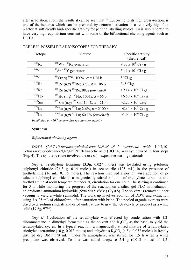
113
after irradiation. From the results it can be seen that 177Lu, owing to its high cross-section, is one of the isotopes which can be prepared by neutron activation in a relatively high flux reactor at sufficiently high specific activity for peptide labelling studies. Lu is also reported to have very high equilibrium constant with some of the bifunctional chelating agents such as DOTA.
TABLE II. POSSIBLE RADIOISOTOPES FOR THERAPY
Isotope Source Specific activity (theoretical)
188Re 188W / 188Re generator 9.80 x 10⁵ Ci / g 90Y 90Sr / 90Y generator 5.44 x 10⁵ Ci / g 90Y 89Y(n,γ) 90Y; 100%, σ = 1.28 b 30Ci /g 186Re 185Re (n,γ) 186Re; 37%, σ = 106 b 345 Ci/g186Re 185Re (n,γ) 186Re; 98% (enriched) >9.14 x 10² Ci /g166Ho 165Ho (n,γ) 166Ho; 100%, σ = 66 b >6.50 x 10² Ci / g153Sm 152Sm (n,γ) 153Sm; 100%,σ = 210 b >2.23 x 10³ Ci/g 177Lu 176Lu (n,γ) 177Lu; 2.6%, σ = 2100 b >8.34 x 10² Ci / g177Lu 176Lu (n,γ) 177Lu; 60.7% (enriched) >1.94 x 10⁴ Ci / g
Irradiation at >1014 neutron flux to saturation activity
Synthesis
Bifunctional chelating agents
DOTA (1,4,7,10-tetraazacyclododecane-N,N’,N’’,N’’’ tetraacetic acid) 1,4,7,10-Tetraazacyclododecane-N,N’,N’’,N’’’tetraacetic acid (DOTA) was synthesized in four steps (Fig. 4). The synthetic route involved the use of inexpensive starting materials.
Step I: Triethylene tetramine (3.3g, 0.027 moles) was tosylated using p-toluene sulphonyl chloride (26.3 g, 0.14 moles) in acetonitrile (125 mL) in the presence of triethylamine (16 mL, 0.115 moles). The reaction involved a portion wise addition of p-toluene sulphonyl chloride to a magnetically stirred solution of triethylene tetramine and triethyl amine at room temperature under N2 circulation for one hour. The stirring is continued for 5 h while monitoring the progress of the reaction on a silica gel TLC in methanol : chloroform : ammonium hydroxide (5:94.5:0.5 v/v/v ) (Rf 0.8). The solvent is removed under vacuum to yield a white semisolid. The work up involves addition of DDW and extraction using 3 x 25 mL of chloroform, after saturation with brine. The pooled organic extracts were dried over sodium sulphate and dried under vacuo to give the tetratosylated product as a white solid (19.8g, 97%).
Step II: Cyclization of the tetratosylate was effected by condensation with 1,2-dibromoethane in dimethyl formamide as the solvent and K2CO3 as the base, to yield the tetratosylated cyclen. In a typical reaction, a magnetically stirred mixture of tetratosylated triethylene tetramine (10 g, 0.013 moles) and anhydrous K2CO3 (4.5g, 0.032 moles) in freshly distilled dry DMF (70 mL), under N2 atmosphere, was stirred for 1.5 h when a white precipitate was observed. To this was added dropwise 2.4 g (0.013 moles) of 1,2-

114
dibromoethane in 30 mL of distilled and dry DMF. The progress of the reaction was monitored by silica gel TLC in methanol:chloroform:ammonium hydroxide (3:96.5:0.5 v/v/v) (Rf 0.6). The reaction was continued for 24 h following which DMF was removed by vacuum distillation. The work-up involved addition of DDW (50 mL) and extraction using 5×25 mL of chloroform, and subsequent removal of chloroform to give the product (8 g, 78%).
Step III: Following the cyclization reaction, detosylation is effected using Conc. H2SO4to yield 1,4,7,10-tetrazacyclododecane (cyclen), the key synthon. 9.3 g (0.012 moles) of tetratosylated cyclen along with 35mL of concentrated H2SO4 was heated to 110oC for 24-28 h. The reaction was monitored by TLC using chloroform: methanol: 3:97 (v/v) (Rf 0.2). The reaction mixture was allowed to cool at room temperature and the pH adjusted to ~12 under cooling (0-5oC) and extracted with 6×25 mL of dichloromethane after saturation with brine. The pooled organic extracts were washed with 3×25 mL brine and solvent removed to yield 0.69g (33%) of cyclen.
Step IV: A mixture of cyclen (0.24g, 0.0014 moles) and chloroacetic acid (0.6g, 0.006 moles), in 25 mL of DDW is stirred for 10 min following which a clear solution results. The pH of the solution is then adjusted to 9.5-10 by dropwise addition of 5N NaOH. The resultant solution is heated to 50–60oC. The progress of the reaction is indicated by decrease in the pH of the solution, which is maintained at approximately 10 after attaining room temperature. After 30 h of reaction, silica gel TLC using ammonium hydroxide:methanol (40:60 v/v) shows completion of the reaction (Rf 0.8). On attaining room temperature, the pH of the solution is adjusted to 2 using concentrated HCl. The reaction mixture is concentrated to dryness under vacuum to yield the crude product. The purification of DOTA was carried out by dissolving the solid in 0.1% TFA in water and loaded on the column. The first solvent used was 100 mL of 0.1% TFA in H2O and 50 mL of 1% TFA in H2O. The second solvent used was 100 mL of 0.1% TFA in acetonitrile and 50 mL of 1% TFA in acetonitrile. The solvent from the fractions collected were removed by vacuum distillation to give the pure product (500 mg).
In another procedure, cyclen 1,2-disulphate (2g, 0.005 moles) is converted to DOTA via a one step reaction involving the liberation of cyclen in alkaline medium and its subsequent conversion to DOTA with chloroacetic acid (2.3g, 0.023 moles) following the same procedure as above. After a similar work-up the yield of DOTA was 90%.
All the reaction conditions were standardized and the intermediates characterized with the help of TLC in suitable solvents. The intermediates and final products were characterized by FT-IR and high resolution 1H-NMR spectroscopy.
1H-NMR, D2O, δ ppm) 2.44, 2.67 (16 H, broad singlets, -CH2 of cyclen) 3.12 (8H, singlet, -CH2COOH)
5-hydroxy-3,7-diazanonan-1,9-dithiol (DAHPES)
To a stirred solution of 2-hydroxy-1,3-diaminopropane (4g, 0.044moles) in 30 mL dry toluene was added to a mixture of ethylene sulphide (5.87g, 0.097 moles) in 20 mL of dry toluene under reflux and nitrogen flushing over a period of 3 h. The refluxing was continued for a period of 40 h, following which silica gel TLC using NH4OH : CH3OH (6:94 v/v) indicated the completion of the reaction (product Rf ~ 0.6). Toluene was removed under vacuum distillation when there was resulting white residue. The residue was washed with a mixture of methanol and dichloromethane (1:1) 5×10mL. The pooled organic extracts were

115
concentrated to yield a viscous liquid, which subsequently solidified to a pale white solid (5.2g, 57%) on storage. The final product was characterized by FT-IR, high-resolution1H-NMR spectroscopy and mass spectroscopy.
IR (KBr, ν cm-1): 3364(-NH), 2960 (-SH); 1H-NMR (CD3OD, δ ppm): 2.64-2.71 [2H dd -NHCHAHB(CHOH)], 2.79-2.93 [6H m-NHCHAHB(CHOH)] and -NHCH2CH2SH, 3.16 (4H t, J = 5.4 Hz , -CH2SH), 3.65-3.80 (1H m,-CHOH); Mass Spectra (EI) m/z:210(M+)
DOTA-lanreotide (Mauritius)
The synthesis of Mauritius was carried out in a three-step reaction (Fig. 5) using commercially supplied lanreotide and locally synthesized DOTA.
Step 1: The reaction in the first step involved the protection of the free amino residue of lysine in Lanreotide (LAN) with ditert butyl dicarbonate (BOC-anhydride) to yield BOC-LAN. To a stirred solution of 5 mg lanreotide (0.0045mM) in 0.1mL dioxane, 0.05mL DDW, was added BOC-anhydride (2.2 equivalents, 2.1 mg), at 0oC. The pH of the solution was checked and found to be ~8. To ensure alkalinity 0.01 mL 1M sodium hydroxide solution was added when pH was found to be 10. The resultant solution is stirred at room temperature for 30 min. Dioxane was removed under a slow stream of nitrogen when a white residue was observed.
Step II: In the second step, the peptide bond formation is effected via the formation of the N-hydroxy succinimide ester of DOTA in the presence of DCC as the condensing agent. The LANBOC-DOTA conjugate is formed at the β-naphthyl amino residue.
To the residue obtained in Step I, 1 mL DDW and 1 mL DMF were added to N-hydroxysuccinimide (5.1 mg), dicyclohexyl carbodiimide (7 mg) and DOTA (6 mg). The pH of the solution was maintained at ~9 by adding 0.02 mL of 1M NaOH. The reaction was kept stirred for 16 h at room temperature following which turbidity was observed. The solvent was removed under a slow stream of nitrogen gas. The progress of the reaction at this stage was checked by silica-gel TLC in NH4OH : methanol (40:60 v/v) (Rf -0.8).
Step III: The final step involved de-blocking of the BOC protected NH2 group to yield the conjugated product LAN-DOTA. The deprotection was carried out by stirring the product obtained from Step II using trifluoroacetic acid (0.13 mL) in methylene chloride (1.5 mL) at room temperature for 30 min. The solvent was removed under nitrogen.
Purification of lanreotide-DOTA (LANDOTA)
Purification of the final product was effected on preparative TLC using NH4OH: methanol (40:60 v/v ) (Rf of LAN-DOTA-0.7, Rf of lanreotide-0.3). The desired zone was scrapped off and extracted in methanol to obtain the pure product.
Characterization
The UV spectra is given in Fig. 6. It is observed that the spectra of the synthesized sample of lanreotide coupled DOTA compared well with that of the authentic sample obtained from Pichem, Finland.

116
Samples for recording NMR were prepared by dissolving 3 mg of the respective peptides in 540 µL H2O and 160 µL of D2O. 1H-NMR spectra was recorded in a Varian Unity Plus 600 MHz FT-NMR spectrometer operating at a 1H resonance frequency of 600 MHz. Preliminary information towards sequence specific 1H resonance assignments were attempted using standard methodology reported by Wuthrich.
The coupling of a carboxyl group in DOTA with the free amino group of the β-naphthyl alanine moiety is observable through the changes in the 1H-NMR signals at the site of the alanine moiety (Table III).
TABLE III. CHANGES OBSERVED IN THE NMR SPECTRA OF LANREOTIDE AND LAN-DOTA
Changes observed at DOTA Lanreotide Lanreotide-DOTA -NH2 - (Ala) - 8.26 d 8.37 d-NH - (Cys) - 8.37 d 8.47 d-Cα-H (Ala) - 4.4 t 5.0 broad t-Cβ-CH2-naphthyl (Ala) - 3.938 t 4.08-4.17 m-CH2COOH (DOTA) 3.12 s - 3.208 broad sLanNHCO-CH2DOTA 3.12 s - 4.012 tRing CH2-DOTA 2.44-2.67 broad s - 3.675 -3.852 m
H
ONH
NH
ONH
SH
O OH
HN
H
H
O
HN
NH2
HNO
H
H
O
NHH3C
CH3
H
H
S
O
HN
HCONH2
H
OHH3C
(Ala)
(Cys)(Cys)
(Tyr)
(Trp)(Lys)
(Val)
(Thr)
LANREOTIDE
NN
NN
COOH
COOH
HOOC
C
O
α
ββ
N
-DOTA

117
3.3. Radiolabelling BFCAs/ligands and characterization of the complexes
90Y-DOTA
The pH of 90YCl3 was adjusted to ∼6.0 using1M NaOH. To ∼500 µL (∼37 MBq) of pH adjusted 90YCl3 added 10 µL of DOTA (1mg/ml in 0.4 M sodium acetate buffer). 100 µL of 0.4 M sodium acetate buffer is added and incubated for 30 min at 60°C. The reaction was also carried out at room temperature to study the reaction kinetics with increase in temperature. Carrier-added studies were also done using 0.2 µg of carrier 89Y (equivalent to ∼110 mCi 90Y). Quality control was performed by paper chromatography using Pyridine:Ethanol:Water (1:2:4) as the developing system. In this solvent system, 90Y acetate remained at the point of application (Rf=0) while 90Y-DOTA moved along the solvent front (Rf =0.9-1.0).
Radiolabelling yields >90% could be achieved with 10 µg of DOTA using trace levels of 90Y (37 MBq). However, on addition of carrier 0.2 µg (equivalent to 110 mCi of 90Y),>95% complexation could be achieved only with 100 µg of DOTA. The time required for complexation was found to be temperature dependant. It was observed that the reaction was slow at room temperature. But when the reaction was carried out at 60°C, the time required for complexation was drastically reduced. At 0.2 µg of carrier 89Y (equivalent to 110 mCi of 90Y) and 100 µg of DOTA, the complexation was complete within 15 min at 60°C (Fig. 7).
177Lu-DOTA
Radiolabelling of DOTA with 177Lu was carried out by dissolving the ligand in 0.5 M NaHCO3 buffer (pH 9) and adding 177LuCl3 solution into it. The reaction mixture was incubated at room temperature after adjusting the volume to 1 mL by adding normal saline. Reaction pH was maintained at 8.5 to 9 (pH of bicarbonate buffer).
Complexation yield was ascertained by paper chromatography in 0.9% saline as well as paper electrophoresis technique. In paper chromatography using 0.9% saline as the solvent, it was observed that 177Lu-DOTA complex moved towards the solvent front (Rf=1.0), while uncomplexed 177Lu under identical conditions remained at the point of spotting (Rf=0).
In order to obtain maximum complexation yield using minimum possible amount of DOTA, several experiments were carried out by varying – such as ligand concentration (10-50 µg/mL), 177Lu concentration (2-20 µg/mL) and reaction time. Results are shown on Table IV.
Paper electrophoresis was carried out using 0.025 M phosphate buffer (pH 7.4) as electrolyte and applying a potential gradient of 10 V/cm for 1 h. It was observed that 177Lu-DOTA complex moved towards the anode indicating that the complex is negatively charged. On the other hand, 177LuCl3 did not show any movement from the point of application under identical conditions.
Complexation yield >90% was achieved using 30 µg/mL of DOTA and 5 µg/mL of 177Lu at pH ~9. Complex was found to retain its stability up to 7 days at room temperature.

118
TABLE IV. OPTIMIZATION OF 177LU-DOTA
% Complexation [DOTA] (µg/mL) 1 h at RT 3 h at RT 24 h at RT
10 7.21 14.23 17.07 20 29.25 48.38 49.22 30 44.53 79.08 70.90 40 61.01 88.34 84.21 50 89.66 - 92.21
177Lu
2 µg/mL
10 7.77 12.17 10.11 20 12.06 9.18 10.24 30 76.05 91.57 90.49 40 86.96 99.07 98.92
5 µg/mL
50 90.69 99.10 99.22
166Ho-DOTA
Complexation studies using 166Ho and 177Lu was carried our identically. Results of the complexation studies with 177Lu are summarized in the Table 5.
TABLE V. COMPLEXATION YIELD OF 166HO WITH DIFFERENTCONCENTRATIONS OF DOTA
% Complexation [DOTA] (µg/mL) 1 h at RT 3 h at RT 24 h at RT
50 49.37 59.68 62.90 70 80.60 97.05 97.43 80 97.07 98.21 97.88
166Ho: 10 µg/mL, pH ~ 9, incubation at RT
3.4. Direct radiolabelling of lanreotide
186/188Re-lanreotide
18 mg of hydroxyethylene diphosphonate (HEDP) was dissolved in 0.2 mL of 0.5 M bicarbonate buffer and the volume was made to 1mL with saline. 20 µL of stannous chloride solution (2.1 mg in concentrated HCl) was added to the HEDP solution followed by addition of 20 µL (10 µg, 0.05µM) of ReO4
-. The reaction mixture was purged with nitrogen for 10 min. The pH of the reaction mixture was adjusted to 2 and was reacted with 250 µg(0.25µM) of lanreotide in a boiling water bath for 90 min. The complex formed was characterized by TLC, paper chromatography and HPLC. TLC was carried out using acetone and saline as two different solvent systems, While paper chromatography was carried out in ethanol: ammonia: water (2: 1: 5).

119
HPLC was carried out using an analytical reverse phase column (Keytech, C 18, 250×4.6 mm). The mobile phase consisted of a gradient system, with solvent A corresponding to water with 0.1% trifluoroacetic acid and solvent B corresponding to acetonitrile with 0.1% trifluoroacetic acid. The mobile phase started with 90% A at time t = 0 followed by a linear gradient from 10%B to 90%B from 0 to 25 min. The flow rate of the mobile phase was 1 mL/min.
Lanreotide was labelled with 186Re by ligand exchange method using HEDP as exchanging ligand. Lanreotide was reacted with 186Re- HEDP in acidic medium at 100°C for 90 min. The labelling yield determined by chromatography technique was found to be 65%. Composition of radiochemical impurities such as reduced hydrolyzed 186Re, unreacted 186Re-HEDP and free 186ReO4
- associated with labelled peptide could be estimated by using combination of different chromatography techniques. In TLC acetone 186Re–peptide, 186Re-HEDP and reduced hydrolyzed 186Re remained at the point of spotting while free perrhenate migrated with the solvent front giving the estimate of free perrhenate. In TLC saline reduced hydrolyzed rhenium and 186Re-lanreotide remained at the point of spotting and 186Re–HEDP migrated with the solvent front indicating the yield of 186Re–HEDP associated with the hydrolyzed peptide. Composition of reduced hydrolyzed species could be obtained from PC results. In PC ethanol: ammonia: water (2: 1: 5) 186Re- HEDP, 186Re lanreotide and free perrhenate migrated with solvent front and reduced hydrolyzed 186Re remained at the point of spotting. Composition of each species associated with 186Re lanreotide is given in Table VI.
TABLE VI. COMPOSITION OF RADIOCHEMICAL SPECIES PRESENT ALONG WITH 186RE-LANREOTIDE
% of 186Re-lanreotide %186Re-HEDP %186ReO4- %186ReO2
65 ± 4 22 ± 3 7 ± 2 6 ± 3 % yields determined by TLC and PC; (n=3)
HPLC pattern of 186Re- lanreotide is depicted in Fig. 8. ~62% of the injected activity was eluted out as 186Re-lanreotide with retention time 15 min. Retention time for Re-HEDP and 186ReO4-was 3 min. The amount of total activity retained on the column was calculated by subtracting the eluted activity from the total activity injected in the column. Activity retained on the column was found to be ~7%. Stability of the complex with time was studied up to 24 h. The complex was found to be stable for this period.
125I-lanreotide
Lanreotide was radioiodinated using chloramineT as the oxidizing agent. 30 µL of 0.5 M PO4
-3, pH 7.5, was added to 2 ug of lanreotide. ~500 µCi of Na125I was added followed by 10 µg of chloramin T in 10 µL of 0.05 MPO4
-3, buffer, pH 7.5. After a reaction time of 90 sec, the reaction mixture was diluted to 0.5 mL with 0.05 M PO4
-3 buffer.
Purification
A Sep Pak column was used for purification. The column was initially washed with 1 mL of 0.05 M PO4
-3. 0.2 mL of the reaction mixture was loaded on to the column. The column was initially eluted with 1 mL of 0.05 M PO4
-3 buffer and later eluted with 2 mL of ethanol. The ethanol was separately collected. Both the reaction mixture as well as the purified fraction were subjected to paper electrophoresis in phosphate buffer, pH 7.5 for 75 min at 10 volts/cm (Fig. 9). The labelling yield was 95% and the radiochemical purity was

120
>98%. The compound was used for biodistribution studies in mice bearing melanoma tumours.
3.5. Radiolabelling of lanreotide-DOTA
90Y-DOTA-lanreotide
To 500 µL of 90YCl3 (37 MBq) was added ∼450 µL of 1 M NaOH to adjust the pH to ∼6. To 500 µL of pH adjusted 90YCl3 (37 MBq) was added 100 µL of gentisic acid (5 mg/ml in 0.4 M sodium acetate) and 50 µg of DOTA-Lanreotide. The reaction mixture was incubated for 30 min at 90°C.
The purification of 90Y-DOTA-lanreotide was carried out using a Sep-Pak C18 column. An aliquot of the 90Y-DOTA-lanreotide was reacted with 100 µg of DOTA. This was then passed through a Sep-Pak C18 column pre-conditioned with 2 mL of methanol. The free yttrium complexed with DOTA was eluted out with 2 ml of 1 M sodium acetate buffer while 90Y-DOTA-Lanreotide was eluted out using 2 ml of methanol. The radiolabelling yield was calculated as the percent radioactivity associated with the methanol fraction and was found to be ∼ 70% (Table VII). The methanol was removed by nitrogen purging and 90Y-DOTA-lanreotide was reconstituted in buffer and used for biodistribution as well as in vitro cell binding studies.
TABLE VII. SEP-PAK PURIFICATION OF 90Y-DOTA-LANREOTIDE
Fraction (2 mL) % Radioactivity Acetate ( free 90Y) 27.9±4.5Methanol (90Y-lanreotide) 72.1±4.5
n=3
4. BIOLOGICAL EVALUATION
4.1.1. In vivo biodistribution in mice models
4.1.1.1. 125I-Lanreotide
Melanoma cells are found to express somatostatin receptors. Therefore, in order to determine the localization of 125I-lanreotide in tumours expressing somatostatin receptors, biodistribution studies were carried out in mice bearing melanoma. Melanoma was induced in C57BL6 mice by injected ∼ 06 cells/animal of melanoma cells. Tumours developed ∼15 days after the induction. ∼150 kBq of 125I-lanreotide in a volume of 0.1 mL was injected through the tail vein. Mice were sacrificed at 3 h and 24 h. Blood (~1 mL) was drawn from heart and the tissues and organs were excised following sacrifice. The radioactivity associated with each specimen was measured in a NaI (Tl) flat type counter and the activity distribution in the various organs and tumour was estimated. Radioactivity in the blood and muscle was estimated by assuming blood and muscle weight as 7% and 40% of total body weight, respectively. It was observed that (Table 8) ∼2.0% ID/g was resident in the tumour at 3 h p.i. There was some amount of activity present in major organs such as liver, intestine and kidney. The activity in the tumour was reduced to ∼0.4% ID/g of tumour while 75% of radioactivity was cleared through excreta at 24 h p.i. Most of the radioactivity in all the organs also drastically decreased.

121
TABLE VIII. BIODISTRIBUTION OF 125I-LANREOTIDE IN C57BL6 MICE BEARING MELANOMA
Tissue/organ %ID/g 3 h
%ID/g 24 h
Blood 1.34±0.05 0.14±0.04Liver 19.26±3.76 3.40±0.40GIT 26.5±2.34 1.17±0.07Kidney 5.82±0.64 1.57±0.16Stomach 9.76±1.21 1.64±0.6Heart 1.54±0.69 0.37±0.19Lungs 3.7±0.04 3.77±0.91Femurs 2.76±0.72 0.55±0.07Muscle 0.09±0.01 0.02±0.0Spleen 4.05±0.53 1.16±0.01Excreta 5.75±2.87 75.4±4.11Tumour 2.03±0.32 0.39±0.04
(n=3)
4.1.1.2. 90Y-DOTA-lanreotide
Biodistribution studies of 90Y-DOTA-lanreotide were carried out in mice bearing melanoma. The results are shown in Table IX. At 24 h p.i., ∼1.3% injected dose was observed to be in the tumour with small amounts of activity in the liver, kidney, intestine and femur.
TABLE IV. BIODISTRIBUTION OF 90Y-DOTA-LANREOTIDE IN C57BL6 MICE BEARING MELANOMA
Tissue/Organ %ID/g 24 h
Tissue/Organ %ID/g 24 h
Blood 0.0 Lungs 5.59±0.71Liver 6.39±0.31 Femurs 6.89±2.41GIT 1.03±0.31 Muscle 0.0 Kidney 17.74±3.26 Spleen 3.44±2.24Stomach 0.98±0.67 Excreta 78.7±4.2Heart 0.0 Tumour 1.30±0.1
4.1.2. In vitro studie s- cell labelling
4.1.2.1 90Y-DOTA-lanreotide
A431 (Epidermoid mammary carcinoma) cell line expressing somatostatin receptors was used for in vitro cell binding studies. The cell line was obtained from the Cancer Research Institute, Mumbai. The cells were maintained in DMEM with 10% Fetal Calf Serum in 5% CO2/95% air at 37 °C. Adherent cells were passaged with trypsin-EDTA solution after

122
confluency was reached. Before being used in binding experiments, the cells were washed with 50 mM Tris HCl buffer (pH 7.5) and resuspended in 50 mM Tris HCl buffer (pH 7.5) containing 5 mM MgCl2, 1 mM CaCl2 and 0.1 M NaCl. Binding was studied by incubating 105 cells with 0.3 µg/tube of 90Y-DOTA-lanreotide for 30 min at 37 °C. In order to determine the specificity of binding of 90Y-DOTA-lanreotide to the receptors, cells were incubated with 0.3 µg of 90Y-DOTA-lanreotide along with cold lanreotide of concentrations 25 µg and 100 µg, respectively. Reaction was also carried out using 90Y-DOTA. After incubation, reaction mixture was diluted 1:10 with assay buffer at 4°C and centrifuged at 4000 rpm for 10 min. The pellet was washed twice with buffer and the pellet as well as the supernatant were counted using NaI(Tl) scintillation counter. Cell binding was determined as the % of total activity bound to the pellet. The results are tabulated in Table X.
14.5% binding was achieved with 90Y-DOTA-lanreotide while binding decreased to 13% and 8.6% on addition of cold lanreotide. 90Y-DOTA showed ∼4.4% binding. Decrease in binding on addition of cold lanreotide showed the binding specificity of 90Y-DOTA-lanreotide to somatostatin receptors expressed by A431 cells.
TABLE X. IN VITRO CELL BINDING STUDIES OF 90Y-DOTA-LANREOTIDE IN A431 CELL LINE
Compound % Binding 90Y-DOTA-Lanreotide 14.5 ± 0.3 90Y-DOTA-Lanreotide + 25 µg cold Lanreotide
13.3 ± 0.7
90Y-DOTA-Lanreotide + 100 µg cold Lanreotide
8.63 ± 0.4
90Y-DOTA 4.4 ± 0.6

123
LABELLED PEPTIDES THROUGH THE DOTA CHELATING AGENT FOR DIAGNOSIS AND TREATMENT OF RECEPTOR POSITIVE TUMOURS
M. CHINOL European Institute of Oncology, Milan, Italy
Abstract
Under the framework of the CRP, I studied the chemistry involved with chelating agent DOTA (1,4,7,10-tetraazacyclododecane-N,N’,N’’,N’’’-tetraacetic acid) which is known to form highly stable complexes with trivalent radiometals such as 111In and 90Y and could be used to tag these radionuclides to tumour seeking biomolecules such as biotin and peptides. In particular, the coupling reaction and labelling with 90Y for targeted therapy were optimized and two radiolabelled DOTA-conjugated peptides were tested in pre-clinical settings.
1. INTRODUCTION
Cells express on their cellular membranes a variety of receptor proteins with high affinity for regulatory peptides, including somatostatin [1]. Somatostatin receptors (SSTR) are membrane glycoproteins with five different subtypes cloned that are distributed in a large variety of tissues throughout the body. The overexpression of SSTR, especially type 2 (SSTR2), in different tumours — classically defined as neuroendocrine — has been exploited for the in vivo localization of these tumours and their metastases using 111In labelled somatostatin analogue scintigraphy [2]. Ongoing multicenter clinical trials, using high doses of 111In-DTPA-octreotide (Octreoscan®) in patients with neuroendocrine tumours [3], have yielded approximately 15% of objective responses, with a 66% overall response rate. These results may be ascribed to the Auger and conversion electrons emitted by 111In. However, 90Yseems more suitable for therapeutic use because of its energetic β particles (Emax = 2.27 MeV) and the long range (R 95 = 5.94 mm), which allow “cross fire” irradiation.
We have developed a new somatostatin analogue containing the metal chelator DOTA, [DOTA-D-Phe1-Tyr3]octreotide, named DOTATOC, which has shown favourable characteristics for therapeutic use: high affinity for somatostatin subtype receptors SSTR2 and SSTR5, high hydrophilicity and ease of labelling and stability with 111In and with 90Y [4].
In fact, for trivalent metallic radionuclides such as 111In and 90Y, the 12 membered ring macrocyclic chelating agent DOTA has shown enhanced thermodynamic and kinetic stability relative to an open-chain analogue. The metal complexes are remarkably stable, especially with yttrium, under physiological conditions and conjugation reactions have been optimized for the linkage of DOTA to monoclonal antibodies and peptides [5].
In order to extend this strategy to other tumours not expressing SSTR, I focused on another natural peptide named oxytocin (OT), which is secreted by the neurohypophysis and binds to several tumours through specific membrane receptors (OTR).
OTR has been described in a high percentage of primary breast cancers and in tumours arising in other organs such as brain and endometrium, where OT is known to exert a physiological function [6]. Moreover, in vitro and in vivo experiments have shown that in breast and endometrial carcinomas, in neuroblastomas and in glioblastomas both OT and synthetic analogues play a biological role by inhibiting cell growth via OTR and the cAMP-PKA pathway [7].
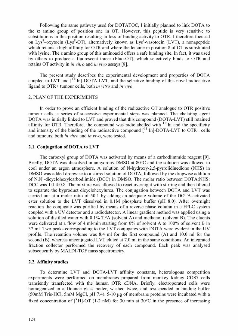
124
Following the same pathway used for DOTATOC, I initially planned to link DOTA to the α amino group of position one in OT. However, this peptide is very sensitive to substitutions in this position resulting in loss of binding activity to OTR. I therefore focused on Lys8–oxytocin (Lys8-OT), alternatively known as Lys8-vasotocin (LVT), a nonapeptide which retains a high affinity for OTR and where the leucine in position 8 of OT is substituted with lysine. The amino group of this aminoacid offers a safe binding site. In fact, it was used by others to produce a fluorescent tracer (Fluo-OT), which selectively binds to OTR and retains OT activity in in vitro and in vivo assays [8].
The present study describes the experimental development and properties of DOTA coupled to LVT and [111In]-DOTA-LVT, and the selective binding of this novel radioactive ligand to OTR+ tumour cells, both in vitro and in vivo.
2. PLAN OF THE EXPERIMENTS
In order to prove an efficient binding of the radioactive OT analogue to OTR positive tumour cells, a series of successive experimental steps was planned. The chelating agent DOTA was initially linked to LVT and proved that this compound (DOTA-LVT) still retained affinity for OTR. Therefore, the compound was radiolabelled with 111In and the specificity and intensity of the binding of the radioactive compound [111In]-DOTA-LVT to OTR+ cells and tumours, both in vitro and in vivo, were tested.
2.1. Conjugation of DOTA to LVT
The carboxyl group of DOTA was activated by means of a carbodiimmide reagent [9]. Briefly, DOTA was dissolved in anhydrous DMSO at 80°C and the solution was allowed to cool under an argon atmosphere. A solution of N-hydroxy-2,5-pyrrolidinedione (NHS) in DMSO was added dropwise to a stirred solution of DOTA, followed by the dropwise addition of N,N’-dicyclohexylcarbodiimide (DCC) in DMSO. The molar ratio between DOTA:NHS: DCC was 1:1.4:0.8. The mixture was allowed to react overnight with stirring and then filtered to separate the byproduct dicyclohexylurea. The conjugation between DOTA and LVT was carried out at a molar ratio of 50:1 by adding an adequate volume of the DOTA-activated ester solution to the LVT dissolved in 0.1M phosphate buffer (pH 8.0). After overnight reaction the conjugate was purified by means of a reverse phase column in a FPLC system coupled with a UV detector and a radiodetector. A linear gradient method was applied using a solution of distilled water with 0.1% TFA (solvent A) and methanol (solvent B). The eluents were delivered at a flow of 4 ml/min starting from 0% of solvent A to 100% of solvent B in 37 ml. Two peaks corresponding to the LVT conjugates with DOTA were evident in the UV profile. The retention volume was 8.4 ml for the first compound (A) and 10.0 ml for the second (B), whereas unconjugated LVT eluted at 7.0 ml in the same conditions. An integrated fraction collector performed the recovery of each compound. Each peak was analysed subsequently by MALDI-TOF mass spectrometry.
2.2. Affinity studies
To determine LVT and DOTA-LVT affinity constants, heterologous competition experiments were performed on membranes prepared from monkey kidney COS7 cells transiently transfected with the human OTR cDNA. Briefly, electroporated cells were homogenized in a Dounce glass potter, washed twice, and resuspended in binding buffer (50mM Tris-HCl, 5mM MgCl, pH 7.4). 5-10 µg of membrane proteins were incubated with a fixed concentration of [3H]-OT (1-2 nM) for 30 min at 30°C in the presence of increasing

125
concentration of unlabelled peptides. Non-specific binding was determined in the presence of 1 mM OT. Bound and free radioactivity were separated by filtration over Whatman GF/C filters pre-soaked in 10 mg/ml BSA. Binding isotherms were analysed with the iterative curve-fitting program LIGAND.
2.3. Radiolabelling of DOTA-LVT (peak A) with [111In]
To prepare [111In]-DOTA-LVT, 3.7 MBq of [111In]Cl3 that is diluted in acetate buffer (0.1 M, pH 5.5), was added to 0.07 µmol of fraction obtained from peak A. The solution was heated for 25 min at 80°C, and the labelling yield was checked by FPLC as above.
2.4. Binding studies on OTR+ and OTR-tumour cells
Human breast carcinoma (MCF7), glioblastoma (MOG-U-V-W) cell lines are OTR+, while colon carcinoma (HT29) cells represent the OTR- control.
All the cells were grown as monolayers in RPMI medium with 10% fetal calf serum, in 25 cm2 T flasks in a 5% CO2 humidified atmosphere, at 37°C. Binding experiments were performed on intact cells. Briefly, 20×105 cells, suspended in 100 µl of culture medium were incubated for 30 min at 4°C in the presence of 1µM [111In]-DOTA-LVT. The cell suspension was then centrifuged for 5 min at 2000 g and the pellet was washed and re-suspended in fresh medium. Centrifugation and cell washing were repeated twice. The entity of the radiolabelling was evaluated measuring the radioactivity bound to cells (cpm/105 cells) by a Packard auto-gamma counter.
Specificity of the binding was determined by evaluating the radioactive displacement. Briefly, the cell suspension was incubated for 5 min in the presence of 100 µM and 1 mM of LVT or OT, or in the presence of unrelated peptides, followed by 20 min of incubation with 1µM [111In]-DOTA-LVT. Cells were then centrifuged and washed twice as reported above. The activity of two different agonists and of the unrelated peptides to compete with the radioligand was evaluated measuring the radioactivity bound to cells. All experiments were performed in triplicate. Statistical analysis was carried out by ANOVA. Cut off for significance was 0.05.
2.5. In vivo studies
To determine the entity and specificity of receptor mediated uptake of [111In]-DOTA-LVT in tumours in comparison with the non-specific compound [125I]-DOTA-TOC, an experimental model of Balb/c mice growing OTR+ and SSTR2- tumour was used. A total of eight mice were injected subcutaneously with mammary carcinima cells. Twenty days later, when the growing tumour reached the size of about 2 cm in diameter, the animals were injected intraperitoneally with a mixture of 1.1 MBq of [111In]-DOTA-LVT and 74 kBq of [125I]-DOTA-TOC. The animals were divided into groups and sacrificed at 3 and 24 h post-injection. Tumour, blood, liver, kidney and brain were removed and weighted. Radioactivity was measured in a gamma ray detector with a well counter geometry together with standards of the injection mixture at two different time points to calculate the contribution of each isotope: (a) immediately after the removal of the tissues; and (b) after 2 weeks, corresponding to five physical half-lives of 111In. Activity was expressed as a percentage of injected doses/mg of tissue and ratios of uptake between tumour and brain vs. blood were calculated.

126
3. SUMMARY OF THE RESULTS
The conjugation between DOTA and LVT was complete as verified by the absence of unconjugated LVT in the reaction mixture. Radiolabelling yields were >95% for [111In]-DOTA-LVT. After HPLC purification, the new conjugate showed hundred-fold decreased affinity for OTR compared to unconjugated LVT by means of heterologous competition experiments on membranes. In particular, LVT inhibited [3H]-OT binding with high affinity (Ki = 1.87 ± 0.41 nM) whereas DOTA-LVT was able to compete [3H]-OT binding with a calculated Ki of 238 ± 52 nM.
However, DOTA-LVT was radiolabelled with 111In and binding experiments showed specific radiolabelling in all the OTR positive cell lines and negligible binding in OTR negative ones. Specificity of the binding was proved by displacement with cold radioligands. In OTR positive cells, more than 90% of the specific 111In-DOTA-LVT binding was displaced by the two agonists OT and LVT.
The results of the biodistribution study in mice bearing OTR+ tumours showed that neither 111In-DOTA-LVT nor 125I-DOTA-TOC accumulated in the brain, an organ where OTRs are known to be expressed.
As expected, kidney uptake was rather high, although lower for 111In-DOTA-LVT than for 125I-DOTA-TOC, especially at 24 h.
The tumour to blood uptake ratios for both radiolabelled peptides are given in Table I.
TABLE I. TUMOUR TO BLOOD RATIOS
3h 24h 111In-DOTALVT 125I-DOTATOC 111In-DOTALVT 125I-DOTATOC
2.7 0.96 2.8 4.86 4.1 1.85 4.4 2.07 1.2 0.81 9.1 2.13
N.A. 0.49 12.6 1.88 mean ± s.d. 2.7 ± 1.4 1.02 ± 0.58 7.2 ± 4.5 2.7 ± 1.4
These experiments showed that 111In activity (specific peptide) in tumour was remarkably higher than 125I (non-specific peptide) demonstrating the receptor mediated binding of 111In-DOTA-LVT to OTR expressing tumours.
4. CONCLUSIONS
The use of radiolabelled peptides in tumour therapy is gaining increasing acceptance. While therapeutic clinical trials with 90Y labelled somatostatin analogue DOTATOC have shown objective therapeutic responses, the development of a new radioligand such as DOTA-LVT still requires further improvements in order to overcome the loss of affinity for OTR of the conjugate.

127
REFERENCES
[1] PATEL, Y.C., et al., “ The somatostatin receptor family (review)”, Life Sci. 57 (1995) 1249-1265.
[2] KRENNING, E.P., et al., “Somatostatin receptor scintigraphy with (111In-DTPA-D-Phe1)- and (123I-Tyr3)-octreotide: the Rotterdam experience with more than 1000 patients”, Eur. J. Nucl. Med. 20 (1993) 716-731.
[3] McCARTY, K.E., et al., “In situ radiotherapy with 111In-pentetreotide”, Q. J. Nucl. Med. 44 (2000) 88-95.
[4] PAGANELLI, G., et al., “Receptor-mediated radionuclide therapy with 90Y-DOTA-D-Phe1-Tyr3-octreotide: preliminary report in cancer patients”, Cancer Biother. Radiopharm. 14 (1999) 477-483.
[5] MOI, M.K., et al., “The peptide way to macrocyclic bifunctional chelating agents: synthesis of 2-(p-nitrobenzyl)-1,4,7,10-tetraazacyclododecane-N,N’,N’’,N’’’-tetra-acetic acid and study of its yttrium (III) complex”, J. Am. Chem. Soc. 110 (1988) 6266-6267.
[6] BUSSOLATI, G., et al., “Immunolocalization and gene expression of oxytocin receptors in carcinomas and non-neoplastic tissues of the breast”, Am. J. Pathol. 148(1996) 1895-1903.
[7] CASSONI, P., et al., “Presence and significance of oxytocin receptors in human neuroblastomas and glial tumours”, Int. J. Cancer 77 (1998).
[8] BOISSONNAS, R.A., et al., “Synthése de la Lys8-Oxytoceine (lysine-vasotocine) et Nouvelle Synthése de la Lysine Vasopressin”, Helv Chim. Acta 43 (1960) 182-185.
[9] BUCKLEY, R.G., et al., “An efficient method for labelling antibodies with 111In”, FEBS Let. 166 (1984) 202-204.

129
LABELLED BIOMOLECULES FOR TARGETED RADIOTHERAPY:BIODISTRIBUTION IN MICE WITH INDUCED MALIGNANT TUMOURS
C. ARTEAGA DE MURPHY, G. FERRO-FLORES, M. PEDRAZA-LÓPEZ, E.M. MOLINA-TRINIDAD, E. MURPHY-STACK, S. HERNÁNDEZ-GUTIÉRREZ Instituto Nacional de Ciencias Médicas y Nutrición Salvador Zubirán, Mexico City, México
Abstract
Antibodies and antibody fragments react against the cell membrane receptors or associated-antigens and can be 188Re-labelled and 153Sm-labelled as potential radioimmunotheraphy (RIT) radiopharmaceuticals. A variety of malignant tumours over-express somatostatin receptors, proteins that can be targeted with radiolabelled biomolecules, like the lanreotide peptide. The direct labelling methods described here are easy to perform from a freeze- dried kit, are reliable and can be carried out in a hospital radiopharmacy. 188Re-lanreotide, thus prepared, shows affinity to HeLa cells and to the SiHa cells, both from a human cervical cancer, to 293 cells from human embryonic renal carcinoma and to B16F cells of murine melanoma, implanted in mice. 188Re-lanreotide standardized tumour uptake value SUV was 1.5 for the cervix cancer, 4.8 for the kidney tumour, and 0.8 for the melanoma metastases. Many of the neuroendocrine tumours which over-express somatostatin receptors are of epidermoid origin and the 4 induced tumours studied have many epidermoid characteristics. The cell origin might be the common factor to all these cancer cells, which bind 188Re-lanreotide. In conclusion: easy labelling methods with 188Re and 153Sm for biomolecules used for targeted therapy have been developed. 188Re-lanreotide binds to 4 different cancer cells implanted into mice.
1. INTRODUCTION
Antibodies and antibody fragments react against the cell membrane associated-antigen or receptors and on occasion are internalized into the malignant cell. These biomolecules can be 188Re-labelled and 153Sm-labelled, as potential radioimmunotheraphy (RIT) radiopharmaceuticals. The pharmacokinetics of antibodies has been studied in animal models and its specificity can be increased using the biotin-avidin pre-targeting strategy in which, an antibody and radiopharmaceutical are administered separately, bringing about a high tumour/non-tumour ratio [1-4].
A variety of malignant tumours over-express somatostatin receptors, proteins that can be targeted with radiolabelled biomolecules. The radionuclides used for the detection and treatment of the malignant cells were gamma and beta emitters, such as 188Re and 153Sm. The most used biomolecules are small molecules like somatostatin analogue peptides.
Since rhenium is a transition metal of the Mn and Tc family the radioisotope 188Re has been used as a biomolecule labelling agent. Its physical half-life is 16.9 h, it decays by a Eßmax. 2.12 MeV beta radiation, adequate for therapy, and emits a gamma radiation of 155 keV, which allows for imaging. The somatostatin analogue peptides labelled with beta emitters have been successfully used for tumour localization and targeted radiotherapy. Labelled octreotide and vapreotide RC 160 appear in the medical literature quite often. Another somatostatin receptor analogue peptide, a long-acting cyclic octapeptide, ß-(2-naphthyl)-D-Ala-Cys-Tyr-D-Trp-Lys-Val-Cys-Thr-amine, ß-naphthyl-peptide or lanreotide can inhibit, in vitro and in vivo, growth of the human small cell lung carcinoma [5]. Lanreotide has been 90Y and 111In labelled, via bifunctional chelators, and used for treatment of somatostatin receptor expressing tumours [6-7]. It has also been 188Re labelled by direct methods [8]. For the direct labelling method a reducing agent is used to reduce rhenium to a lower oxidation state and, reduce the peptide’s cysteine disulphide bridge. The metal-coordinated compound is easily formed by competing with a weak ligand like hydroxyethylidenediphosphonate, HEDP [9].

130
Labelled antibodies show, in general, prolonged circulation and non-specific accumulation in metabolic organs and their biodistribution and elimination routes play a relevant role in the tumour uptake. In order to minimize radiation toxicity it is desirable to have a fast renal excretion or low lipophilicity. The tumour/non tumour radioactive rate could be improved using hydrophilic compounds like 153Sm-DTPA-bis-Biotin with the avidin-biotin strategy [10].
153-samarium is a versatile radionuclide with a physical half life of 46.8 h, a 103 keV gamma emission (23%) for scintigraphic imaging, a therapeutic E�max 0.8 MeV beta emission and can be produced with high specific activity in a fairly low flux nuclear reactor. Due to its radionuclidic properties it is easily handled from the point of view of radiation protection.
The aim of this research was to review our labelling methods for several biomolecules. The methods were designed, considering the need for freeze-dried kits or easy to label methods in a hospital radiopharmacy, and the main advantages offered by 188Re and 153Sm.Reagents´ molar concentration for each biomolecule were determined as were the labelling conditions and the radiochemical purity. The biodistribution in control mice and in malignant tumour-induced laboratory animals was studied for each labelled compound and the radiopharmacokinetic parameters were determined.
2. MATERIALS AND METHODS
2.1. Direct 188Re-antibody labelling
The proteins were: polyclonal IgG (Sandoglobulin); murine monoclonal (MoAb)IgG1 ior cea 1 against carcinoembryonic antigen (CEA), (CIMAB, Cuba), (MoAb); biotinylated MoAb (MoAb-biotin); MoAb fragments (F(ab´)2) and biotinylated MoAb fragments (F(ab´)2-biotin). (2) The labelling method is basically as follows: 2-mercapto-ethanol is used to reduce the disulphide groups and stannous chloride and HEDP are added in molar concentrations specific for each molecule as well as pH and incubation time and temperature. Succinimidyl-6-(biotinamide) hexanoate (PierceCo.) was used to prepare the biotinilated compounds [1-4].
153Sm-DTPA-bis-biotin labelling method
DTPA-bis-biotin (Sigma Chemical Co.). 153SmCl3 was added to the solution of diethylenetriaminepentaacetic acid covalently conjugated to biocytin in bicarbonate buffer at pH 8.0 [10].
2.2. Direct labelling of 188Re-lanreotide
ß-(2-naphthyl)-D-Ala-Cys-Tyr-D-Trp-Lys-Val-Cys-Thr-amine, ß-naphthyl-peptide or lanreotide (PiChem, Austria) was rhenium labelled by a modified direct method [8] using stannous chloride to reduce both rhenium (188W-188Re generator, MAP Medical Technologies Oy, Finland) to a reactive species, and to reduce the peptide’s disulphide bridge for subsequent chelation of the metal with HEDP, as the weak competing ligand [9]. The reaction was carried out at pH 1.7 and 90 min incubation in a boiling water bath (92°C). Citrate buffer pH 7.0 (2.9% sodium citrate, 0.1% EDTA) was used to raise the pH to 6.0 [11].
Quality control
The radiochemical purity of the labelled biomolecules was determined by ITLC, HPLC chromatography and SepPak cartridges.

131
ITLC-SG systems
On 1×10 cm strips (Gelman Sciences, Inc. Ann Arbor, Mich.) the samples were inserted into the mobile phases: saline (0.9% NaCl), acid ethanol (10% HCl 0.3 M). Free perrhenate migrates with the front in the solvents, Rf 1.0. Reduced-hydrolyzed rhenium stays at the origin or seeding point, Rf 0.0.
HPLC
The columns with photodiode array detector and Millenium software were used. The collected fractions (0.5 mL) were counted in a well-type scintillation detector and cpm were plotted against time to obtain a radiochromatogram. The column used for molecular size exclusion was a ProteinPak 125 SW gel filtration column (Waters) and a mobile phase 0.1 M phosphate pH 7.4, at a rate of 1.5 mL/min. For reverse phase HPLC with gradient system, a C-18 µBondaPak 3.9×300 mm column (Waters) with photodiode array detector was used. The mobile phase was A: 0.1% triflouracetic acid-H2O/CH3CN (1:3) and B: 0.1% TFA-H2Owith a gradient 0-15 min 30-50% A and 15-40 min 50-100% A at a 1.3 mL/min rate. The amount of reduced hydrolyzed rhenium in ß-naphthyl-peptide, or column bound radioactivity, was calculated by comparison of recovered activity from the HPLC column with a standard.
SEP-PAK cartridges
Activation
The cartridges were activated with 10 mL absolute ethanol, followed by 10 mL 0.001 N HCl and dried with 10 mL air.
Purification
0.1 mL of labelled biomolecule was placed on top of the cartridge and 10 mL of 0.001N HCl pH 3 were passed to separate free perrhenate. Acidified ethanol (10% 0.01N HCl) was passed to collect the labelled biomolecules. The two fractions plus the cartridge (reduced-hydrolysed radionuclide) were counted in a well type scintillation detector.
Analytical methods
Several of the following methods were used to characterize some of the biomolecules: IR spectroscopy, thermogravimetric analysis TGA, scattering electronic microscopy SEM and elemental dispersion analysis by X-rays EDA.
Theoretical analysis
This was used to visualize the complex and the peptide molecule. Geometric analysis was done by energetic principles, using the MM method of FF and ab initio calculations by DFT were useful for explaining the metal-ligand interactions and their conformational properties.
Cell lines
Cell lines were kept frozen, cultured and prepared for injection in the molecular laboratory of the Universidad Panamericana. All cell lines were plated on 10 cm diameter

132
dishes in Dulbecco´s modified Eagle´s medium (DMEM) supplemented with 10% newborn calf serum and antibiotics: 100 units/mL penicillin and 100 µ/mL streptomycin and incubated at 37°C under 5% CO2 atmosphere and 100% humidity. Semi-confluent cells were detached using trypsin/EDTA and counted in a Neubauer chamber.
HeLa cells
These are cells of epithelial origin and originally derived from an epidermoid human carcinoma of the uterine cervix. They contain the human papilloma virus HPV18 DNA which is integrated into the cellular genome and has been shown to harbour transcriptionally active oncogenic sequence E6 and E7. HPV produces different proteins which are tumour specific antigens and therefore the HeLa cell cancer model provides tools to examine the molecular mechanisms of cervical cancer [2, 10]. Also, allelotype analysis of cervical carcinomas has been performed to localize tumour suppressor loci. The 3p losses are common to cervical, lung, clear cell renal, and nasopharyngeal carcinomas [12].
SiHa cells
These are squamous human cervix cancer cells established from fragments of a primary tissue sample from a patient after surgery. The cell line presents typical desmosomes at the cell junctions and abundant tonofilaments in the cytoplasm. The immortal and tumourigenic cell line is reported to contain 1-2 copies per cell of an integrated human papilloma virus type 16 genome (HPV-16).
Human renal carcinoma 293 cell line
This is an epithelial cell line of primary human embryonal kidney transformed by sheared human adenovirus type 5 (Ad 5) DNA. The cells express an unusual cell surface receptor for vitronectin composed of the integrin beta-1 sub-unit and the vitronectin receptor alfa V sub-unit.
Melanoma B16F cells
These cells are from a C57BL/6 mouse melanoma, tumour. They are epithelial like, produce melanin and are validated by isoenzymes. These cells are tumourigenic and metastasize in syngeneic animals.
Histopathological studies
Solid tumours, from the back of the mice, were studied. The excised tumours were embedded in paraffin blocks, dehydrated and eosin-hematoxilin stained.
Animal subjects
Tumour uptake, biodistribution and radiopharmacokinetics were determined according to the rules and regulations of our Institute for the handling and care of experimental animals.
The rodents used for all the experiments were a) female Balb/c 25 g mice, b) female, athymic, 20 g mice; and c) female B15C57, 25 g mice. The athymic mice were kept in micro-isolators, handled under an airflow cabin and fed irradiated PICO LAB PMI feed and water ad libitum. The other mice were kept in cages at constant temperature and humidity and were

133
fed standard PMI feed and water ad libitum. The animals were injected with the tumour cell line (one million cells suspended in 0.2 mL DMEM) in the right flank, close to the axillary nodes. A smear of all the cell lines injected was taken to pathology for identification.
Biodistribution studies
These were undertaken after a period of time when the tumour in the back of the mice was visible and well formed (7-55 days). The rodents (3 per time point) were injected in the tail vein 1.85 MBq (50 µCi) in 0.1 mL of the radiopharmaceutical and CO2 sacrificed after a determined interval 0.083, 0.25, 0.5, 1, 2, 24 and 48 h. A surgical midline incision was made and blood was drawn into a disposable syringe and approximately 1 mL was placed into a test tube. Then the organs of interest were extracted. Each organ (heart, lung, liver, gall bladder, spleen, stomach, intestines, left kidney, muscle, bone and tumour) were saline rinsed, paper blotted, placed into pre-weighed test tubes and the activity was determined with a well type scintillation detector (Canberra). The mean cpm were used to obtain accumulation in percentage injected activity per gram of tissue% IA/g.
Tumour uptake
The standardized tumour uptake value SUV was calculated as follows [13]:
)(/)(/)(
gweightratactivityinjectedTotalgtumourofmasstumourtheinMBqactivitycorrectedDecaySUV =
3. RESULTS
Labelled antibodies
188Re-polyclonal IgG (188Re-IgG); 188Re-murine anti-CEA monoclonal antibody, (188Re-MoAb); biotinylated 188Re-MoAb (188Re-MoAb-biotin); 188Re-MoAb fragments (188Re-F(ab´)2) and biotinylated 188Re-MoAb fragments (188Re-F(ab´)2-biotin) were produced with specific activities of 1.3 ± 0.2 GBq/mg from freeze-dry kits. The radiochemical purity was >95% and the immunoreactivity was similar to that of 99Tcm radioconjugates. Blood clearance in female Balb/c mice is rapid during the first 5 h after administration and 48 h later the% activity injected/g is less than 3% for all the compounds. Renal clearance is the same for biotinilated MoAb and fragments. Biodistribution of biotinilated and non-biotinilated immunoconjugates was not significant statistically, p>0.5, and the elimination was mainly hepatointestinal and urinary. Injected avidin as a “chase” increased liver accumulation, lowered blood concentration and the accumulated activity in all the organs. The bone marrow absorbed dose, extrapolated to humans, went from 0.06mGy/MBq to 0.02 mGy/MBq for the labelled antibody and for the fragments and, after avidin was injected, the values decreased to 0.013 mGy/MBq-0.005 mGy/MBq respectively (2,4).
153Sm-DTPA-bis-Biotin
The labelling efficiency was >98%, two and 72 h after labelling at pH 8.0 and with a molar ratio DTPA-bis-biotin/Sm from 2 to 4. Spectroscopic and thermal studies show that the complex is formed by 3 carboxylate anions which neutralize the 3 positive charges of the metal and the ligands: carboxylic groups bound to the methylene of the secondary amide

134
groups and the amide function itself completely shield the core. The 2 biotin moieties are free for avidin binding. This complex is stable for three days at 18-25°C. Blood clearance is biexponential and 3 h after the administration of the radiopharmaceutical 88-96% of the injected activity is eliminated in the urine of Balb/c mice. There was no concentration in any other organ (10).
188Re-lanreotide
The radiopharmaceutical was prepared with a labelling efficiency >96 ± 2% and 90 min incubation period at 92°C and a final pH 6. Stability was 90% 24 h after preparation and no radiolytic degradation was observed. Without HEDP in the preparation the labelling efficiency is 10%. The biologically active part of the molecules is free, that is the Tyr-D-Trp-Lys-Val union is not affected by the labelling process and can bind to the tumour receptors. The metal is co-ordinated to five donor atoms: an oxygen, two Cys sulphurs and two nitrogen atoms from Cys and Thr. This fact indicates that the rhenium-188 complex is probably an oxo-rhenium(V) N2S2 penta-coordinated complex, [188ReO-lanreotide]. A model of the molecular structure of both the peptide and the 188Re-ß-naphthyl-peptide was generated and can be seen that the two amino acids (Trp and Lys) which are common to somatostatin are free to bind with the somatostatin receptors [11].
Radiopharmacokinetic Balb/c mice parameters indicated that 188Re-lanreotide is rapidly distributed in all the tissues with a fast distribution constant α=2.1 1/h; and a slow or total elimination constant ß=0.28 1/h; elimination from the central compartment constant k10=0.911/h. The apparent volume of distribution was 21.5 mL and the radio-peptide remained in the body for a mean residence time MRT = 2.49 h. Total clearance is high Cl = 19.7 mL/h. The high activity initially seen in the gallbladder decreases 24 h later and is found in the faeces. The remaining organs had low radioactivity. The biodistribution in athymic mice followed the same pattern except for tumour uptake. There was 6.2% + 2.9% IA/g tumour in the athymic mice. The tumour/muscle ratio in athymic mice, 24 h after radiopeptide injection, was 7.1 and tumour/blood ratio was 3.3 [11].
The histopathological study showed that the HeLa cells used are of human epidermoid origin, positive for cytokeratins and vimentin, and negative for chromogranin. Berruti, et al. mentioned that advanced prostate cancer patients had above normal range chromogranin-A levels and after receiving lanreotide (30 mg×4) showed a decreased plasma concentration [14].
In the human cervix cancer tumour biopsy (SiHa cells) a neoplasic process is identified. It is constituted by flat cells with big nuclei of vacuolated aspect with keratin granules and scarce cytoplasm of pale eosinophilic colour. Many mitoses are identified. The tumour rests on a fibrous stroma and it is surrounded by abundant lymphocytic exudates. The diagnosis was malignant tumour invading soft tissue compatible with epidermoid origin human carcinoma of the uterine cervix. The tumour uptake was 7.4% IA/g tissue at 3 h after injection, which rapidly decreases at 24 h P.I. The tumour/blood ratio is shown in Fig. 1.
The induced human renal tumour in athymic mice showed many epithelial, neoplasic cells with round, pleomorphic and hyperchromatic nuclei. The diagnosis was epithelial tumour of renal cell characteristics. The renal tumour bearing mice presented a similar biodistribution as in control mice except that the kidney had a high 188Re-lanreotide uptake (24.6% IA/g). The kidney/blood ratio was 34, two hours after injection. Biodistribution is shown in Fig. 2. SUV was 4.8.

135
0
5
10
15
20
25
Tum
our/
Blo
od
Rat
io0,083 25 0,5 2 3 24
Time (h)
188Re-lanreotide in athymic mice with human cervix tumour
FIG. 1. Tumour/blood ratio of 188Re-lanreotide in athymic female mice with induced human cervix tumour from 0.083 to 24 h (n=3/time point).
05
101520
25
% a
ctiv
ity p
er
gram
of t
issu
e
0.08 0.25 0.5 1 2 3 24
Time (h)
188Re-lanreotide kidney uptake in control and induced kidney tumour in mice
NormalkidneyRenalTumour
FIG. 2. Uptake, in% injected activity per gram of tissue, of 188Re-lanreotide in control athymic mice and in athymic mice with an induced human renal tumour; n=3 per time point.
The B15C57 mice with the induced melanoma tumour presented, after 10 days, a large flank tumour with adjacent surviving epidermis. In the dermis, hypodermis and soft tissue there was a neoplasic process constituted by proliferation of round, fusiform and polyhedral cells with large hyperchromatic nuclei. In some nuclei there was a prominent nucleolus. The cytoplasm of many cells contained melanin deposits. The tumour destroyed skeletal muscle, its borders are infiltrating and there was invasion of blood vessels. There were multiple black metastases (0.05-0.2 mm in diameter) in the thoracic and peritoneal cavities especially next to the large intestine. Diagnosis: subcutaneous, soft tissue malignant melanoma. Biodistribution is shown in Fig. 3.
4. DISCUSSION AND CONCLUSIONS
MoAb-based radiotherapy of cancer has become very useful because it is an efficient and relatively safe therapy for radiosensitive, and readily accessible neoplasms, like non-Hodgkin lymphoma. Tumour response depends mainly on dose rate, cumulative radiation dose and tumour radiosensitivity. In radiosensitive lymphomas an uptake of 0.001-0.01% of the specific monoclonal activity injected/g of tissue delivers <15 Gy which produces anti-tumour response. A dose of 50 Gy or more is considered necessary for therapeutic response of adenocarcinomas. MoAb and chemotherapy are successful in treating solid tumours. (15).

136
188Re-lanreotide in murine melanoma induced in B15C57 Mouse, 2 h P. I.
4% 8%
16%
4%68%
Blood
Liver
Kidney
Metastases
FIG. 3. 188Re-lanreotide uptake (%IA/g) in murine melanoma induced in B15C57 female mice, two hours post administration, via I.V.
Unlabelled somatostatin analogues also have therapeutic effects. Lanreotide was administered intramuscularly (30 mg) every 14 days for two months to patients with prostatic cancer. The tumour growth was stabilized in 57% of the patients and the lanreotide treatment was very well tolerated and did not cause major toxicity [14]. In another study lanreotide was given: 30 mg/every 14 days for seven months together with octreotide (0.1 mg thrice daily for 17 months) to 35 patients with neuroendocrine tumours also with good response. Somatostatin analogues have been considered an efficient treatment and the authors recommend it as a first-line therapy [16].
The therapeutic effect of somatostatin analogues is increased when labelled with beta emitters (receptor-targeted radionuclide therapy).
A tumour dose >10 Gy/GBq 90Y-DOTA-lanreotide produced stable tumour disease in roughly 35% of the patients with neuroendocrine tumour lesions and did not produce chronic severe hematological toxicity, or modification of renal or liver function values (17). This is in agreement with the data that the maximum activity tolerated by the kidneys is 20-25 Gy meaning the renal tolerated dose (TD) having a 5% probability of late toxicity within five years (TD 5/5). [18].
In pharmacokinetic animal research it is important to recognize the high variability in processes due to genetic, environmental, pathophysiologic and quantitative inter-individual differences [19
Animal models suffer these factors but are useful in extrapolating results to humans. It has been reported that similar biokinetics make the animal model a suitable model for somatostatin analogue therapy evaluation. The fact was that carcinoid tumour material from a patient, who underwent surgery, was cultured and the cells were injected into nude mice. Biokinetics of 111In-DTPA-D-Phe-octreotide were studied in mice at different time intervals and in the patient by sequential scintigraphy and in both the biokinetics was similar (20). It has also been reported that the metabolic properties of 111In-DTPA-octreotide are similar in rat and man (21). Therefore, dosimetric estimates in the chosen animal model could give a close estimate in humans in order to maintain a low TD.
153Sm-DTPA-bis-biotin is a biomolecule with tumour affinity and with the avidin-biotin strategy could be a very promising agent for targeted therapy. Work with hepatoma induced Wistar rats is in progress.

137
Most of the human tumours seem to over-express one or more of the five distinct somatostatin receptor subtypes and many of the neuroendocrine tumours, which over-express somatostatin receptors are of epidermoid origin. 188Re-lanreotide, thus prepared, shows affinity to HeLa cells and to the SiHa cells, both from a human cervical cancer but with different HPV type (18 and 16 respectively) and to human 293 cells from human embryonic kidney and murine melanoma tumours, implanted in mice, all of which have many epidermoid characteristics. The cell origin might be the common factor to all these cancer cells, which bind 188Re-lanreotide.
In conclusion, the four induced tumours tested incorporate the labelled peptide with a tumour standardized uptake value SUV from 0.8 to 4.8. The biomolecule labelling methods described here are easy to perform from a freeze-dried kit, are reliable and can be carried out in a hospital radiopharmacy.
ACKNOWLEDGEMENT
This study was partially supported by the International Atomic Energy Agency.
REFERENCES
[1] GRIFFITHS, G.L.,GOLDENBERG, D.M., JONES, A.L., HANSEN, H.J., “Radio-labelling of monoclonal antibodies and fragments with technetium and rhenium”, Bioconjugate Chem. 3 (1992) 160-168.
[2] FERRO, F.G., HASHIMOTO, K., “Direct labelling of monoclonal antibodies and antibody fragments with 188Re using a weak competing ligand”, Radiochimica Acta 79 (1997) 63-70.
[3] GRANA, C, CHINOL, M, MAGNANI, P, SICCARDI, A.G., PAGANELLI, G., “In vivo targeting based on the avidin-biotin system”, Tumour Targeting 2 (1996) 230-239.
[4] FERRO, F.G., PIMENTEL, G.G., GONZÁLEZ ZMA, MURPHY CA, MELÉNDEZ A.L., et al., “Preparation, biodistribution and dosimetry of 188Re-labelled MoAb ior cea1 and its F(ab’)2 fragments by avidin-biotin strategy”, Nucl. Med. Biol. 26 (1999) 57-62.
[5] TAYLOR, J.E., BOGDEN, A.E., MOREAU, J.P, COY, D.H., “In vitro and in vivoinhibition of human small cell lung carcinoma (NCI-H69) growth by a somatostatin analogue”, Biochem. Biophys. Res. Commun. 153 (1988) 81-86.
[6] SMITH-JONES, P.M., BISCHOF, C., LEIMER, M., GLUDOVACZ, D., ANGELBERGER, P., et al., “DOTA-lanreotide: a novel somatostatin analog for tumour diagnosis and therapy”, Endocrinology 140 (1999) 5136-5148.
[7] VIRGOLINI, I., SZILVASI. I., KURTARAN, A., ANGELBERGER, P., RADERER, M., et al., “Indium-111-DOTA-lanreotide: biodistribution, safety and radiation absorbed dose in tumour patients”, J. Nucl. Med. 39 (1998) 1928-1936.
[8] ZAMORA, P.O., GULKHE, S., BENDER, H., DIEKMANN, D., RODEES, B.A., et al., “Experimental radiotherapy of receptor-positive human prostate adenocarcinoma with 188Re-RC-160: a directly labelled somatostatin analog”, Int J Cancer 65 (1996) 214-220.
[9] MELÉNDEZ-ALAFORT, L., FERRO-FORES, G., ARTEAGA-MURPHY, C., PEDRAZA-LÓPEZ, M., GONZÁLEZ-ZAVALA, M.A., et al., “Labelling peptides with rhenium-188”, Int. J. Pharm. 182 (1999) 165-172.
[10] FERRO, F.G., RAMÍREZ, F.M., TENDILLA, J.I., PIMENTEL, G.G., MURPHY, C.A., et al., “Preparation and pharmacokinetics of Sm(III)-153 labelled DTPA-bis-Biotin. Characterization and theoretical studies of the Sm(III)-152 conjugate”, Bioconjugate Chem. 10 (1999) 726-734.

138
[11] ARTEAGA DE MURPHY, C., PEDRAZA-LÓPEZ, M., CHÁVEZ, L., FERRO-FLORES, G., MURPHY, S.E., et al., “Uptake of 188Re-ß-naphtyl-peptide in cervical carcinoma cells in athymic mice”, Nucl. Med Biol. 28 (2001) 319-326.
[12] CHO, K.R., “Cervical cancer”, The Genetic Bases of Human Cancer (VOGELSTEIN, B., KINZLER, K.W.. Eds.), McGraw Hill, México (1998) 631-637.
[13] WAHL, R., “Positron emission tomography: applications in oncology”, Nuclear Medicine and Clinical Diagnosis and Treatment, (MURRAY, I.P.C, ELL, P.J, STRAUSS, H.W., Eds.), Vol. 2, Churchill Livingstone, New York (1994) 804 pp.
[14] BERRUTI, A., DOGLIOTTI, L., MOSCA, A., TARABUZZI, R., TORTA, M., et al., “Effects of the somatostatin analog lanreotide on the circulating levels of chromogranin-A, prostate-specific antigen, and insulin-Ii growth factor-1 in advanced prostate cancer patients”, Prostate 47 (2001) 204-211.
[15] SERENGULAM, V.G., GOLDENBERG, D.M., HANSEN, H.J., GRIFFITHS, G.L., “Advances in the use of monoclonal antibodies in cancer radiotherapy”, Pharm. Sci. Tech. Today 3 (2000) 90-98.
[16] APARICIO, T., DUCREUX, M., BAUDIN, E., SABOURIN, J.C., DE BAERE, T., et al., “Antitumour activity of somatostatin analogues in progressive metastatic neuroendocrine tumours”, Eur. J. Cancer 37 (2001) 1014-1019.
[17] VIRGOLINI, I., TRAUB, T., NOVOTNY, C., LEIMER, M., FÜGER, B., et al., “New trends in peptide receptor radioligands”, Q. J. Nucl. Med. 45 (2001) 153-159.
[18] PAGANELLI, G., ZOBOLI, S., CREMONESI, M., BODEI, FERRARI, M., et al., “Receptor mediated radiotherapy with 90Y-DOTA-D-Phe1-Tyr3-octreotide”, Eur. J. Nucl. Med. 28 (2001) 426-434.
[19] PÉREZ-URIZAR, J, GRANADOS-SOTO, V, FLORES-MURRIETA, F.J., CASTAÑEDA-HERNÁNDEZ, G., “Pharmacokinetic-pharmacodynamic modeling: Why?”, Arch. Med. Res. 31 (2000) 539-545.
[20] BERNHARDT, P., KOLBY, L., JOHANSON, V., BENJEGARD, S.A., NILSSON, O., et al., “Biokinetics of 111In-DTPA-D-Phe1-octreotide in nude mice transplanted with a human carcinoid tumour”, Nucl. Med. Biol. 28 (2001) 67-73.
[21] KWEKKEBOOM, D.J., KOOIJ, P.P., BAKKER, W.H., MACKE, H.R., KRENNING, E.P., “Comparison of 11In-DOTA-Tyr3-octreotide and 111In-DTPA-octreotide in the same patients: Biodistribution, kinetics, organ and tumour uptake”, J. Nucl. Med. 40(1999) 762-767.

139
LABELLED BIOMOLECULES WITH 153Sm, 188Re, AND 90Y FOR TARGETEDRADIOTHERAPY
A. MUSHTAQ, S. PERVERZ, M. KHALID Pakistan Institute of Nuclear Science and Technology, Isotope Production Division, Islamabad, Pakistan
Abstract
A somatostatin analogue Lanreotide was labelled with 188Re, 99Tcm and 131I. Labelling was accomplished by reduction of cysteine bridge, which provided sulfhydryl groups for chelation with 188Re/99Tcm. Stannous chloride was used as reducing agent, while tartrate acted as transchelating agent. The lower redox potential of ReO4
- than TcO4- required the addition of excess SnCl2 and medium-chelating agent for stabilizing the excess of
SnCl2 in solution. ITLC and HPLC techniques employed for monitoring the labelling yield revealed >95% labelling efficiency. Radioiodination of lanreotide was carried out by Chloramine-T and iodogen methods. The radiolabelling yield varied between 40-80%. Chloramine-T method was found more suitable than Iodogen method, because ~25% of the initial iodine activity was adsorbed on Iodogen coating. Avidin-biotin system is widely used in medical research, especially in pretargeted radioimmunoimaging and therapy. Biotinyl-hydrazine-EDTA (EDTA-B1) was labelled with 153Sm. A molar ratio of 15:1 (EDTA-B1: Sm) resulted in >90% labelling yield. TLC and HPLC techniques were used to determine the labelling and binding efficiency. The 153Sm labelled EDTA-B1 was found to bind to avidin, thereby demonstrating retention of its biological activity. For the labelling of EDTA-B1 with 188Re, SnCl2 and heptagluconate were used as reducing and transchelating agents. Studies relating to development of 90Sr 90Y generator using various inorganic adsorbents were also carried out.
1. INTRODUCTION
Like monoclonal antibodies, peptides are also receptor-specific. Somatostatin has been demonstrated to exhibit a wide spectrum of biological and oncological actions, and the clinical potential of the peptide has been appreciated for several years. Various studies showed the inhibitory effects of somatostatin on a wide range of tumours [1]. However, therapeutic application of somatostatin was limited by its multiple action and the very short half-life in the circulation. Therefore, analogues were synthesized, which were more potent and long acting than somatostatin itself [2-4]. Radiolabelled receptor-specific biomolecules can detect primary sites, identify occult metastatic lesions, guide surgical intervention, stage tumours, predict efficacy of certain therapeutic agents or, when labelled with suitable radionuclides, are useful radiotherapeutic agents. Radiolabelled peptides evaluated for various applications were reviewed by Thakur, et al. [5]. 111In-DTPA-Octreotide has been shown to detect a variety of neuroendocrine tumours with high specificity and sensitivity [6-8]. Although this agent has been a valuable tool in diagnostic imaging, it suffers from at least one major drawback – the cost. The impetus generated by these results have prompted investigator to label peptides with such radionuclides as 99Tcm, 123I, 18F, 67Cu, 67Ga, 186,188Re and 90Y.
To solve the problems of the low level and slow uptake in the tumour and slow clearance from the blood of radiolabelled antibody in the tumour imaging and therapy, improvements were made with pretargeting techniques that, through administering antibodies and chloramines separately, provided an alternative way to get highly selective accumulation of radioactivity in tumours with simultaneous reduction of background radioactivity in non-tumour tissue [9,10]. Avidin or streptavidin and biotin were used for this purpose because of the dissociation constant of the avidin-biotin complex is of the order of 10-15 M [11] and for, practical purposes, the binding of biotin to avidin can be regarded as an irreversible process. Radiolabelling of biotin, avidin or streptavidin is done either directly to the protein (radioiodination) or by covalently linking a chelating agent, which in turn can bind a suitable

140
radionuclide [12]. Various radiobiotin labelled with 131I, 18F, 111In, 99Tcm, and 90Y have been evaluated in vitro and in vivo [13-17].
2. MATERIALS
Lanreotide (8-mer, Cys2-7,cyclo) was a product of piCHEM Austria, while 188W/188Re was fabricated by MAP Finland. Both peptide and generator were supplied by the IAEA. The 99Tcm-pertechnetate was obtained from a 99Mo/99Tcm radionuclide generator Pakgen, PINSTECH, Pakistan. The generator contains fission produced molybdenum-99 adsorbed on alumina. 99Tcm may be eluted asceptically using saline as an eluent. Iodine-131 in carbonate buffer (pH=8.5-9) was obtained from neutron irradiated TeO2 via dry distillation method. 85Sr and 90Y tracers were prepared by irradiation of Sr(NO3)2 and Y2O3 targets in the core of the Pakistan Research Reactor-I. Most of the chemicals, such as anhydrous/hydrated SnCl2,tartaric acid, ascorbic acid, dichloromethane, were purchased from E. Merck, Germany. Iodogen (1,3,4,6-tetrachloro-3a,6a-diphenylglycouril) was a product of Pierce Chemical Company, Rockford, IL. Biotin hydrazide, EDTA-dianhydride and chloramine-T were obtained from Sigma and Aldrich.
3. METHODS
Lanreotide is a cyclic peptide, hence direct labelling method was studied to label the peptide with 188Re and 99Tcm. To determine the labelling yield various factors like pH, reducing agent, temperature, incubation time, etc. were evaluated.
3.1. Labelling of lanreotide with 188Re
3.1.1. Reduction of dicystein bond with 2-ME
(a) 25 µg lanreotide (0.25 mL, pH ~7) (b) Incubation of lanreotide with 2-ME at different molar ratio of 2-ME: lanreotide. 1000: 1
to 5000: 1 at 22 ± 2 oC for 30 min.(c) Addition of SnCl2 solution (2.5 mg – 10mg) and tartaric acid (2mg – 10 mg). (d) Mixing of Na188ReO4 (1-1.5 mL) (e) Monitoring of pH (1.5-3) (f) Agitation or incubation in boiling water for 30 min to 1 h.
3.1.2. Reduction of dicystein bond with ascorbic acid
(a) 25 µg lanreotide (0.25 mL pH ~7) (b) Incubation/agitation with ascorbic acid (20 – 60 mg ) at room temperature of 30 min to
1 h (c) Addition of SnCl2 solution (2.5 – 5 mg ) and tartaric acid (10-15 mg) (d) Mixing of Na188 ReO4 (1-1.5 mL) (e) Monitoring of pH (1.5 – 3) (f) Incubation in boiling water for 1 h.
3.1.3. Reduction of dicystein bond with stannous chloride
(a) 25 or 50 µg lanreotide (0.25 or 0.5 mL, pH ~7). (b) Agitation of lanreotide with tin tartrate [SnCl2 2.5 – 5 mg/tartaric acid 7-15 mg] after
addion of 188Re for 15 min.

141
(c) Addition of Na188ReO4 (1-1.5 mL) (d) pH was 2±0.2 (e) Incubation in boiling water for 1 h.
The amount of radioactivity of 188Re added varied from 111-370 MBq (3-10 mCi).
3.1.4. Radiochemical analysis
To determine the amount of unbound 188Re, the sample was chromatographed on ITLC-SG (Gelman Sciences Inc, USA) using 0.9% saline as a mobile phase. Unbound 188Re migrated with the solvent front. The radioactivity was determined by cutting the strips (1.5×10 cm) into 1 cm pieces and counting in a well type gamma counter. Sometimes 2π scanner (Berthold Germany) was used for scanning the radioactivity on strips. The amount of radiocolloid was determined using 85% acidified ethanol (pH 3.5) to develop the ITLC strip. In this system, colloidal material was retained at the origin.
For reverse phase analysis, C18 cartridges (SepPak, Millipore Inc, USA) were used as reverse-phase adsorbents to evaluate the binding of the radionuclide to the lanreotide. Each cartridge was washed with 100% ethanol followed by 0.001 N HCl. Aliquots of 100 µL of the test sample were loaded onto the cartridge and the unbound material eluted with 10 mL of 0.001 N HCl. The cartridge was then serially eluted with a graded series of acidified ethanol solutions (10 mL of each). The radioactivity in each eluant fraction (0.001N HCl; ethanol solutions of 20,40,60,80, and 100% ethanol) was determined by gamma counting.
Conditions for analytical reverse-phase HPLC analysis are given below:-
Instrument: Hitachi L-6200 Intelligent pump, L-4200 UV-VIS detector. Eluted activity was monitored on Line using a NaI probe (raytest-Steffi), collected fractions were measured by well type gamma counter.
Column: RP-18 (5µm) Lichrosorb 25×0.45 cmGradient: Continuous gradient of acetonitrile (10-90%) and 0.1% aqueous trifluoro-
acetic acid Flow: 1mL /min Inj. Vol: 20 µL
3.1.5. Cysteine challenge
Cysteine solutions were prepared fresh in phosphate buffer saline (PBS) and the pH adjusted to 7.4 with 1.0 M NaOH. Aliquots of 0.5 mL were diluted into separate vials to result in six dilutions. One vial contained only saline. To each vial was added 100 µL of 188Re–Lanreotide. Samples were incubated for 45 min at 37oC. At the end of incubation period each sample was spotted on 1 TLC strip and chromatographed in PBS, pH 7.4. The chromatogram was developed, each strip was cut in half, and the radioactivity in each half determined. The amount of displacement was expressed as the percentage of total radioactivity associated with the solvent front. 188Re displaced by cysteine migrated near the solvent front.
3.1.6. Stability of 188Re-lanreotide
Radiolabelled peptide was kept at room temperature up to 24 h and the stability was checked by HPLC. Pre and post labelling addition of ascorbic acid effect was noted.

142
3.2. Labelling of lanreotide with 99Tcm
Lanreotide in acetate buffer was reduced by addition of SnCl2 2H2O + tartaric acid with incubation at room temperature for certain period and a required radioactivity of 99Tcm was added. Finally the mixture was kept for 30 min in a boiling water bath. Various parameters, such as the mass ratio of peptide and stannous chloride, concentration of 99Tcm and incubation period for the reduction of the peptide were studied. After incubation in a boiling water bath, the reaction mixture was cooled and aliquots were taken for radiochemical analysis.
Radiochemical analysis of 99Tcm-lanreotide was based on similar techniques as mentioned in Sections 3.1.4 and 3.1.5 for 188Re-lanreotide.
3.3. Labelling of lanreotide with 131I
The most widely used radioiodination technique is that of Hunter and Greenwood. It was studied to label lanreotide with 131I. Chloramine-T and iodogen were used as oxidizing agents to form some electrophilic iodine species.
3.3.1. Chloramine-T radioiodination
Radioiodination of lanreotide was performed by the method of Hunter and Greenwood with the modification that dilution with distilled water be used to replace meta bisulphite in order to inhibit competitive reactions with chemically sensitive peptide. The lanreotide (10 µg in 50 µL water), 50 µL of phosphate buffer (pH 6.8) and 10 µL of ~55 MBq no-carrier-added Na131I were added to a small fusion tube. The reaction was initiated by the addition of 10 µg of chloramines-T (10 µL of phosphate buffer) and was allowed to proceed at room temperature for 5 min with occasional agitation. Reactions were terminated prior to purification by adding 0.5-1 mL of distilled water.
3.3.2. Iodogen radioiodination
Iodogen was dissolved in chloroform at a concentration of 0.1 mg/mL and plated onto the bottom of a test tube by evaporating the aliquot with a stream of nitrogen gas. To this tube lanreotide (10 µg in 50 µL water), 50 µL of phosphate buffer (pH 6.8) and 10 µL of ~55 MBq no-carrier-added Na131I were added. The tube was mixed gently and allowed to stand at a room temperature for 30 min. The reaction mixture was then filtered through a glass wool plug to remove any flakes of iodogen that were dislodged from the walls of the test tube. An aliquot of phosphate buffer (0.5-1 mL) was added to rinse the test tube.
3.3.3. Radiochemical analysis of 131I-lanreotide
The labelling yield and radiochemical purity were determined by paper chromatography. For chromatography method the reaction product was spotted on the Whatman No.1 paper and developed in a mixture of CH3CH2CH2CH2OH, CH3CH2OH and NH4OH (v/v/v = 5:2:1). The Rf value of free 131I is 0.5-0.6, while 131I-lanreotide moves with solvent front (0.9-1.0). The purity of labelled peptide was also determined by using the already mentioned HPLC conditions in section 3.1.4.
3.3.4. Purification of 131I-lanreotide
Purification was performed by using a SepPak C18 reversed phase extraction cartridge which was prewashed with 5 mL 70% ethanol and subsequently activated with 5 mL

143
2-propanol. After application of the sample, the cartridge was washed successively with 5 mM ammonium acetate, distilled water, 0.5 M acetic acid (5 mL, each) and finally radioiodinated lanreotide was eluted with 5 mL 96% ethanol. The radioactivities in the fractions eluted from SepPak C18 were measured in a dose calibrator under similar geometric conditions. Removal of free 131I from labelled Lanreotide was also achieved by passing the reaction mixture through a very small column of anion exchange resin AG 1-X8, 100-200 mesh.
3.3.5. Stability of 131I-lanreotide
For comparative purposes purified solutions of 131I-lanreotide containing 10 mg ascorbic acid and without ascorbic acid were monitored by HPLC for a period of five days.
3.4. Preparation of biotinyl-hydrazino-EDTA (EDTA-B1)
Biotinyl-hydrazino-EDTA was prepared by Virzi, et al. method [18]. To a stirred solution of biotin hydrazide (50 mg, 0.194 mmol) in 20 mL of DMF/CHCl3 (1:1, v/v) at 65 oCwas added 20 mL of DMF/CHCl3 (1:1, v/v) containing EDTA bicyclic anhydride (65.0 mg, 0.254 mmol). The mixture was stirred for 30 min, chloramine by the addition of 0.5 mL of water and stirred for additional 10 min. The reaction mixture was poured into 300 mL of CH2Cl2 and then refluxed briefly (1-2 min), cooled to room temperature, and the white solid was filtered, immediately washed with CH3CN, dried, redissolved in 1 mL of 0.2 M sodium bicarbonate pH 8.5, and passed through a 1.4×120 cm gel filteration column of P-2 (Bio-Rad) with water as eluent. The quantitation of biotin and EDTA was carried out, the compound decomposed at196-199oC.
3.4.1. Labelling of biotinyl-hydrazino-EDTA with 153Sm
Solutions of EDTA-B1 (100 µg /mL) in 1 M sodium acetate, pH 6.0 were labelled with 153Sm at a specific activity of 6300 MBq/mg. Different molar ratio of EDTA-B1 and 153Smwere used to determine the labelling yield of EDTA-B1 with 153Sm. Effect of temperature and incubation time on labelling yield was also determined.
3.4.2. Quality control of 153Sm-EDTA-B1
Paper chromatography was used to asses the quantities of labelled compound and 153SmCl3. Whatman 3 MM paper strips were used as support, whereas ammonia:methanol:water (0.2:2:4) was employed as a solvent system. Free 153SmCl3 stayed at the origin and 153Sm- EDTA-B1migrated with the solvent front. Binding of 153Sm- EDTA-B1was determined by incubation for 15 min of an aliquot of the10 µL 153Sm- EDTA-B1 with 10 µL of a solution of avidin in PBS (>15 fold excess) and by analysis of the bound fraction by means of TLC. Thin layer chromatography was carried out on silica gel-impregnated glass fibre sheets (Gelman Sciences Inc., ITLC-SG) with 0.1 M sodium citrate as eluent. Samples of 1-5 µL were loaded on 12×1.5 cm strips, and after elution for 5 min the strips were scanned by 2 scanner, (Berthold, Germany).
For HPLC analyses a 25 cm Lichrosorb 10 RP 18 column was used. For determination of radiolabelled species, the eluent consisted of a 95:5 mixture of EtOH and 0.01 M sodium phosphate buffer (pH 6) solution (eluent A). A was gradually replaced by a 9:1 mixture of MeOH and water (eluent B). The gradient was, as follows: 5 min 100% eluent A at 1 mL/min; linear increase of B to 70% during 15 min at 1 mL/min;5 min 70% eluent B at 1 mL/min.

144
3.5. Labelling of biotinyl-hydrazino-EDTA (EDTA-B1) with 188Re
In order to label a biotin derivative with 188Re, stannous ion was used to reduce perrhenate to lower oxidation state and weak chelating agent glucoheptonate as stabilizer and transchelating agent. Following formulation was employed to determine the labelling yield of EDTA-B1 with 188Re. EDTA-B1(100 µg) in 0,5 mL of acetate buffer (pH 6), 200 mg sodium glucoheptonate, 10 mg gentisic acid, 3 mg anhydrous SnCl2 and 0.5 mL Na188ReO4 (185 MBq) were mixed thoroughly. The reaction vial was evacuated before it was kept in boiling water bath for one hour.
3.5.1. Quality control of 188Re-EDTA-B1
To determine the amount of unbound 188 Re, the sample was chromatographed on ITLC-SG (Gelman Sciences Inc., USA) using 0.9% saline as a mobile phase. Unbound 188Re migrated with the solvent front. The radioactivity was determined by cutting the strips (1.5×10 cm) into 1cm pieces and counting in a well type gamma counter. Sometimes 2π scanner (Berthold, Germany) was used for scanning the radioactivity on strips. The amount of radiocolloid was determined using 85% acidified ethanol (pH 3.5) to develop the ITLC strip. In this system, colloidal material was retained at the origin. Details of HPLC technique for the analyses of different species generated during labelling procedure are already described in Section 3.4.2.
3.6. Studies related to development of 90Sr 90Y generator
3.6.1. Adsorbents The following adsorbents were studied:
– Aluminum oxide-90 active basic, particle size 70-230 mesh ASTM (Merck)
– Silica gel 70-230 mesh, 60 Angstrom for column chromatography (Aldrich)
– SnO2 (hydrous) was prepared by the reaction of high purity tin metal with hot 35% W/W HNO3. The precipitate obtained was insoluble in dilute solutions of H2SO4 and HCl a property typical of the material called β-stannic acid. This precipitate was filtered and washed thoroughly with distilled water until the pH of washings was about 5-6. The product was air dried at 30 0C to constant weight.
– SnO2 powder (Merck)
– MnO2 (hydrous) was prepared by adding a hot solution (~90 0C) of manganese sulphate (71.4 g /L) to a potassium permanganate solution (21.3 g/L). The resulting manganese dioxide was washed with distilled water, dried at 60 0C and sieved. The faction of 200-400 mesh was used in the experiments.
– Celluose for column chromatography (Merck)
– Charcoal activated, 0.5 – 0.8 mm particle size (BDH)
– Hydrous zirconium oxide (HZO) ion exchange crystals, 100-200 mesh (Bio-Rad)
– Titanium molybdate (TIM) was prepared by mixing an aqueous solution of TiCl4 with a solution of ammonium molybdate with stirring. Precipitate was filtered, washed and dried. Dried TIM was ground and sieved. 100-200 mesh TIM was used.
– 85Sr and 90Y tracers were prepared by irradiation of Sr(NO3)2 and Y2O3 targets in the core of Pakistan Rresearch Reactor – I.

145
3.6.2. Batch method
Distribution coefficients (Kd) were measured by means of a batch equilibrium method using 85Sr and 90Y as radioracers at room temperature. 100 mg adsorbent was added to 6 mL solution containing the tracer. The pH of the solutions was adjusted to the desired value by adding HCl or NaOH solution. After one hour of continuous agitation, the solutions were centrifuged and the activities were measured by means of a well type gamma counter, and the distribution coefficient was calculated.
3.6.3. Column method
1-2 gm of adsorbents such as Al2O3, SnO2, Silica gel, HZO were placed in glass columns. Adsorbents were treated with appropriate solvents before loading of an equilibrium mixture of 90Sr and 90Y (µCi level). Different eluants and their compositions were tried to separate 90Y from 90Sr.
4. RESULTS
4.1. 188Re-lanreotide
Lanreotide is a cyclic peptide (D-β-Nal-Cys-Tyr-D-Trp-Lys-Val-Cys-Thr-NH2), hence direct labelling with 99Tcm and 188Re/186Re is applicable. In this method the dicystein bond is reduced with stannous chloride. The sulphahydryls resulted by the reduction serve as strong chelating groups for reduced 188Re. Tartrate serves as transchelating agent. The involvement of tin in protein labelling as tin-protein intermediate was suggested by Rhodes [18] for “pretinning” procedure when the reduction of native disulfide bridges was carried out by incubating the protein with stannous ions. Direct labelling of peptides with 188Re involves, in brief: (1) application of a reducing agent to cleave disulfide bonds in order to expose cysteines residues on the peptide; (2) reduction of 188Re to the required reduced states with the reducing agent, mainly stannous chloride, in the presence of an appropriate complexing agent with simultaneous complexation of reduced 188Re; and (3) transchelation of reduced 188Re from the intermediate complex to the cysteine residues on the peptide.
R-S-S-R+SnX2 → 2R-S –Sn-X ReO-
4+SnX2+Tartrate → Re[Tartrate] Re[Tartrate]+R-S-Sn-X → R-S-ReX2
Paik, et al. [19] proved the existence of both high-and-low-affinity binding sites by measuring the amount of protein labelling in the presence of varying concentration of DTPA, a strong chelator of 99Tcm metal ions. They titrated the free sulfide groups, and as a result, suggested that the high affinity binding was related to the presence of these groups. They also suggested that the pretinning procedure was increasing the number of high affinity binding sites, i.e. the number of reactive sulfides.
A typical elution profile of 188Re-lanreotide from an analytical HPLC at 30 min post labelling is shown in Fig.1. The overall RP-HPLC elution did not change at 24 h post- labelling. Step gradient elutions of 188Re-lanreotide on C18 cartridges were used to confirm the results obtained by analytical HPLC (Fig. 2). Results from ITLC with saline or ethanol as the mobile phases also indicated a high radiolabelling yield. Less than 1% of the 188Re was found to migrate in saline with the solvent front. The amount of radiocolloid was examined by ITLC using 85% acidified ethanol (pH 3.5) as a mobile phase and was frequently less than 4%. The

146
stability of the Re-peptide bond was evaluated by cysteine challenge studies (Fig. 3). The amount of cysteine required to displace 50% of the 188Re from the radiolabelled peptide was approximately 40 mM.
0 5 10 15 20 25
Elution time(min)
Rad
ioac
tivity
Free
188 R
eo4
188 R
e-La
nreo
tide
FIG. 1. Elution profile of 188Re-lanreotide at 30 min post labelling from a C18 RP-HPLC column.
0
2 0
4 0
6 0
8 0
1 0 0
0 2 0 4 0 6 0 8 0 1 0 0 C a r t r id g e
% E t h a n o l in e lu t io n
% A
pplie
d R
adio
activ
ity
FIG. 2. Elution profile of 188Re-lanreotide from a C18 cartridge using a step gradient of acidified ethanol.
020406080
100120
0.1 1 10 100 1000
mM of cysteine
% ra
dioa
ctiv
ity a
t orig
in
FIG. 3. Displacement of 188Re from 188Re-lanreotide using cysteine as a challenge test.

147
Once radiolabelled the preparations were reasonably stable for periods of time up to 24 h post-labelling. The order of addition of the ascorbic acid solution in the labelling procedure was found to be critical. Addition of ascorbic acid (40 mg) after labelling may result in stabilization [20]. In our case. the effect of adding ascorbic acid could not be studied because preparations were found to be table up to 24 h without addition of ascorbic acid. However, when the same amount of ascorbic acid was added prior to addition of the 188Re, lanreotide was not effectively labelled.
Labelling of lanreotide with 188Re varied from 30-70% when dicysteine bridge was reduced with 2-ME. Similarly, when reduction was carried out with ascorbic acid the labelling yield varied from 20-50%.
4.2. 99Tcm-lanreotide
Based on the results of initial experiments 50 µg peptide was used to determine the labelling of lanreotide with 99Tcm lanreotide (50 µg) was dissolved in one mL acetate/acetic acid (pH 2.8) buffer. Stannous chloride/tartaric acid solution (100 µL) containing 50 µgSnCl2.2H2O and 32 µg tartaric acid were added and incubated for various time intervals at room temperature. After a certain period of incubation, freshly eluted pertechnetate solution (37-74 MBq/mL) was added and vortexed. Finally the reaction vial was kept in a boiling water bath for 30 min. After a hr incubation period the labelling yield was 15%, which gradually increased with increase in incubation period. A 5 h incubation period resulted in 80% labelling of lanreotide with 99Tcm To get a labelling yield of >97% the reaction vial containing peptide and stannous tartrate in acetate buffer was kept overnight (∼18h) at room temperature. In each experiment the vial containing the reaction mixture was sealed under vacuum.
The mass ratio of peptide and stannous chloride had a significant effect on the labelling of the peptide with 99Tcm. The best results obtained were near the 1:1 mass ratio of peptide and stannous chloride. When the amount of stannous chloride was doubled, the labelling yield was decreased to 30%. Similarly the labelling yield was also decreased when the mass ratio of peptide and stannous chloride were 1:0.5 or 1:0.75.
The final preparation contained 100 µg Lanreotide, in 1mL acetate/acetic acid buffer (pH 2.8), stannous chloride (100 µg), tartaric acid (64 µg). After overnight incubation at room temperature, 1mL freshly eluted 99Tcm (12 mCi/444 MBq) was added to the reaction vial and finally, the vial was heated in a boiling water bath for 30 min. A typical elution profile of 99Tcm-Lanreotide from an analytical HPLC at 30 min post labelling is shown in Fig.4. No change in the elution profile of 99Tcm-lanreotide was observed at 6 h post labelling when analytical RP-HPLC profiles revealed one primary peak, which accounted for approximately 95% of the radiolabelled material. Step gradient elutions of 99Tcm-lanreotide on C18 cartridges were used to confirm the results obtained by analytical HPLC (Fig. 5). The results from ITLC with saline or acidified ethanol as the mobile phases also indicated a high radiolabelling yield. The amount of radiocolloid was determined by ITLC-SG using acidified ethanol (85%, pH=3.5) as the mobile phase and were frequently less than 1%; less than 2% of the 99Tcm was found to migrate in saline with the solvent front. The stability of the 99Tcm peptide bond was evaluated by cysteine challenge tests. The amount of cysteine required to displace 50% of the 99Tcm from the labelled peptide was approximately 50 mM (Fig. 6).

148
0
20
40
60
80
0 10 20 30Time (min)
Rad
ioac
tivity
FIG. 4. Elution profile of 99Tcm-lanreotide at 30 min post labelling from a C18 RP-HPLC column.
0
20
40
60
80
100
0 20 40 60 100 Column
EtOH (%)
Rad
ioac
tivity
(%)
FIG 5. Elution profile of 99Tcm-lanreotide from a C18 cartridge using a step gradient of acidified ethanol.
020406080
100
0.1 1 10 100
Cysteine [mM]
Rad
ioac
tivity
(%)
FIG. 6. Displacement of 99Tcm from 99Tcm–lanreotide using cysteine as a challenge test.
4.3. 131I-lanreotide
The radiolabelling yield varied between 40-80% using chloramines-T or iodogen method. However ~25% of the initial iodine activity was adsorbed on iodogen coating. Hence further studies were carried out with the product obtained by chloramines-T method. A typical elution profile of 131I-Lanreotide from an analytical HPLC at 20 min post purification is shown in Fig. 7. Step gradient elutions of 131I-Lanreotide on C18 cartridges were used to confirm the results obtained by analytical HPLC (Fig. 8). The radiolabelled 131I-Lanreotide very rapidly degraded. To slow the rate of degradation ascorbic acid was added. Comparative

149
stability with and without ascorbic acid of iodine-131 labelled lanreotide is shown in Fig. 9. The addition of ascorbic acid resulted in a preparations with >90% stability at 24 h post purification. Purification of radiolabelled peptide using anion exchange resin reduces free 131I<2%.
0
10
20
30
40
50
0 10 20 30
Time (min)
Rad
ioac
tivity
FIG. 7. Elution profile of 131I-lanreotide at 20 min post purification from a C18 RP-HPLC column.
010203040506070
NH4OAc
Water
AcOH
EtOH
Column
Rad
ioac
tivity
(% )
FIG. 8. SepPak C18 elution pattern after radioiodination of lanreotide.
0102030405060708090
100
0 20 40 60 80 100 120
Time (h)
Inta
ct la
belli
ng
NilAsc acid
FIG. 9. Comparative stability with and without ascorbic acid of 131I-lanreotide.
4.4. 153Sm-EDTA-B1
To achieve a labelling efficiency of >90% a molar ratio of 1:15 (EDTA-B1:Sm) was essential. The effect of molar ratio of EDTA-B1:Sm is shown in Fig. 10. Labelling of EDTA- B1with 153Sm was quicker in boiling water than at ambient temperature (Fig. 11). After 30

150
min incubation in boiling water the labelling yield reached up to 96%, whereas same labelling efficiency was noted after 1 h incubation at room temperature. A typical elution profile of 153Sm-EDTA-B1 from an analytical HPLC is shown in Fig.12. The labelling yield determined by paper chromatography and HPLC was in good agreement. Binding of 153Sm-EDTA-B1 to avidin was determined by TLC. Rf values were 0.0-0.3 for the labelled biotin-avidin complex and 0.7-1.0 for the labelled unbound conjugate. The binding capacity of 153Sm-EDTA-B1 toavidin ranged from 50 to 65%.
020406080
100
0 5 10 15 20EDTA-B1[M]
yiel
d(%
)
FIG. 10. Complexation of 153Sm-EDTA-B1 with different molar ratios of EDTA-B1 and 153Sm.
0102030405060708090
100
0 10 20 30 40 50 60
Time (min)
Com
plex
atio
n yi
eld
%
Roomtemperasture
Boiling water
FIG. 11. Rate of formation of 153Sm-EDTA-B1.
0
20
40
60
80
0 10 20 30Time (min)
Rad
ioac
tivity
FIG. 12. Elution profile of 153Sm-EDTA-B1 from a C18 RP-HPLC column.
4.5. 188Re-EDTA-B1
The results from ITLC with saline or acidified ethanol as the mobile phases indicated a high radiolabelling yield (95%). The amount of radiocolloid was <4%, and amount of free 188Re was <0.5%. A typical elution profile showed two distinct peaks, amounting 80% and 20% (Fig. 13). The binding capacity of labelled biotin to avidin was 70-80%.

151
010203040506070
0 10 20 30
Time (min)R
adio
activ
ity
FIG. 13. Elution profile of 188Re-EDTA-B1 from a C18 RP-HPLC column.
4.6. Development of 90Sr 90Y generator
The distribution coefficients of Sr and Y as a function of pH on various adsorbents (exchangers) are presented in Table I. In most of the cases Y is more firmly adsorbed than the Sr. In basic media Al2O3, silica gel, SnO2 (hydrous) and TIM (titanium molybdate) show significant Kd values for Sr, along with Y. Therefore in column experiments selective desorption of 90Y was tried by using different concentrations of eluants. These attempts were unsuccessful to elute pure 90Y from 90Sr. Further investigations are warranted to make a successful generator for the separation of 90Y from its parent 90Sr.
TABLE I. DISTRIBUTION COEFFICIENTS (KD VALUES ML/G) OF SR AND Y ON DIFFERENT ADSORBENTS
pHAdsorbent
1 2 4 6 7 8 9 10
Al2O3 basic - -
-1
13001751
1500261
1800219
2000213
3000260
--
Silica gel 6 -
71
92
940
854
-57
14053
300-
SnO2 6-
81
87
98
119
99
129
--
SnO2(hydrous)
5-
72
896
940
750
-45
11550
300-
MnO2 2 1
116
2174
-213
-213
-213
-213
-470
Cellulose - 4
-4
-4
-15
-28
-28
-146
-478
Charcoal 1 4
53
615
-20
527
-20
610
--
HZO 1 2
42
1049
-50
950
2050
-50
-50
TIM 4 1
45
540
643
560
-50
1998
110178
Kd values of “Sr” first row Kd values of “Y” second row

152
5. CONCLUSIONS
Lanreotide was labelled with 188Re/99Tcm using direct labelling method. Labelling was accomplished by reduction of the cysteine bridge, which provided sulfhydryl groups for chelation with 188Re/99Tcm.Stannous chloride was used as reducing agent, while tartrate acted as transchelating agent. More than 95% labelling efficiency was confirmed by RP-HPLC, ITLC-SG and C18 cartridge analysis. For labelling of lanreotide with 131I chloramines-T method was found superior over iodogen method. Simple procedures for labelling of EDTA-B1 with 153Sm and 188Re were developed. Both radiolabelled EDTA-B1 showed high binding capacity to avidin. Receptor binding studies are essential to evaluate the binding affinity of these radiolabelled compounds. Further experiments are warranted for the development of 90Sr 90Y generator based on high radiation resistant inorganic adsorbents.
REFERENCES
[1] SCHALLY, A.V., “Oncological applications of somatostatin analogues”, Cancer. Res. 48 (1988) 6977-6985.
[2] RIEVER, J., BROWN, M., VALE, W., “D-Trp-8 somatostatin: an analog of somatostatin more potent than the native molecule”, Biochem. Biophys. Res. Commun. 65 (1975) 746-751.
[3] BAUER, W., BRINER, U., DOEPFNER, et al., “SMS 201-995: a very potent and selective octapeptide analogue of somatostatin with prolonged action”, Life. Sci. 31(1982) 1133-1140.
[4] MAINA, T., STOLZ, B., ALBERT, R., et al., “Synthesis, radiochemistry and biological evaluation of a new somatostatin analogue (SDZ 219-387) labelled with technetium-99m”, Eur. J. Nucl. Med. 21 (1994) 437-444.
[5] THAKUR, M.L., PALLELA, V.R., “Radiolabelled peptides: experimental and clinical applications, In: Modern trends in radiopharmaceuticals for diagnosis and therapy” (Proc. Symp. Lisbon, 1998), IAEA-TECDOC-1029, IAEA, Vienna (1998) 63-77.
[6] KRENNING, E.P., KWEKKBOOM, D.J., BAKKER, W.H., et al., “Somatostatin receptor scintigraphy with [In-111-DTPA-D-Phe] and [I-123-tyr-3]Octreotide: the Rotterdam experience with more than 1000 patient”, Eur. J. Nucl. Med. 20 (1993) 716-731.
[7] HOFFLAND, L.J., VAN DER PLUIJM, M., VAN KOETSVELD, et al., “A new radiolabelled somatostatin analogue [111In-DTPA-D-Phel]RC-160 preparation, biological activity, receptor scintigraphy in rats and comparision with[111In-DTPA-D-Phel]Octreotide”, Eur. J. Nucl. Med. 21 (1994) 328-335.
[8] TENENBAUM, F., LUMBROSO, J., SCHLUMBERGER, M., et al., “Comparison of radiolabelled octretide and meta-iodobenzylguanidine (MIBG) scintigraphy in malingnant pheochromocytoma”, J. Nucl. Med. 36 (1995) 1-6.
[9] GOODWIN, D.A., “Tumour pre-targeting: almost the bottom line”, J. Nucl. Med. 36 (1995) 876-879.
[10] PAGANELLI, G., MAGNANI, P., SICCARDI, A.G., FAZIO. F., “Clinical application of he avidin-biotin system for tumour targeting, (GOLDENBERG, D.M., Ed.), Cancer Therapy with Radiolabelled Antibodies, CRC Press, USA (1995) 239-254.
[11] GREEN, N.M., Avidin, Adv. Protein. Chem. 29 (1975) 85-133. [12] HNATOWICH, D.J., Recent developments in the radiolabelling of antibodies with
iodine, indium, and technetium, Seminar. Nucl. Med. 20 (1990) 80-91. [13] NAJAFI, A., PETERSON, A., “Preparation and in vivo evaluation of “no-carrier-
added” 18F-labelled biotin”, Nucl. Med. Biol. 20 (1993) 401-405.

153
[14] ROSEBROUGH, S.F., “Pharmokinetics and biodistribution of radiolabelled avidin, strptavidin and biotin”, Nucl. Med. Biol. 20 (1993) 663-668.
[15] VAN GOG, F.B., VISSER, G.W.M., GOWRISING, R.W.A., et al., “Synthesis and evaluation of 99Tcm/99Tc-MAG3-biotin conjugates for antibody pretargeting strategies”, Nucl. Med. Biol. 25 (1998) 611-619.
[16] CHINOL, M., PAGANELLI, G., SUDATI, F., et al., “Bioditribution in tumour bearing mice of two 90Y labelled biotins using three step tumour targeting”, Nucl. Med. Commun. 18 (1997) 176-182.
[17] VIRZI, F., FRITZ, B., RUSCKOWSKI, M., et al., “New indium-111 labelled biotin derivatives for improved immunotargeting”, Nucl. Med. Biol.18 (1991) 719-726.
[18] RHODES, B.A., “Direct labelling of proteins with Tc-99m”, Nucl. Med. Biol. 18(1991) 667-676.
[19] PAIK, C.H., PHAN, L., HONG, J.J., et al., “The labelling of high affinity sites of antibodies with 99Tcm”, Int. J. Nucl. Med. Biol. 12 (1985) 3-8.
[20] ZAMORA, P.O., MAREK, M.J., KNAPP, F.F Jr., “Preparation of 188Re-RC-160somatosatin analog: a peptide for local/regional radiotherapy”, Appl. Radiat. Isot. 48(1997) 305-309.

155
DEVELOPMENT OF 188Re RADIOLABELLING PROCEDURES OFPEPTIDES AND MONOCLONAL ANTIBODIES
V.V. LUNGU, G. MIHAILESCU, D. NICULAE "Horia Hulubei" National Institute for Physics and Nuclear Engineering, Bucharest, Romania
Abstract
In this report are dscribed 188Re radiolabelling methods of anti-CEA Mab and lanreotide 8mer using stannous ions from stannous chloride and stannous tartrate as reducing agents for 188Re-perrhenate. Radiolabelling was done with 2220-2775 MBq (60-75 mCi) with a high labelling efficiency. In vivo stability and tumour affinity were evaluated using tumour bearing rats. A high binding affinity was observed from studies regarding the binding of 188Re-anti CEA-Mab to CEA molecules and the binding of 188Re-lanreotide to the somatostatin receptors.
1. INTRODUCTION
The high specificity of antibodies for antigens and the peptides for receptors determine the new candidate molecules for radiolabelling and new cancer radiotherapy radiopharma-ceuticals obtained. We selected the following biomolecules for 188Re labelling in this study.
– Anti-carcinoembrionic antigen monoclonal antibody (anti-CEA Mab), which reacts specifically with human carcinoebrionic antigen (CEA) from several types of malignant tissues, including colorectal, lung and breast tumours
– Lanreotide 8 mer, somatostatin analogue which reacts specifically with somatostatin receptors.
The goals of this project were:
– 188Re biomolecules labelling with a high labelling efficiency – quality control of labelled biomolecules – studies in vivo regarding post-labelling stability and biospecificity of biomolecules
using bearing tumour rats – studies in vitro regarding the strength of Re to peptide bound – binding of 188Re anti-CEA Mab to CEA and binding of 188Re-lanreotide to somatostatin
receptors.
2. MATERIALS AND METHODS
Studies were performed regarding the kinetics of biomolecule radiolabelling process, focusing on: stannous ions concentration, biomolecule/Sn2+ massic ratio, temperature and time incubation, antioxidant excipients and free radical scavengers. Most of the results were report in the 1st RCM in 1998 in Milan, Italy and in the 2nd RCM in 2000 in Mumbai, India. The reaction conditions for high efficiency labelling were selected and optimized taking account the obtained results and the IAEA recommendations.
In this report we present the labelling methods of biomolecules in our optimization. High quality chemical and biological reagents were used: Lanreotide 8mer, cys2-7, cyclo and DOTA-lanreotide (from piChem Research and Development, Austria); anti-human carcinoembrionic antigen (from Sigma), ELISA, CEA Serozyme (from Biochem. Immunosystem, Italy), 188Re carrier free eluted from a 188W-188Re generator (3700 MBq; 100

156
mCi) with normal saline (10 mL) according to instructions from MAP Medical Technologies Oy, Finland.
2.1. 188Re-radiolabelling of lanreotide 8mer
The preparation of lanreotide-Sn(II) in lyophilized form was carried out in aseptic conditions. The following procedure was developed: 200 µg lanreotide dissolved in 1 mL 0.05 M Na tartrate and 0.9 mL of 0.01 M stannous tartrate (1 mg in stannous ions) was bubbled 15 min with N2 gas. As conditioner excipient was added 10 mg myo-inositol. The final volume was 2 mL and 2.7-3 pH. The vials were sealed after lyophilization under inert gas (argon) and stored at 2-8ºC. The lyophilized lanreotide-Sn(II) samples were labelled by adding 2220-2590 MBq (60-70 mCi) in 4-5 mL 188Re-perrhenate solution and 90ºC incubation for 45 min. 1 mL of 0.1 M Na ascorbate was added by Millipore filter after cooling to room temperature; a 5-6 final pH is reached.
2.2. 188Re-radiolabelling of DOTA-lanreotide
DOTA-lanreotide (MAURITIUS) bioconjugate for indirect radiolabelling method was used. To 100 µg DOTA-lanreotide in 0.3 mL of 0.1 M CH3-COONa was added 1.33 mg stannous ions in 1 mL tartrate buffer pH 4.5 under bubble nitrogen gas. The final solution pH was adjusted to 4.5. and the solution stabilized 30 min under bubble nitrogen. Vials containing 2 mL solution were freeze-dried. The 188Re radiolabelling of Sn(II)-DOTA lanreotide by 4-5 mL adding of 188Re–perrhenate (2220-2590 MBq; 60-70 mCi) followed by 1 hour incubation at 90 0C was done.
2.3. 188Re-radiolabelling of antiCEA-Mab
Samples of 100 µg antiCEA-Mab in H2O and 400 µg ascorbic acid in 200 µL water were incubated for 2 h to 22ºC for the prereduction of the –S-S- bounds to –SH groups. 800 µL stannous chloride dihydrate solution (0.2 g SnCl2x2H2O and 2g citric acid in 100 mL water) was added and the pH was adjusted to 5 with 0.1 M sodium citrate, followed by 30 min incubation at 22ºC, in N2 atmosphere. Then, the solution pH was adjusted to 5.5 with 1M Na tartrate. Vials containing 2mL samples were lyophilized and then reconstituted with 2405-2701 MBq (65-73 mCi) in 5 mL 188Re perrhenate for 188Re-radiolabelling, 3 h incubation time at 37ºC.
3. EXPERIMENTAL RESULTS
After the powder sample reconstitution and radiolabelling with 4-6 mL 188Re-perrhenate (2220-2590 MBq; 60-70 mCi) in vitro and in vivo analyses were achieved for each product characterization.
3.1. Radiochemical analyses
The reducing yield of perrhenate to stannous ions concentration was established by Whatman 1 paper chromatography in acetone solvent. Between 0.6-1 mg concentrations range (Fig. 1) there is the maximum of reducing yield. The concentrations of this above range are used in the labelled products preparation.
For radiochemical purity of final labelled products by Whatman 1 paper chromatography method various solvents has been used (Table I).

157
0.2 0.4 0.6 0.8 1.0 1.2 1.4 1.6
30
40
50
60
70
80
90
100
Perc
ent y
ield
of r
educ
ed 1
88Re
Stannous ion amount (mg)
FIG. 1. Effect of stannous ions quantity on the reducing yield of 188Re-perrhenate.
TABLE I. CHROMATOGRAPHIC SOLVENTS AND RF ESTABLISMENT Rf
Solvent ReO4- Re (Red) Re-
Lanreotide Re-DOTA-Lanreotide
Re- Anti-CEA
85% ethanol pH 3.5
Acetone
Et-ol:NH3:H2O(2:1:5)
0.61-0.72
0,80-1.00
0.70-0.80
0.00-0.10
0.00-0.15
0.00-0.05
0.80-1.00
0.00-0.15
0.80-0.95
0.80-1.00
0.00-0.15
0.80-0.95
-
0.00-0.10
0.80-1.00
The radiochemical purity has been calculated by substraction, using two solvents for the same sample. Also we used the Sephadex G-25 gel chromatography method coupled with spectrometric and radiometric methods for each fraction (Figs 2 and 3).
0 2 4 6 8 10 12 14 16
0
1000
2000
3000
Fraction
Cou
nts/
min
B
0
2
4
Extin
ctio
n
C
FIG. 2. The radioactive ( B, x104 counts) and 280 nm UV absorption (-•C) measurements of gel chromatography188Re-antiCEA fractions.

158
0 2 4 6 8 10 12
0
1
2
3
4
Fractions
Rad
ioac
tivity
(M
Bq)
B
0.00
0.05
0.10
0.15
0.20
0.25
0.30
Extinction at 215 nm
C
FIG. 3. The radioactive (-•C) and 215 nm UV absorption ( B) measurements of gel chromatography fractions.
Table II presents the typical parameters for each product in powder form and the radiochemical purities for the labelled products.
TABLE II. THE MAIN PARAMETERS OF LYOPHILIZED AND LABELLED PRODUCTS
Product Sn2+
(mg) Biomolecule(µg)
Labelling pH
Incubation time (min)
Final pH Radiochemical purity
188Re-lanreotide
1 (as tartrate)
200 3.5-4 45 5 – 6 94 – 97
188Re-DOTA-lanreotide
1.33 (as tartrate)
100 4.5 60 5 – 5.5 85 – 98
188Re-antiCEA-mab
0.8 (as chloride)
100 5.5 180 5.5 – 6 94 – 98
3.2. Cysteine displacement of 188Re from 188Re-lanreotide and 188Re-DOTA-lanreotide
In vitro stability of 188Re-peptide has been studied in presence of cysteine (concentration range: 0.1-100 mM [4]). Fresh solutions of cysteine were prepared in phosphate buffer, pH = 7.4. Aliquots of 100 µL cysteine solution with increasing concentrations were mixed with 100 µL (37 MBq) 188Re-peptides and incubated for 1 hour at 37ºC. After incubation the samples were chromatoraphied in phosphate buffer pH 7.4. Rhenium displaced by cysteine, migrated with the Rf = 0.9-1. Fig. 4 shows that starting with 10 mM cysteine concentration the percent of 188Re suddenly increases.
3.3. Receptor binding studies using rat brain cortex membrane
The biospecificity of 188Re-lanreotide biomolecules for expressive somatostatin receptors was tested using the methods described in the research references [1–3] and the recommendations of IAEA Vienna.

159
1 10 100
0
10
20
30
40
50
B C
188R
e di
spla
cem
ent (
% c
ount
s/min
)
Cysteine concentration (mM)
FIG. 4. Displacement percentage of 188Re from 188 Re-lanreotide (C) and 188Re-DOTA-lanreotide ( B) using increasing concentrations of cysteine.
Receptor binding assays were performed in triplicate test tubes using cortex membrane equivalent to 40µg protein in 50 µL Tris buffer pH 7.5 containing 5 mM MgCl2, 20 mg/L bacitracin, 0.25 M PMSF, 100,000 KIU/L aprotinin and 1000 i.u./mL Rnase inhibitor. 150 µLsolution containing unlabelled lanreotide with concentration between 10-12 to 10-3 M and 22,000 cpm 188Re-lanreotide traces was added to each tube. The obtained samples were gentle mixed and incubated for 45 min to 22ºC. After incubation the samples were rapid filtered by Whatman GF/C filters preliminary treated with 0.1% BSA in 0.9% NaCl. After separation the filters were washed with 1 mL 0.2% BSA in 50 mM Tris buffer 7.5 pH. The radioactivity of the filters and filtrates was counted using gamma spectroscaler counter. Samples without receptors were prepared in the same time only with 150 µL lanreotide solution (22.000 cpm) for determining the percent of radioactivity retained in the filters, which is proved to be 1.2%. The binding to receptors of 188Re-lanreotide and 188Re-DOTA lanreotide are given in Fig. 5. Maximum binding of 188Re-lanreotide to receptors is in 10-11-10-8 mol/L range. For 188Re-DOTA-lanreotide there is not a distinguishable binding level.
-12 -10 -8 -60.0
0.1
0.2
0.3
0.4
0.5
0.6
0.7
0.8
logT
B/T
A
0.0
0.1
0.2
0.3
0.4
0.5
0.6
0.7
0.8
B/T
B
FIG. 5. Binding curves of 188Re-lanreotide ( A) and 188Re-DOTA-lanreotide (B); T is the added lanreotide concentration in mol/L; B is the concentration of 188Re-lanreotide bounded to the somatostatin receptors.

160
3.4. Evaluation of immunoreactivity of 188Re-antiCEA Mab
The immunoreactivity of 188Re-antiCEA Mab was evaluated using the ELISA CEA Serozyme kit. This kit is used for the assessment of carcinoembrionic antigen (CEA) from human ser. Theoretically, the method follows the binding of CEA with two carcinoembrionic antibody enzyme (antiCEAE) and fluorescein labelled (antiCEAFITC) in sandwich complex (AbE-Ag-AbFITC). Our experiments investigate the competition binding of 188Re-antiCEA-Mab in antiCEAE-CEA-antiCEAFITC sandwich complex to CEA increasing concentrations. The method was modified and adapted in next manner: to each tube of 100µL CEA standard solution with 0.5; 13; 33; 67; ng/mL concentrations were added: 100µL antiCEAFITC ; 100µLantiCEAE and 10 µL 188Re-antiCEA (10,000 counter/min) The samples obtained are incubated at 37ºC, 30 min time, for immunological reaction. 200 µL of separation reagent (antifluorescein goat antiserum bonded to the magnetizable particles in Tris buffer pH 7.5 with 0.5% BSA) were added after and incubated 10 min at 370C. After incubation the samples are separated on magnetic support 5 min time and solid complex is washed with TritonX-100 aqueous solution. 300 µL solution of phenolphthalein monophosphate (PMP) as specific enzyme substrate were added to each tube with solid complex. After enzymatic incubation at 370C and 20 min the color development and reaction is stopped with NaOH solution 11 pH. The samples were analysed spectrometric (550 nm) and radiometric measurements. Fig. 6 and Fig. 7 show that for an increasing amount of CEA the extinction and radioactivity of the same sample rise.
0 20 40 60 80
0.0
0.5
1.0
1.5
2.0
2.5
3.0 A B
Extin
ctio
n (a
t 550
)
CEA (ng/mL)
FIG. 6. Immunoreactivity of 188Re-antiCEA (expresses by 550 nm extinction) vs. CEA concentration: A - standard curve; B - experimental curve.
The values of absorbance are with approximation unchangeable in comparison with the standard curves values in 0–33 ng/mL range. The same aspect has the radioactive curve.
3.5. Biodistribution of 188Re-lanreotide
We used for biodistribution studies 256 Walker tumour bearing rats. 188Re-Lanreotide was injected i.v (10 µg Lanreotide/rat; 50 µCi). The animals were sacrificed at 4, 24 and 48 h post injection and various organs were dissected, weighed and counted. The results are shown in Table II. Before sacrification the animals were imaged at 4, 24 and 48h. The maximum accumulation of 188Re-Lanreotide in tumour is 24 and 48 h post injection as we see in Fig. 8.

161
0 20 40 60 802800
3000
3200
3400
3600
3800
4000
4200
4400
4600
Rad
ioac
tivity
(cou
nts/m
in)
CEA (ng/mL)
FIG. 7. Binding curve of 188Re-AntiCEA to CEA.
TABLE III. BIODISTRIBUTION OF 188RE-LANREOTIDE (3 ANIMALS/GROUP) EXPRESSED IN% DOSE/ORGAN
Time (h) postinjection Organ 4 24 48
Tumour 5.6 10.3 9.8 Bone 0.9 1.2 1.1 Blood 5.4 3.7 1.8 Small intestine
2.1 1.3 0.4
Large intestine
2.3 1.2 0.1
Kidney 10.1 6.2 2.3 Spleen 1.1 1.6 3.7 Pituitary gland 2.8 4.5 4.3 Liver 13.1 2.5 2.0
FIG. 8. Images of tumour bearing rats at 4, 24 and 48 h (left to right) after 188Re-lanreotide administration.
3.6. Biodistribution of 188Re-antiCEA-Mab
Wistar London rats with 256 Walker induced breast (mammalian gland) tumour were used for the biodistribution studies. 10 µg of 188Re-antiCEA-Mab with 26 MBq (0.7 mCi) radioactivity were i.v. administrated to tumour bearing rats. The animals were sacrificed at 4, 24 and 48 h post injection and various organs were dissected, weight and counted. The results are shown in Table IV.

162
TABLE IV. BIODISTRIBUTION OF 188Re-ANTICEA (3 ANIMALS/GROUPS) EXPESSED IN% DOSE/GRAM ORGAN
Time (h) postinjection Organ 4 24 48
Tumour 10.5 14.6 13.2 Bone 0.5 0.7 0.2 Blood 8.2 3.3 3.6 Small intestine
1.6 0.9 0.3
Large intestine
2.6 4.1 0.9
Kidney 12.3 7.1 3.4 Liver 14.1 3.7 2.9 Lung 5.2 2.1 0.7
The high tumour accumulation of 188Re-anti CEA still starts from the first hours as it is seen in Fig. 9.
FIG. 9. Images of tumour bearing rats at 4, 24 and 48 h (left to right) after 188Re-antiCEA administration.
4. DISCUSSION AND CONCLUSIONS
The results obtained in this study on the 188Re-radiolabelling of lanreotide and antiCEA-Mab by direct and indirect methods show that the reactions of labelling process are controlled by stannous ion concentration (as reducing agent of perrhenate). A supplemental stannous ion amount is essential and is asked by a low redox potential of perrhenate [5]. When we used 0.6-1.3 mg stannous ions in acid pH, long reaction time and incubation temperature up to 90ºC high efficiency of 188Re radiolabelling was obtained.
In vitro studies show that only lanreotide 8mer has the conformational structure acknowledged by somatostatin receptors in 10-11-10-8 mol/L range, after labelling. For this biomolecules, tumour uptake remains up to 48 h after injection with an over-regulation of somatostatin receptors in the pituitary gland. The binding of 188Re-DOTA-lanreotide to somatostatin receptors has a major alteration as a function of 188Re attachment.
188Re-radiolabelling process of antiCEA-Mab is sustainable by the in vitro and in vivoresults. The immunoreactivity of 188Re-antiCEA-Mab is unalterated in relation to the binding to CEA for 5-33 ng/mL concentrations. The imaging and biodistribution results show us that

163
188Re-antiCEA consistently remains in tumour up to 48 h after injection. At this time the tumour/blood and tumour /liver ratios were approximately 3.6 and 4.5, respectively.
In summary, 188Re-lanreotide and 188Re-antiCEA-Mab may be prepared in kit form. It follows the pre-clinical and clinical studies, which will define the proprieties of this labelled biomolecules as radiotherapeutic agents.
REFERENCES
[1] VARNUM, G.M., et al., “Rhenium labelled somatostatin analog RC-160: H - NMR and computer modeling conformational analysis”, J. Biol. Chem. 269 (1994) 12583-12588.
[2] THAKUR, M.L., et al., “Technetium99m-RC-160. A somatostatin analog”, J. Nucl. Med. 35 (1994) 259 pp.
[3] THAKUR, M.L., et al., “Vapreotide labeld with Tc-99m for imaging tumours: preparation and preliminary evaluation”, Int. J. of Oncology 9 (1996) 445-451.
[4] ZAMORA, P.O., MAREK, M.J., KNAPP, F.F. JR., “Preparation of 188Re-RC-160 somatostatin analog: a peptide for local/regional radiotherapy”, Appl. Radiat. Isot. 48(1997) 305-309.
[5] DEUTSCH, E., et al., “The chemistry of rhenium and technetium as related to the use of isotopes of these elements in therapeutic and diagnostic nuclear medicine”, Nucl. Med. and Biol. 13 (1986) 465-477.
[6] JOHN, E., THAKUR, M.L., DEFLUVIO, J., “Rhenium-186 labelled monoclonal antibodies for radioimmuntherapy, preparation and evaluation”, J. Nucl. Med. 34 2(1993) 260-267.
[7] RHODES, B.A., ZAMORO, P.O., MAREK, M.I., “Direct labelling of antibodies with Re-186”, Diagn. Oncol. 3 (1993), 29 pp.
[8] GRIFFITS, G.L., GOLDENBERG, D.M., KNAPP, F.F. JR., “Direct radiolabelling of monoclonal antibodies with generator-produced Rhenium-188 for Radioimmuno-herapy: labelling and animal biodistribution studies”, Cancer Res. 51 (1991) 4594-4602.
[9] GRIFFITS, G.L., GOLDENBERG, D.M., “Technetium-99m, Rhenium-186 and Rhenium-188 direct labelled antibodies”, 4th Conference on Radioimmunodetection and Radioimmunotherapy of Cancer, Sept. 1992, Princeton, NJ (1992).
[10] FRANK, B., VISSER, G.W., STROONER, J.W., “High dose Rhenium-186 labelling of monoclonal antibodies for clinical application”, 6th Conference on Radioimmunodetection and Radioimmunotherapy of Cancer, Oct. 1996, Princeton, NJ, (1996).
[11] ESCOBAR, N.I., “188Re direct labelling of monoclonal antibodies for radioimmuno-therapy of solid tumours: biodistribution, normal organ dosimetry and toxicology”, Nucl. Med. & Biol. 25 (1998) 441-447.
[12] RHODES, B.A., LAMBERT, C.R., MAREK, M.J., “Re-188 labelled antibodies”, Appl. Radiat. Isot. 47 (1) (1996) 7-14.
[13] MUNSON, P.J., RODBARD, D., LIGAND, “A versatile computerized approach for characterization of ligand-binding systems”, Analytical Biochemistry 107 (1980) 220-239.

165
RADIOLABELLING OF MONOCLONAL ANTIBODIES FOR RADIOTHERAPY
J. SANGSURIYAN, C. WARDWILAI, W. IAMSAM-ANG, T. NGAMPRAYAD, P. PICHA, M. DANGPRASET Isotope Production Division, Office of Atomic Energy for Peace, Bangkok, Thailand
Abstract
Preparation of beta emitting radioisotope and development of labelling techniques with antibodies, peptides and their conjugates was investigated. Samarium–153 was labelled to DTPA – antibody conjugate giving low yields due to instability of labelled products and low specific activity of 153Sm produced. Rhenium-188 was labelled to antibodies and lanreotide peptides by direct method and indirect method via MAG3–conjugate giving maximum yields of 86.1% and 79.5% labelling for 188Re–labelled antibody and 99.3% and 85.0% labelling for 188Re–labelled lanreotide, respectively. Synthesis of DOTA and DOTA–lanreotide was performed and the products were characterized by instrumental analysis and compared with standard sample. Yttrium–90 was produced by 90Sr/90Y generator system and was labelled to DOTA–lanreotide with more than 97% labelling yields. All radiolabelled products were determined for radiochemical purity by ITLC and HPLC, in vitro and serum stability, immunoreactivity or receptor binding affinity and some of them were evaluated for in vivo biodistribution in model animals.
1. INTRODUCTION
Targeted radiotherapy is a new form of radiotherapy that is very useful in the treatment of many tumour types. This technique involves the use of radiolabelled tumour-seeking biomolecules to deliver a cytotoxic dose of radiation to tumour cell. A number of anti tumour-associated antigen monoclonal antibodies, some specific proteins or somatostatin analogue peptides are the examples of biomolecules usually used. After labelling with beta emitting radionuclide, which have suitable chemical and physical properties, the labelled products are formulated as radiopharmaceuticals and administered to the patients. Samarium-153 (153Sm), Rhenium–188 (188Re) and Yttrium–90 (90Y) are the most attractive radionuclides usually used in this application and their physical characteristics were summarized in Table I.
TABLE I. PHYSICAL CHARACTERISTICS OF 153SM, 188RE AND 90Y
Radionuclide Physical half life Decay mode (MeV) Particle maximum range in soft tissue
153Sm 188Re
90Y
1.95 days 16.95 h
2.67 days
β (0.8), γ (0.103) β (2.13), γ (0.155)
β (2.27)
3.0 mm 11.0 mm 12.0 mm
Radiolabelling of these radiometals with biomolecules had been developed for many years. The labelling techniques were mostly based on two methods: the first is the direct attachment of radiometal to the biomolecules [1, 2, 3]; the second is labelling via suitable bifunctional chelating agent or ligand, with one site that attaches to tumour specific biomolecules and the other site that binds to radiometal [4, 5]. The selected ligand should have to bind very tightly to radiometal to yield stable radiolabelled products both in vitro and in vivo under physiological conditions. Besides the chemical stability of labelled products,

166
their biological activity has to be preserved as much as possible during the processes to ensure their ability to bind to the target.
The objective of this project is to develop laboratory procedures to label monoclonal antibodies, peptides and other proteins with beta emitting radionuclides in order to prepare radiopharmaceuticals for therapeutic purpose. Preparation of 153Sm and 90Y radioisotope, synthesis and radiolabelling of 153Sm–DTPA antibody, 188Re antibody, 188Re–MAG3
antibody, 188Re–lanreotide peptide, 188Re–MAG3 lanreotide, 90Y–DOTA lanreotide (Fig. 1), radiochemical quality control and evaluation of specific biological characteristics will be described.
2. MATERIALS AND METHODS
2.1. Antibody, protein, peptide and chemical
Goat IgG (Sigma), goat anti human IgG (Sigma), anti human prostate antigen MoAb. (Fitzgerald, USA.), PSA antigen (Fitzgerald, USA.), lanreotide and DOTA–lanreotide (Pi Chem, Austria) were used as model antibodies and model peptides.
All other reagents and chemicals were obtained from commercial sources except benzoyl–MAG3 and DOTA, which were locally synthesized. Standard sample of DOTA was provided by Dr. M. Chinol.
2.2. Radionuclide and radioactivity calibration
Iodine–125 was a product of Amercham. 188W/188Re generators were provided by the IAEA (product of MAP Medical Technology Oy, Finland). Rhenium–188 radioactivity was measured in Victoreen Isotope Calibrator II (Model 34–056) dial setting 025.4. Samarium-153 was produced by neutron irradiation in the TRIGA III Research Reactor (OAEP) using a thermal neutron flux of 2×10³ n. cm- 2. sec- 1 and was formulated in 0.9% NaCl or acetate buffer solution depending on experimental condition.
Y–90: The Yttrium–90 was produced by 90Sr/90Y generator system (Fig. 2). The generators were prepared by ion exchange method described previously by W.J. Skraba, et al. [6] and M. Chinol, et al. [7]. Strontium-90 was purchased from CIAE China. The nuclear characteristics of the decay system for this pair are shown below.
Lanreotide = (D) β - Nal – Cys – Tyr – (D) Trp NH2.Thr – Cys – Val – Lys
MAG3 – lanreotide = MAG3 – (D) β - Nal – Cys – Tyr – (D) Trp NH2.Thr – Cys – Val – Lys
DOTA – lanreotide = DOTA – (D) β - Nal – Cys – Tyr – (D) Trp NH2.Thr – Cys – Val – Lys
FIG. 1. Structure formulas of lanreotide peptide, MAG3 — lanreotide, and dota — lanreotide.

167
90Sr 90Y 90Zn (stable)
FIG. 2. Schematic diagram of 90Sr–90Y generator system.
The 90Y radioactivity was measured in Capintec Dose Calibrator (Pittsburgh, PA) dial setting 048 multiplied by 10 [8]. The generator yield of 90Y was determined by measuring the count rate of the product by Liquid Scintillation Counter (LSC, ALOKA 5000 series, Japan) in the energy range above 0.54 MeV, comparing the count rate with that of a standard 90Sr–90Y sample at equilibrium.
The Yttrium–90 radionuclidic purity and 90Sr breakthrough in the 90Y–product were estimated by paper chromatographic technique on Whatman No. 1 or ITLC–SG with 0.9% NaCl as eluant, 90Sr migrates with solvent front with Rf ~ 0.8 – 1.0 while 90Y as acetate remains at origin at Rf ~ 0.0 – 0.1 The results were confirmed by counting the remaining trace of 90Sr in GM counter after allowing 90Y to decay for more than 30 days or determined by Doering’s method [9].
2.3. Synthesis of bifunctional chelating agents
2.3.1. Bz–MAG3; The s– benzoyl MAG3 was synthesized by the method described previously by Fritzburg, A. R. et al. [10] and the chemical purity was confirmed by mp., IR and 1H–NMR spectrometry.
2.3.2. DOTA; 1, 4, 7, 10 – Tetraazacyclododecane 1, 4, 7, 10 – Tetraacetic acid was synthesized by two methods.
Method I: The synthetic route were shown in the following chemical reactions [11] (Scheme 1).
β - (0.54 MeV) β - (2.27 MeV)
28 64.2 hrs.
Plastic tubing
Active resin
Glass column
Inactive resin
0.003 M EDTA (pH 4.6)
Peristaltic pump
Lead shielding
Glass frit
Plastic tubing

168
BrCH2COCl BrCH2COOEt
FIG. 3. Scheme 1: synthesis of DOTA by Method I. Method II: The synthesis was the modification of the method described previously by
M. Studer and T.A. Kaden (1986). The chemical reaction was only one step [12] as shown in scheme 2 and the chemical structure of DOTA was identified by IR and 1H – NMR.
FIG. 4. Scheme 2: synthesis of DOTA by Method II.
2.4. Conjugation of antibodies and peptide
2.4.1. DTPA antibody
Cyclic DTPA anhydride (cDTPAa) was conjugated to antibody according to a modification of Hnatowich’s method [13] at optimum ratio cDTPAa: antibody 10: 1 in 0.1 M
EtOH / H+
cyclene + BrCH2COOEt anh. Na2CO3 / DMF
reflux 60 °C 18 h95 °C 3 h
DOTA
Conc. HCl (4 ml.) / H2O (3 ml.)
(4.71 mmole)
(27.05 mmole)
CH2COOH
CH2COOH
HOOCCH2
HOOCCH2 N
N N
N
nh. Na2CO3 / DMF
warm 45°C 30 min 65°C 6 h
Cyclene + BrCH2COOH (0.99 mmole) 6.47 mmole
CH2COOEt
CH2COOEt
EtOOCCH2
EtOOCCH2N
N N
N
bromoacetyl bromide ethyl bromoacetate

169
NaHCO3 pH 8.2 at room temperature for 1 h. After incubation, the DTPA-antibody conjugate was separated from free DTPA by Sephadex G-50 gel column chromatography or by dialysis against 1 l of 0.9% NaCl.
2.4.2. MAG3 antibody
Conjugation of MAG3 to antibody was done after 188Re–MAG3 labelling took place and the details were described in the labelling processes.
2.4.3. MAG3 lanreotide
The conjugation was performed using the method described previously by P. Smith Jone [14] and by Decristoforo and Mather, S.J. [15]. It was divided into three steps: step 1 – protection with Boc; step 2 – MAG3 conjugation; step 3 – deprotection as shown in Scheme 3.
The correct structures of Boc lanreotide and MAG3 lanreotide were confirmed by means of ESI–MS and their purity by RPC18–HPLC.
Lan Boc – Lan MAG3–Boc–Lan
MAG3 lanreotide
FIG. 5. Scheme 3: synthesis of Bz–Mag3–lanreotide.
2.4.4. DOTA lanreotide. The conjugation of DOTA to Boc–lanreotide was carried out as the method described previously by Peter Smith–Jones [15] (Scheme 4).
DOTA + Boc – lanreotide DOTA – lanreotide
FIG. 6. Scheme 4: synthesis of DOTA–lanreotide.
HPLC conditions: Column — Sphericlone ODS 5 µ 4.6 mm×250 mm. (Phenomenex). Mobile phase — A 1% TFA/H2O B 1% TFA/acetonitrile gradient — 0 – 3 mins. 100% A, 3 – 13 mins. 0 – 50% B, 13 – 23 mins. 50% B, 23 – 26 mins. 50 – 70% B, 26 – 30 mins. 70% B, 30 – 32 mins. 70 – 0% B, 33 – 40 mins. 0% B. flow rate ; 1 ml/min, detector UV 280 nm.
1. DCC/NHS/H2O/DMF
2. TFA/CH2Cl2
3. HPLC purify
1. (Boc)2O/DMF/r.t.
2. HPLC purify
1. Bz-MAG3 / HATU / DIEA in DMF
2. HPLC purify
1. TFA/thioanisole 2% in ACN2. HPLC purify

170
The product concentration was determined by UV spectrometric assay. The chemical purity was analysed by HPLC in comparison with standard sample.
2.5. Radiolabelling and quality control
2.5.1. 153Sm–DTPA antibody
Purified DTPA antibody conjugate (500 µg) was used for Sm-153 labelling in three difference buffer solutions in order to find optimum condition. The buffer used were 0.1 M acetate buffer pH 4.6 – 5.0, 0.1 M citrate buffer pH 5.6 and 0.1 M Hepes buffer pH 7.0 plus excess EDTA. The mole ratio of Sm: DTPA was adjusted to be 5: 1 and the reaction mixtures were incubated at room temperature for 1 h (Scheme 5). After the buffer was optimized, the mole ratios of Sm: DTPA were increased to 50: 1 and 100:1.
DTPA antibody + 153SmCl3 153Sm–DTPA antibody
FIG. 7. Scheme 5: radiolabelling of 153Sm-DTPA antibody.
The labelling yields were determined by two ITLC systems.
System 1: ITLC-SG / 0.9% NaCl, 153Sm DTPA antibody and 153Sm colloid remained at origin (Rf ~ 0.0).
System 2: ITLC-SG presoaking with 0.5% BSA/saline develop in solvent EtOH: NH3: H2O1: 2: 10 153Sm colloid remained at origin (Rf ~ 0.0) while 153Sm-DTPA antibody and 153Sm-DTPA moved with solvent front (Rf ~ 0.9 – 1.0).
The labelled product, 153Sm-DTPA antibody, was purified by Sephadex G-50 gel column and its radiochemical purity confirmed by ITLC, paper electrophoresis on cellulose acetate paper and size exclusion HPLC; column SEC-3000 (Phenomenex) mobile phase; 0.2 M Tris buffer pH 7.0, flow rate ; 1 ml/min detector ; UV 280 nm and gamma (NaI) detector.
2.5.2. 188Re antibody
Antibodies were labelled with 188Re by both direct and indirect method. For direct labelling, the antibodies were reduced with 3500-fold excess of ascorbic acid (AA) at pH 5.0 for 1 h [16] or modified by 2–iminothiolane (2IT) at molar ratio 2IT: Ab = 100: 1 for 30 min [17] and then labelled with 188Re by tranchelation with pre-form 188Re(V) gluconate or 188Re(V) citrate at room temperature for 1 h (Scheme 6). By indirect labelling, MAG3 ligand was labelled with 188Re in the first step by solid phase synthesis of 188Re–MAG3 [5], which was converted to 188Re–MAG3–TFP ester before being conjugated to antibody (Scheme 7). The 188Re labelled antibodies were purified by Sephadex G-50 using 0.9 NaCl as eluent or by
0.1 M buffer solution
r.t., 1 h

171
188Re – gluconate or 188Re – citrate
dialysis against 1 litre of 0.9% NaCl for 2–6 h with 2–3 changes. Quality controls were determined by ITLC and HPLC.
2.5.3. 188Re lanreotide
Lanreotide peptide was labelled directly with 188Re by exchange reaction with 188Re gluconate using SnCl2 as reducing agent (Scheme 8).
Antibody AA–reduced antibody (AA – Ab) or
2IT – modified antibody (2IT – Ab)
gluconic acid or citric acid + 188ReO4- 188Re–gluconate or 188Re– citrate
AA–Ab or 2IT–Ab 188Re AA–Ab or 188Re–2IT–Ab
r.t., 1h
FIG. 8. Scheme 6: 188Re direct labelling of antibody.
MAG3 + 188ReO4- 188Re–MAG3
188Re–MAG3 antibody 188Re–MAG3–TFP
FIG. 9. Scheme 7: 188Re indirect labelling of antibody.
ascorbic acid (AA)
2-iminothiolane (2IT)
1 mg SnCl2, pH 5.6
70ºC, 1 h
SnCl2, pH 11.7
100 OC, 30 min 1. H2O / pH 5.5 2. TFP / EDC, r.t., 30 min 3. Sep – Pak purify
Ab / 0.9% NaCl, pH 9.5

172
Lanreotide + 188ReO4- 188Re – lanreotide
FIG. 10. Scheme 8: 188Re direct labelling of lanreotide.
To a reaction vial containing 250 µl of SnCl2 gluconate solution (0.5-2 mg SnCl2, 7.5-30 mg gluconate) pH 5.6, was added 10 µg lanreotide and 3–5 mCi 188ReO4
- (0.2-1 ml), N2–filled and closed with rubber stopper and aluminium cap. The vial was heat at 70 OC for 1 h in water bath. After cooling, the labelling yield was determined by ITLC – SG / 0.9% NaCl.
The 188Re lanreotide product was purified by Sep-Pak, free 188ReO4- and 188Re–
gluconate were washed out with 5 ml dilute (0.001 N) HCl and eluted 188Re lanreotide with acidified (10% 0.01 N acetic acid) EtOH. The radiochemical purity was checked by ITLC– SG/0.9% NaCl and by HPLC employing RPC18 column with water/acetonitrile/ 0.1% TFA solvent system.
2.5.4. 188Re-MAG3 lanreotide
188Re-MAG3 lanreotide was labelled by exchange reaction with pre-formed 188Re (V)–gluconate at 90oC for 10 min and cooled down for 5 min (Scheme 9).
gluconic acid + 188ReO4 188Re (V)–gluconate
MAG3–lanreotide + 188Re (V)–gluconate 188Re–MAG3–lanreotide
FIG. 11. Scheme 9: 188 Re indirect labelling of MAG3 lanreotide conjugate.
The product 188Re–MAG3 lanreotide was purified by Sep-Pak with 80% ACN or 80% ethanol as eluant. The labelling yield and radiochemical purity were determined by ITLC/ 0.9% NaCl and RPC18–HPLC with UV and gamma detector (NaI). 2.5.6. 90Y–DOTA lanreotide
Labelling conditions of 90Y-DOTA lanreotide; pH, ligand concentration, reaction temperature and incubation time, were optimized under limited amount of 90Y activity (Scheme 10).
DOTA – lanreotide + 90YCl 90Y–DOTA–lanreotide
FIG. 12. Scheme 10: 90Y labelling of DOTA lanreotide.
SnCl2/gluconate
pH 5.6, 70ºC, 60 min
SnCl2 (1 mg)/pH 5.6 70oC, 1 h
90 oC
10 min
0.2 M NH4OAc
heat

173
One to forty micrograms (0.60–26.5 nmole) of DOTA lanreotide in 150 µl 0.2 M NH4OAc buffer pH ranging from pH 4 to pH 8, was reacted with 20–30 µCi (4–6×10⁻⁴
nmole) 90YCl in acid washed glass vial. The solutions were allowed to react at 100oC in water bath for 30 min. The labelling yields were determined by ITLC-SG using 0.004 M EDTA pH 4.0 as solvent, the 90Y-DOTA lanreotide remained at origin with Rf ~ 0.0 – 0.1 while unbounded 90Y moved with the solvent front as EDTA complex with Rf ~ 1.0. The reaction time and incubation temperature were also optimized and the labelling yield was determined in the same manner.
Low labelling yield products were purified by silica gel column; Sep-Pak, using 80% acetonitrile as eluant, and the radiochemical purity were confirmed by ITLC/0.004 M EDTA pH 4.0 and/or HPLC RPC18 column Spherisorb ODS 4.5×150 nm. (5 µ); UV detection λ = 280; flow rate 1 ml/min; mobile phase A: 0.1% TFA in H2O; mobile phase B: 0.1% TFA in H2O/CH3CN (20/80); linear gradient 0% to 100% B over 20 min.
2.6. In vitro stability studies
2.6.1. In vitro and serum stability studies of Re-labelled antibodies
In vitro stability of Re–labelled antibodies was checked by cysteine challenge (mole ratio cysteine: Ab 0–500: 1 in 0.4 M phosphate buffer pH 7.0) at 37oC for 1 h and by incubation in fresh human serum (~ 10 µg Ab/ml) at 37oC for 24 h using PBS as control.
2.6.2. In vitro and serum stability studies of 188Re lanreotide, 188Re–MAG3 lanreotide and 90Y–DOTA lanreotide
Purified 188Re and 90Y labelled lanreotides were formulated in 0.075 M NaCl, 0.05 M NH4OAc and 0.2 M ascorbic acid for in vitro stability test at 4oC for 24 h. For serum stability, small volume of this solution was added to 500 µl of fresh human serum to final concentration of 0.3–1 µg peptide/ml and incubated at 37oC, 5% CO2 for 24 h. The stability was determined at each time interval by ITLC/0.9% NaCl for free 188ReO4 and / or 188Re–MAG3 dissociated or by ITLC-SG/0.004 M EDTA pH 4.0 for free Y-90.
2.7. Immunoreactivity test of 188Re labelled antibodies
Immunoreactivity of labelled antibodies was determined as the method described by T. Lindmo [18]. The assay was performed with model antibody (human IgG as antigen and goat anti-hIgG as antibody) labelled with 188Re by both direct and indirect methods and comparing to its immunoreactivity after being labelled with I-125 (chloramines-T technique).
2.8. Receptor binding of 188Re and 90Y labelled peptide
The binding affinity of 188Re lanreotide, 188Re–MAG3 lanreotide and 90Y–DOTA lanreotide was tested in saturation binding and competition binding assay as methods described previously by J.C. Reubi (1985) using rat brain cortex membrane as a source of somatostatin receptors. The membrane was prepared using a modified method of Raynor and Reisine (1989). In case of 188Re lanreotide and 188Re–MAG3 lanreotide, the final pellet was counted for radioactivity by HPGe detector equipped with multichannel �nalysed. For 90Y –

174
DOTA – Lnareotide, the membrane was solubilized in concentrated hydrochloric acid (~1 ml.) for one night and the solution was heated to evaporate the excess acid out before mixing with Scintillation cocktail (Optiphase Hisafe’2, Wallac, UK) and measured the radioactively in LSC (Wallac wind spectra 1441, UK). The data were analysed using the method of Scatchard (1949) and specific binding is total binding minus binding persisting in the presence of 1 µM sandostatin (non–specific).
2.9. Biodistribution studies
2.9.1. Biodistribution of 188Re–MoAb and 188Re–MAG3–MoAb in normal animals (mice)
Biodistribution of 188Re labelled antibody were first evaluated in normal animal with model antibody, anti Prostate Specific Antigen monoclonal antibody (anti. PSA. MoAb), labelled by both direct labelling (188Re-AA-anti PSA. MoAb) and indirect labelling (188Re– MAG3 anti PSA MoAb). Two group of mice (weight 24-26 g) were injected with each Re-labelled MoAb. After 2 and 24 h post injection, the animals were killed and the organs dissected. Samples were weighed and counted in gamma counter and the results were expressed as percentage of injected dose per gram of tissue.
2.9.2. Biodistribution of 188Re lanreotide and 188Re-MAG3 lanreotide in normal animals (rats)
In order to study receptor uptake, the rats were divided into two groups, the control group was injected 150–160 µg unlabelled peptide to block the receptor. Thirty minutes later all rats were injected with 188Re lanreotide or 188Re–MAG3 lanreotide (50–70 µCi). Two hours after injection, the animals were killed, dissected the organs and the sample of blood, muscle and bone are taken. Samples were weighed and counted in gamma counter and the results were expressed as percent of injected dose per gram of tissue.
3. RESULTS
3.1. Sm-153 and Y-90 production
Samarium–153 yields were 205–269 mCi for 1 day (12 h) irradiation with specific activity 85.3–99.0 mCi/mg and 10,11 mCi for 3 days (36 h, 12 h/day) irradiation with specific activity 152 mCi/mg. The generator yields (Fig. 3) of Y-90 for every seven day elution were 297–453 µCi for 1 mCi 90Sr–loaded generators (59.8–76.6%) and 1,360–1,485 µCi for 5 mCi 90Sr–loaded generator (48.5 – 54.7%). The 90Y–radionuclidic purities determined by ITLC (Fig. 4) were greater than 99% and 90Sr–breakthrough were less than 0.1% (1.2–3.0 x 10- 2%).
FIG. 13. Beta ray spectra of 90Sr–90 Y at equilibrium (a) and pure 90Y (b) by LSC.

175
FIG. 14. Radiochromatogram of 90 Y (acetate) by ITLC – SG / 0.9% NaCl.
3.2. Ligand synthesis and peptide conjugation
The yields for s–Bz–MAG3 synthesis were 1.44–2.28 g/batch mp. 194–196 °C; IR and 1H–NMR were corresponded to the previous work [10].
Synthesis of DOTA by Method I gave low yield, 90 mg. or 5.57%, from 4.71 mmole cyclene and 27 mmole ethyl bromoacetate while good yield was obtained by method II, 400 mg. (96.2%) of pure DOTA was recovered (using 0.99 mmole cyclene). Instrumental analysis by IR (cm- 1): 3407.2 (sharp, broad, -COOH), 2,847.3 (sharp, C-O) and by 1H–NMR (ppm.): 3.6 (s, 8H, CH2COO), 3.27 (s, 16H, CH2N).
DTPA antibody: modified method of Hnatowich [13] was applied to estimate DTPA incorporated into the antibody molecule and found to be 0.883 mole DTPA/mole antibody for conjugation ratio cDTPAa: antibody 10: 1.
MAG3 lanreotide; Boc lanreotide was synthesized (Fig. 5A) with maximum yield of 54.2% (914 µg) and this product (183–400 µg) was conjugated to MAG3 chelator (Fig. 5B) giving recovery yields of MAG3–lanreotide, after deprotection and HPLC purified process (Fig. 5C), ranging between 30 µg (16.49%) and 64.5 µg (26.7%).
FIG. 15. HPLC (UV – detector) oF Boc – LAN (A), MAG3–Boc–LAN (B) and MAG3–LAN (C).
DOTA lanreotide: Mole ratio and sequence of reaction had been optimized. The mole ratio was found to be Boc–lan: DOTA: DCC: NHS 1: 5: 10 : 5 and the sequences of addition were as follow: DOTA was mixed with NHS for 5 mins. before addition of DCC and mixed

176
well, and then added this solution dropwise to the vial of Boc–lan, mixed and stirred at room temperature for 16 h as show in scheme 4. DOTA lanreotide yield, after deprotection and HPLC purified (Fig. 6), was 90.4 µg (29.2%) from 0.2089 mmol Boc–lan comparing to 60% by peak integration. Chemical purity was identified by HPLC using standard sample of DOTA-lanreotide as reference.
FIG. 16. HPLC (UV detector) of DOTA–Bo–LAN conjugation (A) and pure DOTA–LAN (B).
3.3. Labelling yields
3.3.1. 153Sm-DTPA antibody
Labelling yields of 153Sm-DTPA antibody in 0.1 M acetate buffer, 0.1 M citrate buffer and 0.1 M Hepes buffer and mole ratio Sm: DTPA = 5: 1 were 41.3, 19.4 and 24.6 µCi Sm-153/mg. antibody or their chemical yields were 49.5, 23.3 and 29.6%, respectively. When the mole ratio Sm: DTPA was increased to 500: 1, the recovery yield after purification was only 58.1 µCi/mg. Radiochemical purity analysis by ITLC and PE were 94% and 93.8%, respectively, and by HPLC, it gave a peak at Tr 5.28 min.
3.3.2. 188Re–labelled antibody 188Re antibody by direct labelling: Ascorbic acid (AA) and 2–iminothiolane (2IT)
reduced antibodies were labelled with 188Re by exchanged with 188Re–gluconate or 188Re–citrate. 188Re–Antibody had Tr 7.5 min y HPLC and Rf 0.0–0.1 by ITLC/0.9% NaCl.
188Re–MAG3–Antibody by indirect labelling: MAG3 was first labelled with 188Re under optimized condition; 10 µg MAG3, 1 mg. SnCl2, pH 11.0 and 100 °C reaction temperature, giving maximum labelling yield of 79.5% determined by ITLC. The formation of 188Re–MAG3–TFP ester was optimized at pH 5.5–5.7 with 61.85% yield. 188Re–MAG3–Antibody was purified efficiency by dialysis. ITLC analysis gave Rf 0.75–0.9 for 188Re–MAG3 in ITLC–SG/MeOH, Rf 0.1–0.2 for 188Re–MaG3–TFP and Rf 0.75–0.9 for 188Re–MAG3 in ITLC–SG/20% NaCl, Rf 0.0–0.1 for 188Re – MAG3–Antibody in ITLC–SG/0.9% NaCl. HPLC determination of 88Re–MAG3–antibody gave Tr 7.61 min.
Labelling yield and radiochemical purity analysis of 188Re–Antibody (direct labelling) and 188Re–MAG3–Antibody (indirect labelling) are summarized in Table II.
3.3.3. 188Re labelled peptide 188Re–lanreotide and 188Re–MAG3–lanreotide had Rf 0.0 in ITLC–SG/0.9% NaCl
analysis and by HPLC gave Tr 21 min and 17.2 min respectively. Labelling yields and radiochemical analysis of both were summarized in Table III.
A B
DOTA – lanreotide

177
TABLE II. LABELLING YIELDS AND RADIOCHEMICAL PURITY OF 188RE- LABELLED ANTIBODIES BY ITLC AND HPLC
Labelling yield Radiochemical purity Specific activity (ITLC) ITLC HPLC µCi / µg Ab.
Direct labelling 188Re-AA-Ab 188Re-2IT-Ab
79.6 – 84.2%
27.3 – 86.1%
80.5 – 99.0%
90.05 – 97.4%
94.15%
94.01%
5.8 – 7.1
5.3 – 6.5
Indirect labelling 188Re-MAG3-Ab
10.0 – 79.5% 87.0 – 98.6% 95.25% 2.1 – 5.7
TABLE III. LABELLING YIELDS AND RADIOCHEMICAL PURITY OF 188RELANREOTIDE AND 188RE–MAG3 LANREOTIDE
% labelling (ITLC) %RCP (ITLC) Sp. Act. (µCi / µg) 188Re–lan 74.94 – 99.34 85.00 – 98.90 0.018 – 0.365
188Re–MAG3–lan 53.60 – 82.40 89.03 – 95.31 0.016 – 0.093
3.3.4. 90Y–DOTA lanreotide
Labelling yields of 90Y–DOTA lanreotide depended mainly on reaction temperature, pH and incubation time. At optimum condition, 10 µg DOTA–Lanreotide, pH 5–5.5 0.2 M NH4OAc buffer, 100 °C reaction temperature and 25–30 min, the labelling efficiencies were greater than 97% (98.00 ± 0.4, n=10) even though at reaction temperature 90 °C or at low ligand concentration to 22.4 µm. Radiochemical purity analysis by ITLC, 90Y–DOTA–Lanreotide had Rf 0.0–0.2, by HPLC had Tr 18.5–19.5 mins. with 97.83% purity (Fig. 7).
FIG. 17. Radiochromatogram of 90Y-DOTA-Lanreotide by ITLC (A) and by HPLC (B).
3.4. In vitro stability
3.4.1.188Re labelled antibody
188Re antibody and 188Re–MAG3 antibody had good stability under cysteine challlenging test, less than 10% of 188Re activity was dissociated. In fresh human serum incubation at 37°Cfor 24 h, 26–36% of 188Re activity dissociated comparing to 32–44% lost in PBS.
90Y–DOTA–lanreotide 90Y–DOTA–lanreotide

178
3.4.1. 188Re labelled peptides
Both 188Re lanreotide and 188Re–MAG3 lanreotide showed good in vitro stability at 4°Cupto 4 h, that is less than 3% of 188Re activity dissociated. In fresh human serum at 37°C, 5% CO2, for 24 h, the dissociations of Re–activity were 38.4% and 39.3%, respectively.
3.4.2. 90Y–DOTA lanreotide
90Y–DOTA lanreotide had very high chemical stability both in vitro at 4°C for 72 h incubation or in fresh human serum at 37°C for 24 h, in which less than 0.3% of free 90Yactivity dissociated.
3.5. Immunoreactivity and receptor binding
Immunoreactivity test with model antibody (anti h. IgG) gave the immunoreactive reaction (r) for 188Re–AA antibody (direct), 188Re-2IT antibody (direct) and 188Re-MAG3
antibody (indirect) 0.51, 0.57 and 0.28, respectively, or 71.8%, 80.2% and 39.4% when comparing to 125I antibody (r = 0.71).
Receptor binding: Receptor binding assay of 188Re-lanreotide, 188Re-MAG3 lanreotide and 90Y-DOTA lanreotide are shown in Figs 18 to 20.
FIG. 18. Saturation binding (A), scatchard plot (B) and competitive binding (C) 90Y-DOTA lanreotide.
FIG. 19. Saturation binding (A), scatchard plot (B) and competitive binding (C) 188Re-MAG3 lanreotide.
0
100
200
300
400
0.0 0.5 1.0 1.5 2.0 2.5 3.0
Y-DOTA-Lanreotide concentration (pM)
Bou
nd (f
mol
/ mg
prot
ein)
Total
Specific
Non-specific0.000
0.020
0.040
0.060
0.080
0.100
0 20 40 60 80 100
Bound (fM)
B/F
0
10
20
30
40
1 100 10000 1000000 100000000
Sandostatin concentration (pM)
%R
elat
ive
activ
ity
C
0
50
100
150
200
0 100 200 300
Re-MAG-Lanreotide concentration (pM)
Bou
nd (f
mol
/mg
prot
ein)
Total binding
Non-specificbindingSpecific
0.00
0.02
0.04
0.06
0.08
0.10
0 10 20 30 40
Bound (pM)
B/F
0102030405060708090
100
1.0E+00 1.0E+04 1.0E+08 1.0E+12
Sandostatin concentration (fM)
% R
elat
ive
acti
vity
A B

179
FIG. 20. Saturation binding (A), scatchard plot (B) and competitive binding (C) 188Re lanreotide.
FIG. 21. Biodistribution in normal mice of 188Re – labelled anti PSA. MoAb by direct and indirect labelling at 4 h (a) and 24 h (b) p.i.
0
50
100
150
200
0 100 200 300
Re-Lanreotide concentration (pM)
Boun
d (fm
ol/m
g pr
otei
n)
Total binding
Non-specificbindingSpecific 0.00
0.05
0.10
0.15
0.20
0.25
0.30
0 10 20 30 40
Bound (pM)
B/F
0102030405060708090
100
1.0E+00 1.0E+04 1.0E+08 1.0E+12
Sandostatin concentration (fM)
% R
elat
ive
activ
ity
0
5
10
15
20
25
30
35
40
Liver Kidney Bone Lungs Blood Urine Thyroid
Organ
% ID
/g ti
ssue
Re-188-AA-anti PSA.MoAb
Re-188-MAG3-antiPSA.MoAb
0
20
40
60
80
100
120
140
160
180
200
Liver Kidney Bone Lungs Blood Urine ThyroidOrgan
% I
D/g
tis
sue
Re-188-AA-anti PSA.MoAb
Re-188-MAG3-antiPSA.MoAb
A (4 h)
B (24 h)

180
3.6. Animal biodistribution
3.6.1. 188Re labelled MoAb Biodistribution of 188Re–labelled MoAb by direct labelling (188Re–AA anti PSA
MoAb) and by indirect labelling (188Re–MAG3 anti PSA MoAb) in normal mice at 4 and 24 h P.I. are shown in Fig. 11 (A) and (B). At 24 h, both 188Re labelled MoAb’s had low uptake in most organs accepted in bone (3.3% ID / g.) for 188Re–MAG3 anti PSA MoAb, and the activity was excreted mainly by kidney and liver.
3.6.2. 188R–labelled peptide Normal animal biodistribution of 188Re lanreotide and 188Re-MAG3 lanreotide had
been evaluated in normal rats to demonstrate its ability to bind to somatostatin receptor–positive organ in vivo. The results are shown in Table IV.
TABLE IV. TISSUE DISTRIBUTION (% ID/g TISSUE, MEAN±SD, N = 3) OF 188RE LANREOTIDE AND 188RE–MAG3 LANREOTIDE IN NORMAL RATS AT 2 H P.I
188Re lanreotide 188Re–MAG3 lanreotide
Tissue Blocked Unblocked Blocked Unblocked
Liver
Spleen
Kidney
Muscle
Bone
Heart
Blood
Urine
Brain
Pancreas
Adrenal
1.24±0.10
0.57±0.09
0.66±0.08
0.00
0.00
0.25±0.08
0.13±0.02
19.37±10.11
0.00
0.789±0.20
0.00
2.28±0.32
2.10±0.51
1.60±0.16
0.00
0.00
0.96±0.09
0.33±0.50
18.43±4.70
0.011±0.009
3.60±0.40
0.81±0.57
1.07±0.08
0.28±0.14
0.81±0.11
0.02±0.02
0.12±0.01
0.15±0.06
0.14±0.04
22.82±15.21
0.03±0.02
1.17±0.30
0.78±0.98
1.53±0.02
0.74±0.07
1.26±0.24
0.21±0.05
0.26±0.10
0.41±0.08
0.17±0.01
46.50±13.30
0.06±0.02
1.85±0.10
2.73±0.54
4. DISCUSSION AND CONCLUSION
Samarium–153 antibody labelling has been investigated via DTPA antibody conjugate at conjugation ratio cDTPAa: Antibody 10: 1 and was labelled with 153Sm acetate. The maximum recovery yields of 153Sm–DTPA antibody after Sephadex G-50 column purification were in the range of 41.3–58.1 µCi/mg (antibody) with 94% radiochemical purity. The labelling yields were low because of low specific activity of Sm–153 produced (<160 mCi/mg. Sm) and instability of product.

181
Rhenium–188 antibody labelling was performed with three different labelling techniques, two by direct labelling and one by indirect, with their quality controls determined. The maximum labelling yields for direct and indirect labelling were 86.1% and 79.5% and maximum radiochemical purities were 99.0% and 98.6%, respectively. Rhenium–188 labelled antibodies had good in vitro stability under cysteine challenging test, less than 10% of Re-188 activity was dissociated. In fresh human serum incubation at 37oC for 24 h, the dissociation of Re–188 activity was 26–36%. Labelling by direct method was more efficient and simpler than indirect method. Labelling by the addition of 188Re–MAG3 chelate to form 188Re–MAG3
antibody conjugate led to loss of immunoreactivity and required several steps of formulation more than the labelling by direct approach.
188Re lanreotide and 188Re–MAG3 lanreotide were synthesized and labelled with Re-188 by ligand exchange through 188Re (V) gluconate. The labelling yields of 188Re lanreotide were ranging between 74.9–99.34% and the radiochemical purity, 85.0–98.9%. For MAG3
lanreotide conjugation, the overall yields of the final product were low (16.4–26.7%) due to multisteps of synthesis and purification. The labelling yield of 188Re–MAG3 lanreotide depended directly on the yield of 188Re (V) gluconate that formed earlier before exchange to MAG3 lanreotide. In most cases, the exchange was higher than 90% (data not shown) but the overall yields were in the range of 53.6–82.4% and the radiochemical purities were 89.0-95.3%.
Construction of 90Sr/90Y generators succeeded. Three generators were prepared and all of them were working well. The 90Y elution efficiencies were about 50–70% yield with more than 99% radionuclidic purity and less than 0.1% 90Sr breakthrough. Yttrium–90 was labelled to DOTA lanreotide giving 90Y DOTA lanreotide radioligand of maximum labelling yields higher than 97%. Radiochemical purity analysis of 90Y DOTA lanreotide was 99.76% by ITLC and 97.83% by HPLC.
188Re lanreotide and 188Re–MAG3 lanreotide had good in vitro stability for 4 h at 4ºC but had moderate stability in fresh human serum after incubation at 37ºC for 24 h, 90Y DOTA lanreotide had very high chemical stability both in vitro at 4°C and in fresh human serum incubation.
Receptor binding studies of 188Re lanreotide, 188Re–MAG3 lanreotide and 90Y DOTA lanreotide showed binding affinity for somatostatin receptor in rat cortex membrane under saturation binding and competition binding tests. Scatchard analysis gave the dissociation constant (kd) of 0.15 nM, 0.38 nM and 1.25 pM and maximum binding (Bmax) of 112, 100 and 101.5 fmole/mg. protein, respectively. 188Re lanreotide and 188Re–MAG3 lanreotide had been tested for in vivo study in normal rat and showed specific uptake in somatostatin–positive organs (pancreas, adrenal and brain).
ACKNOWLEDGEMENTS
The authors wish to thank Dr. M. Chinol for the helpful discussions, Dr. T. Vilaiwan. of the Faculty of Science, Chulalongkorn University for the mass–spectrometry analysis, Mr. P. Soonthornmondha for the LSC counting, and the Isotope Production Division Staff of OAEP for their help in all the experiments. This work was supported in part by the IAEA.

182
REFERENCES [1] JOHN, E., THAKUR, M.L., et al., “Rhenium–186 labelled monoclonal antibodies for
radioimmunotherapy: preparation and evaluation”, J. Nucl. Med. 34 (1993) 260–267. [2] SYKES, T.R., SOMAYAJI, V.V., et al., “Radiolabelling of monoclonal antibody
B 43.13 with Rhenium–188 for immunoradiotherapy”, Appl. Radiat. Isot. 48 (1997) 899–906.
[3] RHODES, B.A., LAMBERT, C.R., et al., “Re–188 labelled antibodies”, Appl. Radiat. Isot. 47 (1996) 7–14.
[4] BONIFACE, G.R., IZARD, M.E., et al., “Labelling of monoclonal antibodies with Samarium–153 for combined radioimmunoscintigraphy and radioimmunotherapy”, J. Nucl. Med. 30 (1989) 683–691.
[5] VISSER, G.W.M., GERRESTSEN, M., et al., “Labelling of monoclonal antibodies with Rhenium–186 using the MAG3 chelate for radioimmunotherapy of cancer: a technical protocol”, J. Nucl. Med. 34 (1993) 1953–1963.
[6] SKRABA, W.J., ARINO, H., KRAMER, H.H., “A New 90Sr/90Y radioisotope generator”, Int. J. Appl. Radiation and Isotopes 19 (1979) 91–96.
[7] CHINOL, M., HNATOWICH, D.J., “Generator–produced Yttrium-90 for radioimmunotherapy”, J. Nucl. Med. 28 (1987) 465–1470.
[8] SALAKO, Q.A., DE NARDO, S.J., et al., “Radioassay of Yttrium-90 radiation using the radionuclide dose calibrator”, J. Nucl. Med. 38 (1997) 723–726.
[9] DOERING, R.F., W.D., TUKER, L.G., STANG, JR., “A rapid method for the detection of 90Sr contamination in 90Y”, J. Inorg. Nucl. Chem. 15 (1960) 215–221.
[10] FRITZBERG, A.R., KASINA, S., et al., “Synthesis and biological evaluation of Technetium–99m MAG3 as a hippuran replacement”, J. Nucl. Med. 27 (1986) 111–116.
[11] E′ VA TO′ TH., BRUCHER, E., et al., “Kinetics of formation and dissociation of lathanide (III)–DOTA complexes”, Inorg. Chem. 33 (1994) 4070–4076.
[12] MONTEMBAULT, et al., “Synthesis of dicarboxymethyl tetraazacyclododecane derivatives for polycondensation”, Synthetic Communication 29: 23 (1999) 4279–4294.
[13] HNATOWICH, D.J., LAYNE, W.W., CHILDS, R.L., “The preparation and labelling of DTPA–coupled albumin”, Int. J. Appl. Radiat. Isot. 33 (1982) 327–332.
[14] SMITH-JONES, P.M., STOLZ, B., et al., “Gallium–67/Gallium–68 [DFO] Octreotide – a potential radiopharmaceutical for PET imaging of somatostatin receptor–positive tumours: synthesis and radiolabelling in vitro and preliminary in vivo studies”, J. Nucl. Med. 35 (1994) 317–325.
[15] DECRISTOFORO, C., MATHER, S.J., “Technetium-99m somatostatin analogues: effect of labelling methods and peptide sequence”, Eur. J. Nucl. Med. 26 (1999) 869-876.
[16] JOHN, E., THAKUR, M.L., et al., “Rhenium–186 labelled monoclonal antibodies for radioimmunotherapy: preparation and evaluation, J. Nucl. Med. 34 (1993) 260–267.
[17] GOEDERMAN, W.T., PANEK, K.J., ENSING, G.J., DE JONG, M.T.M., “A new simple method for labelling of proteins with 99Tcm by derivatization with 1–imimo–4–mercaptobutyl groups”, (M. NICOLINI, et. al., Eds.), Technetium and Rhenium in Chemistry and Nuclear Medicine 3 Raven Press, New York. (1990).
[18] LINDMO, T., BUUN, P.A., JR., “Determination of the true immunoreactive fraction of monoclonal antibodies after radiolabelling”, Methods in Enzymology 121 (1986) 678-691.

183
LABELLING AND CONTROL OF BIOMOLECULES WITH 188Re AND 153Sm
H.S. BALTER, E.S. VERDERA, G. RODRÍGUEZ, P. OLIVER, B. SOUTO, L. MALLO, A. ROBLES Departamento de Radiofarmacia, Centro de Investigaciones Nucleares, Montevideo, Uruguay
Abstract
The aim of this work was to develope and/or standardize the labelling of different biomolecules with beta emitters as well as their radiochemical and biological quality control methods. Lanreotide was labelled with 188Re with yields >90% with good stability using SnF2 in the presence of ascorbic acid and HEDP (molar ratio HEDP/lanreotide=260 and SnF2/lanreotide=40) at pH 1-2. In order to conduct indirect labelling, the synthesis of 188Re-MAG3-TFP ester was done. Lanreotide was also labelled with 125I by chloramine-T method and Bolton-Hunter method with yield >95% and RCP >98% after purification. Somatostatin receptors from rat brain cortex were prepared, their control with 125I-somatostatin gave maximum binding capacity near to 100%. 188Re-lanreotide binding was low and could be inhibited with unlabelled lanreotide but not with somatostatin. 153Sm-EB1 was obtained with RCP >90%, without affecting the binding site for avidin-biotin complex using molar ratio EB1-153Sm 20:1, at 80ºC, 10 min. Conjugation of 83D4 and IgG bovine with cDTPA was done and labelled with 153Sm (yield <20%). Radioiodination (chloramine-T method) of native and biotinylated 83D4, scFv and VH with BMCC-biotin in 1:50, 1:25 and 1:25 molar ratios respectively, were achieved with yields >50%. Their binding capacity with respect to Tn structure and avidin by immnunoradiochemical analysis and by formation of triple complex [AcB]-[avidin]-[111In-DTPA-biotin] was higher for 83D4 than for the recombinant fragments:%B/T=36 for native and%B/T=38 for 83D4-biotin. The highest affinity constant, determined by surface plasmon resonance, was obtained for 83D4-biotin, KA=1x10¹⁰ M-1, while the value for the 83D4 was 7×10⁹ M-1. In order to gain experience and then switch to therapeutic applications by replacing 99Tcm with 188Re, the labelling conditions of 99Tcm-N4-Lys-Biotin were investigated. The best results were obtained using 50 µg of the ligand (61 µmol), 50 µg of SnCl2 (224µmol) at pH 12, incubating a RT. A diagnostic study in a patient with colon carcinoma by using a pretargeting system showed the location of the tumour at 2 h and a predominant renal excretion of the labelled molecule. Bifuntional chelating agents cDTPA and TETA were synthesized.
1. INTRODUCTION
In view of the potential of targeted radiotherapy to treat a wide range of malignant conditions, biomolecules such as monoclonal antibodies, peptides and other receptor avid molecules as carriers have been strongly investigated during the last decade. The over-expression of somatostatin receptors on most of tumours has made somatostatin analogues favourable target molecules, i.e. lanreotide. Also, alterations in carbohidrate profile has been demonstrated in a wide range of cancer cells. One of them, the Tn structure (N-acetilgalactosamina-Ser/Thr) is aberrantly expressed in breast, ovarian and other cancers. So, the use of molecules directed towards Tn, could have potential application in this field.
On the other hand, the three step pre-targetting approach based on the avidin-biotin system, makes it important to develop new biotin conjugates suitable for labelling with beta emitters.
In addition to the therapeutic nuclides of the first generation, many other beta emitting radionuclides with relatively short half lives such as 153Sm, 188Re have also been made available for therapy and used with good results.
So, the aim of the project is centred in the search of chemical and radiochemical approaches for labelling selected biomolecules with 188Re and 153Sm, while preserving the in vivo capability of recognising their specific receptors or antigens, as well as to develop and/or standardize quality control methods. 125I, 111In and 99Tcm were used for molecule characterization and/or to gain experience looking their replacement/analogy with terapheutic radionuclides.

184
2. MATERIALS AND EQUIPMENT
2.1. Radioisotopes Na188 ReO4 was obtained from a 188W/188Re generator system (MAP Medical
Technologies Oy, Finland; OAK Ridge) 153SmCl3 was received from IPEN, Perú, under the scope of the IAEA ARCAL
Programme Na125I high specific activity, highly concentrated, was obtained from Amersham and
CIS biointernational 111InCl3 was obtained from CIS biointernational, France.
2.2. Biomolecules – Lanreotide (8 mer,Cys2-7, cyclo) D-ß-NaI-Cys-Tyr-D-Trp-Lys-Val-Cys-Thr.NH2, were
provided by piChem Research Development, ustria through the IAEA – 3-[125I] iodotyrosyl11Somatostatin-14 (125I-SMT), specific activity approx. 74
TBq/mmol (2000 Ci/mmol) was from Amersham, England – Somatostatine (SMT) and IgG bovine was from Sigma Chemical Co. – EDTA-Biotin (EB1) was a gift from Silvia Castiglia, CNEA, Argentina. – Anti Tn proteins: IgM Mab (83D4), scFv and VH fragments were provided by LOBBM,
Faculty of Medicine, Uruguay – Anti-CEA monoclonal antibody, avidin and biotinylated-HSA were provided by Dr.
Chinol, IEO, Milan, Italy.
2.3. Chemicals – Reagents were purchased from Aldrich Sigma Chemical Co. except where otherwise
stated and used as they were received.
2.4. Equipment – A solvent module (Varian Associates, model 5000) with UV detector – coupled to and on-line (NaI(Tl) detector, was used for HPLC analysis. – A dose calibrator (Capintec CRC-7, USA), a solid scintillation counter (ORTEC, USA)
with plane (3 x 3”) NaI(Tl) detector, an automatic well-type gamma counter (Compac-120, Picker, USA) and a multichannel analyser coupled with a NaI(Tl) 3” detector were used for radioactive measures.
3. METHODS
3.1. Labelling and quality control of biomolecules with 188 Re
188W/188Re generator was evaluated until six months after calibration, analysing the elution yield and profile, pH, radiochemical purity (RCP) by paper chromatography, levels of some radionuclidic impurities using a multichannel analyser (with an efficiency of 1,76% for photons of 290 keV and 8.8% for photons of 155 keV), and presence of Al+3 as chemical impurity (CP).
3.1.1. Lanreotide
Labelling by direct method using SnF2, SnCl2.H20 or potasium borohydrure as reducing agents, and HEDP, MDP or pyrophosphate as transchelating agents were

185
investigated. Different molar ratios and pH were studied, incubating at 100oC during 30 min. Sep-pak (C18) purification and chromatography systems were standardized. HPLC controls for the labelled molecule before and after purification were done using MCH-5-n-capp (C18) column with radiometric and UV detection. The solvent programme was linear gradient from 90% H20 (TFA 0,1%):10% ACN (TFA 0,1%) up to 10% H20 (TFA 0,1%):90%ACN (TFA 0.1%) in 30 min with flow 1mL/min.
In order to conduct binding studies, rat brain membrane somatostatin receptors were prepared according a modification of the IAEA protocol: 25 mM Tris-Mg buffer was used without adding antibiotics. Tissues were disected in cold environment, homogenization was done with a Polytron mixer and centrifugation at 500 g during 10 min was used to avoid all big tissue debris. Tissues were centrifuged 4 times at 2500 g during 30 min each and keeping precipitate for further homogenization. Supernatants were discarded. Precipitates were resuspended in Hepes buffer. Protein concentration was determined by Lowry method. These tissue preparations were controlled by challenging with 125I-SMT. The assay procedure was also modified: an overnight incubation at room temperature was preferred instead of 2 h at 37°C. Separation of bound activity to receptor was done by addition of polyethylene glycol solution at 6% concentration, with incubation at 4°C during 30 min. After this step, centrifugation at 4°C was done at 2500 g during 50 min.
Inhibition of binding of labelled biomolecules was done by addition of SMT concentrations from 3pM to 8µM to a receptor preparation of 800 µg/mL in HEPES buffer.
Determination of maximum binding capacity (MBC) was performed by challenging a fixed amount of 125I-SMT (aprox. 20000cpm) to various dilutions of receptor preparation (from 100µg/mL to 800µg/mL) in a reaction volume of 250 µL, and measurement of the bound activity (B), as extrapolation at y-axys of inverse corrected bound activity versus inverse receptor volume. Non-specific binding was estimated by evaluation of the bound activity in the presence of 8µM cold somatostatin. MBC was also estimated for 125I-SMT localy prepared; for 125I lanreotide labelled by chloramine-T method and Bolton-Hunter method, as well as for 188Re lanreotide using cold somatostatin or cold lanreotide.
3.1.2. IgG bovine Forty µg/mL of reduced immunoglobulin with 2-mercaptoethanol (equal volume of
10% solution; 30 min.; RT) purified by PD-10 column (Sephadex G-25) was added to 1 mL of Na188 ReO4 in the presence of SnCl2.2H2O (17 µmoles), pH 3,5. Labelling yield and stability of the final product was tested by ITLC-SG chromatography control systems at different incubation conditions.
3.1.3. MAG3 and synthesis of 188 Re-MAG3-TFP ester, coupled to amines (preconjugate approach)
Six mg of SnCl2.H20 was dissolved in 1 mL of 0.1 M citrate buffer, pH 5,5 (sol. A). 1,5 mg of benzoil-MAG3 was dissolved in CNCH3:H2O (500 µL, 9:1 v/v) (sol. B).
250 µL of solution A (1,5 mg SnCl2.H20) and 150 µL of solution B was added to 26,4 mCi of Na188ReO4 fresh solution previously evaporated through dryness under nitrogen atmosphere. The reaction mixture was heated for 60 min at 90°C and chromatographic quality control in ITLC-SG using saline solution and octanol as a solvents were done.

186
To the 188Re-MAG3 solution, 50 µL (30 µmol) of 2,3,5,6-tetrafluorophenol (TFP) (100 µg/mL CNCH3:H2O; 9:1) and 20 mg (106µmol) of 1 ethyl-3-(3-dimethylaminopropyl)-carboiimide (EDC) were added. Incubation for 40 min at RT was done with gently agitation. The molar ratios used were 70:1 (TFP:MAG3) and 1:3,5 (TFP:EDC).
HPLC analysis was done using MCH-5-n-capp (C18) column with radiometric and UV detection. The solvent programme was: 0-3 min 100% H20 (TFA 0,1%); linear gradient from 3 to 13 min up to 50%ACN:50% H20 (TFA 0,1%); isocratic mode up to 18 min following by linear gradient 18- 20 min up to 70% ACN: 30% H20 (TFA 0,1%) at flow 1 mL/min.
3.2. Labelling and quality control of biomolecules with 153Sm
3.2.1. EDTA-biotin
Based on the strategy of administration of biotin conjugated Mabs followed by avidin and final addition of biotin labelled with beta emitter, labelling of EB1 at different reagent ratios and incubation conditions was investigated. Different thin layer chromatography systems were evaluated. HPLC in TSK-Gel column Type G3000SWXL 7,8mm×300mm with isocratic programme (flow 1mL/min) with phosphate buffer 0.1M, pH 6,9 sodium azide 0.01% with and without EDTA 0.002M were done.
3.2.2. 83D4 (IgM) and IgG bovine For that purpose, the synthesis of cDTPA was performed using as starting material
DTPA according to the Hnatowich procedure (J. Nucl. Med. (1985) 26:503).
Conjugation to immnunoglobulins was done, as follows: 5 mg of cDTPA were dissolved in 10 mL chloroform. Different volumes of this solution were added to a solution of 83D4 1,1mg/mL (1,2 µM) to achieve a molar ratio of cDTPA to 83D4 20:1, 100:1 and 2500:1.
For the preparation of IgG conjugate, a solution of cDTPA in CHCl3 was added to 1 mg of IgG bovine in 100 µL (69 mM). In both cases pH reaction was adjusted to 8,3 by adding NaHCO3 0.1M and incubated 30 min at RT. Purification was done by gel filtration on PD10 column (Sephadex G-25) eluting with acetate buffer 0.2M, pH 5,5 measuring protein concentration by Lowry method.
Labelling was attempted by mixing equal volumes of 153Sm-acetate and protein conjugates. Labelling yield was estimated by ITLC-SG chromatography using saline solution, MeOH 85% and trichloroacetic acid (TCA) 10% as solvents and gel filtration in PD10 column.
3.3. Biotinylization and control of biomolecules 1
Sulphydril groups were introduced into proteins by reaction with 2-Iminothiolane (2-IT) in the following molar ratios 83D4 to 2-IT: 1:100, 1:500 and 1:1000; scFv and VH to 2-IT: 1:10.
1 Work performed by H. Balter during a stage in Unité INSERM 463, Nantes, France, in collaboration with A. Faivre-Chauvert, J.Le Boterff and P. Vusio.

187
Conjugation with BMCC-biotin in 1:50 molar ratio for 83D4 and 1:25 for scFv and VH. Purification in both steps was done by gel permeation in PD-10 columns (Sephadex G-25) and concentration was done by ultracentrifugation in C-10 or C-30 cartridges depending on the molecular weigth. Binding capacity of native and biotinylated products respect to Tn structure, was done by immunoradiochemical analysis. Affinity constant was determined by surface plasmon resonance (Biacore) system immobilizing asialo bovine submaxilar mucin (aBSM) in the sensorchip.
3.4. Labelling and quality control of biomolecules with 125I
3.4.1. 83D4, scFv and VH
The 83D4 (15 µg; 1.7x10⁻¹¹ mol) was labelled with 227 to 540 µCi of 125I (100 mCi/mL; 17400 Ci/g without reducing agents) by chloramine-T limiting method and purification was done by gel filtration in PD 10 column. Binding studies with coated tubes with aOSM (5µg/mL), a Tn rich mucin, were conducted in order to verify its capability to recognize them after labelling. Biodistribution studies were performed in normal rats and with breast tumours induced with N-nitroso methyl urea.
Radiodination of native and biotinylated 83D4, scFv and VH was done by means of chloramine-T limiting method [Robles, et al: Nucl. Med. Biol. (2002)]. Specific binding to aBSM or avidin immobilized to solid phase was determined. Avidin was immobilized to poliestirene tubes (Nunc maxisorp) by incubating 0,2mL of 3.175x10⁻³ U/mL (1U binds 1,0 µg biotin) solutions in carbonate buffer 0,1M, pH 9.6 during 14-18h and washing two times with PBS 0.05M with Tween 20, 0.05%. Radiolabelled biomolecules (30000-100000cpm, 0.2mL) were incubated 14-18hs at RT under continuous gentle agitation. Unbound activity was removed by decantation and tubes were washed twice with 1mL PBS 0.05M, Tween 20, 0.05%. Bound activity was measured in a well gamma counter.
3.5. Labelling and quality control of biomolecules with 111In
DTPA-biotin was labelled with 111In and purified by Sep-pak C18. RCP was determined by chromatography on CCM silice using MeOH-NH4Ac. Specific binding was determined using avidin immobilized to solid phase.
Formation of triple complex “in vitro”: [Ab-B]-[Avidin2]-[B-DTPA-111In] was done by secuencial incubation and determination of binding to Tn-FS.
Also, with the expertise of Dr. Chinol of CNEA, Argentina, participation of our staff allowed gained some experience in the biotin labelling with 111In and the radiochemical and biological controls.
3.6. Preparation of 99Tcm -N4-Lys-biotin
The labelling of this biotin derivative provided by Dr. Chinol, was carried out in aqueous media (pH: 10-12) at low ligand amounts (50 µg) and using Sn (30-50µg as tin chloride or fluoride) in presence of citrate for the reduction of 99TcmO4
– (20-40 mCi in 0.2-0.4 mL). Also, a commercial kit manufactured by a local radiopharmaceutical company containing tin and tartrate was tested. The labelling was conducted either at room temperature
2 And/or streptavidin

188
or at 60°C. After that, an aliquot was mixed with 20 µL of avidin solution and the labelling yields were checked by ITLC-SG (saline eluant) and HPLC (size exclusion column). Gel filtration on Sephadex G-25 was also performed.
Patient study: During the mission of Dr. Chinol, a patient with a CEA positive colon carcinoma was injected on Day 1 with 2.0 mg of biotiynilated anti-CEA monoclonal antibody. On the Day 2, the patient received 2.0 mg of avidin in bolus “chase” followed by other 10,0 mg of avidin as slow infusion. On Day 3, the patient was injected with 2.6 mg of biotinylated-HS (2° chase) followed 15 min later by 31,8 mCi of 99Tcm-N4-Lys-Biotin. Planar and SPECT images were acquired at 2 h and 4 h post-injection.
3.7. Synthesis of TETA
1,4,8,11-tetraazacyclotetradecane-N,N’,N’’,N’’’-tetraaceticacid (TETA) synthesis and control was performed according technique for DOTA synthesis reported by Desreux (Inorg. Chem., 1980, 19:1319-1324) and the advise of Dr. Varvarighou, Greece. Briefly, 1 g of 1,4,8,11-tetrazacyclo-tetradecane (cyclam) was suspended in cold water. In a three mouth balon, a solution of 2,2 g of chloroacetic acetic was prepared (10% excess respect to cyclam) and neutralized with NaOH 10M in an ice bath. Cyclam is added to the balon and then heated at 80ºC with stirring during 24 h maintaining pH range between 9–10. After this time, mixture is cooled and acidified with HCl 12N until pH 2,5. A white precipitate is formed. Purification was carried out by cationic exchange (Dowex). The obtained precipitate is dissolved in NaOH and incubated for 1 h with stirring in a beaker with ten-fold resin excess. Solution is then filtered and evaporated in a rotavapor. After this the product is dryed in phosphorus penthoxid desicator and analysed by melting point, IR and NMR spectra.
4. RESULTS 188W/188Re generator (MAP) quality control indicate that it is according to specifications
until six month after calibration in view of pH, levels of Al+3 (<5ppm), levels of radionuclidic impurities (60Co: 2,3x10⁻⁴ µCi/mL of eluate; 1,6x10⁻⁵%; 134Cs: 2,5x10⁻⁵ to 1,4x10⁻⁴ µCi/mL of eluate; 4.3x10⁻⁶ to 5,9x10⁻⁶% and presence of 188W ranged from 5,2 to 9,6x10⁻³%). The elution yield was 71±4% with the maximum of activity in the first three mililliters (88% of total activity).
The best results for direct labelling of Lanreotide with 188Re were obtained with the use of SnF2 as reducing agent in the presence of ascorbic acid, HEDP as transchelating agent at pH 1-2 and molar ratios HEDP/lanreotide and SnF2/lanreotide of 260 and 40 respectively. The chromatography quality control systems selected are shown in Table I. The labelling yield were more than 90%. Sep-pak (C18) purification allow the separation of radiochemical impurities with good yield. Figure 1 shows a representative elution profile obtained. Figure 2 shows the HPLC profiles for the labelled lanreotide with an without purification (radiometric detection). Stability of the purified peptide at RT revealed that after 24 h the RCP is in the order of 90%. Specific activity of labelled lanreotide was about 1,74x10¹⁴ Bq/mol.
The in-house somatostatin receptor tissue membranes tested with a reference material is presented in Fig. 3. The locally prepared iodinated tracers in all cases did not show any biological activity. The receptors protein concentration by Lowry for the two tissue batches were 3.8 mg/mL and 3.06 mg/mL. Fig. 4 shows the inhibition test. For 8µM mass of cold SMT, inhibition of tracer binding was 66%, considering bound counts at zero mass added as 100% binding. Representation of MBC shows a steady straight line with positive slope with

189
aproximately 100% of binding activity for 125I-SMT Amersham and cold somatostatin (Sigma). Assays using cold lanreotide as mass show a decrease of the MBC to 16%. The other iodinated tracers prepared at laboratory gave no specific binding. About 125I-lanreotide, tagging of high amounts of tracer was observed but no specific inhibition with SMT. Nevertheless, when MBC assays of 188Re lanreotide were done, there was a very low binding capacity which was inhibited by cold lanreotide but not by cold somatostatine.
TABLE I. RF VALUES OF LABELLED PRODUCTS IN THE SPECIFIED SYSTEMS
MEKWhatman 1
NaCl 0.9% Whatman 3MM
EtOH-HCl0.01N 90:10 ITLC-SG
188Re-Lanreotide 0 0 0.9-1.0 188Re-HEDP 0 0.9-1.0 0 188ReO4
- 0.9-1.0 0.9-1.0 0.9-1.0 188Re red-hidr. 0 0 0
Labelling of IgG bovine with 188Re was achieved with very low yield (less than 20%) and very poor stability of the labelled compound.
The procedure conducted for labelling MAG3 with 188Re was succesfully achieved with quantitative yields. Fig. 5 shows a typical HPLC chromatogram of the 188Re-MAG3. Synthesis of 188Re-MAG3-TFP ester was achieved with low yield due probably for hydrolysis of the EDC.
Fig. 6 shows the labelling yield obtained for 153Sm-EB1 at different EB1: 153Sm molar ratios where the influence of temperature for 20:1 molar ratio is presented in Fig. 7.
ITLC-SG chromatography in the different solvent systems assay: a) EtOH:NH3:H2O(3:1:5), b) MeOH:NH3:H2O (10:1:20) and c) saline allowed to determine the labelling yield of 153Sm-EB1. HPLC in TSK-Gel column, Type G3000SWXL 7,8 mm×300 mm with isocratic program, flow 1 mL/min with phosphate buffer 0,1M, pH 6.9, sodium azide 0,01% with and without EDTA 0,002M allowed to resolve the species 153Sm-acetate from 153Sm-EB1 and complex 153Sm-EB1-avidine. Considering all the conditions analysed, the best yield for the labelling of 153Sm-EB1 was obtained at 80ºC during 10 min with a molar ratio of EB1-153Sm 20:1. Up to 5 mCi of 153Sm were used in the labellings. The incorporation of 153Sm did not affect the binding site for avidin-biotin complex.
Labelling of 83D4 with 153Sm was achieved with very low yields (less than 10%) in all cases. For the labelling with 125I, the yield was higher than 50%, obtaining a product with RCP>90% after purification by gel filtration (PD-10, sephadex G25). Binding studies with coated tubes with aOSM (5µg/mL), a rich Tn mucin, gave 28% B/T. Table II shows the biodistribution results obtained in rats with breast tumours induced with N-nitroso methyl urea as well as in normal rats. Although the tumour/muscle ratio is promising, blood clearance must be improved in order to evaluate the potential application of this antibody in 3-step pretargetting; further studies are in course.
Related to biotinilization of biomolecules, the Table III depict the results obtained for the diverse biomolecules studied in function of the molar ratio used in each case. Immunoreactivity results are expresed considering the direct binding to aBSM and avidin for

190
the respective radioiodinated molecules and, in the case of formation of triple complex the activity due to [Ab-B]-[Avidin3]-[B-DTPA-111In].
TABLE II. BIODISTRIBUTION OF 125I-83D4 IN RATS
Rat bearing tumour
2 h
Rat bearing tumour 24 h
Normal rat
2 h
% Activity in tumour 5.65 1.19 ---
% Act/g in tumour 4.50 1.08 ---
% Act/g in muscle 0.60 0.26 0.30
% Act/g in blood 4.64 1.24 4.80
% Act/g in thyroid 1.90 0.45 1.22
Tumour/Muscle ratio 7.5 4.2
TABLE III. IMMUNOREACTIVITY OF NATIVE AND BIOTINYLATED MOLECULES
Protein Molar ratio Immunoreactivity (%B/T)
KA (Biacore)
(M-1)
P:2-IT P/BMCC-B ABSM125I-P
aBSM 111In-B
Avidin125I-P
VH native 6 2x10⁸
VH-biotin 1:10 1:25 3 8 57 ND
ScFv D native 5 2x10⁷
ScFv D-biotin 1:10 1:25 3 2 51 ND
83D4 native 36 7x10⁹
83D4-biotin 1:100 1:50 38 3 7 1x10¹⁰
83D4-biotin 1:500 1:50 34 1 7 4x10⁹
83D4-biotin 1:1000 1:50 18 6 18 2x10⁹
P: protein; B: biotin; ND: non detectable
The best labelling yield in the 99Tcm-N4-Lys-Biotin preparation was obtained using 224 µmol of stannous chloride with 61 µmol of N4-Lys-Biotin, adjusting to pH 12 and incubating 1 h at RT. Fig. 8 shows a representative gel filtration profile for the labelled molecule incubated with avidin. The patient study revealed that although, the location of the tumour was in close proximity of the bladder, the 2h images showed already a mass separated by the bladder (Fig. 9). The radiopharmaceutical showed a predominant renal excretion although the presence of some free pertechnetate disturbed the images in the bowel area.
3 And/or streptavidin

191
The synthesis of TETA was performed with 95% yield obtaining a product with melting point of 211-214ºC. IR spectra showed and intense band in 1735cm-1 and 3413cm-1
corresponding to the functional groups C=O and O-H, respectively. NMR peaks correspond to the structure proposed with four carboxylic groups.
5. CONCLUSIONS
188Re lanreotide, 125I lanreotide, 153Sm-EB1, 99Tcm-N4-Lys-biotin were obtained with high radiochemical purity, high in vitro stability and their biological characteristics were determined. On the other hand, the indirect labelling of lanreotide with 188Re must be improved for what availability of chemicals represent a problem for our laboratory work.
A technique for efficient conjugation of biotine to antibodies and fragments by means of introduction of SH groups was set up. This technique could be adapted to other bifunctional agents capable to be attached to sulphydryl groups. The binding capacity to Tn was higher for the integer antibody (83D4) than for the recombinant fragments produced by genetic engeneering and further studies would indicate its potential as radiotherapeutic agent in three steps RIT. Research shold be continued in order to achieve fragments with immunoreactivity compatible for RIT.
It was possible to prepare by a modified method rat brain cortex membrane somatostatine receptors which not require an ultra centrifuge although is more time consuming, and whose bioactivity was confirmed by commercial reference material. Locally produced tracers did not exhibit significant specific binding and in the case of lanreotide (cold and labelled) a high non specific tagging was observed. So, more work is needed to conclude our findings.
In summary, significant progress have been made acquiring and applying different techniques including direct labelling of biomolecules with 188Re, biotin and bifunctional chelate conjugation, purification of conjugates and their labelling with 153Sm as well as with 125I and 111In, which can be easily replaced by others radionuclides for therapeutic applications. Valuable experience was also gained on the application of analytical techniques for the separation and detection of different components. Methods for preparing rat brain membrane receptors and for measuring radioligand binding assays were also acquired. Progress in bifunctional chelating agents synthesis was achieved.
The possibility of local preparation of 99Tcm-N4-Lys-Biotin and its clinical use by 3-steps prettargeting approach beside the promotion activities carried out during the expertise of Dr. Chinol, has increased the scientists and clinicians interest in the development of new radiolabelled biomolecules as well as in their clinical application.
National interdisciplinary collaboration, regional co-operation and expertise from other participants of the CRP, which have enabled the transfer of materials and information, made possible for us to gain insight into many aspects of the research that was done in our laboratory.

192
0
20
40
60
80
100
188ReO4- 188Re-Lanreotide 188Re-red-hidr.
% A
ctiv
ity
FIG. 1. Sep-pak purification pattern of 188Re lanreotide.
FIG. 2. HPLC profiles of 188Re-lanreotide purified, not purified and 188ReO4-.

193
125I-SMT
y = 0.8365x - 0.0005R2 = 0.9886
0.00E+00
1.00E-03
2.00E-03
3.00E-03
4.00E-03
0 0.001 0.002 0.003 0.004 0.005 0.006
1/[receptor] (ug/mL)-1
1/B
ound
FIG. 3. Maximum binding capacity for rat brain cortex membrane receptors.
0
5
10
15
20
25
30
35
40
0.001 0.01 0.1 1 10 100 1000
SMT (nM)
%In
hibi
tion
FIG. 4. Inhibition test for rat brain cortex membrane receptors.
FIG. 5. HPLC profile of OF 188Re-MAG3.

194
51±2
82±892±2
0102030405060708090
100
% L
abel
led
mol
ecul
e
1,4:1 10:1 20:1
Molar ratio EB1:153Sm
FIG. 6. 153Sm-EB1 labelling yields at different molar ratios, incubated at 80º C, 10 min.
86±4 88±2 92±2 88±2
0102030405060708090
100
% L
abel
led
mol
ecul
e
20 37 80 100Temperature (ºC)
FIG. 7. 153Sm-EB1 labelling yields at different incubation temperatures, 10 min, molar ratio EB1/153Sm 20:1.
0
5
10
15
20
25
30
35
0 2 4 6 8 10 12 14 16
Eluted volume (mL)
% A
ctiv
ity
FIG. 8. Gel filtration profile of 99n Tc-N4-Lys-biotin incubated with avidin.

195
FIG. 9. SPECT images obtained 2 h after injection of 99Tcm-N4–biotin in a patient with colorectal carcinoma. Although the lesion was located in proximity of the bladder was clearly visible in the tomographic images.
ACKNOWLEDGEMENTS
We thank the IAEA for inviting us to participate in the CRP. Also we are grateful to Dr. Marco Chinol for his advice and for sharing his knowledge with us, to S. Castiglia and José Crudo, Comisión Nacional de Energía Atómica, Ezeiza, República Argentina, to A. Varvarighou, Demokritos NCSR, Greece.
Also, we thank Prof. Dr. J.F. Chatal for accepting the scientific stage of Balter, at Unité INSERM 463, Nantes, France, to A. Faivre-Chauvert, J. Le Boterff and P. Vusio for their advise and collaboration, and to Dr. E. Osinaga, LOBBM for promoting this fellowship.
This work was done with the collaboration of Z. Goncalvez, A. López, J. Berbejillo and J. Laiz, Departamento de Radiofarmacia, C.I.N., Montevideo, Uruguay.

197
LIST OF PARTICIPANTS
Ahmad, M. Pakistan Institute of Nuclear Science and Technology, Isotope Production Division, P.O. Nilore, Islamabad, Pakistan
Andreae, F. Prodotti Informatizatti per la Chimica (PiCHEM), Research and Development, A-8045 Graz, Austria
Archimandritis, S. "Demokritos" National Centre for Scientific Research, Institute of Radioisotopes and Radiodiagnostic Products, Aghia Paraskevi 15310, Greece
Arteaga de Murphy, C. Instituto Nacional de Ciencias Médicas y Nutricíon, Salvador Zubirán, Departamento de Medicina Nuclear, 14000 México City, México
Bai, H. Institute of Atomic Energy, Radiopharmaceutical Laboratory, Beijing, China
Balter, H. Universidad de la Republica, Centro de Investigaciones Nucleares, Montevideo, Uruguay
Chinol, M. European Institute of Oncology, Nuclear Medicine, I-20141 Milan, Italy
Gomez de Castiglia, S.I. Comisíon Nacional de Energía Atómica, Centro Atómico Ezeiza, Unidad de Actividad Radioquímica, CP 1429, Buenos Aires, Argentina
Hiltunen, J. MAP Medical Technologies Oy, SF-41160 Tikkakoski, Finland
Janoki, G. Frederic Joliot-Curie National Research Institute for Radiobiology and Radiohygiene, Department of Applied Radioisotopes, 1775 Budapest, Hungary
Linkowski-Faintuch, B. Comissão Nacional de Energia Nuclear, Instituto de Pesquisas Energeticas e Nucleares, Cidade Universitaria, 05508-900 Sao Paulo, Brazil
Lungu, V.V. Institute of Physics and Nuclear Engineering “Horia Hulubei”, 76900 Bucharest, Romania
Mihailescu, G. Institute of Physics and Nuclear Engineering “Horia Hulubei”, 76900 Bucharest, Romania
Pillai, M.R.A. Bhabha Atomic Research Centre, Radiopharmaceuticals Section, Isotope Division, Mumbai 400 085, India

198
Pimentel, J.G. Instituto Nacional de Oncología y Radiobiología, Departamento de Medicina Nuclear, Havana 10400, Cuba
Sangsuriyan, J. Office of Atomic Energy for Peace, Isotope Production Division, Bangkok, Thailand
Vera Ruiz, H. International Atomic Energy Agency, Wagramer Strasse 5, P.O. Box 100, A-1400 Vienna, Austria
Verdera Presto, S. Centro de Investigaciones Nucleares, Departamento de Radiofarmacía, Montevideo, Uruguay