Skeletal Tissue Mechanics - Home - Springer978-1-4757-2968-9/1.pdf · Skeletal Tissue Mechanics...
13
Skeletal Tissue Mechanics
-
Upload
nguyendung -
Category
Documents
-
view
229 -
download
0
Transcript of Skeletal Tissue Mechanics - Home - Springer978-1-4757-2968-9/1.pdf · Skeletal Tissue Mechanics...
Skeletal Tissue Mechanics

Springer Science+Business Media, LLC

R. Bruce Martin David B. Burr Neil A. Sharkey
Skeletal Tissue Mechanics
With 175 Illustrations
, Springer

R. Bruce Martin David B. Burr University of California Davis Medical
Center Indiana University School of Medicine Department of Anatomy, MS 259
Orthopaedics Research Laboratories Research Building I 4635 Second Avenue Sacramento, CA 95817 USA
Neil A. Sharkey The Pennsylvania State University Center for Locomotion Studies 29 Recreation Building University Park, PA 16802-5702 USA
635 Barnhill Drive Indianapolis, IN 46202 USA
Library of Congress Cataloging in Publication Data Martin, R. Bruce, 1940-
Skeletal tissue mechanics I R. Bruce Martin, David B. Burr, Neil A. Sharkey
p. cm. Includes bibliographical references and index.
1. Bones---Mechanical properties. 2. Cartilage-Mechanical properties. I. Burr, David B. II. Sharkey, Neil A. III. Title.
[DNLM: 1. Bone and Bones-physiology. 2. Bone and Bonesphysiology examination questions. 3. Joints---physiology. 4. Joints-physiology examination questions. 5. Cartilagephysiology. 6. Cartilage-physiology examination questions. 7. Biomechanics. WE 200 M382s 1998) QP88.2.Ml84 1998 612.7'5--dc21 DNLM/DLC
Printed on acid-free paper.
ISBN 978-1-4419-3128-3 ISBN 978-1-4757-2968-9 (eBook) DOI 10.1007/978-1-4757-2968-9
©1998 Springer Science+Business Media New York
Originally published by Springer-Verlag New York Inc. in 1998. Softcover reprint ofthe hardcover 1 st edition 1998
98-2906
All rights reserved. This work may not be translated or copied in whole or in part without the written permission of the publisher Springer Science+Business Media, LLC except for brief excerpts in connection with reviews or scholarly analysis. Use in connection with any form of information and retrieval, electronic adaptation, computer software, or by similar or dissimilar methodology now known or hereafter developed is forbidden. The use of general descriptive names, trade names, trademarks, etc., in this publication, even if the former are not especially identified, is not to be taken as a sign that such names, as understood by the Trade Marks and Merchandise Marks Act, may accordingly be used freely by anyone.
Acquiring Editor: Robin Smith Production coordinated by Laura Carlson Co. and managed by Terry Kornak; manufacturing supervised by Joe Quatela. Typeset by Laura Carlson Co., Yellow Springs, OH, from the authors' electronic files.
987654321
SPIN 106'.i7906

Dedication
Many books are dedicated to the authors' children, but in this case it seems all the more appropriate because of the cornucopia of biology and mechanics present in a child's developing skeletal tissues. Rather than just listing our children's names, we have chosen to present them in the context of this book by using the accompanying graph. As you will learn, teams of cells called Basic Multicellular Units continually repair and remodel the interiors of our bones. The graph plots the activity of these multicellular units as a function of age. Bone remodeling is most rapid in toddlers like Corrine, and decreases logarithmically on the way to adulthood. Allowing for individual variations, everyone slides down this curve in the process of growing up.
We want to express here our heart-felt gratitude to our particular tribe of deCidedly unbasic "multicellular units." Activated varying numbers of years ago, they have repaired our fatigue damage, adapted us to stress, and generally remodeled our lives for the better ever since. In an age of information technology, new and wondrous instruments of learning appear almost daily, but not one compares with the ability of children to educate the adults around them. We have learned much from our children, and dedicate this book to them.
100
"7 ~
'";' e e 0- 10 ~
"" t> -< ;::J ::?] a:l
o 10 15 20 25 30
Age, yrs.
v

Preface
This book was written primarily as a textbook for graduate and advanced undergraduate students. It grew out of the lecture notes for a course called Skeletal Tissue Mechanics, which is part of the biomedical engineering curriculum at the University of California at Davis. Most of the many books available on skeletal biomechanics fall into one of two categories: textbooks dealing primarily with the biomechanics of locomotion and sport, or edited collections of reviews or tutorials on tissue mechanics. Although several of the latter are excellent resources for teaching at the graduate level, they generally lack a didactic presentation of the various topics and do not include exercises for the student. There are also textbooks aimed at teaching orthopaedic biomechanics to clinicians. However, very few of these attempt to analyze the biologic processes and mechanical behaviors of skeletal tissues in an integrated fashion. The integration of anatomy and physiology with structural and material behavior was one of this book's principal objectives.
Another objective was to develop an introduction to tissue mechanics that can be shared by students from several diSciplines, including human and veterinary medicine, physical anthropology, mechanical engineering, and zoology, as well as bioengineering and biomechanics. In the interest of this diverse audience, time is taken to introduce basic mechanical and biologic concepts, and the approaches used for some of the engineering analyses are purposefully limited. We have tried to make the book an effective bridge between these classically dissociated disciplines. In the classroom we have observed spontaneous and enlightening discussions between students from dissimilar backgrounds. If the book sparks similar discussions in other classrooms, then we will have achieved at least a portion of our goal. In recent years, "interdisciplinary" has become a byword of productive research, and nowhere is this more evident than in the field of skeletal biomechanics. Traditional boundaries have dissolved, and today's graduate students, be they engineers or phYSiologists, clinicians or basic scientists, are required to syntheSize knowledge across diSCiplines to become tomorrow's successful researchers. We hope that this text helps students to be more receptive
vii

viii Preface
to the possibilities for scientific enlightenment in disciplines beyond their chosen area of specialization.
The exercises at the end of each chapter endeavor to maintain this diversity of application. Also, it should be noted that few of the exercises are simply practice at solving problems demonstrated in the book. Instead, most of the exercises are designed to stimulate the student to synthesize concepts based on material discussed in the text and to develop analytical problem-solving skills that will serve them in the future. Thus, it is important for the student to work out a reasonable number of the exercises.
The third reason for writing this book was a selfish one: it has given us the benefit of stretching our brain cells again, in the way that only this kind of a writing project can. It is an exciting challenge to write a textbook in which the goal is to weave a fabric from the many disparate threads of a broad field, and to do so in a manner that is comprehensible to students as well as researchers. This statement does not imply that we suffer the illusion of having accomplished so much. We hope we have a proper sense of the limitations of this book, but at the same time we cannot help but believe that we have learned much in the writing of it. We look forward to learning still more as we invite comments from other instructors and students that may improve future editions.
We hope the book also serves as a useful resource for researchers. In a sense, it is a sequel to the monograph The Structure, Function and Adaptation of Cortical Bone (Martin and Burr, 1989). The present volume covers much of the same material, but has been updated and expanded to include trabecular bone, cartilage, synovial joints, and tendons and ligaments as well.
We thank the many colleagues who have contributed directly or indirectly to this effort; your insights and critiques were invaluable. James Paul Maganito was extremely helpful, and patient, in preparing the illustrations, as were the editors and production staff at Springer-Verlag in bringing the book to print. Finally, many thanks are owed to the students in the skeletal tissue mechanics course at UC Davis who helped refine earlier versions of this work.
R. Bruce Martin Neil A. Sharkey David B. Burr

Contents
Preface
Chapter 1. Forces in Joints
1.1 Introduction 1.2 Static Analysis of Forces in Joints
Forces in the Elbow Joint Forces in the Hip Joint Clinical Significance of High Joint Forces
1.3 Hip Forces in Human Ancestors 1.4 The Three-Force Rule 1.5 Indeterminate Joint Problems 1.6 Equine Fetlock Forces 1. 7 Summary and Further Reading 1.8 Exercises
Chapter 2. Skeletal Biology
2.1 Introduction 2.2 The Shapes of Bones 2.3 Types of Bone Tissue
Trabecular Vs. Compact Bone Lamellar Vs. Woven Bone Primary Vs. Secondary Bone
2.4 Composition of Bone Quantitative Representation of Bone Composition Basic Stereology
2.5 Bone Cells 2.6 Cartilage
Composition of Cartilage Mechanical Significance of Cartilage Organization of Articular Cartilage The Role of Cartilage in Growth
2.7 Longitudinal Growth of Bones
vii
1
1 1 1 6
10 11 14 16 20 23 24
29 29 30 31 32 34 37 39 40 42 44 50 50 52 53 55 55
IX
x Contents
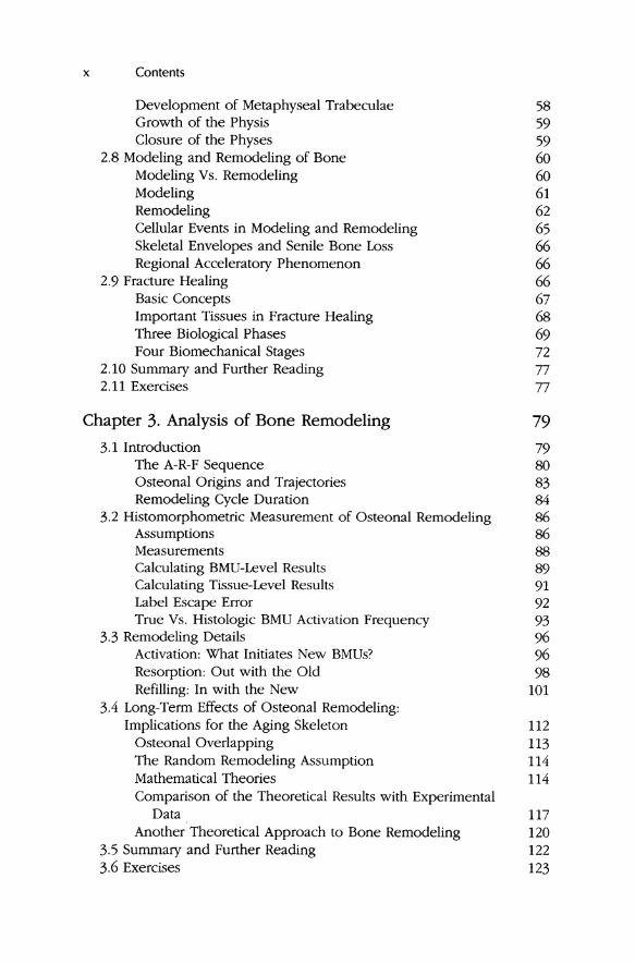
Development of Metaphyseal Trabeculae 58 Growth of the Physis 59 Closure of the Physes 59
2.8 Modeling and Remodeling of Bone 60 Modeling Vs. Remodeling 60 Modeling 61 Remodeling 62 Cellular Events in Modeling and Remodeling 65 Skeletal Envelopes and Senile Bone Loss 66 Regional Acceleratory Phenomenon 66
2.9 Fracture Healing 66 Basic Concepts 67 Important Tissues in Fracture Healing 68 Three Biological Phases 69 Four Biomechanical Stages 72
2.10 Summary and Further Reading 77 2.11 Exercises 77
Chapter 3. Analysis of Bone Remodeling 79
3.1 Introduction 79 The A-R-F Sequence 80 Osteonal Origins and Trajectories 83 Remodeling Cycle Duration 84
3.2 Histomorphometric Measurement of Osteonal Remodeling 86 Assumptions 86 Measurements 88 Calculating BMU-Level Results 89 Calculating Tissue-Level Results 91 Label Escape Error 92 True Vs. Histologic BMU Activation Frequency 93
3.3 Remodeling Details 96 Activation: What Initiates New BMUs? 96 Resorption: Out with the Old 98 Refilling: In with the New 101
3.4 Long-Term Effects of Osteonal Remodeling: Implications for the Aging Skeleton 112
Osteonal Overlapping 113 The Random Remodeling Assumption 114 Mathematical Theories 114 Comparison of the Theoretical Results with Experimental
Data 117 Another Theoretical Approach to Bone Remodeling 120
3.5 Summary and Further Reading 122 3.6 Exercises 123

Contents xi
Chapter 4. Mechanical Properties of Bone 127
4.1 Introduction 127 4.2 Fundamentals of Solid Mechanics 127
Strength and Stiffness of a Structure 128 Stress and Strain 129 Principal Directions 131 Strength and Stiffness of a Material 131 Generalized Hooke's Law: Anisotropy 133
4.3 Determinants of the Strength of a Whole Bone 134 Mechanics 134 Examples 136 Mechanical Failure of Whole Bones 139 Relationship to Modeling and Remodeling 142
4.4 Material Properties of Cortical Bone 143 Properties of Individual Secondary Osteons 144 Effects of Osteons on Bone Mechanical Properties 148 Anisotropy of Cortical Bone Mechanical Properties 151 Determinants of Osteonal Bone's Mechanical Properties 156
4.5 Material Properties of Cancellous Bone 165 Stress-Strain Curves for Cancellous Bone 165 The Three Determinants of Cancellous Bone Mechanical
Properties 168 Modeling Cancellous Bone as a Cellular System of
Plates or Struts 174 Invariance of Yield Strain 175
4.6 Predicting Material Properties: Bone as a Composite Material 176 4.7 Summary and Additional Reading 177 4.8 Exercises 178
Chapter 5. Fatigue and Fracture Resistance of Bone 181
5.1 Introduction 181 5.2 Basic Fracture Mechanics 182
Linear Elastic Fracture Mechanics 183 It Takes Energy to Propagate a Crack 188 Beyond the Linear Theory: Real Cracks Have Ears 189 Crack Growth and Fatigue 191
5.3 Fatigue Behavior of Bone 191 The S-N Curve 191 Fatigue Damage in Bone 192
5.4 Creep Behavior of Bone and its Relationship to Fatigue 193 5.5 Fatigue Behavior of Fiber-Reinforced Composite Laminates 196
The Birth and Growth of Cracks 197 Material Strength and Fiber Diameter 197 The Road to Failure 198

xii Contents
5.6 Osteonal Bone as a Fibrous Lamellar Composite Material 200 Osteonal Bone's Road to Failure 200 Bone Toughness 202 Controlled Crack Propagation Studies 202 Mathematical Analysis of Osteonal Pullout 203 Comparing Theory with Data 204 Crack Initiation 206 Crack Stopping 207 Strain Rate 208 Effect of Remodeling 208
5.7 Modeling Fatigue Damage Effects in Osteonal Bone 209 Why Would Cracks Be Stopped in Tension but Not in
Compression? 212 If Cracks Are Self-Limiting in Tensile Fatigue but Not in
Compressive Fatigue, Why Is Fatigue Life Longer in Compression Than in Tension? 213
5.8 The Role of Fatigue in Activating Bone Remodeling 214 Random or Directed Repair 214 The Historical Perspective 215 Microdamage and Bone Fragility in the Elderly 216
5.9 Modeling Stress Fractures 217 5.10 Summary and Additional Reading 222 5.12 Exercises 223
Chapter 6. Mechanical Adaptability of the Skeleton 225
6.1 Introduction 225 6.2 The Historical Context 226
The Mechanical Adaptability Hypothesis 230 Clinical Problems and Mechanical Adaptability 232
6.3 Self-Correction of Abnormally Curved Bones 233 Frost's Flexural Neutralization Theory 233 Stress Gradients and Fluid Flows 234 Related Experimental Results 236
6.4 Some Important Experiments 237 Measurement of Strain in Living Animals 237 Osteotomy Experiments: Surgically Overloaded Bones 240 Canine Disuse Experiments 241 The Porcine Exercise Experiment 242 Avian Isolated Ulna Experiments 242 Rat Tibia Bending Experiments 243 Summary 244
6.5 Some Additional Theories 245 Pauwels' Stress Magnitude Theory: Control of Modeling Drifts 245 Adaptive Elasticity Theory: Control of Density or
Modeling Drifts 247

Contents xiii
What Controls Osteonal Tunneling Directions? 248 Adaptive Finite-Element Models: Control of Density 251 Self-Trabeculating Models: Control of Density and
Trabecular Alignment 254 SyntheSiS 259 Frost's Mechanostat Theory 260 Relationship of Mechanically Adaptive Responses to
Other Control Factors 262 Mechanical Adaptability and Damage Repair 263
6.6 Mechanical Adaptability in Cartilage 264 The Carter-Wong Chondral Calcification Theory 264 Frost's Chondral Modeling Theory 266
6.7 Mechanical Adaptability and Evolutionary Adaptability 268 Somatic Vs. Evolutionary Adaptation 268 A Nonskeletal Example 269 Somatic Change in the Skeleton 270 Somatic Vs. Evolutionary Effects 270
6.8 Summary and Further Reading 271 6.9 Exercises 271
Chapter 7. Synovial Joint Mechanics 275
7.1 Introduction 275 Functions of a Joint 276 Joint Diseases 276
7.2 Mechanical Properties of Cartilage 278 Initial Points 278 Structure of Articular Cartilage 279 Permeability 280 Indentation Testing 281 Tensile Tests 281 Biphasic Theory of Articular Cartilage 283
7.3 Lubrication of Joints 289 Friction 289 Wear 290 Types of Lubrication 291 Synovial Joint Lubrication 300 Diversity of Joint Architecture 303
7.4 Summary and Further Reading 305 7.5 Exercises 305
Chapter 8. Mechanical Properties of ligament and Tendon 309
8.1 Introduction 8.2 Functional Considerations
Ligament
309 310 310

xiv Contents
Tendon 310 8.3 Structure and Composition 311
Ligament 311 Tendon 312
8.4 Mechanical Behavior 315 Quasistatic Tensile Properties 315 Viscoelastic Properties 318 Mathematical Modeling 320 Age and Mechanical Behavior 323
8.5 Mechanical Testing 324 Determination of Resting Length 326 Architectural Implications 326 Testing Environment 327
8.6 Functions at the Junctions 329 The Myotendinous Junction 329 Insertions; Sites of Ligament-to-Bone and Tendon-to-Bone
Attachment 330 8.7 Functional Adaptation and Specialization 332
Determinants of Tendon Architecture 333 Sites of Tendon Compression 335 Flexors and Extensors 338
8.8 Pathology and Healing 339 Ligament 339 Tendon 340 Immobilization, ExerCise, and Passive Motion 340
8.9 Surgical Repair 342 Anterior Cruciate Ligament 342 Supraspinatus Tendon (Rotator Cuff) 344 Flexor Tendons of the Hand 345
8.10 Summary and Further Reading 346 8.11 Exercises 347
Bibliography 349
Index 381