

Skeletal Muscles 10 Notecards. Head and Neck Muscles There are two main groups of head muscles....
74
Skeletal Muscles 10 Notecards
-
Upload
harvey-white -
Category
Documents
-
view
220 -
download
1
Transcript of Skeletal Muscles 10 Notecards. Head and Neck Muscles There are two main groups of head muscles....
Skeletal Muscles10 Notecards
Skeletal Muscles10 Notecards
Head and Neck Muscles• There are two main groups of head
muscles.–Facial muscles• Inserted into soft tissues
• Pull on the skin of the face
–Chewing muscles•Responsible for breaking down the foods we
eat

Frontalis muscle - Facial
• Originates at the cranial aponeurosis and inserts to the skin of the eyebrows.
• Raises your eyebrows.
• Wrinkles your forehead
Orbicularis Oculi - Facial
• Fibers run circles around the eyes.
• Allows you to close your eyes, squint, blink, and wink.

Orbicularis Oris
• Fibers run circles around the mouth.
• Closes the mouth and protrudes the lips
• Often called the “kissing” muscle.
Buccinator – Facial and Chewing
• Runs horizontally across the cheek and inserts into the orbicularis oris.
• Flattens the cheek (whistling or blowing a trumpet).
• Compresses the cheek to hold the food between the teeth during chewing.
Zygomaticus - Facial
• Extends from the corner of the mouth to the cheekbone.
• Called the smiling muscle because it raises the corners of the mouth upward.
Masseter – Chewing
• Covers the angle of the lower jaw.
• Runs from the zygomatic process of the temporal bone to the mandible.
• Closes the jaw by elevating the mandible.
Temporalis – Chewing
• Fan-shaped muscle overlying the temporal bone.
• Inserts into the mandible.
• Acts as synergist of the masseter in closing the jaw.
Platysma – Neck
• Sheetlike muscle that covers the anterolateral neck
• Originates from the connective tissue covering of the chest
• Inserts into the area of the mouth.
• Pulls on the corners of the mouth to produce a downward sag.
Sternocleidomastoid – Neck• Two-headed paired muscle.• One head originates at the sternum and the
second head originates at the clavicle.• Heads fuse before inserting into the mastoid
process of the temporal bone.• When both are contracted, they flex the neck
(often called the prayer muscle).• When one contracts, it turns the head the
opposite direction.
Anterior and Posterior Trunk Muscles
14 Notecards

Trunk Muscles
1. Those that move the vertebral column
2. Anterior thorax muscles (moving the ribs, head, and arms)
3. Abdominal wall (also move the vertebral column and form the natural girdle of the abdominal wall).
Anterior Thorax (cont.)
• Pectoralis Major–Large fan-shaped muscle covering the
upper part of the chest.
–Origin is from the shoulder girdle and the first six ribs
–Inserts on the proximal end of the humerus
–Acts to adduct and flex the arm.
Anterior Thorax (cont.)• Deltoid–Triangular shaped the forms the rounded
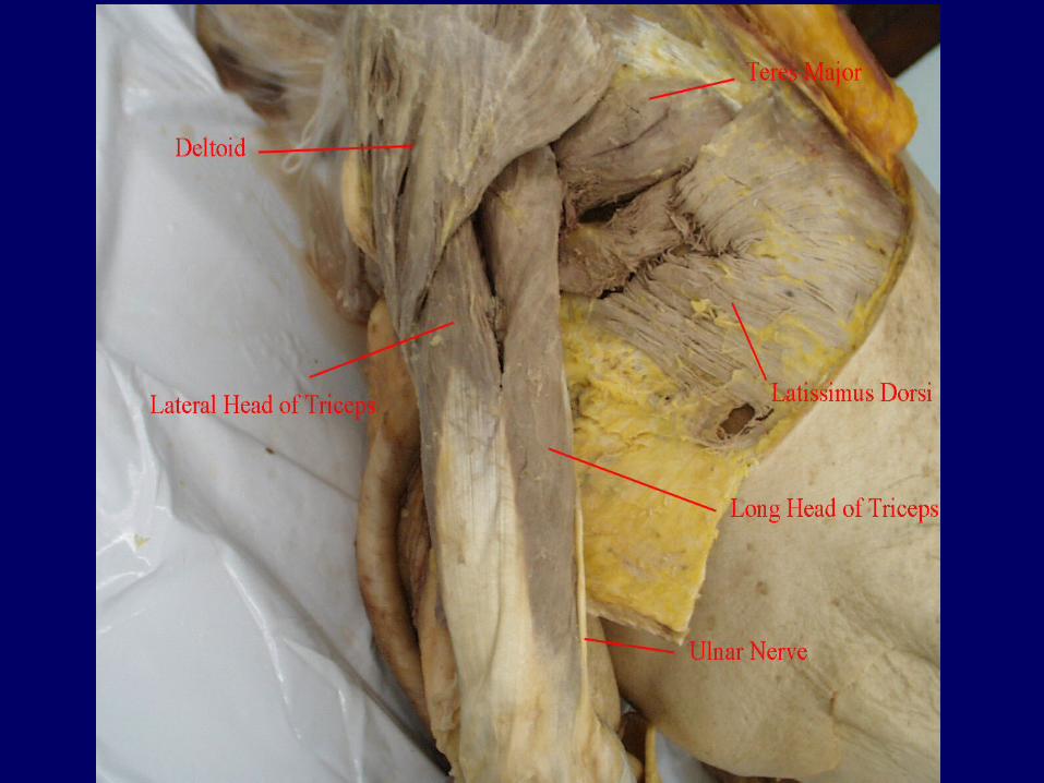
shape of your shoulders.–Favorite injection site (less than 5mL).–Originates at the scapular spine and clavicle.–Inserts into the proximal humerus.–Prime mover of arm abduction.
Anterior Thorax (cont.)
• Intercostal Muscles–Deep muscles between the ribs.
–External intercostals responsible for raise the rib cage for breathing air in.
–Internal intercostals, deep to the externals, depress the rib cage to move air out.
Anterior Thorax (cont.)
• Diaphragm–Single thin dome-shaped
muscle.
–Separates the thoracic cavity from the abdominal cavity.
–Primary muscle of inspiration
Anterior Thorax – Abdominal Girdle• Rectus abdominis–Paired straplike muscle–Most superficial of abdominal muscles–Run from the pubis to the rib cage–Main function is to flex the vertebral
column.–They also compress the abdominal
contents during defecation and childbirth.
Anterior Thorax – Abdominal Girdle (cont.)
• External Oblique–Paired superficial muscle that make up the
lateral walls of the abdomen.–Fibers run downward and medially form the
last eight ribs and insert into the ilium.–Flex the vertebral column, but also rotate
the trunk and bend laterally.

Anterior Thorax – Abdominal Girdle (cont.)
• Internal Oblique–Paired muscle deep to the external
oblique.–Fibers run at right angles to external
obliques.–Originate from the iliac crest and insert
into the last three ribs.–Functions are same as the external
obliques
Anterior Thorax – Abdominal Girdle (cont.)
• Transversus abdominis–Deepest muscle of the abdominal wall.–Fibers run horizontally across the abdomen.–Originates from the lower ribs and iliac crest
and inserts into the pubis.–Function to compress the abdominal
contents.
Anterior Thorax (cont.)• Deltoid–Triangular shaped the forms the rounded
shape of your shoulders.–Favorite injection site (less than 5mL).–Originates at the scapular spine and
clavicle.–Inserts into the proximal humerus.–Prime mover of arm abduction.

Posterior Thorax (cont.)• Trapezius–Most superficial muscles of the posterior
neck and upper trunk.–Together, they are diamond- or kite-
shaped.–Both originate at the occipital bone and
insert to the end of the thoracic vertebrae.–(CONTINUED ON THE NEXT SLIDE)
Posterior Thorax (cont.)
• Trapezius (cont.)–They flare laterally to insert on the
scapular spine and clavicle.
–They extend the head and can also elevate, depress, adduct, and stabilize the scapula (antagonist of sternocleidomastoids).
Posterior Thorax (cont.)
• Latissimus Dorsi–Large flat muscle pair the covers the lower
back.–Originates on the lower spine and ilium and
sweeps upward to insert on the proximal humerus.–Extends and adducts the humerus.–Powerful muscle for swimmers on the
power stroke.
Posterior Thorax (cont.)
• Erector Spinae–Group of three deep back muscles
resembling columns along the spine.–Prime mover of back extension.–Also provide resistance that helps control
the action of bending over at the waist.–When in spasm, represent a common source
of low back pain.

Muscles of the Hip, Thigh, and Leg
Muscles of the hip, thigh, and leg
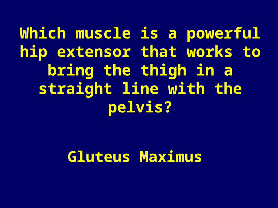
• Gluteus Maximus– Large superficial muscle of the hip.– Originates from the sacrum and iliac
bones and inserts on the gluteal tuberosity of the femur.
– Powerful hip extensor that brings the thigh in a straight line with the pelvis.
– Used when climbing stairs and jumping.

Muscles of the hip, thigh, and leg (cont.)
• Gluteus Medius–Runs from the ilium to the femur
under the gluteus maximus
–It is a hip abductor and important in steadying the pelvis while walking.
–Site for IM injections more than 5mL.
Muscles of the hip, thigh, and leg (cont.)
• Iliopsoas–Originates at the iliac bone and lower
vertebrae deep in the pelvis and inserts on the lesser trochanter of the femur.–Prime mover of hip flexion.–Acts to keep the upper torso from
bending backward.
Muscles of the hip, thigh, and leg (cont.)
• Adductor muscles (or group)–Originate on the pelvis and insert on the
proximal aspect of the femur.–Form the muscle mass on the medial aspect
of the thigh.–They adduct the thigh (press the thighs
together).–They tend to become flabby very easily.

Muscles of the hip, thigh, and leg (cont.)
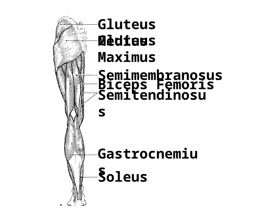
• Hamstring group (3 muscles)–Biceps femoris
–Semimembranosus
–Semitendinosus
• Originate on the ischial tuberosity.• Insert on both sides of the proximal
tibia.
Muscles of the hip, thigh, and leg (cont.)
• Sartorius–Thin, straplike muscle that is not very
important.
–Most superficial muscle of the thigh.
–Runs obliquely across the thigh from the anterior iliac crest to the medial side of the tibia.
–Is a weak thigh flexor.

Muscles of the hip, thigh, and leg (cont.)
• Quadriceps group (4 muscles)–Rectus femoris
–3 vastus muscles
• Vastus muscles originate from the femur while the rectus femoris originates on the pelvis.
• (Continued on next slide)

Muscles of the hip, thigh, and leg (cont.)
• Quadriceps group (4 muscles) (Cont.)
• All four insert on the tibial tuberosity via the patellar ligament.
• As a whole, act to extend the knee.
• The rectus femoris can also act to flex the hip.
Muscles of the hip, thigh, and leg (cont.)
• Tibialis anterior.–Superficial muscle on the anterior
leg.
–Originates on the proximal tibia and inserts into the tarsal bones by a long tendon.
–Dorsiflexes and inverts the foot.
Muscles of the hip, thigh, and leg (cont.)
• Extensor Digitorum Longus–Lateral to the tibialis anterior
–Originates from the lateral tibial condyle and proximal radius and inserts into the phalanges of toes 2-5 via a long tendon.
–Prime mover of toe extension and a dorsiflexor of the foot.
Muscles of the hip, thigh, and leg (cont.)
• Fibularis muscles.–Originate from the fibula and
insert into the metatarsal bones of the foot.
–Act to plantar flex and evert the foot.
Muscles of the hip, thigh, and leg (cont.)
• Gastrocnemius–Two-bellied muscle of the posterior leg.
–Originates on each side of the distal femur and insert by calcaneal tendon into the calcaneus.
–Prime mover for plantar flexion of the foot.
–Often called the “toe dancers” muscle.
Muscles of the hip, thigh, and leg (cont.)
• Soleus–Deep to the gastrocnemius.
–Originates on the tibia.
–A strong plantar flexor of the foot.
Leg Muscles ReviewLeg Muscles Review
What muscle inserts through the Achilles tendon to cause plantar flexion of the foot?
Gastrocnemius
Which muscle acts to dorsiflex and invert the
foot?
Anterior Tibialis

What group of muscles act to powerfully extend
the knee?
Quadriceps group
Which muscle is a powerful hip extensor that works to bring the thigh in a straight line with the
pelvis?
Gluteus Maximus

Which muscle of the lower limb is a prime mover of hip flexion and also
acts as a postural muscle to keep the upper body from falling backward
when standing erect?
Iliopsoas

Gluteus MediusGluteus Maximus
SemitendinosusBiceps FemorisSemimembranosus
Gastrocnemius
Soleus

SartoriusRectus FemorisVastus MedialisVastus Lateralis
Fibularis MusclesTibialis Anterior
Gluteus MediusGluteus Maximus
SemitendinosusBiceps FemorisSemimembranosus
Gastrocnemius
Soleus
SartoriusRectus FemorisVastus MedialisVastus Lateralis
Fibularis MusclesTibialis Anterior
Muscles of the Arm and Forearm
Muscles of the arm and forearm
• Biceps Brachii–Originates by two heads from the
shoulder girdle and inserts into the radial tuberosity.
–Prime mover for flexion of the forearm
–Also act to supinate the forearm.
Muscles of the arm and forearm (cont.)
• Brachialis–Lies deep to the biceps muscle
–Also acts to cause elbow flexion.
Muscles of the arm and forearm (cont.)
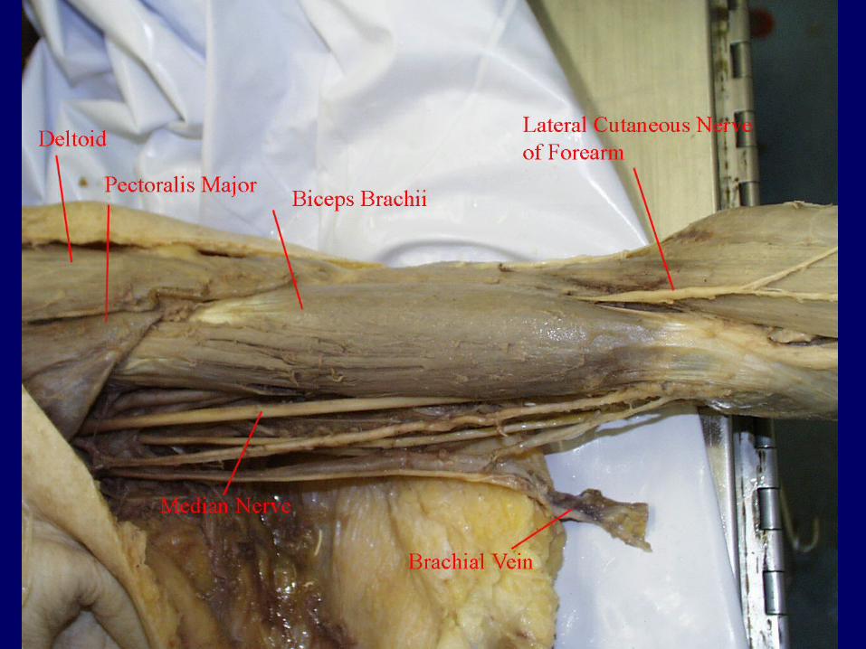
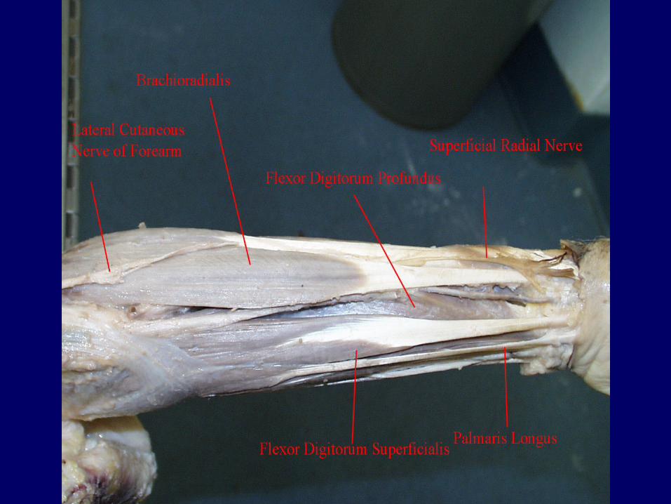
• Brachioradialis–Originates on the humerus and
inserts into the distal forearm.
–Assists in flexion at the elbow and also supinates/pronates the forearm.
Muscles of the arm and forearm (cont.)
• Triceps Brachii–Originates from the shoulder girdle
and proximal humerus.
–Inserts into the olecranon process of the ulna.
–Prime mover of elbow extension (also antagonist of Biceps Brachii).
–Often called the “boxer’s” muscle.
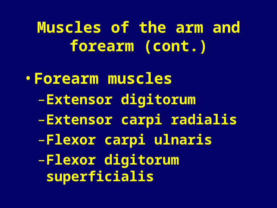
Muscles of the arm and forearm (cont.)
• Forearm muscles–Extensor digitorum
–Extensor carpi radialis
–Flexor carpi ulnaris
–Flexor digitorum superficialis

Arm and Forearm Muscles Review
Arm and Forearm Muscles Review
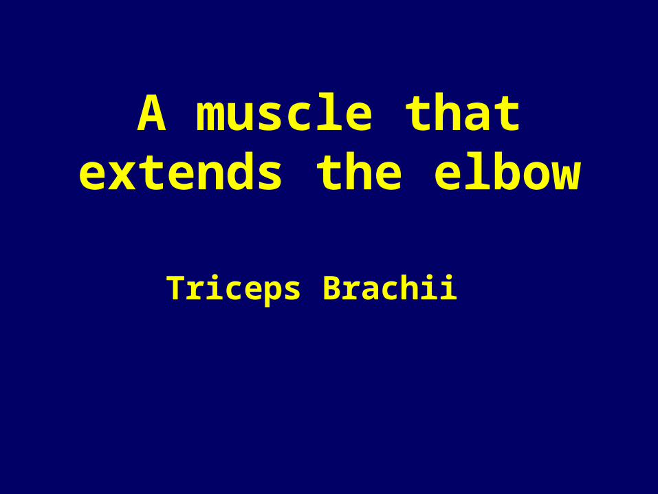
A muscle that extends the elbow
Triceps Brachii

Shoulder abductor, used to raise the arm
overhead
Deltoid
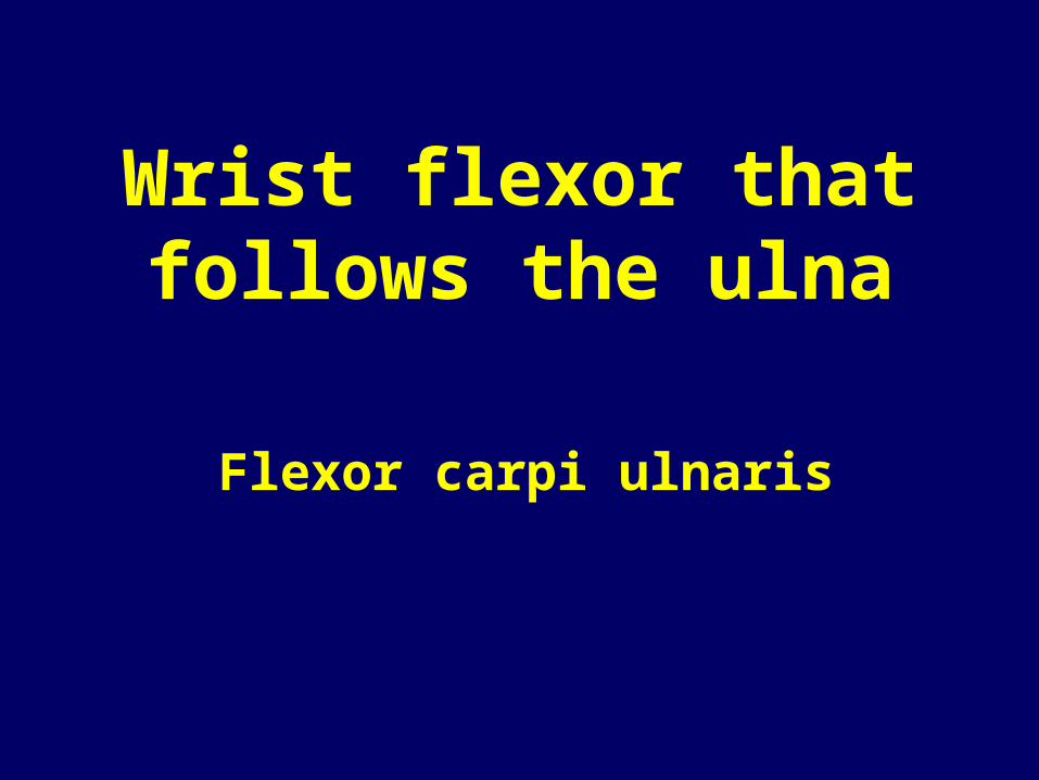
Wrist flexor that follows the ulna
Flexor carpi ulnaris

Muscle that extends the fingers
Extensor digitorum
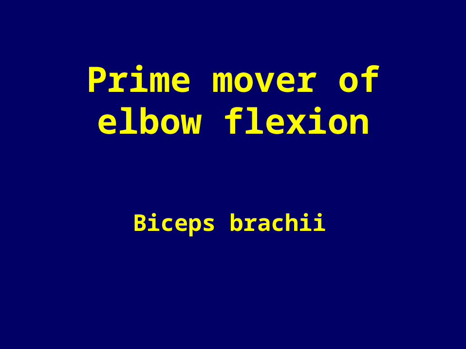
Prime mover of elbow flexion
Biceps brachii
Muscle that flexes the fingers
Flexor digitorum superficialis
What is the antagonist of the biceps brachii?
The triceps brachii

Digit flexor muscles are located on the _____
aspect of the forearm.
Anterior
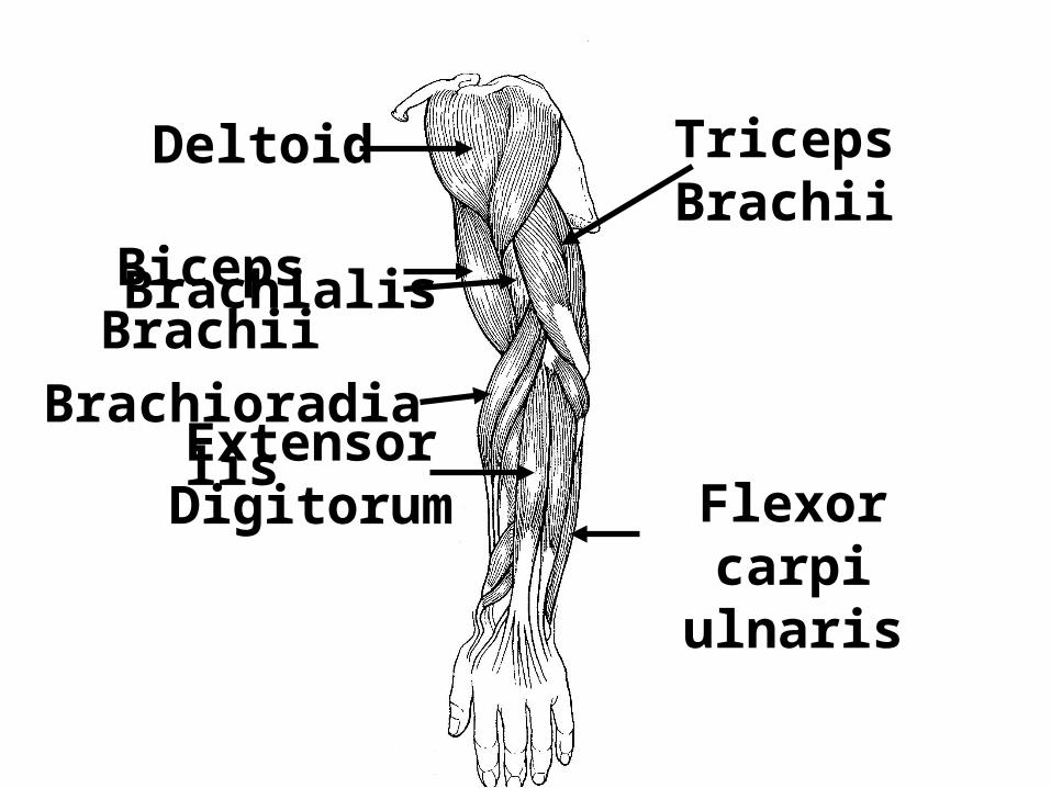
Deltoid Triceps Brachii
Biceps BrachiiBrachialis
BrachioradialisExtensor Digitorum Flexor carpi
ulnaris
![Genetics Notecards[1]](https://static.fdocuments.net/doc/165x107/577d23c11a28ab4e1e9ab0a6/genetics-notecards1.jpg)