Size, charge and concentration dependent uptake of iron oxide particles by non-phagocytic cells
8
Size, charge and concentration dependent uptake of iron oxide particles by non-phagocytic cells Daniel L.J. Thorek, Andrew Tsourkas * Department of Bioengineering, University of Pennsylvania, 210 South 33rd Street, 240 Skirkanich Hall, Philadelphia, PA 19104, USA article info Article history: Received 25 March 2008 Accepted 16 May 2008 Available online 3 June 2008 Keywords: Molecular imaging MRI Ultrasmall superparamagnetic iron oxide Standard superparamagnetic iron oxide Micron-sized paramagnetic iron oxide Nanoparticles abstract A promising new direction for contrast-enhanced magnetic resonance (MR) imaging involves tracking the migration and biodistribution of superparamagnetic iron oxide (SPIO)-labeled cells in vivo. Despite the large number of cell labeling studies that have been performed with SPIO particles of differing size and surface charge, it remains unclear which SPIO configuration provides optimal contrast in non- phagocytic cells. This is largely because contradictory findings have stemmed from the variability and imprecise control over surface charge, the general need and complexity of transfection and/or targeting agents, and the limited number of particle configurations examined in any given study. In the present study, we systematically evaluated the cellular uptake of SPIO in non-phagocytic T cells over a continuum of particle sizes ranging from 33 nm to nearly 1.5 mm, with precisely controlled surface properties, and without the need for transfection agents. SPIO labeling of T cells was analyzed by flow cytometry and contrast enhancement was determined by relaxometry. SPIO uptake was dose-dependent and exhibited sigmoidal charge dependence, which was shown to saturate at different levels of functionalization. Efficient labeling of cells was observed for particles up to 300 nm, however, micron-sized particle uptake was limited. Our results show that an unconventional highly cationic particle configuration at 107 nm maximized MR contrast of T cells, outperforming the widely utilized USPIO (<50 nm). Ó 2008 Elsevier Ltd. All rights reserved. 1. Introduction Continuing advancements in cell-based therapies have recently led to the emergence of cellular imaging as a strategy to track the migration and biodistribution of target cells in living organisms. Pre-clinical studies have already shown that cellular imaging can be used to evaluate stem cell distribution and homing in cell-based regenerative therapies [1,2]. Recently, cellular imaging has also allowed for improved assessment of functional efficacy and appli- cability of immunotherapeutic treatments in disease models for cancer [3–5] and AIDS [6]. In addition to evaluating cell-based therapies, cellular imaging also promises to provide a great deal of insight into diverse physio- and pathological phenomena. Interesting applications include the observation of monocyte recruitment to atherosclerotic lesions for the mapping of disease development and therapeutic intervention [7], imaging embryonic stem cell movement during embryonic [8] and organ development [9] and monitoring the dynamics of met- astatic cellular extravasation and tissue invasion [10,11]. Tracking of labeled cells has been accomplished with a variety of imaging modalities including optical methods, positron emission tomography (PET), single photon emission computed tomography (SPECT), and magnetic resonance (MR) imaging [12–14]. MR im- aging presents a particularly promising approach because of its high spatial resolution in three dimensions and exquisite soft tissue contrast, which can be acquired concomitantly with the contrast- enhanced cellular distribution. MR detection of cells in vivo is often accomplished following labeling with superparamagnetic iron ox- ide (SPIO) particles. SPIO are negative contrast agents that are typically composed of an iron oxide crystal core surrounded by a polymer or polysaccharide shell [15]. A variety of manifestations of SPIO have been used to track cells, which can be broadly cate- gorized as (1) ultrasmall SPIO (USPIO) with an overall diameter of 30–50 nm [16], (2) standard SPIO (SSPIO) with a diameter of 50– 150 nm and (3) micron-sized paramagnetic iron oxide (MPIO) having a diameter approaching or greater than 1 mm [17]. To date, USPIO has perhaps been the most widely utilized SPIO configuration for cell labeling. Although they provide less contrast enhancement per particle compared with SSPIO and MPIO, large numbers of particles can be loaded into each cell [18,19]. As cationic surfaces have been shown to facilitate cellular internalization [20,21], USPIO is often modified with polycationic cell permeating peptides (CPPs) such as HIV transactivator (TAT) [22] or protamine [23]. Other transfection techniques, sometimes in concert with CPPs, are also used [24,25]. * Corresponding author. Tel.: þ1 (215)898 8167; fax: þ1 (215)573 2071. E-mail address: [email protected] (Andrew Tsourkas). Contents lists available at ScienceDirect Biomaterials journal homepage: www.elsevier.com/locate/biomaterials 0142-9612/$ – see front matter Ó 2008 Elsevier Ltd. All rights reserved. doi:10.1016/j.biomaterials.2008.05.015 Biomaterials 29 (2008) 3583–3590
-
Upload
blaghlargh -
Category
Documents
-
view
20 -
download
1
description
Paper describing factors that affect uptake of iron oxide particles into noon-phagocytic cells
Transcript of Size, charge and concentration dependent uptake of iron oxide particles by non-phagocytic cells

lable at ScienceDirect
Biomaterials 29 (2008) 3583–3590
Contents lists avai
Biomaterials
journal homepage: www.elsevier .com/locate/biomateria ls
Size, charge and concentration dependent uptake of ironoxide particles by non-phagocytic cells
Daniel L.J. Thorek, Andrew Tsourkas*
Department of Bioengineering, University of Pennsylvania, 210 South 33rd Street, 240 Skirkanich Hall, Philadelphia, PA 19104, USA
a r t i c l e i n f o
Article history:Received 25 March 2008Accepted 16 May 2008Available online 3 June 2008
Keywords:Molecular imagingMRIUltrasmall superparamagnetic iron oxideStandard superparamagnetic iron oxideMicron-sized paramagnetic iron oxideNanoparticles
* Corresponding author. Tel.: þ1 (215)898 8167; faE-mail address: [email protected] (Andrew
0142-9612/$ – see front matter � 2008 Elsevier Ltd.doi:10.1016/j.biomaterials.2008.05.015
a b s t r a c t
A promising new direction for contrast-enhanced magnetic resonance (MR) imaging involves trackingthe migration and biodistribution of superparamagnetic iron oxide (SPIO)-labeled cells in vivo. Despitethe large number of cell labeling studies that have been performed with SPIO particles of differing sizeand surface charge, it remains unclear which SPIO configuration provides optimal contrast in non-phagocytic cells. This is largely because contradictory findings have stemmed from the variability andimprecise control over surface charge, the general need and complexity of transfection and/or targetingagents, and the limited number of particle configurations examined in any given study. In the presentstudy, we systematically evaluated the cellular uptake of SPIO in non-phagocytic T cells over a continuumof particle sizes ranging from 33 nm to nearly 1.5 mm, with precisely controlled surface properties, andwithout the need for transfection agents. SPIO labeling of T cells was analyzed by flow cytometry andcontrast enhancement was determined by relaxometry. SPIO uptake was dose-dependent and exhibitedsigmoidal charge dependence, which was shown to saturate at different levels of functionalization.Efficient labeling of cells was observed for particles up to 300 nm, however, micron-sized particle uptakewas limited. Our results show that an unconventional highly cationic particle configuration at 107 nmmaximized MR contrast of T cells, outperforming the widely utilized USPIO (<50 nm).
� 2008 Elsevier Ltd. All rights reserved.
1. Introduction
Continuing advancements in cell-based therapies have recentlyled to the emergence of cellular imaging as a strategy to track themigration and biodistribution of target cells in living organisms.Pre-clinical studies have already shown that cellular imaging can beused to evaluate stem cell distribution and homing in cell-basedregenerative therapies [1,2]. Recently, cellular imaging has alsoallowed for improved assessment of functional efficacy and appli-cability of immunotherapeutic treatments in disease models forcancer [3–5] and AIDS [6].
In addition to evaluating cell-based therapies, cellular imagingalso promises to provide a great deal of insight into diverse physio-and pathological phenomena. Interesting applications include theobservation of monocyte recruitment to atherosclerotic lesions forthe mapping of disease development and therapeutic intervention[7], imaging embryonic stem cell movement during embryonic [8]and organ development [9] and monitoring the dynamics of met-astatic cellular extravasation and tissue invasion [10,11].
Tracking of labeled cells has been accomplished with a variety ofimaging modalities including optical methods, positron emission
x: þ1 (215)573 2071.Tsourkas).
All rights reserved.
tomography (PET), single photon emission computed tomography(SPECT), and magnetic resonance (MR) imaging [12–14]. MR im-aging presents a particularly promising approach because of itshigh spatial resolution in three dimensions and exquisite soft tissuecontrast, which can be acquired concomitantly with the contrast-enhanced cellular distribution. MR detection of cells in vivo is oftenaccomplished following labeling with superparamagnetic iron ox-ide (SPIO) particles. SPIO are negative contrast agents that aretypically composed of an iron oxide crystal core surrounded bya polymer or polysaccharide shell [15]. A variety of manifestationsof SPIO have been used to track cells, which can be broadly cate-gorized as (1) ultrasmall SPIO (USPIO) with an overall diameter of30–50 nm [16], (2) standard SPIO (SSPIO) with a diameter of 50–150 nm and (3) micron-sized paramagnetic iron oxide (MPIO)having a diameter approaching or greater than 1 mm [17].
To date, USPIO has perhaps been the most widely utilized SPIOconfiguration for cell labeling. Although they provide less contrastenhancement per particle compared with SSPIO and MPIO, largenumbers of particles can be loaded into each cell [18,19]. As cationicsurfaces have been shown to facilitate cellular internalization[20,21], USPIO is often modified with polycationic cell permeatingpeptides (CPPs) such as HIV transactivator (TAT) [22] or protamine[23]. Other transfection techniques, sometimes in concert withCPPs, are also used [24,25].

D.L.J. Thorek, Andrew Tsourkas / Biomaterials 29 (2008) 3583–35903584
An exciting new direction for cell tracking involves labeling cellswith MPIO [26]. The large iron oxide cores present in these particlesprovide enough contrast for single cells to be imaged by MR.However, work with such large particles generally confines appli-cation of iron oxide labeling to phenotypes such as macrophages[18], dendritic cells [27] or hepatocytes that actively internalizeforeign material. MPIO uptake in non-phagocytic cells has beenaccomplished, but is limited by the additional conjugation workand cost of using an antibody-mediated approach [28], which mustbe species specific and may induce adverse cellular events.
Recently, several studies have attempted to define an optimizedparticle configuration for iron oxide labeling of both phagocytic andnon-phagocytic cell types. Although MPIO was excluded from all ofthese studies, it was found that phagocytic monocytes are moreeffectively labeled with SSPIO (150 nm) compared with USPIO(30 nm) [18,29]. Further, it was found that ionic carboxydextran-coated SSPIO (i.e. ferucarbotran) performed better than non-ionicdextran-coated SSPIO (i.e. ferumoxide) [18]. It remains unclear howMPIO compares with these agents; however, single cell detectionhas been achieved in phagocytic cells with both SPIO configurations[30,31].
The optimal SPIO configuration for labeling non-phagocytic cellshas been much more elusive and findings have been contradictory.For example, in one study it was found that the delivery of car-boxydextran USPIO and dextran-labeled SSPIO into non-phagocyticcancer cells and leukocytes (with the assistance of lipofectionagents) was similar in terms of iron uptake [21]. Both particles ledto higher iron uptake than USPIO. This indirectly suggests thatlarger particles with ionic coatings are superior to non-ionic USPIO.However, in a different study it was found that, in the presence ofpoly-L-lysine, ionic (aminated) USPIO exhibited significantly higheriron uptake in non-phagocytic cells compared with SSPIO. Thesedata suggest that smaller ionic particles are internalized into non-phagocytic cells more efficiently [32]. These contradictory findingslikely stem from the variability and imprecise control over surfacecharge and the limited number of particle configurations examined,particularly with respect to diameter (ranging only from w17 nm to150 nm).
In the present study we systematically evaluated the cellularuptake of SPIO in non-phagocytic T cells over a continuum of par-ticle sizes ranging from 33 nm to nearly 1.5 mm and with preciselycontrolled surface properties. T cells were selected as a model non-phagocytic phenotype since visualization of their distribution isexpected to be of importance for adoptive T cell therapy for cancerand T cell homing in autoimmune diseases. Extremely fine controlwas exerted on the surface properties of SPIO by direct chemicalmodification of particle surfaces rather than attempting to modu-late the density of supplemental transfection agents. Concentrationeffects and incubation times were also tested in the interest ofisolating the role particle size exerts on individual cell uptake andoverall contrast enhancement. Our work shows that in a spacebetween USPIO and MPIO exist configurations of relatively smallparticles (w100 nm) that efficiently label non-adherent, non-phagocytic T cells and generate higher relaxivity (per cell) relativeto particles of other sizes.
2. Materials and methods
2.1. Nanoparticle synthesis
Three different formulations of dextran-coated superparamagnetic iron oxidenanoparticles were prepared using the co-precipitation method [33]. All three for-mulations were prepared following the same procedure, as described below, withthe only difference being the amount of FeCl2 and FeCl3 added. Specifically, 25 g ofdextran T10 (GE Healthcare, Piscataway, NJ) was dissolved in 50 mL of dH2O andheated to 80 �C for 1 h. The solution was allowed to return to room temperatureand continued to mix overnight. Subsequently, the dextran was cooled to 4 �C on iceand degassed with N2 for 1 h. FeCl2 (0.7313 g, 1.5 g, or 2.2 g) and FeCl3 (1.97 g, 4 g, or
6 g, respectively) were each rapidly dissolved in 12.5 mL of degassed dH2O and kepton ice for approximately 10 min. The iron solutions were added to the dextransimultaneously and allowed to mix for 30 min. Keeping this mixing solution at 4 �C,15 mL of ammonium hydroxide was added. The resulting black viscous solution wasthen heated to 90 �C for 1 h then cooled overnight, followed by ultracentrifugationat 20 k rcf for 30 min. Pellets were discarded and the supernatant was continuallydiafiltrated using a 100-kDa MWCO cartridge (GE Healthcare) on a peristaltic pump(E323, Watson Marlow Bredel, Wilmington, MA). The particles were exchanged into0.02 M citrate, 0.15 M sodium chloride buffer until all unreacted products had beenremoved. Aminated silica-coated iron oxide micro-particles were purchased fromBioclone Inc. (San Diego, CA). Amine functionalized styrene copolymer-coated ironoxide particles (Adembeads) were purchased from Ademtech SA (Pessac, France).
2.2. Amination of particles
Amination and crosslinking of the coating on the dextran–SPIO were accom-plished through reaction of the SPIO with 25% 10 M NaOH and 33% epichlorohydrin[34]. After mixing for 24 h, additional ammonium hydroxide was added to the so-lution, bringing the volume fraction to 25% ammonium hydroxide, and the reactionwas allowed to proceed for another 24 h. The particles were then exhaustively pu-rified via diafiltration. The resulting particles were amine functionalized crosslinkediron oxide.
2.3. FITC labeling and amine-blocking of particles
All SPIO particles were labeled with FITC at a FITC-to-iron molar ratio of 19.2:1.FITC was reacted with particles for 4 h followed by two rounds of gel purification,once on a NAP-5 column and then on a PD10 column (GE Healthcare), both equili-brated with PBS. The FITC-labeled SPIO was subsequently reacted with variousvolumes of glycidol (0.01–50%) to produce populations of particles with differentamine content. The particles were cleaned of excess glycidol through repeatedprecipitation in isopropanol and resuspension in PBS. Amine-blocking was alsoattempted with particles of 200 nm and greater, but this modification impelledimmediate particle insolubility.
2.4. Measurement of particle size
The hydrodynamic diameter of the dextran-coated and commercial iron oxideparticles was measured using a Zetasizer Nano-z (Malvern Instruments, Malvern,UK) through dynamic light scattering (DLS). The dextran-coated SPIO particles werediluted in PBS to a concentration of approximately 0.5 mg/mL and read in triplicate.The commercial particle diameters were read in the same manner, but only afterundergoing three washes by precipitation in the presence of a strong magnet andresuspension in PBS. The values reported for all samples are the intensity peakvalues.
2.5. Measurement of particle cores
Transmission electron micrographs of all iron oxide particles were taken usinga JEOL 2010 at 200 kV. Samples were prepared for imaging by evaporating theparticles onto a carbon-coated copper grid (Holey carbon – mesh 200, StructureProbe Inc., West Chester, PA). Salt was removed from all of the samples prior toevaporation by exchanging the particles into dH2O. Images of particle cores wereanalyzed using ImageJ (National Institutes of Health, Bethesda, MD). Since many ofthe particles were found to be composed of a cluster of multiple iron oxide cores, theaverage diameter of each core and the average number of cores per particle weredetermined. Assuming each core to be spherical, the amount of iron per particle typewas determined from the aggregate core volume.
2.6. Measurement of particle relaxivity (R1 and R2)
The longitudinal (R1) and transverse (R2) relaxivity of each particle was calcu-lated as the slope of the curves 1/T1 and 1/T2 against iron concentration, respectively.T1 and T2 relaxation times were determined using a Bruker mq60 MR relaxometeroperating at 1.41 T (60 MHz). T1 measurements were performed by collecting 12data points from 5.0 ms to 1000 ms with a total measurement duration of 1.49 min.T2 measurements were made using s¼ 1.5 ms and two dummy echoes, and fittedassuming monoexponential decay.
2.7. Measurement of number of amines per particle
The number of amines per particle was determined following the general pro-cedure described by Zhao et al. [35]. Briefly, iron oxide particles at a concentration of2 mg/mL Fe were reacted with excess N-succinimidyl 3-(2-pyridyldithio) propionate(SPDP, Calbiochem, San Diego, CA) for 4 h. SPIO was washed of excess SPDP throughrepeated precipitation in isopropanol and resuspension in PBS. The particles werethen run through a 50-kDa MWCO centrifugal filter (YM-50, Millipore, Billerica, MA)either with or without the addition of disulfide cleavage agent TCEP. The differenceof the absorbance of these two samples at 343 nm was used to determine the

Fig. 1. Hydrodynamic diameter of SPIO. The hydrodynamic diameter of SPIO particleswas determined by DLS. Intensity measurements are reported and the peak intensity isprovided for each distribution.
D.L.J. Thorek, Andrew Tsourkas / Biomaterials 29 (2008) 3583–3590 3585
concentration of SPDP in the filter flow. Adjusting for dilution, the number of aminesper particle was determined.
2.8. Cell culture and labeling
Immortalized human T cells, Jurkat Clone E6-1 (ATCC), were maintained at 37 �Cin 5% CO2 in RPMI 1640 (Mediatech, Manassas, VA) media supplemented with 10%FBS (Hyclone, Logan, UT) and penicillin/streptomycin (Mediatech). T cells were la-beled with iron oxide particles by incubating the commercial and lab-made particleswith 2�106 cells in 400 mL of fully supplemented media for 1 h or 4 h, at 37 �C in 5%CO2. Cells were washed of non-internalized particles through two methods. Syn-thesized dextran-coated particles were washed from cells using centrifugation.Specifically, cells were pelleted at 0.5 rcf for 5 min and resuspended in PBS. This wasrepeated three times. The dextran-coated particles are highly soluble in aqueoussolvents and do not precipitate at these centrifugation speeds. Removal of non-internalized commercial particles was accomplished through a density gradient. Thecells and particles were diluted to 1 mL with PBS and overlayed on 4 mL of roomtemperature Ficoll-Paque PLUS (GE Healthcare). The sample was centrifuged at0.4 rcf for 40 min. Cells loaded with particles were retrieved from the interface layer.To determine if particles were internalized or merely adsorbed on the cell exterior,surface receptor cleavage enzyme trypsin was used. Following particle incubation, asdescribed above, cells were exposed to 0.025% trypsin–EDTA (Invitrogen) for 5 min.Purification of non-internalized particles was carried out as detailed. No statisticaldifference was seen in either flow cytometry or relaxometry between groupswashed with or without enzyme.
2.9. Flow cytometry and relaxation measurements
Immediately after non-internalized iron oxide particles were removed from Tcell samples, flow cytometry was performed on a Guava Easycyte (Guava Technol-ogies, Hayward, CA). For labeling and viability experiments, forward and side scat-terings were used to identify the entire population of cells. Data analysis of flowcytometry data was accomplished with FlowJo (TreeStar, Ashland, OR). Viability of Tcells was determined using the LIVE/DEAD cytotoxicity kit for mammalian cells(Invitrogen, Carlsbad, CA) according to the manufacturer’s instructions. In order toevaluate the decrease in T2 relaxation time of iron oxide internalized T cells, purifiedcells were lysed for 30 min in 0.1% SDS in PBS at 37 �C. Samples were diluted to0.5�106 cells/mL in 300 uL and T2 relaxation times were measured using thebenchtop relaxometer. All flow and magnetic resonance measurements were madein triplicate on at least two separate occasions.
3. Results and discussion
3.1. Particle synthesis and characterization
Three different formulations of dextran-coated super-paramagnetic iron oxide nanoparticles were prepared via co-pre-cipitation. All three syntheses utilized a ratio of approximatelythree ferrous to ferric iron chloride; however, the total amount ofiron was increased by whole numbers, i.e. 2� and 3� irons, re-spectively. This deviation in the amount of iron present duringsynthesis allowed for the manufacture of SPIO with a range ofdifferent sizes and properties. Specifically, DLS of the SPIO, fol-lowing crosslinking and amination of the dextran coating, indicatedaverage hydrodynamic radii of 33.4 nm, 53.5 nm and 107 nm, re-spectively, with the larger nanoparticles corresponding to synthe-ses that utilized more iron. When the total amount of iron wasincreased further, the co-precipitation solution became extremelyviscous and yielded highly dispersed aggregates that precipitatedout of solution. Therefore, nanoparticles ranging from 200 nm to1 mm in diameter were acquired from commercial sources. Specif-ically, superparamagnetic iron oxide particles of 200 nm and300 nm diameter with an amine functionalized styrene copolymercoating (Amino–Adembeads) were purchased from Ademtech,while amine functionalized silica-coated 1 mm diameter particleswere purchased from Bioclone. This allowed particle sizes acrossnearly three orders of magnitude to be compared.
The particle sizes as determined by DLS, peak intensity values,are compared in Fig. 1. The 33.4 nm, 53.5 nm and 107 nm dextran-coated SPIO samples were fully soluble at physiological conditions.Conversely, it was found that the large size of the 289 nm and1430 nm particles led to rapid precipitation. Settling was also
a concern for the 207 nm particles; however, full precipitationgenerally took several hours.
Analysis of the iron oxide core size and structure of the magneticparticles was conducted using TEM. Representative micrographsare shown in Fig. 2. Aggregation of particles in salt free solution wasa problem during TEM sample preparation; however, reduction insample concentration allowed for imaging of discretely distributedparticles. Iron cores were easily distinguished from carbon-coatedcopper grids, while dextran and styrene copolymer were not visiblebecause of their low electron density.
An interesting feature of the dextran-coated nanoparticles isthat each particle consists of a cluster of one or more iron oxidecores, with each core being approximately equal in size. Specifi-cally, the distribution of cores is centered at approximately 6 nm forall three dextran-coated nanoparticles (Fig. 3); however, the aver-age number of cores per particle increases with overall hydrody-namic diameter. In contrast, the larger 207 nm and 289 nm styrenecopolymer-coated particles exhibited a single large spherical ironoxide core, while the 1.43 mm silica-coated particles exhibited anamorphous iron oxide core of no discrete size or shape. A summaryof the properties of each SPIO is provided in Table 1.
The R1 and R2 data (Figs. 4 and 5), also summarized in Table 1,indicate that there is a trend of increasing R2 and decreasing R1 withsize up to the 107 nm particles. For particles of greater size, thesingle large core of the 207 nm and 289 nm particles does nottranslate into proportionately higher R2. This likely reflects lowercrystallinity of the larger single iron oxide cores in comparison tosmaller crystals [36]. Furthermore, according to the Solomon–Bloembergen theory, which relates the relaxation rate to particleproperties, the total size of the particle is not critical to the mag-nitude of R2 as the susceptibility effect falls off from the surfacewith an exponential (r6) dependence [37,38]. It should be notedthat the R1 values reported for particles greater than 200 nm arelikely underestimates due to precipitation of the particles during T1
measurements. For instance, determining T1 relaxation times re-quired more than 100 s per sample, which was an ample time forthe micrometer-sized particles to precipitate out of solution.
3.2. Cell loading
The extent to which T cells internalize iron oxide particles is notonly dependent on particle size but also various other particlecharacteristics and cell loading conditions, including surfacecharge, particle concentration, and incubation time. Thus, before it
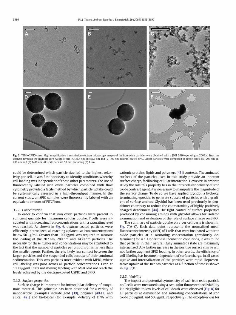
Fig. 2. TEM of SPIO cores. High magnification transmission electron microscopy images of the iron oxide particles were obtained with a JEOL 2010 operating at 200 kV. Structureanalysis revealed the multiple core nature of the (A) 33.4 nm, (B) 53.5 nm and (C) 107 nm dextran-coated SPIO. Larger particles were composed of single cores; (D) 207 nm, (E)289 nm and (F) 1430 nm. All scale bars are 50 nm, excluding (F) 1 mm.
D.L.J. Thorek, Andrew Tsourkas / Biomaterials 29 (2008) 3583–35903586
could be determined which particle size led to the highest relax-ivity per cell, it was first necessary to identify conditions wherebycell loading was independent of these other parameters. The use offluorescently labeled iron oxide particles combined with flowcytometry provided a facile method by which particle uptake couldbe systematically assessed in a high-throughput manner. In thecurrent study, all SPIO samples were fluorescently labeled with anequivalent amount of FITC/iron.
3.2.1. ConcentrationIn order to confirm that iron oxide particles were present in
sufficient quantity for maximum cellular uptake, T cells were in-cubated with increasing iron concentrations until a saturating levelwas reached. As shown in Fig. 6, dextran-coated particles wereefficiently internalized, all reaching a plateau at iron concentrationsbelow 50 mg/mL. Greater than 100 mg/mL was required to saturatethe loading of the 207 nm, 289 nm and 1430 nm particles. Thenecessity for these higher iron concentrations may be attributed tothe fact that the number of particles per unit of iron is far less thanthe smaller agents. Further, there is likely less contact between thelarger particles and the suspended cells because of their continualsedimentation. This was perhaps most evident with MPIO, wherecell labeling was poor across all particle concentrations. Even at1000 mg/mL (data not shown) labeling with MPIO did not reach thelevels achieved by the dextran-coated USPIO and SPIO.
3.2.2. Surface propertiesSurface charge is important for intracellular delivery of exoge-
nous material. This principle has been described for a variety ofnanoparticle (examples include gold [39], polymer [40,41] andsilica [42]) and biological (for example, delivery of DNA with
cationic proteins, lipids and polymers [43]) contexts. The aminatedsurfaces of the particles used in this study provide an inherentsurface charge, facilitating cellular interaction. However, in order tostudy the role this property has in the intracellular delivery of ironoxide contrast agent, it is necessary to manipulate the magnitude ofthe surface charge. To do so we have applied glycidol, a hydroxylterminating epoxide, to generate subsets of particles with a gradi-ent of surface amines. Glycidol has been used previously in den-drimer chemistry to reduce the chemotoxicity of highly-positivelycharged dendrimers [44]. The tight control of surface propertiesproduced by consuming amines with glycidol allows for isolatedexamination and evaluation of the role of surface charge on SPIO.
The summary of particle uptake on a per cell basis is shown inFig. 7(A–C). Each data point represents the normalized meanfluorescence intensity (MFI) of T cells that were incubated with ironoxide particles at a saturating concentration (previously de-termined) for 4 h. Under these incubation conditions, it was foundthat particles in their natural (fully aminated) state are maximallyinternalized. Any further increase in the positive surface charge willnot further augment SPIO loading. In other words, the efficiency ofcell labeling has become independent of surface charge. In all cases,uptake and internalization of the particles were rapid. Represen-tative uptake of the 107 nm particles as a function of time is shownin Fig. 7(D).
3.2.3. ViabilityThe impact and potential cytotoxicity of each iron oxide particle
on T cells were measured using a two-color fluorescent cell viabilitykit. Negligible to low levels of cell death were observed (Fig. 8) forall particles at diminished and saturating concentrations of ironoxide (10 mg/mL and 50 mg/mL, respectively). The exception was for

Fig. 3. Size distribution of SPIO core diameters. TEM measurements of the SPIO core diameter for (A) 33.4 nm, (B) 53.5 nm, (C) 107 nm and (D) all cores. The cores diameters wereanalyzed assuming that they were spherical and the frequency and cumulative distributions are plotted. Particle size appears to be determined by the number of cores per particlerather than the size of those constituent cores.
Table 1Physical and magnetic properties of SPIO
Hydrodynamic diameter (nm) Core diameter (nm) Number of cores R2 (/mM/s) R1 (/mM/s)a R2/R1 NH2/particle Fe (atoms)/particleb Coating material
33 6.067 1.9 71.00 13.56 5.24 185 8924 Dextran53 5.603 5.3 82.25 9.97 8.25 631 20,065 Dextran107 6.534 11.2 381.00 7.24 52.66 1024 66,729 Dextran207 175.4 1 176.58 0.51 344.48 6.0� 105 6.3� 107 Styrene copolymer289 289.6 1 115.20 0.34 337.43 2.2� 106 2.6� 108 Styrene copolymer1430 – 1 64.32 0.41 156.49 8.5� 108 1.3� 107 Silica
a R1 values for 207 nm, 289 nm and 1430 nm particles may be underestimated due to precipitation during measurements.b Measurement of Fe (atoms)/particle for the commercial particles was made using the company provided relative iron mass per particle data, rather than the core size
determination from TEM.
D.L.J. Thorek, Andrew Tsourkas / Biomaterials 29 (2008) 3583–3590 3587
the 107 nm SPIO, which exhibited some adverse cell influence evenat 10 mg/mL. This effect was exacerbated at increased concentra-tions. When the amines on the 107 nm particle were completelyblocked, cell death was reduced to negligible levels; however, in-ternalization was also reduced to negligible levels (Fig. 7C). T celldeath is likely attributable to the high positive surface chargepossessed by the SPIO. Similar results have been seen with amine-terminated poly(amidoamine) dendrimers [45]. The extremelyhigh driving force for cell internalization imparted by positive SPIOsurface charge can lead to cell death.
In order to minimize the toxicity of the 107 nm particles, theincubation time with T cells was decreased to 1 h. As shown inFig. 7D, particle uptake is still saturated within this time frame,therefore exposing T cells to excess SPIO for longer periods of timewas deemed unnecessary. No toxicity was observed with the107 nm particles after just 1 h of incubation.
3.2.4. Magnetic contrast enhancementFlow cytometry was utilized to determine the saturating con-
ditions for each SPIO; however, these single cell measurementswere conducted with some variation between the number offluorescent labels per particle making it difficult to accuratelyquantify the number of particles per cell. Also, after labeling cellswith superparamagnetic tracking agents the critical assessment ofability to track cells is their relaxivity. Therefore, a benchtop NMRminispectrometer, near the clinical field strength of 1.5 T, was uti-lized for evaluating in vitro loading. As shown in Fig. 9, T cellsloaded with particles showed a dose-dependent, negative contrastenhancement.
As befits their widespread application in the literature, theUSPIO proved effective at lowering the spin–spin relaxation time(T2). Despite delivering only a small payload of iron per particle, thelarge numbers of 33.4 nm and 53.5 nm particles that accumulate in

Fig. 6. Dependence of SPIO loading on particle concentration. Fluorescently labeled SPIO of various sizes and across a range of concentrations was incubated with 2�106 T cells/mLat 37 �C for 4 h (excluding the 107 nm particle as indicated). SPIO uptake was then measured by flow cytometry. Each experiment was conducted in triplicate on at least twoseparate occasions and each data point represents the average value for the mean fluorescent intensity (MFI). Note the difference in x- and y-axes for (A) and (B).
Fig. 4. T1 relaxivity (R1) measurements of SPIO. SPIO of various sizes were diluted in PBS to iron concentrations between (A) 0.1 mM and 2 mM or (B) 1 mM and 6 mM. T1 values werethen obtained using the minimum time sequence required to get reproducible values, because of precipitation issues. The inverse of the T1 time, in seconds, was linearly fit againstconcentration to yield the particle R1.
Fig. 5. T2 relaxivity (R2) measurements of SPIO. SPIO of various sizes were diluted in PBS to iron concentrations between (A) 0.1 mM and 2 mM or (B) 0.01 mM and 0.5 mM. The T2
values were then obtained using a monoexponential curve fit. The inverse of these values, plotted against concentration, gives the R2. Precipitation of the 1430 nm particles resultedin nonlinearity.
D.L.J. Thorek, Andrew Tsourkas / Biomaterials 29 (2008) 3583–35903588
the cells allow for a strong aggregate effect, producing an average T2
signal of 126.05 ms and 51.5 ms under saturating conditions, re-spectively. These reduced signal values correlate to an 8.04 and19.68 times reduction in signal from T cells without any contrastagent (T2¼1013 ms).
Performance of particles greater than 200 nm was ranked in-versely with diameter. Greater concentrations of large particles
continued to reduce the T2 signal; however, when the iron con-centration was increased above 500 mg/mL the methods used todistinctly separate loaded-cells from free particles became less re-liable. It should be noted that this drawback does not exist for theflow cytometry measurements, as the particles themselves could beexcluded from the cells based on forward and side scatter. At150 mg/mL Fe, the spin–spin relaxation signal from the 207 nm,

Fig. 7. Dependence of SPIO loading on surface charge. T cell uptake of fluorescently labeled SPIO as a function of surface charge was examined by modulating the number of aminesper particle for the (A) 33.4 nm, (B) 53.5 nm and (C) 107 nm particles. A gradient in the degree of functionalization was produced by glycidol blocking of amines. SPIO was incubatedwith T cells at saturating concentrations, 50 mg/mL, under identical conditions. Flow cytometry was then performed to assess the relative uptake of each SPIO. Each data pointrepresents the mean fluorescent intensity (MFI). The loading of SPIO was rapid; Fig. 7(D) shows the representative uptake of fully-aminated 107 nm particles.
Fig. 8. Viability of T cells incubated with SPIO. SPIO was incubated with T cells atvarious iron concentrations: 10 mg/mL [black], 50 mg/mL [white] and 100 mg/mL [grey].After 4 h (unless otherwise noted), viability was measured and normalized to cellsgrown in the absence of any particles (blank). All SPIO exhibited negligible impact oncell survival after 4 h, excluding the 107 nm diameter particles. Reducing incubationtime of these particles to 1 h eliminated adverse effects at both low and saturatingconcentrations.
Fig. 9. T2 relaxation times of T cells labeled with SPIO. T cells were labeled with SPIO ofvarious sizes and across a range of concentrations. The T2 relaxivity of 0.5�106 SPIO-loaded T cells/mL in 300 mL was measured on a Bruker mq60 MR relaxometer oper-ating at 1.41 T (60 MHz). The signal decrease observed following internalization of SPIOis dose-dependent and saturation correlates well with values determined by flowcytometry. The 107 nm SSPIO produced maximum signal decrease.
D.L.J. Thorek, Andrew Tsourkas / Biomaterials 29 (2008) 3583–3590 3589
289 nm and 1430 nm particles was 149.75 ms, 224.3 ms and398 ms. These findings suggest that despite their high R2 values andlarge iron content, particles greater than 200 nm seem to havelimited applicability in labeling non-phagocytic cells.
The highly-aminated SPIO with a diameter of 107 nm producedthe greatest contrast enhancement. These particles combined the
high degree of internalization of the USPIO with the superiorrelaxivity of larger particles. At the 1 h loading time, to avoid anylonger term cytotoxic events, these SSPIO were able to reducesignal approximately two orders of magnitude, providing T2 signalof only 12.25 ms, or an 82.74 times reduction in signal from control.This reduction in signal was approximately five and 10 timesgreater than that produced by the 53.5 nm and 33.4 nm SPIO (forthe same concentration).

D.L.J. Thorek, Andrew Tsourkas / Biomaterials 29 (2008) 3583–35903590
4. Conclusions
In this work, efficient iron oxide labeling, without the use of cellpenetrating peptides or transfection agents, was accomplished ina clinically relevant non-phagocytic cellular system. The level ofSPIO loading in T cells was determined by flow cytometry andverified through evaluation of MR contrast enhancement. Usingconditions under which cell loading was independent of particleconcentration, chemical surface modification, and incubation time,particle size was isolated as an attribute to affect nano- andmicroparticle loading. Large particles, over 200 nm in diameter,possess much greater amounts of iron per particle, and thus theo-retically require few particles or a single particle per cell in order tobe used. However, they suffered from gravitational sedimentation,decreased efficiency of cell labeling, and in some cases free particleswere incompletely removed from labeled cells. This may not bea problem with adherent and/or phagocytic cell systems, butsignificantly hampered their efficacy as magnetic labeling probesfor non-phagocytic suspended cells. The vastly greater number ofUSPIO that accumulate within the cells made up for their weaker R2
values. While a general trend correlating increased or decreasedparticle size with labeling was not observed, it was clear that the107 nm SPIO manifestation led to the largest T2 signal decrease.
Acknowledgments
D.L.J.T. was supported by NIH T32 HL007954-07, MultidisciplinaryTraining in Cardiovascular Biology. This work was supported in partby Wyeth Pharmaceuticals, the Transdisciplinary Program in Trans-lational Medicine and Therapeutics, the Lupus Research Institute, andthe DOD Breast Cancer Research Program of the Office of the Con-gressionally Directed Medical Research Programs (BC061856).
References
[1] Bulte JW, Douglas T, Witwer B, Zhang SC, Strable E, Lewis BK, et al. Magne-todendrimers allow endosomal magnetic labeling and in vivo tracking of stemcells. Nat Biotechnol 2001;19(12):1141–7.
[2] Rogers WJ, Meyer CH, Kramer CM. Technology insight: in vivo cell tracking byuse of MRI. Nat Clin Pract Cardiovasc Med 2006;3(10):554–62.
[3] Ahrens ET, Flores R, Xu HY, Morel PA. In vivo imaging platform for trackingimmunotherapeutic cells. Nat Biotechnol 2005;23(8):983–7.
[4] Pittet MJ, Grimm J, Berger CR, Tamura T, Wojtkiewicz G, Nahrendorf M, et al. Invivo imaging of T cell delivery to tumors after adoptive transfer therapy. ProcNatl Acad Sci U S A 2007;104(30):12457–61.
[5] Sauer MG, Ericson ME, Weigel BJ, Herron MJ, Panoskaltsis-Mortari A, Kren BT,et al. A novel system for simultaneous in vivo tracking and biological assess-ment of leukemia cells and ex vivo generated leukemia-reactive cytotoxic Tcells. Cancer Res 2004;64(11):3914–21.
[6] Sundstrom JB, Mao H, Santoianni R, Villinger F, Little DM, Huynh TT, et al.Magnetic resonance imaging of activated proliferating rhesus macaque T cellslabeled with superparamagnetic monocrystalline iron oxide nanoparticles. JAcquir Immune Defic Syndr 2004;35(1):9–21.
[7] Kircher MF, Grimm J, Swirski FK, Libby P, Gerszten RE, Allport JR, et al. Non-invasive in vivo imaging of monocyte trafficking to atherosclerotic lesions.Circulation 2008;117(3):388–95.
[8] Hadjantonakis AK, Papaioannou VE. Dynamic in vivo imaging and cell trackingusing a histone fluorescent protein fusion in mice. BMC Biotechnol 2004;4.
[9] Puri S, Hebrok M. Dynamics of embryonic pancreas development using real-time imaging. Dev Biol 2007;306(1):82–93.
[10] Voura EB, Jaiswal JK, Mattoussi H, Simon SM. Tracking metastatic tumor cellextravasation with quantum dot nanocrystals and fluorescence emission-scanning microscopy. Nat Med 2004;10(9):993–8.
[11] Cahill KS, Gaidosh G, Huard J, Silver X, Byrne BJ, Walter GA. Noninvasivemonitoring and tracking of muscle stem cell transplants. Transplantation2004;78(11):1626–33.
[12] Yoneyama R, Chemaly ER, Hajjar RJ. Tracking stem cells in vivo. Ernst ScheringRes Found Workshop 2006;60:99–109.
[13] Zhang SJ, Wu JC. Comparison of imaging techniques for tracking cardiac stemcell therapy. J Nucl Med 2007;48(12):1916–9.
[14] Hoshino K, Ly HQ, Frangioni JV, Hajjar RJ. In vivo tracking in cardiac stemcell-based therapy. Prog Cardiovasc Dis 2007;49(6):414–20.
[15] Weissleder R, Hahn PF, Stark DD, Elizondo G, Saini S, Todd LE, et al. Super-paramagnetic iron oxide: enhanced detection of focal splenic tumors with MRimaging. Radiology 1988;169(2):399–403.
[16] Weissleder R, Elizondo G, Wittenberg J, Rabito CA, Bengele HH, Josephson L.Ultrasmall superparamagnetic iron oxide: characterization of a new class ofcontrast agents for MR imaging. Radiology 1990;175(2):489–93.
[17] Shapiro EM, Skrtic S, Koretsky AP. Sizing it up: cellular MRI using micron-sizediron oxide particles. Magn Reson Med 2005;53(2):329–38.
[18] Metz S, Bonaterra G, Rudelius M, Settles M, Rummeny EJ, Daldrup-Link HE.Capacity of human monocytes to phagocytose approved iron oxide MRcontrast agents in vitro. Eur Radiol 2004;14(10):1851–8.
[19] Montet-Abou K, Montet X, Weissleder R, Josephson L. Cell internalization ofmagnetic nanoparticles using transfection agents. Mol Imaging 2007;6(1):1–9.
[20] Petri-Fink A, Hofmann H. Superparamagnetic iron oxide nanoparticles(SPIONs): from synthesis to in vivo studies – a summary of the synthesis,characterization, in vitro, and in vivo investigations of SPIONs with particularfocus on surface and colloidal properties. IEEE Trans Nanobioscience 2007;6(4):289–97.
[21] Matuszewski L, Persigehl T, Wall A, Schwindt W, Tombach B, Fobker M, et al.Cell tagging with clinically approved iron oxides: feasibility and effect of lip-ofection, particle size, and surface coating on labeling efficiency. Radiology2005;235(1):155–61.
[22] Lewin M, Carlesso N, Tung CH, Tang XW, Cory D, Scadden DT, et al. Tat pep-tide-derivatized magnetic nanoparticles allow in vivo tracking and recovery ofprogenitor cells. Nat Biotechnol 2000;18(4):410–4.
[23] Arbab AS, Yocum GT, Kalish H, Jordan EK, Anderson SA, Khakoo AY, et al.Efficient magnetic cell labeling with protamine sulfate complexed to ferum-oxides for cellular MRI. Blood 2004;104(4):1217–23.
[24] Tai JH, Foster P, Rosales A, Feng B, Hasilo C, Martinez V, et al. Imaging islets labeledwith magnetic nanoparticles at 1.5 Tesla. Diabetes 2006;55(11):2931–8.
[25] Walczak P, Kedziorek DA, Gilad AA, Lin S, Bulte JWM. Instant MR labeling of stemcells using magnetoelectroporation. Magn Reson Med 2005;54(4):769–74.
[26] Shapiro EM, Skrtic S, Sharer K, Hill JM, Dunbar CE, Koretsky AP. MRI detectionof single particles for cellular imaging. Proc Natl Acad Sci U S A 2004;101(30):10901–6.
[27] de Vries IJ, Lesterhuis WJ, Barentsz JO, Verdijk P, van Krieken JH, Boerman OC,et al. Magnetic resonance tracking of dendritic cells in melanoma patients formonitoring of cellular therapy. Nat Biotechnol 2005;23(11):1407–13.
[28] Shapiro EM, Medford-Davis LN, Fahmy TM, Dunbar CE, Koretsky AP. Antibody-mediated cell labeling of peripheral T cells with micron-sized iron oxideparticles (MPIOs) allows single cell detection by MRI. Contrast Media MolImaging 2007;2(3):147–53.
[29] Oude Engberink RD, van der Pol SM, Dopp EA, de Vries HE, Blezer EL. Com-parison of SPIO and USPIO for in vitro labeling of human monocytes: MRdetection and cell function. Radiology 2007;243(2):467–74.
[30] Foster-Gareau P, Heyn C, Alejski A, Rutt BK. Imaging single mammalian cellswith a 1.5 T clinical MRI scanner. Magn Reson Med 2003;49(5):968–71.
[31] Shapiro EM, Sharer K, Skrtic S, Koretsky AP. In vivo detection of single cells byMRI. Magn Reson Med 2006;55(2):242–9.
[32] Song M, Moon WK, Kim Y, Lim D, Song IC, Yoon BW. Labeling efficacy of su-perparamagnetic iron oxide nanoparticles to human neural stem cells: com-parison of ferumoxides, monocrystalline iron oxide, cross-linked iron oxide(CLIO)–NH2 and tat-CLIO. Korean J Radiol 2007;8(5):365–71.
[33] Shen T, Weissleder R, Papisov M, Bogdanov A, Brady TJ. Monocrystalline iron-oxide nanocompounds (Mion) – physicochemical properties. Magn ResonMed 1993;29(5):599–604.
[34] Pittet MJ, Swirski FK, Reynolds F, Josephson L, Weissleder R. Labeling ofimmune cells for in vivo imaging using magnetofluorescent nanoparticles. NatProtoc 2006;1(1):73–9.
[35] Zhao M, Kircher MF, Josephson L, Weissleder R. Differential conjugation of tatpeptide to superparamagnetic nanoparticles and its effect on cellular uptake.Bioconjug Chem 2002;13(4):840–4.
[36] Ayyub P, Palkar VR, Chattopadhyay S, Multani M. Effect of crystal size-reduction on lattice symmetry and cooperative properties. Phys Rev B 1995;51(9):6135–8.
[37] Solomon I. Relaxation processes in a system of two spins. Phys Rev 1955;99:559.[38] Bloembergen N. Proton relaxation times in paramagnetic solutions. J Chem
Phys 1957;27(2):572–3.[39] Giljohann DA, Seferos DS, Patel PC, Millstone JE, Rosi NL, Mirkin CA. Oligo-
nucleotide loading determines cellular uptake of DNA-modified gold nano-particles. Nano Lett 2007;7(12):3818–21.
[40] Duncan R, Izzo L. Dendrimer biocompatibility and toxicity. Adv Drug Deliv Rev2005;57(15):2215–37.
[41] Foged C, Brodin B, Frokjaer S, Sundblad A. Particle size and surface chargeaffect particle uptake by human dendritic cells in an in vitro model. Int JPharm 2005;298(2):315–22.
[42] Chung TH, Wu SH, Yao M, Lu CW, Lin YS, Hung Y, et al. The effect of surfacecharge on the uptake and biological function of mesoporous silica nano-particles 3T3-L1 cells and human mesenchymal stem cells. Biomaterials 2007;28(19):2959–66.
[43] Luo D, Saltzman WM. Synthetic DNA delivery systems. Nat Biotechnol 2000;18(1):33–7.
[44] Shi XY, Wang SH, Sun HP, Baker JR. Improved biocompatibility of surfacefunctionalized dendrimer entrapped gold nanoparticles. Soft Matter 2007;3(1):71–4.
[45] Malik N, Wiwattanapatapee R, Klopsch R, Lorenz K, Frey H, Weener JW, et al.Dendrimers: relationship between structure and biocompatibility in vitro, andpreliminary studies on the biodistribution of I-125-labelled polyamidoaminedendrimers in vivo. 2000;65:133. J Control Release 2000;68(2):299–302.