Failure of Nitro Blue Tetrazolium Reduction Phagocytic Vacuoles of
10
Failure of Nitro Blue Tetrazolium Reduction in the Phagocytic Vacuoles of Leukocytes in Chronic Granulomatous Disease DAVID G. NATHAN, ROBERT L. BAEHNEm, and DON K. WEAVER From the Division of Hematology of the Department of Medicine, Children's Hospital Medical Center, and the Departments of Pediatrics and Anatomy, Harvard Medical School, Boston, Massachusetts 02115 A B S T R A C T The leukocytes of patients with chronic granulomatous disease (CGD) may be identified by their failure to reduce Nitro Blue Tetrazolium (NBT) during phagocytosis. This reaction, normally detected in the phagocytic vacuole, is absent or delayed in CGD mono- cytes and eosinophils as well as in neutrophils, even though sonicates of normal and CGD leukocytes contain equal activities of a cyanide insensitive enzyme system capable of reduction of NBT in the presence of pyridine nucleotide. Enlargement of CGD phagocytic vacuoles appears to be inhibited. Histochemical estimates of the rate of re- lease of alkaline phosphatase are normal in CGD cells. Peroxidase activity is released from CGD cells, but the rate appears to be somewhat slower than normal in some cases. The latter observation may be explained by the increased intensity of the peroxidase stain in resting and phagocytizing CGD cells. The severity of the defect in NBT reduction within the phagocytic vacuoles of the leukocytes of patients and carriers is more variable than was previously appre- ciated. Some female carriers have profoundly reduced dye reduction and others are nearly indistinguishable from normal. Three brothers with CGD demonstrated significant, albeit delayed, NBT reduction in phago- cytic vacuoles during prolonged incubation of their leukocytes. No obvious relationship exists, however, be- tween the rate of reduction of NBT in vacuoles and the clinical severity of the disease. INTRODUCTION During phagocytosis, normal granulocytes exhibit aug- mented aerobic metabolism (1), which may be governed Some of these data have been presented in abstract form in J. Clin. Invest. 47: 3a. Received for publication 12 December 1968 and in revised form 6 June 1969. by NADH 1 oxidase activity (2, 3), and promptly reduce Nitro Blue Tetrazolium (NBT) to blue formazan (4-7). The granulocytes of patients with chronic granulo- matous disease (CGD) ingest particles normally (8), but fail to kill certain bacteria (8-10), are deficient in NADH oxidase activity (3, 7), lack the respiratory burst (5, 11), and do not reduce NBT (4-7). The latter important difference has formed the basis of an accurate quantitative dye test (5) for the detection of this in- herited disease and its carrier state (6, 7). Since the decreased NBT reaction which characterizes the CGD cell is likely to be intimately related to the metabolic and bactericidal deficiencies of these cells, closer exami- nation of the characteristics of NBT reduction during phagocytosis might provide further information concern- ing both the pathogenesis of this severe disease and normal leukocyte metabolism. The experiments presented in this paper are based on the fact that human leukocytes avidly engulf zymosan particles (12-14). This process, with concomitant per- oxidase and alkaline phosphatase release, and NBT reduction, can be observed by bright-field and phase microscopy or with histochemical methods. When these techniques were applied to normal and CGD leukocytes, it was found that reduced NBT is localized mainly within or around the phagocytic vacuoles of normal neutrophils, eosinophils, and monocytes. The reduced dye appears very slowly, if at all, in phagocytic vacuoles of CGD granulocytes and monocytes despite the fact that CGD cell sonicates contain normal pyridine nucleotide: "NBT reductase" activity. Thus, the present results indicate that NBT reduction in normal leukocytes depends upon acti- vation of an enzyme system in the phagocytic vacuole. 'Abbreviations used are: NADH, nicotinamide adenine di- nucleotide, reduced form; NADPH, nicotinamide adenine dinucleotide phosphate, reduced form; CGD, chronic granulo- matous disease; NBT, Nitro Blue Tetrazolium. The Journal of Clinical Investigation Volume 48 1969 1895
Transcript of Failure of Nitro Blue Tetrazolium Reduction Phagocytic Vacuoles of

Failure of Nitro Blue Tetrazolium Reduction
in the Phagocytic Vacuoles of
Leukocytes in Chronic Granulomatous Disease
DAVID G. NATHAN, ROBERTL. BAEHNEm,and DONK. WEAVER
From the Division of Hematology of the Department of Medicine, Children'sHospital Medical Center, and the Departments of Pediatrics and Anatomy,Harvard Medical School, Boston, Massachusetts 02115
A B S T R A C T The leukocytes of patients with chronicgranulomatous disease (CGD) may be identified by theirfailure to reduce Nitro Blue Tetrazolium (NBT) duringphagocytosis. This reaction, normally detected in thephagocytic vacuole, is absent or delayed in CGDmono-cytes and eosinophils as well as in neutrophils, eventhough sonicates of normal and CGDleukocytes containequal activities of a cyanide insensitive enzyme systemcapable of reduction of NBT in the presence of pyridinenucleotide.
Enlargement of CGDphagocytic vacuoles appears tobe inhibited. Histochemical estimates of the rate of re-lease of alkaline phosphatase are normal in CGDcells.Peroxidase activity is released from CGDcells, but therate appears to be somewhat slower than normal in somecases. The latter observation may be explained by theincreased intensity of the peroxidase stain in resting andphagocytizing CGDcells.
The severity of the defect in NBT reduction withinthe phagocytic vacuoles of the leukocytes of patients andcarriers is more variable than was previously appre-ciated. Some female carriers have profoundly reduceddye reduction and others are nearly indistinguishablefrom normal. Three brothers with CGD demonstratedsignificant, albeit delayed, NBT reduction in phago-cytic vacuoles during prolonged incubation of theirleukocytes. No obvious relationship exists, however, be-tween the rate of reduction of NBT in vacuoles and theclinical severity of the disease.
INTRODUCTIONDuring phagocytosis, normal granulocytes exhibit aug-mented aerobic metabolism (1), which may be governed
Some of these data have been presented in abstract formin J. Clin. Invest. 47: 3a.
Received for publication 12 December 1968 and in revisedform 6 June 1969.
by NADH1 oxidase activity (2, 3), and promptly reduceNitro Blue Tetrazolium (NBT) to blue formazan (4-7).The granulocytes of patients with chronic granulo-matous disease (CGD) ingest particles normally (8),but fail to kill certain bacteria (8-10), are deficient inNADH oxidase activity (3, 7), lack the respiratoryburst (5, 11), and do not reduce NBT (4-7). The latterimportant difference has formed the basis of an accuratequantitative dye test (5) for the detection of this in-herited disease and its carrier state (6, 7). Since thedecreased NBT reaction which characterizes the CGDcell is likely to be intimately related to the metabolicand bactericidal deficiencies of these cells, closer exami-nation of the characteristics of NBT reduction duringphagocytosis might provide further information concern-ing both the pathogenesis of this severe disease andnormal leukocyte metabolism.
The experiments presented in this paper are based onthe fact that human leukocytes avidly engulf zymosanparticles (12-14). This process, with concomitant per-oxidase and alkaline phosphatase release, and NBTreduction, can be observed by bright-field and phasemicroscopy or with histochemical methods. When thesetechniques were applied to normal and CGDleukocytes,it was found that reduced NBT is localized mainly withinor around the phagocytic vacuoles of normal neutrophils,eosinophils, and monocytes. The reduced dye appearsvery slowly, if at all, in phagocytic vacuoles of CGDgranulocytes and monocytes despite the fact that CGDcell sonicates contain normal pyridine nucleotide: "NBTreductase" activity. Thus, the present results indicate thatNBT reduction in normal leukocytes depends upon acti-vation of an enzyme system in the phagocytic vacuole.
'Abbreviations used are: NADH, nicotinamide adenine di-nucleotide, reduced form; NADPH, nicotinamide adeninedinucleotide phosphate, reduced form; CGD, chronic granulo-matous disease; NBT, Nitro Blue Tetrazolium.
The Journal of Clinical Investigation Volume 48 1969 1895

4 * .. S .
i tai t r
iSS#4S
!:I.::;1t'-:-:'
,,.j;,

Such a system is present in CGDcells but it fails tooperate within their phagocytic vacuoles. On the otherhand, histochemical analyses of degranulation rates util-izing peroxidase and alkaline phosphatase stains havefailed to provide convincing evidence of a delay inrelease of these enzymes in CGD.
The zymosan: leukocyte: NBT system provides valua-ble practical information regarding the number and typeof phagocytes which contain reduced dye. It identifiesCGDpatients and certain female carriers and emphasizesthe wide variation in severity of the cellular lesion.The system also reveals that CGDmonocytes and eosino-phils share the defect observed in CGDneutrophils.
METHODSLeukocytes were harvested from normal individuals, patientswith CGD, and their parents by simple sedimentation meth-ods. The blood was drawn in a heparinized 30 ml plasticsyringe. The syringe was inverted and incubated for 30 60min at 37'C. The white cell and platelet-rich plasma wasexpressed from the syringe into a plastic centrifuge tubewhich was then centrifuged at 500 g for 10 min at 4VC toprovide a leukocyte button. The plasma was then removedand centrifuged at 3000 g for 10 min to decrease the numberof platelets. The leukocytes were washed twice in Krebs-Henseleit buffer, pH 7.4, and then resuspended at a con-centration of approximately 100,000 cells/mm3 either inplatelet-poor plasma, in fresh frozen (80'C) AB+ serumor in phosphate-buffered isotonic sodium chloride, pH 7.4, towhich bovine serum albumin at a concentration of 1 g/100ml was added. These cells suspensions were utilized in sub-sequent manipulations.
Leukocyte sonicates. Normal and CGD leukocyte soni-cates were prepared by suspending 0.2 ml of cell concentratein 1 ml of 0.34 M sucrose. This preparation was exposed tosonic disruption using a Medical Science Electronics Inc.ultrasonic power unit set at 110 v and 1.8 amp for 1 min.Normal and CGD cells were completely disrupted by thistechnique.
Lcukocyte incubation. Incubations of leukocytes were per-formed either in plasma, serum, or buffer. NBT manufacturedby Sigma was added to some of the incubates from a freshlyprepared solution of 10 mg NBT dissolved in 10 ml physio-logical saline buffered with phosphate ion at pH 7.4. Thefinal concentration of NBT when it was added to theincubates was 25 mg/100 ml. Powdered zymosan was pur-chased from Nutritional Biochemicals and boiled and washedaccording to the methods outlined by Hirsch (12). It wasfinally suspended in physiological saline and a volume ofconcentrated zymosan particles was added to some of the cellsuspensions such that the numerical ratio of zymosan to leu-kocytes was approximately 10. For certain studies zymosan-bound NBT was prepared in the following manner. Zymosanwas boiled, washed, and then suspended in buffered salineto which 0.1%o NBT had been added. After incubation for30 min at 37'C, the zymosan particles were twice washed in
the buffer so as to remove excess NBT. The presence ofresidual NBT bound to the washed zymosan particles wasconfirmed by the addition of isotonic alkaline sodium ascor-bate to the particles, a procedure which promptly turnedthem blue. The plastic tubes which contained the cell suspen-sions with or without NBT and with or without zymosanor zymosan-bound NBT were incubated in a shaking waterbath at 37'C for variable lengths of time.
Morphology. a. Direct observation and photomicrographyof the cellular sites of NBT reduction were performed byphase, cine-phase, or bright-field microscopy of wet prepara-tions of leukocytes during or after ingestion of zymosanparticles in the presence of excess NBT or after ingestion ofzymosan bound NBT. In the latter incubations excess NBTwas not added to the medium.
b. Smears of CGDcells which had ingested zymosan-boundNBT were prepared on glass slides. They were then exposedto formalin vapor for 30 min, washed, and finally incubatedfor 2 min under an isotonic solution of alkaline sodiumascorbate. The cells were then examined microscopically forthe presence of ingested blue zymosan.
c. Aliquots of normal and CGDcell incubates were eithersmeared and air dried on glass slides and cover slips, orfixed in 2% v/v glutaraldehyde in phosphate-buffered iso-tonic sucrose (pH 7.0), or frozen on enamel plates whichwere mounted on solid carbon dioxide and sectioned in acryostat. Some of the smears were fixed in formalin vaporor under 95% methanol for 30 min at 23'C.. The formalinvapor-fixed and the frozen sectioned resting cells were incu-bated for 1 hr at 370C under an isotonic phosphate-bufferedsodium chloride solution (pH 7.0) which contained 10-' 3I,NBT and 10-' M NADH. Some of the glutaraldehyde-fixedcells were washed with phosphate-buffered isotonic sucrosecontaining calcium chloride at a concentration of 50 mg/liter. The cells were then exposed to osmium, dehydratedwith ethanol, and embedded in Araldite for sectioning witha glass knife on Porter Blum MT-1 microtome. The thicksections were stained with toluidine blue for light microscopy.Some of the thin sections were stained with lead citrate forelectron microscopy.2 Other sections were not stained.
d. The methanol-fixed cell smears were counter stainedwith safranin for enumeration of the number and kinds ofleukocytes which contained reduced NBT in and aroundphagocytic vacuoles. Others of these preparations werestained for peroxidase and alkaline phosphatase activities bythe methods of Kaplow (15, 16). The intensity of stain wasclassified either as zero or from 1 to 3+ in each of at least100 cells by an unbiased observer and a staining score wasthereby established. The per cent of the mean resting score ateach period of incubation was determined. The average num-ber of ingested zymosan particles in phagocytizing CGDand normal cells were indistiguishable at all intervals ofincubation.
Determination of "NBT reductase" activity in cell somi-cates. Whole sonicates containing 0.1-0.5 mg protein were
2The Araldite sections were prepared in the laboratory ofDoctors Elizabeth Hay and Jean-Paul Revel and the electronmicrographs prepared in an RCAelectron microscope in theDepartment of Anatomy, Harvard Medical School.
FIGURE 1 Nearly simultaneous phase (left) and bright field (right) photomicrographs of nor-mal neutrophil (a), eosinophil and neutrophil (b), neutrophil and monocyte (c), and neutrophiland two monocytes (d) 20 min after incubation with zymosan and NBT on a glass slide undera cover slip. Dye reduction is observed only on and around phagocytized zymosan. Extracellularzymosan is not stained.
NBT Reduction in Chronic Granulomatous Disease 1 (St 7

) w vt.*V~~~~~~~~.
e4.)A '
40 rb , E, LS
.. . +, .* ,* < f- *.,I
1.
_ vt ** ,' :c ':g4*.1898< Z... i
I Iz . . I
:<,
.:::
:e.t ff
I.s
0 4%. 0
.1
'k
added to 1.0 ml of 0.1 M pH 7.0 phosphate buffer, 1 X 10' MNADHor NADPH, 1.0 X 10' M KCN and 1.0 X 10' MNBT. After incubation for 30 min at 37'C, the reaction wasstopped with 1 ml of 1 N HCL. The blue-colored formazanwvas extracted with 2 ml of pyridine and the absorbancedetermined at 560 m~u. A standard curve was constructedfrom known amounts of formazan prepared by treatment ofNBT with alkaline sodium ascorbate.
RESULTS
Morphology and NBT reduction. Microscopic obser-vations of wet preparations of normal leukocytes ingest-ing zymosan in the presence of NBT revealed that blueformazan first appeared in neutrophils, eosinophils, andmonocytes only in and around the phagocytic vacuoles.The localization within vacuoles was established by thefact that the ingested zymosan was stained (Fig. 1).After more than 15 min, there was progressive deposi-tion of formazan in some cells around the ingestedparticles and a hazy veil of dye was sometimes observedto radiate from the vacuole throughout the cytoplasmand over the nucleus. Zymosan particles to which NBTwas bound were stained blue after they were ingestedby normal cells. CGDleukocytes ingested zymosan par-ticles very readily, but failed to stain the ingestedzymosan blue either in the presence of excess NBT orwhen zymosan-bound NBT was ingested (Fig. 2). Onthe other hand, the addition of alkaline sodium ascorbateto formalin vapor-fixed CGDcells which had ingestedzymosan-bound NBT provoked light blue staining of theingested particles.
Resting fresh normal neutrophils and eosinop)hils werenot internally stained by NBT, although some of thecells had precipitates of blue formazan adherant to theirouter membranes. A few normal monocytes were inter-nally stained.
Internal punctate deposits of formazan were detectedwithin normal resting leukocytes after they were eitherfrozen sectioned or fixed with glutaraldehyde or formalinvapor and incubated in the presence of NBT and NADH(17).
Examination of fixed smears of leukocyte: zymosan:NBT incubates permitted quantitation of the number andtype of cells with formazan-tinted zymosan particles inphagocytic vacuoles. Granulocytes frequently containedas many as four or five zymosan particles. Monocytesfrequently contained no zymosan or only one or twoparticles. Within 5 min of incubation, nearly 100% of thenormal phagocytizing neutrophils, eosinophils, or mono-cytes contained at least one blue-stained zymosan par-
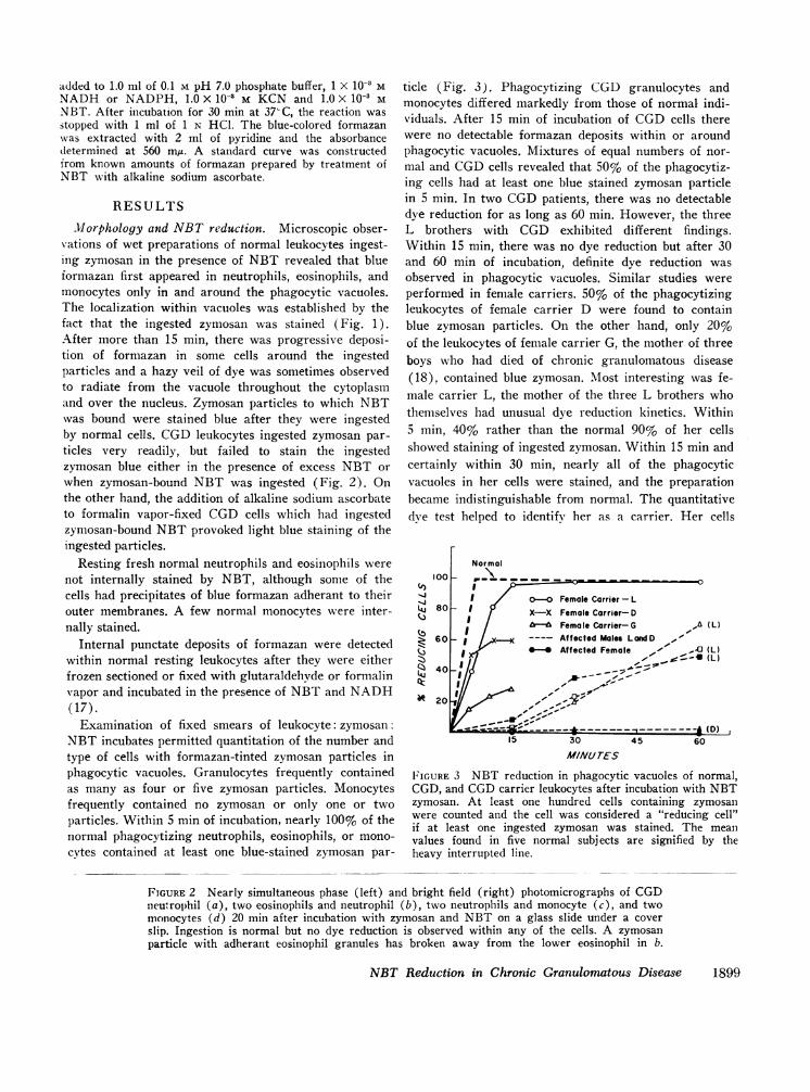
ticle (Fig. 3). Phagocytizing CGD granulocytes andmonocytes differed markedly from those of normal indi-viduals. After 15 min of incubation of CGDcells therewere no detectable formazan deposits within or aroundphagocytic vacuoles. Mixtures of equal numbers of nor-mal and CGDcells revealed that 50% of the phagocytiz-ing cells had at least one blue stained zymosan particlein 5 min. In two CGDpatients, there was no detectabledye reduction for as long as 60 min. However, the threeL brothers with CGD exhibited different findings.Within 15 min, there was no dye reduction but after 30and 60 min of incubation, definite dye reduction wasobserved in phagocytic vacuoles. Similar studies wereperformed in female carriers. 50% of the phagocytizingleukocytes of female carrier D were found to containblue zymosan particles. On the other hand, only 20%of the leukocytes of female carrier G, the mother of threeboys who had died of chronic granulomatous disease(18), contained blue zymosan. Most interesting was fe-male carrier L, the mother of the three L brothers whothemselves had unusual dye reduction kinetics. Within5 min, 40% rather than the normal 90% of her cellsshowed staining of ingested zymosan. Within 15 min andcertainly within 30 min, nearly all of the phagocyticvacuoles in her cells were stained, and the preparationbecame indistinguishable from normal. The quantitativedye test helped to identify her as a carrier. Her cells
1001
Li 80
o 60
' 40
0 20
Normal
o-o Female Carrier - LX-X Female Carrier- DXXFemale Carrier-DGfr-s Female Carrier-G ,A IL)
Affected Males LandD ,'Affected Female A,(' _ IL)
-.-p iii li}Dl45 60
MINUTES
FIGURE 3 NBT reduction in phagocytic vacuoles of normal,CGD, and CGDcarrier leukocytes after incubation with NBTzymosan. At least one hundred cells containing zymosanwere counted and the cell was considered a "reducing cell"if at least one ingested zymosan was stained. The meanvalues found in five normal subj ects are signified by theheavy interrupted line.
FIGURE 2 Nearly simultaneous phase (left) and bright field (right) photomicrographs of CGDneutrophil (a), two eosinophils and neutrophil (b), two neutrophils and monocyte (c), and twomonocytes (d) 20 min after incubation with zymosan and NBT on a glass slide under a coverslip. Ingestion is normal but no dye reduction is observed within any of the cells. A zymosanparticle with adherant eosinophil granules has broken away from the lower eosinophil in b.
NBT Reduction in Chronic Granulomatous Disease 1899

reduced 41 ,ug NBT/1.25 X 107 cells per 15 min, thenormal range being 52-88 Ag NBT reduced.
"NBT reductase" activity in cell sonicates. Despitethe fact that dye reduction during particle ingestion wasimpaired in CGDand in CGDfemale carrier cells, totalcyanide insensitive 'NBT reductase" activity with eitherNADHor NADPHas precursor was normal in CGDand carrier cell sonicates (Table I).
Vacutole formation and degranulation. Araldite sec-tions stained with toluidine blue revealed that the phago-cytic vacuoles in CGDtended to be considerably smallerthan the vacuoles present in normal leukocytes (Fig. 4).This observation, previously made by Quie, White,Holmes, and Good (9) and by Andersen, Koch, Vejls-gaard, and Wilken-Jenses (19) was a variable phe-nomenon from cell to cell and also depended to a largeextent upon the plane of section. But, in general, thelarge phagocytic vacuoles which often completely dis-torted normal leukocytes were not observed in the CGDcells. Electron micrographs of CGD and normal cellsfailed to demonstrate any obvious differences in granule
distribution in and around the phagocytic vacuoles.Actually, this technique revealed so much variation inthe extent of "degranulation" among different normalphagocytizing cells, after incubation times which variedfrom 15 min to 2 hr, that a quantitative morphologic dif-ference between normal and CGDcells would be nearlyimpossible to define.
Rates of release of alkaline phosphatase and peroxi-dase. Figs. 5 and 6 provide data on the rate of releaseof stainable alkaline phosphatase and peroxidase activi-ties from normal cells and patients' cells by the scoringmethod described above. In only one CGDpatient wasthere apparent delay in the release of alkaline phospha-tase. The remaining curves fell within the range ob-served in five normal subjects. Peroxidase activity wasreleased from CGDcells but the rate of release appearedto be somewhat slower than that observed in normals.On the other hand, it must be emphasized that this scor-ing technique, especially with regard to peroxidaseactivity, was subject to misinterpretation. Inspection ofresting CGD leukocytes stained for peroxidase activity
A v- w* r
f~e 'Sfi'' is@ZW ^t~ / *Ab
V
a Pt
FIGURE 4 Thick Araldite sections of glutaraldehyde: osmium fixed normal (a) and CGD(b) leukocytes after 15 min incubation with zymosan. Note large distorting phagocytic vacuolesin normal cells and smaller vacuoles in CGDcells.
1900 D. G. Nathan, R. L. Baehner, and D. K. Weaver
.....IX
*
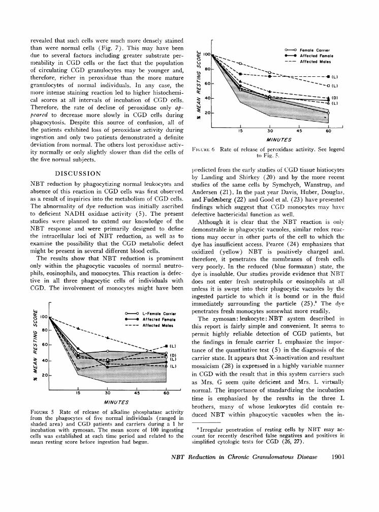
revealed that such cells were much more densely stainedthan were normal cells (Fig. 7). This may have beendue to several factors including greater substrate per-meability in CGDcells or the fact that the populationof circulating CGDgranulocytes may be younger and,therefore, richer in peroxidase than the more maturegranulocytes of normal individuals. In any case, themore intense staining reaction led to higher histochemi-cal scores at all intervals of incubation of CGD cells.Therefore, the rate of decline of peroxidase only ap-peared to decrease more slowly in CGD cells duringphagocytosis. Despite this source of confusion, all ofthe patients exhibited loss of peroxidase activity duringingestion and only two patients demonstrated a definitedeviation from normal. The others lost peroxidase activ-ity normally or only slightly slower than did the cells ofthe five normal subjects.
100
(i) 80
,1 60
40
20
at
F4-
)k
°-O Female Carrier*-* Affected Female
-- Affected Moles
- _ _ _ _ _ -____x- (L)
I_ O (L)
fi(Dl~--Z(L)
1e
15 30
M/AJurES
45 60
Fi(URE 6 Rate of release of peroxidase activity. See legendto Fig. 5.
predicted from the early studies of CGDtissue histiocytesby Landing and Sbirkey (20) and by the more recent
reduction by phagocytizing normal leukocytes and studies of the same cells by Symchych, Wanstrup, ande of this reaction in CGDcells was first observed Andersen (21). In the past year Davis, Huber, Douglas,sult of inquiries into the metabolism of CGDcells. and Fudonberg (22) and Good et al. (23) have presentedbnormality of dye reduction was initially ascribed findings which suggest that CGDmonocytes mav haveIcient NADHoxidase activity (5). The present defective bactericidal function as well.
were planned to extend our knowledge of the Although it is clear that the NBT reaction is onlyresponse and were primarily designed to define demonstrable in phagocytic vacuoles, similar redox reac-tracellular loci of NBT reduction, as well as to tions may occur in other parts of the cell to which thee the possibility that the CGDmetabolic defect dye has insufficient access. Pearce (24) emphasizes that
be present in several different blood cells. oxidized (yellow) NBT is positively charged and.results show that NBT reduction is prominent therefore, it penetrates the membranes of fresh cells
within the phagocytic vacuoles of normal neutro- very poorly. In the reduced (blue formazan) state, theeosinophils, and monocytes. This reaction is defec- dye is insoluble. Our studies provide evidence that NBTl all three phagocytic cells of individuals with does not enter fresh neutrophils or eosinophils at allThe involvement of monocytes might have been unless it is swept into their phagocytic vacuoles by the
ingested particle to which it is bound or in the fluidimmediately surrounding the particle (25).' The dye
0-0 L-Female Carrier penetrates fresh monocytes somewhat more readily.-4 Affected Female The zymosan: leukocyte: NBT system described in
\~ --- Affected Males this report is fairly simple and convenient. It seems topermit highly reliable detection of CGD patients, but
W\~~-_the findings in female carrier L emphasize the impor-tance of the quantitative test (5) in the diagnosis of the
(D ):. (L) carrier state. It appears that X-inactivation and resultantImL) mosaicism (28) is expressed in a highly variable manner
gin CGDwith the result that in this system carriers such.as Mrs. G seem quite deficient and Mrs. Lvirtuall
15 30 45 60 normal. The importance of standardizing the incubation
M/NU7(5 ~~time is emphasized by the results in the three Lbrothers, many of whose leukoeytes did contain re-
; Rate of release of alkaline phosphatase activity duced NBT within phagocytic vacuoles whenthein-he phagocytes of five normal individuals (trangedin
shaded area) and CUDlJ patients and carriers during a 1 hrincubation with zymosan. The mean score of 100 ingestingcells was established at each time period and related to themean resting score before ingestion had begun.
'Irregular penetration of resting cells by NBT may ac-
count for recently described false negatives and positives in
simplified cytologic tests for CGD (26, 27).
NBT Reduction in Chronic Granulomatous Disease
NBT i
absenc4as a reThe alto defistudiesNBTthe intexamirmight
Theonly Mphils, etive inCGD.
FIGUREfrom ti
100.J(r).3 80
;.t(f 60LuQZ;. 40IR
20iR
1901
cubation was prolonged. (The clinical state of thesebrothers is not particulary benign nor do their cellscontain more NADHoxidase activity than the cells ofmale patient D.)
The failure of CGDcell phagocytic vacuoles to sup-port NBT reduction is a particularly challenging prob-lem because the three components which provoke dyereduction are present in CGDcells. CGDcell sonicatescontain normal amounts of "NBT reductase," an en-zyme system(s) capable of catalyzing the reduction ofNBT in the presence of pyridine nucleotide. NBT en-ters the phagocytic vacuole of the CGDcell bound to orclosely associated with the ingested zymosan, and thecell is capable of providing reduced pyridine nucleotidethrough glycolysis (5, 11, 29). A possible explanationfor the absence of dye reduction might be that "NBTreductase" activity, though present in the whole cell, is
deficient within the phagocytic vacuole of the CGDcell.Deficiency of enzyme activity within the phagocyticvacuole might be due to an error of degranulation. Infact, the pathogenesis of CGDhas been ascribed to de-fective degranulation by Quie and his coworkers (9).This theory is based in part upon White's observations(9) of diminished vacuole size in CGDcells which weand other investigators (19) have confirmed. The con-cept is not supported, however, by the histochemicalstudies reported here or by recently described histochem-ical analyses of Kauder, Kahle, Moreno, and Partin(30) and of Mandel and Hook (31). Admittedly, his-tochemical studies or routine electron micrographicanalyses of degranulation provide very rough estimates,and the former are plagued by artifacts such as the dif-ferences in the staining characteristics of peroxidaseactivity in CGD cells described above. In this regard,
0..:: .:. .: :4e.x .... : .* : : ...
.: .... .:* :: :: ... . : : . . .::...000:X::..:. :.t- * .....
;hIS0 850"t00 s-S i_ _w1!_. _ . .w.: S
FIGURE 7 Peroxidase stain of resting normal (a) and CGD (b) neutrophils. Notethe more intense stain in the CGDcells.
1902 D. G. Nathan, R. L. Baehner, and D. K. Weaver

TABLE INADH-NBT Reductase of Sonicates of lI hole Cells*
Subject NBT reduced
yg/30 min per mg sonicate
Male-CGI) 58.2Male-CGD 66.6Male CGD 60.0
Female carrier 67.6
Control 78.0Control 57.6Control 62.0
* The values found when NADPHwas utilized in place ofNADHwere essentially identical.
the studies of Baehner, Karnovsky, and Karnovsky (32)are of particular importance since these workers utilizedobjective measurements of enzyme release and providedevidence that CGDcells degranulate perfectly normally.
Of course, it is entirely possible that only a smallfraction of total leukocyte "NBT reductase" activitycontributes to NBT reduction in phagocytic vacuolesand this small but critical fraction could be deficient inCGD. NADHoxidase activity, a system which can ac-count for most of the oxygen consumption and some ofthe bactericidal activity of normal human leukocytes, isdeficient in CGD cells (3). As previously suggested(5) this system might also qualify as the critical miss-ing factor which results in the failure of NBT reduc-tion in CGD phagocytic vacuoles. Evaluation of thistheory awaits further clarification of the intricacies ofthe respiratory response to phagocytosis, studies inwhich the CGDcell will play a vital role.
ACKNOWLEDGMENTSThe authors are grateful to their colleagues who helped tomake this work possible. Dr. Jean-Paul Revel and Dr.Elizabeth Hay of the Department of Anatomy and Dr. Mor-ris Karnovsky of the Department of Pathology, HarvardMedical School provided laboratory space, instructions inelectron micrographic techniques, and stimulating discussions.Dr. Arnold Weinberg, Department of Medicine, Massachu-setts General Hospital and Harvard Medical School, madeavailable the clinical studies and the blood of female carrierG. Dr. John M. Craig, Department of Pathology, BostonHospital for Women and Harvard Medical School, madepossible the studies of the NBT reaction in frozen sectionsof leukocytes. Finally, Dr. Manfred L. Karnovsky of theDepartment of Biological Chemistry, Harvard MedicalSchool, gave freely of his advice, laboratory methods, andinvaluable criticism.
This work was supported by U. S. Public Health ServiceGrants Al 08173, GM00451, and T1 AM 05581 and by agrant from the John A. Hartford Foundation. Dr. Nathan isthe recipient of U. S. Public Health Service Research CareerDevelopment Award 5 K03-AM35371. Some of these clinicalstudies were performed in a clinical research center at the
Children's Hospital Medical Center supported by U. S.Public Health Service Grant FR 00128.
REFERENCES1. Karnovsky, M. L. 1962. Metabolic basis of phagocytic
activity. Physiol. Rev. 42: 143.2. Cagan, R. H., and M. L. Karnovsky. 1964. Enzymatic
basis of the respiratory stimulation during phagocytosis.Nature (London). 204: 255.
3. Baehner, R. L., and M. L. Karnovsky. 1968. Deficiencyof reduced nicotinamide-adenine dinucleotide oxidase inchronic granulomatous disease. Science (Washington).162: 1277.
4. Baehner, R. L., and D. G. Nathan. 1966. Deficient glu-cose oxidation in intact leukocytes of chronic granulo-matous disease. Blood. 28: 1010.
5. Baehner, R. L., and D. G. Nathan. 1967. Leukocyte oxi-dase: defective activity in chronic granulomatous disease.Science (Washington). 155: 835.
6. Windhorst, D. B., B. Holmes, and R. A. Good. 1967.A newly defined X-linked trait in man with demonstra-tion of the Lyon effect in carrier females. Lancet. 1: 737.
7. Baehner, R. L., and D. G. Nathan. 1968. Quantitativenitroblue tetrazolium test in chronic granulomatous dis-ease. N. Engl. J. Med. 278: 971.
8. Holmes, B., P. G. Quie, D. B. Windhorst, and R. A.Good. 1966. Fatal granulomatous disease of childhood.An inborn abnormality of phagocytic function. Lancet.1:1225.
9. Quie, P. G., J. G. White, B. Holmes, and R. A. Good.1967. In vitro bactericidal capacity of human polymorpho-nuclear leukocytes: diminished activity in chronic granu-lomatous disease of childhood. J. CGn. Invest. 46: 668.
10. MacFarlane, P. S., A. L. Speirs, and R. G. Sommerville.1967. Fatal granulomatous disease of childhood and be-nign lymphocytic infiltration of the skin (congenitaldysphagocytosis). Lancet. 1: 408.
11. Holmes, B., A. R. Page, and R. A. Good. 1967. Studiesof metabolic activity of leukocytes from patients witha genetic abnormality of phagocytic function. J. Clin.Invest. 46: 1422.
12. Hirsch, J. G. 1962. Cinemicrophotographic observationson granule lysis in polymorphonuclear leukocytes duringphagocytosis. J. Exp. Med. 116: 827.
13. Cohn, Z. A., and E. Wiener. 1963. The particulate hy-drolases of macrophages. II. Biochemical and morphologi-cal response to particle ingestion. J. Exp. Med. 118: 1009.
14. Zucker-Franklin, D., and J. G. Hirsch. 1964. Electronmicroscope studies on the degranulation of rabbit peri-toneal leukocytes during phagocytosis. J. Exp. Med. 120:569.
15. Kaplow, L. S. 1965. Simplified myeloperoxidase stainusing benezidine dihydrochloride. Blood. 26: 215.
16. Kaplow, L. S. 1963. Cytochemistry of leukocyte alka-line phosphatase: use of complex napthol as phosphatesin AZO dye coupling technics. Amer. J. Clin. Pathol.39: 439.
17. Rozenszajn, L., and D. Shoham. 1967. The demonstrationof dehydrogenases and diaphorases in cells of peripheralblood and bone marrow. Blood. 29: 737.
18. Janeway, C. A., J. Craig, M. Davidson, W. Downey,D. Gitlin, and J. C. Sullivan. 1954. Hypergammaglobu-linemia associated with severe recurrent and chronicnonspecific infection. Amer. J. Dis. Child. 88: 388.
NBT Reduction in Chronic Granulomatous Disease 1903

1'). Andersen, V., C. Koch, R. Vejlsgaard, and K. Wilkein-Jenses. 1968. Fatal granulomatous disease. Acta Paediat.Scand. 57: 110.
20. Landing, B. H., and H. S. Shirkey. 1957. Syndrome ofrecurrent infection and infiltration of viscera by pig-mented lipid histiocytes. Pediatrics. 20: 431.
21. Symchych, P. S., J. Wanstrup, and V. Andersen. 1968.Chronic granulomatous disease of childhood. A morpho-logic study. Acta Pathol. Microbiol. 74: 179.
22. Davis, W. C., H. Huber, S. D. Douglas, and H. H.Fudenberg. 1968. A defect in circulating mononuclearphagocytes in chronic granulomatous disease of child-hood. J. Immunol. 101: 1093.
23. Good, R. A., P. G. Quie, D. B. Windhorst, A. R. Page,G. E. Rodey, J. White, J. J. Wolfson, and B. H. Holmes.1968. Fatal (chronic) granulomatous disease of child-hood: a hereditary defect of leukocyte function. SeminarsHematol. 5: 215.
24. Pearse, A. G. E. 1960. Histo-hemistry, Theoretical andApplied. Little, Brown & Co., Boston. 2nd edition. 538.
25. Berger, R. R., and M. L. Karnovsky. 1966. Biochemicalbasis of phagocytosis. V. Effect of phagocytosis on cel-lular uptake of extracellular fluid, and on the intracellularpool of L-a-glycerophosphate. Fed. Proc. 25: 840.
26. Park, B. H., S. M. Fikrig, and E. M. Smithwick. 1968.Infection and nitroblue-tetrazolium reduction by neutro-phils. Lancet. 7: 532.
27. Park, B. H., B. M. Holmes, G. E. Rodey, and R. A.Good. 1969. Nitroblue-tetrazolium test in children withfatal granulomatous disease and newborn infants. Lancet.1:157.
28. Lyon, M. F. 1962. Sex chromatin and gene action in themammalian X-chromosome. Amer. J. Hum. Genet. 14:135.
29. Holmes, B., and A. R. Page. 1966. Studies on the meta-bolic activity of leukocytes from patients with a geneticabnormality of phagocytic function. J. Cell. Biol. 31: 48a.(Abstr.)
30. Kauder, E., L. L. Kahle, H. Moreno, and J. C. Partin.1968. Leukocyte degranulation and vacuole formation inpatients with chronic granulomatous disease of child-hood. J. Clin. Invest. 47: 1753.
31. Mandell, G., and E. Hook. 1968. Studies of the leuko-cyte defect in chronic granulomatous disease of child-hood. Clin. Res. 16: 332. (Abstr.)
32. Baehner, R. L., M. J. Karnovsky, and M. L. Karnovsky.1969. Degranulation of leukocytes in chronic granulo-matous disease. J. Clin. Invest. 48: 187.
1904 D. G. Nathan, R. L. Baehner, and D. K. Weaver


![Evaluation of a Tetrazolium-based Semiautomated ... · (CANCER RESEARCH 47, 936-942, February 15, 1987] Evaluation of a Tetrazolium-based Semiautomated Colorimetrie Assay: Assessment](https://static.fdocuments.net/doc/165x107/5f5765572ff1b503ec225aa6/evaluation-of-a-tetrazolium-based-semiautomated-cancer-research-47-936-942.jpg)