Single Cut Distal Femoral Varus Osteotomy )SCFO(: A...
6
)322( COPYRIGHT 2017 © BY THE ARCHIVES OF BONE AND JOINT SURGERY Arch Bone Jt Surg. 2017; 5(5): 322-327. http://abjs.mums.ac.ir the online version of this article abjs.mums.ac.ir Reza Abdi, MD; Ramin Hajzargarbashi, MD; Mohammad H. Ebrahimzadeh, MD Research performed at Pediatrics Center of Excellence, Tehran University of Medical Sciences, Tehran, Iran Corresponding Author: Ramin Hajzargarbashi, Pediatrics Center of Excellence, Tehran University of Medical Sciences, Tehran, Iran Email: [email protected] RESEARCH ARTICLE Received: 19 November 2016 Accepted: 01 July 2017 Single Cut Distal Femoral Varus Osteotomy )SCFO(: A Preliminary Study Abstract Background: Genu valgum usually originates from a deformity of distal femur that is often corrected by distal femoral varus osteotomy. The osteotomy includes both components of angulationcorrection and translation because the site of osteotomy is not commonly at the apex of deformity. Improvement of patellar tracking not only depends on valgus correction, but also it may be partially due to centralization of the trochlear groove under the femoral anatomical axis (FAA). We asked whether we could accurately correct the deformities based on our preoperative goals for the correction of the mechanical axis and centralization of the trochlear groove under the FAA by using a single bone cut. This study describes a new lateral single cut distal femoral osteotomy (SCFO) that enables concurrent correction of angulation and translation. Methods: This study was done on 12 young adult patients with femoral juxta-articular genu valgum deformity using SCFO. The average age at operation was 21 years (range: 16-25). SCFO is a type of closing-opening distal femoral osteotomy that corrects the valgus deformity of the distal femur while the translation of the distal fragment is done using one oblique cut. It centralizes the trochlear groove under the FAA. We compared the pre and postoperative radiographic and clinical variables including mechanical tibiofemoral angle, knee range of motion (ROM), International knee documentation committee (IKDC) score and the time to union. Mean follow-up of the patients was 24 months. Results: The average mechanical tibiofemoral angle improved from 16 degrees (10-23) to 1 degrees (-2 to +2). IKDC subjective score slightly improved from preoperative (65) to 2-year follow-up (71). Centralization of the trochlea was achieved in all patients. Conclusion: SCFO can be a reasonable alternative for correction of the distal femur genu valgum deformity. It can centralize the patellar groove under the FAA with satisfactory clinical outcomes. Keywords: Femoral osteotomy, Genu valgum, SCFO, Single cut, Valgus Introduction V algus knee is a condition that may originate from the distal femur, proximal tibia, or the knee joint (1-4). Distal femoral varus realignment osteotomy is mostly the treatment of choice to correct the valgus deformity, which can eliminate pain and other functional and cosmetic problems up to 83% (4, 5). Generally, the results of various method of distal femoral osteotomy are satisfactory (3, 6-10). Paley described the normal patellar-tracking that depends on the angulation and alignment of extensor mechanism. Accordingly, the most accurate correction of the angulation and alignment can be achieved through a metaphyseal osteotomy for femoral juxta-articular deformity. This osteotomy must include both angulation
Transcript of Single Cut Distal Femoral Varus Osteotomy )SCFO(: A...

)322( COPYRIGHT 2017 © BY THE ARCHIVES OF BONE AND JOINT SURGERY
Arch Bone Jt Surg. 2017; 5(5): 322-327. http://abjs.mums.ac.ir
the online version of this article abjs.mums.ac.ir
Reza Abdi, MD; Ramin Hajzargarbashi, MD; Mohammad H. Ebrahimzadeh, MD
Research performed at Pediatrics Center of Excellence, Tehran University of Medical Sciences, Tehran, Iran
Corresponding Author: Ramin Hajzargarbashi, Pediatrics Center of Excellence, Tehran University of Medical Sciences, Tehran, IranEmail: [email protected]
RESEARCH ARTICLE
Received: 19 November 2016 Accepted: 01 July 2017
Single Cut Distal Femoral Varus Osteotomy )SCFO(: A Preliminary Study
Abstract
Background: Genu valgum usually originates from a deformity of distal femur that is often corrected by distal femoral varus osteotomy. The osteotomy includes both components of angulationcorrection and translation because the site of osteotomy is not commonly at the apex of deformity. Improvement of patellar tracking not only depends on valgus correction, but also it may be partially due to centralization of the trochlear groove under the femoral anatomical axis (FAA). We asked whether we could accurately correct the deformities based on our preoperative goals for the correction of the mechanical axis and centralization of the trochlear groove under the FAA by using a single bone cut.This study describes a new lateral single cut distal femoral osteotomy (SCFO) that enables concurrent correction of angulation and translation. Methods: This study was done on 12 young adult patients with femoral juxta-articular genu valgum deformity using SCFO. The average age at operation was 21 years (range: 16-25). SCFO is a type of closing-opening distal femoral osteotomy that corrects the valgus deformity of the distal femur while the translation of the distal fragment is done using one oblique cut. It centralizes the trochlear groove under the FAA. We compared the pre and postoperative radiographic and clinical variables including mechanical tibiofemoral angle, knee range of motion (ROM), International knee documentation committee (IKDC) score and the time to union. Mean follow-up of the patients was 24 months.
Results: The average mechanical tibiofemoral angle improved from 16 degrees (10-23) to 1 degrees (-2 to +2). IKDC subjective score slightly improved from preoperative (65) to 2-year follow-up (71). Centralization of the trochlea was achieved in all patients. Conclusion: SCFO can be a reasonable alternative for correction of the distal femur genu valgum deformity. It can centralize the patellar groove under the FAA with satisfactory clinical outcomes.
Keywords: Femoral osteotomy, Genu valgum, SCFO, Single cut, Valgus
Introduction
Valgus knee is a condition that may originate from the distal femur, proximal tibia, or the knee joint (1-4). Distal femoral varus realignment osteotomy
is mostly the treatment of choice to correct the valgus deformity, which can eliminate pain and other functional and cosmetic problems up to 83% (4, 5). Generally, the results of various method of distal femoral osteotomy are
satisfactory (3, 6-10). Paley described the normal patellar-tracking that
depends on the angulation and alignment of extensor mechanism. Accordingly, the most accurate correction of the angulation and alignment can be achieved through a metaphyseal osteotomy for femoral juxta-articular deformity. This osteotomy must include both angulation

SINGLE CUT DISTAL FEMORAL VARUS OSTEOTOMY (SCFO)THE ARCHIVES OF BONE AND JOINT SURGERY. ABJS.MUMS.AC.IRVOLUME 5. NUMBER 5. SEPTEMBER 2017
)323(
and translation components because the site of osteotomy is hardly at the apex of the deformity (8).
Wang reported that the distal femoral closed wedge osteotomy can improve patellar-tracking and congruence angle when it was fixed by a medial blade plate (11). He believed that in medial closed wedge osteotomy and medial blade plate fixation displaces the diaphysis toward the blade plate, which can centralize the trochlear groove under the femoral anatomical axis (FAA) and improves patellar-tracking.
In the lateral open wedge osteotomy, the position of trochlear groove has not been widely studied, however it seems that centralization of the trochlear groove under FAA would not occur because of leaving the medial cortex intact leading to the absence of translation [Figure 1].
The purpose of this study was to assess the results of a modified distal femoral varus osteotomy through the lateral approach that corrects valgus and centralizes the trochlear groove under the FAA simultaneously by correction of angulation and translation of the femoral
condyles.
Materials and MethodsThe study is prospectively conducted at a tertiary care
referral, teaching hospital between 2013 and 2014 and is registered in IRCT council (Iranian Registry of Clinical Trial, code number: Ir.bums.1395.62). We included 12 young adults presenting with a genu valgum deformity with a mean mechanical tibiofemoral angle of 16 degrees (range: 10-23 degree), mean mechanical lateral distal femoral angle of 73 degrees (range: 67-78 degrees), and an intermalleolar distance of more than 10 cm (4). The average age at operation was 21 years (range: 16-25 years).
The correction of mechanical tibiofemoral angle was planned according to the method described by Paley and Dugdale (12, 13). The weight-bearing line was placed at a selected position 48% to 50% across the width of the tibial plateau from medial to lateral (12). Clinical experience has shown that overcorrection to varus is contraindicated. Patients who had severe collateral ligament instability, unstable knee, sagittal plane deformity (fixed flexion deformity >20 degree or genu recurvatum), osteoarthritis, osteomalacia, multiple epiphyseal dysplasia, or renal disease were excluded from the study (4).
The mechanical axis of the lower limb was defined as a line drawn from the center of the femoral head to the center of the ankle. The distance between the mechanical axis and the center of the knee in the frontal plane was calculated as the mechanical axis deviation (MAD). The malalignment test (MAT) was used to assess the severity and location of the deformity. According to this method, all of the patients had femoral juxta-articular valgus deformity.
The correction angle between mechanical axis of femur and tibia was measured on the standing anteroposterior three joint radiograph of the lower extremity. The mechanical axes of all patients passed trough the zone II or III according to the method described by Stevens who divided the knee joint into four radiographic quadrants [Figure 2] (14).
Standing radiographs in both anteroposterior and lateral views were taken in the early post-operative period and at 4-week intervals. The patients were evaluated clinically and radiologically for the alignment, union, and knee range of motion at each subsequent visits. Patients were followed up for 24 months. At the last visit, International knee documentation committee (IKDC) subjective score was used to assess the functional outcome.
Surgical techniqueThe surgery was performed under general anesthesia
in a supine position under tourniquet control. The knee was flexed 45 degrees during the surgery to avoid pressure on the popliteal vessels. Through the lateral distal femoral approach, lateral femoral condyle and diaphysis were exposed, and then under the guidance of fluoroscopy, a guide wire was advanced in an oblique direction from point A (just proximal to the lateral
Figure1. Lateral open wedge osteotomy medialized the trochlear groove in relation to the femoral anatomical axis especially when it is done proximal to the CORA (center of rotation of angulation) and/or femoral anatomical plate is used.
SINGLE CUT DISTAL FEMORAL VARUS OSTEOTOMY (SCFO)THE ARCHIVES OF BONE AND JOINT SURGERY. ABJS.MUMS.AC.IRVOLUME 5. NUMBER 5. SEPTEMBER 2017
)324(
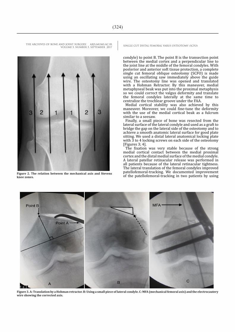
condyle) to point B. The point B is the transection point between the medial cortex and a perpendicular line to the joint line at the middle of the femoral condyles. With posterior and anterior soft tissue protection, a complete single cut femoral oblique osteotomy (SCFO) is made using an oscillating saw immediately above the guide wire. The osteotomy line was opened and translated with a Hohman Retractor. By this maneuver, medial metaphyseal beak was put into the proximal metaphysis so we could correct the valgus deformity and translate the femoral condyles laterally at the same time to centralize the trochlear groove under the FAA.
Medial cortical stability was also achieved by this maneuver. Moreover, we could fine-tune the deformity with the use of the medial cortical beak as a fulcrum similar to a seesaw.
Finally, a small piece of bone was resected from the lateral surface of the lateral condyle and used as a graft to bridge the gap on the lateral side of the osteotomy and to achieve a smooth anatomic lateral surface for good plate sitting. We used a distal lateral anatomical locking plate with 3 to 4 locking screws on each side of the osteotomy [Figures 3; 4].
The fixation was very stable because of the strong medial cortical contact between the medial proximal cortex and the distal medial surface of the medial condyle. A lateral patellar retinacular release was performed in all patients because of the lateral retinacular tightness. The lateral translation of the femoral condyles improved patellofemoral-tracking. We documented improvement of the patellofemoral-tracking in two patients by using Figure 2. The relation between the mechanical axis and Stevens
knee zones.
Figure 3. A: Translation by a Hohman retractor. B: Using a small piece of lateral condyle. C: MFA (mechanical femoral axis) and the electrocautery wire showing the corrected axis.

SINGLE CUT DISTAL FEMORAL VARUS OSTEOTOMY (SCFO)THE ARCHIVES OF BONE AND JOINT SURGERY. ABJS.MUMS.AC.IRVOLUME 5. NUMBER 5. SEPTEMBER 2017
)325(
knee arthroscopy before and after osteotomy, and before the lateral patellar retinacular release. Lateral translation of the femoral condyles is mandatory in varus osteotomy through the medial femoral approach but characteristic specificity of a single cut femoral oblique osteotomy (SCFO) is simplicity of osteotomy, stable fixation by anatomical plate and capability of lateral translation of condyles through a lateral femoral approach.
After closing the incision, the patient’s knee was put in a hinged knee brace. Patients were instructed to bear no weight for 2 weeks, followed by partial weight bearing with 2 crutches as tolerated. Passive and active assisted range of motion of the knee was permitted one week after surgery. Patients were allowed full weight bearing 8 weeks after surgery.
Results Twelve limbs were operated in 12 patients consisted
of 8 females and 4 males. The majority of the patients (9 out of 12) mentioned the cosmetic deformity of the knee as the presenting complaint and 3 patients were
Figure 4. Mechanical axis after union of the osteotomy in the same patient.
complaining of lateral knee pain. In 11 patients, the deformity was considered primary while one patient had an old femoral valgus malunion. The average amount of blood loss was 60 ml during surgery (range, 40 ml - 150 ml). The mean duration of hospital stay was 2 days (range: 2- 4 days). The mean follow-up was 24 months. All osteotomies healed at an average of 12 weeks (range: 10-15 weeks). There was no loss of fixation, loss of initial correction, infection, loss of knee range of motion, and any knee pain at the last visit in all patients. The mean mechanical tibiofemoral angle of the lower limb was 15.5 degrees (range: 10-23 degree) before SCFO that improved to 1 degree (range: -2 to +2 degrees). All mechanical axes passed through zone 1 at the last follow-up radiograph.
All patients had slight improvement according to the IKDC subjective score from preoperative (65) to 2-year follow-up (71). The average femoral shortening after SCFO was 5 millimeter (range: -3 to 7) on AP standing radiograph of the lower limbs at the last follow-up. All of the knees regained a normal ROM after the surgery.
DiscussionVarus distal femoral osteotomy for the correction of
genu valgum and prevention of DJD in patients under the age of 60 years is widely supported in the literature (3). Although femoral supracondylar varus osteotomy does not preclude total knee arthroplasty but makes it technically easier (1, 6, 9, 15).
Various methods of osteotomy and fixation devices was used for varus distal femoral osteotomy including 1) a lateral opening wedge osteotomy and fixation with a 95-degree blade plate, DCS, or a Puddu plate, 2) a medial closed wedge osteotomy and leaving a lateral hinge intact with removing a medially based wedge and fixation by a 95-degree offset dynamic compression blade plate or fixation by a TomoFix plate, 3) a medial closed wedge osteotomy by reciprocating ledge technique, Wedgeless ‘V’ shaped osteotomy, percutaneous distal femoral curved (dome) osteotomy and fixation by a monolateral Hex-Fix external fixator, biplane derotation osteotomy and etc (3, 4, 6, 7, 10, 12, 16-24).
Numerous techniques for osteotomy are used with a medial or lateral hinge for the correction of angular deformity of the distal fragment (with or without hinge) that can displace the trochlear groove medially or laterally in regards to the FAA that can change the patellar congruence angle and tracking. In reviewing the literatures, there is little about the relation between the type of distal femoral osteotomy and patellar-tracking (11).
In vivo studies showed that the least amount of motion with the highest stiffness was achieved in a medial oblique closing-wedge osteotomy fixed by an angled blade plate. The lateral open wedge techniques were less stable and had a lower stiffness compared to the medial one (15). Furthermore, the oblique cut was more stable and had a higher stiffness than the perpendicular cut (15).
We believe that if the translation and centralization of the trochlear groove is not achieved during surgery, the

SINGLE CUT DISTAL FEMORAL VARUS OSTEOTOMY (SCFO)THE ARCHIVES OF BONE AND JOINT SURGERY. ABJS.MUMS.AC.IRVOLUME 5. NUMBER 5. SEPTEMBER 2017
)326(
Reza Abdi MDBirjand University of Medical Sciences, Birjand, Iran
Ramin Hajzargarbashi MDPediatrics Center of Excellence, Tehran University of Medical Sciences,Tehran, Iran.
Mohammad H. Ebrahimzadeh MDOrthopedic Research Center, Department of Orthopedic Surgery, Qhaem Hospital, Mashad University of Medical Sciences, Mashad, Iran
results would be inferior especially after correction of a severe valgus knee. For instance in one of our patients after a lateral open wedge osteotomy and fixation by a lateral blade plate, we found out that the mechanical axis was in varus position and the patella was dislocated out of the groove. However, after the revision surgery two days later with translation of the distal fragment using the SCFO technique, the patella was relocated with no need for subsequent patellar realignment procedure [Figure 5].
In this study all mechanical axes passed through zone 1 at the last follow-up in AP standing X-ray of lower limbs. Because the patients were young adults with no significant functional problem, IKDC subjective score did not change significantly after the procedure.
The beneficial aspects of SCFO can be summarized as below.
1. It is a single cut osteotomy, which is easier than a double cut closed wedge medial osteotomy.
2. Lateral translation of the trochlear groove occurs but medial stability is preserved. This translation can centralize the trochlear groove under the FAA.
3. Fine-tuned correction of the deformity can be done after osteotomy by seesaw movement of the distal fragment spike beneath the medullary canal edges.
4. Fixation is straightforward by using a lateral anatomic plate.
5. Iliac bone grafting is not necessary.6. Femoral shortening is minimal.
SCFO is a reasonable alternative for the correction of the femoral juxta-articular genu valgum deformity. It seems to improve the patellar-tracking by centralization of the patellar groove under the FAA; however, future studies are necessary to confirm this finding. We did not assess the amount of improvement in patellar-tracking, which is a point for the future studies.
Figure 5. A: Patellar dislocation after excessive varus correction with a lateral open wedge osteotomy.B: centralization of femoral condyles by revision SCFO and using the same blade plate.C: Patellar relocation.

SINGLE CUT DISTAL FEMORAL VARUS OSTEOTOMY (SCFO)THE ARCHIVES OF BONE AND JOINT SURGERY. ABJS.MUMS.AC.IRVOLUME 5. NUMBER 5. SEPTEMBER 2017
)327(
References
1. Saithna A, Kundra R, Modi CS, Getgood A, Spalding T. Distal femoral varus osteotomy for lateral compartment osteoarthritis in the valgus knee. A systematic review of the literature. Open Orthop J. 2012; 6(2):313-9.
2. Omidi-Kashani F, Hasankhani IG, Mazlumi M, Ebrahimzadeh MH. Varus distal femoral osteotomy in young adults with valgus knee. J Orthop Surg Res. 2009; (4):15.
3. Paccola CA. Pre-operative planning and surgical technique of the open wedge supracondylar osteotomy for correction of valgus knee and fixation with a fixed-angle implant. Rev Bras Ortop . 2015; 45(6):627-35.
4. Gupta V, Kamra G, Singh D, Pandey K, Arora S. Wedgeless ‘V’ shaped distal femoral osteotomy with internal fixation for genu valgum in adolescents and young adults. Acta Orthop Belg. 2014; 80(2):234-40.
5. Luna-Pizarro D, Moreno-Delgado F, De la Fuente-Zuno JC, Meraz-Lares G. Distal femoral dome varus osteotomy: surgical technique with minimal dissection and external fixation. Knee. 2012; 19(2):99-102.
6. Puddu G, Cipolla M, Cerullo G, Franco V, Gianni E. Which osteotomy for a valgus knee? Int Orthop. 2010; 34(2):239-47.
7. Dhar SA, Butt MF, Mir MR, Dar TA, Sultan A. A reciprocating ledge technique in closing wedge osteotomy for genu valgum in adolescents. J Orthop Surg (Hong Kong) . 2009; 17(3):313-6.
8. Paley D, Tetsworth K. Mechanical axis deviation of the lower limbs: preoperative planning of uniapical angular deformities of the tibia or femur. Clinical orthopaedics and related research. 1992; (280):48-64.
9. Mathews J, Cobb AG, Richardson S, Bentley G. Distal femoral osteotomy for lateral compartment osteoarthritis of the knee. Orthopedics. 1998; 21(4):437-40.
10. Marti RK, Schroder J, Witteveen A . The closed wedge varus supracondylar osteotomy. Oper Tech Sports Med . 2000; 8(1):48-55.
11. Grelsamer RP. Distal femoral varus osteotomy for osteoarthritis of the knee. J Bone Joint Surg Am. 2005; 87(8):1886.
12. Puddu G, Cipolla M, Cerullo G, Franco V, Gianni E. Osteotomies: the surgical treatment of the valgus knee. Sports Med Arthrosc. 2007; 15(1):15-22.
13. Dugdale TW, Noyes FR, Styer D. Preoperative Planning
for High Tibial Osteotomy: The Effect of Lateral Tibiofemoral Separation and Tibiofemoral Length. Clin Orthop Relat Res. 1992; 274:248-64.
14. Stevens PM, Maguire M, Dales MD, Robins AJ. Physeal stapling for idiopathic genu valgum. J Pediatr Orthop . 1999; 19(5):645-9
15. Brinkman JM. Fixation stability and new surgical concepts of osteotomies around the knee.[ Doctoral dissertation]. Utrecht: Utrecht University; 2013.
16. Hinterwimmer S, Minzlaff P, Saier T, Niemeyer P, Imhoff AB, Feucht MJ. Biplanar supracondylar femoral derotation osteotomy for patellofemoral malalignment: the anterior closed-wedge technique. Knee Surg Sports Traumatol Arthrosc. 2014; 22(10):2518-21.
17. Andrade MAPd, Gomes DC, Portugal AL, Silva GM. Osteotomia femoral distal de varização para osteoartrose no joelho valgo: seguimento em longo prazo. Rev Bras Ortop. 2009; 44(4):346-50.
18. Postel M, Langlais F. Osteotomies du genou pour gonarthrose. Encyclopédie médico-chirurgicale: techniques chirurgicales, orthopédie Paris: Editions Techniques; 1977. P. 1-17.
19. Thein R, Bronak S, Thein R, Haviv B. Distal femoral osteotomy for valgus arthritic knees. J Orthop Sci. 2012; 17(6):745-9.
20. Gugenheim JJ Jr., Brinker MR. Bone realignment with use of temporary external fixation for distal femoral valgus and varus deformities. J Bone Joint Surg Am. 2003; 85-A(7):1229-37.
21. Seah KT, Shafi R, Fragomen AT, Rozbruch SR. Distal femoral osteotomy: is internal fixation better than external? Clin Orthop Relat Res. 2011; 469(7):2003-11.
22. Omidi-Kashani F, Hasankhani IG, Mazlumi M, Ebrahimzadeh MH. Varus distal femoral osteotomy in young adults with valgus knee. J Orthop Surg Res. 2009; 13: 4:15.
23. Cameron JI, McCauley JC, Kermanshahi AY, Bugbee WD. Lateral Opening-wedge Distal Femoral Osteotomy: Pain Relief, Functional Improvement, and Survivorship at 5 Years. Clin Orthop Relat Res. 2015; 473(6):2009-15.
24. Chahla J, Mitchell JJ, Liechti DJ, Moatshe G, Menge TJ, Dean CS, LaPrade RF. Opening- and Closing-Wedge Distal Femoral Osteotomy: A Systematic Review of Outcomes for Isolated Lateral Compartment Osteoarthritis. Orthop J Sports Med. 2016; 6:4(6):2325967116649901.