Shedding of Infectious Borna Disease Virus-1 in Living Bicolored ...
14
RESEARCH ARTICLE Shedding of Infectious Borna Disease Virus-1 in Living Bicolored White-Toothed Shrews Daniel Nobach 1 , Manon Bourg 1 , Sibylle Herzog 2 , Hildburg Lange-Herbst 2 , Jorge A. Encarnação 3 , Markus Eickmann 4 , Christiane Herden 1 * 1 Institute of Veterinary Pathology, Justus-Liebig-University, Giessen, Germany, 2 Institute of Virology, Justus-Liebig-University, Giessen, Germany, 3 Mammalian Ecology Group, Department of Animal Ecology and Systematics, Justus-Liebig-University, Giessen, Germany, 4 Institute of Virology, Philipps-University, Marburg, Germany * [email protected] Abstract Background Many RNA viruses arise from animal reservoirs, namely bats, rodents and insectivores but mechanisms of virus maintenance and transmission still need to be addressed. The bicol- ored white-toothed shrew (Crocidura leucodon) has recently been identified as reservoir of the neurotropic Borna disease virus 1 (BoDV-1). Principal Findings Six out of eleven wild living bicoloured white-toothed shrews were trapped and revealed to be naturally infected with BoDV-1. All shrews were monitored in captivity in a long-term study over a time period up to 600 days that differed between the individual shrews. Inter- estingly, all six animals showed an asymptomatic course of infection despite virus shedding via various routes indicating a highly adapted host-pathogen interaction. Infectious virus and viral RNA were demonstrated in saliva, urine, skin swabs, lacrimal fluid and faeces, both during the first 8 weeks of the investigation period and for long time shedding after more than 250 days in captivity. Conclusions The various ways of shedding ensure successful virus maintenance in the reservoir popula- tion but also transmission to accidental hosts such as horses and sheep. Naturally BoDV-1- infected living shrews serve as excellent tool to unravel host and pathogen factors responsi- ble for persistent viral co-existence in reservoir species while maintaining their physiological integrity despite high viral load in many organ systems. PLOS ONE | DOI:10.1371/journal.pone.0137018 August 27, 2015 1 / 14 OPEN ACCESS Citation: Nobach D, Bourg M, Herzog S, Lange- Herbst H, Encarnação JA, Eickmann M, et al. (2015) Shedding of Infectious Borna Disease Virus-1 in Living Bicolored White-Toothed Shrews. PLoS ONE 10(8): e0137018. doi:10.1371/journal.pone.0137018 Editor: Jens H. Kuhn, Division of Clinical Research, UNITED STATES Received: March 9, 2015 Accepted: August 10, 2015 Published: August 27, 2015 Copyright: © 2015 Nobach et al. This is an open access article distributed under the terms of the Creative Commons Attribution License, which permits unrestricted use, distribution, and reproduction in any medium, provided the original author and source are credited. Data Availability Statement: DNA sequences are available from the Genbank database (http://www. ncbi.nlm.nih.gov/genbank; accession number KM349818, KM 349819). All other relevant data are within the paper and its Supporting Information files. Funding: HLH was funded by the Margarete Ammon Stiftung (http://www.ammon-stiftung.de). The funders had no role in study design, data collection and analysis, decision to publish, or preparation of the manuscript. Competing Interests: The authors have declared that no competing interests exist.
Transcript of Shedding of Infectious Borna Disease Virus-1 in Living Bicolored ...

RESEARCH ARTICLE
Shedding of Infectious Borna Disease Virus-1in Living Bicolored White-Toothed ShrewsDaniel Nobach1, Manon Bourg1, Sibylle Herzog2, Hildburg Lange-Herbst2, JorgeA. Encarnação3, Markus Eickmann4, Christiane Herden1*
1 Institute of Veterinary Pathology, Justus-Liebig-University, Giessen, Germany, 2 Institute of Virology,Justus-Liebig-University, Giessen, Germany, 3 Mammalian Ecology Group, Department of Animal Ecologyand Systematics, Justus-Liebig-University, Giessen, Germany, 4 Institute of Virology, Philipps-University,Marburg, Germany
Abstract
Background
Many RNA viruses arise from animal reservoirs, namely bats, rodents and insectivores but
mechanisms of virus maintenance and transmission still need to be addressed. The bicol-
ored white-toothed shrew (Crocidura leucodon) has recently been identified as reservoir of
the neurotropic Borna disease virus 1 (BoDV-1).
Principal Findings
Six out of eleven wild living bicoloured white-toothed shrews were trapped and revealed to
be naturally infected with BoDV-1. All shrews were monitored in captivity in a long-term
study over a time period up to 600 days that differed between the individual shrews. Inter-
estingly, all six animals showed an asymptomatic course of infection despite virus shedding
via various routes indicating a highly adapted host-pathogen interaction. Infectious virus
and viral RNA were demonstrated in saliva, urine, skin swabs, lacrimal fluid and faeces,
both during the first 8 weeks of the investigation period and for long time shedding after
more than 250 days in captivity.
Conclusions
The various ways of shedding ensure successful virus maintenance in the reservoir popula-
tion but also transmission to accidental hosts such as horses and sheep. Naturally BoDV-1-
infected living shrews serve as excellent tool to unravel host and pathogen factors responsi-
ble for persistent viral co-existence in reservoir species while maintaining their physiological
integrity despite high viral load in many organ systems.
PLOS ONE | DOI:10.1371/journal.pone.0137018 August 27, 2015 1 / 14
OPEN ACCESS
Citation: Nobach D, Bourg M, Herzog S, Lange-Herbst H, Encarnação JA, Eickmann M, et al. (2015)Shedding of Infectious Borna Disease Virus-1 inLiving Bicolored White-Toothed Shrews. PLoS ONE10(8): e0137018. doi:10.1371/journal.pone.0137018
Editor: Jens H. Kuhn, Division of Clinical Research,UNITED STATES
Received: March 9, 2015
Accepted: August 10, 2015
Published: August 27, 2015
Copyright: © 2015 Nobach et al. This is an openaccess article distributed under the terms of theCreative Commons Attribution License, which permitsunrestricted use, distribution, and reproduction in anymedium, provided the original author and source arecredited.
Data Availability Statement: DNA sequences areavailable from the Genbank database (http://www.ncbi.nlm.nih.gov/genbank; accession numberKM349818, KM 349819). All other relevant data arewithin the paper and its Supporting Information files.
Funding: HLH was funded by the Margarete AmmonStiftung (http://www.ammon-stiftung.de). The fundershad no role in study design, data collection andanalysis, decision to publish, or preparation of themanuscript.
Competing Interests: The authors have declaredthat no competing interests exist.

IntroductionMost of emerging viruses that are continuously detected belong to the RNA viruses and areoften zoonotic in nature with epidemic or epizootic potential in case of transmission to live-stock or humans [1–3]. Interestingly, approximately 50% of the highly pathogenic diseasescaused by these agents affect the central nervous system [4–6]. Emerging viruses and alsoviruses highly pathogenic for animal species often arise from animal reservoirs, namely bats,rodents and insectivores. Thus, reliable animal models for the in vivo analysis of host-pathogeninteractions in respective reservoir species and the mechanisms that drive crossing of speciesbarriers are urgently needed. This could also allow characterization of transmission routes andmaintenance in reservoir populations of these viruses. The orderMononegavirales comprisesnon segmented negative stranded RNA viruses with a considerable number of highly patho-genic viruses which reside inconspicuously in natural reservoirs, e.g. lyssaviruses, paramyxovi-ruses and henipaviruses in bats. In case of transmission to susceptible animals or humans theycause fatal disease [7, 8]. Borna disease virus-1 (BoDV-1) also belongs to the orderMononega-virales and was classified within an own and currently growing family named Bornaviridae. Anew classification of this family with subdivision into 5 species has been proposed with theclassical Borna disease virus-1 as part of the speciesMammalian 1 bornavirus [9]. Recently, avariegated squirrel-derived bornavirus (VSBV-1) was found in association with the death ofthree people indicating the zoonotic potential for this newly discovered bornavirus [10]. Com-parably to other reservoir-bound viruses of the orderMononegavirales, BoDV-1 infection canlead to a lethal neurological disorder in accidental hosts such as horses and sheep due to asevere immune mediated non purulent meningoencephalitis [11]. The strictly endemic courseof Borna disease with seasonal appearance in spring and early summer, the varying incidencebetween years with peaks every three to five years as well as the highly conserved viral genomepointed to a natural reservoir for BoDV-1 already for a long time [12]. However, many studiesin wild rodents did not reveal any signs of BoDV-1 infection in these species [13]. First evi-dence of natural BoDV-1 infection in small mammals was provided by the detection of BoDV-1 antigen and RNA in bicolored white-toothed shrews (Crocidura leucodon) originating froman endemic area in Switzerland [14, 15]. This was substantiated by a study based on a geo-graphic information system analysis which connects the prevalence of Borna disease and thedistribution of C. leucodon [16]. Recently, similar occurrence of BoDV-1 infection in C. leuco-don in endemic areas in Bavaria and in Saxony-Anhalt [17, 18] further underlines the role ofthis shrew species as BoDV-1 reservoir. Overlapping feature of all BoDV-1-infected shrews—regardless of their endemic origin—is the widespread virus distribution not only in the centralnervous system (CNS) but also in peripheral organs capable of shedding virus in secretions andexcretions [15, 17, 18]. Experimental BoDV-1 infection of neonatal immune incompetent ratsleads to a quite comparable mode of virus distribution [19]. In these animals, persistent infec-tion is achieved by immune tolerance. Obvious neurological signs are lacking but behaviouraldeficiencies have been noted. In contrast, adult Lewis rats exhibit a severe neurological biphasicdisease due to a non purulent meningoencephalitis closely resembling the accidental host situa-tion. Certain mice strains develop a fatal neurological disease only after intracerebral infectionof newborns [20, 21]. Thus, outcome of experimental BoDV-1 infection in rodents such asmice and rats depend on the species and even the particular strain and, the age at time point ofinfection. The latter is most likely explainable by the status of the immune system. This leads tosignificant differences in virus-host interactions resulting in variable clinical outcome and fatal-ity of disease, reaction pattern of the immune system, virus distribution and shedding.
Whether natural BoDV-1 infection of C. leucodonmay fit to any of the known experimentalcourses or even run a different and so far unknown way of infection remains unknown. Thus,
Shedding of Borna Disease Virus-1 in Shrews
PLOSONE | DOI:10.1371/journal.pone.0137018 August 27, 2015 2 / 14

clinical outcome, routes of virus shedding including demonstration of infectivity was charac-terized in BoDV-1-infected C. leucodon. This contributes to understand not only BoDV-1pathogenesis but also serve as in vivo model for the analysis of general mechanisms of viral co-existence of reservoir-bound neurotropic viruses in physiologically normal appearing hosts.
Material and Methods
AnimalsTo further characterize viral maintenance in reservoir species, bicolored white-toothed shrewswere caught alive. Trapping was performed at two sites in the administrative district of Swabia(permission No. 55.1-8646-2/75), a known endemic area for BoDV-1 infections and clinicalapparent disease (Borna disease) in horses. After trapping, shrews were transported separatelyand put in husbandry. The animals were kept isolated from each other in single cages. Theywere kept in adapted standard cages type 4 and with respect to the natural requirements theywere fed with a mixture of chicken heart muscle, chicken liver and insects. After an adaptionperiod of 4 weeks and the initial veterinary care the animals stayed in husbandry for breeding.With respect to the high vulnerability to stress of wild-born animals, a health monitoring wasinstalled. Once a day a visual examination of body condition, haired skin and behaviour wascarried out and food intake was measured. Once a week body mass was recorded. The animalswere monitored for any sign of direct abnormal behaviour or any indirect evidence like alteredfood intake, altered skin care or loss of body weight. In case of suffering, an early humane end-point scheme adopted from laboratory rodents could be applied including euthanasia by anaes-thesia conducted with CO2 and decapitating. A postmortem examination scheme withevaluation of gross and histologic lesions could be performed to evaluate the cause of death.
For comparative analysis, tissues from three naturally BoDV-1-infected dead bicoloredwhite-toothed shrews from pest control (#2001 and #5017, [17] and another animal #5072from the same stable as #5017) were used.
MethodsAt trapping, infection status of the animals was unknown therefore high hygiene standardswere applied to avoid accidental transmission. To detect naturally BoDV-1-infected animals,first samples of skin surface were taken directly on first day in husbandry and screened for thepresence of BoDV-1 RNA as described below. Non-infected animals were sampled in the sameway. In animals caught in 2013 (group 1: female #2, male #5, female #6), after an adaptionphase of one month, samples of saliva, lacrimal fluid, skin surface, urine and excrements fromthe BoDV-1-infected shrews were taken weekly over a period of 4 weeks as necessary veterinarycare. Initial veterinary care could be reduced in animals caught in 2014 (#9, #10, #12) and onlyan initial sampling was performed. As health monitoring, possibility of long lasting virus shed-ding after at least more than 250 days in the husbandry was investigated and infected shrews(group 2: female #2, male #9, male #10, male #12) were sampled again. Quantitative amplifica-tion of BoDV-1 RNA was carried out by real time RT-PCR as described elsewhere [22] byusing commercially available kits for RNA extraction and real-time reverse transcription poly-merase chain reaction (QIAsymphony RNA Kit, OneStep RT-PCR kit, Qiagen).
Qualitative isolation of infectious virus was performed on rabbit embryonic brain cells (REBcells) according to Herzog et al., 1980 [23]. Briefly, cells were incubated with diluted samplesfrom shrews no #2, #5, #6 and virus replication in REB cells was visualized by indirect immu-nofluorescence test [24]. Viral RNA was extracted from REB cells persistently infected with theisolated BoDV-1 by using commercially available kits for RNA-extraction (RNeasy Mini Kit,
Shedding of Borna Disease Virus-1 in Shrews
PLOSONE | DOI:10.1371/journal.pone.0137018 August 27, 2015 3 / 14

Qiagen) and was sequenced according to previous protocols [25]. The nucleotide sequenceswere submitted to GenBank database.
Phylogenetic studies were performed as described elsewhere [25] using the Phylogeny Infer-ence Program package, PHYLIP [26]. Representative sequences of all five regional BoDV-1subclusters were obtained from GenBank (Group 1A: L27077, AY374524; Group 1B:AY374551, AY374550; Group 2: AY374521, AY374531; Group 3: AY374519, AY374534;Group 4: U04608, AY374522; Borna Disease Virus-2 AJ311524). Firstly, SEQBOOT programwas used for testing stability of the trees by bootstrap resampling analysis of 100 replicates. Sec-ondly, genetic distances between each pair of sequences were calculated based on the Kimuratwo-parameter model, transition/transversion ratio of 2, computed with the DNADIST pro-gram. Thirdly, using the neighbour-joining method of the NEIGHBOR program a phyloge-netic tree was generated and printed out as a consensus tree by the CONSENSE program.Finally, the phylogenetic tree was displayed using SEAVIEW [27].
Immunohistochemistry (IHC) was carried out using the monoclonal anti-BoDV-1 nucleo-protein (BoDV-1-N) antibody Bo18 as described elsewhere [28, 29]. In-situ hybridization(ISH) to detect genomic RNA and respective mRNA sequences of the BoDV-1-N gene was per-formed additionally applying established protocols [29].
All statistical analyses were performed using Statistica 10 software package (StatSoft, Tulsa,Oklahoma, USA). Female shrew #3 was excluded from the statistical analyses of relative bodymass trend as it was used for breeding during the observation period and could therefore showchanges of body mass due to pregnancy. Relative body mass trend was calculated by ratio ofbody mass of an individual at time point x to the body mass at day 1 of husbandry. Normalityof data was assessed using Kolmogorov-Smirnov and Lilliefors tests. We used a Kruskal-Wallistest to assess significant differences in weekly body mass trend between individuals. We usedMann-Whitney-U test to test for significant differences relative in body mass trend betweennon-infected and infected shrews within the same week in husbandry. The criterion to acceptstatistical significance was p< 0.05.
Ethics StatementAnimal husbandry and health management were performed in accordance with the Germanlaw and were declared to the Animal Welfare Officer of the University, additional ethicalwaiver of an ethical Animal Care and Use Committee was not required. Prior to animals cap-ture, capture protocol and gathering of animals were approved and permitted by the adminis-trative district of Swabia (permission No. 55.1-8646-2/75) for establishing an insectivoreanimal model. Additional approval by an animal ethics committee for capture of wild animalswas not required. Capture of wild animals was performed by skilled veterinarians according tothe “Guidelines for the capture, handling and care of mammals as approved by the AmericanSociety of Mammalogists” of the Animal Care Use Committee [30]. Animals were kept in ananimal facility of the Philipps-University in Marburg, animal housing was licenced (Az LRVFD 83.4.1-19c 20/21) by the administrative district of Marburg-Biedenkopf according to thelaw (Animal Welfare Act = “Tierschutzgesetz”, §11) and to the guidelines of the VeterinarianAssociation for animal welfare (= “Tierärztliche Vereinigung für Tierschutz e.V.”). Only non-invasive diagnostic sampling procedures during routine veterinary care were applied that didnot need to be additionally approved by an animal ethics committee.
Rabbit embryonic brain cells were generated in the early 1990s by S. Herzog and frozenuntil usage. Generation of these primary cells was licenced (Gi 23-1/89) by the administrativedistrict of Giessen.
Shedding of Borna Disease Virus-1 in Shrews
PLOSONE | DOI:10.1371/journal.pone.0137018 August 27, 2015 4 / 14

ResultsEleven bicolored white-toothed shrews were caught (4 females [#2, #3, #6, #8], 7 males [#1, #5,#7, #9, #10, #12, #13]). An overview about the different shrews is given in S1 Table. As animalswere integrated into husbandry at different time points, the observation period varied betweenthe animals. In totally six out of eleven shrews (female #2, male #5, female #6, male #9, male#10, male #12) natural BoDV-1-infection was confirmed by detection of viral RNA, in threeout of eleven shrews (female #2, male #5, female #6) additionally by detection of infectiousvirus. Two of the naturally infected shrews (male #5, female #6) died about 9 weeks after thestart of the observation period (see below). The five other shrews did not exhibit any evidencefor BoDV-1-infection, neither infectious virus nor viral RNA was detected at any time pointinvestigated. During the whole observation period up to 600 days, activity and behaviourduring day or night light regime and food intake did not differ between infected and non-infected animals. Furthermore, there was no significant difference of relative body mass trendbetween infected and non-infected individuals (Mann-Whitney U-test, p> 0.06 for each com-parison) (Fig 1). There was also no significant difference of relative body mass trend betweendifferent weeks in husbandry of non-infected animals (Kruskal-Wallis-Test: H(10;42) =4.3123; p = 0.9322) and between different weeks in husbandry of infected animals (Kruskal-Wallis-Test: H(10;50) = 6,8237; p = 0.7420). Body mass of the individual shrews are shown inS1 Fig. Six shrews (#2, #7, #9, #10, #12, #13), both infected and non-infected ones, developedfocal alopecia after 4 to 5 months. Two animals of group 1 (#5, #6) were found dead withoutprevious symptoms shortly after the initial health monitoring. Post mortem examinationrevealed intestinal invagination as cause of death in one case and hepatitis and pneumoniawithout known etiology in the other case.
Infected animals caught in 2013 received an intensive initial health monitoring includingshedding of the virus for 4 weeks. In these three naturally infected shrews (#2, #5, #6), viralRNA was present in saliva, lacrimal fluid, skin swabs, urine and faeces as well as in the groundsubstrate from their lairs (Table 1). During the observation period viral RNA was consistentlypresent in swabs from saliva and skin, however detection varied in urine, lacrimal fluid andwas solely sporadically possible in faeces. Ct-values were lowest in samples of saliva.
For the investigation of long lasting virus shedding BoDV-1 infected animals were sampledagain after at least more than 250 days in the husbandry. In these four naturally infectedshrews(#2, #9, #10, #12), viral RNA was present in swabs from saliva, lacrimal fluid, skin andurine, but was not detectable in faeces (Table 2). Ct-values varied between different animalsand between the samples but were lowest in saliva in two animals.
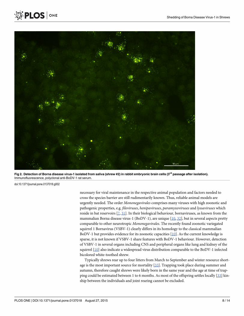
Furthermore, infectious virus was successfully isolated on REB cells from all of the BoDV-1-positive shrews caught in 2013 (#2, #5, #6) in samples from saliva (#2, #6), skin/sebum (#2,#6) and urine (#5, #6) (Fig 2). Viral RNA of isolates from saliva of #2 and saliva of #6 wassequenced (Genbank accession no. KM349818, KM 349819). In a 2150 nucleotide stretch(nt 17 to 2161 covering the N, X, P, half of M-protein-encoding regions) sequences of bothisolates revealed a homology of 99% compared to a recent BoDV-1-sequence (GenBank acces-sion no. KF275185) obtained from a shrew of the same endemic area and to an equine BoDV-1-sequence from a horse housed in the same region (GenBank accession no. KF275184)[17].Both isolates are part of the regional BoDV-1 subcluster 1a. (Fig 3)
The results obtained from the living shrews were compared to the organ distribution of viralantigen and BoDV-1-RNA in three naturally BoDV-1-infected C. leucodon from pest control(#2001 and #5017, [17] and another animal #5072 from the same stable as #5017) and in thetwo deceased shrews. Detailed information about organ distribution is given in S3 Fig. In all ofthese animals, mRNA, genomic RNA and/or viral antigen were detected in the nervous system
Shedding of Borna Disease Virus-1 in Shrews
PLOSONE | DOI:10.1371/journal.pone.0137018 August 27, 2015 5 / 14

and widespread in peripheral organs (e.g. epithelial cells of the parotid gland, lacrimal gland,sebaceous glands, bronchi, kidney tubules, esophagus and epidermal keratocytes) [17] (Fig 4).Interestingly, viral antigen was also present in the uterus in one shrew. Thus, detection of viralRNA and infectious virus from secretions and excretions in the living shrews (saliva, lacrimalfluid, skin swabs, urine and faeces) fits well with the morphological demonstration of viral anti-gen and RNA in the respective organ systems and further confirms successful viral replicationin peripheral organs. Beside virus shedding via secretions and excretions shedding of BoDV-1seems also to be possible via scaling of epidermal epithelial cells.
DiscussionReservoir-bound RNA viruses reside typically inconspicuously in animal reservoirs such asbats, rodents and insectivores. However, transmission routes, host-pathogen interactions
Fig 1. Relative bodymass trend of non-infected and infected shrews.Relative body mass trend of non-infected and infected shrews at 11 time points(weeks in husbandry 2, 4, 8, 12, 16, 20, 24, 28, 32, 36, 40) with no differences in non-infected (demonstrated in green, Kruskal-Wallis-Test: H(10;42) =4.3123; p = 0.9322) and infected animals (demonstrated in red, Kruskal-Wallis-Test: H(10;50) = 6.8237; p = 0.7420) and between groups (Mann-Whitney U-test, p > 0.06)
doi:10.1371/journal.pone.0137018.g001
Shedding of Borna Disease Virus-1 in Shrews
PLOSONE | DOI:10.1371/journal.pone.0137018 August 27, 2015 6 / 14

Table 1. Detection of BoDV-1* RNA in naturally infected bicolored white-toothed shrews over a period of 4 weeks.
Shrew Sample Week 1 Week 2 Week 3 Week 4
#2 Saliva 38 a 29,8 b 27,85 c 28,99 c
Lacrimal fluid 34 a nd* 27,3 c - c,*
Skin swab 36 a 34,5 b 29,48 c 30,2 c
Urine 39 a - b 30,07 c nd
Faeces - a 36,3 b 31,59 c - c
Lair nd nd nd 32,96 c
#5 Saliva 28,53 d 30,36 d 30,57 d 32,50 d
Lacrimal fluid 32,15 d 33,12 d 34,48 d 36,38 d
Skin swab 35,02 d 34,91 d 34,10 d 36,06 d
Urine 34,68 d 38,86 d 34,83 d 35,59 d
Faeces - d - d - d - d
Lair - d 35,50 d - d 38,92 d
#6 Saliva 31,12 d 33,31 d 33,35 d 31,27 d
Lacrimal fluid 35,06 d 35,61 d 36,60 d 32,95 d
Skin swab 36,4 d 36,97 d 36,89 d 32,97 d
Urine 37,34 d 36,41 d - d 34,47 d
Faeces - d - d - d - d
Lair 35,64 d - d - d 33,04 d
Footnote Table 1: * BoDV-1 = Borna disease virus;— = negative; nd = not done
Results are presented as ct-values of different real time RT-PCR runs: a = first run; b = second run; c = third run; d = fourth run
doi:10.1371/journal.pone.0137018.t001
Table 2. Detection of BoDV-1* RNA in naturally infected bicolored white-toothed shrews after morethan at least 250 days in husbandry.
Shrew time point of sampling Sample ct-value
#2 634 days a.c. * Saliva 32,44
Lacrimal fluid 34,93
Skin swab nd*
Urine 33,94
Faeces -*
#9 315 days a.c. Saliva 27,4
Lacrimal fluid 26,76
Skin swab 31,64
Urine nd
Faeces -
#10 315 days a.c. Saliva 25,95
Lacrimal fluid 29,62
Skin swab 33,93
Urine nd
Faeces -
#12 284 days a.c. Saliva 34,47
Lacrimal fluid 32,21
Skin swab 33
Urine 32,19
Faeces -
Footnote Table 2: * BoDV-1 = Borna disease virus; a.c. = after capture;— = negative; nd = not done;
Results are presented as ct-values
doi:10.1371/journal.pone.0137018.t002
Shedding of Borna Disease Virus-1 in Shrews
PLOSONE | DOI:10.1371/journal.pone.0137018 August 27, 2015 7 / 14
necessary for viral maintenance in the respective animal population and factors needed tocross the species barrier are still rudimentarily known. Thus, reliable animal models areurgently needed. The orderMononegavirales comprises many viruses with high zoonotic andpathogenic properties, e.g. filoviruses, henipaviruses, paramyxoviruses and lyssaviruses whichreside in bat reservoirs [7, 31]. In their biological behaviour, bornaviruses, as known from themammalian Borna disease virus-1 (BoDV-1), are unique [10, 32], but in several aspects prettycomparable to other neurotropicMononegavirales. The recently found zoonotic variegatedsquirrel 1 Bornavirus (VSBV-1) clearly differs in its homology to the classical mammalianBoDV-1 but provides evidence for its zoonotic capacities [10]. As the current knowledge issparse, it is not known if VSBV-1 share features with BoDV-1 behaviour. However, detectionof VSBV-1 in several organs including CNS and peripheral organs like lung and kidney of thesquirrel [10] also indicate a widespread virus distribution comparable to the BoDV-1 infectedbicolored white-toothed shrew.
Typically shrews rear up to four litters fromMarch to September and winter resource short-age is the most important source for mortality [33]. Trapping took place during summer andautumn, therefore caught shrews were likely born in the same year and the age at time of trap-ping could be estimated between 1 to 6 months. As most of the offspring settles locally [33] kin-ship between the individuals and joint rearing cannot be excluded.
Fig 2. Detection of Borna disease virus-1 isolated from saliva (shrew #2) in rabbit embryonic brain cells (3rd passage after isolation).Immunofluorescence, polyclonal anti-BoDV-1 rat serum.
doi:10.1371/journal.pone.0137018.g002
Shedding of Borna Disease Virus-1 in Shrews
PLOSONE | DOI:10.1371/journal.pone.0137018 August 27, 2015 8 / 14

During trapping, infection status of the individuals was unknown. Previous studies showeddifferent infection prevalence of shrews that also differed between the trapping sites in thestudy. Hilbe et al [14] found only infected shrews (100%), Puorger et al. [15] detected 2/6infected shrews (33%), Bourg et al. [17] showed 1/1 infected shrews (100%) at one site und 1/19 infected shrews (5%) at the other site whereas Dürrwald et al. [18] found an amount of 9/17infected shrews (53%) at one site with a variance between the years from 25% to 100%. Thesedifferences can be due to the small number of animals in the respective population or representthe natural variation within the shrew population between sites and years. Since examinationof larger cohorts has not been carried out so far, the percentage of naturally infected shrewsamong the trapped animals could not be predicted in detail. Six naturally infected shrews outof eleven shrews implies a percentage of 55% of infected shrews with variations between thesites and years from 50% (site A, year 2013 3/6, year 2014 1/2) to 66% (site B, year 2014 2/3).As all non-infected animals did not show any shedding during the whole observation period,transmission of the virus in the husbandry could be successfully prevented in captivity.
Current data from living shrews provide reliable evidence that natural BoDV-1-infection inthese animals is indeed clinically inconspicuous over a long time period as already previouslyassumed [15, 18] despite persistent infection with shedding of infectious virus via various sites.During the observation period of up to 600 days, only two naturally infected animals were lostdue to an intestinal invagination in one case and hepatitis/pneumonia in the other case whichdid not seem to be directly related to BoDV-1 infection. In the bronchial epithelium of the ani-mal suffering from hepatitis/pneumonia only few cells harboured BoDV-1 nucleoprotein,BoDV-1 mRNA and genomic RNA without associated distribution to the pneumonia and inthe liver only genomic RNA was detected in very few cells.
Interestingly, shedding of viral RNA was continuously present.As shrews were naturallyinfected before trapping, the time between the infection and first virus release remain
Fig 3. Phylogenetic analysis of BoDV-1 sequences obtained from isolated virus. 2150 nt long nucleic sequences comprising N, P, X genes of twoisolated virus isolates (shrew #2, shrew #6), two sequences obtained from a shrew and a horse of the same region (KF275184, KF275185 [17]) and otherrepresentative BoDV-1 of endemic subclusters [25]. Cluster 1: Southwest Germany and Southern Rhine valley group with Cluster 1a: Baden-Wurttembergand parts of Bavaria, Germany (L27077, AY374524) and Cluster 1b: Switzerland, The Principality of Liechtenstein and Austria (AY374550, AY374551),Cluster 2: South German Group (AY374521, AY374531), Cluster 3: Southern Saxony-Anhalt and Saxony (AY374519, AY374534), Cluster 4: CentralGerman group (U04608, AY374522). Tree is rooted with BoDV-2 (AJ311524).
doi:10.1371/journal.pone.0137018.g003
Shedding of Borna Disease Virus-1 in Shrews
PLOSONE | DOI:10.1371/journal.pone.0137018 August 27, 2015 9 / 14

unknown. However, low ct-values were found in samples taken at time points at least morethan 4 to 8 weeks after infection and at time points at least more than 200 days after infection.This indicates a persistent BoDV-1 infection as known from other animals [11, 13] with longlasting and continuous virus release. There was certain variability in the amount of viral RNA,sites of shedding, between individual animals and for the time points of sampling. Some ofthese variations can be due to variations in sample size, as gathering of samples had to be per-formed non-invasive on non-anaesthesized animals. However, several shrews exhibited lowestct-values in saliva and lacrimal fluid regardless of time point of sampling. Whether this mighthave a role for virus transmission, e.g. combating, needs to be further investigated.
Fig 4. Demonstration of BoDV-1 nucleoprotein, messenger RNA and genomic RNA. (A) Demonstration of BoDV-1 nucleoprotein by immunohistochemistry(IHC) in the brain ofC. leucodon #5017; (B) Demonstration of BoDV-1messenger RNA by in-situ hybridization (ISH) in the brain ofC. leucodon #5017; (C)Demonstration of genomic BoDV-1 RNA by ISH in the brain ofC. leucodon #5017; (D) Demonstration of BoDV-1 nucleoprotein by IHC in the trigeminal ganglionofC. leucodon #2001; (E) Demonstration of BoDV-1 messenger RNA by in-situ hybridization (ISH) in the trigeminal ganglion ofC. leucodon #2001; (F)Demonstration of genomic BoDV-1 RNA by ISH in the trigeminal ganglion ofC. leucodon #2001; (G) Demonstration of BoDV-1 nucleoprotein by IHC in the skin,mainly in the sebaceous glands ofC. leucodon #2001; (H) Demonstration of BoDV-1messenger RNA by in-situ hybridization (ISH) in the skin, mainly in thesebaceous glands ofC. leucodon #2001; (I) Demonstration of genomic BoDV-1 RNA by ISH in the skin, mainly in the sebaceous glands ofC. leucodon #2001;
doi:10.1371/journal.pone.0137018.g004
Shedding of Borna Disease Virus-1 in Shrews
PLOSONE | DOI:10.1371/journal.pone.0137018 August 27, 2015 10 / 14

The simultaneous presence of viral antigen, viral mRNA and genomic RNA in CNS andperipheral tissues points to many sites of viral replication thereby enhancing probability of suc-cessful virus transmission to other animals [17]. Horizontal transmission of BoDV-1 in shrewsmight be either achieved via direct contact with secretions or excretions or even via contami-nated environment. Since shrews are known to behave territorially, infection by infected salivaduring combating for a habitat might also occur. Vertical transmission of BoDV-1 in shrewscannot be excluded as viral antigen has been detected in the uterus. However, the route of entryin the reservoir still remains unknown. Offspring might already be infected early by theirmothers due to the various sites of viral shedding even from the skin. The underlying viralmechanisms of maintenance in the reservoir are still incompletely understood but mightinclude adjusted viral life cycle possibly with attenuated pathogenicity, differences in viralentry and circumvention of the antiviral host immune system [4, 34, 35]. The latter could beachieved best in specific situations of the host immune system. Infection of animals in animmune-incompetent stage can lead to persistent, immune-tolerant virus infections, oftenassociated with shedding of high doses of infectious virus and without any severe clinical signsand notable inflammatory lesions. To date it remains unknown whether disseminated BoDV-1infection of shrews is only possible when infected in an immune incompetent state as knownfor rats [19]. However, the clinical inconspicuous course could point to an immune tolerantinfection and a highly adapted viral-host interaction. Neonatally BoDV-1 infected rats displayno neurological signs but increased motor activity, learning deficits and subtle changes in socialbehaviour and memory [36, 37]. Moreover, experimental BoDV-1 infection of the prosimiantree shrew (Tupaia glis) leads to a persistent infection and transient mild encephalitis, resultingin a disorder characterized primarily by hyperactivity and pronounced disturbances in socialand breeding behavior rather than neurological signs [38]. In the neonatally BoDV-1 infectedrat the behavioral changes were attributed to lesions in the hippocampus and cerebellum andin the tree shrew to alterations of the limbic system. Whether naturally BoDV-1 infectedshrews also display subtle deficits in learning, memory and/or social behavior, especially mat-ing, needs to be addressed in further behavioral and breeding experiments. As known so far, C.leucodon did not exhibit any morphological changes in cerebellum, hippocampus or elsewherein the brain as noted for the neonatally infected rat. However, any behavioral changes mightcontribute to higher contact frequency or increased aggressive and territorial behavior therebyfacilitating viral transmission and maintenance in the reservoir.
Characteristics of the shrew population correspond well to the epidemiologic pattern ofBorna disease. The distribution of C. leucodon in Bavaria and the prevalence of Borna diseaseseem to be connected [16]. The yearly varying peaks of Borna disease in accidental hosts andthe decline of Borna disease within the last decades could be related to population dynamics ofthe shrews between the years and the restriction of habitats indirectly caused by modern agri-culture [18]. Inbreeding and low dispersal distance of the offspring correlates with the limiteddistribution of BoDV-1 within endemic territories [18].
Moreover, the continuous secretion and excretion of infectious BoDV-1 and the detectionof viral RNA in the lair substantiates the hypothesis that “infectious dust” is responsible forBoDV-1 transmission to accidental hosts through the intranasal route as known for hantavirusinfections [39]. In this scenario, the BoDV-1 infection of horses and sheep might rather repre-sent an accidental occasion. To date it still remains to be solved whether and which factors areresponsible for successful crossing the species barrier. Amount of infectious virus, virulence,immune status and age of reservoir and accidental host as well as their genetic makeup mightfunction as essential co-factors.
Taken together, shedding of BoDV-1 in the bicolored white-toothed shrew is achieved viavarious routes which enable successful viral maintenance in the reservoir population and even
Shedding of Borna Disease Virus-1 in Shrews
PLOSONE | DOI:10.1371/journal.pone.0137018 August 27, 2015 11 / 14

fatal transmission to susceptible accidental hosts such as horses and sheep. Moreover, theseanimals serve as suitable model to investigate host and pathogen factors that enable persistentviral co-existence in apparently healthy carriers.
Supporting InformationS1 Fig. Body mass change infected individuals [g].(TIF)
S2 Fig. Body mass change non-infected animals [g].(TIF)
S3 Fig. Organ distribution of BoDV-1 nucleoprotein, mRNA and genomic BoDV-1 RNAof individual shrews.(PDF)
S4 Fig. No detection of BoDV-1 antigen or RNA in a non infected shrew by immunohisto-chemy and in-situ hybridization, shrew was trapped dead by pest control.(TIF)
S1 Table. Overview of individual shrews of the study.(DOCX)
AcknowledgmentsWe thank Franz Schmid (Veterinäramt Günzburg) for his helpful support and cooperation. H.Lange-Herbst is supported by the Margarete Ammon-Stiftung.
Author ContributionsConceived and designed the experiments: DNMB SH HLH JAE ME CH. Performed the exper-iments: DNMB SH HLHME. Analyzed the data: DNMB SH HLH JAE ME CH. Contributedreagents/materials/analysis tools: SH JAE ME CH. Wrote the paper: DNMB SH HLH JAE MECH.
References1. Cutler SJ, Fooks AR, van der Poel WH (2010) Public health threat of new, reemerging and neglected
zoonoses in the industrialized world. Emerg Infect Dis 16: 1–7. doi: 10.3201/eid1601.081467 PMID:20031035
2. Jones KE, Patel NG, Levy MA, Storeygard A, Balk D, Gittleman JL, et al. (2008) Global trends in emerg-ing infectious diseases. Nature 451: 990–993. doi: 10.1038/nature06536 PMID: 18288193
3. Eickmann M, Becker S, Klenk HD, Doerr HW, Stadler K, Censini S, et al. (2003) Phylogeny of theSARS coronavirus. Science 302: 1504–1505.
4. Griffin DE (2009) Emergence and re-emergence of viral disease of the central nervous system. Prog-ress in Neurobiology doi: 10.1016/j.pneurobio.2009.12.003
5. Sejvar JJ (2006) The evolving epidemiology of viral encephalitis. Current Opinion in Neurology 19:350–357. PMID: 16914972
6. Tyler KL (2009) Emerging viral infections of the central nervous system. Arch Neurol 66: Part 1: 939–948, Part 2: 1065–1074.
7. Calisher CD, Childs JE, Field HE, Holmes KV, Schountz T (2006) Bats: important reservoir hosts ofemerging viruses. Clin Microbiol Rev 19:531–45. PMID: 16847084
8. Drexler JF, Corman VM, Müller MA, Maganga GD, Vallo P, Binger T, et al (2012) Bats host major mam-malian paramyxoviruses. Nat Commun 3: 796 doi: 10.1038/ncomms1796 PMID: 22531181
Shedding of Borna Disease Virus-1 in Shrews
PLOSONE | DOI:10.1371/journal.pone.0137018 August 27, 2015 12 / 14

9. Kuhn JH, Durrwald R, Bao Y, Briese T, Carbone K, Clawson AN, et al (2015) Taxonomic reorganizationof the family Bornaviridae. Arch Virol (2015) 160:621–632 doi: 10.1007/s00705-014-2276-z PMID:25449305
10. Hoffmann B, Tappe D, Höper D, Herden C, Boldt A, Mawrin C, et al (2015) A Variegated Squirrel Borna-virus associated with fatal human encephalitis. N Engl J Med 2015; 373:154–62. doi: 10.1056/NEJMoa1415627 PMID: 26154788
11. Herden C, et al. (2013) Bornaviridae. In: Knipe DM, Howley PM, editors. Fields of Virology Philadel-phia: Wolters Kluwer Health/Lippincott Williams &Wilkins. pp. 1124–1150.
12. Dürrwald R, Kolodziejek J, Muluneh A, Herzog S, Nowotny N (2006) Epidemiological pattern of classi-cal Borna disease and regional genetic clustering of Borna disease virus point towards the existence ofto-date unknown endemic reservoir host populations. Microbes Infect 8: 917–929. PMID: 16469519
13. Vahlenkamp TW, Kourath A, Weber M, Müller H (2002) Persistence of Borna disease virus in naturallyinfected sheep. J Virol 76: 9735–9743. PMID: 12208952
14. Hilbe M, Herrsche R, Kolodziejek J, Nowotny N, Ehrensperger F (2006) Shrews as Reservoir Hosts ofBorna Disease Virus. Emerg Infect Dis; 12(4):675–7. PMID: 16704819
15. Puorger ME, Hilbe M, Müller JP, Kolodziejek J, Nowotny N, Zlinszky K, et al. (2010) Distribution ofBorna Disease Virus Antigen and RNA in Tissues of naturally infected BicoloredWhite-ToothedShrews,Crocidura leucodon, supporting their role as Reservoir Host Species. Vet Pathol 47(2): 236–244. doi: 10.1177/0300985809351849 PMID: 20133953
16. Encarnação JA, Herzog S, Eickmann M, Becker NI, Hermes N, Herden C (2013) Landscape featuresand reservoir occurrence affecting the risk for equine infection with Borna disease virus. J Wildl Dis 49(4):860–8. doi: 10.7589/2012-10-262 PMID: 24502713
17. Bourg M, Herzog S, Encarnação JA, Nobach D, Lange-Herbst H, Eickmann M, et al. (2013) Bicoloredwhite-toothed shrews as reservoir for Borna disease virus, Bavaria, Germany. Emerg Infect Dis doi: 10.3201/eid1912.131076
18. Dürrwald R, Kolodziejek J, Weissenböck H, Nowotny N (2014) The Bicolored white-toothed shrew Cro-cidura leucodon is an indigenous host of Mammalian Borna Disease Virus. PLoS One. 2014 Apr. 3; 9(4):e93659. doi: 10.1371/journal.pone.0093659 PMID: 24699636
19. Herzog S, Kompter C, Frese K, Rott R (1984) Replication of Borna disease virus in rats: age dependentdifferences in tissue distribution. Med Microbiol Immunol 173 (4): 171–7. PMID: 6439986
20. HallenslebenW, Schwemmle M, Hausmann J, Stitz L, Volk B, Pagenstecker A, et al. (1998) Borna Dis-ease Virus-Induced Neurological Disorder in Mice: Infection of Neonates results in Immunopathology. JVirol 72(5):4379–86. PMID: 9557728
21. Kramer K, Schaudien D, Eisel UL, Herzog S, Richt JA, Baumgärtner W, et al. (2012) TNF-Overexpres-sion in Borna Disease Virus-Infected Mouse Brains Triggers Inflammatory Reaction and Epileptic Sei-zures. PloS One 2012: 7(7):e41476, doi: 10.1371/journal.pone.0041476 PMID: 22848506
22. Schindler AR, Vogtlin A, Hilbe M, Puorger M, Zlinszky K, Ackermann M, et al. (2007) Reverse transcrip-tion real-time PCR assays for detection and quantification of Borna disease virus in diseased hosts.Mol Cell Probes 2007; 21:47–55. PMID: 17014984
23. Herzog S, Rott R (1980) Replication of Borna Disease Virus in Cell Cultures Med Microbiol Immunol168, 153–158. PMID: 6772932
24. Herzog S, Enderlein D, Heffels-Redmann U, Piepenbring A, Neumann D, Kaleta EF, et al. (2010) Indi-rect Immunofluorescence Assay for Intra Vitam Diagnosis of Avian Bornavirus Infection in PsittacineBirds. J Clin Microbiol; 48 (6): 2282–4. doi: 10.1128/JCM.00145-10 PMID: 20392921
25. Kolodziejek J, Dürrwald R, Herzog S, Ehrensperger F, Lussy H, Nowotny N (2005) Genetic clusteringof Borna disease virus natural animal isolates, laboratory and vaccine strains strongly reflects theirregional geographical origin. J Gen Virol, 86, 385–398. PMID: 15659758
26. Felsenstein J (2005). PHYLIP (phylogeny inference package), version 3.6. Distributed by the author.Department of Genome Sciences, University of Washington, Seattle.
27. Gouy M, Guindon S, Gascuel O (2010) SeaView version 4: a multiplatform graphical user interface forsequence alignment and phylogenetic tree building. Molecular Biology and Evolution 27(2): 221–224doi: 10.1093/molbev/msp259 PMID: 19854763
28. Herden C, Herzog S, Richt JA, Nesseler A, Christ M, Failing K, et al. (2000) Distribution of Borna Dis-ease Virus in the Brain of Rats Infected with an Obesity-inducing Virus Strain. Brain Pathol; 10:39–48.PMID: 10668894
29. Werner-Keišs N, GartenW, Richt JA, Porombka D, Algermissen D, Herzog S, et al. (2008) Restrictedexpression of Borna Disease virus glycoprotein in brains of experimentally infected Lewis rats. Neuro-pathol Appl Neurobiol; 34:590–602. doi: 10.1111/j.1365-2990.2008.00940.x PMID: 18282160
Shedding of Borna Disease Virus-1 in Shrews
PLOSONE | DOI:10.1371/journal.pone.0137018 August 27, 2015 13 / 14

30. Animal Care Use Committee (1998) Guidelines for the capture, handling and care of mammals asapproved by the American Society of Mammalogists. J Mammal 79:1416–1431
31. Wong S, Lau S, Woo P, Yuen KY (2007) Bats as a continuing source of emerging infections in humans.Rev Med Virol 17: 67–91e. PMID: 17042030
32. De la Torre JC (2006) Reverse-genetic approaches to the study of Borna disease. Nature Rev Microbiol4: 777–83.
33. Duarte LC, Bouteiller C, Fontanillas P, Petit E, Perrin N (2003) Inbreeding in the greater white-toothedshrew, Crocidura russula. Evolution 57:638–645 PMID: 12703953
34. Brandes G, Khayami M, Peck CT, Baumgärtner W, Bugday H, Wewetzer K (2011) Cell surface expres-sion of 27C7 by neonatal rat olfactory ensheathing cells in situ and in vitro is independent of axonal con-tact. Histochem Cell Biol 135: 397–408. doi: 10.1007/s00418-011-0796-0 PMID: 21437623
35. Schneider DS, Ayres JS (2008) Two ways to survive infection: what resistance and tolerance can teachus about treating infectious diseases. Nat Rev Immunol 8: 889–95. doi: 10.1038/nri2432 PMID:18927577
36. Bautista JR, Schwartz GJ, De La Torre JC, Moran TH, Carbone KM (1994) Early and persistent abnor-malities in rats with neonatally acquired Borna disease virus infection. Brain Res Bull 34:31–40. PMID:8193931
37. Rubin SA, Sylves P, Vogel M, Pletnikov M, Moran TH, Schwartz GJ, et al. (1999) Borna disease virus-induced hippocampal dentate gyrus damage is associated with spatial learning and memory deficits.Brain Res Bull 48: 23–30. PMID: 10210164
38. Sprankel H, Richarz K, Ludwig H, Rott R (1978) Behavior alterations in tree shrews (Tupaia glis)induced by Borna disease virus. Med Microbiol Immunol 165:1–18. PMID: 566371
39. Schwarz AC, Ranft U, Piechotowski I, Childs JE, Brockmann SO (2009) Risk factors for human infec-tion with Puumala virus, southwestern Germany. Emerg Infect Dis 15(7):1032–9. doi: 10.3201/eid1507.081413 PMID: 19624917
Shedding of Borna Disease Virus-1 in Shrews
PLOSONE | DOI:10.1371/journal.pone.0137018 August 27, 2015 14 / 14