Shc Epidermal Growth Factor Receptor and through a Signaling ...
12
1998, 18(9):5178. Mol. Cell. Biol. Yusen Liu Wei Chen, Jennifer L. Martindale, Nikki J. Holbrook and Shc Epidermal Growth Factor Receptor and through a Signaling Pathway Mediated by Extracellular Signal-Regulated Kinase Tumor Promoter Arsenite Activates http://mcb.asm.org/content/18/9/5178 Updated information and services can be found at: These include: REFERENCES http://mcb.asm.org/content/18/9/5178#ref-list-1 at: This article cites 64 articles, 29 of which can be accessed free CONTENT ALERTS more» articles cite this article), Receive: RSS Feeds, eTOCs, free email alerts (when new http://journals.asm.org/site/misc/reprints.xhtml Information about commercial reprint orders: http://journals.asm.org/site/subscriptions/ To subscribe to to another ASM Journal go to: on February 21, 2013 by PENN STATE UNIV http://mcb.asm.org/ Downloaded from
Transcript of Shc Epidermal Growth Factor Receptor and through a Signaling ...
1998, 18(9):5178. Mol. Cell. Biol.
Yusen LiuWei Chen, Jennifer L. Martindale, Nikki J. Holbrook and ShcEpidermal Growth Factor Receptor and through a Signaling Pathway Mediated byExtracellular Signal-Regulated Kinase Tumor Promoter Arsenite Activates
http://mcb.asm.org/content/18/9/5178Updated information and services can be found at:
These include:
REFERENCEShttp://mcb.asm.org/content/18/9/5178#ref-list-1at:
This article cites 64 articles, 29 of which can be accessed free
CONTENT ALERTS more»articles cite this article),
Receive: RSS Feeds, eTOCs, free email alerts (when new
http://journals.asm.org/site/misc/reprints.xhtmlInformation about commercial reprint orders: http://journals.asm.org/site/subscriptions/To subscribe to to another ASM Journal go to:
on February 21, 2013 by P
EN
N S
TA
TE
UN
IVhttp://m
cb.asm.org/
Dow
nloaded from
MOLECULAR AND CELLULAR BIOLOGY,0270-7306/98/$04.0010
Sept. 1998, p. 5178–5188 Vol. 18, No. 9
Tumor Promoter Arsenite Activates Extracellular Signal-RegulatedKinase through a Signaling Pathway Mediated by Epidermal
Growth Factor Receptor and ShcWEI CHEN, JENNIFER L. MARTINDALE, NIKKI J. HOLBROOK, AND YUSEN LIU*
Gene Expression and Aging Section, Laboratory of Biological Chemistry,National Institute on Aging, Baltimore, Maryland 21224
Received 29 December 1997/Returned for modification 18 February 1998/Accepted 18 June 1998
Although arsenite is an established carcinogen, the mechanisms underlying its tumor-promoting propertiesare poorly understood. Previously, we reported that arsenite treatment leads to the activation of the extracel-lular signal-regulated kinase (ERK) in rat PC12 cells through a Ras-dependent pathway. To identify potentialmediators of the upstream signaling cascade, we examined the tyrosine phosphorylation profile in cells exposedto arsenite. Arsenite treatment rapidly stimulated tyrosine phosphorylation of several proteins in a Ras-inde-pendent manner, with a pattern similar to that seen in response to epidermal growth factor (EGF) treatment.Among these phosphorylated proteins were three isoforms of the proto-oncoprotein Shc as well as the EGFreceptor (EGFR). Tyrosine phosphorylation of Shc allowed for enhanced interactions between Shc and Grb2as identified by coimmunoprecipitation experiments. The arsenite-induced tyrosine phosphorylation of Shc, en-hancement of Shc and Grb2 interactions, and activation of ERK were all drastically reduced by treatment ofcells with either the general growth factor receptor poison suramin or the EGFR-selective inhibitor tyrphostinAG1478. Down-regulation of EGFR expression through pretreatment of cells with EGF also attenuated ERKactivation and Shc tyrosine phosphorylation in response to arsenite treatment. These results demonstrate thatthe EGFR and Shc are critical mediators in the activation of the Ras/ERK signaling cascade by arsenite andsuggest that arsenite acts as a tumor promoter largely by usurping this growth factor signaling pathway.
Arsenite, the trivalent arsenic compound, is a potent carcin-ogen to which there is significant worldwide exposure throughnatural contamination of food and drinking water (3). In hu-mans, chronic exposure is associated with an increased inci-dence of skin and bladder cancers (3, 16). Although it appearsto be nonmutagenic alone, in cultured cells arsenite potenti-ates the mutagenic effects of short-wavelength UV radiation(UV-C) (23, 34, 63). Hence, arsenite has been suggested to actas a tumor promoter in the carcinogenic process (6, 59). Whilethe basis for such tumor-promoting activity is unclear, theability of arsenite to interact with protein thiol groups andthereby alter the activities of key regulatory proteins is likely tocontribute to its carcinogenic properties (6, 37).
The mitogen-activated protein (MAP) kinase cascade lead-ing to the activation of ERK is crucial for regulating cellgrowth and differentiation. Initiation of this signaling pathwayvia growth factor receptors has been studied extensively (2, 11,56). Ligand-mediated dimerization of growth factor receptorstriggers the activation of receptor-type tyrosine kinases, result-ing in autophosphorylation of tyrosine residues (2, 24, 53, 56).These residues then serve as docking sites for the recruitmentof downstream signaling mediators necessary for the activationof membrane-localized Ras (15, 53). For example, the adapterprotein Grb2 binds to the phosphotyrosine residues through itsSrc homology 2 (SH2) domain to bring the Ras guanine nu-cleotide exchange factor, Son of Sevenless (Sos), from thecytoplasm to the vicinity of Ras through its two Src homology3 domains. In addition, the Grb2-Sos complexes can be re-
cruited to the phosphotyrosine sites through another adaptorprotein, Shc (4). Shc binds to certain receptor phosphotyrosinesites through its phosphotyrosine-binding domain; Shc thenbecomes tyrosine phosphorylated itself and thereby providesadditional docking sites for Grb2 (3, 4, 15, 53). The recruit-ment of Grb2-Sos complexes to the phosphotyrosine sites leadsto Ras activation, which then initiates the phosphorylationcascade starting with the activation of the proto-oncoproteinRaf, a serine kinase that phosphorylates and activates ERKkinase (MEK), which in turn activates ERK (2, 56). ERK isresponsible for the phosphorylation of a variety of cellularproteins including downstream kinases, transcription factors,and components of the protein synthesis machinery (11).
In cultured cells, dysregulation of the ERK pathway throughalterations in any one of several mediators involved in the cas-cade (e.g., mutation of growth factor receptors, Shc, Ras, Raf,and MEK) can lead to cellular transformation (7, 14, 15, 33,40). Indeed, many tumors exhibit elevated ERK activity (32,54, 57). It is not surprising, therefore, that a common feature ofknown tumor promoters is their ability to perturb the ERK sig-naling pathway. For example, although acting through differentmechanisms, the tumor promoters phorbol ester, okadaic acid,and butylated hydroxytoluene hydroperoxide all lead to the ac-tivation of ERK. Phorbol esters act through activation of pro-tein kinase C (leading to activation of Raf) (29), while okadaicacid acts through the inhibition of serine/threonine phospha-tases (18). The mechanism responsible for ERK activation bybutylated hydroxytoluene hydroperoxide is not fully under-stood but appears to involve steps upstream of Ras, as the ac-tivation of ERK by this agent is Ras dependent (20). In keep-ing with arsenite’s putative role as a tumor promoter, we haverecently observed that arsenite treatment leads to a significantelevation in ERK activity in Rat1 and PC12 cells (37). Thisactivation is dependent on Ras and is sensitive to the presence
* Corresponding author. Mailing address: Gene Expression and Ag-ing Section, Laboratory of Biological Chemistry, National Institute onAging, Intramural Research Program, Gerontology Research Center,5600 Nathan Shock Dr., Box 12, Baltimore, MD 21224. Phone: (410)558-8442. Fax: (410) 558-8335. E-mail: [email protected].
5178
on February 21, 2013 by P
EN
N S
TA
TE
UN
IVhttp://m
cb.asm.org/
Dow
nloaded from
of the growth factor receptor poison suramin (60). These find-ings suggested that arsenite may act early in the pathway,either at a step involving the growth factor receptors directlyor during the events serving to activate the growth factorreceptors (37). Better understanding of the signal transduc-tion pathway leading to ERK activation by arsenite could pro-vide important insight into its carcinogenic mechanisms. In thepresent report, we focus on the early events in the signalingpathway leading to ERK activation. We provide evidence thatarsenite treatment results in tyrosine phosphorylation of theShc adapter protein, leading to its enhanced interaction withGrb2. We further demonstrate that epidermal growth factor(EGF) receptor (EGFR) tyrosine kinase is required forthese interactions and the subsequent activation of ERK.The ability of arsenite to usurp this growth regulatory path-way is likely to contribute to its tumor-promoting properties.
MATERIALS AND METHODS
Cell culture and treatments. Wild-type and Ras N17 PC12 cells, kindly pro-vided by G. Cooper (61), were grown in a 37°C humidified atmosphere contain-ing 5% CO2 in Dulbecco’s modified Eagle’s medium (DMEM) (Life Tech-nologies, Inc., Gaithersburg, Md.) supplemented with 10% fetal bovine serum(HyClone, Logan, Utah) and 5% horse serum (Life Technologies, Inc.). Cellswere serum starved by placement in serum-free medium for at least 16 h prior totreatment with arsenite or EGF. Sodium arsenite (Sigma Chemical Co., St.Louis, Mo.) was added directly into the medium to a final concentration of 400mM. EGF stimulation was carried out by directly adding EGF (Life Technolo-gies) into the medium to a final concentration of 100 ng/ml. Suramin (300 mM)and tyrphostin AG1478 (30 or 40 nM) (Calbiochem, San Diego, Calif.) wereadded to the medium immediately before addition of sodium arsenite. All re-agents were left in the medium until cells were harvested.
Down-modulation of EGFR. Down-modulation of EGFR was carried out byincubating PC12 cells with EGF essentially as previously described by Carpenterand Cohen (5). PC12 cells cultured in serum-free medium overnight were stim-ulated with EGF (1 mg/ml) for 1 h. The EGF-containing medium was thenremoved, and the cells were washed twice with phosphate-buffered saline (PBS).The cells were then kept in serum-free medium for another 2 h at 37°C to allowthe internalization of EGF-EGFR complexes before a second treatment withEGF, nerve growth factor (NGF), or arsenite. Replenishment of EGFR wasaccomplished by incubation of PC12 cells with down-regulated EGFR in DMEMcontaining 10% fetal bovine serum for 10 h. After replenishment of EGFR, cellswere serum starved for another 14 h and subsequently treated with arsenite,EGF, or NGF.
In vitro binding assays. The glutathione S-transferase (GST)–Grb2 SH2 do-main-expressing plasmid was generously provided by H. Gram (43). The GST-Grb2 SH2 domain fusion protein was produced in Escherichia coli and purifiedby glutathione-Sepharose affinity chromatography followed by fast protein liquidchromatography (FPLC) purification procedures. Twenty micrograms of the glu-tathione-free GST-Grb2 SH2 domain fusion protein was incubated with 400 mgof soluble cell lysates at 4°C for 2 h. Glutathione-Sepharose beads (PharmaciaBiotech Inc., Piscataway, N.J.) were then added to the samples, and the sampleswere incubated for an additional hour with gentle rotation. The Sepharose beadswere recovered through gentle centrifugation and washed extensively with thelysis buffer. Proteins recovered with the GST-Grb2 SH2 domain fusion proteinwere separated through a 10% NuPAGE Bis-Tris gel (Novex, San Diego, Calif.)and subjected to Western blot analysis.
Antibodies, immunoprecipitation, and immunoblotting. Polyclonal anti-Shc,monoclonal anti-Grb2, and anti-pan ERK antibodies were purchased from Trans-duction Laboratories (Lexington, Ky.). Polyclonal antibodies against EGFR andERK2 were purchased from Santa Cruz Biotechnology, Inc. (Santa Cruz, Calif.).Polyclonal anti-phospho-p42/p44 ERK antibody was purchased from PromegaCo. (Madison, Wis.). Antiphosphotyrosine monoclonal antibody 4G10 was pur-chased from Upstate Biotechnology Inc. (Lake Placid, N.Y.). Horseradish per-oxidase-conjugated anti-mouse and anti-rabbit secondary antibodies were pur-chased from Amersham Co. (Arlington Heights, Ill.).
Immunoblotting was performed as described previously (35–37). Briefly, PC12cells were washed twice with ice-cold PBS and lysed in 1 ml of lysis buffer (20 mMHEPES [pH 7.4], 50 mM b-glycerophosphate, 1% Triton X-100, 10% glycerol,2 mM EGTA, 1 mM dithiothreitol, 10 mM sodium fluoride, 1 mM sodiumorthovanadate, 2 mM leupeptin, 2 mM aprotinin, 1 mM phenylmethylsulfonylfluoride, 0.5 mM okadaic acid). The cell lysates were clarified by centrifugationat 14,000 rpm for 10 min. Samples normalized for total protein content wereresolved by electrophoresis through either straight or gradient NuPAGE Bis-Trisgels (10% or 4 to 12%) (Novex) and transferred onto a polyvinylidene difluoridemembrane (Millipore, Bedford, Mass.). Enhanced chemiluminescence reagent(Amersham Co.) was used for the detection of the immunoreactive bands. For
reprobing, blots were stripped with a buffer containing 50 mM Tris-HCl (pH 6.8),2% sodium dodecyl sulfate (SDS), and 0.1 M b-mercaptoethanol.
For immunoprecipitation, soluble cell lysates normalized for total proteincontent were precleaned by incubation with 20 ml of protein A-Sepharose beads(Pharmacia Biotech) at 4°C for 30 min. After the incubation, the supernatant wascollected by brief centrifugation and incubated with the indicated antibody and20 ml of protein A-Sepharose beads for 16 h at 4°C while gently rotating. Theimmunoprecipitates were washed four times with 1 ml of lysis buffer and sepa-rated through the NuPAGE gel system (Novex). Immunoblotting analysis of theimmunoprecipitates was carried out as described above.
Immune complex kinase assay. The kinase activity of ERK was assessed by animmune complex kinase assay as previously described, using myelin basic protein(MBP) as a substrate (35–37). Briefly, soluble cell lysates containing 400 mg ofprotein were incubated with 1 mg of rabbit polyclonal anti-ERK2 antibody and 20ml of protein A-Sepharose beads at 4°C for 16 h with gentle rotation. Theimmunoprecipitates were then washed three times with lysis buffer, three timeswith wash buffer (500 mM LiCl, 100 mM Tris-HCl [pH 7.6], 0.1% Triton X-100,1 mM dithiothreitol), and three times with kinase assay buffer (20 mM morpho-linepropanesulfonic acid [pH 7.2], 2 mM EGTA, 10 mM MgCl2, 1 mM dithio-threitol, 0.1% Triton X-100). The kinase reactions were carried out at 30°C for20 min in 55 ml of kinase assay buffer containing 10 mM ATP, 10 mCi of [g-32P]ATP, 20 mM MgCl2, and 6 mg of MBP (Sigma).
Northern blot analysis. Northern blot analysis was carried out as previouslydescribed (36). Briefly, serum-starved PC12 cells were pretreated with or withouttyrphostin AG1478 (40 nM) for 30 min prior to arsenite treatment. Arsenite wasdirectly added to the medium to a final concentration of 400 mM. Cells wereharvested 30 min after the addition of arsenite, and total RNA was extracted byusing Stat 60 (Teltest “B,” Friendswood, Tex.). Northern blot analysis was per-formed with rat c-fos and c-jun cDNAs and 18S oligonucleotides as probes.
Cell cycle distribution analysis. PC12 cells were plated into six-well plates andcultured until reaching 50 to 60% confluence. Cells were serum starved by beingplaced in serum-free medium for 48 h and were then treated with 400 mMarsenite for another 24 h. Cells were harvested before and after the treatment.Cell cycle distribution was analyzed by flow cytometry as previously described(17). Briefly, 106 to 2 3 106 cells were collected and fixed in 70% ethanol for 30min. Fixed cells were washed with PBS once, incubated with 1 mg of RNase A perml for 30 min at 37°C, and then stained with propidium iodide (BoehringerMannheim, Indianapolis, Ind.). The stained cells were analyzed on a FACscanflow cytometer for relative DNA content based on red fluorescence. The per-centages of the cells in the various cell cycle compartments were determined byusing the MULTICYCLE software program (Phoenix Flow Systems, San Diego,Calif.).
RESULTS
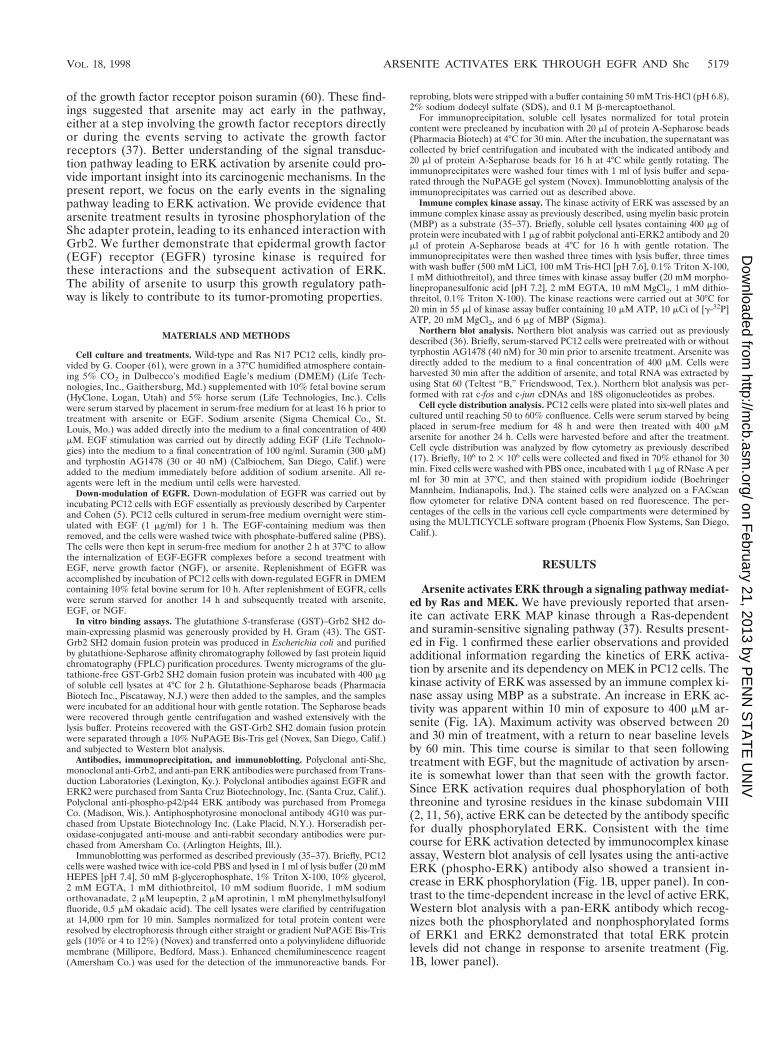
Arsenite activates ERK through a signaling pathway mediat-ed by Ras and MEK. We have previously reported that arsen-ite can activate ERK MAP kinase through a Ras-dependentand suramin-sensitive signaling pathway (37). Results present-ed in Fig. 1 confirmed these earlier observations and providedadditional information regarding the kinetics of ERK activa-tion by arsenite and its dependency on MEK in PC12 cells. Thekinase activity of ERK was assessed by an immune complex ki-nase assay using MBP as a substrate. An increase in ERK ac-tivity was apparent within 10 min of exposure to 400 mM ar-senite (Fig. 1A). Maximum activity was observed between 20and 30 min of treatment, with a return to near baseline levelsby 60 min. This time course is similar to that seen followingtreatment with EGF, but the magnitude of activation by arsen-ite is somewhat lower than that seen with the growth factor.Since ERK activation requires dual phosphorylation of boththreonine and tyrosine residues in the kinase subdomain VIII(2, 11, 56), active ERK can be detected by the antibody specificfor dually phosphorylated ERK. Consistent with the timecourse for ERK activation detected by immunocomplex kinaseassay, Western blot analysis of cell lysates using the anti-activeERK (phospho-ERK) antibody also showed a transient in-crease in ERK phosphorylation (Fig. 1B, upper panel). In con-trast to the time-dependent increase in the level of active ERK,Western blot analysis with a pan-ERK antibody which recog-nizes both the phosphorylated and nonphosphorylated formsof ERK1 and ERK2 demonstrated that total ERK proteinlevels did not change in response to arsenite treatment (Fig.1B, lower panel).
VOL. 18, 1998 ARSENITE ACTIVATES ERK THROUGH EGFR AND Shc 5179
on February 21, 2013 by P
EN
N S
TA
TE
UN
IVhttp://m
cb.asm.org/
Dow
nloaded from
Recent studies have suggested that arsenite activates JNKthrough inhibition of a JNK phosphatase rather than throughactivation of upstream kinases (6). In an effort to determinewhether upstream mediators were involved in ERK activationby arsenite, we examined the effect of the MEK-specific inhib-itor PD98059 (13, 44) on arsenite-induced ERK phosphoryla-tion and activation. Pretreatment of PC12 cells with PD98059resulted in complete inhibition of arsenite-induced ERK phos-phorylation at the 20-min time point following arsenite expo-sure and greatly reduced ERK activity at 30 min (Fig. 1C). Thesensitivity of arsenite-triggered ERK activation to the MEKinhibitor indicated that it was mediated through this upstreamkinase. Supporting this observation, Fig. 1D shows a directcomparison of ERK activity in arsenite-treated wild-type PC12cells with that in PC12 cells expressing a dominant negativeRas mutant (Ras N17) (15, 61), which confirmed our earlierobservation that Ras was required for arsenite-induced ERKactivation (37). Thus, these results showed the ERK activationwas dependent on both MEK and Ras and further establishedthat an upstream signal(s) was involved in the activation of theERK pathway by arsenite.
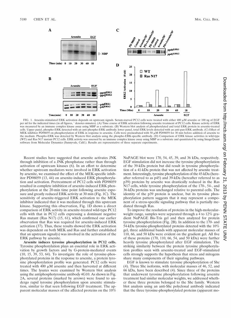
Arsenite induces tyrosine phosphorylation in PC12 cells.Tyrosine phosphorylation plays an essential role in ERK acti-vation by growth factors and by G-protein-mediated events(10, 15, 39, 53, 64). To investigate the role of tyrosine-phos-phorylated proteins in the response to arsenite, a protein tyro-sine phosphorylation profile was generated. PC12 cells weretreated with 400 mM arsenite and then harvested at differenttimes. The lysates were examined by Western blot analysisusing the antiphosphotyrosine antibody 4G10. As shown in Fig.2A, several proteins (marked by arrows) were found to un-dergo rapid tyrosine phosphorylation upon arsenite stimula-tion, similar to that seen following EGF treatment. The ap-parent molecular masses of the affected proteins on the 10%
NuPAGE blot were 170, 54, 45, 39, and 36 kDa, respectively.EGF stimulation did not increase the tyrosine phosphorylationof the 39-kDa protein but did result in tyrosine phosphoryla-tion of a 41-kDa protein that was not affected by arsenite treat-ment. Interestingly, tyrosine phosphorylation of the 45-kDa (here-after referred to as p45) and 39-kDa (hereafter referred to asp39) proteins by arsenite was drastically reduced in the RasN17 cells, while tyrosine phosphorylation of the 170-, 54-, and36-kDa proteins was unchanged relative to parental cells. Theidentity of the p39 protein is unclear, but its tyrosine phos-phorylation pattern suggests that it may represent a compo-nent of a stress-specific signaling pathway that is partially me-diated through Ras.
To improve the resolution of proteins in the high-molecular-weight range, samples were separated through a 4 to 12% gra-dient NuPAGE Bis-Tris gel and then analyzed for proteintyrosine phosphorylation (Fig. 2B). In addition to the 170- and54-kDa tyrosine-phosphorylated proteins detected with the 10%gel, three additional bands with apparent molecular masses of110, 66, and 50 kDa were evident on the gradient gel. All fiveof these proteins (170, 110, 66, 54, and 50 kDa) were furtherheavily tyrosine phosphorylated after EGF stimulation. Thestriking similarity between the protein tyrosine phosphoryla-tion profiles seen with arsenite-treated and EGF-stimulatedcells strongly supports the hypothesis that stress and mitogensshare many components of their signaling pathways.
EGF is known to stimulate tyrosine phosphorylation of Shc(4). Three Shc isoforms, with molecular masses of 46, 52, and66 kDa, have been described (4). Since three of the proteinsthat underwent tyrosine phosphorylation following arsenitetreatment had similar molecular weights, we addressed wheth-er these three proteins belonged to the Shc family. Westernblot analysis using an anti-Shc polyclonal antibody indicatedthat the three tyrosine-phosphorylated proteins (apparent mo-
FIG. 1. Arsenite-stimulated ERK activation depends on upstream signals. Serum-starved PC12 cells were treated with either 400 mM arsenite or 100 ng of EGFper ml for the indicated times (in all figures, 9 denotes minutes). (A) Time course of ERK activation following arsenite treatment of PC12 cells. Kinase activity of ERKwas measured by an immune complex kinase assay using MBP as a substrate. (B) Western blot analysis of phosphorylated and total ERK protein in arsenite-treatedcells. Upper panel, phospho-ERK detected with an anti-phospho-ERK antibody; lower panel, total ERK levels detected with an anti-pan-ERK antibody. (C) Effect ofMEK inhibitor PD98059 on phosphorylation of ERK in response to arsenite. Cells were preincubated with 50 mM PD98059 for 30 min before addition of arsenite tothe medium. Phospho-ERK was detected by Western blot analysis using the phospho-ERK-specific antibody. (D) Comparison of ERK kinase activities in wild-type(WT) and Ras N17 mutant PC12 cells. ERK activity was assessed by an immune complex kinase assay using MBP as a substrate and quantitated by using ImageQuantsoftware from Molecular Dynamics (Sunnyvale, Calif.). Results are representative of three separate experiments.
5180 CHEN ET AL. MOL. CELL. BIOL.
on February 21, 2013 by P
EN
N S
TA
TE
UN
IVhttp://m
cb.asm.org/
Dow
nloaded from

lecular weights of 66,000, 54,000, and 50,000) comigrated withthe three isoforms of Shc (Fig. 2C). Given that Shc plays animportant role in linking growth factor receptor tyrosine ki-nases to the Ras/ERK pathway (4), and that Shc was potentlytyrosine phosphorylated in arsenite-treated cells in a Ras-in-dependent manner, it is likely that Shc serves a similar role intransducing the arsenite signal to the Ras/ERK MAP kinasecascade.
Arsenite induces tyrosine phosphorylation of Shc and en-hances its association with Grb2. Shc serves as an adapterprotein in growth factor signaling through its interaction withGrb2 (4). To determine if it provides a similar function dur-ing stress signaling, we examined whether Shc indeed associ-ated with Grb2 following arsenite treatment. Shc was immuno-precipitated from the cell lysates by using the rabbit anti-Shcantibody that recognizes all three Shc isoforms. The immuno-precipitates were then separated by NuPAGE Bis-Tris gelelectrophoresis, and tyrosine-phosphorylated proteins in theimmune complexes were detected by Western blot analysis us-ing the mouse monoclonal antiphosphotyrosine antibody 4G10.As shown in the upper panel of Fig. 3A, arsenite treatmentled to an enrichment in tyrosine-phosphorylated Shc proteins(p52Shc and p46Shc) which were absent in control cells. Inter-estingly, a 170-kDa tyrosine-phosphorylated protein (p170)was also detected in the Shc immune complexes isolated fromthe arsenite-treated cells but not from the control cells (Fig.3A, upper panel). This result suggested that arsenite treatmentled to an interaction between the tyrosine-phosphorylated pro-tein p170 and Shc. Western blot analysis of the anti-Shc im-munoprecipitates with an anti-Grb2 antibody indicated thatGrb2 did coimmunoprecipitate with Shc in the arsenite-treatedsamples (Fig. 3A, lower panel). Although a small amount ofGrb2 in the Shc immune complexes could be detected in theShc immune complexes as early as 10 min following arseniteexposure, the amount of Grb2 in the Shc immunocomplexesincreased markedly after 20 min (Fig. 3A, lower panel; com-pare the first three lanes). Again, little Grb2 protein was de-tectable in the negative controls (time zero). Importantly, theenrichment of Grb2 protein in the Shc immune complexescorrelated precisely with the kinetics of Shc tyrosine phosphor-ylation (compare the upper and lower panels in Fig. 3A), while
the amounts of Shc and Grb2 detected by Western blot analysisin the total cell lysates remained unchanged throughout theentire treatment period (Fig. 3B).
In response to growth factor stimulation, Grb2 interacts withphosphotyrosine residues of both growth factor receptor tyro-sine kinases and Shc through its SH2 domain, bridging thetyrosine phosphorylation signal to the Ras/ERK pathway (4,53). To determine whether Shc plays a prominent role in re-cruiting Grb2 to the phosphotyrosine sites in arsenite-treatedcells, an experiment was carried out to detect all of the tyro-sine-phosphorylated proteins capable of interacting with theSH2 domain of Grb2 in arsenite-treated cells. A recombinantGST-Grb2 SH2 domain fusion protein was produced in E. colias previously described (43) and purified by glutathione-Seph-arose chromatography followed by passage through an FPLCion-exchange column to remove glutathione from the fusionprotein (Fig. 3C). Twenty micrograms of the GST-Grb2 SH2domain fusion protein was then incubated with 400 mg ofsoluble cell lysate at 4°C for 2 h. The GST-Grb2 SH2 domainfusion protein and proteins interacting with it were recoveredby incubation with glutathione-Sepharose beads, separatedthrough a 10% NuPAGE Bis-Tris gel, and subjected to immu-noblotting analysis using the antiphosphotyrosine antibody4G10. Three major tyrosine-phosphorylated proteins with mo-lecular weights of about 170,000, 54,000, and 50,000 were de-tected in preparations from arsenite-treated cells (Fig. 3D,upper panel). Essentially no tyrosine-phosphorylated proteinswere brought down by the GST-Grb2 SH2 domain fusion pro-tein from untreated control cells (Fig. 3D, upper panel, firstlane). Reprobing the same blot with the polyclonal anti-Shc an-tibody indicated that the two smaller tyrosine-phosphorylatedproteins corresponded to the p52 and the p46 isoforms of Shc(Fig. 3D, lower panel). This in vitro binding experiment furtherestablished that Shc constituted the major tyrosine-phosphor-ylated protein interacting with the SH2 domain of Grb2 inarsenite-treated PC12 cells.
Activation of EGFR is required for arsenite-induced Shctyrosine phosphorylation and ERK activation. Tyrosine phos-phorylation of Shc can be regulated by both receptor-type andnonreceptor-type protein tyrosine kinases (4). It has beenshown that the EGFR tyrosine kinase can directly phosphor-
FIG. 2. Protein tyrosine phosphorylation profile of arsenite-treated PC12 cells. (A) Wild-type and Ras N17 PC12 cells were treated with 400 mM arsenite or 100ng of EGF per ml for the indicated times. The cell extracts were separated on 10% NuPAGE Bis-Tris gel. Tyrosine-phosphorylated proteins were detected byimmunoblotting with the antiphosphotyrosine antibody 4G10. The major tyrosine-phosphorylated bands induced by arsenite or EGF are indicated by arrows. (B)Protein tyrosine phosphorylation profile in arsenite-treated PC12 cells was generated on a 4 to 12% gradient NuPAGE Bis-Tris gel. Tyrosine-phosphorylated proteinswere detected by immunoblotting with the antiphosphotyrosine antibody 4G10. Arrows indicate the major tyrosine-phosphorylated bands induced by arsenite or EGF.(C) Shc isoforms comigrate with tyrosine-phosphorylated proteins induced by arsenite or EGF. The blot shown in panel B was stripped and reprobed with anShc-specific rabbit polyclonal antibody. Shown is a single lane from this blot (wild-type, arsenite-treated cells), as all lanes showed identical patterns.
VOL. 18, 1998 ARSENITE ACTIVATES ERK THROUGH EGFR AND Shc 5181
on February 21, 2013 by P
EN
N S
TA
TE
UN
IVhttp://m
cb.asm.org/
Dow
nloaded from

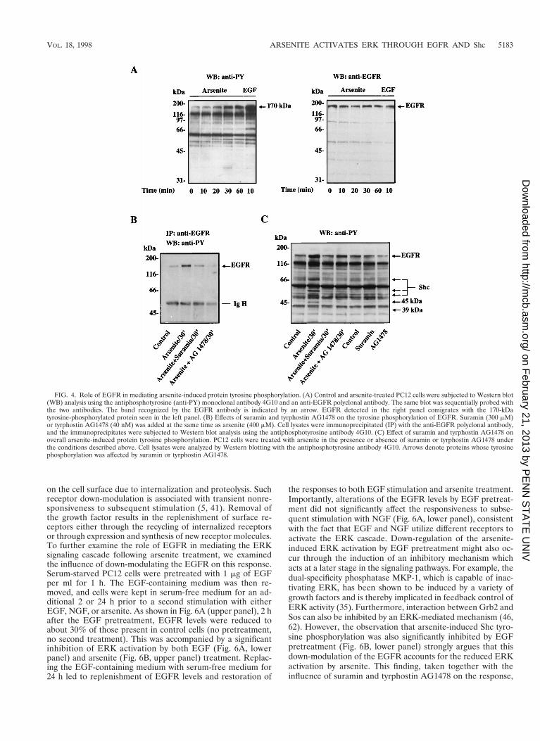
ylate Shc (55). The observations that the tyrosine-phosphory-lated protein p170 was found to coimmunoprecipitate withtyrosine-phosphorylated Shc in arsenite-treated cells (Fig. 3A)and also to copurify with tyrosine-phosphorylated Shc by us-ing the Grb2 SH2 domain fusion protein (Fig. 3D) raised thepossibility that the 170-kDa tyrosine-phosphorylated proteinp170 might be the tyrosine kinase responsible for Shc phosphor-ylation. Because EGF treatment also enhanced the tyrosinephosphorylation of this protein and its size was similar to thatof the EGFR, we investigated whether EGFR was involvedin the arsenite-triggered signaling cascade. Western blot anal-ysis using a polyclonal anti-EGFR antibody indicated that EGFRdid indeed comigrate with the 170-kDa tyrosine-phosphory-lated protein seen in both arsenite- and EGF-treated cells (Fig.4A, right panel).
To more directly examine the role of the EGFR in theresponse to arsenite, we sought to inhibit the function of theEGFR. Two inhibitory agents were used for this purpose:suramin, a general growth factor receptor inhibitor (60), andtyrphostin AG1478, a selective inhibitor of EGFR (33). Theability of suramin and tyrphostin AG1478 to inhibit the ty-rosine phosphorylation of EGFR was demonstrated by immu-noprecipitation of EGFR followed by Western blot analysisusing the antiphosphotyrosine antibody 4G10 (Fig. 4B). Ar-senite treatment of PC12 cells stimulated the phosphorylationof EGFR on tyrosine residues, but this was significantly dimin-ished in the presence of 300 mM suramin and completelyabolished in the presence of 40 nM tyrphostin AG1478. Figure
4C shows the overall protein tyrosine phosphorylation profileobtained after arsenite treatment in the presence or absence ofthese inhibitors. As noted earlier, in the absence of these in-hibitors, arsenite treatment resulted in tyrosine phosphoryla-tion of the p170/EGFR protein as well as p66Shc, p52Shc,p46Shc, p45, and p39. Treatment of cells with suramin or tyr-phostin AG1478 not only greatly diminished the tyrosine phos-phorylation of p170/EGFR induced by arsenite but also pre-vented arsenite-triggered tyrosine phosphorylation of the otherfive proteins. This finding provides strong evidence that EGFRis the major tyrosine kinase involved in transducing the arsen-ite signal.
The importance of EGFR in arsenite-induced ERK activa-tion was further indicated by the ERK immunocomplex kinaseassays and Western blot analysis using the phospho-ERK-spe-cific antibody. Treatment of PC12 cells with suramin reducedarsenite-induced ERK activation by about 85% (Fig. 5A). Tyr-phostin AG1478 treatment resulted in even greater inhibitionof arsenite-triggered ERK activation (93% inhibition) (Fig.5A). The inhibition on ERK kinase activity by suramin andtyrphostin AG1478 was consistent with their inhibitory effectson arsenite-induced ERK phosphorylation (Fig. 5B). Neithersuramin nor tyrphostin AG1478 alone had any effect on ERKactivity (data not shown). Taken together, our data stronglysuggest that the activation of EGFR by arsenite is critical fordownstream ERK activation.
The continued presence of a growth factor such as EGF inthe culture medium often results in decreased receptor levels
FIG. 3. Arsenite stimulates Shc tyrosine phosphorylation and its interaction with Grb2. (A) Upper panel, arsenite-induced tyrosine phosphorylation of Shc. Cellularextracts from arsenite-treated PC12 cells were immunoprecipitated with anti-Shc polyclonal antibody (pAb) and protein (Prot or Pro) A-Sepharose beads. Shcimmunoprecipitates were resolved by NuPAGE Bis-Tris gel and Western blotted (WB) with the antiphosphotyrosine (anti-PY) antibody 4G10. Cell lysates (60-minarsenite treatment) and immunoprecipitates obtained in the absence of either Shc antibody or lysates were run as controls. Arrows indicate the major tyrosine-phosphorylated bands detected. Lower panel, arsenite-enhanced interaction between Grb2 and Shc. The immunoblot used in the upper panel was stripped and thenblotted with a monoclonal anti-Grb2 antibody. The position of Grb2 is indicated. (B) Western blot analysis of total Shc and Grb2 levels in control and arsenite-treatedcells. (C) Coomassie blue staining of the GST-Grb2 SH2 domain fusion protein. Recombinant GST-Grb2 SH2 domain fusion protein was produced in E. coli andpurified by glutathione-Sepharose affinity column followed by FPLC. The fusion protein was then separated by SDS-polyacrylamide gel electrophoresis and stained.(D) Arsenite stimulates interaction between Grb2 SH2 domain and Shc in vitro. Cell lysates prepared from arsenite- or EGF-treated cells were incubated with theglutathione-free GST-Grb2 SH2 fusion protein for 2 h at 4°C. GST-Grb2 SH2 domain fusion protein and proteins associated with it were recovered by glutathione-Sepharose affinity chromatography and subjected to immunoblotting with the antiphosphotyrosine 4G10 or the anti-Shc antibodies.
5182 CHEN ET AL. MOL. CELL. BIOL.
on February 21, 2013 by P
EN
N S
TA
TE
UN
IVhttp://m
cb.asm.org/
Dow
nloaded from
on the cell surface due to internalization and proteolysis. Suchreceptor down-modulation is associated with transient nonre-sponsiveness to subsequent stimulation (5, 41). Removal ofthe growth factor results in the replenishment of surface re-ceptors either through the recycling of internalized receptorsor through expression and synthesis of new receptor molecules.To further examine the role of EGFR in mediating the ERKsignaling cascade following arsenite treatment, we examinedthe influence of down-modulating the EGFR on this response.Serum-starved PC12 cells were pretreated with 1 mg of EGFper ml for 1 h. The EGF-containing medium was then re-moved, and cells were kept in serum-free medium for an ad-ditional 2 or 24 h prior to a second stimulation with eitherEGF, NGF, or arsenite. As shown in Fig. 6A (upper panel), 2 hafter the EGF pretreatment, EGFR levels were reduced toabout 30% of those present in control cells (no pretreatment,no second treatment). This was accompanied by a significantinhibition of ERK activation by both EGF (Fig. 6A, lowerpanel) and arsenite (Fig. 6B, upper panel) treatment. Replac-ing the EGF-containing medium with serum-free medium for24 h led to replenishment of EGFR levels and restoration of
the responses to both EGF stimulation and arsenite treatment.Importantly, alterations of the EGFR levels by EGF pretreat-ment did not significantly affect the responsiveness to subse-quent stimulation with NGF (Fig. 6A, lower panel), consistentwith the fact that EGF and NGF utilize different receptors toactivate the ERK cascade. Down-regulation of the arsenite-induced ERK activation by EGF pretreatment might also oc-cur through the induction of an inhibitory mechanism whichacts at a later stage in the signaling pathways. For example, thedual-specificity phosphatase MKP-1, which is capable of inac-tivating ERK, has been shown to be induced by a variety ofgrowth factors and is thereby implicated in feedback control ofERK activity (35). Furthermore, interaction between Grb2 andSos can also be inhibited by an ERK-mediated mechanism (46,62). However, the observation that arsenite-induced Shc tyro-sine phosphorylation was also significantly inhibited by EGFpretreatment (Fig. 6B, lower panel) strongly argues that thisdown-modulation of the EGFR accounts for the reduced ERKactivation by arsenite. This finding, taken together with theinfluence of suramin and tyrphostin AG1478 on the response,
FIG. 4. Role of EGFR in mediating arsenite-induced protein tyrosine phosphorylation. (A) Control and arsenite-treated PC12 cells were subjected to Western blot(WB) analysis using the antiphosphotyrosine (anti-PY) monoclonal antibody 4G10 and an anti-EGFR polyclonal antibody. The same blot was sequentially probed withthe two antibodies. The band recognized by the EGFR antibody is indicated by an arrow. EGFR detected in the right panel comigrates with the 170-kDatyrosine-phosphorylated protein seen in the left panel. (B) Effects of suramin and tyrphostin AG1478 on the tyrosine phosphorylation of EGFR. Suramin (300 mM)or tyrphostin AG1478 (40 nM) was added at the same time as arsenite (400 mM). Cell lysates were immunoprecipitated (IP) with the anti-EGFR polyclonal antibody,and the immunoprecipitates were subjected to Western blot analysis using the antiphosphotyrosine antibody 4G10. (C) Effect of suramin and tyrphostin AG1478 onoverall arsenite-induced protein tyrosine phosphorylation. PC12 cells were treated with arsenite in the presence or absence of suramin or tyrphostin AG1478 underthe conditions described above. Cell lysates were analyzed by Western blotting with the antiphosphotyrosine antibody 4G10. Arrows denote proteins whose tyrosinephosphorylation was affected by suramin or tyrphostin AG1478.
VOL. 18, 1998 ARSENITE ACTIVATES ERK THROUGH EGFR AND Shc 5183
on February 21, 2013 by P
EN
N S
TA
TE
UN
IVhttp://m
cb.asm.org/
Dow
nloaded from

supports the conclusion that the EGFR plays a central role ininitiating arsenite-induced ERK activation.
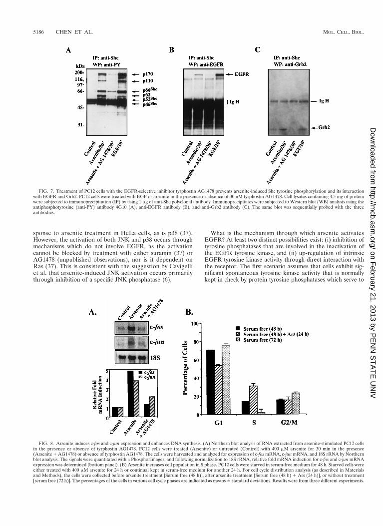
We next examined whether the tyrosine kinase activity ofEGFR was required for EGFR-Shc interaction, Shc tyrosinephosphorylation, and/or Grb2 translocation to the membranein response to arsenite treatment. PC12 cells were treated witharsenite for 30 min in the presence or absence of tyrphostinAG1478, after which Shc was immunoprecipitated from thecell extracts with the polyclonal Shc antibody. As a comparison,PC12 cells treated with EGF for 10 min were also examined.The coimmunoprecipitating tyrosine-phosphorylated proteinsin the Shc immune complexes were detected with the antiphos-photyrosine antibody 4G10 (Fig. 7A). Six tyrosine-phosphory-lated proteins, p46Shc, p52Shc, p66Shc, and three proteins withmolecular weights of 170,000, 110,000, and 62,000, were de-tected in the Shc immunoprecipitates from arsenite-treatedcells. This pattern was very similar to that observed in EGF-treated samples. In contrast, only residual tyrosine phosphor-ylation of p46Shc and p52Shc was observed in Shc immuno-precipitates from untreated control cells. In the presence oftyrphostin AG1478 (30 nM), the arsenite-induced tyrosinephosphorylation of the proteins was reduced to near basallevels (Fig. 7A). Immunoblotting of the same membrane withanti-EGFR antibody detected the p170EGFR in both thearsenite-treated and EGF-stimulated samples, although morep170EGFR was complexed with Shc in EGF-stimulated cellsthan in arsenite-treated cells (Fig. 7B). No p170EGFR was de-tected in Shc immunoprecipitates from either the control cellsor cells treated with arsenite in the presence of tyrphostinAG1478. Finally, in addition to its effects on Shc tyrosinephosphorylation and EGFR association, tyrphostin AG1478prevented the enrichment of Grb2 in the Shc immunocom-plexes (Fig. 7C). These data indicate that arsenite activates theRas/ERK cascade primarily through the EGFR tyrosine kinaseand the adapter protein Shc.
Arsenite induces expression of c-fos and c-jun and stimu-lates DNA synthesis. Growth factors and other mitogens havebeen shown to stimulate the expression of a number of imme-
diate-early genes that play a critical role in mediating prolif-eration (1). Among these are the c-fos and c-jun genes, whoseinductions are largely dependent on MAP kinase pathways (1,11, 27). Since arsenite treatment is also known to induce theexpression of c-fos and c-jun (6), we investigated the potentialrole of EGFR in mediating this response. Arsenite treatmentof PC12 cells resulted in the rapid induction of both c-fos andc-jun mRNA levels (4.8- and 3.8-fold, respectively [Fig. 8A]).Pretreatment of cells with tyrphostin AG1478 almost com-pletely abolished c-fos induction and partially inhibited c-junexpression, supporting a role for the EGFR in mediating theseeffects (Fig. 8A). To gain further evidence for the ability ofarsenite to influence cell proliferation, cells were examined byfluorescence-activated cell sorting analysis for changes incell cycle distribution following treatment with the agent. Cellswere first subjected to 48 h of serum starvation to enrich forcells in the G1 phase. At this point only a small number of cellswere present in S phase (14%). Cells were then either leftuntreated or treated with arsenite and examined 24 h later. Asshown in Fig. 8B, in the absence of arsenite treatment therewas a further reduction in the number of cells in S phase. Incontrast, arsenite treatment led to a significant increase in thenumber of cells in S phase (31%), indicative of increased DNAsynthesis. Thus, arsenite does appear to enhance cell prolifer-ation, consistent with its role as a tumor promoter.
DISCUSSION
We have previously demonstrated that arsenite treatmentresults in the activation of ERK MAP kinase through a Ras-dependent, suramin-sensitive pathway (37). Based on thisobservation, we hypothesized that the arsenite signal wastransmitted through a pathway mediated by growth factor re-ceptor-type tyrosine kinase(s). In this report, we have provideddirect evidence to support this view. In particular, we haveshown that EGFR undergoes tyrosine phosphorylation follow-ing arsenite treatment and that this event is associated withenhanced tyrosine phosphorylation of the Shc adapter protein,allowing for its increased interaction with Grb2.
The EGFR has been implicated in ERK activation by otherextracellular stress signals including UV-C, H2O2, and asbestos(22, 25, 28, 47, 50, 52, 65). However, in none of the previousstudies was the link between EGFR phosphorylation and ac-tivation of the ERK pathway established. Phosphorylation ofShc has been demonstrated to be a crucial step in the acti-vation of the Ras/MEK/ERK pathway in response to growthfactor stimulation (4). Our findings indicate that Shc is also animportant mediator in arsenite-induced ERK activation, serv-ing as an adapter for the recruitment of Grb2 and Sos to themembrane. Shc can be phosphorylated by both receptor-typeand nonreceptor-type tyrosine kinases (4). Since a variety ofstresses are known to activate nonreceptor-type tyrosine ki-nases (e.g., Src), it remains possible that these, rather than theintrinsic EGFR kinase activity, are responsible for Shc phos-phorylation following arsenite treatment (12, 52). It is alsoclear that EGFR can be phosphorylated by nonreceptor-typetyrosine kinases, and in certain instances, EGFR does not relyon its intrinsic kinase activity for its signaling functions (39, 64).For example, Src family tyrosine kinases have been shown tobe responsible for tyrosine phosphorylation of EGFR regu-lated by G-protein-coupled receptors (39). Similarly, JAK2,another nonreceptor tyrosine kinase, was found to phosphor-ylate EGFR in response to growth hormone treatment (64). Inboth instances, the phosphorylated EGFR was shown to beimportant for mediating downstream events, including Shc ty-rosine phosphorylation and activation of the ERK pathway.
FIG. 5. Central role of EGFR in mediating arsenite-induced ERK activation.(A) Effects of suramin and tyrphostin AG1478 on arsenite-induced ERK acti-vation. PC12 cells were treated with arsenite (400 mM) in the presence or ab-sence of 300 mM suramin or 40 nM tyrphostin AG1478 for 30 min. ERK activitywas analyzed by an immune complex kinase assay using MBP as a substrate andquantitated by using ImageQuant software (Molecular Dynamics). (B) Effects ofsuramin and tyrphostin AG1478 on arsenite-induced ERK phosphorylation. Celllysates from cells treated with arsenite in the absence or presence of suramin ortyrphostin AG1478 were analyzed by Western blotting using the phospho-ERK-specific antibody.
5184 CHEN ET AL. MOL. CELL. BIOL.
on February 21, 2013 by P
EN
N S
TA
TE
UN
IVhttp://m
cb.asm.org/
Dow
nloaded from

In our case, abrogation of EGFR autophosphorylation bytreatment with the highly specific EGFR inhibitor tyrphos-tin AG1478 also prevented the arsenite-induced Shc tyrosinephosphorylation and ERK activation. These findings arguestrongly that EGFR phosphorylation is crucial for activation ofthe ERK signaling cascade in response to arsenite and also thatintrinsic EGFR tyrosine kinase activity is required.
In addition to EGFR, other growth factor receptors includ-ing the T-cell receptor complex, interleukin-1a receptor, andthe NGF receptor TrkA have been implicated in the transduc-tion of stress signals by other toxic agents (21, 28, 30, 48, 50,51). In particular, we have found that overexpression of TrkAin PC12 cells leads to enhanced activation of ERK in responseto hydrogen peroxide treatment (21). Hence, it was somewhatsurprising in this study to find that the EGFR tyrosine kinase-selective inhibitor tyrphostin AG1478 completely abolished theERK activation in response to arsenite. This observation sug-gests that at least for PC12 cells, the response to arsenite ishighly dependent on EGFR. Further support for this notion
was obtained in experiments in which down-regulation ofEGFR led to an attenuation of Shc tyrosine phosphorylationand ERK activation.
The arsenite-induced activation of ERK is not restricted toPC12 cells, as we have observed that arsenite produces similareffects in other cell types, including Rat1 fibroblasts (37), hu-man epidermoid carcinoma A431 cells, and transformed hu-man embryonal kidney 293 cells (data not shown). However,our findings do contrast with several reports by others. Forexample, Rouse et al. (49) failed to detect ERK activation inarsenite-treated PC12 cells. Similarly, Cavigelli et al. (6) didnot observe activation of ERK in arsenite-treated HeLa cells,although JNK was highly activated. While the reasons for thedifferences between our studies and these remain unclear, theymay reflect differences in the EGFR content of the differentcell lines or strains used. In support of this notion, we haveobserved that compared to our PC12 cells, HeLa cells containvery low levels of EGFR (data not shown). Like Cavigelli et al.(6), we too have reported that JNK is highly activated in re-
FIG. 6. Pretreatment of cells with EGF inhibits a subsequent arsenite response. (A) PC12 cells were pretreated with EGF (1 mg/ml) for 1 h (1) or not pretreated(2), as indicated at the bottom. The cells were then washed and incubated in EGF-free medium for the time intervals shown at the bottom. The cells were furthertreated with EGF (100 ng/ml) for 10 min or NGF (100 ng/ml) for 5 min (second treatment, shown at the top). Cell lysates following these different treatments weresubjected to Western blot analysis using anti-EGFR polyclonal antibody (upper panel). The same blot was sequentially probed with anti-phospho-ERK-specific antibody(lower panel). (B) PC12 cells were pretreated with EGF (1 mg/ml) for 1 h (1) or not pretreated (2), as indicated at the bottom. The cells were then washed andincubated in EGF-free medium for the time intervals shown at the bottom. The cells were further treated (second treatment) with 400 mM arsenite for an additional30 min. Total cellular proteins were subjected to Western blot (WB) analysis using anti-phospho-ERK-specific antibody (upper panel) or the antiphosphotyrosine(anti-PY) monoclonal antibody 4G10 (lower panel). EGFR-mediated protein tyrosine phosphorylation stimulated by arsenite is indicated by arrows.
VOL. 18, 1998 ARSENITE ACTIVATES ERK THROUGH EGFR AND Shc 5185
on February 21, 2013 by P
EN
N S
TA
TE
UN
IVhttp://m
cb.asm.org/
Dow
nloaded from
sponse to arsenite treatment in HeLa cells, as is p38 (37).However, the activation of both JNK and p38 occurs throughmechanisms which do not involve EGFR, as the activationcannot be blocked by treatment with either suramin (37) orAG1478 (unpublished observations), nor is it dependent onRas (37). This is consistent with the suggestion by Cavigelliet al. that arsenite-induced JNK activation occurs primarilythrough inhibition of a specific JNK phosphatase (6).
What is the mechanism through which arsenite activatesEGFR? At least two distinct possibilities exist: (i) inhibition oftyrosine phosphatases that are involved in the inactivation ofthe EGFR tyrosine kinase, and (ii) up-regulation of intrinsicEGFR tyrosine kinase activity through direct interaction withthe receptor. The first scenario assumes that cells exhibit sig-nificant spontaneous tyrosine kinase activity that is normallykept in check by protein tyrosine phosphatases which serve to
FIG. 7. Treatment of PC12 cells with the EGFR-selective inhibitor tyrphostin AG1478 prevents arsenite-induced Shc tyrosine phosphorylation and its interactionwith EGFR and Grb2. PC12 cells were treated with EGF or arsenite in the presence or absence of 30 nM tyrphostin AG1478. Cell lysates containing 4.5 mg of proteinwere subjected to immunoprecipitation (IP) by using 1 mg of anti-Shc polyclonal antibody. Immunoprecipitates were subjected to Western blot (WB) analysis using theantiphosphotyrosine (anti-PY) antibody 4G10 (A), anti-EGFR antibody (B), and anti-Grb2 antibody (C). The same blot was sequentially probed with the threeantibodies.
FIG. 8. Arsenite induces c-fos and c-jun expression and enhances DNA synthesis. (A) Northern blot analysis of RNA extracted from arsenite-stimulated PC12 cellsin the presence or absence of tyrphostin AG1478. PC12 cells were treated (Arsenite) or untreated (Control) with 400 mM arsenite for 30 min in the presence(Arsenite 1 AG1478) or absence of tyrphostin AG1478. The cells were harvested and analyzed for expression of c-fos mRNA, c-jun mRNA, and 18S rRNA by Northernblot analysis. The signals were quantitated with a PhosphorImager, and following normalization to 18S rRNA, relative fold mRNA induction for c-fos and c-jun mRNAexpression was determined (bottom panel). (B) Arsenite increases cell population in S phase. PC12 cells were starved in serum-free medium for 48 h. Starved cells wereeither treated with 400 mM arsenite for 24 h or continual kept in serum-free medium for another 24 h. For cell cycle distribution analysis (as described in Materialsand Methods), the cells were collected before arsenite treatment [Serum free (48 h)], after arsenite treatment [Serum free (48 h) 1 Ars (24 h)], or without treatment[serum free (72 h)]. The percentages of the cells in various cell cycle phases are indicated as means 6 standard deviations. Results were from three different experiments.
5186 CHEN ET AL. MOL. CELL. BIOL.
on February 21, 2013 by P
EN
N S
TA
TE
UN
IVhttp://m
cb.asm.org/
Dow
nloaded from

inactivate the kinase. Studies of Knebel et al. (28) have pro-vided strong evidence that certain treatments such as radiation,oxidants, and alkylating agents act through such a mechanism.Essential thiol groups on the tyrosine phosphatases are thepresumed targets of the adverse agents, but the specific phos-phatases involved have not yet been identified. Given the highreactivity of arsenite for vicinal dithiols, it is possible thatarsenite also produces such effects, leading to elevated EGFRtyrosine kinase activity. The second possibility is that arsenitecan actually mimic the action of ligands to activate the receptortyrosine kinase. EGFR contains extracellular cysteine-rich do-mains that are important for ligand-triggered dimerization (24,53). By reacting with vicinal dithiols in these domains, arsenitecould alter the conformation of EGFR, resulting in an increaseof its intrinsic tyrosine kinase activity. In support of this model,we observed that cotreatment of cells with arsenite and or-thovanadate, a general tyrosine phosphatase inhibitor, syner-gistically enhanced the protein tyrosine phosphorylation (datanot shown).
Arsenite is a potent carcinogen (3) that is believed to play animportant role in the development of certain cancers such asskin and bladder tumors (3, 8, 9, 26). For example, arseniteingested through contaminated water supplies was found to beassociated with a high rate of bladder and skin cancer in re-gions of developing countries. In addition, a high rate of thesecancers has also been reported in patients receiving Fowler’ssolution (potassium arsenite) for the treatment of psoriasisin the 1940s to 1970s (8, 31). The potential role of EFGR inmediating arsenite’s carcinogenic effects is intriguing. Consis-tent with its tumor-promoting properties, arsenite treatmentled to enhanced DNA synthesis and induction of the prolifer-ation-associated genes c-fos and c-jun (Fig. 8). That the induc-tion of c-fos was almost completely abolished in the presence oftyrphostin AG1478 highlights the essential role of the EGFR inmediating this response. Abnormal activation of EGFR andrelated tyrosine kinases such as Neu, ErbB-3, and ErbB-4 hasbeen reported for a variety of human cancers (19, 33, 38, 42,58). In many cases, elevated Shc tyrosine phosphorylation hasalso been observed (45). Finally, suramin as well as antibodiestargeting members of the EGFR tyrosine kinase family haveproven effective in the treatment of certain tumors (33, 60).Our findings indicating that arsenite usurps the EGF/EGFRsignaling pathway to activate ERK provides a mechanism forits tumor-promoting properties and offers a potential target fortherapeutic strategies aimed at preventing or inhibiting arsen-ite-induced tumor growth.
ACKNOWLEDGMENTS
We are very grateful to H. Gram and G. Cooper for providing valu-able reagents. We are grateful to R. Wange, J. Staros, and M. Bernierfor stimulating discussion and valuable suggestions. We thank F. Chrestfor technical support.
REFERENCES1. Angel, P., and M. Karin. 1991. The role of Jun, Fos and the AP-1 complex
in cell-proliferation and transformation. Biochim. Biophys. Acta 1072:129–157.
2. Avruch, J., X. F. Zhang, and J. M. Kyriakis. 1994. Raf meets Ras: complet-ing the framework of a signal transduction pathway. Trends Biochem. Sci. 19:279–283.
3. Bagla, P., and J. Kaiser. 1996. India’s spreading health crisis draws globalarsenic experts. Science 274:174–175.
4. Bonfini, L., E. Migliaccio, G. Pelicci, L. Lanfrancone, and P. G. Pelicci. 1996.Not all Shc’s roads lead to Ras. Trends Biochem. Sci. 21:257–261.
5. Carpenter, G., and S. Cohen. 1976. 125I-labeled human epidermal growthfactor. Binding, internalization, and degradation in human fibroblasts. J. CellBiol. 71:159–171.
6. Cavigelli, M., W. W. Li, A. Lin, B. Su, K. Yoshioka, and M. Karin. 1996. Thetumor promoter arsenite stimulates AP-1 activity by inhibiting a JNK phos-phatase. EMBO J. 15:6269–6279.
7. Cowley, S., H. Paterson, P. Kemp, and C. J. Marshall. 1994. Activation ofMAP kinase kinase is necessary and sufficient for PC12 differentiation andfor transformation of NIH 3T3 cells. Cell 77:841–852.
8. Cuzick, J., S. Evans, M. Gillman, and E. D. Price. 1982. Medicinal arsenicand internal malignancies. Br. J. Cancer 45:904–911.
9. Cuzick, J., P. Sasieni, and S. Evans. 1992. Ingested arsenic, keratoses, andbladder cancer. Am. J. Epidemiol. 136:417–421.
10. Daub, H., F. U. Weiss, C. Wallasch, and A. Ullrich. 1996. Role of transac-tivation of the EGF receptor in signalling by G-protein-coupled receptors.Nature 379:557–560.
11. Davis, R. J. 1993. The mitogen-activated protein kinase signal transductionpathway. J. Biol. Chem. 268:14553–14556.
12. Devary, Y., R. A. Gottlieb, T. Smeal, and M. Karin. 1992. The mammalianultraviolet response is triggered by activation of Src tyrosine kinases. Cell 71:1081–1091.
13. Dudley, D. T., L. Pang, S. J. Decker, A. J. Bridges, and A. R. Saltiel. 1995. Asynthetic inhibitor of the mitogen-activated protein kinase cascade. Proc.Natl. Acad. Sci. USA 92:7686–7689.
14. Egan, S. E., and R. A. Weinberg. 1993. The pathway to signal achievement.Nature 365:781–783.
15. Feig, L. A. 1993. The many roads that lead to Ras. Science 260:767–768.16. Germolec, D. R., T. Yoshida, K. Gaido, J. L. Wilmer, P. P. Simeonova, F.
Kayama, F. Burleson, W. Dong, R. W. Lange, and M. I. Luster. 1996. Arsenicinduces overexpression of growth factors in human keratinocytes. Toxicol.Appl. Pharmacol. 141:308–318.
17. Gorospe, M., Y. Liu, Q. Xu, F. J. Chrest, and N. J. Holbrook. 1996. Inhibitionof G1 cyclin-dependent kinase activity during growth arrest of human breastcarcinoma cells by prostaglandin A2. Mol. Cell. Biol. 16:762–770.
18. Gotoh, Y., E. Nishida, and H. Sakai. 1990. Okadaic acid activates microtu-bule-associated protein kinase in quiescent fibroblastic cells. Eur. J. Bio-chem. 193:671–674.
19. Gullick, W. J. 1996. The c-erbB3/HER3 receptor in human cancer. CancerSurv. 27:339–349.
20. Guyton, K. Z., M. Gorospe, T. W. Kensler, and N. J. Holbrook. 1996.Mitogen-activated protein kinase (MAPK) activation by butylated hydroxy-toluene hydroperoxide: implications for cellular survival and tumor promo-tion. Cancer Res. 56:3480–3485.
21. Guyton, K. Z., M. Gorospe, X. Wang, Y. D. Mock, G. C. Kokkonen, Y. Liu,G. S. Roth, and N. J. Holbrook. Age-related changes in activation of mitogenactivated protein kinase cascades by oxidative stress. J. Invest. Dermatol.Symp. Proc., in press.
22. Guyton, K. Z., Y. Liu, M. Gorospe, Q. Xu, and N. J. Holbrook. 1996.Activation of mitogen-activated protein kinase by H2O2. Role in cell survivalfollowing oxidant injury. J. Biol. Chem. 271:4138–4142.
23. Hartwig, A., U. D. Groblinghoff, D. Beyersmann, A. T. Natarajan, R. Filon,and L. H. Mullenders. 1997. Interaction of arsenic (III) with nucleotideexcision repair in UV-irradiated human fibroblasts. Carcinogenesis 18:399–405.
24. Heldin, C. H. 1995. Dimerization of cell surface receptors in signal trans-duction. Cell 80:213–223.
25. Huang, R. P., J. X. Wu, Y. Fan, and E. D. Adamson. 1996. UV activatesgrowth factor receptors via reactive oxygen intermediates. J. Cell Biol. 133:211–220.
26. Johansson, S. L., and S. M. Cohen. 1997. Epidemiology and etiology ofbladder cancer. Semin. Surg. Oncol. 13:291–298.
27. Karin, M. 1995. The regulation of AP-1 activity by mitogen-activated proteinkinases. J. Biol. Chem. 270:16483–16486.
28. Knebel, A., H. J. Rahmsdorf, A. Ullrich, and P. Herrlich. 1996. Dephos-phorylation of receptor tyrosine kinases as target of regulation by radiation,oxidants or alkylating agents. EMBO J. 15:5314–5325.
29. Kolch, W., G. Heidecker, G. Kochs, R. Hummel, H. Vahidi, H. Mischak, G.Finkenzeller, D. Marme, and U. R. Rapp. 1993. Protein kinase C alphaactivates RAF-1 by direct phosphorylation. Nature 364:249–252.
30. Kyriakis, J. M., and J. Avruch. 1996. Protein kinase cascades activated bystress and inflammatory cytokines. Bioessays 18:567–577.
31. Lander, J. J., R. J. Stanley, H. W. Sumner, D. C. Boswell, and R. D. Aach.1975. Angiosarcoma of the liver associated with Fowler’s solution (potassiumarsenite). Gastroenterology 68:1582–1586.
32. Lengyel, E., H. Wang, R. Gum, C. Simon, Y. Wang, and D. Boyd. 1997.Elevated urokinase-type plasminogen activator receptor expression in a co-lon cancer cell line is due to a constitutively activated extracellular signal-regulated kinase-1-dependent signaling cascade. Oncogene 14:2563–2573.
33. Levitzki, A., and A. Gazit. 1995. Tyrosine kinase inhibition: an approach todrug development. Science 267:1782–1788.
34. Li, J. H., and T. G. Rossman. 1989. Mechanism of comutagenesis of sodiumarsenite with n-methyl-n-nitrosourea. Biol. Trace Elem. Res. 21:373–381.
35. Liu, Y., M. Gorospe, C. Yang, and N. J. Holbrook. 1995. Role of mitogen-activated protein kinase phosphatase during the cellular response to geno-toxic stress. Inhibition of c-Jun N-terminal kinase activity and AP-1-depen-dent gene activation. J. Biol. Chem. 270:8377–8380.
36. Liu, Y., K. Z. Guyton, M. Gorospe, Q. Xu, G. C. Kokkonen, Y. D. Mock, G. S.Roth, and N. J. Holbrook. 1996. Age-related decline in mitogen-activated
VOL. 18, 1998 ARSENITE ACTIVATES ERK THROUGH EGFR AND Shc 5187
on February 21, 2013 by P
EN
N S
TA
TE
UN
IVhttp://m
cb.asm.org/
Dow
nloaded from
protein kinase activity in epidermal growth factor-stimulated rat hepatocytes.J. Biol. Chem. 271:3604–3607.
37. Liu, Y., K. Z. Guyton, M. Gorospe, Q. Xu, J. C. Lee, and N. J. Holbrook.1996. Differential activation of ERK, JNK/SAPK and P38/CSBP/RK mapkinase family members during the cellular response to arsenite. Free RadicalBiol. Med. 21:771–781.
38. Lupu, R., M. Cardillo, L. Harris, M. Hijazi, and K. Rosenberg. 1995. Inter-action between erbB-receptors and heregulin in breast cancer tumor pro-gression and drug resistance. Semin. Cancer Biol. 6:135–145.
39. Luttrell, L. M., R. G. Della, B. T. van, D. K. Luttrell, and R. J. Lefkowitz.1997. Gbg subunits mediate Src-dependent phosphorylation of the epider-mal growth factor receptor. A scaffold for G protein-coupled receptor-mediated Ras activation. J. Biol. Chem. 272:4637–4644.
40. Mansour, S. J., W. T. Matten, A. S. Hermann, J. M. Candia, S. Rong,K. Fukasawa, W. G. Vande, and N. G. Ahn. 1994. Transformation of mam-malian cells by constitutively active MAP kinase kinase. Science 265:966–970.
41. Masui, H., L. Castro, and J. Mendelsohn. 1993. Consumption of EGF byA431 cells: evidence for receptor recycling. J. Cell Biol. 120:85–93.
42. Mitra, A. B., V. V. Murty, M. Pratap, P. Sodhani, and R. S. Chaganti. 1994.ERBB2 (HER2/neu) oncogene is frequently amplified in squamous cellcarcinoma of the uterine cervix. Cancer Res. 54:637–639.
43. Muller, K., F. O. Gombert, U. Manning, F. Grossmuller, P. Graff, H. Zaegel,J. F. Zuber, F. Freuler, C. Tschopp, and G. Baumann. 1996. Rapid identi-fication of phosphopeptide ligands for SH2 domains. Screening of peptidelibraries by fluorescence-activated bead sorting. J. Biol. Chem. 271:16500–16505.
44. Pang, L., T. Sawada, S. J. Decker, and A. R. Saltiel. 1995. Inhibition of MAPkinase kinase blocks the differentiation of PC-12 cells induced by nervegrowth factor. J. Biol. Chem. 270:13585–13588.
45. Pelicci, G., L. Lanfrancone, A. E. Salcini, A. Romano, S. Mele, B. M. Grazia,O. Segatto, F. P. Di, and P. G. Pelicci. 1995. Constitutive phosphorylation ofShc proteins in human tumors. Oncogene 11:899–907.
46. Porfiri, E., and F. McCormick. 1996. Regulation of epidermal growth factorreceptor signaling by phosphorylation of the ras exchange factor hSOS1.J. Biol. Chem. 271:5871–5877.
47. Rao, G. N. 1996. Hydrogen peroxide induces complex formation of SHC-Grb2-SOS with receptor tyrosine kinase and activates Ras and extracellularsignal-regulated protein kinases group of mitogen-activated protein kinases.Oncogene 13:713–719.
48. Rosette, C., and M. Karin. 1996. Ultraviolet light and osmotic stress: acti-vation of the JNK cascade through multiple growth factor and cytokinereceptors. Science 274:1194–1197.
49. Rouse, J., P. Cohen, S. Trigon, M. Morange, A. Alonso-Llamazares, D.Zamanillo, T. Hunt, and A. R. Nebreda. 1994. A novel kinase cascadetriggered by stress and heat shock that stimulates MAPKAP kinase-2 andphosphorylation of the small heat shock proteins. Cell 78:1027–1037.
50. Sachsenmaier, C., A. Radler-Pohl, R. Zinck, A. Nordheim, P. Herrlich, and
H. J. Rahmsdorf. 1994. Involvement of growth factor receptors in the mam-malian UVC response. Cell 78:963–972.
51. Schieven, G. L., and J. A. Ledbetter. 1994. Activation of tyrosine kinasesignal pathways by radiation and oxidative stress. Trends. Endocrinol.Metab. 5:383–387.
52. Schieven, G. L., R. S. Mittler, S. G. Nadler, J. M. Kirihara, J. B. Bolen, S. B.Kanner, and J. A. Ledbetter. 1994. ZAP-70 tyrosine kinase, CD45, and T cellreceptor involvement in UV- and H2O2-induced T cell signal transduction.J. Biol. Chem. 269:20718–20726.
53. Schlessinger, J. 1993. How receptor tyrosine kinases activate Ras. TrendsBiochem. Sci. 18:273–275.
54. Schmidt, C. M., I. H. McKillop, P. A. Cahill, and J. V. Sitzmann. 1997.Increased MAPK expression and activity in primary human hepatocellularcarcinoma. Biochem. Biophys. Res. Commun. 236:54–58.
55. Segatto, O., G. Pelicci, S. Giuli, G. Digiesi, F. P. Di, J. McGlade, T. Pawson,and P. G. Pelicci. 1993. Shc products are substrates of erbB-2 kinase. On-cogene 8:2105–2112.
56. Seger, R., and E. G. Krebs. 1995. The MAPK signaling cascade. FASEB J.9:726–735.
57. Sivaraman, V. S., H. Wang, G. J. Nuovo, and C. C. Malbon. 1997. Hyper-expression of mitogen-activated protein kinase in human breast cancer.J. Clin. Invest. 99:1478–1483.
58. Slamon, D. J., W. Godolphin, L. A. Jones, J. A. Holt, S. G. Wong, D. E. Keith,W. J. Levin, S. G. Stuart, J. Udove, and A. Ullrich. 1989. Studies of theHER-2/neu proto-oncogene in human breast and ovarian cancer. Science244:707–712.
59. Snow, E. T. 1992. Metal carcinogenesis: mechanistic implications. Pharma-col. Ther. 53:31–65.
60. Stein, C. A. 1993. Suramin: a novel antineoplastic agent with multiple po-tential mechanisms of action. Cancer Res. 53:2239–2248.
61. Szeberenyi, J., H. Cai, and G. M. Cooper. 1990. Effect of a dominant inhib-itory Ha-ras mutation on neuronal differentiation of PC12 cells. Mol. Cell.Biol. 10:5324–5332.
62. Waters, S. B., K. H. Holt, S. E. Ross, L. J. Syu, K. L. Guan, A. R. Saltiel, G. A.Koretzky, and J. E. Pessin. 1995. Desensitization of Ras activation by a feed-back disassociation of the SOS-Grb2 complex. J. Biol. Chem. 270:20883–20886.
63. Wiencke, J. K., J. W. Yager, A. Varkonyi, M. Hultner, and L. H. Lutze. 1997.Study of arsenic mutagenesis using the plasmid shuttle vector pZ189 prop-agated in DNA repair proficient human cells. Mutat. Res. 386:335–344.
64. Yamauchi, T., K. Ueki, K. Tobe, H. Tamemoto, N. Sekine, M. Wada, M.Honjo, M. Takahashi, T. Takahashi, H. Hirai, T. Tushima, Y. Akanuma, T.Fujita, I. Komuro, Y. Yazaki, and T. Kadowaki. 1997. Tyrosine phosphory-lation of the EGF receptor by the kinase Jak2 is induced by growth hormone.Nature 390:91–96.
65. Zanella, C. L., J. Posada, T. R. Tritton, and B. T. Mossman. 1996. Asbestoscauses stimulation of the extracellular signal-regulated kinase 1 mitogen-activated protein kinase cascade after phosphorylation of the epidermalgrowth factor receptor. Cancer Res. 56:5334–5338.
5188 CHEN ET AL. MOL. CELL. BIOL.
on February 21, 2013 by P
EN
N S
TA
TE
UN
IVhttp://m
cb.asm.org/
Dow
nloaded from