ebi regulates epidermal growth factor receptor signaling...
13
ebi regulates epidermal growth factor receptor signaling pathways in Drosophila Xinzhong Dong, 1,4 Leo Tsuda, 1,4 Kenton H. Zavitz, 1 Michael Lin, 1 Songhui Li, 3 Richard W. Carthew, 3 and S. Lawrence Zipursky 1,2,5 1 Department of Biological Chemistry, Molecular Biology Institute and 2 Howard Hughes Medical Institute, The School of Medicine, University of California, Los Angeles, California 90095 USA; 3 Department of Biological Sciences, University of Pittsburgh, Pittsburgh, Pennsylvania 15260 USA ebi regulates the epidermal growth factor receptor (EGFR) signaling pathway at multiple steps in Drosophila development. Mutations in ebi and Egfr lead to similar phenotypes and show genetic interactions. However, ebi does not show genetic interactions with other RTKs (e.g., torso) or with components of the canonical Ras/MAP kinase pathway. ebi encodes an evolutionarily conserved protein with a unique amino terminus, distantly related to F-box sequences, and six tandemly arranged carboxy-terminal WD40 repeats. The existence of closely related proteins in yeast, plants, and humans suggests that ebi functions in a highly conserved biochemical pathway. Proteins with related structures regulate protein degradation. Similarly, in the developing eye, ebi promotes EGFR-dependent down-regulation of Tramtrack88, an antagonist of neuronal development. [Key Words: ebi; EGFR; tramtrack88; F box; WD40 repeats; protein degradation] Received January 13, 1999; revised version accepted Feburary 22, 1999. Epidermal growth factor receptors (EGFRs) have a cen- tral role in vertebrate and invertebrate development (for review, see van der Geer et al. 1994; Wassarman et al. 1995; Freeman 1998). Biochemical studies largely in mammalian systems and genetic studies in Caenorhab- ditis elegans and Drosophila have led to a detailed de- scription of the signal transduction pathways elicited by activation of the EGFR (for review, see Kayne and Stern- berg 1995; Schwietzer and Shilo 1997). These include the Ras/MAP kinase (MAPK), Ca 2+ , and phosphatidyl inosi- tol-dependent signaling pathways (for review, see Kaz- lauskas 1994). In Drosophila, the Ras/MAP kinase cas- cade is the prominent signaling pathway triggered by the EGFR. Two other fly receptor tyrosine kinases (RTKs) that control patterning and cell fate specification, Torso (for review, see Duffy and Perrimon 1994) and Sevenless (Sev) (for review, see Zipursky and Rubin 1994), do so largely, if not exclusively, through activation of this pathway. The development of the R7 photoreceptor neuron in the fly eye has proved to be a system amenable to de- tailed genetic dissection of RTK signaling pathways (Wassarman et al. 1995). Whereas the Sev RTK is re- quired for the development of R7 only, EGFR is essential for the development of most, if not all, cells, including R7 (for review, see Freeman 1996a). This dual RTK re- quirement is intriguing. Constitutively active overex- pressed forms of both Sev and EGFR are sufficient to induce R7 development. Furthermore, overexpression of Spitz, a ligand for EGFR, can partially rescue R7 devel- opment in a sev null mutant background. These findings have led Freeman (1996b) to propose that signals from Sev and EGFR are qualitatively equivalent. This is con- sistent with previous findings that overexpression of ac- tivated forms of proteins in the Ras/MAP kinase path- way induce receptor-independent R7 development (For- tini et al. 1992; Dickson et al. 1992a; Brunner et al. 1994). These observations support the view that activation of the Ras/MAPK pathway is sufficient to induce R7 devel- opment during normal development. The activities of three transcription factors are modu- lated by the MAPK signaling pathway in the R7 precur- sor cell. Two ETS-domain-containing transcription fac- tors, Yan and Pointed, are direct targets of MAPK phos- phorylation: Pointed is activated by phosphorylation and promotes R7 induction (O’Neil et al. 1994), whereas Yan is inhibited by phosphorylation and acts as a transcrip- tional repressor (Rebay and Rubin 1995). Inactivation of a second repressor, Tramtrack88 (Ttk88), also is required for R7 development (Xiong and Montell 1993). Ttk88 is down-regulated via a MAPK-induced protein degradation 4 These authors contributed equally to this work and are listed alphabeti- cally. 5 Corresponding author. E-MAIL [email protected]; FAX (310) 206-3800. 954 GENES & DEVELOPMENT 13:954–965 © 1999 by Cold Spring Harbor Laboratory Press ISSN 0890-9369/99 $5.00; www.genesdev.org Cold Spring Harbor Laboratory Press on June 22, 2018 - Published by genesdev.cshlp.org Downloaded from
Transcript of ebi regulates epidermal growth factor receptor signaling...
ebi regulates epidermal growth factorreceptor signaling pathwaysin DrosophilaXinzhong Dong,1,4 Leo Tsuda,1,4 Kenton H. Zavitz,1 Michael Lin,1 Songhui Li,3
Richard W. Carthew,3 and S. Lawrence Zipursky1,2,5
1Department of Biological Chemistry, Molecular Biology Institute and 2Howard Hughes Medical Institute, The Schoolof Medicine, University of California, Los Angeles, California 90095 USA; 3Department of Biological Sciences, Universityof Pittsburgh, Pittsburgh, Pennsylvania 15260 USA
ebi regulates the epidermal growth factor receptor (EGFR) signaling pathway at multiple steps in Drosophiladevelopment. Mutations in ebi and Egfr lead to similar phenotypes and show genetic interactions. However,ebi does not show genetic interactions with other RTKs (e.g., torso) or with components of the canonicalRas/MAP kinase pathway. ebi encodes an evolutionarily conserved protein with a unique amino terminus,distantly related to F-box sequences, and six tandemly arranged carboxy-terminal WD40 repeats. The existenceof closely related proteins in yeast, plants, and humans suggests that ebi functions in a highly conservedbiochemical pathway. Proteins with related structures regulate protein degradation. Similarly, in thedeveloping eye, ebi promotes EGFR-dependent down-regulation of Tramtrack88, an antagonist of neuronaldevelopment.
[Key Words: ebi; EGFR; tramtrack88; F box; WD40 repeats; protein degradation]
Received January 13, 1999; revised version accepted Feburary 22, 1999.
Epidermal growth factor receptors (EGFRs) have a cen-tral role in vertebrate and invertebrate development (forreview, see van der Geer et al. 1994; Wassarman et al.1995; Freeman 1998). Biochemical studies largely inmammalian systems and genetic studies in Caenorhab-ditis elegans and Drosophila have led to a detailed de-scription of the signal transduction pathways elicited byactivation of the EGFR (for review, see Kayne and Stern-berg 1995; Schwietzer and Shilo 1997). These include theRas/MAP kinase (MAPK), Ca2+, and phosphatidyl inosi-tol-dependent signaling pathways (for review, see Kaz-lauskas 1994). In Drosophila, the Ras/MAP kinase cas-cade is the prominent signaling pathway triggered by theEGFR. Two other fly receptor tyrosine kinases (RTKs)that control patterning and cell fate specification, Torso(for review, see Duffy and Perrimon 1994) and Sevenless(Sev) (for review, see Zipursky and Rubin 1994), do solargely, if not exclusively, through activation of thispathway.
The development of the R7 photoreceptor neuron inthe fly eye has proved to be a system amenable to de-tailed genetic dissection of RTK signaling pathways(Wassarman et al. 1995). Whereas the Sev RTK is re-
quired for the development of R7 only, EGFR is essentialfor the development of most, if not all, cells, includingR7 (for review, see Freeman 1996a). This dual RTK re-quirement is intriguing. Constitutively active overex-pressed forms of both Sev and EGFR are sufficient toinduce R7 development. Furthermore, overexpression ofSpitz, a ligand for EGFR, can partially rescue R7 devel-opment in a sev null mutant background. These findingshave led Freeman (1996b) to propose that signals fromSev and EGFR are qualitatively equivalent. This is con-sistent with previous findings that overexpression of ac-tivated forms of proteins in the Ras/MAP kinase path-way induce receptor-independent R7 development (For-tini et al. 1992; Dickson et al. 1992a; Brunner et al. 1994).These observations support the view that activation ofthe Ras/MAPK pathway is sufficient to induce R7 devel-opment during normal development.
The activities of three transcription factors are modu-lated by the MAPK signaling pathway in the R7 precur-sor cell. Two ETS-domain-containing transcription fac-tors, Yan and Pointed, are direct targets of MAPK phos-phorylation: Pointed is activated by phosphorylation andpromotes R7 induction (O’Neil et al. 1994), whereas Yanis inhibited by phosphorylation and acts as a transcrip-tional repressor (Rebay and Rubin 1995). Inactivation ofa second repressor, Tramtrack88 (Ttk88), also is requiredfor R7 development (Xiong and Montell 1993). Ttk88 isdown-regulated via a MAPK-induced protein degradation
4These authors contributed equally to this work and are listed alphabeti-cally.5Corresponding author.E-MAIL [email protected]; FAX (310) 206-3800.
954 GENES & DEVELOPMENT 13:954–965 © 1999 by Cold Spring Harbor Laboratory Press ISSN 0890-9369/99 $5.00; www.genesdev.org
Cold Spring Harbor Laboratory Press on June 22, 2018 - Published by genesdev.cshlp.orgDownloaded from
pathway. RTK signaling induces the expression of phyl-lopod (phyl) (Dickson et al. 1995). Phyl and Sina, theproduct of the seven in absentia gene, then bind toTtk88 and promote its ubiquitin-dependent degradation(Li et al. 1997; Tang et al. 1997).
Proteolysis has an important role in signaling down-stream of activated receptors in several developmentalsystems. Activation of the NF-kB transcription factorand its Rel homologs in Drosophila (i.e., Dorsal and Dif)requires signal-dependent phosphorylation of the inhibi-tor, IkB or Cactus, followed by its degradation via a ubiq-uitin-dependent pathway (for review, see Verma et al.1995). Degradation of the inhibitor allows NF-kB to enterthe nucleus and directly regulate gene expression. Prote-olysis also is important in transmitting signals in theWingless (Wg), Hedgehog (Hh), and Notch (N) pathways.Slimb is required for Wg and Hh signaling and regulatesthe processing of intracellular components in these path-ways, Cubitus interruptus and Armadillo, respectively(Jiang and Struhl 1998). On the basis of genetic and pro-tein-binding studies, C. elegans Sel-10 was proposed tobe a direct negative regulator of Notch/Lin-12 signaling(Hubbard et al. 1997). Both sel-10 and slimb encode F-box/WD40 repeat proteins homologous to Saccharomy-ces cerevisiae Cdc4, which itself targets a specific phos-phorylated cell cycle-related substrate, Sic1, to ubiqui-tin-mediated protein degradation (for review, see Pattonet al. 1998).
During the course of a screen for mutations affectingeye development, we identified mutations in a gene wenow designate ebi (‘shrimp’ in Japanese). Genetic analy-sis revealed that ebi functions in EGFR pathways regu-lating aspects of oogenesis and embryogenesis, and inimaginal disc development. ebi is highly homologous toproteins in yeast, plants, and humans and is structurallyrelated to Cdc4/Sel-10/Slimb. We demonstrate that ebiis required in R7 development for down-regulation ofTtk88 in response to EGFR signaling.
Results
Loss-of-function mutations in ebi and Egfr havesimilar phenotypes
ebi mutations were identified in a screen for enhancersof an eye mutant called roughex that plays a key role inregulating cell cycle progression in the developing eye(see Materials and Methods; Dong et al. 1997). As a con-sequence of cell cycle defects, photoreceptor differentia-tion and pattern formation in the eye are disrupted.Whereas cell cycle regulators enhance and suppress theprimary cell cycle phenotype (Thomas et al. 1994; Donget al. 1997; K.H. Zavitz and S.L. Zipursky, unpubl.), mu-tations in other loci, such as Star and Egfr (i.e., encodingthe EGF receptor), modify the differentiation phenotypeonly and not the earlier cell cycle defects. Like Star andEgfr, ebi enhances the differentiation phenotype. Theseobservations led us to consider the relationship betweenthe EGFR signaling pathway and ebi. In this section wepresent evidence that ebi participates in EGFR signaling
pathways. ebiE4, ebiE90, and ebiP7 are null, strong, andweak alleles, respectively (see below).
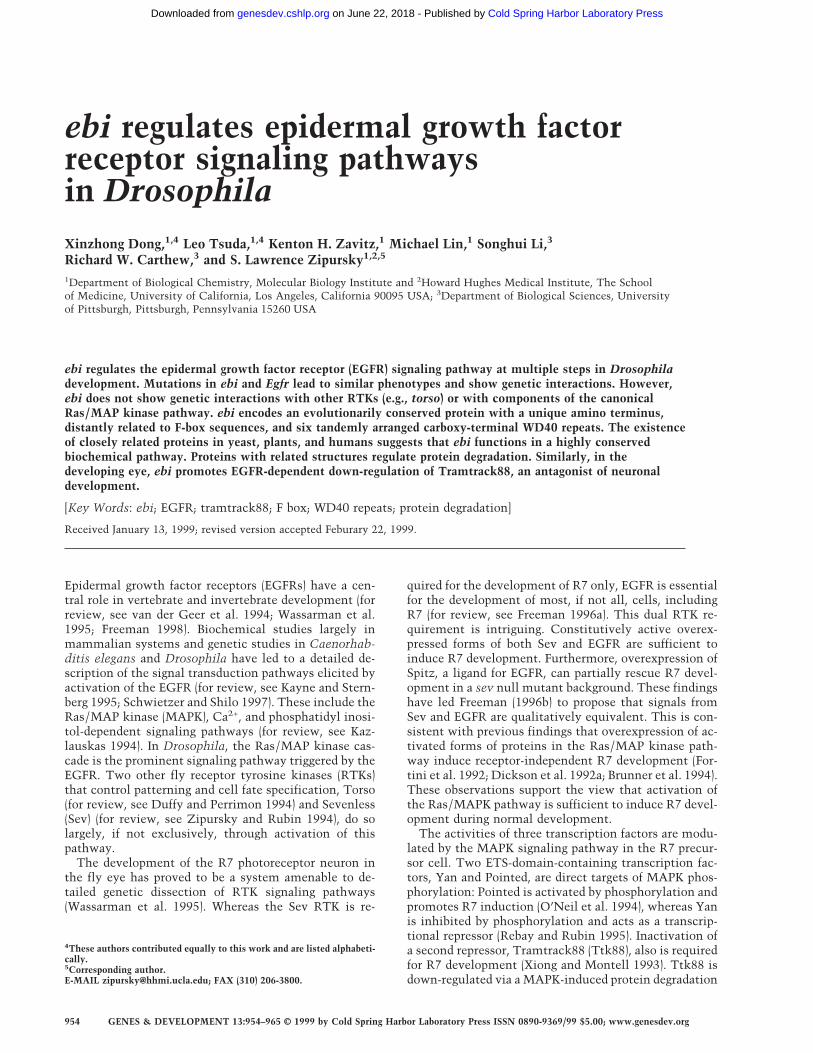
That ebi functions in the EGFR pathway was sug-gested initially by phenotypes of a viable heteroalleliccombination of ebi (i.e., ebiP7/ebiE90). These flies exhibitphenotypes similar to weak loss-of-function Egfr alleles(i.e., Egfrtop1/Egfrf2) including partial female sterility re-sulting from partially ventralized eggs (Fig. 1A–C), wingvein defects, short bristles, and abnormal eyes (i.e., rougheyes) (data not shown). Further evidence that ebi partici-pates in the EGFR pathway was provided by genetic in-teractions between them (also see below). For instance,flies carrying two different alleles of Egfr (Egfrtop1/Egfrf2)have a weak rough-eye phenotype, which is enhanced inflies that are heterozygous for ebi (data not shown).
ebi and Egfr mutant embryos are similar also. Homo-zygous ebi null mutant embryos (ebiE4) exhibit a tail-upor U-shaped embryo with head defects (Fig. 1E). Embryoslacking both the zygotic and maternal contributions ofebi were created using ovoD and FRT/FLP-induced re-combination (see Materials and Methods). This resultedin a more severe phenotype, including the loss of ventraldenticle belt structures and a tightly curled morphologyindicating a marked failure in germ-band retraction (Fig.1F). Severe head defects were also observed. In contrastto Egfr mutants, some residual ventral cuticular struc-
Figure 1. ebi mutants exhibit egg and embryonic cuticle phe-notypes. (A) Two dorsal appendages (arrow) are located at thedorsal anterior region of the wild-type egg. (B) Females harbor-ing a weak Egfr allele, Egfrtop1/Egfrtop1, produce ventralized eggswith fused dorsal appendages (arrow). (C) Eggs laid by femalesmutant for ebi (ebiE90/ebiP7) are also ventralized (arrow). (D)Wild-type embryos. (E) Embryos lacking maternal, but not zy-gotic, contribution of ebi exhibit a tail-up embryo, indicatingfailure of germ-band retraction. These embryos also have headdefects. Embryos lacking maternal ebi were generated using theovoD method (see Materials and Methods). (F) Embryos lackingmaternal and zygotic ebi exhibit more severe phenotypes, in-cluding loss of ventral denticle belt (db) structures and a tightlycurled morphology. In addition, severe head defects and mal-formed posterior filzkorper (Fk) were observed. Anterior is tothe right.
ebi and Ttk88 in R7 development
GENES & DEVELOPMENT 955
Cold Spring Harbor Laboratory Press on June 22, 2018 - Published by genesdev.cshlp.orgDownloaded from
tures remain in embryos lacking both the zygotic andmaternal contributions of ebi (Fig. 1F).
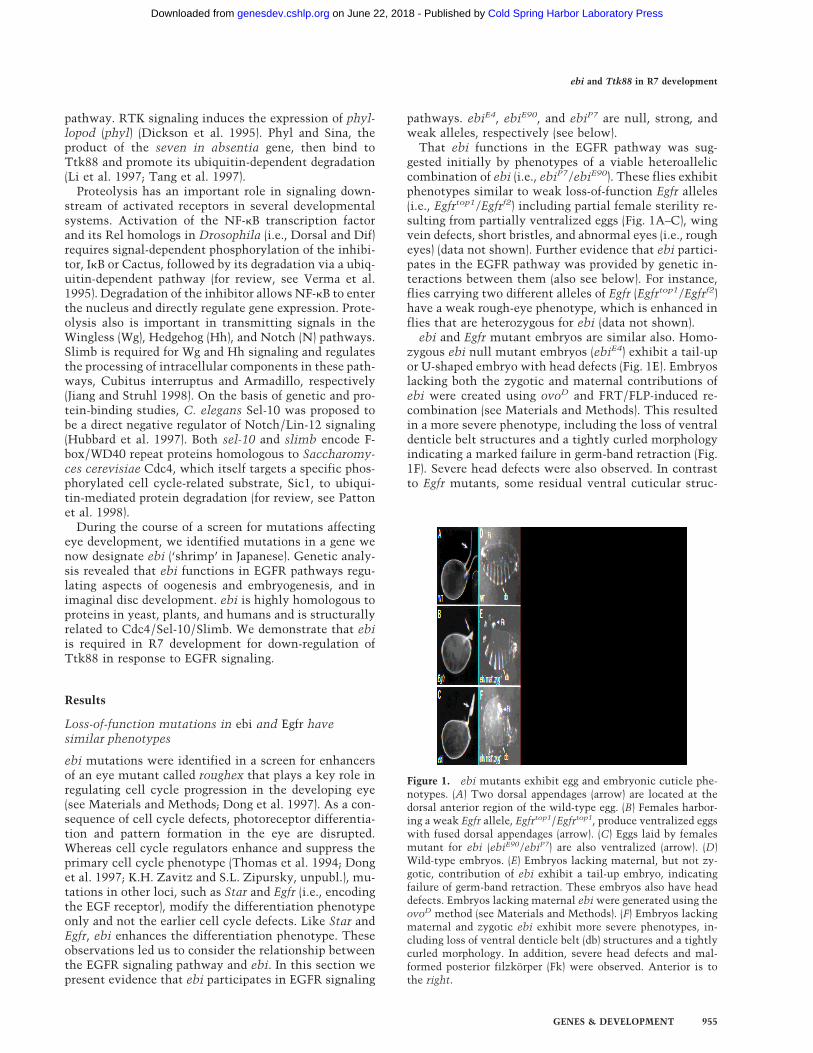
Loss of ebi also affects Egfr-dependent expression ofgenes in the embryo. The EGFR ligand Spitz is expressedalong the ventral midline and induces expression of dif-ferent target genes, including fasciclin III (fasIII) and or-thodenticle (otd), in cells located in more lateral posi-tions. In zygotic null Egfr mutants both otd and FasIIIexpression are lost (Kim and Crews 1993; Raz and Shilo1993). In wild-type stage 11/12 embryos, FasIII protein isbroadly distributed in the visceral mesoderm and in abilaterally symmetric cluster of cells flanking the mid-line of the ventral ectoderm (Fig. 2A). In ebi mutant em-bryos lacking both maternal and zygotic contribution,FasIII expression is largely abolished, although some re-sidual patches of staining remain (Fig. 2C). Egfr-indepen-
dent expression of FasIII in the anterior-most region ofthe embryo is unaffected in ebi mutants (data notshown). In wild-type stage 10/11 embryos, otd mRNA isexpressed in the preantennal head region and in the ven-tral-most ectoderm (Fig. 2B). In ebi mutant embryos, otdexpression was markedly reduced (Fig. 2D). These datasuggested that ebi may be a component in the EGFRsignal transduction pathway.
To assess whether ebi encoded a hitherto unidentifiedregulator in the Ras/MAP kinase pathway, we assessedits role in the Torso RTK pathway. Torso controls thedevelopment of the anterior and posterior termini of theembryo (for review, see Duffy and Perrimon 1994). Ras,Raf, MEK, and MAPK participate in both the EGFR andTorso RTK pathways. The expression of Torso targetgenes huckebein (hkb) and tailless (tll) in embryos en-tirely deficient in ebi (i.e., lacking both maternal andzygotic ebi) were indistinguishable from wild type (Fig.2E–H). Although filzkorper material is present, it is mal-formed. It is unlikely that this cuticular defect is due toabnormal Torso signaling but, rather, arises later in de-velopment as a result of a disruption in the EGFR sig-naling pathway.
In summary, ebi mutant phenotypes assessed usingboth molecular and morphological criteria are similar toEgfr mutations. Furthermore, ebi does not function in allRTK pathways, as Torso-induced terminal developmentis ebi independent. These data indicate that ebi, eitherdirectly or indirectly, regulates EGFR signaling. As a steptoward understanding the role of ebi in the context of aspecific developmental process, we have assessed therole of ebi in R7 development in the compound eyethrough both genetic and molecular studies.
ebi is required for R7 development
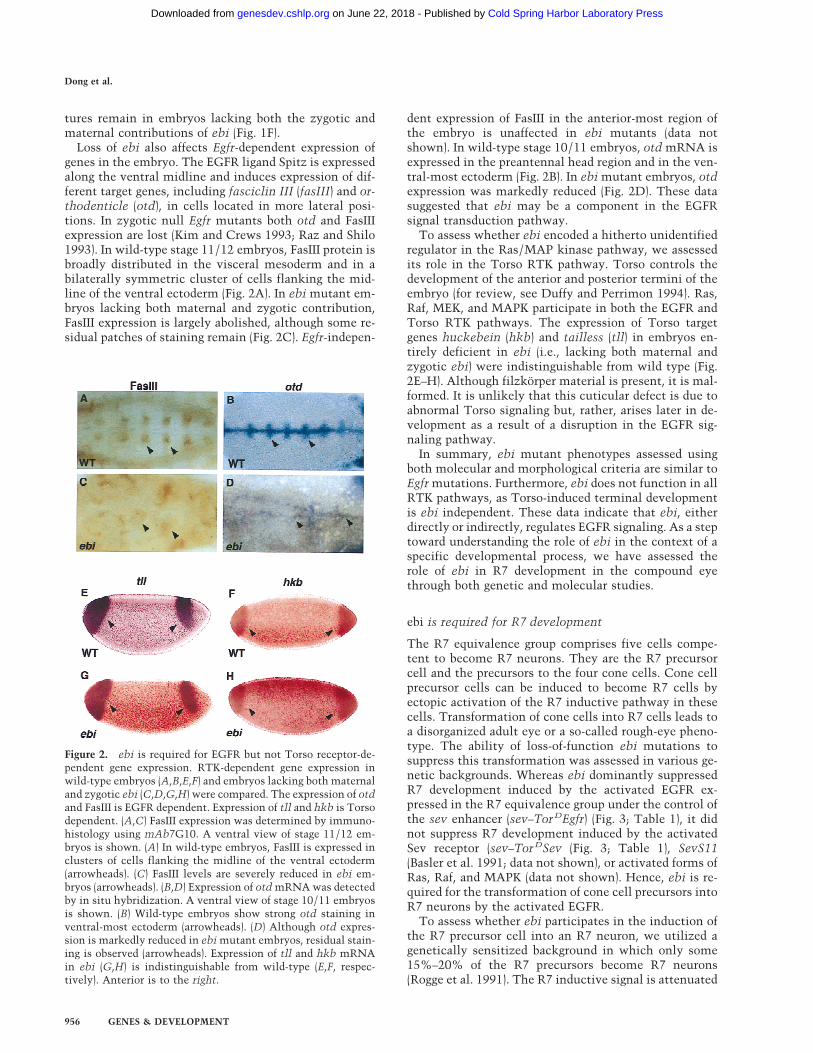
The R7 equivalence group comprises five cells compe-tent to become R7 neurons. They are the R7 precursorcell and the precursors to the four cone cells. Cone cellprecursor cells can be induced to become R7 cells byectopic activation of the R7 inductive pathway in thesecells. Transformation of cone cells into R7 cells leads toa disorganized adult eye or a so-called rough-eye pheno-type. The ability of loss-of-function ebi mutations tosuppress this transformation was assessed in various ge-netic backgrounds. Whereas ebi dominantly suppressedR7 development induced by the activated EGFR ex-pressed in the R7 equivalence group under the control ofthe sev enhancer (sev–TorDEgfr) (Fig. 3; Table 1), it didnot suppress R7 development induced by the activatedSev receptor (sev–TorDSev (Fig. 3; Table 1), SevS11(Basler et al. 1991; data not shown), or activated forms ofRas, Raf, and MAPK (data not shown). Hence, ebi is re-quired for the transformation of cone cell precursors intoR7 neurons by the activated EGFR.
To assess whether ebi participates in the induction ofthe R7 precursor cell into an R7 neuron, we utilized agenetically sensitized background in which only some15%–20% of the R7 precursors become R7 neurons(Rogge et al. 1991). The R7 inductive signal is attenuated
Figure 2. ebi is required for EGFR but not Torso receptor-de-pendent gene expression. RTK-dependent gene expression inwild-type embryos (A,B,E,F) and embryos lacking both maternaland zygotic ebi (C,D,G,H) were compared. The expression of otdand FasIII is EGFR dependent. Expression of tll and hkb is Torsodependent. (A,C) FasIII expression was determined by immuno-histology using mAb7G10. A ventral view of stage 11/12 em-bryos is shown. (A) In wild-type embryos, FasIII is expressed inclusters of cells flanking the midline of the ventral ectoderm(arrowheads). (C) FasIII levels are severely reduced in ebi em-bryos (arrowheads). (B,D) Expression of otd mRNA was detectedby in situ hybridization. A ventral view of stage 10/11 embryosis shown. (B) Wild-type embryos show strong otd staining inventral-most ectoderm (arrowheads). (D) Although otd expres-sion is markedly reduced in ebi mutant embryos, residual stain-ing is observed (arrowheads). Expression of tll and hkb mRNAin ebi (G,H) is indistinguishable from wild-type (E,F, respec-tively). Anterior is to the right.
Dong et al.
956 GENES & DEVELOPMENT
Cold Spring Harbor Laboratory Press on June 22, 2018 - Published by genesdev.cshlp.orgDownloaded from
by using a strong hypomorphic allele of sev (sevE4) and aweak gain-of-function mutation in the Ras activator, en-coded by the son-of-sevenless gene, SosJC2. Aside fromthe loss of the majority of the R7 cells, development ofthe eye in this genetic background is otherwise indistin-guishable from wild type. ebi is a dominant enhancer ofthis phenotype, as are Egfr loss-of-function mutations(Table 2). These data are consistent with previous stud-ies by Freeman (1996b), demonstrating a requirement forboth the EGFR and Sev receptor in R7 induction. Hence,ebi is required for induction of the R7 precursor cell intoan R7 neuron and for transformation of cone cell precur-sors into R7 in response to ectopic activation of EGFR.That Ttk88 down-regulation is required for R7 inductionof the R7 precursor cell is supported by the finding thatTtk88 mutations are dominant suppressors of theSevE4;SosJC2/+ phenotype (Table 2).
To assess the role of ebi on R7 development in anotherwise wild-type background, we sought to generatehomozygous null mutant clones. We were unable to gen-erate such clones using X-ray and heat shock Flp-induced
mitotic recombination. Hence, like Egfr (Xu and Rubin1993; Dominguez et al. 1998; Kumar et al. 1998 ), ebi isrequired for cell proliferation and/or survival during theproliferative phase of disc development. To increase theefficiency of Flp-induced mitotic recombination, weused a Flp source driven by the eyeless (ey) promoter.The ey promoter drives expression from the earliest cell
Figure 3. ebi is a dominant suppressor of ectopic R7 develop-ment induced by an activated EGFR. (A) A schematic represen-tation of the sev–TorDEgfr construct (Reichman-Fried et al.1994). (B–D) Scanning electron micrographs of adult eyes show-ing sev–TorDEgfr alone (B), in an SosSF15/+ background (C), andin an ebiE90/+ background. Like Sos, ebi is a dominant suppres-sor of sev–TorDEgfr. (E) A schematic representation of sev–TorD
-
Sev (Dickson et al. 1992b). (F–H) Scanning electron micrographsof adult eyes showing sev-TorDSev alone (F), in an SosSF15/+background (G) and in an ebiE90/+ background (H). AlthoughSos suppresses the sev–TorDSev phenotype, ebi does not (seeTable 1). Anterior is to the right.
Table 1. ebi suppresses sev–TorD Egfr
GenotypeExtra R cells
per ommatidia
sev–TorDEgfr;++
1.8 (n = 159)
sev–TorDEgfr;SosSF15
+
0.5 (n = 142)
sev–TorDEgfr;ebiE90
+
0.5 (n = 122)
sev–TorDSev+
2.9 (n = 89)
sev–TorDSev
SosSF15
1.9 (n =113)
sev–TorDSev
ebiE90
3.0 (n = 97)
SosSF15 is a null allele (Rogge et al. 1991). (n) The number ofommatidia scored. Between four and five eyes were analyzed insemithin sections for each genotype.
Table 2. ebi and ttk regulate R7 development
Genotype
Ommatidiacontaining
R7 cells (%)
sevE4
Y;SosJC2
+
14.7 (n = 2527)
sevE4
Y;SosJC2
Egfrf2
0.1 (n = 1045)
sevE4
Y;SosJC2
ebiE4
0.2 (n = 2036)
sevE4
Y;SosJC2
ebiE90
0.3 (n = 1144)
sevE4
Y;SosJC2
+ ;ttk1
+
29.6 (n = 2527)
sevE4
Y;SosJC2
+ ;ttk0219
+
31.1 (n = 1219)
In ttk1, mRNA encoding p88 is missing (Xiong and Montell1993). ttk0219 is an enhancer trap allele with a P element in-serted upstream of the ttk transcription unit (Lai et al. 1996).Egfrf2 is a null allele (Clifford and Schupbach 1994). (n) Thenumber of ommatidia scored. Between 7 and 12 eyes were ana-lyzed for each genotype.
ebi and Ttk88 in R7 development
GENES & DEVELOPMENT 957
Cold Spring Harbor Laboratory Press on June 22, 2018 - Published by genesdev.cshlp.orgDownloaded from
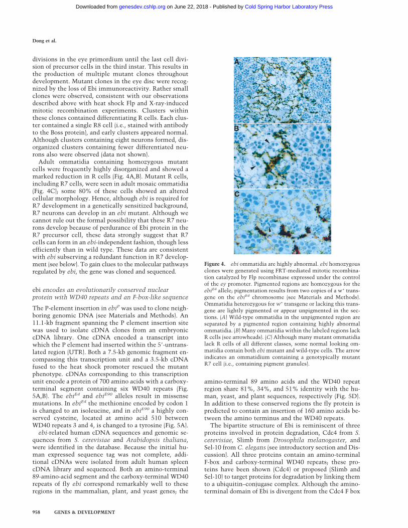
divisions in the eye primordium until the last cell divi-sion of precursor cells in the third instar. This results inthe production of multiple mutant clones throughoutdevelopment. Mutant clones in the eye disc were recog-nized by the loss of Ebi immunoreactivity. Rather smallclones were observed, consistent with our observationsdescribed above with heat shock Flp and X-ray-inducedmitotic recombination experiments. Clusters withinthese clones contained differentiating R cells. Each clus-ter contained a single R8 cell (i.e., stained with antibodyto the Boss protein), and early clusters appeared normal.Although clusters containing eight neurons formed, dis-organized clusters containing fewer differentiated neu-rons also were observed (data not shown).
Adult ommatidia containing homozygous mutantcells were frequently highly disorganized and showed amarked reduction in R cells (Fig. 4A,B). Mutant R cells,including R7 cells, were seen in adult mosaic ommatidia(Fig. 4C); some 80% of these cells showed an alteredcellular morphology. Hence, although ebi is required forR7 development in a genetically sensitized background,R7 neurons can develop in an ebi mutant. Although wecannot rule out the formal possibility that these R7 neu-rons develop because of perdurance of Ebi protein in theR7 precursor cell, these data strongly suggest that R7cells can form in an ebi-independent fashion, though lessefficiently than in wild type. These data are consistentwith ebi subserving a redundant function in R7 develop-ment (see below). To gain clues to the molecular pathwaysregulated by ebi, the gene was cloned and sequenced.
ebi encodes an evolutionarily conserved nuclearprotein with WD40 repeats and an F-box-like sequence
The P-element insertion in ebiP was used to clone neigh-boring genomic DNA (see Materials and Methods). An11.1-kb fragment spanning the P element insertion sitewas used to isolate cDNA clones from an embryoniccDNA library. One cDNA encoded a transcript intowhich the P element had inserted within the 58-untrans-lated region (UTR). Both a 7.5-kb genomic fragment en-compassing this transcription unit and a 3.5-kb cDNAfused to the heat shock promoter rescued the mutantphenotype. cDNAs corresponding to this transcriptionunit encode a protein of 700 amino acids with a carboxy-terminal segment containing six WD40 repeats (Fig.5A,B). The ebiE4 and ebiE90 alleles result in missensemutations. In ebiE4 the methionine encoded by codon 1is changed to an isoleucine, and in ebiE90 a highly con-served cysteine, located at amino acid 510 betweenWD40 repeats 3 and 4, is changed to a tyrosine (Fig. 5A).
ebi-related human cDNA sequences and genomic se-quences from S. cerevisiae and Arabidopsis thaliana,were identified in the database. Because the initial hu-man expressed sequence tag was not complete, addi-tional cDNAs were isolated from adult human spleencDNA library and sequenced. Both an amino-terminal89-amino-acid segment and the carboxy-terminal WD40repeats of fly ebi correspond remarkably well to theseregions in the mammalian, plant, and yeast genes; the
amino-terminal 89 amino acids and the WD40 repeatregion share 81%, 34%, and 51% identity with the hu-man, yeast, and plant sequences, respectively (Fig. 5D).In addition to these conserved regions the fly protein ispredicted to contain an insertion of 160 amino acids be-tween the amino terminus and the WD40 repeats.
The bipartite structure of Ebi is reminiscent of threeproteins involved in protein degradation, Cdc4 from S.cerevisiae, Slimb from Drosophila melanogaster, andSel-10 from C. elegans (see introductory section and Dis-cussion). All three proteins contain an amino-terminalF-box and carboxy-terminal WD40 repeats; these pro-teins have been shown (Cdc4) or proposed (Slimb andSel-10) to target proteins for degradation by linking themto a ubiquitin–conjugase complex. Although the amino-terminal domain of Ebi is divergent from the Cdc4 F box
Figure 4. ebi ommatidia are highly abnormal. ebi homozygousclones were generated using FRT-mediated mitotic recombina-tion catalyzed by Flp recombinase expressed under the controlof the ey promoter. Pigmented regions are homozygous for theebiE4 allele; pigmentation results from two copies of a w+ trans-gene on the ebiE4 chromosome (see Materials and Methods).Ommatidia heterozygous for w+ transgene or lacking this trans-gene are lightly pigmented or appear unpigmented in the sec-tions. (A) Wild-type ommatidia in the unpigmented region areseparated by a pigmented region containing highly abnormalommatidia. (B) Many ommatidia within the labeled regions lackR cells (see arrowheads). (C) Although many mutant ommatidialack R cells of all different classes, some normal looking om-matidia contain both ebi mutant and wild-type cells. The arrowindicates an ommatidium containing a genotypically mutantR7 cell (i.e., containing pigment granules).
Dong et al.
958 GENES & DEVELOPMENT
Cold Spring Harbor Laboratory Press on June 22, 2018 - Published by genesdev.cshlp.orgDownloaded from
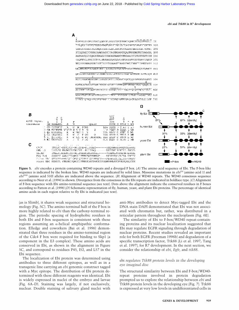
(as is Slimb), it shares weak sequence and structural ho-mology (Fig. 5C). The amino-terminal half of the F box ismore highly related to ebi than the carboxy-terminal re-gion. The periodic spacing of hydrophobic residues inboth Ebi and F-box sequences is consistent with theseregions assuming an a-helical amphipathic conforma-tion. Elledge and coworkers (Bai et al. 1996) demon-strated that three residues in the amino-terminal regionof the Cdc4 F box were required for binding to Skp1 (acomponent in the E3 complex). These amino acids areconserved in Ebi, as shown in the alignment in Figure5C, and correspond to residues P45, I52, and L57 in theEbi sequence.
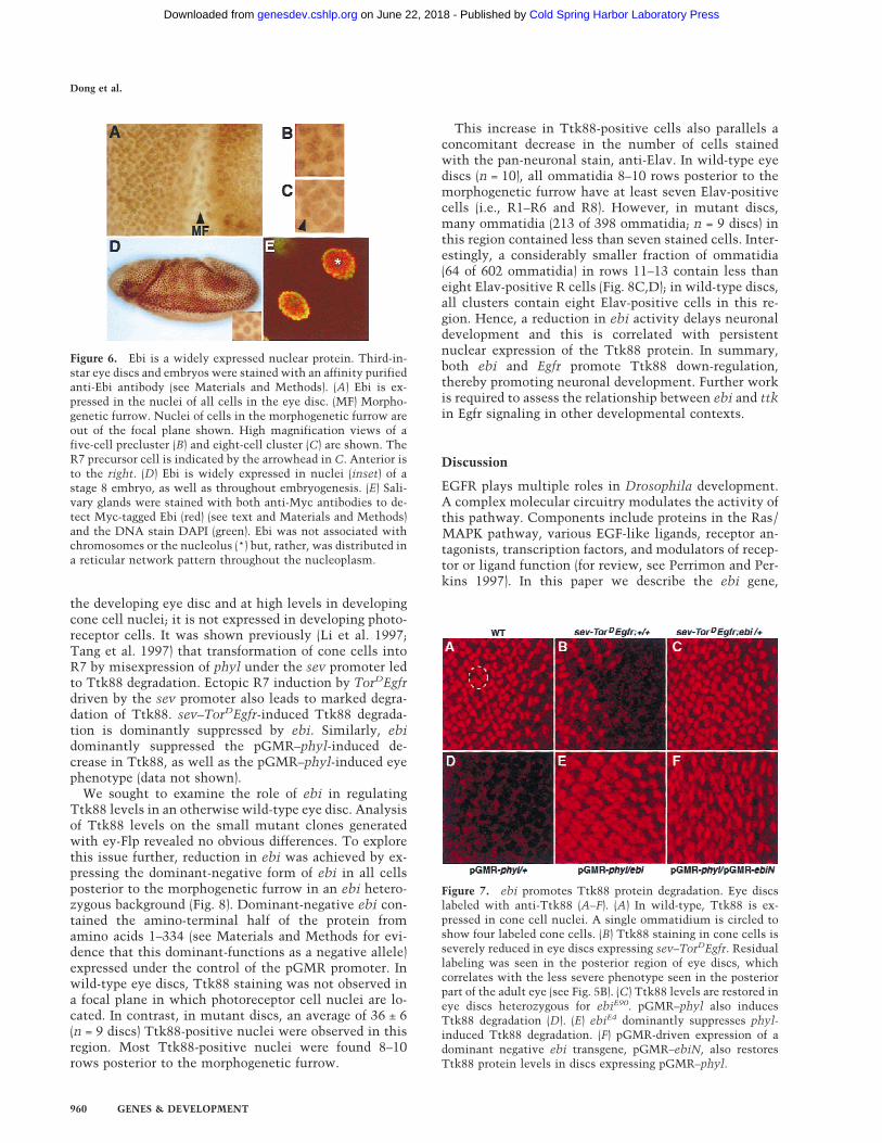
The localization of Ebi protein was determined usingantibodies to three different epitopes, as well as in atransgenic line carrying an ebi genomic construct taggedwith a Myc epitope. The distribution of Ebi protein de-termined with these different reagents was identical. Ebiis widely expressed in nuclei of the embryo and larvae(Fig. 6A–D). Staining was largely, if not exclusively,nuclear. Double staining of salivary gland nuclei with
anti-Myc antibodies to detect Myc-tagged Ebi and theDNA stain DAPI demonstrated that Ebi was not associ-ated with chromatin but, rather, was distributed in areticular pattern throughout the nucleoplasm (Fig. 6E).
The similarity of Ebi to F-box/WD40 repeat-contain-ing proteins and its nuclear localization suggested thatEbi may regulate EGFR signaling through degradation ofnuclear proteins. Recent studies revealed an importantrole for both EGFR (Freeman 1996b) and degradation of aspecific transcription factor, Ttk88 (Li et al. 1997; Tanget al. 1997), for R7 development. In the next section, weconsider the relationship of ebi, Egfr, and ttk88.
ebi regulates Ttk88 protein levels in the developingeye imaginal disc
The structural similarity between Ebi and F-box/WD40-repeat proteins involved in protein degradationprompted us to explore the relationship between ebi andTtk88 protein levels in the developing eye (Fig. 7). Ttk88is expressed at very low levels in undifferentiated cells in
Figure 5. ebi encodes a protein containing WD40 repeats and a diverged F box. (A) The amino acid sequence of Ebi. The F-box-likesequence is indicated by the broken line. WD40 repeats are indicated by solid lines. Missense mutations in ebiE4 (amino acid 1) andebiE90 (amino acid 510) alleles are indicated above the sequence. (B) Alignment of WD40 repeats. The WD40 consensus sequenceaccording to Neer et al. (1994) is shown. Divergence from the consensus in the Ebi repeats are indicated in boldface type. (C) Alignmentof F-box sequence with Ebi amino-terminal sequence (see text). Dots above the alignment indicate the conserved residues in F boxesaccording to Patton et al. (1998) (D) Schematic representation of fly, human, yeast, and plant Ebi proteins. The percentage of identicalamino acids in each region relative to fly Ebi is indicated (see text).
ebi and Ttk88 in R7 development
GENES & DEVELOPMENT 959
Cold Spring Harbor Laboratory Press on June 22, 2018 - Published by genesdev.cshlp.orgDownloaded from
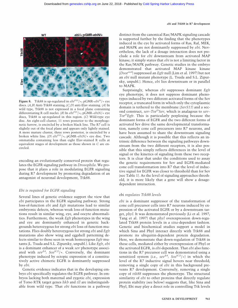
the developing eye disc and at high levels in developingcone cell nuclei; it is not expressed in developing photo-receptor cells. It was shown previously (Li et al. 1997;Tang et al. 1997) that transformation of cone cells intoR7 by misexpression of phyl under the sev promoter ledto Ttk88 degradation. Ectopic R7 induction by TorDEgfrdriven by the sev promoter also leads to marked degra-dation of Ttk88. sev–TorDEgfr-induced Ttk88 degrada-tion is dominantly suppressed by ebi. Similarly, ebidominantly suppressed the pGMR–phyl-induced de-crease in Ttk88, as well as the pGMR–phyl-induced eyephenotype (data not shown).
We sought to examine the role of ebi in regulatingTtk88 levels in an otherwise wild-type eye disc. Analysisof Ttk88 levels on the small mutant clones generatedwith ey-Flp revealed no obvious differences. To explorethis issue further, reduction in ebi was achieved by ex-pressing the dominant-negative form of ebi in all cellsposterior to the morphogenetic furrow in an ebi hetero-zygous background (Fig. 8). Dominant-negative ebi con-tained the amino-terminal half of the protein fromamino acids 1–334 (see Materials and Methods for evi-dence that this dominant-functions as a negative allele)expressed under the control of the pGMR promoter. Inwild-type eye discs, Ttk88 staining was not observed ina focal plane in which photoreceptor cell nuclei are lo-cated. In contrast, in mutant discs, an average of 36 ± 6(n = 9 discs) Ttk88-positive nuclei were observed in thisregion. Most Ttk88-positive nuclei were found 8–10rows posterior to the morphogenetic furrow.
This increase in Ttk88-positive cells also parallels aconcomitant decrease in the number of cells stainedwith the pan-neuronal stain, anti-Elav. In wild-type eyediscs (n = 10), all ommatidia 8–10 rows posterior to themorphogenetic furrow have at least seven Elav-positivecells (i.e., R1–R6 and R8). However, in mutant discs,many ommatidia (213 of 398 ommatidia; n = 9 discs) inthis region contained less than seven stained cells. Inter-estingly, a considerably smaller fraction of ommatidia(64 of 602 ommatidia) in rows 11–13 contain less thaneight Elav-positive R cells (Fig. 8C,D); in wild-type discs,all clusters contain eight Elav-positive cells in this re-gion. Hence, a reduction in ebi activity delays neuronaldevelopment and this is correlated with persistentnuclear expression of the Ttk88 protein. In summary,both ebi and Egfr promote Ttk88 down-regulation,thereby promoting neuronal development. Further workis required to assess the relationship between ebi and ttkin Egfr signaling in other developmental contexts.
Discussion
EGFR plays multiple roles in Drosophila development.A complex molecular circuitry modulates the activity ofthis pathway. Components include proteins in the Ras/MAPK pathway, various EGF-like ligands, receptor an-tagonists, transcription factors, and modulators of recep-tor or ligand function (for review, see Perrimon and Per-kins 1997). In this paper we describe the ebi gene,
Figure 6. Ebi is a widely expressed nuclear protein. Third-in-star eye discs and embryos were stained with an affinity purifiedanti-Ebi antibody (see Materials and Methods). (A) Ebi is ex-pressed in the nuclei of all cells in the eye disc. (MF) Morpho-genetic furrow. Nuclei of cells in the morphogenetic furrow areout of the focal plane shown. High magnification views of afive-cell precluster (B) and eight-cell cluster (C) are shown. TheR7 precursor cell is indicated by the arrowhead in C. Anterior isto the right. (D) Ebi is widely expressed in nuclei (inset) of astage 8 embryo, as well as throughout embryogenesis. (E) Sali-vary glands were stained with both anti-Myc antibodies to de-tect Myc-tagged Ebi (red) (see text and Materials and Methods)and the DNA stain DAPI (green). Ebi was not associated withchromosomes or the nucleolus (*) but, rather, was distributed ina reticular network pattern throughout the nucleoplasm.
Figure 7. ebi promotes Ttk88 protein degradation. Eye discslabeled with anti-Ttk88 (A–F). (A) In wild-type, Ttk88 is ex-pressed in cone cell nuclei. A single ommatidium is circled toshow four labeled cone cells. (B) Ttk88 staining in cone cells isseverely reduced in eye discs expressing sev–TorDEgfr. Residuallabeling was seen in the posterior region of eye discs, whichcorrelates with the less severe phenotype seen in the posteriorpart of the adult eye (see Fig. 5B). (C) Ttk88 levels are restored ineye discs heterozygous for ebiE90. pGMR–phyl also inducesTtk88 degradation (D). (E) ebiE4 dominantly suppresses phyl-induced Ttk88 degradation. (F) pGMR-driven expression of adominant negative ebi transgene, pGMR–ebiN, also restoresTtk88 protein levels in discs expressing pGMR–phyl.
Dong et al.
960 GENES & DEVELOPMENT
Cold Spring Harbor Laboratory Press on June 22, 2018 - Published by genesdev.cshlp.orgDownloaded from
encoding an evolutionarily conserved protein that regu-lates the EGFR signaling pathway in Drosophila. We pro-pose that it plays a role in modulating EGFR signalingduring R7 development by promoting degradation of anantagonist of neuronal development, Ttk88.
Ebi is required for EGFR signaling
Several lines of genetic evidence support the view thatebi participates in the EGFR signaling pathway. Strongloss-of-function ebi and Egfr mutations lead to similarembryonic defects, whereas weak loss-of-function muta-tions result in similar wing, eye, and oocyte abnormali-ties. Furthermore, the weak Egfr phenotypes in the wingand eye are dominantly enhanced in genetic back-grounds heterozygous for strong ebi loss-of-function mu-tations. Flies doubly heterozygous for strong ebi and Egfrmutations also show wing and eggshell patterning de-fects similar to those seen in weak homozygous Egfr mu-tants (L. Tsuda and S.L. Zipursky, unpubl.). Like Egfr, ebiis a dominant enhancer of a weak sev phenotype associ-ated with sevE4; SosJC2/+. Conversely, the dominantphenotype induced by ectopic expression of a constitu-tively active chimeric EGFR is dominantly suppressedby ebi.
Genetic evidence indicates that in the developing em-bryo ebi specifically regulates the EGFR pathway. In em-bryos lacking both maternal and zygotic ebi, expressionof Torso RTK target genes hkb and tll are indistinguish-able from wild type. That ebi functions in a pathway
distinct from the canonical Ras/MAPK signaling cascadeis supported further by the finding that the phenotypesinduced in the eye by activated forms of Ras, Raf, MEK,and MAPK are not dominantly suppressed by ebi. Nev-ertheless, the lack of a dosage interaction does not pre-clude a role for ebi downstream from activated MAPkinase; it simply states that ebi is not a limiting factor inthe Ras/MAPK pathway. Genetic studies in the embryodemonstrated that activated MAP kinase kinase(Dsorsu4) suppressed an Egfr null (Lim et al. 1997) but notan ebi null mutant phenotype (L. Tsuda and S.L. Zipur-sky, unpubl.). Hence, ebi lies downstream or in parallelto MAPK.
Surprisingly, whereas ebi suppresses dominant Egfreye phenotype, it does not suppress dominant pheno-types induced by two different activated forms of the Sevreceptor, a truncated form in which only the cytoplasmicdomain is tethered to the membrane (SevS11) and a sec-ond construct, sev–TorDSev, which is analagous to sev–TorDEgfr. This is particularly perplexing because thedominant forms of EGFR and the two different forms ofactivated Sev drive the same developmental transforma-tion, namely cone cell precursors into R7 neurons, andhave been assumed to share the downstream signalingcascade. Although it is possible that this reflects an in-trinsic difference between the signaling pathways down-stream from the two different receptors, it is also pos-sible that this simply reflects differences in the level ofsignal or the kinetics of signaling from these two recep-tors. It is clear that under the conditions used to assaythe genetic requirements for Sev and EGFR-mediatedcone cell transformation into R7 that the level of induc-tive signal for EGFR was closer to threshold than for Sev(see Table 1). As the level of signaling approaches thresh-old, it is more likely that a gene will show a dosage-dependent interaction.
ebi regulates Ttk88 levels
ebi is a dominant suppressor of the transformation ofcone cell precursor cells into R7 neurons induced by ex-pression of the activated EGFR or by a downstream tar-get, phyl. It was demonstrated previously (Li et al. 1997;Tang et al. 1997) that phyl overexpression down-regu-lated Ttk88 protein levels in a sina-dependent fashion.Genetic and biochemical studies support a model inwhich Sina and Phyl interact directly with Ttk88 andpromote its ubiquitin-dependent protein degradation.Here, we demonstrate that down-regulation of Ttk88 inthese cells, mediated either by overexpression of Phyl orthe activated EGFR, is ebi-dependent. That ebi also func-tions in the R7 precursor cell was demonstrated using asensitized system (i.e., sevE4; SosJC2/+) in which thelevel of the R7 inductive signal hovers near threshold;removing a single copy of ebi in this background pre-vents R7 development. Conversely, removing a singlecopy of ttk88 suppresses the phenotype. The structuralsimilarity of ebi to other proteins that directly regulateprotein stability (see below) suggests that, like Sina andPhyl, Ebi may play a direct role in controlling Ttk levels
Figure 8. Ttk88 is up-regulated in ebiP25/+; pGMR–ebiN/+ eyediscs. (A,B) Anti-Ttk88 staining; (C,D) anti-Elav staining. (A) Inwild type, Ttk88 is not expressed in a focal plane containingdifferentiating R cell nuclei. (B) In ebiP25/+; pGMR–ebiN/+ eyediscs, Ttk88 is up-regulated in this region. (C) Wild-type eyedisc. An eight-cell cluster, 11 rows posterior to the morphoge-netic furrow, is encircled by a broken black line. The R7 cell isslightly out of the focal plane and appears only lightly stained.A more mature cluster, three rows posterior, is encircled by abroken white line. (D) ebiP25/+; pGMR–ebiN/+ eye disc. Twoommatidia containing less than eight Elav-stained R cells atequivalent stages of development as those shown in C are en-circled.
ebi and Ttk88 in R7 development
GENES & DEVELOPMENT 961
Cold Spring Harbor Laboratory Press on June 22, 2018 - Published by genesdev.cshlp.orgDownloaded from
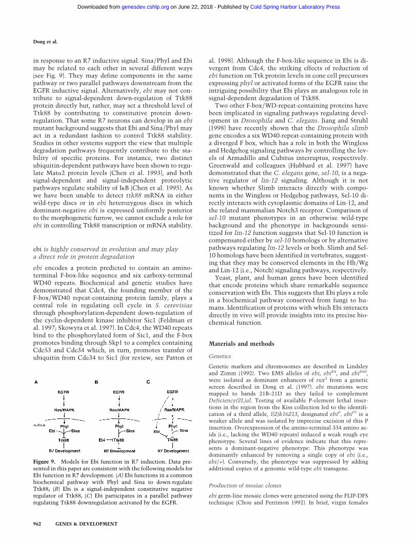
in response to an R7 inductive signal. Sina/Phyl and Ebimay be related to each other in several different ways(see Fig. 9). They may define components in the samepathway or two parallel pathways downstream from theEGFR inductive signal. Alternatively, ebi may not con-tribute to signal-dependent down-regulation of Ttk88protein directly but, rather, may set a threshold level ofTtk88 by contributing to constitutive protein down-regulation. That some R7 neurons can develop in an ebimutant background suggests that Ebi and Sina/Phyl mayact in a redundant fashion to control Ttk88 stability.Studies in other systems support the view that multipledegradation pathways frequently contribute to the sta-bility of specific proteins. For instance, two distinctubiquitin-dependent pathways have been shown to regu-late Mata2 protein levels (Chen et al. 1993), and bothsignal-dependent and signal-independent proteolyticpathways regulate stability of IkB (Chen et al. 1995). Aswe have been unable to detect ttk88 mRNA in eitherwild-type discs or in ebi heterozygous discs in whichdominant-negative ebi is expressed uniformly posteriorto the morphogenetic furrow, we cannot exclude a role forebi in controlling Ttk88 transcription or mRNA stability.
ebi is highly conserved in evolution and may playa direct role in protein degradation
ebi encodes a protein predicted to contain an amino-terminal F-box-like sequence and six carboxy-terminalWD40 repeats. Biochemical and genetic studies havedemonstrated that Cdc4, the founding member of theF-box/WD40 repeat-containing protein family, plays acentral role in regulating cell cycle in S. cerevisiaethrough phosphorylation-dependent down-regulation ofthe cyclin-dependent kinase inhibitor Sic1 (Feldman etal. 1997; Skowyra et al. 1997). In Cdc4, the WD40 repeatsbind to the phosphorylated form of Sic1, and the F-boxpromotes binding through Skp1 to a complex containingCdc53 and Cdc34 which, in turn, promotes transfer ofubiquitin from Cdc34 to Sic1 (for review, see Patton et
al. 1998). Although the F-box-like sequence in Ebi is di-vergent from Cdc4, the striking effects of reduction ofebi function on Ttk protein levels in cone cell precursorsexpressing phyl or activated forms of the EGFR raise theintriguing possibility that Ebi plays an analogous role insignal-dependent degradation of Ttk88.
Two other F-box/WD-repeat-containing proteins havebeen implicated in signaling pathways regulating devel-opment in Drosophila and C. elegans. Jiang and Struhl(1998) have recently shown that the Drosophila slimbgene encodes a six WD40-repeat-containing protein witha diverged F box, which has a role in both the Winglessand Hedgehog signaling pathways by controlling the lev-els of Armadillo and Cubitus interruptus, respectively.Greenwald and colleagues (Hubbard et al. 1997) havedemonstrated that the C. elegans gene, sel-10, is a nega-tive regulator of lin-12 signaling. Although it is notknown whether Slimb interacts directly with compo-nents in the Wingless or Hedgehog pathways, Sel-10 di-rectly interacts with cytoplasmic domains of Lin-12, andthe related mammalian Notch3 receptor. Comparison ofsel-10 mutant phenotypes in an otherwise wild-typebackground and the phenotype in backgrounds sensi-tized for lin-12 function suggests that Sel-10 function iscompensated either by sel-10 homologs or by alternativepathways regulating lin-12 levels or both. Slimb and Sel-10 homologs have been identified in vertebrates, suggest-ing that they may be conserved elements in the Hh/Wgand Lin-12 (i.e., Notch) signaling pathways, respectively.
Yeast, plant, and human genes have been identifiedthat encode proteins which share remarkable sequenceconservation with Ebi. This suggests that Ebi plays a rolein a biochemical pathway conserved from fungi to hu-mans. Identification of proteins with which Ebi interactsdirectly in vivo will provide insights into its precise bio-chemical function.
Materials and methods
Genetics
Genetic markers and chromosomes are described in Lindsleyand Zimm (1992). Two EMS alleles of ebi, ebiE4, and ebiE90,were isolated as dominant enhancers of rux3 from a geneticscreen described in Dong et al. (1997). ebi mutations weremapped to bands 21B–21D as they failed to complementDeficiency(2L)al. Testing of available P-element lethal inser-tions in the region from the Kiss collection led to the identifi-cation of a third allele, l(2)k16213, designated ebiP. ebiP7 is aweaker allele and was isolated by imprecise excision of this Pinsertion. Overexpression of the amino-terminal 334 amino ac-ids (i.e., lacking the WD40 repeats) induced a weak rough eyephenotype. Several lines of evidence indicate that this repre-sents a dominant-negative phenotype: This phenotype wasdominantly enhanced by removing a single copy of ebi (i.e.,ebi/+). Conversely, the phenotype was suppressed by addingadditional copies of a genomic wild-type ebi transgene.
Production of mosiac clones
ebi germ-line mosaic clones were generated using the FLIP-DFStechnique (Chou and Perrimon 1992). In brief, virgin females
Figure 9. Models for Ebi function in R7 induction. Data pre-sented in this paper are consistent with the following models forEbi function in R7 development. (A) Ebi functions in a commonbiochemical pathway with Phyl and Sina to down-regulateTtk88; (B) Ebi is a signal-independent constitutive negativeregulator of Ttk88; (C) Ebi participates in a parallel pathwayregulating Ttk88 downregulation activated by the EGFR.
Dong et al.
962 GENES & DEVELOPMENT
Cold Spring Harbor Laboratory Press on June 22, 2018 - Published by genesdev.cshlp.orgDownloaded from
carrying the ebiE4 FRT40A/CyO were crossed with hs-FLP/Y;ovoD1, FRT40A/Elp1, Bc, Gla males. Progeny were heat-shocked for 1 hr at 37°C during larval stages, and females ofgenotypes hs-FLP/+; ebiE4 FRT40A/ovoD1, FRT40A were exam-ined for the presence of germ-line clones. Both null (ebiE4/ebiE4)and paternally rescued (ebiE4/+) animals, derived from femaleslacking maternal ebi activity during oogenesis, die during em-bryogenesis. To distinguish between null and paternally rescuedembryos, mosaic females possessing ebi germ-line clones werecrossed with males carrying ebiE4/CyO, ftz-lacZ, a balancerchromosome that contains a lacZ gene under the control of thefushi tarazu promoter. The genotypes of embryos were deter-mined by following the expression pattern of the lacZ gene,which was detected by b-galactosidase activity. Embryos with-out the lacZ marker are referred to as maternal−/zygotic−. Theirsiblings, which express the lacZ gene, are referred to as mater-nal−/zygotic+. Larval cuticles were prepared in Hoyer’s mount.Cuticles were examined using dark-field optics. Clones in theeye were generated in flies heterozygous for ebiE4 using FRT-mediated recombination catalyzed by Flp recombinase (pro-vided by expression of flp under the ey promoter. The genotypeof these flies was: y, w P[ry, eyflp]/w; ebiE4, P[w+]27F, FRT40A/FRT40A. Mosaic eyes contained patchy pigmentation with re-gions of dark red (mutant clones) in an otherwise light yelloweye. Analysis of clones was as described previously (Reinke andZipursky 1988).
In situ hybridization and immunocytochemistry
In situ hybrydization on whole-mount Drosophila embryos wasperformed according to Tautz and Pfeifle (1989). Single-strandedantisense digoxigenin-containing RNA probes were preparedusing the Genius kit (Boehringer Mannheim). Probes were pre-pared from plasmids containing the tll (Pignoni et al. 1990), hkb(Bronner et al. 1994), and otd (Wieschaus et al. 1992) cDNAs.For visualization, embryos were mounted in glycerol and wereanalyzed and photographed with a Zeiss Axiophot equippedwith Nomarski optics.
Immunocytochemistry was performed as described in Patel etal. (1987). Antibody was used at dilutions 1:50. The FasIII mono-clonal antibody (7G10) was obtained from C. Goodman (Uni-versity of California, Berkeley). Confocal microscopy was doneusing a Bio-Rad MRC 1024.
SEM and plastic eye section were carried out as describedpreviously (Cheyette et al. 1994). In Table 1 the number of om-matidia containing R7 cells was determined using the pseudo-pupil method (Banerjee et al. 1987). To determine the ebi ex-pression pattern, a Myc epitope tag sequence was inserted in-frame with the start codon of the ebi genomic rescue construct.Anti-Myc antibody (mAb1-9E10.2 from the ATCC, final dilu-tion 1:25) staining was performed in lines carrying this con-struct.
Molecular biology
A 5.5-kb BamHI genomic fragment flanking the P-element siteof ebiP was isolated by plasmid rescue and subsequently used asa probe to screen a Drosophila genomic library in lEMBL3(Tamkun et al. 1992). An 11.1-kb DNA flanking the P-elementinsertion site was isolated and used to screen a 0- to 24-hr em-bryo cDNA library in lEXLX(+) (Palazzolo et al. 1989). FourteencDNAs corresponding to four classes of transcription units wereisolated. The P element was inserted into the 58 UTR of one ofthe four transcripts. The longest cDNA corresponding to thistranscript is 3.5 kb. It has two small introns (65 and 79 bp). ebicDNAs were sequenced by ABI automated sequencing. ebiE4
and ebiE90 mutant chromosomes were amplified by PCR andsequenced to determine the molecular lesions. In ebiE4 the ATGstart codon is changed to ATC (codon for isoleucine), and inebiE90 the codon TGC encoding cysteine 510 is changed to TACencoding tyrosine. ebiE4 is likely to be a null allele based onsequencing. DNA and protein databases were searched for ho-mologous sequences using the BLAST program (Altschul et al.1990).
Isolation of ebi homologs
A human EST clone (114411), which encodes a partial ORF withsignificant similarity to fly ebi, was obtained from ResearchGenetics, Inc. The 1.0-kb EST clone was used to screen humanadult spleen cDNA library (a gift of Dr. Owen Witte, UCLA). A2.6-kb human cDNA was isolated and sequenced.
Rescue and expression
Germ-line transformation of Drosophila was performed usingstandard methods (Spradling and Rubin 1982). A 7.5-kb genomicfragment encompassing the ebi transcription unit and a 3.5-kbebi cDNA were inserted into pCaspeR4 and CaspeR-hs trans-formation vectors, respectively. Heat shock at 37°C was admin-istered for 30 min every 12 hr. A DNA fragment encoding thefirst 334 amino acids of Ebi was inserted into the pGMR trans-formation vector. This functioned as a dominant-negative con-struct (see above). More than three independent lines of eachconstruct were generated.
Generation of antibodies
Three peptides corresponding to amino-terminal (TEVEWS-VGEDGEVA), carboxy-terminal (WNSKGTKVGASASDG), andmiddle (SQKKSQNSNEAGSSS) regions of Ebi were synthesizedand injected into rabbits to raise antibodies by Chiron Tech-nologies Ltd. Antisera were purified using the peptides as affin-ity reagents. Ebi staining was performed using one of the puri-fied antisera, 2903, which was raised against the peptide corre-sponding to middle region of Ebi. The final dilution for theantibodies was 1:300.
Acknowledgments
We thank Gerry Rubin and Corey Goodman for anti-Elav, andanti-FasIII antibodies, respectively, Owen Witte for a humanspleen cDNA library, Trudi Schupbach for Egfr alleles, andBarry Dickson for ey-Flp. We also thank Drs. Owen Witte, UtpalBanerjee, Mike Tyers, Tom Clandinin, and Chi-Hon Lee, as wellas members of the Zipursky laboratory, for helpful discussionsand for reading various versions of this manuscript. This workwas supported by a National Research Service Award (NRSA)training grant (GM07185) (X.D.), a Human Frontiers ResearchFellowship (L.T.), a senior research fellowship from the Leuke-mia Society of America (K.Z.), a Medical Scientist Training Pro-gram training grant (M.L.), and a grant from the National Insti-tutes of Health (S.L.Z.). S.L.Z. is an Investigator of the HowardHughes Medical Institute.
The publication costs of this article were defrayed in part bypayment of page charges. This article must therefore be herebymarked ‘advertisement’ in accordance with 18 USC section1734 solely to indicate this fact.
References
Altschul, S.F., W. Gish, W. Miller, E.W. Myers, and D.J. Lipman.
ebi and Ttk88 in R7 development
GENES & DEVELOPMENT 963
Cold Spring Harbor Laboratory Press on June 22, 2018 - Published by genesdev.cshlp.orgDownloaded from
1990. Basic local alignment search tool. J. Mol. Biol.215: 403–410.
Bai, C., P. Sen, K. Hofman, L. Ma, M. Goebl, J.W. Harper, and S.Elledge. 1996. Skp1 connects cell cycle regulators to theubiquitin proteolysis machinery through a novel motif, theF-box. Cell 86: 263–274.
Banerjee, U., P.J. Renfranz, J.A. Pollock, and S. Benzer. 1987.Molecular characterization and expression of sevenless, agene involved in neuronal pattern formation in the Dro-sophila eye. Cell 49: 281–291.
Basler K., B. Christen, and E. Hafen. 1991. Ligand-independentactivation of the sevenless Receptor tyrosine kinase changesthe fate of cells in the developing Drosophila eye. Cell64: 1069–1081.
Bronner, G., Q. Chu-LaGraff, C.Q. Doe, B. Cohen, D. Weigel, H.Taubert, and H. Jackle. 1994. Sp1/egr-like zinc-finger pro-tein required for endoderm specification and germ-layer for-mation in Drosophila. Nature 369: 664–668.
Brunner, D., N. Oellers, J. Szabad, W.H. Biggs, S.L. Zipursky,and E. Hafen. 1994. A gain-of-function mutation in Dro-sophila MAP kinase activates multiple receptor tyrosine ki-nase signaling pathways. Cell 76: 875–888.
Chen, P., P. Johnson, T. Sommer, S. Jentsch, and M. Hochstras-ser. 1993. Multiple ubiquitin-conjugating enzymes partici-pate in the in vivo degradation of the yeast MAT a 2 repres-sor. Cell 74: 357–369.
Chen, Z., J. Hagler, V.J. Palombella, F. Melandri, D. Scherer, D.Ballard, and T. Maniatis. 1995. Signal-induced site-specificphosphorylation targets IkBa to the ubiquitin-proteasomepathway. Genes & Dev. 9: 1586–1597.
Cheyette, B.N., P.J. Green, K. Martin, H. Garren, V. Harten-stein, and S.L. Zipursky. 1994. The Drosophila sine oculislocus encodes a homeodomain-containing protein requiredfor the development of the entire visual system. Neuron12: 977–996.
Chou, T.B. and N. Perrimon. 1992. Use of a yeast site-specificrecombinase to produce female germline chimeras in Dro-sophila. Genetics 131: 643–653.
Clifford, R. and T. Schupbach. 1994. Molecular analysis of theDrosophila EGF receptor homolog reveals that several ge-netically defined classes of alleles cluster in subdomains ofthe receptor protein. Genetics 137: 531–550.
Dickson, B., F. Sprenger, D. Morrison and E. Hafen. 1992a. Raffunctions downstream of Ras1 in the Sevenless signal trans-duction pathway. Nature 360: 600–603.
Dickson, B., F. Sprenger, and E. Hafen. 1992b. Prepattern in thedeveloping Drosophila eye revealed by an activated torso-sevenless chimeric receptor. Genes & Dev. 6: 2327–2339.
Dickson, B.J., M. Dominguez, A. van der Straten, and E. Hafen.1995. Control of Drosophila photoreceptor cell fates byPhollopod, a novel nuclear protein acting downstream of theRaf kinase. Cell 80: 453–462.
Dominguez, M., J.D. Wasserman, and M. Freeman. 1998. Mul-tiple functions of the EGF receptor in Drosophila eye devel-opment. Curr. Biol. 8: 1039–1048.
Dong, X., K.H. Zavitz, B.J. Thomas, M. Lin, S. Campbell, andS.L. Zipursky. 1997. Control of G1 in the developing Dro-sophila eye: rca1 regulates Cyclin A. Genes. & Dev. 11: 94–105.
Duffy, J.B. and N. Perrimon. 1994. The torso pathway in Dro-sophila: Lessons on receptor tyrosine kinase signaling andpattern formation. Dev. Biol. 166: 380–395.
Feldman, R.M.R., C.C. Correll, K.B. Kaplan, and R.J. Deshaies.1997. A complex of Cdc4p, Skp1p, and Cdc53p/Cullin cata-lyzes ubiquitination of the phosphorylated CDK inhibitorSic1p. Cell 91: 221–230.
Fortini, M.E., M.A. Simon, and G.M. Rubin. 1992. Signalling bythe sevenless protein tyrosine kinase is mimicked by Ras1activation. Nature 355: 559–561.
Freeman, M. 1996a. Cell determination strategies in the Dro-sophila eye. Development 124: 261–270.
———. 1996b. Reiterative use of the EGF receptor triggers dif-ferentiation of all cell types in the Drosophila eye. Cell87: 651–660.
———. 1998. Complexity of EGF receptor signalling revealed inDrosophila. Curr. Opin. Genet. Dev. 8: 407–411.
Hubbard, E.J.A., G. Wu, J. Kitajewski, and I. Greenwald. 1997.sel-10 a negative regulator of lin-12 activity in Caenorhab-ditis elegans, encodes a member of the CDC4 family of pro-teins. Genes & Dev. 11: 3182–3193.
Jiang, J. and G. Struhl. 1998. Regulation of the Hedgehog andWingless signaling pathways by the F-box/WD40-repeat pro-tein Slimb. Nature 391: 493–496.
Kazlauskas, A. 1994. Receptor tyrosine kinases and their tar-gets. Curr. Opin. Genet. Dev. 4: 5–14.
Kayne, P.S. and P.W. Sternberg. 1995. Ras pathways in Cae-norhabditis elegans. Curr. Opin. Genet. Dev. 5: 38–43.
Kim, S.H. and S.T. Crews. 1993. Influence of Drosophila ventralepidermal development by the CNS midline cell and spitzclass genes. Development 118: 893–901.
Kumar, J.P., M. Tio, F. Hsiung, S. Akopyan, L. Gabay, R. Seger,B.-Z. Shilo, and K. Moses. 1998. Dissecting the roles of theDrosophila EGF receptor in eye development and MAP ki-nase activation. Development 125: 3875–3885.
Lai Z.C., S.D. Harrison, F. Karim, Y. Li, and G.M. Rubin. 1996.Loss of tramtrack gene activity results in ectopic R7 cellformation, even in a sina mutant background. Proc. Natl.Acad. Sci. 93: 5025–5030.
Li, S., Y. Li, R.W. Carthew, and Z. Lai. 1997. Photoreceptor celldifferentiation requires regulated proteolysis of the tran-scription repressor Tramtrack. Cell 90: 469–478.
Lim Y., L. Tsuda, Y.H. Inoue, K. Irie, T.A. Yamada, M. Hata, Y.Nishi, K. Matsumoto, and Y. Nishida. 1997. Dominant mu-tations of Drosophila MAP Kinase Kinase and their acitivi-ties in Drosophila and yeast MAP Kinase cascades. Genetics146: 263–273.
Lindsley, D.L. and G.G. Zimm. 1992. The genome of Drosophilamelanogaster. Academic Press, San Diego, CA.
Neer, E.J., C.J. Schmidt, R. Nambudripad, and T.F. Smith. 1994.The ancient regulatory-protein family of WD-repeat pro-teins. Nature 371: 298–300.
O’Neill, E.M., I. Rebay, R. Tjian, and G.M. Rubin. 1994. Theactivities of two Ets-related transcription factors required forDrosophila eye development are modulated by the Ras/MAPK pathway. Cell 78: 137–147.
Palazzolo, M.J., D.R. Hyde, K. VijayRaghavan, K. Mecklenburg,S. Benzer, and E. Meyerowitz. 1989. Use of a new strategy toisolate and characterize 436 Drosophila cDNA clones corre-sponding to RNAs detected in adult heads but not in earlyembryos. Neuron 3: 527–539.
Patel, N.H., P.M. Snow, and C.S. Goodman. 1987. Character-ization and cloning of fasciclin III: A glycoprotein expressedon a subset of neurons and axon pathways in Drosophila.Cell 48: 975–988.
Patton E.E., A.R. Willems, and M. Tyers. 1998. Combinationalcontrol in ubiquitin-dependent proteolysis: Don’t Skp theF-box hypothesis. Trends Genet. 14: 236–243.
Perrimon, N. and L.A. Perkins. 1997. There must be 50 ways torule the signal: The case of the Drosophila EGF receptor.Cell 89: 13–16.
Pignoni, F., R.M. Baldarelli, E. Steingrimsson, R.J. Diaz, A. Pata-poutian, J.R. Merriam, and J.A. Lengyel. 1990. The Dro-
Dong et al.
964 GENES & DEVELOPMENT
Cold Spring Harbor Laboratory Press on June 22, 2018 - Published by genesdev.cshlp.orgDownloaded from
sophila gene tailless is expressed at the embryonic terminiand is a member of the steroid receptor superfamily. Cell62: 151–163.
Raz, E. and B. Shilo. 1993. Establishment of ventral cell fates inthe Drosophila embryonic ectoderm requires DER, the EGFreceptor homolog. Genes & Dev. 7: 1937–1948.
Rebay, I. and G.M. Rubin. 1995. Yan functions as a generalinhibitor of differentiation and is negatively regulated byactivation of the Ras/MAPK pathway. Cell 81: 857–866.
Reichman-Fried, M., B. Dickson, E. Hafen, and B. Shilo. 1994.Elucidation of the role of breathless, a Drosophila FGF re-ceptor homolog, in tracheal cell migration. Gene & Dev.8: 428–439.
Reinke, R. and S.L. Zipursky. 1988. Cell-cell interaction in theDrosophila retina: The bride of sevenless gene is required inphotoreceptor R8 for R7 development. Cell 55: 321–330.
Rogge, R.D., C.A. Karlovich, and U. Banerjee. 1991. Geneticdissection of a neurodevelopmental pathway: Son of seven-less functions downstream of the sevenless and EGF receptortyrosine kinases. Cell 64: 39–48.
Schweitzer, R. and B. Shio. 1997. A thousand and one roles forthe Drosophila EGF receptor. Trends Genet. 13: 191–196.
Skowyra, D., K.L. Craig, M. Tyers, S.J. Elledge, and J.W. Harper.1997. F-box proteins are receptors that recruit phosophory-lated substrates to the SCF ubiquitin-ligase complex. Cell91: 209–219.
Spradling, A.C. and G.M. Rubin. 1982. Transposition of clonedP elements into Drosophila germ line chromosomes. Science218: 341–347.
Tamkun, J.W., R. Deuring, M.P. Scott, M. Kissinger, A.M. Pat-tatucci, T.C. Kaufman, and J.A. Kennison. 1992. brahma: Aregulator of Drosophila homeotic genes structurally relatedto the yeast transcriptional activator SNF2/SWI2. Cell68: 561–572.
Tang A.H., T.P. Neufeld, E. Kwan, and G.M. Rubin. 1997. PHYLacts to down-regulate TTK88, a transcriptional repressor ofneuronal cell fates, by a SINA-dependent mechanism. Cell90: 459–467.
Tautz, D. and C. Pfeifle. 1989. A non-radioactive in situ hybrid-ization method for the localization of specific RNAs in Dro-sophila embryos reveals translational control of the segmen-tation gene hunchback. Chromosoma 98: 81–85.
Thomas, B.J., D.A. Gunning, J. Cho, and S.L. Zipursky. 1994.Cell-cycle progression in the Developing Drosophila Eve:roughex encodes a novel protein required for the establish-ment of G1. Cell 77: 1003–1014.
van der Geer, P., T. Hunter, and P.A. Lindberg. 1994. Receptorprotein-tyrosine kinases and their signal transduction path-ways. Annu. Rev. Cell Biol. 10: 251–337.
Verma, I.M., J.K. Stevenson, E.M. Schwarz, D. Van Antwep, andS. Miyamoto. 1995. Rel/NF-kB/IkB family: intimate tales ofassociation and dissociation. Genes & Dev. 9: 2723–2735.
Wassarman, D.A., M. Therrien, and G.M. Rubin. 1995. The Rassignaling pathway in Drosophila. Curr. Opin. Genet. Dev.5: 44–50.
Wieschaus, E., N. Perrimon, and R. Finkelstein. 1992. Ortho-denticle activity is required for the development of medialstructures in the larval and adult epidermis of Drosophila.Development 115: 801–811.
Xiong, W. and C. Montell. 1993. tramtrack is a transcriptionalrepressor required for cell fate determination in the Dro-sophila eye. Genes & Dev. 7: 1085–1096.
Xu, T. and G.M. Rubin. 1993. Analysis of genetic mosaics indeveloping and adult Drosophila tissues. Development117: 1223–1237.
Zipursky, S.L. and G.M. Rubin. 1994. Determination of neuro-
nal cell fate: Lessons from the R7 neuron of Drosophila.Annu. Rev. Neurosci. 17: 373–397.
ebi and Ttk88 in R7 development
GENES & DEVELOPMENT 965
Cold Spring Harbor Laboratory Press on June 22, 2018 - Published by genesdev.cshlp.orgDownloaded from
13:1999, Genes Dev. Xinzhong Dong, Leo Tsuda, Kenton H. Zavitz, et al.
Drosophila regulates epidermal growth factor receptor signaling pathways in ebi
References
http://genesdev.cshlp.org/content/13/8/954.full.html#ref-list-1
This article cites 57 articles, 18 of which can be accessed free at:
License
ServiceEmail Alerting
click here.right corner of the article or
Receive free email alerts when new articles cite this article - sign up in the box at the top
Cold Spring Harbor Laboratory Press
Cold Spring Harbor Laboratory Press on June 22, 2018 - Published by genesdev.cshlp.orgDownloaded from