SEROLOGICAL DIAGNOSIS OF BRUCELLOSIS Nielsen … · laboratory tests for serology diagnosis of ......
25
Prilozi, Odd. biol. med. nauki, MANU, XXXI, 1, s. 65‡89 (2010) Contributions, Sec. Biol. Med. Sci., MASA, XXXI, 1, p. 65–89 (2010) ISSN 0351–3254 UDK: 616.98:579.841.93.083.33 SEROLOGICAL DIAGNOSIS OF BRUCELLOSIS Nielsen K., Yu WL. Ottawa Laboratories (Fallowfield), Canadian Food Inspection Agency, Nepean, Ontario, Canada Abstract: Aim: To present a review and to describe the most widely used laboratory tests for serology diagnosis of brucellosis along with their pros and cons. Methods: Review the recent literature on brucellosis serology diagnostic tests. The choice of the testing strategy depends on the prevailing brucellosis epidemiological situation and the goal of testing. Results: The ‘gold standard’ for the diagnosis of brucellosis is isolation and identification of the causative bacterium, a member of Brucella sp. Isolation of Brucella sp. requires high security laboratory facilities (biological containment level 3), highly skilled personnel, an extended turnaround time for results and it is considered a hazar- dous procedure. Hence brucellosis is generally diagnosed by detection of an elevated le- vel of antibody in serum or other body fluid. This is a presumptive diagnosis as other mi- croorganisms and perhaps environmental factors can also cause increased antibody levels. Conclusion: A large number of serological tests for brucellosis have been devi- sed over the 100+ years since its initial isolation, starting with a simple agglutination test and progressing to sophisticated primary binding assays available today. However, no test devised to date is 100% accurate so generally serological diagnosis consists of testing sera by several tests, usually a screening test of high sensitivity, followed by a confirmatory test of high specificity. Key words: Brucellosis, serology, diagnosis, conventional tests, primary binding assays. Introduction Intermittent fever in man has been recognized in the Mediterranean area since Hippocrates described it in 450 BC. Further evidence of the presence of the disease in the area was presented by Capasso [1], finding typical brucellosis
Transcript of SEROLOGICAL DIAGNOSIS OF BRUCELLOSIS Nielsen … · laboratory tests for serology diagnosis of ......

Prilozi, Odd. biol. med. nauki, MANU, XXXI, 1, s. 65‡89 (2010) Contributions, Sec. Biol. Med. Sci., MASA, XXXI, 1, p. 65–89 (2010)
ISSN 0351–3254 UDK: 616.98:579.841.93.083.33
SEROLOGICAL DIAGNOSIS OF BRUCELLOSIS
Nielsen K., Yu WL.
Ottawa Laboratories (Fallowfield), Canadian Food Inspection Agency, Nepean, Ontario, Canada
A b s t r a c t: Aim: To present a review and to describe the most widely used laboratory tests for serology diagnosis of brucellosis along with their pros and cons.
Methods: Review the recent literature on brucellosis serology diagnostic tests. The choice of the testing strategy depends on the prevailing brucellosis epidemiological situation and the goal of testing.
Results: The ‘gold standard’ for the diagnosis of brucellosis is isolation and identification of the causative bacterium, a member of Brucella sp. Isolation of Brucella sp. requires high security laboratory facilities (biological containment level 3), highly skilled personnel, an extended turnaround time for results and it is considered a hazar-dous procedure. Hence brucellosis is generally diagnosed by detection of an elevated le-vel of antibody in serum or other body fluid. This is a presumptive diagnosis as other mi-croorganisms and perhaps environmental factors can also cause increased antibody levels.
Conclusion: A large number of serological tests for brucellosis have been devi-sed over the 100+ years since its initial isolation, starting with a simple agglutination test and progressing to sophisticated primary binding assays available today. However, no test devised to date is 100% accurate so generally serological diagnosis consists of testing sera by several tests, usually a screening test of high sensitivity, followed by a confirmatory test of high specificity. Key words: Brucellosis, serology, diagnosis, conventional tests, primary binding assays.
Introduction
Intermittent fever in man has been recognized in the Mediterranean area since Hippocrates described it in 450 BC. Further evidence of the presence of the disease in the area was presented by Capasso [1], finding typical brucellosis
66 Nielsen K., Yu W.L.
Contributions, Sec. Biol. Med. Sci., XXXI/1 (2010), 65–89
lesions in the bones of people killed in the eruption of Vesuvius in 79AD as well as the presence of coccoid cells consistent in morphology with Brucella sp. in carbonized cheese. These findings are in keeping with the nature of Roman diets of the day, containing both milk and cheese derived from small ruminants. Sir William Burnett, surgeon general to the British navy, differentiated the va-rious fevers affecting British troops sent to Malta to recuperate in 1810. A Bri-tish army surgeon, Jeffery Marston, contracted the disease and described his own symptoms in considerable detail in 1861. Sir David Bruce, a medical of-ficer of the British army and after whom Brucella was later named, provided the first description of this pathogen. Considerable morbidity and at least one case of mortality of British soldiers stationed at garrison on Malta arose as it turned out because of consumption of fresh goat’s milk. Dr. Bruce organized a team of scientists and clinicians who succeeded in isolating Micrococcus melitensis as the causative agent of the problem [2, 3]. The organism was later renamed Bru-cella melitensis. These findings helped to explain the epidemiology of the disease. For example, private soldiers were less likely to become ill because they drank less milk than the officers. Other species of Brucella include B. abortus isolated by Bang in 1897 [4], resulting in the term Bang’s Disease and B. suis first described by Taum [5]. In terms of human public health and agricultural economics, these three species are the most important. There are several other species, including B. ovis, B. canis, B. neotomae, B. microti and of which only B. canis has been reported to infect man. Two other, B. ceti and B. pinnipedialis infect marine mammals and are potential human pathogens as well.
Brucella ovis and B. canis contain rough lipopolysaccharide (RLPS) in their outer cell wall whereas all the other species contain smooth lipopolysac-charide (SLPS). Smooth lipopolysaccharide contains a lipid A anchor to the cell wall, in the intermediate core region, and an immunodominant O-polysaccha-ride (OPS) which has been chemically defined as a homopolymer of 4,6-dideoxy-4-formamide-alpha-D-mannose linked via glycosidic linkages [6]. B. ovis and B. canis lack the OPS component [7]. Because all smooth species share common epitopes in the OPS, virtually all serological tests for an antibody to these bacteria use B. abortus antigen in the form of whole cells, SLPS or OPS [8] while RLPS is commonly used as the main antigen for detection of antibody to B. ovis and B. canis [7, 9]. Most recently developed tests use either SLPS or OPS antigens although some attempts at using protein antigen have been made.
Cattle infected with B. abortus generally produce an early IgM isotype antibody response, the amplitude of which is governed by a multiplicity of factors. It usually appears 5 to 15 days post exposure but may be delayed [10–12]. The IgM antibody response is followed very shortly by production of IgG1 isotype of antibody and subsequently by IgG2 and IgA [11–15]. Because of the IgM response commences early, theoretically it would be most suitable to measure this isotype as an indicator of exposure. There is, however, a number of other microorganisms containing antigens with epitopes similar to those of OPS

Serological diagnosis of brucellosis 67
Прилози, Одд. биол. мед. науки, XXXI/1 (2010), 65–89
and the main antibody response to these cross-reacting antigens is IgM [16]. The-refore, measurement of IgM antibody may result in a false positive reaction in serological tests. False positive reactivity would lead to specificity problems which would be of considerable consequence in an early control programme re-sulting in unnecessary slaughter; in the last stages of an eradication programme and in free areas, resulting in expensive follow-ups. Production of IgG2 and IgA isotypes occurs later in infection and, as a result, measurement of these antibodies would generally lower assay sensitivity. Based on these observations, the most useful antibody for serological testing for brucellosis is IgG1 [12, 15, 17, 18].
An antibody produced in response to smooth vaccines may also result in positive serological reactions which may lead to misdiagnosis. Specifically, B. abortus S19, a vaccine used in many areas, may be retained over an extended period, causing problems [19]. Smooth vaccine SLPS is antigenically identical to that of pathogenic strains of B. abortus; however, administration of the vac-cine to young animals, usually between 3 and 8 months of age or by the conjun-ctival route generally results in insufficient antibody levels to cause diagnostic problems by the time animals reach sexual maturity and are tested for brucello-sis [20]. However, some animals do have residual antibody resulting in allowan-ces for higher antibody levels in vaccinated animals. Most of these problems have been overcome by the development of improved serological tests, for example, the competitive enzyme immunoassay and fluorescence polarization assay [21] and the development of a live vaccine devoid of OPS (B. abortus RB51 developed by Schurig) [22].
Serological tests
Brucellosis was first diagnosed by a serological test by Wright and Smith in 1897 [23] using a simple tube agglutination test. Subsequently, various modifications to the tube agglutination test and numerous other tests have been developed to increase test accuracy. The procedures are divided into 2 catego-ries, the Conventional Tests and Primary Binding Assays. All conventional tests rely on the antibody performing a secondary function, for instance fixation of complement while in primary binding assays the only function of the antibody is attachment to its antigen.
Conventional Tests
Agglutination tests: Slow tests requiring incubation from 8 to 24 hours • Standard tube (SAT)
68 Nielsen K., Yu W.L.
Contributions, Sec. Biol. Med. Sci., XXXI/1 (2010), 65–89
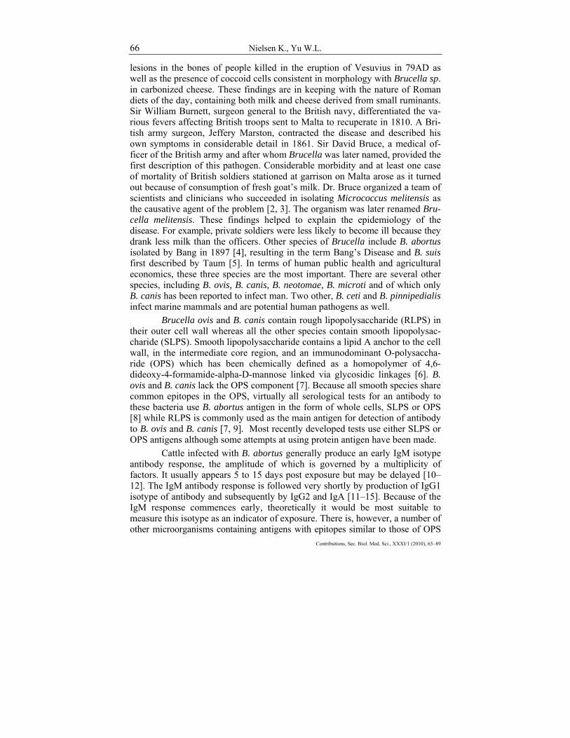
• SAT with added reducing agents such as 2-mercaptoethanol or dit-hiothreitol
• SAT with addition of rivanol to precipitate glycoproteins • SAT with addition of ethylene diamine tetraacetic acid to reduce IgM
binding (EDTA) • SAT with antiglobulin added to enhance agglutination • Milk ring test
Rapid agglutination tests performed in minutes: • Rose Bengal • Modified Rose Bengal • Buffered antigen plate agglutination • Card • Antigen with rivanol added • Heat treatment of serum • Addition of 10% sodium chloride
Precipitation tests: • Agar gel immunodiffusion • Radial immunodiffusion
Complement fixation tests: • Warm • Cold • Haemolysis in gel • Indirect haemolysis
Primary Binding Assays: • Radioimmunoassay • Fluorescence immunoassay • Particle counting fluorescence immunoassay • Indirect enzyme immunoassay • Competitive enzyme immunoassay • Fluorescence polarization assay
Each category of tests will be described and their performance will be
discussed. There are variations on some of these tests and there are several other tests not commonly used which will be beyond the scope of this review.

Serological diagnosis of brucellosis 69
Прилози, Одд. биол. мед. науки, XXXI/1 (2010), 65–89
Agglutination tests
In 1897, Smith and Wright [23] published the first description of a test for the serological diagnosis of brucellosis in man. This test used a mixture of bacterial cell antigens incubated with the patient’s serum in a glass tube and if a ‘mantle’ pattern of cell sediment was observed, it was considered as an indication of infection while a ‘button’ pattern was considered as negative. This test is virtually the identical test still used in some countries, except that only B. abortus cells are used as the antigen. This test is performed at a near neutral pH and therefore detects IgM isotype of antibody efficiently and is therefore very sensitive. The SAT detects IgG less efficiently, especially IgG1, resulting in low assay specificity [13, 15, 24]. Therefore, the SAT is generally not used as a single test but rather in combination with other tests.
The production of IgM in response to cross-reacting antigens often induces significant levels of agglutinating antibody which causes specificity problems in the SAT. As a result, a number of modifications have been made to the SAT to lower the IgM levels thereby increasing the assay specificity. The most commonly used methods of IgM destruction is chemical treatment with 2-mercaptoethanol or dithiothreitol which reduce disulfide bridges in the molecule resulting in monomeric units of the pentameric molecule. The monomers are much less efficient agglutinins. Other methods include precipitation of glycop-roteins using rivanol and addition of divalent cheating agents.
A number of rapid agglutination tests have been devised. Some of these tests use a stained whole cell antigen stored in an acid buffer. This antigen is mixed with undiluted serum resulting in an acid test environment which in turn discourages agglutination by IgM and enhances agglutination by IgG1. Other tests use heat-treated serum or a high salt concentration to diminish reactivity by IgM.
Agglutination tests are generally not used for the diagnosis of infection with B. ovis and B. canis, rough species of Brucella, as the whole cell antigens tends to autoagglutinate. However, rapid slide agglutination tests have been developed for the serological diagnosis of B. canis infection [25–28] as well as a microagglutination test [29].
Acidified antigen modifications
Because of the cross-reaction of the LPS of B. abortus, B. melitensis and B. suis, only one antigen is required for serological diagnosis. Virtually all agglutination tests use the B. abortus antigen although in some cases different strains are used. The Buffered Antigen Plate Agglutination test (BPAT) has

70 Nielsen K., Yu W.L.
Contributions, Sec. Biol. Med. Sci., XXXI/1 (2010), 65–89
been widely used [30] as has the Rose Bengal test (RBT) [31]. In these tests, B. abortus S99 or S1119.3 cell antigen, stained with Rose Bengal or Brilliant Green and Crystal Violet, respectively, and suspended in a buffer which when mixed with the appropriate volume of serum results in a final pH of 3.65. After thorough mixing of the serum and antigen, agglutination must be visible be wit-hin the specified time for each test (4 minutes for the RBT and 8 minutes for the BPAT). Incubation for extended periods of time may sometimes result in false reactions, often due to the formation of fibrin clots. The acid pH diminishes ag-glutination by IgM but encourages agglutination by IgG1, generally reducing cross-reactions [12, 13]. False negative reactions can occur in the acidified antigen tests, especially in the RBT, due to prozoning with sera containing very high levels of antibody. These tests are considered as suitable screening tests for brucellosis, followed by confirmatory testing. Antibody resulting from B. abor-tus S19 vaccination will react in these tests [8].
Reducing agents
Dithiotreitol [32] and 2-mercaptoethanol [33] have both been used for the serological diagnosis of brucellosis. Either chemical may be added to serum as a diluent, using dilutions starting at 1 : 25 and increasing. For the diagnosis of brucellosis, a reaction at a 1 : 25 serum dilution is considered significant. In general, reduction of IgM increases specificity. However, some false negative reactions may occur as some IgG molecules are also susceptible to reduction of disulfide bridges, rendering them unable to agglutinate.
Care must be taken when using 2-mercaptoethanol as it is quite toxic and should only be used in a well ventilated area or a chemical hood. Test em-ploying reducing agents are usually used as confirmatory tests, however, anti-body resulting from B. abortus S19 vaccination may interfere [34].
Precipitation of glycoproteins
Reduction of non-specific reactivity by precipitation of high molecular weight serum glycoproteins has been applied to serological diagnosis of brucel-losis [34, 35]. This is commonly done by addition of rivanol (2-ethoxy-6,9-dia-minoacridine lactate) to serum followed by removal of the precipitate by centri-fugation and either a rapid plate type agglutination test with undiluted serum or a tube test using serum dilutions starting at 1: 25. Because the protocol is fairly labour-intensive, precipitation tests are generally used as confirmatory tests.

Serological diagnosis of brucellosis 71
Прилози, Одд. биол. мед. науки, XXXI/1 (2010), 65–89
Use of EDTA
Addition of ethylene diamino tetraacetic acid disodium salt (EDTA) has proven to significantly increase SAT specificity [36–38]. The mechanism by which EDTA reduces non-specificity is not understood; however, it appears to eliminate attachment of immunoglobulins to the Brucella cell wall via the Fc piece. The modified SAT may be used in tubes or 96 well plates and incubation is usually overnight after which the cell sediment pattern is observed. The modi-fied SAT has been used mainly as a screening test.
Milk ring test
The agglutination test has been adapted to test milk for antibody to Brucella sp. The format of the milk ring test (MRT) is a little different in that haematoxylin stained Brucella cells are mixed with whole milk or whole milk with cream added [35, 39, 40]. Immunoglobulins present in the milk will in part be attached to fat globules via the Fc portion of the molecule. If antibody to Brucella sp. is present, antigen will attach to it, resulting in a purple band in the cream layer. If no antibody is present, the fat layer will remain a buff colour and the purple antigen will be evenly distributed throughout the milk. This test may be applied to individual animals or to pooled milk samples using a larger vo-lume of milk relative to the pool size. The milk ring test is prone to false reac-tions caused by abnormal milk such as mastitic milk, colostrums and late lacta-tion cycle milk. Still, in spite of its problems, it may be used as an inexpensive screening test in conjunction with other tests.
Precipitin tests
Precipitin tests were shown to distinguish B. abortus S19 vaccinal anti-body from the antibody resulting from infection with pathogenic strains [41, 42]. There are two basic formats: agar gel immunodiffusion in which soluble antigen(s) and test serum are inserted into adjacent wells, cut in an agar matrix 0.5 to 1.0 cm apart. The reagents diffuse into the agar for a period of time, resulting in the formation of a visible precipitin band where they intersect if the serum contains antibody. The second format, radial immunodiffusion, utilizes antigen placed directly in the agar matrix, pipetting test serum in a well cut in the agar and allowing the serum to diffuse radially to form a precipitin ring if antibody is present in the serum. Both tests use OPS antigens derived from B. melitensis [41] or native hapten [43]. Both formats proved to be relatively in-sensitive with OPS antigen [44] while the sensitivity was better with native

72 Nielsen K., Yu W.L.
Contributions, Sec. Biol. Med. Sci., XXXI/1 (2010), 65–89
hapten antigen [43]. The tests are quite labour-intensive but provide results not available by any other test procedure at the time. Neither test is currently used extensively.
Precipitin tests are widely used for the diagnosis of B. ovis infection in sheep using RLPS or hot saline extracted antigens [8].
Complement fixation tests
The complement fixation test (CFT) requires a multitude of reagents and is technically challenging. However, in spite of this, it is a widely used confirmatory test for brucellosis. The basic test consists of B. abortus whole cell antigen incubated with dilutions of heat-inactivated serum (heated to destroy indigenous complement) and a titrated source of complement, usually guinea-pig serum. After a suitable time a pretitrated amount of sheep erythrocytes coated with rabbit antibody is added. If a primary immune complex (B. abortus cells and test serum) formed due to the presence of certain antibody isotypes mainly IgG1, in the serum, complement was activated and therefore not available to react with the secondary immune complex of sheep erythrocytes and rabbit antibody, resulting in no or only slight lysis of the erythrocytes. Alternately, if no primary immune complex was formed, complement would cause all the sensitized sheep erythrocytes to lyse. Thus the amount of haemoglobin in solution is a measure of anti-Brucella antibody activity. The complement fixation assay has been standardized [45, 46].
Because a number of reagents must be titrated daily and a number of controls for all the reagents and reactions are required, the test is time-consu-ming and technically challenging. It is also an expensive test because of the number of reagents used in the test and because it is labour-intensive, especially the daily titration routines. Since only IgG1 isotype of antibody fixes complement efficiently, the test specificity is high. The test does not allow for discrimination of B. abortus S19 derived antibody. Other problems include the subjectivity of the interpretation of results, occasional direct activation of complement by se-rum (anticomplementary activity), prozoning resulting in false negative results and the inability of the test for use with haemolysed serum samples. In spite of the shortcomings, the (CFT) has been and is a widely used as a confirmatory test in control/eradication programmes.
There are a number of variations of the test, including the indirect hae-molysis test and the haemolysis in gel test [47–51]. These tests were not used extensively as diagnostic tests.
The (CFT) using a hot saline extracted antigen preparation has been used for the diagnosis of B. ovis infection in sheep [52–55].

Serological diagnosis of brucellosis 73
Прилози, Одд. биол. мед. науки, XXXI/1 (2010), 65–89
Primary Binding Assays
Indirect formats Indirect primary binding assays rely only on an antibody present in the
test serum (or other body fluids) reacting with its antigen and then detection of the immune complex using a detection system with a ‘marker’ molecule. The tracer system usually comes in one of three formats: antiglobulins or bacterial cell receptors labelled with isotopes [48, 49, 50, 56–59]; fluorochromes [60–67] or enzymes (described initially by Carlsson et al, 1976) [68] and reviewed by Nielsen and Gall, 1994 [69–82].
The most commonly used system depends on enzyme conjugates for detection of antibody to SLPS preparations which are passively attached to a polystyrene matrix (usually in a 96 well format) to which diluted serum or milk is added. The detection system varies considerably but often a monoclonal antibody specific for an immunoglobulin heavy chain epitope of the test species and conjugated with peroxidase is used. Variation in the detection system inclu-des the use of cellular receptors such as protein A, protein G, protein A/G or po-lyclonal anti-immunoglobulin reagents. Alkaline phosphatase or other enzymes can be used as well. Peroxide is the substrate used for peroxidase enzyme and a number of different chromogens (hydrogen acceptors) are available including ABTS and TMB.
A multistep washing procedure is used between each stage of the assay. A number of other antigens have been used, including RLPS, used mos-
tly for the diagnosis of B. ovis and B. canis infection [55, 81–86]. Numerous protein antigens have also been employed with various success in indirect assays [87–98].
The indirect enzyme immunoassays generally have very high sensitivity but because they are largely unable to distinguish B. abortus S19 vaccinal anti-body and cross reacting antibody, the specificity can be slightly lower. These assays are available as commercial kits from numerous sources and while there is some variation in their accuracy, the kits as well as individually developed assays are excellent screening assays for the diagnosis of brucellosis, especially in individual animal tests of serum or milk.
Competitive immunoassays
There are two types of competitive assays used for brucellosis serology. In both cases, antigen is immobilized, a competing antibody, specific for OPS, with or without an incorporated detection system, is added at a predetermined dilution, followed by diluted test serum and in some cases by a separate detec-tion system.

74 Nielsen K., Yu W.L.
Contributions, Sec. Biol. Med. Sci., XXXI/1 (2010), 65–89
The particle concentration fluorescent immunoassay has been widely used only in the US [99,100]. Antigen coated polystyrene beads are added to test serum and polyclonal Brucella specific antibody labelled with a fluorochro-me. Excess reagents are removed with washing through a filter in the bottom of the 96 well plate. The amount of fluorochrome labelled antibody attached to the beads is inversely related to the amount of antibody present in the serum. This assay can be automated for high throughput.
A second and more widely used competitive assay type uses SLPS pas-sively immobilized in 96 well polystyrene plates. Competition between a mono-clonal antibody specific for a common epitope of OPS and test serum, both ap-propriately diluted and added to the well, takes place. The monoclonal antibody may be labelled directly with enzyme or a secondary anti-mouse antibody label-led with enzyme may be added [75, 101–118].
Competitive enzyme immunoassays were developed in order to over-come some of the problems arising from residual B. abortus S19 vaccinal anti-body and from cross reacting antibody. By selecting a monoclonal antibody with slightly higher affinity for the antigen than most of the vaccinal/cross-reac-ting antibody but with lower affinity than most antibody arising from infection, reactivity by vaccinal antibody could be eliminated in the majority of cases. The specificity of the competitive enzyme immunoassay is very high; however, it is slightly less sensitive than the indirect enzyme immunoassay. This assay is an excellent confirmatory assay for the diagnosis of brucellosis in most mamma-lian species. Competitive assay kits are available commercially from various so-urces.
Fluorescence polarization assay
The basis for the fluorescence polarization assay (FPA) is that the rate of rotation of a molecule in solution is inversely proportional to its size. A small molecule will rotate rapidly while larger molecules rotate more slowly. By attaching a fluorescing molecule to a small molecular weight antigen molecule, the time of rotation through a given angle can be measured using polarized light. For brucellosis serology, a small molecular weight subunit of OPS, labelled with fluoroescein isothiocyanate is used as the antigen. If antibody to the OPS is present in diluted serum, milk or whole blood to which the antigen has been added, the rate of rotation of the labelled antigen will be reduced. The rate of reduction is proportional to the amount of antibody present. The (FPA) was developed in 1996 [119] and has since been extensively validated [111, 119–127].
The (FPA) is a homogeneous assay, requiring no washing steps or removal of unreacted components. It can be performed in a 96 well format or in a tube format. The tube format can be used in the field for rapid diagnosis.

Serological diagnosis of brucellosis 75
Прилози, Одд. биол. мед. науки, XXXI/1 (2010), 65–89
When testing the serum or milk, the incubation time is a minimum of 2 minutes while the whole blood assay requires a maximum of 15 seconds of incubation. Because only 2 reagents, antigen and diluent buffer are required, the test is technically simple and relatively inexpensive. It does require a fluorescence polarization analyzer of which several are available at various costs. Diagnostic kits are also commercially available from several sources at various prices and accuracy.
The (FPA) is very accurate and the sensitivity/specificity can be mani-pulated by altering the cut-off value between positive and negative reactions to provide a highly sensitive screening test as well as a highly specific confirma-tory test. The FPA can distinguish vaccinal antibody in most vaccinated animals and it can eliminate reactivity by some cross-reacting antibodies as well.
Primary binding assays in general are highly sensitive and specific as-says for detection of antibody in various species.
Their main advantages include: • Electronic data assessment, precluding subjective observation errors; • Easy data transmission; • Adjustable sensitivity/specificity values depending on their use in
disease control, eradication or surveillance; • Commercial availability; • Easily adapted to continuous quality control schemes; • Some can distinguish vaccinal antibody; • Most formats can be used to test multiple species of hosts; • Some formats are rapid and may be used in the field; • Some formats are simple to perform; • Easily automated.
The main disadvantages include: • Some commercial kits are very highly priced; • Some commercial kits are more accurate than others; • Expensive equipment but may be used for multiple tests; • May be technically challenging due to high dilutions of reagents and
multiple steps.
Other serological tests for brucellosis
The development of new test formats for serological diagnosis of infec-tious diseases is ongoing. The threat of bioterrorism has resulted in the infusion of funds for technology that detects minute quantities of biological agents. This

76 Nielsen K., Yu W.L.
Contributions, Sec. Biol. Med. Sci., XXXI/1 (2010), 65–89
technology has also been applied to the serological diagnosis of brucellosis, resulting in tests that use modern technology for efficient antibody detection.
Fluorescence immunoassay using a capture and elution technique to measure antibody eluted from antigen with cyanine-5 was developed by Silva et al. 2004 [128]. This versatile, portable assay gave good specificity and sensiti-vity values at a low cost.
Chemiluminescence assays have also been developed both in a homo-geneous format [118, 129] and in a format including washing procedures [129]. The homogeneous type of assay used a competitive based format in which 2 types of beads, a donor and an acceptor are pulled together by a reaction betwe-en their conjugates. Using laser excitation, singlet oxygen is formed in a posi-tive reaction resulting in conversion to light emission by the acceptor. This as-say was shown to have a performance index comparable to other primary bin-ding assays. The assay format which included wash steps apparently did not im-prove assay performance.
Lateral flow assays have also been developed. These assays utilized co-loured beads conjugated with a detection reagent for antibody bound to an im-mobilized antigen on a cellulose membrane matrix [130, 131]. This type of as-say has a definite advantage because it requires no equipment for its performan-ce; however, the interpretation is subjective, depending on the formation of a visible coloured line of reaction and the assay itself tends to be expensive be-cause of the multiple ingredients/components included.
False Serological Reactors
Inaccurate serological results causing incorrect diagnoses are a conti-nuous problem when testing for infectious disease agents in an outbred popula-tion of animals or in human beings. Because of the genetic diversity of popula-tions, some animals will respond with low antibody levels to exposure to Bru-cella sp., resulting in false negative results. Other animals will respond with very high levels of antibody which may cause prozoning in some of the older assay types. High responders may also have elevated antibody levels to natu-rally occurring antibody caused by exposure to cross-reacting microorganisms. Exposure to cross-reacting microorganisms may also cause elevated antibody levels for various periods of time, some prolonged. Both scenarios will result in a false positive serological reaction, a major diagnostic problem in some areas where such microorganisms are endemic.
As described above, many modifications of various serological tests have been made to overcome the false positive reactor problem, some with li-mited success, some a little better. Virtually all serological tests for antibody to

Serological diagnosis of brucellosis 77
Прилози, Одд. биол. мед. науки, XXXI/1 (2010), 65–89
smooth Brucella sp. use LPS, part of LPS or whole cells as the antigen. The im-munodominant epitope on the surface of the smooth cell is O-polysaccharide (OPS) the outermost portion of LPS. O-polysaccharide is a homopolymer of 4-formamide-4,6-dideoxymannose. Most of the problems but not all arise from an immune response of the animal to another miccroorganism which shares epito-pes with Brucella sp. OPS. The various cross-reactions have been reviewed in considerable detail by Corbel [132].
Many serological tests cannot distinguish these antibody responses, ho-wever, because often the cross-reacting antibody is of the IgM isotype, limiting the agglutinability of this antibody class somewhat diminishes the number of false positive reactors. Examples of IgM agglutination reduction include the use of dithiotreitol [133], 2-mercaptoethanol [33] and divalent cations [36].
A second line of reasoning has been to look for alternate antigens for serological tests. A number of protein antigens have been tried with limited suc-cess. For instance, Brucella Protein 26 (BP26) was cloned and the recombinant protein assessed for its value in the diagnosis of brucellosis. It was found to be of some potential using a western blotting method [134]. Further examination has demonstrated that while BP 26 may be useful, it requires combination with other tests for accuracy [135, 136]. Other candidate antigens include rough lipo-polysaccharide (RLPS) part of which is unique to Brucella sp. This antigen which is very hydrophobic and difficult to prepare was shown to be capable of some discrimination of antibody due to Yersinia enterocolitica O: 9 and other cross-reacting microorganisms [137–140]. Similarly, RLPS of Yersinia sp. was shown to eliminate Brucella cross-reacting antibody in some cases [138].
Skin testing using a protein antigen derived from Brucella (Bruceller-gene, Brucellin or equivalent) is another approach to elimination of false reac-tions. While skin testing has certain logistical drawbacks, the test, in combina-tion with serological tests, can provide part of a sensitive and specific protocol for detection of infected animals, especially latently infected animals devoid of measurable antibody. It was shown to be able to eliminate most false positive serological reactors [141, 142], however, in a recent review [143], both B. abor-tus vaccinated animals and animals infected with cross-reacting microorganisms gave skin tests reactions for a period of time.
Another method of detection cell mediated immunity involves the measurement of cell proliferation or gamma interferon produced in response to antigenic stimulation of sensitized peripheral lymphocytes. Thus Brucella or Yersinia experimentally infected cattle could be clearly differentiated by either blastogenesis or kin testing while both gave measurable serological responses [144]. These results were disputed [145] using a Brucellergene gamma inter-feron production assay, however, in more recent studies, the gamma interferon test also using Brucellergene as the lymphocyte stimulant have been shown to
78 Nielsen K., Yu W.L.
Contributions, Sec. Biol. Med. Sci., XXXI/1 (2010), 65–89
discriminate Y. enterocolitica O: 9 infection in pigs with high specificity com-pared to serological tests [146, 147].
Conclusion
Accurate diagnosis of brucellosis in any species is generally fairly straightforward but may be very difficult in some cases. The only finite diagno-sis, the ‘gold standard’, is the recovery of the causative agent from the host. Be-cause of inherent problems with isolation of Brucella sp.: inefficiency, cost, danger and other factors, most laboratories prefer to use other, more cost-effec-tive methods. Molecular biology as a diagnostic tool is advancing and will soon be at the point of replacing actual bacterial isolation. It is rapid, safe and cost-effective, the only real problems being some uncertainties regarding specificity.
Serological tests for the diagnosis of brucellosis have advanced consi-derably since their inception by Wright and Smith in 1897. The accuracy of mo-dern assays has improved diagnosis resulting in more efficient control of the disease. However, the perfect test has still not been developed and probably never will be. In the meantime, the use of a vaccine that does not interfere with most serological tests and the validation and extensive use of primary binding assays has made diagnosis more manageable. Most likely the solution to the problems with accurate diagnosis will involve several tests for different func-tions of the immune response.
There are more than 5000 published manuscripts dealing with the diag-nosis of brucellosis with approximately half describing serological diagnosis. Because of space limitations, only a sample of manuscripts have been cited. Omission of any reference is not a reflection of its quality.
R E F E R E N C E S
1. Capasso L. (2002): Bacteria in two-millenia-old cheese and related epizo-onoses in Roman populations J Infect; 45: 122–7.
2. Bruce D. (1887): Note on the discovery of a microorganism in Malta fever. The Practicioner; 39: 161–70.
3. Bruce D. (1893): Sur une nouvelle forme de fievre. Ann. Inst. Pasteur; 4: 289–304.
4. Bang B. (1897): Die Aetiologie des Seuchenhaften (‘Infectiosen’) Verwer-fens. Zeit. Tiermed; 1: 241–78.
5. Taum J. (1914): Report of the Chief of the Bureau of Animal Industry. USDA. Washington, DC; 30–5.
Serological diagnosis of brucellosis 79
Прилози, Одд. биол. мед. науки, XXXI/1 (2010), 65–89
6. Bundle DR. (1987): Cherwonogrodzky J, Caroff M, Perry M. The lipopo-lysaccharides of Brucella abortus and B. melitensis. Ann Inst Pasteur Microbiol; 138: 92–8.
7. Blasco JM. (1990): Brucella ovis. In: Nielsen K., Duncan JR., (Editors). Animal Brucellosis. CRC Press, Boca Raton Fl.; 351–78.
8. OIE. (2008): Manual of Standards for Diagnostic Tests and Vaccines-Bo-vine brucellosis. OIE, Paris: 624–59.
9. Carmichael LE. (1990): Brucella canis. In: Nielsen K and Duncan JR (Editors), Animal Brucellosis. CRC Press, Boca Raton FL; 335–50.
10. Beh KJ. (1973): Distribution of Brucella antibody among immunoglobulin classes and a low molecular weight antibody fraction in serum and whey of cattle. Res. Vet. Sci.; 14: 381–4.
11. Beh KJ. (1974): Quantitative distribution of Brucella antibody amongst immunoglobulin classes in vaccinated and infected cattle. Res. Vet. Sci.; 17: 1–4.
12. Allan G., Chappel R., Williamson P., McNaught D. (1976): A quantitative comparison of the sensitivity of serological tests for bovine brucellosis to different antibody classes. J Hyg; 76: 287–98.
13. Corbel MJ. (1972): Characterization of antibodies active in the Rose Bengal plate test for bovine brucellosis. Vet. Rec.; 90: 484–5.
14. Levieux D. (1978): Bovine immunoglobulins and brucellosis. 3. Activity of IgG1, IgG2 and IgM versus different batches of Rose Bengal antigen. Ann. Rech. Vet.; 9: 489–93.
15. Nielsen K., Heck F., Wagner G., Stiller J., Rosenbaum B., Pugh R. et al. (1984): Comparative assessment of antibody isotypes to Brucella abortus by primary and secondary binding assays. Prev. Vet. Med.; 2: 197–204.
16. Corbel MJ. (1985): Recent advances in the study of Brucella antigens and their serological cross-reactions. Vet. Bull.; 55: 927–42.
17. Lamb V., Jones L., Schurig G., Berman D. (1979): Enzyme linked immu-nosorbent assay for bovine immunoglobulin subclass-specific response to Brucella abortus lipopolysaccharides. Infect. Immunity; 26: 240–7.
18. Butler J., Seawright G., McGivern P., Gilsdorf M. (1986): Preliminary evidence for a diagnostic Immunoglobulin G1 antibody response among culture-posi-tive cows vaccinated with Brucella abortus strain 19 and challenge exposed with strain 2308. Am. J. Vet. Res.; 47: 1258–64.
19. Buck JM. (1930): Studies of vaccination during calfhood to prevent bo-vine infectious abortion. J. Agric. Res.; 41: 667–85.
20. Nicoletti P. (1990): Vaccination. In: Nielsen K and Duncan JR (Editors). Animal Brucellosis. CRC Press, Boca Raton Fl.: 283–300.
21. Nielsen K. (2002): Diagnosis of brucellosis by serology. Vet Microbiol; 90: 447–59.
22. Schurig GG., Roop RM., Buhrman D., Boyle S., Bagchi T., Srirangana-than N. (1991): Biological properties of RB51, a stable, O-chain deficiant mutant of Brucella abortus. Vet. Microbiol.; 28: 171–88.

80 Nielsen K., Yu W.L.
Contributions, Sec. Biol. Med. Sci., XXXI/1 (2010), 65–89
23. Wright AE., Smith F. (1897): On the application of the serum test to the differential diagnosis of typhoid and Malta fever. Lancet; 1: 656–9.
24. Rice C., Boyes B. (1971): Serum immunoglobulins in bovine brucellosis. New Zea. Vet. J.; 19: 146–54.
25. George LW., Carmichael LE. (1987): Development of a rose bengal stai-ned antigen for the rapid diagnosis of Brucella canis infection. Cornell Vet; 68: 530–43.
26. Badakhsh F., Carmichael L., Douglass J. (1982): Improved rapid slide ag-glutination test for presumptive diagnosis of canine brucellosis. Clin Microbiol.; 15: 286–9
27. Carmichael L., Joubert J. (1987): A rapid slide agglutination test for the serodiagnosis of Brucella canis that employs a variant (M) organism as antigen. Cornell Vet; 77: 3–12.
28. Reid L., Soares R., Vasconcellos S., Megid J., Salgado V., Richtzenstein L. (2009): Comparison of agar gel immunodiffusion test, rapid slide agglutination test, microbial cultura and PCR for the diagnosis of canine brucellosis. Res Vet Sci; 86: 22–7.
29. Kimura M., Imaoka K., Suzuki M., Kamiyama T., Yamada A. (2008): Evaluation of a microagglutination test (MAT) for serological diagnosis of canine bru-cellosis. J Vet Med Sci; 70: 707–9.
30. Angus R., Barton C. (1984): The production and evaluation of a buffered plate antigen for use in the presumptive test for brucellosis. Dev. Biol. Std.; 56: 349–58.
31. Morgan W., MacKinnon D., Lawson J., Cullen G. (1969): The Rose Ben-gal plate agglutination test in the diagnosis of brucellosis. Vet. Rec.; 85: 636–7.
32. Klein G., Behan K. (1981): Determination of Brucella immunoglobulin G agglutinating antibody titer with dithiothreitol. J. Clin. Microbiol.; 14: 24–5.
33. Rose J., Roepke M. (1964): Physiochemical properties of nonspecific bo-vine seroagglutinins for Brucella abortus. Am. J. Vet. Res.; 25: 325–8.
34. Nicoletti P. (1969): Further evaluation of serologic procedures used to diagnose brucellosis. Am. J. Vet. Res.; 30: 1811–21.
35. Huber J., Nicoletti P. (1986): Comparison of the results of card, rivanol, complement fixation and milk ring test with the isolation rate of Brucella abortus from cattle. Am. J. Vet. Res.; 47: 1529–31.
36. Nielsen K., Samagh B., Speckman G., Stemshorn B. (1979): The bovine immune response to Brucella abortus. Elimination of some sporadic serological reac-tions by chelation of divalent cations. Can J comp Med; 43: 420–5.
37. Garin B., Trapp D., Gaumont R. (1985): Assessment of the EDTA seroag-glutination test for the diagnosis of bovine brucellosis. Vet Rec; 117: 444–5.
38. MacMillan A., Cockrem D. (1985): Reduction of non-specific reactions to Brucella abortus serum agglutination test y the addition of EDTA. Res Vet Sci; 38: 288–91.
39. Hunter D., Allan J. (1972): An evaluation of milk and blood tests used to diagnose brucellosis. Vet. Rec.; 88: 310–2.
40. Sutra L., Caffin J., Dubray G. (1986): Role of immunoglobulins in the Brucella milk ring test. Vet Microbiol.; 12: 359–66.
Serological diagnosis of brucellosis 81
Прилози, Одд. биол. мед. науки, XXXI/1 (2010), 65–89
41. Diaz R., Jones L., Leong D., Wilson J. (1968): Surface antigens of smooth Brucellae. J. Bacteriol.; 96: 893–900.
42. Diaz R., Garatea P., Jones L., Moriyon I. (1979): Radial immunodiffusion test with Brucella polysaccharide antigen for differentiating infected from vaccinated cattle. J. Clin. Microbiol.; 10: 37–46.
43. Asarta A. (1989): Erradicacion de la brucellosis en el ganado vacuno de Navarra. In Actas del XII Congreso Nacional de Microbiologia. Sociedad Española Microbiologia. SEM. Pamplona; Pp 371.
44. Jones L., Berman D., Moreno E., Deyoe B., Gilsdorf M., Huber J. et al. (1980): Evaluation of a radial immunodiffusion with polysaccharide B antigen for diag-nosis of bovine brucellosis. J. Clin. Microbiol.; 12: 753–8.
45. Hill W. (1963): Standardization of the complement fixation test for brucel-losis. Bull. OIE; 60: 401–10.
46. MacKinnon D. (1963): The complement fixation test in brucellosis. Bull. OIE; 60: 383–400.
47. Plackett P., Cottew G., Best S. (1976): An indirect haemolysis test (IHLT) for bovine brucellosis. Aust Vet J Mar.; 136–40.
48. Chappel R., Hayes J., Brain G., McNaught D. (1982): A modified radio-immunoassay for antibodies against Brucella abortus. J. Hyg; 88: 1–9.
49. Chappel R., Hayes J., Rogerson B., Shenfield L. (1982): The serological response of cattle to vaccines against brucellosis, as measured by radioimmunoassay and other tests. J Hyg; 88: 11–9.
50. Sutherland SS., Le Cras D., Evans R. (1982): Comparison of the comple-ment fixation test and the indirect hemolysis test for cattle vaccinated with Brucella abortus. J Clin Microbiol; 16: 599–603.
51. Nielsen K., Heck F., Stiller J., Rosenbaum B. (1983): Interaction of specifically purified isotypes of bovine antibody to Brucella abortus in the haemolysis in gel test and enzyme linked immunosorbent assay. Res Vet Sci; 35: 14–8.
52. Myers D., Jones L., Varela-Diaz V. (1972): Studies of antigens for com-plement fixation and gel diffusion tests in the diagnosis of infections caused by Brucella ovis and other Brucella. Appl. Microbiol.; 23: 894–902.
53. Burgess G., Norris M. (1982): Evaluation of the cold complement fixation test for diagnosis of ovine brucellosis. Aust. Vet. J.; 57: 479–80.
54. Searson J. (1982): Sensitivity and specificity of two microtitre comple-ment fixation tests for the diagnosis of Brucella ovis infection in rams. Aust. Vet. J.; 36: 194–8.
55. Marin C., Jiminez de Bagues M., Blasco J., Gamazo C., Moriyon I., Diaz R. (1989): Comparison of three serological tests for Brucella ovis infection of rams using different antigenic extracts. Vet. Rec.; 125: 504–8.
56. Parratt D., Nielsen K., White RG. (1977): Radioimmunoassay of IgM, IgG and IgA Brucella antibodies. Lancet; 1: 1075–8.
57. Wilson D., Thornley M., Coombs R. (1977): A solid phase assay with radioactively labelled antibody for the detection of Brucella abortus. J Med Micorbiol; 10: 281–92.
82 Nielsen K., Yu W.L.
Contributions, Sec. Biol. Med. Sci., XXXI/1 (2010), 65–89
58. Lawman M., Thurmond M., Reis K., Gauntlett D., Boyle M. (1984): Solid phase radioimmunoassay for detection of immunoglobulins against bovine Brucella abortus. Vet Immunol Immunopathol; 6: 291–305.
59. Devlin J., Redington F., Stephenson M. (1986): A study using an isotope probe comparing immunoassay with serology detection of Brucella abortus antibody. Ir J Med Sci; 155: 349–52.
60. Hajdu S. (1966): Efficiency of the fluorescence method in the serological diagnosis of cattle and swine brucellosis. Arch Exp Vetmed; 20: 293–306.
61. Freibourg-Blanc A. (1970): Serologic diagnosis of brucellosis by the im-muno-fluorescent technic. Presse Med; 78: 271–2.
62. Edwards J., Tannahill A., Bradstreet C. (1970): Comparison of the indirect fluorescent antibody test with agglutination, complement fixation and Coombs test for Brucella antibody. J Clin Pathol; 23: 161–5.
63. Corbel MJ., Day CA. (1973): Assessment of fluorescent antibody absorp-tion procedures for differentiation of the serological response to Yersinia enterocolitica serotype IX and Brucella abortus in cattle. Br. Vet J; 129: 27–31.
64. Fenske G. (1977): Value of antiglobulin test and indirect immunofluores-cence test in the serological diagnosis of bovine brucellosis. Arch Exp Vetmed; 31: 211–26.
65. Navarro Rodriguez A., Pachon Diaz J., Torronteras Santiago R., Cuello Contreras J., Viciana Fernandez P., Lopez Cortes L. et al. (1984): Usefulness of the indirect immunofluorescence test and the rose bengal test for the diagnosis of brucellosis. Rev Clin Esp; 175: 27–31.
66. Hall SM., Confer AW., Tabatabai L., Deyoe BL. (1984): Detection of serum antibody to Brucella abortus in cattle using a quantitative fluorometric immu-noassay. J Clin Microbiol; 20: 1023–7.
67. Colmenero J., Reguera J., Cabrera F., Hernandez S., Porras J., Manchado P. et al. (1989): Combined use of rose Bengal and indirect immunofluorescence in the diagnosis of brucellosis. Enferm Infecc Microbiol Clin; 7: 316–20.
68. Carlsson H., Hurvell B., Lindberg A. (1976): Enzyme linked immunosor-bent assay (ELISA) for titration of antibodies against Brucella abortus and Yersinia enterocolitica. Acta. Pathol. Microbiol. Scand.; 84C: 168–76.
69. Nielsen K., Gall D. (1994): Advances in the diagnosis of bovine brucello-sis: use of enzyme immunoassays. Genetic Engineer Biotech.; 14: 25–39.
70. Uzal F., Carrasco A., Echaide S., Nielsen K. (1995): Evaluation of an indirect ELISA for the diagnosis of bovine brucellosis. J Vet Diagn Invest; 7: 473–5.
71. Uzal F., Carrasco A., Nielsen K., Echaide S., Cabrera R. (1996): An indi-rect ELISA using a monoclonal IgG1 enzyme conjugate for the diagnosis of bovine brucellosis. Vet Microbiol; 52: 175–80.
72. Vanzini V., Aguirre N., Lugaresi C., De Echaide S., De Canavesio V., Guglielmone A. et al. (1998): Evaluation of an indirect ELISA for the diagnosis of bovine brucellosis in milk and serum simples in Argentina. Prev Vet Med; 36: 211–17.
73. Vanzini V., Aguirre N., Valentini B., Torioni de Schaide S., Lugaresi C., Marchesino M. et al. (2001): Comparison of an indirect ELISA with the Brucella milk

Serological diagnosis of brucellosis 83
Прилози, Одд. биол. мед. науки, XXXI/1 (2010), 65–89
ring test for the detection of antibodies to Brucella abortus in bulk milk samples. Vet Microbiol; 82: 55–60.
74. Alonso-Urmeneta B., Marin C., Aragon V., Blasco J., Diaz R., Moriyon I. (1998): Evaluation of lipopolysaccharidesand polysaccharides of different epitopic structures in the indirect enzyme linked immunosorbent assay for the diagnosis of brucellosis in small ruminants and cattle. Clin Diagn Lab Immunol; 5: 749–54.
75. Samartino L., Gall D., Gregoret R., Nielsen K. (1999): Validation of en-zyme linked immunosorbent assays for the diagnosis of bovine brucellosis. Vet Micro-biol; 70: 193–200.
76. Cassataro J., Pasquevich K., Bruno L., Wallach J., Fossati C., Baldi P. (2004): Antibody reactivity to OMP31 from Brucella melitensis in human and animal infections by smooth and rough Brucellae. Clin Diagn Lab Immunol; 11: 111–4.
77. Nielsen K., Smith P., Widdison J., Gall D., Kelly L., Kelly W. et al. (2004): Serological relationship between cattle exposed to Brucella abortus, Yersinia enterocolitica O: 9 and Eschericia coli O157: H7. Vet Microbiol; 100: 25–30.
78. Nielsen K., Smith P., Yu W., Nicoletti P., Elzer P., Vigliocco A. et al. (2004): Enzyme immunoassay for the diagnosis of brucellosis: chimeric Protein A-Protein G as a common enzyme labelled detection reagent for sera from different animal species. Vet Microbiol; 101: 123–9.
79. Nielsen K., Smith P., Yu W., Nicoletti P., Elzer P., Robles C. et al. (2005): Towards a single screening test for brucellosis. Rev Sci Tech; 24: 1027–37.
80. Nielsen K., Smith P., Yu W., Nicoletti P., Jurgersen G., Stack J. et al. (2006): Serological discrimination by indirect enzyme immunoassay between the antibody response to Brucella sp. and Yersinia enterocolitica O: 9 in cattle and pigs. Vet Immunol Immunopathol; 109: 69–78.
81. Barrio M., Grillo M., Munoz P., Jacques I., Gonzales D., de Miguel M. et al. (2009): Rough mutants defective in core and O-polysaccharide synthesis and export induce antibodies reacting in an indirect ELISA with smooth lipopolysaccahride and are less effective than Rev 1 vaccine against Brucella melitensis infection in sheep. Vaccine; 27: 1741–9.
82. Worthington R., Weddell W., Penrose M. (1984): A comparison of three serological tests for the diagnosis of Brucella ovis infection in rams. New Zea. Vet. J.; 32: 58–60.
83. Vigliocco A., Silva P., Mestre J., Briones G., Draghi G., Tossi M. et al. (1997): Development and validation of an indirect enzyme immunoassay for detection of ovine antibody to Brucella ovis. Vet. Microbiol.; 54: 357–68.
84. Lucero N., Escobar G., Ayala S., Lopez G. (2002): Sensitivity and specifi-city of an indirect enzyme linked immunoassay for the diagnosis of Brucella canis infection in dogs. J Med Microbiol; 51: 656–60.
85. Lucero N., Eacobar G., Ayala S., Jacob N. (2005): Diagnosis of human brucellosis caused by Brucella canis. J Med Microbiol; 54: 457–61.
86. Lopez G., Ayala S., Escobar G., Lucero N. (2004): Use of Brucella canis antigen for detection of ovine serum antiboies against Brucella ovis. Vet Microbiol; 105: 181–7.
84 Nielsen K., Yu W.L.
Contributions, Sec. Biol. Med. Sci., XXXI/1 (2010), 65–89
87. Nielsen K., Smith P., Conde S., Draghi de Benitez G., Gall D., Halbert G. et al. (2004): Rough lipopolysaccharide of Brucella abortus for serological detection of B. ovis, B. canis and B. abortus RB51 exposure using indirect enzyme immunoassay and fluorescence polarization assay. J immunoassay Immunochem; 25: 171–82.
88. Barrouin-Melo S., Poester F., Ribeiro M., De Alcantara A., Agiuiar P., Nascimento R. et al. (2007): Diagnosis of canine brucellosis by ELISA using an antigen obtained from wild Brucella canis. Res Vet Sci; 83: 340–6.
89. Robles C., Nielsen K., Gall D., Willems P. (2009): An indirect enzyme linked immunoassay for detection of antibodies against Brucella abortus RB51 in vaccinated heifers. Vet immunol Immunopathol; 127: 153–5.
90. Limet JN., Cloeckaert A., Bezard G., Van Broeck J., Dubray G. (1993): Antibody response to the 89 kDa outer membrane protein of Brucella in bovine brucellosis. J Med Microbiol; 30: 403–7.
91. Baldi P., Wanke M., Loza M., Fossati C. (1994): Brucella abortus cytop-lasmic proteins used as antigens in an ELISA potentially useful for the diagnosis of canine brucellosis. Vet Microbiol; 41: 127–34.
92. Baldi P., Wanke M., Loza M., Monachesi N., Fossati C. (1997): Diagnosis of canine brucellosis by detection of serum antibodies against an 18 kDa cytoplasmic protein of Brucella sp. Vet microbial; 57: 273–81.
93. Bowden R., Cloeckaert A., Zygmunt M., Dubray G. (1998): Evaluation of immunigenecity and protective activity in BALB/c mice of the 25 kDa major outer membrane protein of Brucella melitensis (Omp 25) expressed in Eschericia coli. J Med Microbiol; 47: 39–48.
94. Kittelberger R., Diack D., Vizcaino N., Zygmunt M., Cloeckaert A. (1998): Characterization of an immuno-dominant antigen in Brucella ovis and evaluation of its use in enzyme linked immunosorbent assay. Vet microbial; 59: 213–27.
95. Cloeckaert A., Baucheron S., Vizcaino N., Zygmunt M. (2001): Use of recombinant BP26 protein in serological diagnosis of Brucella melitensis in sheep. Clin Diagn Lab Immunol; 8: 722–75.
96. Delpino M., Cassataro J., Fossati C., Baldi P. (2003): Antibodies to CP24 protein of Brucella melitensis lack diagnostic usefulness in ovine brucellosis. Vet Microbiol; 93: 101–7.
97. Cassataro J., Pasquevich K., Bruno L., Wallach J., Fossati C., Baldi P. (2004): Antibody reactivity to Omp31 from Brucella melitensis in human and animal infections by smooth and rough Brucellae. Clin Diagn Lab Immunol; 11: 111–4.
98. Mahajan N., Kulshreshtha R., Malik G., Dahiya J. (2005): Immunoge-necity of major cell surface proteins of Brucella melitensis Rev 1. Vet Res Commun; 29: 189–99.
99. Snyder M., McMahon P., Workman E. (1990): An automated fluorescence based brucellosis test system with a proven track record. In Nielsen KH and Duncan JR. Animal Brucellosis. CRC Press, Boca Raton, Florida; Pp 237–82.
100. Nicoletti P., Tanya V. (1993): Comparison of enzyme labelled immuno-sorbent assay and particle concentration fluorescence immunoassay with standard

Serological diagnosis of brucellosis 85
Прилози, Одд. биол. мед. науки, XXXI/1 (2010), 65–89
serologic methods and bacterial culture for detection of Brucella sp. infected cows in herds with brucellosis. J Am Vet Med Assoc; 202: 1975–7.
101. Rylatt D., Wyatt D., Bundesen P. (1985): A competitive enzyme immu-noassay for the detection of bovine abtibodies to Brucella abortus using monoclonal antibodies. Vet Immunol Immunopathol; 8: 261–71.
102. Nielsen K., Cherwonogrodsky J., Duncan R., Bundle D. (1989): Enzyme immunoassay for the differentiation of antibody response of Brucella abortus infected and vaccinated cattle. Am. J. Vet. Res.; 50: 5–9.
103. MacMillan A., Greiser-Wilde I., Moenning V., Mathias L. (1990): A competitive enzyme immunoassay for brucellosis diagnosis. Dtsch. Tierarztl. Wchns-chr.; 97: 83–5.
104. Nielsen K., Kelly L., Gall D., Nicoletti P., Kelly W. (1995): Improved CELISA for the diagnosis of bovine brucellosis. Vet. Immunol. Immunopathol.; 48: 285–91.
105. Uzal F., Carrasco A., Nielsen K. (1996): Evaluation of a competitive ELISA for the diagnosis of bovine brucellosis. Vet Res Commun; 20: 421–26
106. Debbarh H., Zygmunt M., Dubray G., Cloeckaert A. (1996): Competitive enzyme linked immunosorbent assay using monoclonal antibodies to the Brucella melitensis BP26 protein to evaluate antibody responses in infected and B. melitensis Rev 1 vaccinated sheep. Vet Microbiol; 53: 325–37.
107. Gall D., Colling A., Marino O., Moreno E., Nielsen K., Perez B. et al. (1998): Enzyme immunoassays for serological diagnosis of bovine brucellosis: a trial in Latin America. Clin Diagn Lab Immunol; 5: 654–61.
108. Marin C., Moreno E., Moriyon I., Diaz R., Blasco J. (1999): Performance of competitive and indirect enzyme linked immunosorbent assays, gel immunoprecipi-tation with native hapten polysaccharide and standard serological test in the diagnosis of sheep brucellosis. Clin Diagn Lab Immunol; 6: 269–72.
109. Lucero N., Foglia L., Alaya S., Gall D., Nielsen K. (1999): Competitive enzyme immunoassay for diagnosis of human brucellosis. J Clin Microbiol; 37: 3245–8.
110. Biancifiori F., Garrido F., Nielsen K., Moscati L., Duran M., Gall D. (2000): Assessment of a monoclonal antibody based competitive enzyme linked immu-nosorbent assay (cELISA) for diagnosis of brucellosis in infected and Rev 1 vaccinated sheep and goats. New Microbiol; 23: 399–407.
111. McGiven J., Tucker J., Perrett L., Stack J., Brew S., MacMillan A. (2003): Validation of FPA and cELISA for the detection of antibodies to Brucella abortus in cattle sera and comparison to SAT, CFT and iELISA. J Immunol Meth; 278: 171–8.
112. Duran-Ferrer M., Leon L., Nielsen K., Caporale V., Mendoza J., Osuna A. et al. (2004): Antibody response and antigen specific gamma interferon profiles of vaccinated and unvaccinated pregnant sheep experimentally infected with Brucella melitensis. Vet Microbiol; 100: 219–31.
113. Nielsen K., Gall D., Smith P., Balsevicius S., Garrido F., Duran Ferrer M. et al. (2004): Comparison of serological tests for detection of ovine and caprine anti-body to Brucella melitensis. Rev sci tech Off int Epiz; 23: 979–87.
86 Nielsen K., Yu W.L.
Contributions, Sec. Biol. Med. Sci., XXXI/1 (2010), 65–89
114. Portani O., Tittarelli M., Di Febo T., Luciani M., Mercante M., Conte A. et al. (2006): Development and validation of a competitive ELISA for the serological diagnosis of Ovine, caprine and bovine brucellosis. J Vet Med B Infect Dis Vet Public Hlth; 53: 494–8.
115. Nielsen K., Smith P., Yu WL., Elmgren C., Nicoletti P., Perez B. et al. (2007): Second generation competitive enzyme immunoassay for detection of bovine antibody to Brucella abortus. Vet Microbiol.; 124: 173–7.
116. Nielsen K., Smith P., Yu WL., Elmgren C., Halbert G., Nicoletti P. et al. (2008): Validation of a second generation competitive enzyme immunoassay (CELISA) for the diagnosis of brucellosis in various species of domestic animals. Vet Immunol Immunopathol; 125: 246–50.
117. Muma JB., Toft N., Oloya J., Lund A., Nielsen K., Samui K. et al. (2007): Evaluation of three serological tests for brucellosis in naturally infected cattle using latent class analysis. Vet Microbiol.; 125: 187–92.
118. Thompson I., McGiven J., Sawyer J., Thirwall R., Commander N., Stack J. (2009): Competitive electrochemiluminescence wash and no-wash immunoassays for detection of serum antibodies to smooth Brucella strains. Clin Vaccine Immunol; 16: 765–71.
119. Nielsen K., Gall D., Jolley M., Leishman G., Balsevicius S., Smith P. et al. (1996): A homogeneous fluorescence polarization assay for detection of antibody to Brucella abortus. J. Immunol. Meth.; 195: 161–8.
120. Samartino L., Gregoret R., Gall D., Nielsen K. (1999): Fluorescence polarization assay: application to the diagnosis of bovine brucellosis in Argentina. J. Immunoassay; 20: 115–20.
121. Nielsen K., Lin M., Gall D., Jolley M. (2000): Fluorescence polarization immunoassay: detection of antibody to Brucella abortus. In Methods (Ed. TT Ngo). Academic Press; 22: 71–6.
122. Nielsen K., Smith P., Gall D., Perez B., Samartino L., Nicoletti P. et al. (2001): Validation of the fluorescence polarization assay for detection of milk antibody to Brucella abortus. J. Immunoassay Immunochem.; 22: 203–11.
123. Bahn P., Nockler K. (2005): Validation of the fluorescence polarization assay (FPA) for the serological diagnosis of brucellosis. Berl Tierarztl Wochenschr; 118: 372–6.
124. Minas A., Stournara A., Minas M., Papaioannou A., Krikelis V., Tsele-pidis S. (2005): Validation of fluorescence polarization assay (FPA) and comparison with other tests used for diagnosis of B. melitensis infection in sheep. Vet Microbiol; 111: 211–21.
125. Nielsen O., Nielsen K., Braun R., Kelly L. (2005): A comparison of four serological assays for screening for Brucella exposure in Hawaiian monk seals. J Wildlife Dis; 41: 126–33.
126. Ramirez-Pfeiffer C., Nielsen K., Marin-Ricalde F., Rodrigues-Padilla C., Gomez-Flores R. (2006): Comparison of fluorescence polarization assay with card and complement fixation tests for the diagnosis of goat brucellosis in a high prevalence area. Vet. Immunol. Immunopathol.; 110: 121–7.

Serological diagnosis of brucellosis 87
Прилози, Одд. биол. мед. науки, XXXI/1 (2010), 65–89
127. Stournara A., Minas A., Bourtzi-Chatzopoulou E., Stack J., Koptopoulos G., Petridou E. et al. (2007): Assessment of serological responses of young and adult sheep to conjunctivival vaccination with Rev 1 vaccine by fluorescence polarization assay (FPA) and other serological tests for B. melitensis. Vet Microbiol; 119: 53–64.
128. Silva M., Cruz H., Rosatti O., Arese A., Oliva A. (2004): Development of an optical immunosensor based on the fluorescent of cyanini-5 for veterinarian diagnos-tics. Biotech Lett; 26: 993–7.
129. McGiven J., Sawyer J., Perrett L., Brew S., Commander N., Fisher A. et al. (2008): A new homogeneous assay for high throughput serological diagnosis of brucellosis in ruminants. J Immunol meth; 337: 7–15.
130. Smits H., Abdoel T., Solera J., Clavijo E., Diaz R. (2003): Immunochro-matographic Brucella specific immunoglobulin M and G lateral flow assays for rapid serodiagnosis of human brucellosis. Clin Diagn Lab Immunol; 10: 1141–6.
131. Kim J., Lee Y., Han M., Bae D., Jung S., Oh J. et al. (2007): Evaluation of immunochromatographic assay for serodiagnosis of Brucella canis. J Vet Med Sci; 69: 1103–7.
132. Corbel MJ. (1985): Recent advances if the study of Brucella antigens and their serological cross reactions. Vet Bull; 55: 928–42.
133. Behan K., Kelin G. (1982): Reduction of Brucella species and Francisella tularensis cross-reacting agglutinins by dithiothreitol. J Clin Microbiol; 16: 756–7.
134. Rosetti O., Arese A., Boschiroli M., Cravero S. (1996): Cloning of Bru-cella abortus gene and characterization of expressed 26-kilodalton periplasmic protein: potential use for diagnosis. J Clin Microbiol; 34: 165–9.
135. Seco-Mediavilla P., Verger J., Grayon M., Cloeckaert A., Marin C., Zyg-munt M. et al. (2003): Epitope mapping of the Brucella melitensis BP16 immunogenic protein: usefulness for diagnosis of sheep brucellosis. Clin Diagn Lab Immunol; 10: 647–51.
136. Cloeckaert A., Jacques I., Grillo M., Marin C., Grayon M., Blasco J. et al. (2004): Development and evaluation as vaccines in mice of Brucella melitensis Rev 1 single and double deletion mutants of the bp26 and omp31 genes coding for antigens of diagnostic significance in ovine brucellosis. Vaccine; 22: 2827–35.
137. Nielsen K., Smith P., Yu W., Halbert G. (2007): Salmonella enterica Serotype Urbana interferes with brucellosis serology. J Immunoassay Immunochem; 28: 289–96.
138. Nielsen K., Smith P., Widdison J., Gall D., Kelly L., Kelly W. et al. (2004) Serological relationship between cattle exposed to Brucella abortus, Yersinia enterocolitica O: 9 and Escherichia coli O157: H7. Vet Microbiol; 100: 25–30.
139. Munoz P., Marin C., Monreal D., Gonzales D., Garin-Bastuji B., Diaz R. et al. (2005): Efficacy of several serological tests and antigens for diagnosis of bovine brucellosis in the presence of false-positive results due to Yersinia enterocolitica O: 9. Clin Diagn Lab Immunol; 12: 141–51.
140. Staak C., Draeger A., Bahn P., Nockler K. (2000): Contribution to the dif-ferentiation of cross-reacting antibodies in brucellosis serology-1. Reactions with va-
88 Nielsen K., Yu W.L.
Contributions, Sec. Biol. Med. Sci., XXXI/1 (2010), 65–89
rious Yersinia serotypes and antibody avidity. Berl Munch Tierarztl Wochenschr; 113: 361–7.
141. Bercovich Z., Haagsma J., Van Lipzig J., Taaijke R. (1993): Specificity of the skin delayed-type hypersensitivity test in brucellosis free cattle tested with a Brucella allergen. Zentralbl Veterinarmed B; 40: 582–8.
142. Saegerman C., Vo T., De Waele L., Glison D., Bastin A., Dubray G. et al. (1999): Diagnosis of bovine brucellosis by skin test: conditions for the test and evaluation of its performance. Vet Rec; 145; 214–8.
143. Bercovich Z. (2000): The use of skin delayed-type hypersensitivity as an adjunct test to diagnose brucellosis in cattle: a review. Vet Q; 22: 123–30.
144. Chukwu CC. (1987): Differentiation of Brucella abortus and Yersinia enterocolitica serotype O9 infections in cattle: the use of specific lymphocyte transformation and brucellin skin tests. Vet Q; 9: 134–42.
145. Kittelberger R., Reichel M., Joyce M., Staak C. (1997): Serological cros-sreactivity between Brucella abortus and Yersinia enterocolitica O: 9. III. Specificity of the in vitro antigen specific gamma interferon test for bovine brucellosis diagnosis in experimentally Yersinia enterocolitica O: 9 infected cattle. Vet Microbiol; 57: 361–71.
146. Riber U., Jungersen G. (2007): Cell mediated immune responses differen-tiate infections with Brucella suis from serotype O: 9 in pigs. Vet Immunol Immuno-pathol; 116: 13–25.
147. Thirwall R., Commander N., Brew S., Cutler S., McGiven J., Stack J. (2008): Improving the specificity of immunodiagnosis for porcine brucellosis. Vet Res Commun; 32: 209–13.
R e z i m e
SEROLO[KA DIJAGNOZA NA BRUCELOZA
Nilsen K., Ju V.L.
Otava laboratorii (Fallowfield), Kanadska inspekcija za hrana, Nepean, Ontario, Kanada
Cel: Da se dade pregled i da se opi{at najkoristenite laborato-riski testovi za serolo{ka dijagnoza na brucelozata zaedno so nivnite pozitivni i negativni strani.
Metodi: Pregled na ponovata nau~na literatura za serolo{kite dijagnosti~ki testovi za brucelozata. Izborot na strategijata za testi-rawe zavisi od aktuelnata epidemiolo{ka situacija na brucelozata i od celta na testiraweto.
Rezultati: Zlaten standard vo dijagnozata na brucelozata e izola-cijata i identifikacijata na bakterijata predizvikuva~ koja pripa|a na
Serological diagnosis of brucellosis 89
Прилози, Одд. биол. мед. науки, XXXI/1 (2010), 65–89
vidot Brucella. Za izolacija na Brucella sp. potrebni se labaratoriski uslovi so visoka sigurnost (biolo{ka bezbednost nivo 3), visoko obu~en personal, dovolno vreme za dobivawe rezultati i istata se smeta za rizi~na pro-cedura. Zaradi toa brucelozata glavno se dijagnosticira so detekcija na poka~eni nivoa na antitela vo serumot ili druga telesna te~nost. Ova pretstavuva verojatna dijagnoza zatoa {to i drugi mikroorganizmi i mo`ebi faktori od okolinata mo`at isto taka da gi poka~at nivoata na antitelata.
Zaklu~ok: Pronajdeni se golem broj na serolo{ki testovi za bru-celoza vo poslednite 100 godini od nejzinata prvi~na izolacija, po~nuvaj}i od prost test na aglutinacija pa sè do primarni vrzuva~ki testovi {to se dostapni denes. Me|utoa nitu eden test otkrien do denes ne e 100% siguren, pa glavno serolo{kata dijagnoza se sostoi vo testirawe na serumi so nekolku testovi, obi~no skrining testovi so visoka osetlivost, sledeni od testovi za potvrda so visoka specifi~nost. Klu~ni zborovi: bruceloza, serologija, dijagnoza, konvencionalni testovi, primarni vrzuva~ki testovi. Corresponding Author: Klaus Nielsen Ottawa Laboratories (Fallowfield) Canadian Food Inspection Agency 3851 Fallowfield Road. Nepean, Ontario, K2H 8P9, Canada. Tel + 1 613-228-6698, Fax +1 613-228-6669 E-mail: [email protected]