
SERCA2a controls the mode of agonist-induced intracellular Ca2+ signal, transcription factor NFAT...
13
Original article SERCA2a controls the mode of agonist-induced intracellular Ca 2+ signal, transcription factor NFAT and proliferation in human vascular smooth muscle cells Regis Bobe a , Lahouaria Hadri b,1 , Jose J. Lopez a,1 , Yassine Sassi c , Fabrice Atassi c , Ioannis Karakikes b , Lifan Liang b , Isabelle Limon d , Anne-Marie Lompré c , Stephane N. Hatem c , Roger J. Hajjar b , Larissa Lipskaia b,c, ⁎ a INSERM U770; Univ Paris Sud, Le Kremlin-Bicêtre, 94276, France b Mount Sinai School of Medicine, Department of Cardiology, New York, NY 10029-6574, USA c INSERM UMRS 956, UPMC-Paris 6, Paris, 75013 France d Univ-Paris 06, UR4, Paris, France abstract article info Article history: Received 19 July 2010 Received in revised form 23 November 2010 Accepted 21 December 2010 Available online 29 December 2010 Keywords: SERCA NFAT Proliferation Signal transduction Calcium oscillations Store-operated calcium entry In blood vessels, tone is maintained by agonist-induced cytosolic Ca 2+ oscillations of quiescent/contractile vascular smooth muscle cells (VSMCs). However, in synthetic/proliferative VSMCs, Gq/phosphoinositide receptor-coupled agonists trigger a steady-state increase in cytosolic Ca 2+ followed by a Store Operated Calcium Entry (SOCE) which translates into activation of the proliferation-associated transcription factor NFAT. Here, we report that in human coronary artery smooth muscle cells (hCASMCs), the sarco/endoplasmic reticulum calcium ATPase type 2a (SERCA2a) expressed in the contractile form of the hCASMCs, controls the nature of the agonist-induced Ca 2+ transient and the resulting down-stream signaling pathway. Indeed, restoring SERCA2a expression by gene transfer in synthetic hCASMCs 1) increased Ca 2+ storage capacity; 2) modified agonist-induced IP 3 R Ca 2+ release from steady-state to oscillatory mode (the frequency of agonist- induced IP 3 R Ca 2+ signal was 11.66 ± 1.40/100 s in SERCA2a-expressing cells (n = 39) vs 1.37 ± 0.20/100 s in control cells (n = 45), p b 0.01); 3) suppressed SOCE by preventing interactions between SR calcium sensor STIM1 and pore forming unit ORAI1; 4) inhibited calcium regulated transcription factor NFAT and its down- stream physiological function such as proliferation and migration. This study provides evidence for the first time that oscillatory and steady-state patterns of Ca 2+ transients have different effects on calcium-dependent physiological functions in smooth muscle cells. © 2010 Elsevier Ltd. All rights reserved. 1. Introduction The primary function of vascular smooth muscle cells (VSMCs) in mature vessels is to control the vascular tone [1]. In differentiated VSMCs, contraction is triggered by entry of the Ca 2+ through voltage- dependent L-type Ca 2+ channels (LTCC) [2]. However, VSMCs maintain also considerable plasticity throughout life and can exhibit a diverse range of phenotypes in response to changes in local environment [3]. During vascular pathologies including atheroscle- rosis and post-angioplasty restenosis, VSMCs transit towards a synthetic/proliferating status characterized by the down-regulation of contractile proteins [3] as well as proteins regulating the excitation- contraction coupling process. These include the L-type Ca 2+ channels [4,5], the sarco(endo)plasmic reticulum (SR/ER) Ca 2+ channel, the ryanodine receptor 2 (RyR2) and the sarco(endo)plasmic reticulum calcium ATPase type 2a (SERCA2a) [6–9]. Interestingly, the synthetic/ proliferating status of VSMCs is also associated with an up-regulation of certain molecular entities, particularly those interfering directly or indirectly with the plasma membrane-localized Ca 2+ release-activat- ed Ca 2+ channel (CRAC) 2 [10,11]; we refer to the inositol-1,4,5- triphosphate (IP 3 ) receptor (IP 3 R), proteins form the CRAC complex and in turn regulate the I CRAC (such as the pore forming units ORAI1-3 and the SR/ER sensor of [Ca 2+ ]i–stromal interaction molecule 1 (STIM1) [12,13]). Similar observations have been made in the transient receptor potential protein C (TRPC) 1/3/4/5/6, involved in the formation of multi-protein complexes responsible for store- operated Ca 2+ entry (SOCE) [12–14]. These data suggested a change in calcium handling in synthetic/proliferating VSMCs. IP 3 /Ca 2+ signaling pathway leading to VSMC proliferation trans- lates into the transcription factor NFAT (standing for nuclear factor of activated T-lymphocytes) translocation to the nucleus [6,7] and its Journal of Molecular and Cellular Cardiology 50 (2011) 621–633 ⁎ Corresponding author. Mount Sinai School of Medicine, Atran Laboratory Building, One Gustave L. Levy Place, Box 1030, New York, NY 10029-6574, USA. Tel.: +1 212 241 5737; fax: +1 212 241 4080. E-mail address: [email protected] (L. Lipskaia). 1 These authors contributed equally to this work. 2 The CRAC is known to be responsible for the 2h cytosolic Ca 2+ increase required to induce proliferation [10]. 0022-2828/$ – see front matter © 2010 Elsevier Ltd. All rights reserved. doi:10.1016/j.yjmcc.2010.12.016 Contents lists available at ScienceDirect Journal of Molecular and Cellular Cardiology journal homepage: www.elsevier.com/locate/yjmcc
-
Upload
regis-bobe -
Category
Documents
-
view
213 -
download
0
Transcript of SERCA2a controls the mode of agonist-induced intracellular Ca2+ signal, transcription factor NFAT...
Journal of Molecular and Cellular Cardiology 50 (2011) 621–633
Contents lists available at ScienceDirect
Journal of Molecular and Cellular Cardiology
j ourna l homepage: www.e lsev ie r.com/ locate /y jmcc
Original article
SERCA2a controls the mode of agonist-induced intracellular Ca2+ signal,transcription factor NFAT and proliferation in human vascular smooth muscle cells
Regis Bobe a, Lahouaria Hadri b,1, Jose J. Lopez a,1, Yassine Sassi c, Fabrice Atassi c, Ioannis Karakikes b,Lifan Liang b, Isabelle Limon d, Anne-Marie Lompré c, Stephane N. Hatem c,Roger J. Hajjar b, Larissa Lipskaia b,c,⁎a INSERM U770; Univ Paris Sud, Le Kremlin-Bicêtre, 94276, Franceb Mount Sinai School of Medicine, Department of Cardiology, New York, NY 10029-6574, USAc INSERM UMRS 956, UPMC-Paris 6, Paris, 75013 Franced Univ-Paris 06, UR4, Paris, France
⁎ Corresponding author. Mount Sinai School of MedicOne Gustave L. Levy Place, Box 1030, New York, NY 10025737; fax: +1 212 241 4080.
E-mail address: [email protected] (L. Lipska1 These authors contributed equally to this work.
0022-2828/$ – see front matter © 2010 Elsevier Ltd. Aldoi:10.1016/j.yjmcc.2010.12.016
a b s t r a c t
a r t i c l e i n f oArticle history:Received 19 July 2010Received in revised form 23 November 2010Accepted 21 December 2010Available online 29 December 2010
Keywords:SERCANFATProliferationSignal transductionCalcium oscillationsStore-operated calcium entry
In blood vessels, tone is maintained by agonist-induced cytosolic Ca2+ oscillations of quiescent/contractilevascular smooth muscle cells (VSMCs). However, in synthetic/proliferative VSMCs, Gq/phosphoinositidereceptor-coupled agonists trigger a steady-state increase in cytosolic Ca2+ followed by a Store OperatedCalcium Entry (SOCE) which translates into activation of the proliferation-associated transcription factorNFAT. Here, we report that in human coronary artery smooth muscle cells (hCASMCs), the sarco/endoplasmicreticulum calcium ATPase type 2a (SERCA2a) expressed in the contractile form of the hCASMCs, controls thenature of the agonist-induced Ca2+ transient and the resulting down-stream signaling pathway. Indeed,restoring SERCA2a expression by gene transfer in synthetic hCASMCs 1) increased Ca2+ storage capacity; 2)modified agonist-induced IP3R Ca2+ release from steady-state to oscillatory mode (the frequency of agonist-induced IP3R Ca2+ signal was 11.66±1.40/100 s in SERCA2a-expressing cells (n=39) vs 1.37±0.20/100 s incontrol cells (n=45), pb0.01); 3) suppressed SOCE by preventing interactions between SR calcium sensorSTIM1 and pore forming unit ORAI1; 4) inhibited calcium regulated transcription factor NFAT and its down-stream physiological function such as proliferation and migration. This study provides evidence for the firsttime that oscillatory and steady-state patterns of Ca2+ transients have different effects on calcium-dependentphysiological functions in smooth muscle cells.
ine, Atran Laboratory Building,9-6574, USA. Tel.: +1 212 241
ia). 2 The CRAC is knowinduce proliferation
l rights reserved.
© 2010 Elsevier Ltd. All rights reserved.
1. Introduction
The primary function of vascular smooth muscle cells (VSMCs) inmature vessels is to control the vascular tone [1]. In differentiatedVSMCs, contraction is triggered by entry of the Ca2+ through voltage-dependent L-type Ca2+ channels (LTCC) [2]. However, VSMCsmaintain also considerable plasticity throughout life and can exhibita diverse range of phenotypes in response to changes in localenvironment [3]. During vascular pathologies including atheroscle-rosis and post-angioplasty restenosis, VSMCs transit towards asynthetic/proliferating status characterized by the down-regulationof contractile proteins [3] as well as proteins regulating the excitation-contraction coupling process. These include the L-type Ca2+ channels[4,5], the sarco(endo)plasmic reticulum (SR/ER) Ca2+ channel, the
ryanodine receptor 2 (RyR2) and the sarco(endo)plasmic reticulumcalcium ATPase type 2a (SERCA2a) [6–9]. Interestingly, the synthetic/proliferating status of VSMCs is also associated with an up-regulationof certain molecular entities, particularly those interfering directly orindirectly with the plasma membrane-localized Ca2+ release-activat-ed Ca2+ channel (CRAC)2 [10,11]; we refer to the inositol-1,4,5-triphosphate (IP3) receptor (IP3R), proteins form the CRAC complexand in turn regulate the ICRAC (such as the pore forming units ORAI1-3and the SR/ER sensor of [Ca2+]i–stromal interaction molecule 1(STIM1) [12,13]). Similar observations have been made in thetransient receptor potential protein C (TRPC) 1/3/4/5/6, involved inthe formation of multi-protein complexes responsible for store-operated Ca2+ entry (SOCE) [12–14]. These data suggested a changein calcium handling in synthetic/proliferating VSMCs.
IP3/Ca2+ signaling pathway leading to VSMC proliferation trans-lates into the transcription factor NFAT (standing for nuclear factor ofactivated T-lymphocytes) translocation to the nucleus [6,7] and its
n to be responsible for the 2h cytosolic Ca2+ increase required to[10].

622 R. Bobe et al. / Journal of Molecular and Cellular Cardiology 50 (2011) 621–633
subsequent activation. This occurs through its dephosphorylationmediated by the Ca2+/calmodulin-activated phosphatase PP2B (cal-cineurin) induced by a low steady-state increase in cytosolic Ca2+ [15]and allows the NFAT control of cell-cycle-related proteins such asCyclin D1, Cyclin D2, c-myc and pRb, required for the passage of G1/Scheckpoint [6,16]). Consistent with NFAT involvement in VSMCproliferation, in vitro disruption of NFAT signaling pathway either bysilencing STIM1, ORAI1 or TRPC1 or by expressing the NFATcompeting peptide VIVIT, inhibits this cell response [6,17–19].Because SERCA2a gene transfer inhibited VSMC proliferation in vitroand prevented restenosis in animal models in a very similar way towhat has been observed using VIVIT or shSTIM gene transfer[6,16,20,21], a role for SERCA2a in control of NFAT has been suggested.We previously reported that, in rat VSMCs, SERCA2a inhibits NFATtranscriptional activity preventing the formation of active PP2B/calmodulin complex required for NFAT dephosphorylation [6].However, how SERCA2a modifies Ca2+ homeostasis to specificallyinhibit proliferation-associated PP2B/NFAT signaling pathwayremained largely unknown.
Here, we provide evidence that SERCA2a increases the rate of Ca2+
store refilling, maintaining a high SR Ca2+ concentration. Further-more, we demonstrated that SERCA2a inhibits the activation ofSTIM1/ORAI1 dependent SOCE and the downstream PP2B/NFATsignaling pathway by modifying the agonist-induced intracellularCa2+ transient from steady-state to an oscillatory mode.
2. Materials and methods
2.1. Human samples
Fragments of left anterior descending coronary artery weredissected from human explanted hearts. The artery segments wereimmediately immersed in physiological saline solution, placed at 4 °Cand used within a few hours.
2.2. Materials
The following primary antibodies were used: IID8 (sc-53010, SantaCruz Biotechnology), anti-SERCA2a and anti-SERCA2b [22], anti-RyR2[23], anti-non-muscularmyosin heavy chain B (NM-B) (Ab 684, Abcam),anti-smooth muscle myosin heavy chain (MHC) (M3558, Dako Cytoma-tion), anti-Cyclin D1 (556470, BD Biosciences), anti-PP2B (calcineurin,556350, BD Biosciences), anti-STIM1 (ACC-63, Alomone labs), anti-Orai2(ACC-061,Alomone labs), anti-ORAI1 (ACC-60,Alomone lab), anti-ORAI1(sc-68895), anti-Cav1.2 calcium channel (L-type Ca2+ channel α1C
subunit) (75053, NeuroMab); anti-h-calponin (C2687, Sigma-Aldrich),anti-caldesmon (C4562, Sigma-Aldrich); anti-glyceraldehyde 3-phos-phate dehydrogenase (GAPDH) (sc-47424, Santa Cruz Biotechnology).
2.3. Adenovirus
The following adenovirus were used: Ad-S2a, encoding humanSERCA2a and green fluorescence protein (GFP) under cytomegalovirus(CMV) promoter [24]; Ad-βGal, encoding β-galactosidase and GFPunder CMVpromoter [24]; Ad-VIVIT, encodingNFAT competing peptideVIVIT and GFP under CMV promoter [25,26]; AdNFAT–GFP, encodinghuman cDNA for NFATc1 fused to GFP under CMV promoter (Seven HillBioreagent, JMAd-98). Cellswere infectedwithadenovirus at1 to10pfu/cell. The efficacy of infection was controlled by GFP fluorescence.
2.4. Confocal microscopy
Immunocytochemistry was performed on acetone-fixed sectionsaccording to a standard protocol. Slides were examined with a LeicaTCS4D confocal scanning laser microscope equipped with a 25 mWargon laser anda1 mWhelium-neon laser, usinga PlanApochromat 63×
objective (NA 1.40, oil immersion). Green fluorescence was observedwith a 505–550 nm band-pass emission filter under 488 nm laserillumination. Red fluorescence was observed with a 560 nm long-passemission filter under 543 nm laser illumination. Pinholes were set at 1.0Airy units. Stacks of images were collected every 0.4 μmalong the z-axis.All settings were kept constant to allow comparison. For doubleimmunofluorescence, dual excitationusing themultitrackmode (imagestaken sequentially) was achieved using the argon and He/Ne lasers.
2.5. Culture of hCASMCs
Human coronary artery smooth muscle cells (hCASMCs) wereisolated from the medial layer of coronary by enzymatic digestion.After dissection, the fragments of media were incubated in SMCBM2medium (Promocell) with collagenase (CLS2, 50 U/mL, Worthington)and pancreatic elastase (0.25 mg/mL, Sigma) for 4–6 h at 37 °C. Afterperiods of 30 min, the suspension was centrifuged at 1000 rpm for3 min, and the cells were collected and placed in SMCBM2+20%SupplementMix (SM). The cells obtained in thefirst 30 minperiodwerediscarded. Those obtained in the other cycles were pooled and culturedin SMCBM2 containing SM (5%) and antibiotics at 37 °C and 5% of CO2.Cells were used between passages 2 and 8. Proliferation was measuredby BrdU incorporation during 24 or 48 h using Cell Proliferation ELISA,BrdU (colorimetric) assay kit (Roche) or by using the CellTiter96® CellProliferation Assay kit (Promega), according to manufacture instruc-tions. Migration of hCASMCs was assessed using a micro BoydenChamberQCM™ 24-Well Colorimetric CellMigration Assay (ECDM508,Chemicon International). Briefly, different concentrations of serummediumwere added to the lower chamber of the apparatus. Cells wereinfected for 3 dayswith virus and then serum-starved and spread to theupper chamber (105/300 μl). The Transwell chambers were thenincubated in a humidified incubator with 5% CO2 for 18 h. Afterincubation, the insertswere incubatedwith cell stain solution for 20 minand rinsedwithwater and swabbedwith a cotton swab to remove non-migrated cells. Subsequently migrated cells were extracted anddetected on a microplate reader at 560 nm by colorimetric assay. Allexperiments were performed in triplicate and expressed as thepercentages of βGal infected cells. For NFAT-reporter gene assay, cellswere transfected with NFAT-promoter-luciferase construct by electro-poration using Basic Nucleofector® Kit Prim. Smooth Muscle Cells(Amaxa). The luciferase activity was measured by using “the luciferaseassay kit” (Promega) and normalized to total protein. It was expressedas percent of control in relative luciferase units (RLU).
2.6. Co-immunoprecipitation and Western blot
Total cell lysates were prepared according to a standard protocol(Upstate) and were separated by SDS-PAGE to perform Western blotanalysis. Proteins were visualized by using the SuperSignal West PicoChemiluminescent Substrate (Pierce Biotechnology). For co-immuno-precipitation total lysates were incubated with prewashed protein A-agarose beads (50 μl, Sigma Aldrich Corp. St. Louis, MO) for 1 h, prior toincubation with primary antibody anti-STIM1 antibody (5 μg/mL,Alomone labs) overnight at 4 °Cwith gentle shaking. Prewashed agarosebeadswere further incubatedwith lysate/antibodymixture for 1–2 handsubsequently washed three times in ice-cold washing buffer. Proteinswere resolved by 7.5% SDS-PAGE and subsequent Western blot analysis.
2.7. Measurement of intracellular free Ca2+ concentration ([Ca2+]i)
Cells were loaded with 2 μM Fura-2-AM for 45 min at 37 °C andkept in serum free medium for 30 min before the experiment. HEPESbuffer (inmmol/L: 116 NaCl, 5.6 KCl, 1.2 MgCl2, 5 NaHCO3, 1 NaH2PO4,20 HEPES pH 7.4) was used for the experiments. Single images offluorescent emission at 510 nm under excitation at 340 and 380 nmwere taken every 5 s. To record Ca2+ oscillations, single images of

Fig. 1. Characterization of SMCs of healthy human coronary arteries (CA). A. Representative haematoxylin/eosin staining of human CA cross-sections. a— adventitia, m—media, si—subendothelial intima, ec — endothelial cells, iel — internal elastic lamina. B. Confocal immunofluorescence (red) of human CA cross sections. Antibodies used are given inexperimental procedure. Abbreviations antibodies MHC— anti-smoothmuscle myosin heavy chains 1 and 2; NM-B, anti- non-muscular myosin heavy chain B; RyRII, anti-RyanodineReceptor isoform 2; SERCA2a or SERCA2b, anti-sarco/endoplasmic reticulum calcium ATPase 2a or 2b; IID8, pan anti-SERCA2 (a and b); GAPDH, anti-glyceraldehyde 3-phosphatedehydrogenase. Green — elastin autofluorescence. Abbreviations for the different compartments of the vessel wall are the same as that of mentioned in A. C. Quantitative real-timePCR of SERCA2a and 2b mRNA expression in human CA and cultured hCASMCs. Values represent the mean of values obtained from 3 donors. Levels of mRNA are normalized to thevalue obtained in coronary arteries. D. Western blot of SERCA2 isoform expression in freshly dissociated and cultured hCASMCs. Total protein extracts (50 μg) were loaded. Upperpanel shows a representative immunoblot; lower panel: histograms showing the relative ratio of SERCA normalized to GAPDH in three independent experiments.
623R. Bobe et al. / Journal of Molecular and Cellular Cardiology 50 (2011) 621–633
624 R. Bobe et al. / Journal of Molecular and Cellular Cardiology 50 (2011) 621–633
fluorescent emission at 510 nm under single excitation at 380 nmwere recorded at the rate of 7 images per second. Changes in [Ca2+]iin response to the indicated agonist were calculated using the Fura-2340/380 fluorescence ratio according to the equation of Grynkeiwiczor using the ratio 380 em/380 em (basal).
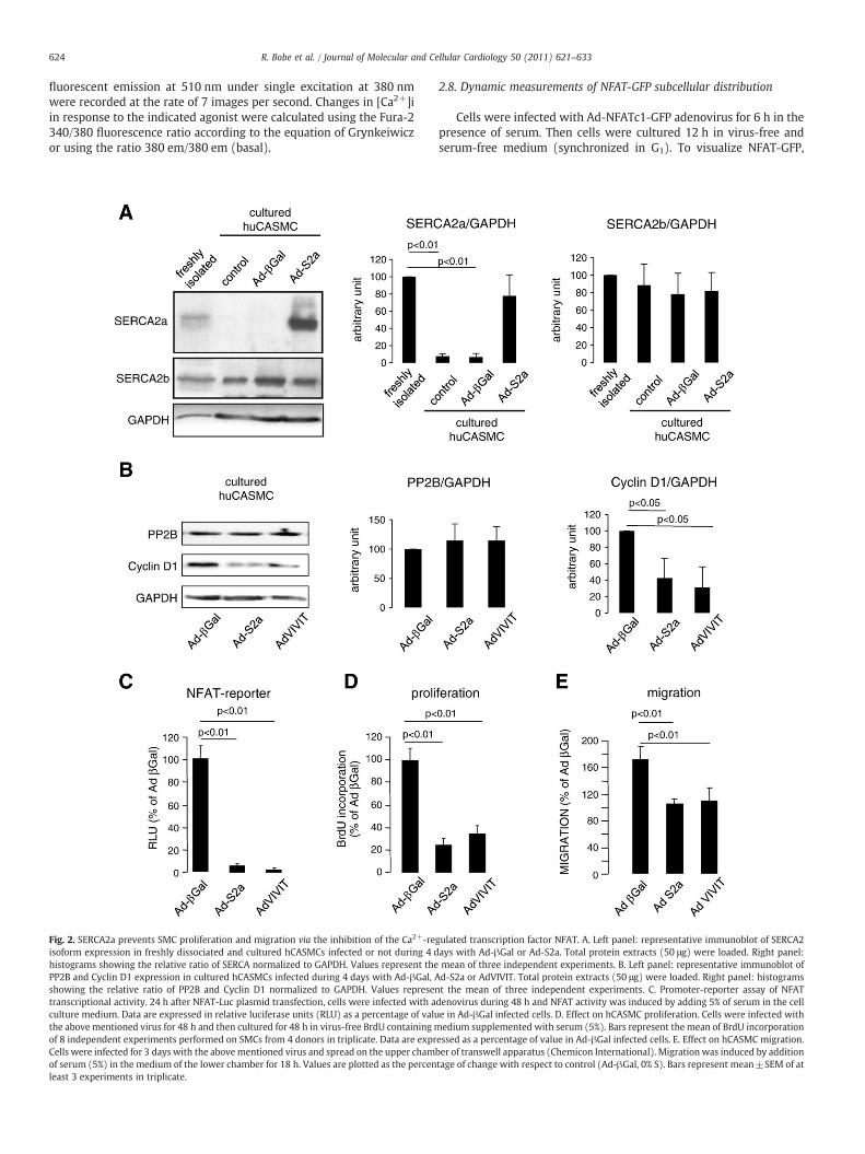
Fig. 2. SERCA2a prevents SMC proliferation and migration via the inhibition of the Ca2+-reisoform expression in freshly dissociated and cultured hCASMCs infected or not during 4 dhistograms showing the relative ratio of SERCA normalized to GAPDH. Values represent thePP2B and Cyclin D1 expression in cultured hCASMCs infected during 4 days with Ad-βGal, Ashowing the relative ratio of PP2B and Cyclin D1 normalized to GAPDH. Values representranscriptional activity. 24 h after NFAT-Luc plasmid transfection, cells were infected with aculture medium. Data are expressed in relative luciferase units (RLU) as a percentage of valuthe above mentioned virus for 48 h and then cultured for 48 h in virus-free BrdU containing mof 8 independent experiments performed on SMCs from 4 donors in triplicate. Data are exprCells were infected for 3 days with the abovementioned virus and spread on the upper chamof serum (5%) in the medium of the lower chamber for 18 h. Values are plotted as the percenleast 3 experiments in triplicate.
2.8. Dynamic measurements of NFAT-GFP subcellular distribution
Cells were infected with Ad-NFATc1-GFP adenovirus for 6 h in thepresence of serum. Then cells were cultured 12 h in virus-free andserum-free medium (synchronized in G1). To visualize NFAT-GFP,
gulated transcription factor NFAT. A. Left panel: representative immunoblot of SERCA2ays with Ad-βGal or Ad-S2a. Total protein extracts (50 μg) were loaded. Right panel:mean of three independent experiments. B. Left panel: representative immunoblot ofd-S2a or AdVIVIT. Total protein extracts (50 μg) were loaded. Right panel: histogramst the mean of three independent experiments. C. Promoter-reporter assay of NFATdenovirus during 48 h and NFAT activity was induced by adding 5% of serum in the celle in Ad-βGal infected cells. D. Effect on hCASMC proliferation. Cells were infected withedium supplemented with serum (5%). Bars represent the mean of BrdU incorporation
essed as a percentage of value in Ad-βGal infected cells. E. Effect on hCASMC migration.ber of transwell apparatus (Chemicon International). Migrationwas induced by additiontage of change with respect to control (Ad-βGal, 0% S). Bars represent mean±SEM of at

625R. Bobe et al. / Journal of Molecular and Cellular Cardiology 50 (2011) 621–633
cells were exited at 488 nm and cellular emission was recordedbetween 500 nm and 520 nm. Subcellular distribution of NFAT–GFPwas quantified as the ratio NFATNUC/NFATCYT using a region of interest(ROI) that covered the area of the nucleus (NFATNUC) and acytoplasmic ROI (NFATCYT) as described [27]. The average fluores-cence of a particular ROI was analyzed using Metamorph. Theindividual cellular ratio NFATNUC/NFATCYT was measured every 10 s.
0% S
erum
Dil
50
10 1
2APB
10 5 1
CsA (µM)
***
***
CAI
50
10 1 50
10 1
***
0
100
200
300
***
*
***
5% Serum+Growth factors
Aproliferation
C
E
0100200300400500600700800900
1000
0 100 200 300 400 500 600 700
Serum
Ca2+
300µM
Time (in sec)
0200400600800
100012001400160018002000
0 100 200 300 400 500 600 700
THR
Ca2+
300µM
Time (in sec)
[Ca2
+]i
(in n
M)
[Ca2
+]i
(in n
M)
Fig. 3. Analysis of Ca2+ signal required for induction of proliferation in synthetic hCASMCs. Efactivity (B) of hCASMCs. Abbreviations used are: Dil— diltiazem, CAI— carboxyamidotriazoleindicated in the figure. For proliferation assay, cells were cultured in presence of BrdU and diplasmid, cultured 24 h in serum-free medium andwere stimulated by adding 5% serum and dcontrol wells (0% serum). Bars represent mean±SEM of at least 3 experiments in triplicate.Typical traces representative of the cytosolic Ca2+ concentration ([Ca2+]i) recorded in singlemedium for 24 h before experiments. Cells were treated with serum (C) or thrombin (THR,cells were treated with 5% serum (D) (the Ca2+ present in the serumwas buffered with 2 mMand then (Ca2+ 300 μM) was then added (D and F).
2.9. Quantitative real-time PCR
Total RNAwaspreparedwithRNeasyMini kits (Invitrogen), and1 μgwas reverse transcribedusinga standardprotocol. Gene specific primerswere used to amplify mRNA by quantitative PCR on an Mx3005apparatus (Stratagen) using Qiagen SYBR Green Master Mix using thefollowing conditions: 95 °C for 15 min and 40 cycles, each at 94 °C for
B
0
100
200
300
400
500
600
*** **
****
***
0% S
erum
Dil
50
2APB10
CsA (µM)CAI50 50
5% Serum+Growth factors
NFAT-reporter
D
F
0100200300400500600700800900
1000
0 100 200 300 400 500 600 700
Serum
EGTA 100µM
Ca2+
Time (in sec)
0200400600800
100012001400160018002000
0 100 200 300 400 500 600 700
THR
EGTA 100µM
Ca2+
Time (in sec)
[Ca2
+]i
(in n
M)
[Ca2
+]i
(in n
M)
fect of various Ca2+ channels blockers on serum-induced proliferation (A) and NFAT-Luc, 2APB— 2-aminoethoxydiphenyl borate, CsA— cyclosporine A. Concentrations used arefferent drugs during 48 h. For NFAT activity assay, cells were transfected with NFAT-Lucifferent drugs during 4h. Data (A and B) are expressed as a percentage of value issued of***Pb0.001; *Pb0.05 vs 5%S. C–F. Intracellular calcium imaging in FURA-2 loaded cells.cell. Cells were synchronized in G1 phase of cell cycle by removing of serum from culture1 U/mL) (E) in the presence of extracellular Ca2+ (300 μM). To record SOCE activation,EGTA) extracellular Ca2+ or THR (1 U/mL) (F) in the absence of calcium (EGTA 100 μM)
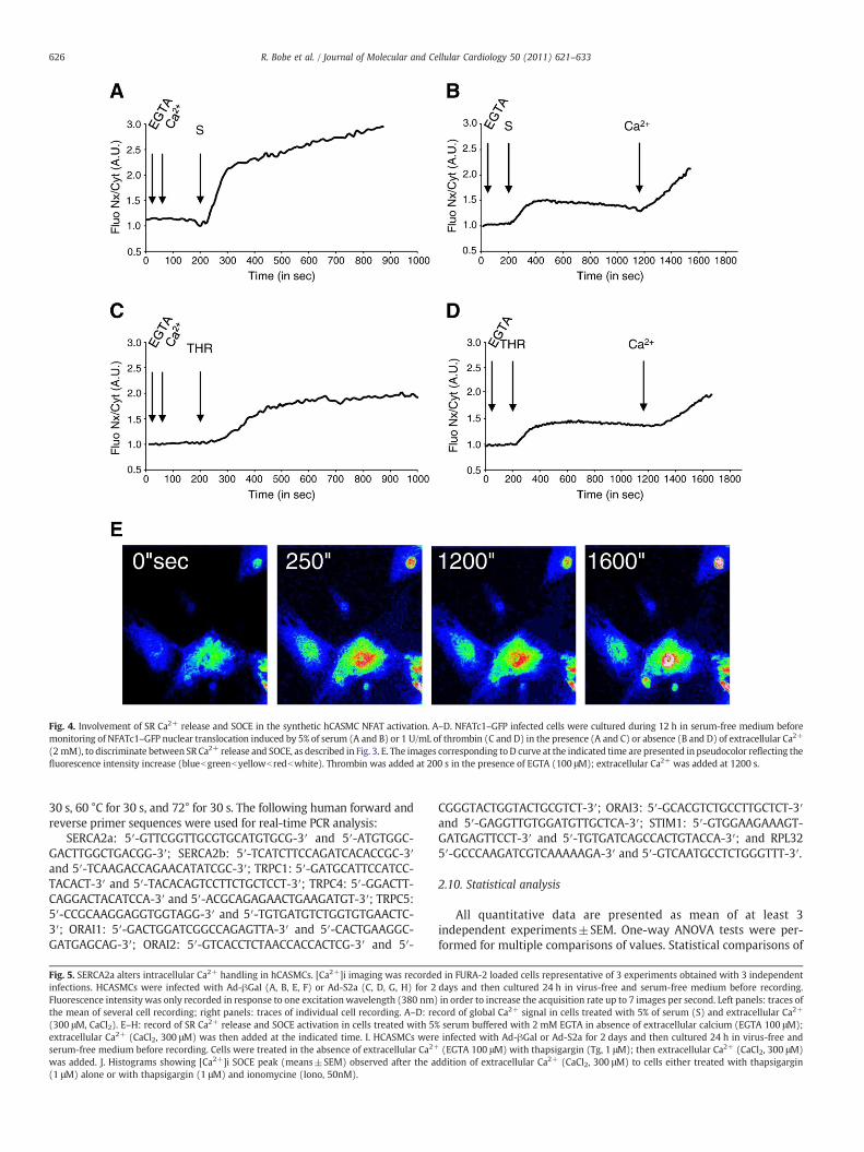
Fig. 4. Involvement of SR Ca2+ release and SOCE in the synthetic hCASMC NFAT activation. A–D. NFATc1–GFP infected cells were cultured during 12 h in serum-free medium beforemonitoring of NFATc1–GFP nuclear translocation induced by 5% of serum (A and B) or 1 U/mL of thrombin (C and D) in the presence (A and C) or absence (B and D) of extracellular Ca2+
(2 mM), to discriminate between SR Ca2+ release and SOCE, as described in Fig. 3. E. The images corresponding toD curve at the indicated time are presented in pseudocolor reflecting thefluorescence intensity increase (bluebgreenbyellowb redbwhite). Thrombin was added at 200 s in the presence of EGTA (100 μM); extracellular Ca2+ was added at 1200 s.
626 R. Bobe et al. / Journal of Molecular and Cellular Cardiology 50 (2011) 621–633
30 s, 60 °C for 30 s, and 72° for 30 s. The following human forward andreverse primer sequences were used for real-time PCR analysis:
SERCA2a: 5′-GTTCGGTTGCGTGCATGTGCG-3′ and 5′-ATGTGGC-GACTTGGCTGACGG-3′; SERCA2b: 5′-TCATCTTCCAGATCACACCGC-3′and 5′-TCAAGACCAGAACATATCGC-3′; TRPC1: 5′-GATGCATTCCATCC-TACACT-3′ and 5′-TACACAGTCCTTCTGCTCCT-3′; TRPC4: 5′-GGACTT-CAGGACTACATCCA-3′ and 5′-ACGCAGAGAACTGAAGATGT-3′; TRPC5:5′-CCGCAAGGAGGTGGTAGG-3′ and 5′-TGTGATGTCTGGTGTGAACTC-3′; ORAI1: 5′-GACTGGATCGGCCAGAGTTA-3′ and 5′-CACTGAAGGC-GATGAGCAG-3′; ORAI2: 5′-GTCACCTCTAACCACCACTCG-3′ and 5′-
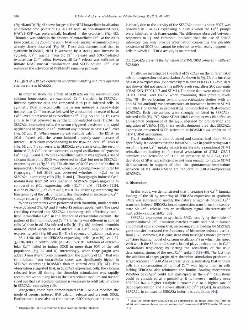
Fig. 5. SERCA2a alters intracellular Ca2+ handling in hCASMCs. [Ca2+]i imaging was recordeinfections. HCASMCs were infected with Ad-βGal (A, B, E, F) or Ad-S2a (C, D, G, H) for 2Fluorescence intensity was only recorded in response to one excitation wavelength (380 nm)the mean of several cell recording; right panels: traces of individual cell recording. A–D: re(300 μM, CaCl2). E–H: record of SR Ca2+ release and SOCE activation in cells treated with 5%extracellular Ca2+ (CaCl2, 300 μM) was then added at the indicated time. I. HCASMCs wereserum-free medium before recording. Cells were treated in the absence of extracellular Ca2+
was added. J. Histograms showing [Ca2+]i SOCE peak (means±SEM) observed after the a(1 μM) alone or with thapsigargin (1 μM) and ionomycine (Iono, 50nM).
CGGGTACTGGTACTGCGTCT-3′; ORAI3: 5′-GCACGTCTGCCTTGCTCT-3′and 5′-GAGGTTGTGGATGTTGCTCA-3′; STIM1: 5′-GTGGAAGAAAGT-GATGAGTTCCT-3′ and 5′-TGTGATCAGCCACTGTACCA-3′; and RPL325′-GCCCAAGATCGTCAAAAAGA-3′ and 5′-GTCAATGCCTCTGGGTTT-3′.
2.10. Statistical analysis
All quantitative data are presented as mean of at least 3independent experiments±SEM. One-way ANOVA tests were per-formed for multiple comparisons of values. Statistical comparisons of
d in FURA-2 loaded cells representative of 3 experiments obtained with 3 independentdays and then cultured 24 h in virus-free and serum-free medium before recording.in order to increase the acquisition rate up to 7 images per second. Left panels: traces ofcord of global Ca2+ signal in cells treated with 5% of serum (S) and extracellular Ca2+
serum buffered with 2 mM EGTA in absence of extracellular calcium (EGTA 100 μM);infected with Ad-βGal or Ad-S2a for 2 days and then cultured 24 h in virus-free and(EGTA 100 μM) with thapsigargin (Tg, 1 μM); then extracellular Ca2+ (CaCl2, 300 μM)
ddition of extracellular Ca2+ (CaCl2, 300 μM) to cells either treated with thapsigargin

IAd-S2a
Tg
0100200300400500600700800900
0 100 200 300 400 500 600
[Ca2+
]i (
in n
M)
Time (in Sec)
Ca2+
C DAd-S2a (mean of 18 cells)
0
1
2
3
4
5
6
0 100 200 300 400 350
Ad-S2a (one cell)
0
1
2
3
4
5
6
0 100 200 300 400 500
E F
0
1
2
3
4
5
6
0 100 200 300 400 500
SEGTA Ca2+
0
1
2
3
4
5
6
0 100 200 300 400 500
SEGTA Ca2+
B
0
1
2
3
4
5
6
0 100 200 300 400 500
SCa2+
SCa2+ SCa2+
AAd-βGal (mean of 22 cells) Ad-βGal (one cell)
Ad-βGal (mean of 22 cells) Ad-βGal (one cell)
Ad-βGal
0
1
2
3
4
5
6
0 100 200 300 400 500
1/F
ura2
fluo
resc
ent
inte
nsity
at 3
80nm
1/
Fur
a2 fl
uore
scen
tin
tens
ity a
t 380
nm
1/F
ura2
fluo
resc
ent
inte
nsity
at 3
80nm
1/
Fur
a2 fl
uore
scen
tin
tens
ity a
t 380
nm
1/F
ura2
fluo
resc
ent
inte
nsity
at 3
80nm
1/
Fur
a2 fl
uore
scen
tin
tens
ity a
t 380
nm
1/F
ura2
fluo
resc
ent
inte
nsity
at 3
80nm
1/
Fur
a2 fl
uore
scen
tin
tens
ity a
t 380
nm
SCa2+
Time (in Sec) Time (in Sec)
Time (in Sec) Time (in Sec)
Time (in Sec) Time (in Sec)
GAd-S2a (mean of 15 cell)
0
1
2
3
4
5
6
0 100 200 300 400 500
SEGTA Ca2+
Time (in Sec)
HAd-S2a (one cell)
0
1
2
3
4
5
6
0 100 200 300 400 500
SEGTA Ca2+
Time (in Sec)
J
Tg (1μM)
050
100150200250300350400450
Ca2+
SO
CE
pea
k (in
nM
)
Tg (1μM) +Iono (50 nM)
627R. Bobe et al. / Journal of Molecular and Cellular Cardiology 50 (2011) 621–633

628 R. Bobe et al. / Journal of Molecular and Cellular Cardiology 50 (2011) 621–633
2 groups were done by an unpaired Student's t-test. Differences wereconsidered significant when Pb0.05.
3. Results
3.1. SR Ca2+ handling proteins expression in SMCs from healthy coronaryarteries
Two distinct populations of SMCs have been described in humannormal and pathological coronary arteries (CA): the “contractile”(differentiated) and “synthetic” (proliferative) [28]. These popula-tions of VSMCs are respectively present in the media and thesubendothelial intima.3 To identify whether in human coronaryarteries SERCA2a expression is associated with specific SMC pheno-type, healthy segments of CA obtained from 5 patients with dilatedcardiomyopathy were studied. Adventitia (a), media (m) andsubendothelial intima (si) were identified on cross sections byhematoxylin/eosin staining (Fig. 1A) and elastin autofluorescence(Fig. 1B). The non-muscular myosin heavy chain B, NM-B, wasvizualized in the media and the subendothelial layers; the smoothmuscle myosin heavy chain (MHC), a marker of terminal differenti-ation [3], was only detected in the media. Thus, SMCs from the mediapresented a “contractile” phenotype whereas those of the suben-dothelial space displayed a “synthetic/proliferative” phenotype.Consistent with this, the intima exhibited a considerable amount ofCyclin D1-positive cells (Fig. 1B). Of note: Cyclin D1 expression insubendothelial intima was heterogeneous along the arteries, withzones of high expression and zones of low expression, in accordancewith previous observations [32]. RyR2 and SERCA2a were bothexpressed in medial contractile SMCs but no positive labeling could bedetected in the subendothelial intima; conversely, the ubiquitousSERCA2b isoform was present in both types of SMCs. As attested bythe expression of contractile SMCmarkers [3], such asMHC, h-calponinand caldesmon, freshly isolated medial hCASMCs displayed a differen-tiated phenotype (Fig. S1A and B); they also expressed SERCA2a,SERCA2b andRyR2 (Fig. S1A andB). Noteworthy, no difference betweenSERCA2a and SERCA2b subcellular localizationwas detected by confocalmicroscopy (Fig. S1A). When cultured in the presence of serum,hCASMCs both proliferated — as attested by BrdU incorporation (notshown) and the emerging expression of CyclinD1— anddedifferentiatedas illustrated by the loss of MHC, h-calponin and caldesmon (Fig. S1Aand B). Consistentwith previous observationsmade for rat VSMCs [4,5],the expression of L-type Ca2+ channel α1C subunit dramaticallydecreased (Fig. S1B). Moreover, the expression of PP2B increased inkeeping with the activation of PP2B/NFAT signaling [7]. Real-time-PCRanalysis revealed a similar pattern of expression of SERCA2a andSERCA2b when compared to that visualized on coronary arterysegments. Indeed, SERCA2a mRNA expression significantly diminishesin proliferating hCASMCs comparing to coronary artery hCASMCs,whereas that of SERCA2b remains unchanged (Fig. 1C). Similarobservations could bemadewhen examining SERCA protein expression(Fig. 1D, 2A, and S1). Of note: total SERCA2 protein (IID8) was notsignificantly modified.
3.2. SERCA2a Controls hCASMC proliferation and migration via theinhibition of PP2B/NFAT signaling pathway
To assess the role of SERCA2a in controlling proliferation andmigration, we studied the consequences of restoring its expression insynthetic hCASMCs on PP2B/NFAT signaling pathway and cyclin D1
3 Of note: the subendothelial SMCs present even in normal human coronary arteriesare believed the most likely source of intimal growth in atherosclerosis, restenosis, andbypass graft intima hyperplasia [29,30]; normal human coronary arteries contain asubendothelial intima, composed of 5 to 10 layers of SMCs and extracellular matrix[31].
expression. Cells were transduced by means of an adenovirus encodingSERCA2a (Ad-S2a); importantly the infection was pursued untilobtaining a detectable level of terminally differentiated human SMCsin coronary arteries (Fig. 2A). Adenovirus encoding β-galactosidase,(Ad-βGal) (used as control) altered neither the expression of SERCA2anor that of SERCA2b (Fig. 2A). Moreover, Ad-βGal did not alter theproliferation of these cells (BrdU incorporation: 100.00±4.48, incontrol vs 84.83±4.38, in Ad-βGal infected cells, ns). Hence, Ad-βGalinfected cells were used as a baseline in these experiments (Fig. 2B–E).
As shown in Fig. 2B, the expression of cyclin D1 was decreased inSERCA2a infected cells while that of PP2B remains unaltered.Adenovirus delivering the NFAT competing peptide VIVIT4 providedsimilar results (Fig. 2B). NFAT-reporter assay showed that NFATtranscriptional activity was blocked in both SERCA2a infected andVIVIT infected cells compared to β-Gal infected cells (Fig. 2C). Asexpected, NFAT-dependent physiological functions, proliferation andmigration induced by serum, were significantly inhibited in SERCA2aand VIVIT-infected cells when compared to β-Gal infected cells(Fig. 2D and E). These data suggested that in hCASMCs SERCA2acontrols NFAT-dependent proliferation andmigration via inhibition ofPP2B and are consistent with previously reported results obtained inrats aortic SMC [6].
3.3. PP2B/NFAT signaling pathway is controlled by both SR Ca2+ releaseand influx of extracellular SOCE Ca2+
To identify the source of calcium regulating PP2B/NFAT signalingpathway, we analyzed the effects of various Ca2+ channel inhibitors onhCASMCproliferation andNFAT-luciferase activity inducedbyserum.Asshown in Fig. 3A and B, Diltiazem (Dil), a specific L-type Ca2+ channelinhibitor did not have any effect either on cell proliferation or NFATactivity. This was consistent with the down-regulation of LTCC insynthetic hCASMCs. In contrast, carboxyamidotriazole (CAI) and 2-aminoethoxydiphenyl borate (2-APB), two inhibitors of SOCE, inhibitedboth NFAT-luciferase activity and cell proliferation similarly to the PP2Binhibitor cyclosporine A (CsA), used here as a control (Fig. 3A and B). Ofnote: SOCE inhibitors were effective only at 50 μM, a concentrationwhich is supposed to inhibit IP3R Ca2+ release and SOCE [33,34]. Serum-induced proliferation translated into a long lasting increase in cytosolicCa2+ followed by an increase of cytosolic Ca2+ basal levels as recordedby the use of a fluorescent calcium probe FURA-2 (Fig. 3C). In theabsence of extracellular Ca2+, the [Ca2+]i increase was systematicallylower (Fig. 3D); addition of extracellular Ca2+ results in a rise ofcytosolic [Ca2+]i related to extracellular Ca2+ influx. The same resultswere obtained using the Gq/phosphoinositide receptor-coupled agonistthrombin (THR) (Fig. 3E and F) except that cytological [Ca2+]i seems toreturn to the basal level after stimulation. Of note: thrombin has beenpreviously shown to induce NFAT activation and proliferation of VSMCs[35]. These data indicated that, in synthetic hCASMCs, the serum-induced steady-state calcium signal activating NFAT-luciferase activityand cell proliferation consistedof SRCa2+ release and extracellular Ca2+
influx. Next, the identification of the calcium signal required for PP2B/NFAT activation was evaluated by discriminating calcium originbuffering extracellular calcium with EGTA (100 μM) or by adding Ca2+
in the cell media. Here, PP2B/NFAT activation was measured bycalculating NFATNUC/NFATCYT ratio as an index of NFAT nucleartranslocation. This was performed in cells stimulated either by serumor by thrombin, using a NFAT–GFP fusion protein in the presence ofextracellular calcium. The NFATNUC/NFATCYT ratio was arbitrary set to1.00 for each cell at the beginning of the recording. As displayed in(Fig. 4A) the NFATNUC/NFATCYT ratio reached 1.99±0.20 (n=26),within 4 min after the addition of serum; a slight increase could also be
4 Synthetic peptide VIVIT interacts with PP2B in competition with NFAT [25,26].

629R. Bobe et al. / Journal of Molecular and Cellular Cardiology 50 (2011) 621–633
noticed until the end of recording (15 min). The increase of NFAT-luciferase activity was observed rapidly 30 min after stimulation (datanot shown). Although the rate of translocation was lower (NFATNUC/NFATCYT ratio reached 1.58±0.09 (n=19)), similar datawere obtainedwhen the cellswere incubatedwith thrombin (Fig. 4C). In the absenceofextracellular Ca2+ (EGTA 100 μM), stimulation with serum (Fig. 4B) orthrombin (Fig. 4D) caused IP3Rmediated Ca2+ release from SR and rapid
Fig. 6. SERCA2a modifies the nature of thrombin-induced Ca2+ transient and inhibits Sfluorescence (lower) was similar in both infected and non-infected cells. Two areas wereSERCA2a-infected cells. B. Typical traces (representative of the [Ca2+]i) recorded in single n100 μM), with 1 U/mL of thrombin and Ca2+ (CaCl2, 300 μM). In order to detect [Ca2+]iwavelength (380 nm) as in Fig. 5. The full recording is presented as Online supplement Virecorded when extracellular Ca2+ (300 μM) was added after stimulation with thrombin (1 Uexperiments (**pb0.01 vs control). D. Bar graphs comparing the percentage of SERCA2a-expra SOCE upon extracellular Ca2+ (300 μM) addition. This has been recorded during 6 experimthe absence of extracellular Ca2+ (EGTA, 100 μM), with 1 U/mL thrombin for 3 min. Then, thΣ[Ca2+]i⁎ time (in seconds)/number of measurements (δ[Ca2+]Tg⁎s). The data are mean±
(within 3 min) NFAT1c–GFP nuclear translocation. Indeed, NFATNUC/NFATCYT ratioswere increasedup to1.33±0.04 (n=36), and1.54±0.05(n=101), respectively. When influx of extracellular Ca2+ was activated(by adding Ca2+ in the extracellular medium), a second spurt of NFATnuclear accumulation was recorded in both cases (NFATNUC/NFATCYT:with serum, 2.02±0.16, n=36; with thrombin, 1.94±0.08, n=101).These data were calculated 3 min after extracellular calcium addition
OCE. A. Ad-S2a-infected cells were identified by GFP fluorescence (upper); FURA-2monitored for FURA-2 fluorescence recording: (a) for non-infected cells and (b) for
on-infected (a) or infected cell (b). Cells were treated, in the absence of calcium (EGTA,oscillations, fluorescence intensity was only recorded in response to one excitationdeo. C. Bar graphs comparing the [Ca2+]i peak corresponding to SOCE (means±SEM)/mL) of hCASMCs infected or not with ad-S2a or ad-βGal. The data are mean±SEM of 3essing to that of and control cells that displayed an oscillatory response to thrombin andents. E. Ca2+ store content after thrombin-induced Ca2+ response. Cells were treated, inapsigargin (Tg, 1 μM) was added and the following Ca2+ mobilization was quantified asSEM for 3 experiments (**pb0.01 vs control).
630 R. Bobe et al. / Journal of Molecular and Cellular Cardiology 50 (2011) 621–633
(Fig. 4BandD). Fig. 4E shows imagesof theNFAT intracellular localizationat different time points of Fig. 4D. Of note: in non-stimulated cells,NFATc1–GFP was preferentially localized in the cytoplasm (Fig. 4E).Thrombin was added in the absence of extracellular Ca2+ at the 200 stime point; at the 250 s time point, NFAT–GFP nuclear accumulationwasalready clearly observed (Fig. 4E). These data demonstrated that, insynthetic hCASMCs, NFAT is activated by a steady-state increase incytosolic Ca2+ arising from SR Ca2+ release and SOC-mediatedextracellular Ca2+ influx. However, SR Ca2+ release was sufficient toinitiate NFAT nuclear translocation and SOCE-induced Ca2+ riseenhanced the activation of PP2B/NFAT signaling pathway.
6 SERCA2b differs from SERCA2a by an extension of 46 amino acids that form an
3.4. Effect of SERCA2a expression on calcium handling and store operatedcalcium entry in hCASMCs
In order to study the effects of SERCA2a on the serum-inducedcalcium homeostasis, we examined Ca2+ transient in SERCA2a-infected synthetic cells and compared it to βGal infected cells. Insynthetic βGal infected cells, the serum induced a steady-stateintracellular Ca2+ increase followed by a sustained increase of basalCa2+ level in presence of extracellular Ca2+ (Fig. 5A and B). This wassimilar to that observed in synthetic non-infected cells (Fig.3A). InSERCA2a expressing cells, the serum triggered persistent and rapidoscillations of cytosolic Ca2+ without any increase in basal Ca2+ level(Fig. 5C and D). When removing extracellular calcium (by EGTA) inβGal-infected cells, the serum induced a steady-state increase ofintracellular calcium corresponding to the IP3R-induced Ca2+ release(Fig. 5E and F); conversely, in SERCA2a expressing cells, the serum-induced IP3R Ca2+ release occurred as rapid oscillations of cytosolicCa2+ (Fig. 5G andH).When extracellular calciumwas applied, a rise ofcalcium illustrating SOCE was observed in βGal- but not in SERCA2a-expressing cells (Fig.5E–H). The absence of SOCE could not be due toimpaired SOC function; indeed, when SERCA pumpswere inhibited bythapsigargin5 full SOCE was observed whether in βGal- or inSERCA2a- expressing cells (Fig. 5I and J). Thapsigargin-induced Ca2+
mobilization from SR was higher in SERCA2a expressing-cells ascompared to βGal expressing cells ([Ca2+]i, nM: 443.40±52.24,n=72 vs 282.80±27.28, n=62, P=0.01). Besides guaranteeing thefunctionality of the calcium pump, this illustrated an increase of Ca2+
storage capacity in SERCA2a-expressing cells.When experiments were performed with thrombin, similar results
were obtained (Fig. 6A and B, video 1S online supplement). The rapidrecording revealed that SERCA2a expressing cells effectively mobi-lized intracellular Ca2+ in the absence of extracellular calcium. Thepattern of thrombin-induced Ca2+ transients was different in controlcells (a) than in Ad-S2a infected cells (b) (Fig. 6B); indeed, thrombininduced rapid oscillations of intracellular Ca2+ only in SERCA2aexpressing cells (Fig. 6B and D). The frequency of calcium peak was11.66±1.40/100 s in SERCA2a-expressing cells (n=39) vs 1.37±0.20/100 s in control cells (n=45), pb0.01. Addition of extracel-lular Ca2+ failed to induce SOCE in more than 90% of the cellpopulation (Fig. 6C and D). Interestingly, when thapsigargin wasadded 3 min after thrombin stimulation, the quantity of Ca2+ that wasre-mobilized from intracellular store, was significantly higher inSERCA2a expressing hCASMCs than in control cells (Fig. 6E). Thisobservation suggested that, in SERCA2a expressing cells, the calciumreleased from SR during the thrombin stimulation was rapidlyrecaptured without any loss in the extracellular medium and furtherruled out that extracellular calcium is necessary to refill calcium storein SERCA2a-expressing cells.
Altogether, these data demonstrated that SERCA2a modifies themode of agonist induced IP3R calcium release and prevents SOCE.Furthermore, it reveals that the absence of SOC response in these cells
5 Thapsigargin (Tg) is a common SERCA (calcium pump) inhibitor.
is clearly due to the activity of the SERCA2a proteins since SOCE wasobserved in SERCA2a expressing hCASMCs when the Ca2+ pumpswere inhibited with thapsigargin. The difference observed betweenresponses to Tg and thrombin indicated that the use of SERCAinhibitors can only provide information concerning the possibleexistence of SOCE but cannot be relevant to what really happens incells in which all SERCA activity is maintained.
3.5. SERCA2a prevents the formation of STIM1/ORAI complex in culturedhCASMCs
Finally, we investigated the effect of SERCA2a on the different SOCsub-unit expression and association. As shown in Fig. 7A, the increaseof SERCA2a expression (evidenced by real-time PCR as ~100-fold, datanot shown) did not modify the mRNA levels regardless SOC sub-units(ORAI1/2/3, TRPC1/4/5 and STIM1). The same data were obtained forSTIM1, ORAI1 and ORAI2 when examining their protein levels(Fig. 7B). By performing co-immunoprecipitation studies with ananti-STIM1 antibody, we demonstrated an interaction between STIM1and ORAI1 or ORAI2, in proliferating non-infected or βGal-infectedhCASMCs. Both interactions were strongly inhibited in SERCA2a-infected cells (Fig. 7C). Since STIM1/ORAI1 complex was identified asan essential component of the ICRAC, required for proliferation andmigration of VSMCs [13], these results demonstrated that SERCA2aexpression prevented SOCE activation in hCASMCs via inhibition ofSTIM1/ORAI association.
Fig. 8 combines the data obtained and summarized them. Morespecifically, it evidences that the loss of SERCA2a in proliferating SMCsresult in lesser Ca2+ uptake which translate into a peripheral STIM1relocalization leading to functional association of STIM1–ORAI1complex and activation of SOCE. In presence of SERCA2a, Ca2+
depletion of SR is not sufficient or not long enough to induce STIM1delocalization. In support of that, the spontaneous interactionsbetween STIM1 and/ORAI1/2 are reduced in SERCA2a-expressinghCASMCs.
4. Discussion
In this study, we demonstrated that increasing the Ca2+ luminalloading of the SR by restoring of SERCA2a expression in syntheticSMCs was sufficient to modify the nature of agonist-induced Ca2+
transient. Indeed, SERCA2a forced expression transforms the steady-state SR Ca2+ release into an oscillatory signal, characteristic ofcontractile vascular SMCs [36].
SERCA2a expression in synthetic SMCs modifying the mode ofagonist-induced Ca2+ transient matches results obtained in humanendothelial cells showing that, increasing store loading by SERCA2agene transfer increased the frequency of histamine-induced oscilla-tions [37]. Moreover, it is consistent with Berridge's model (referredas “store loading model of calcium oscillations”) in which the speedwith which the SR internal store is loaded plays a critical role in Ca2+
oscillations frequency; by setting the sensitivity of the IP3R,determining timing of the next Ca2+ spike [10,38–40]. The fact thatthe addition of thapsigargin after thrombin stimulation produced alarger response in SERCA2a expressing cells, indicating that in thesecells the concentration of luminal Ca2+ was higher than in cellslacking SERCA2a, also reinforced the luminal loading mechanism.Whether SERCA2b6 could also participate in the Ca2+ oscillationscould be considered as a possibility. It is, however, unlikely sinceSERCA2a has a higher catalytic turnover due to a higher rate ofdephosphorylation and a lower affinity to Ca2+ [42,43]. In addition,SERCA2b, in contrast to SERCA2a isoform, is ubiquitous [44].
additional transmembrane domain setting the C terminus of SERCA2b in the SR lumen[41].

Fig. 7. SERCA2a prevents the formation of STIM-1/ORAI1 complex in cultured hCASMCs. A. Effect of SERCA2a gene transfer on the expression of SOC sub-units. mRNA level quantifiedby real-time PCRwasnormalized to thevalue obtained inAd-βGal-infected cells. Histograms showthemeans±SEMof three experiments. B. Cellswere infected for 4 dayswithAd-βGal orAd-S2a. Total protein extracts (50 μg)were loaded. Left panel:western blot showing the expression of ORAI1, ORAI2 and STIM1 inwhole-cell lysates. Right panel: histograms showing therelative ratio of ORAI1, ORAI2 and STIM1 normalized to GAPDH in three experiments. C.Whole-cell lysates were immunoprecipitated (IP) with an anti-STIM1 antibody, resolved on SDS/PAGE and immunobloted for ORAI1 or ORAI2. Membranes were reprobed for STIM1 for protein loading control. Histograms showing themean (n=4) relative ratio of ORAI1 (left panel)and ORAI2 (right panel) normalized to STIM1 and arbitrary considered as 100% for Ad-βGal infected cells.
631R. Bobe et al. / Journal of Molecular and Cellular Cardiology 50 (2011) 621–633
Our study also shows that serum or thrombin-induced persistentoscillations could occur in SERCA2a expressing synthetic SMCswithout any extracellular calcium, in absence of SOCE and STIM1/ORAI1 complex. These data highlight that Ca2+ oscillations can persistwithout extracellular Ca2+ influx in a closed system based solely onlumen/cytosol Ca2+ turnover, challenging the idea as to whetherextracellular Ca2+ influx is absolutely required for refilling the storesbetween each oscillatory cycle [10].
Several studies have reported that the extracellular Ca2+ influx isrequired for transcriptional activation of NFAT [45]. Based on ourmeasurements of NFAT–GFP subcellular localization clearly demonstrat-ing that the initial IP3-induced Ca2+ release from SR intracellular store issufficient to induceNFATnuclear translocation,wenowsuggest that Ca2+
influx through plasma membrane channels acts as an enhancer— ratheras an inductor — of NFAT mobilization. This mechanism is further
reinforced by the fact that modifying the mode of IP3R-induced Ca2+
transient expressing SERCA2a prevented NFAT activation.In our study, we found that diltiazem had no effect on NFAT
signaling pathway in hCASMCs. Our results are different than those ofNieves-Cintron and coworkers who showed that diltiazem blockedPP2B/NFAT signaling through the inhibition of persistant calciumsparklets in mouse and rat arterial myocytes. In contractile arterialSMC, persistent Ca2+ sparklets refer to sustained Ca2+ influx and aremediated by clusters of L-type Ca2+ channels operating in a high openprobability mode. In fact persistent Ca2+ sparklet activity is requiredfor activation of PP2B/NFAT signaling in contractile SMCs [46–49].Considering that hCASMCs, as opposed to the rodent arterial VSMCs,display low level of L-type Ca2+ channels, and that the major route ofextracellular calcium influx is the SOCs, this may explain the differingresults in our study.

Fig. 8. Schematic representation of the involvement of SERCA2a in the physiological control of SOCE. GPCR — G-protein coupled receptor; PLC — phospholipase C; NFAT — nuclearfactor of activated T lymphocytes; P— phosphate; IP3 — inositol-1,4,5-trisphosphate, IP3R— IP3 receptor; SR/ER sarco/endoplasmic reticulum; SERCA— SE/ER Ca2+ATPase; STIM1—
Stromal Interaction Molecule 1, ORAI1 — the pore forming unit.
632 R. Bobe et al. / Journal of Molecular and Cellular Cardiology 50 (2011) 621–633
In addition to modifying the intracellular agonist-induced Ca2+
transient, the rescue of SERCA2a expression in synthetic hCASMCsdisrupted functional association of STIM1/ORAI1 and that of STIM1/ORAI2. SERCA2a control of ORAI isoform activity is consistent withSERCA2a regulating STIM function. Though, it is generally admittedthat STIM1/ORA1 complex is responsible for SOCE in VSMCs [13], thefunctional properties of STIM1/ORAI2 protein complex remainscontroversial. Potier et al. (2009) reported that silencing of eitherSTIM1 or ORAI1 in synthetic VSMCs greatly reduced SOCE, whereasthat of ORAI2, ORAI3, had no effect [13]; however, Mercer et al. (2006)showed that the co-expression of ORAI1 or ORA2 in combination withSTIM1 resulted in substantial increase in ICRAC, ORAI3 failing toproduce any detectable Ca2+ selective currents [50]. Additionalexperiments would be needed to clarify the role of STIM1/ORAI2association in synthetic hCASMCs.
In conclusion, we have demonstrated that SERCA2a is involved inthe frequency dependence of intracellular Ca2+ signaling which leadsto the control of SOCE and NFAT pathways and eventually in theproliferation of VSMCs. This study is, to our knowledge, the firstevidence for oscillatory and steady-state increases in cytosoliccalcium having different effects on calcium dependent signalingprocesses in muscle cells.
Sources of funding
This work is supported by AHA SDG 0930116N (LL), by NIH R01HL080498, and HL083156 (RJH), by Leducq Foundation through theCaerus network (05 CVD 03, AML and RJH); the Association FrançaiseContre les Myopathies, AFM (RB); by MEC-FEDER BFU2010-C02-01(RB and JJL), by K01 HL1031176-01 (LH); JJL was supported by apostdoctoral fellowship from the Junta de Extremadura (POS0922).
Acknowledgments
We thank Michael J. Berridge (The Babraham Institute, Cambridge,UK) for helpful discussion, Bruno Constantin (University of Poitiers,France) for critical reading of this manuscript, Susan Kraner andChristopher M. Norris (Sanders-Brown Center on Aging, Lexington,
KY-USA) for providing AdVIVIT, FrankWuytack (University of Leuven,Belgium) for the anti-SERCA2a and anti-SERCA2b antibodies.
Appendix A. Supplementary data
Supplementary data to this article can be found online atdoi:10.1016/j.yjmcc.2010.12.016.
References
[1] Wamhoff BR, Bowles DK, Owens GK. Excitation–transcription coupling in arterialsmooth muscle. Circ Res 2006 Apr 14;98(7):868–78.
[2] House SJ, Potier M, Bisaillon J, Singer HA, Trebak M. The non-excitable smoothmuscle: calcium signaling and phenotypic switching during vascular disease.Pflugers Arch Aug 2008;456(5):769–85.
[3] Owens GK, Kumar MS, Wamhoff BR. Molecular regulation of vascular smoothmuscle cell differentiation in development and disease. Physiol Rev Jul 2004;84(3):767–801.
[4] Gollasch M, Haase H, Ried C, Lindschau C, Morano I, Luft FC, et al. L-type calciumchannel expression depends on the differentiated state of vascular smooth musclecells. FASEB J May 1998;12(7):593–601.
[5] Quignard JF, Harricane MC, Menard C, Lory P, Nargeot J, Capron L, et al. Transientdown-regulation of L-type Ca(2+) channel and dystrophin expression afterballoon injury in rat aortic cells. Cardiovasc Res Jan 2001;49(1):177–88.
[6] Lipskaia L, del Monte F, Capiod T, Yacoubi S, Hadri L, Hours M, et al. Sarco/endoplasmic reticulum Ca2+-ATPase gene transfer reduces vascular smoothmuscle cell proliferation and neointima formation in the rat. Circ Res Sep 22005;97(5):488–95.
[7] Lipskaia L, Pourci ML, Delomenie C, Combettes L, Goudouneche D, Paul JL, et al.Phosphatidylinositol 3-kinase and calcium-activated transcription pathways arerequired for VLDL-induced smooth muscle cell proliferation. Circ Res May 302003;92(10):1115–22.
[8] Massaeli H, Austria JA, Pierce GN. Lesions in ryanodine channels in smooth musclecells exposed to oxidized low density lipoprotein. Arterioscler Thromb Vasc BiolFeb 2000;20(2):328–34.
[9] Lipskaia L, Pinet C, Fromes Y, Hatem S, Cantaloube I, Coulombe A, et al. Mutation ofdelta-sarcoglycan is associated with Ca(2+)-dependent vascular remodeling in theSyrian hamster. Am J Pathol Jul 2007;171(1):162–71.
[10] Berridge MJ. Inositol trisphosphate and calcium signalling mechanisms. BiochimBiophys Acta Jun 2009;1793(6):933–40.
[11] Lewis RS. The molecular choreography of a store-operated calcium channel.Nature Mar 15 2007;446(7133):284–7.
[12] Berra-Romani R, Mazzocco-Spezzia A, Pulina MV, Golovina VA. Ca2+ handling isaltered when arterial myocytes progress from a contractile to a proliferativephenotype in culture. Am J Physiol Sep 2008;295(3):C779–90.
[13] Potier M, Gonzalez JC, Motiani RK, Abdullaev IF, Bisaillon JM, Singer HA, et al.Evidence for STIM1- and Orai1-dependent store-operated calcium influx through

633R. Bobe et al. / Journal of Molecular and Cellular Cardiology 50 (2011) 621–633
ICRAC in vascular smooth muscle cells: role in proliferation andmigration. FASEB JAug 2009;23(8):2425–37.
[14] Albert AP, Saleh SN, Peppiatt-Wildman CM, Large WA. Multiple activationmechanisms of store-operated TRPC channels in smooth muscle cells. J PhysiolAug 15 2007;583(Pt 1):25–36.
[15] Dolmetsch RE, Lewis RS, Goodnow CC, Healy JI. Differential activation oftranscription factors induced by Ca2+ response amplitude and duration. NatureApr 24 1997;386(6627):855–8.
[16] Liu Z, Zhang C, Dronadula N, Li Q, Rao GN. Blockade of nuclear factor of activated Tcells activation signaling suppresses balloon injury-induced neointima formationin a rat carotid artery model. J Biol Chem Apr 15 2005;280(15):14700–8.
[17] Peel SE, Liu B, Hall IP. ORAI and store-operated calcium influx in human airwaysmooth muscle cells. Am J Respir Cell Mol Biol Jun 2008;38(6):744–9.
[18] Takahashi Y, Watanabe H, Murakami M, Ono K, Munehisa Y, Koyama T, et al.Functional role of stromal interaction molecule 1 (STIM1) in vascular smoothmuscle cells. Biochem Biophys Res Commun Oct 5 2007;361(4):934–40.
[19] Sweeney M, Yu Y, Platoshyn O, Zhang S, McDaniel SS, Yuan JX. Inhibition ofendogenous TRP1 decreases capacitative Ca2+ entry and attenuates pulmonaryartery smooth muscle cell proliferation. Am J Physiol Lung Cell Mol Physiol Jul2002;283(1):L144–55.
[20] Yu H, Sliedregt-Bol K, Overkleeft H, van der Marel GA, van Berkel TJ, Biessen EA.Therapeutic potential of a synthetic peptide inhibitor of nuclear factor of activated Tcells as antirestenotic agent. Arterioscler Thromb Vasc Biol Jul 2006;26(7):1531–7.
[21] Aubart FC, Sassi Y, Coulombe A, Mougenot N, Vrignaud C, Leprince P, et al. RNAinterference targeting STIM1 suppresses vascular smooth muscle cell proliferationand neointima formation in the rat. Mol Ther Mar 2009;17(3):455–62.
[22] Eggermont JA, Wuytack F, Verbist J, Casteels R. Expression of endoplasmic-reticulum Ca2(+)-pump isoforms and of phospholamban in pig smooth-muscletissues. Biochem J Nov 1 1990;271(3):649–53.
[23] Marty I, Robert M, Villaz M, De Jongh K, Lai Y, Catterall WA, et al. Biochemicalevidence for a complex involving dihydropyridine receptor and ryanodinereceptor in triad junctions of skeletal muscle. Proc Natl Acad Sci USA Mar 151994;91(6):2270–4.
[24] del Monte F, Harding SE, Schmidt U, Matsui T, Kang ZB, Dec GW, et al. Restorationof contractile function in isolated cardiomyocytes from failing human hearts bygene transfer of SERCA2a. Circulation Dec 7 1999;100(23):2308–11.
[25] Aramburu J, Garcia-Cozar F, Raghavan A, Okamura H, Rao A, Hogan PG. Selectiveinhibition of NFAT activation by a peptide spanning the calcineurin targeting siteof NFAT. Mol Cell Apr 1998;1(5):627–37.
[26] Aramburu J, Yaffe MB, Lopez-Rodriguez C, Cantley LC, Hogan PG, Rao A. Affinity-driven peptide selection of an NFAT inhibitor more selective than cyclosporin A.Science Sep 24 1999;285(5436):2129–33 New York, NY.
[27] Rinne A, Banach K, Blatter LA. Regulation of nuclear factor of activated T cells(NFAT) in vascular endothelial cells. J Mol Cell Cardiol Sep 2009;47(3):400–10.
[28] Hao H, Gabbiani G, Bochaton-Piallat ML. Arterial smooth muscle cell heteroge-neity: implications for atherosclerosis and restenosis development. ArteriosclerThromb Vasc Biol Sep 1 2003;23(9):1510–20.
[29] Schwartz SM, deBlois D, O'Brien ER. The intima. Soil for atherosclerosis andrestenosis. Circ Res Sep 1995;77(3):445–65.
[30] Stary HC, Blankenhorn DH, Chandler AB, Glagov S, Insull Jr W, Richardson M, et al. Adefinition of the intima of human arteries and of its atherosclerosis-prone regions. Areport from the Committee on Vascular Lesions of the Council on Arteriosclerosis,American Heart Association. Arterioscler Thromb Jan 1992;12(1):120–34.
[31] Rekhter MD, Simari RD, Work CW, Nabel GJ, Nabel EG, Gordon D. Gene transfer intonormal and atherosclerotic human blood vessels. Circ Res Jun 29 1998;82(12):1243–52.
[32] Gueguen M, Keuylian Z, Mateo V, Mougenot N, Lompre AM, Michel JB, et al.Implication of adenylyl cyclase 8 in pathological smooth muscle cell migrationoccurring in rat and human vascular remodelling. The Journal of pathology. Jul2010;221(3): 331–42.
[33] Bootman MD, Collins TJ, Mackenzie L, Roderick HL, Berridge MJ, Peppiatt CM. 2-aminoethoxydiphenyl borate (2-APB) is a reliable blocker of store-operated Ca2+
entry but an inconsistent inhibitor of InsP3-induced Ca2+ release. FASEB J Aug2002;16(10):1145–50.
[34] Faehling M, Kroll J, Fohr KJ, Fellbrich G, Mayr U, Trischler G, et al. Essential role ofcalcium in vascular endothelial growth factor A-induced signaling: mechanism ofthe antiangiogenic effect of carboxyamidotriazole. FASEB J Nov 2002;16(13):1805–7.
[35] Yellaturu CR, Ghosh SK, Rao RK, Jennings LK, Hassid A, Rao GN. A potential role fornuclear factor of activated T-cells in receptor tyrosine kinase and G-protein-coupled receptor agonist-induced cell proliferation. Biochem J Nov 15 2002;368(Pt 1):183–90.
[36] Berridge MJ. Smooth muscle cell calcium activation mechanisms. J Physiol Nov 12008;586(Pt 21):5047–61.
[37] Hadri L, Bobe R, Kawase Y, Ladage D, Ishikawa K, Atassi F, et al. SERCA2a genetransfer enhances eNOS expression and activity in endothelial cells. Mol Ther. Jul2010;18(7): 1284–92.
[38] Berridge MJ. Inositol trisphosphate and calcium signalling. Nature Jan 281993;361(6410):315–25.
[39] Berridge MJ. Inositol trisphosphate and calcium oscillations. Biochem Soc Symp2007(74):1–7.
[40] Berridge MJ, Dupont G. Spatial and temporal signalling by calcium. Curr Opin CellBiol Apr 1994;6(2):267–74.
[41] Campbell AM, Kessler PD, Fambrough DM. The alternative carboxyl termini ofavian cardiac and brain sarcoplasmic reticulum/endoplasmic reticulum Ca(2+)-ATPases are on opposite sides of the membrane. J Biol ChemMay 5 1992;267(13):9321–5.
[42] Dode L, Andersen JP, Leslie N, Dhitavat J, Vilsen B, Hovnanian A. Dissection of thefunctional differences between sarco(endo)plasmic reticulum Ca2+-ATPase(SERCA) 1 and 2 isoforms and characterization of Darier disease (SERCA2)mutants by steady-state and transient kinetic analyses. J Biol Chem Nov 282003;278(48):47877–89.
[43] Dally S, Bredoux R, Corvazier E, Andersen JP, Clausen JD, Dode L, et al. Ca2+-ATPases in non-failing and failing heart: evidence for a novel cardiac sarco/endoplasmic reticulum Ca2+-ATPase 2 isoform (SERCA2c). Biochem J Apr 152006;395(2):249–58.
[44] Bobe R, Bredoux R, Corvazier E, Lacabaratz-Porret C, Martin V, Kovacs T, et al.How many Ca(2)+ATPase isoforms are expressed in a cell type? A growingfamily of membrane proteins illustrated by studies in platelets. Platelets May–Jun 2005;16(3–4):133–50.
[45] Gwack Y, Feske S, Srikanth S, Hogan PG, Rao A. Signalling to transcription: store-operated Ca2+ entry and NFAT activation in lymphocytes. Cell Calcium Aug2007;42(2):145–56.
[46] Navedo MF, Amberg GC, Nieves M, Molkentin JD, Santana LF. Mechanismsunderlying heterogeneous Ca2+ sparklet activity in arterial smooth muscle. J GenPhysiol Jun 2006;127(6):611–22.
[47] Nieves-Cintron M, Amberg GC, Navedo MF, Molkentin JD, Santana LF. The controlof Ca2+ influx and NFATc3 signaling in arterial smooth muscle duringhypertension. Proc Natl Acad Sci USA Oct 7 2008;105(40):15623–8.
[48] Amberg GC, Bonev AD, Rossow CF, Nelson MT, Santana LF. Modulation of themolecular composition of large conductance, Ca(2+) activated K(+) channels invascular smooth muscle during hypertension. J Clin Investig Sep 2003;112(5):717–24.
[49] Wellman GC, Santana LF, Bonev AD, Nelson MT. Role of phospholamban in themodulation of arterial Ca(2+) sparks and Ca(2+)-activated K(+) channels bycAMP. Am J Physiol Sep 2001;281(3):C1029–37.
[50] Mercer JC, Dehaven WI, Smyth JT, Wedel B, Boyles RR, Bird GS, et al. Large store-operated calcium selective currents due to co-expression of Orai1 or Orai2 withthe intracellular calcium sensor, Stim1. J Biol Chem Aug 25 2006;281(34):24979–90.