Sensors and Actuators B: Chemical - SKKUshb.skku.edu/_res/nems/etc/2017-6.pdf · 2017-09-25 · 974...
7
Sensors and Actuators B 248 (2017) 973–979 Contents lists available at ScienceDirect Sensors and Actuators B: Chemical jo ur nal home page: www.elsevier.com/locate/snb Detection of protein kinase using an aptamer on a microchip integrated electrolyte-insulator-semiconductor sensor Rohit Chand a,b , Dawoon Han a , Suresh Neethirajan b , Yong-Sang Kim a,∗ a School of Electronic and Electrical Engineering, Sungkyunkwan University, Suwon, Gyeonggi, 16419, South Korea b BioNano Laboratory, School of Engineering, University of Guelph, Guelph, Ontario, N1G 2W1, Canada a r t i c l e i n f o Article history: Received 15 October 2016 Received in revised form 22 January 2017 Accepted 22 February 2017 Available online 24 February 2017 Keywords: Aptamer Biosensor Capacitance Electrolyte-insulator-semiconductor Microchip Protein kinase A a b s t r a c t Herein, we developed a microchip electrolyte-insulator-semiconductor (EIS) sensor for the capacitive detection of protein kinase A (PKA). EIS sensing is customarily performed in a Teflon cell to define the sensing area. However, in this work, a rapid prototyping technique was followed to integrate polymeric microchip, a reference electrode, and the EIS sensor. The aptameric peptide was used for one-step and label-free detection of PKA enzyme. The thiolated PKA-specific aptamer was immobilized on the gold nanoparticles decorated EIS sensor surface. The detection of PKA in microchip was based on the change in surface charge of EIS sensor. We also analyzed the ability of microchip-EIS sensor to distinguish between buffers at different pH. An average sensitivity of 96 mV/pH for a pH range of 5–9 was obtained. The quanti- tative detection of PKA was performed by analyzing the capacitance-voltage curve after the aptamer-PKA interaction. The EIS sensor showed a detection limit of 2 U/mL with a relative linearity from 10 U/mL to 80 U/mL for the detection of PKA. This study proposes an integrated and point-of-care applicable biosensor for the rapid diagnosis. © 2017 Elsevier B.V. All rights reserved. 1. Introduction Protein kinases are enzymes that mediate the transfer of a phos- phate group from adenosine triphosphate to a target protein, thus monitoring the cellular life. Regulation of protein function through phosphorylation is an important part of the post-translational mod- ification. Altering the phosphorylation of intracellular proteins is a common mode of action of many toxins and pathogens. Several medical conditions, including cancers, Alzheimer’s, and autoim- mune diseases are linked to irregular phosphorylation of protein [1,2]. The extracellular protein kinase A level rises up to 20 U/mL, whereas the total kinase concentration is much higher in the cancer patients [3]. Therefore, determination of protein kinase is crucial in terms of biochemical diagnosis. Conventional techniques like radioisotope labeling, mass spec- troscopy, or enzyme-linked immunosorbent assays based kinase detection consume many expensive and hazardous reagents and typically require long durations for analysis [4,5]. Additionally, miniaturization of these techniques for the point-of-care use is quite not possible. Electrochemical and optical based detec- tion are modern techniques and has proved to be an efficient ∗ Corresponding author. E-mail addresses: [email protected], [email protected] (Y.-S. Kim). approach [6,7]. However, most of these modern approaches depend on the kinase-catalyzed phosphorylation of peptide and require extensive labeling steps. Therefore, detection of a kinase enzyme through phosphorylation of peptide, involving multiple washing, incubation, reaction, and detection sequence is time-consuming, expensive, and laborious. Besides, use of multiple biomolecules for recognizing and reporting reduces the shelf life of biosensors. To overcome these limitations, in our previous work, we designed one-step capacitive aptasensor on metal-insulator- semiconductor (MIS) and electrolyte-insulator-semiconductor (EIS) platforms [8]. On comparison of the two sensing platforms, we concluded that the EIS based sensors are highly sensitive for biosensing. EIS sensors are simple ion-sensitive field-effect tran- sistors (FET) with capacitive detection [9]. EIS sensing is based on the change in gate voltage ensuing due to the release of protons (change in local pH) or intrinsic charge of the biomolecules dur- ing bio-molecular interactions [10,11]. Focusing on the isoelectric point (pI) of the biomolecule can increase or reverse the polarity of biomolecules. The change in the local surface charge modulates the space-charge of the semiconductor-insulator interface leading to a shift in the gate voltage [10]. In the last few years, a number of EIS sensors for detecting bio- molecular interactions were reported which rely on the release of protons or change in the surface charge. Schoning et al. have exten- sively studied and reviewed the application of EIS sensors [9]. EIS http://dx.doi.org/10.1016/j.snb.2017.02.140 0925-4005/© 2017 Elsevier B.V. All rights reserved.
Transcript of Sensors and Actuators B: Chemical - SKKUshb.skku.edu/_res/nems/etc/2017-6.pdf · 2017-09-25 · 974...

Di
Ra
b
a
ARRAA
KABCEMP
1
pmpicmm[wpt
tdtmit
h0
Sensors and Actuators B 248 (2017) 973–979
Contents lists available at ScienceDirect
Sensors and Actuators B: Chemical
jo ur nal home page: www.elsev ier .com/ locate /snb
etection of protein kinase using an aptamer on a microchipntegrated electrolyte-insulator-semiconductor sensor
ohit Chanda,b, Dawoon Hana, Suresh Neethirajanb, Yong-Sang Kima,∗
School of Electronic and Electrical Engineering, Sungkyunkwan University, Suwon, Gyeonggi, 16419, South KoreaBioNano Laboratory, School of Engineering, University of Guelph, Guelph, Ontario, N1G 2W1, Canada
r t i c l e i n f o
rticle history:eceived 15 October 2016eceived in revised form 22 January 2017ccepted 22 February 2017vailable online 24 February 2017
eywords:ptamer
a b s t r a c t
Herein, we developed a microchip electrolyte-insulator-semiconductor (EIS) sensor for the capacitivedetection of protein kinase A (PKA). EIS sensing is customarily performed in a Teflon cell to define thesensing area. However, in this work, a rapid prototyping technique was followed to integrate polymericmicrochip, a reference electrode, and the EIS sensor. The aptameric peptide was used for one-step andlabel-free detection of PKA enzyme. The thiolated PKA-specific aptamer was immobilized on the goldnanoparticles decorated EIS sensor surface. The detection of PKA in microchip was based on the change insurface charge of EIS sensor. We also analyzed the ability of microchip-EIS sensor to distinguish between
iosensorapacitancelectrolyte-insulator-semiconductoricrochip
rotein kinase A
buffers at different pH. An average sensitivity of 96 mV/pH for a pH range of 5–9 was obtained. The quanti-tative detection of PKA was performed by analyzing the capacitance-voltage curve after the aptamer-PKAinteraction. The EIS sensor showed a detection limit of 2 U/mL with a relative linearity from 10 U/mLto 80 U/mL for the detection of PKA. This study proposes an integrated and point-of-care applicablebiosensor for the rapid diagnosis.
© 2017 Elsevier B.V. All rights reserved.
. Introduction
Protein kinases are enzymes that mediate the transfer of a phos-hate group from adenosine triphosphate to a target protein, thusonitoring the cellular life. Regulation of protein function through
hosphorylation is an important part of the post-translational mod-fication. Altering the phosphorylation of intracellular proteins is aommon mode of action of many toxins and pathogens. Severaledical conditions, including cancers, Alzheimer’s, and autoim-une diseases are linked to irregular phosphorylation of protein
1,2]. The extracellular protein kinase A level rises up to 20 U/mL,hereas the total kinase concentration is much higher in the canceratients [3]. Therefore, determination of protein kinase is crucial inerms of biochemical diagnosis.
Conventional techniques like radioisotope labeling, mass spec-roscopy, or enzyme-linked immunosorbent assays based kinaseetection consume many expensive and hazardous reagents andypically require long durations for analysis [4,5]. Additionally,
iniaturization of these techniques for the point-of-care uses quite not possible. Electrochemical and optical based detec-ion are modern techniques and has proved to be an efficient
∗ Corresponding author.E-mail addresses: [email protected], [email protected] (Y.-S. Kim).
ttp://dx.doi.org/10.1016/j.snb.2017.02.140925-4005/© 2017 Elsevier B.V. All rights reserved.
approach [6,7]. However, most of these modern approaches dependon the kinase-catalyzed phosphorylation of peptide and requireextensive labeling steps. Therefore, detection of a kinase enzymethrough phosphorylation of peptide, involving multiple washing,incubation, reaction, and detection sequence is time-consuming,expensive, and laborious. Besides, use of multiple biomolecules forrecognizing and reporting reduces the shelf life of biosensors.
To overcome these limitations, in our previous work, wedesigned one-step capacitive aptasensor on metal-insulator-semiconductor (MIS) and electrolyte-insulator-semiconductor(EIS) platforms [8]. On comparison of the two sensing platforms,we concluded that the EIS based sensors are highly sensitive forbiosensing. EIS sensors are simple ion-sensitive field-effect tran-sistors (FET) with capacitive detection [9]. EIS sensing is based onthe change in gate voltage ensuing due to the release of protons(change in local pH) or intrinsic charge of the biomolecules dur-ing bio-molecular interactions [10,11]. Focusing on the isoelectricpoint (pI) of the biomolecule can increase or reverse the polarityof biomolecules. The change in the local surface charge modulatesthe space-charge of the semiconductor-insulator interface leadingto a shift in the gate voltage [10].
In the last few years, a number of EIS sensors for detecting bio-molecular interactions were reported which rely on the release ofprotons or change in the surface charge. Schoning et al. have exten-sively studied and reviewed the application of EIS sensors [9]. EIS

9 Actua
sgEfptIAmttbiskw
fuptacbtiaspgTdaaardspo
2
2
sthAokEafsaCtoKft
74 R. Chand et al. / Sensors and
ensors have been employed to detect rheumatoid arthritis, urea,lucose, DNA amplification, and KRAS gene [12–16]. Recently, anIS sensor for protein kinase C, ion-sensitive field effect transistoror creatine kinase II, and carbon nanotube field effect transistor forrotein kinase A were reported [17–19]. Their detection depends onhe time consuming kinase-catalyzed phosphorylation of peptide.n addition, these FET based sensors have a small detection range.nother drawback of these EIS sensors is the lack of an integratedicrochip-based sensing platform. In the majority of EIS sensors,
he sensing area is defined by a Teflon cell or patterned SU-8 struc-ure. For the analysis, an Ag/AgCl reference electrode is inserted intouffer reservoir on the sensor surface or the total sensor is dipped
n the electrolyte-containing beaker [13,15]. This makes the sen-or unfit for disposable and point-of-care use. To the extent of ournowledge, only one group has developed an EIS sensor integratedith the microfluidic device for capacitive biosensing [14,20].
In this study, we developed a polymeric microchip-EIS sensoror the label-free and one-step detection of protein kinase A (PKA)sing PKA-specific aptameric peptide. The EIS sensor was com-rised of silicon dioxide (SiO2) grown on the p-type silicon (Si) ashe substrate. The surface of the EIS sensor was functionalized withptamer to make it selective for the PKA and to prevent nonspe-ific interaction. Aptamers have attracted a considerable attentionecause of several advantages over antibodies [21]. Aptamers arehermally stable, easy to design and manufacture, and have unlim-ted applications. The fabrication of polymeric microchip follows
low cost and effortless procedure for integrating the EIS sen-or. A rapid prototyping technique for a microfluidic system usingolymer film and double-sided tape, in place of commonly usedlass and polydimethylsiloxane, was developed to detect the PKA.he detection was based on the change in local surface chargeue to aptamer-PKA interaction. Scanning electron microscopy,tomic force microscopy, Fourier transform-infrared spectroscopy,nd capacitive analysis was used to study the surface modificationnd validation of the EIS sensor. Capacitance-voltage curves wereecorded to detect the presence of PKA. As a proof of concept, weetected PKA in the spiked human cell sample. The microchip-EISensor benefits in a reduced reagent consumption, integrated andoint-of-care analysis, and has the possibility of multiplexing withther biosensors.
. Materials and methods
.1. Materials
Hydrogen tetrachloroaurate(III) hydrate (HAuCl4·3H2O),odium citrate, 3-Mercaptopropyl)trimethoxysilane (MPTS),ris(hydroxymethyl)aminomethane (Tris), sodium chloride, andydrochloric acid of analytical grade were purchased from Sigma-ldrich (St. Louis, MO, USA). Polyethersulfone (PES) films werebtained from Fine chemicals (Korea). cAMP–dependent proteininase A and protein kinase buffer was purchased from Newngland Biolabs (Ipswich, MA, USA) and stored at −20 ◦C. Thiolatedptameric peptide (Mpr-TTYADFIASGRTGRRNAIHD) was obtainedrom AnyGen co. Ltd (Korea). Fourier transform-infrared (FT-IR)pectra of the samples were collected between wavenumbers 400nd 4000 cm−1 at room temperature, using a Agilent Technologiesary 630 FT-IR spectrophotometer coupled with an attenuatedotal reflectance (ATR) device. Atomic force microscopic analysis
f sensor surface was performed using XE-100 (Park systems,orea). All other reagents were of analytical grade and purchasedrom Sigma-Aldrich. Ultrapure de-ionized (DI) water was usedhroughout the experiment.
tors B 248 (2017) 973–979
2.2. Synthesis of gold nanoparticles
The AuNPs (d ≈ 16 nm) were synthesized using a seedlessmethod as described before [22]. Briefly, 20 mL of 1.0 mM aque-ous HAuCl4·3H2O solution was first brought to a boil. Next, 2 mL of38.8 mM aqueous solution of sodium citrate was added, which wasthen boiled for 10 min until the color changed to deep red. The syn-thesized particles were characterized using UV–vis spectroscopyand scanning electrode microscope.
2.3. Fabrication of EIS sensors
p-doped silicon substrate with a resistivity of 10 W-cm wasused for the fabrication of sensors. The Si wafer was cleaned usinga standard Radio Corporation of America (RCA) process. A 50 nmthick SiO2 was grown on the Si substrate through plasma-enhancedchemical vapor deposition. Next, the back of Si wafer was primedusing the wafer back grinding process to obtain a 200 �m thicksubstrate. After priming, 100 nm thick aluminum (Al) was ther-mally deposited on the back of Si wafer using a vacuum thermalevaporator to serve as a back contact.
2.4. Surface functionalization of the EIS sensors
The SiO2 surface of EIS sensor was first treated the with O2plasma for 5 min at 50 sccm flow rate and 5 × 10⁻² Torr pressure.The O2 plasma treatment activated the hydroxyl groups on the SiO2surface, which then reacts with the silane molecule. Silanization ofthe SiO2 surface was performed by dipping the EIS sensor in 1%MPTS-toluene solution for 3 h. The reaction between the hydroxylgroup of the EIS surface and the silane group of the MPTS formeda self-aligned monolayer, leaving free thiol group on the top. Thesilanized surface was thoroughly rinsed with the toluene followedby the ethanol. The EIS sensor was heated at 110 ◦C for 15 min tostrengthen the silane bonds and activate thiol groups. Next, the syn-thesized AuNPs were allowed to anchor on the MPTS modified EISsensor for 6 h. The reaction between thiol groups of the MPTS andAu captured the AuNPs on the EIS sensor. The AuNPs modified EISsensor was characterized by the scanning electron microscopy andcapacitance–voltage (C–V) analysis. The functionalization of the EISsensor was finalized after immobilizing the aptamer solution on theAuNPs for 6 h at 4 ◦C.
2.5. Fabrication of EIS microchip
The rapid prototyping technique for microchip based on a poly-mer film and the double-sided tape was employed (Fig. 1(a)). Thefabrication technique was adopted from the previously reportedworks [1,23]. For this purpose, we used polyethersulfone (PES) filmsand 3MTM double-sided tape. The chip was fabricated in three parts,where, the first bottom PES film layer contained the 100 nm thickAl electrode for the back contact with the EIS sensor. The aptamerfunctionalized EIS was bonded on the Al electrode using a thin layerof conductive silver gel. The middle double-sided tape containedthe fluid network. The tape formed a reservoir with an active areaof 5 mm × 5 mm. The top PES film layer contained the inlet andoutlet holes for the sample. For laying the reference electrode, ashadow mask containing the electrode pattern was attached to thetop PES film. Then, in a thermal vacuum evaporator, titanium layerwas deposited on the PES film as an adhesion layer, followed bya layer of silver. The silver electrode was treated with 50 mM fer-ric chloride for 50 s followed by rinsing with DI water, to obtain
a thin AgCl over the deposited Ag electrode. The three parts werealigned under a simple optical microscope and kept under pres-sure for 30 min. The above technique enabled a rapid prototypingof microchip without the use of a clean room facility and sophisti-
R. Chand et al. / Sensors and Actuators B 248 (2017) 973–979 975
F (b) Strm
ci
2
EwtsETTs
2
oftw
2
AR3smP
ig. 1. (a) Schematic of the polymeric microchip, E1 = electrode for back contact,
icrochip-EIS sensor.
ated instruments. The aptamer terminated EIS sensor integratednto polymeric microchip was further used for the detection of PKA.
.6. pH sensing using microchip-EIS sensors
To verify the pH sensing ability of gold nanoparticle terminatedIS sensors, 2 mM Tris-HCl buffers with pH ranging from 5 to 9ere used. The Tris-HCl buffer was injected using a syringe into
he fluid channel of the microchip and electrical response of the EISensor was measured for each pH. The C–V analysis on microchip-IS was performed using a Hewlett-Packard (HP) 4284A LCR meter.he thin film Ag/AgCl electrode was used as the reference electrode.he gate voltage (VG) was swept at a frequency of 1000 Hz with auperimposed AC signal of 10 mV.
.7. Protein kinase A detection on microchip-EIS sensors
The one-step detection of PKA was performed by adding 10 �Lf different concentrations of PKA in 1X PKA buffer on the aptamerunctionalized EIS sensors. The PKA was allowed to interact withhe aptamer at 25 ◦C. After the interaction, the reservoir was filledith 2 mM Tris-HCl buffer (pH 7) for the C-V analysis.
.8. Detection of PKA in cell sample
Human prostate cancer cell line DU145 was obtained frommerican Type Culture Collection (USA). Cells were maintained inPMI-1640 medium supplemented with 10% fetal bovine serum at
7 ◦C in a humidified 5% CO2 incubator. Cells from subcultures wereupplemented with 0.01% trypsin-EDTA (Sigma-Aldrich, USA) andixed with 1X PKA buffer containing different concentrations ofKA.
ucture of the EIS sensor, and (c) Scheme of aptamer-based PKA detection on the
3. Results and discussion
3.1. Characterization of microchip-EIS sensor
The structure of the fabricated microchip-EIS sensor is shownin Fig. 1(a). The bottom and top PES film contained electrode forthe back contact and Ag/AgCl thin-film reference electrode, respec-tively. The tape provided fluidic connection and reservoir for theanalyte and buffer during analysis. The structure of the EIS sensoris elaborated in Fig. 1(b). The EIS sensor consists of a SiO2 layer asan active material and silicon as a semiconducting layer. A mono-layer of citrate-capped AuNPs was generated on the SiO2 film usingMPTS as the linker molecule. The monolayer serves as a supportinglayer for the thiolated PKA aptamer and additional gating layer. Thepresented fabrication methodology makes it easy to independentlyfunctionalize the EIS sensor in bulk and quickly integrate with thepolymeric microchip. Each modification step of the EIS sensor sur-face was characterized by means of contact-angle measurementswith a 12 �L drop of DI water. Fig. 3(a) presents the results ofthe water contact-angle measurements of a bare SiO2 surface, O2plasma treated SiO2 surface, and MPTS functionalized surface. Thehydrophilicity of the surface increased significantly to a contactangle of ∼7.8◦ after the plasma treatment from a contact angle of∼42◦ for a bare SiO2. The increased surface hydrophilicity was dueto the increase in hydroxyl groups. The sensor surface modifiedwith MPTS regained the hydrophobicity (contact angle = ∼37◦). Thechange in the surface property of SiO2 confirms the silanization ofsensor. Fig. S1 shows the scanning electron microscopic analysisof EIS sensor surface to confirm the attachment of AuNPs. As evi-dent from the image, highly resolved AuNPs with an average sizeof 16 ± 2 nm were attached on the SiO2 surface. Fig. S2 shows theC-V response of EIS sensor after immobilizing negatively chargedcitrate-capped AuNPs. The immobilization of AuNPs altered theVG of EIS sensor. The local charge on the surface of gate insula-
tor affects the depletion region, thereby shifting the C-V curve ofthe EIS sensor. A positive shift in the VG was seen due to the attach-ment of negatively charged AuNPs to the SiO2 surface. The schemefor PKA analysis using EIS sensor is summarized in Fig. 1(c). First, the
9 Actua
tg1w
3
cfibs5CalaTtttcs
3
d
FAp
76 R. Chand et al. / Sensors and
hiolated aptamers were immobilized on the AuNPs through thiol-old chemistry. Next, different concentrations of PKA solution inX reaction buffer was added to the sensor surface for interactionith the aptamer.
.2. pH sensing using microchip-EIS sensors
The detection of PKA on microchip-EIS sensor was based on thehange in the surface charge and local surface pH. Therefore, atrst, we analyzed the ability of microchip-EIS sensor to distinguishetween buffers at different pH. For the analysis, we fabricated EISensor with AuNPs terminated surface. The buffers from pH range–9 were injected through the microchannel and the corresponding-V response was measured. The ionic groups present in the bufferdsorbs on the SiO2 surface, thus changing the surface charge. Thiseads to a shift in the C-V curve of the EIS sensor. A response char-cteristic of EIS sensor at different pH buffers is shown in Fig. 2.he gate voltage (VG) shifted towards negative and positive whenhe sensor surface was exposed to acidic and basic buffer, respec-ively. A good relationship was seen between the pH and shift inhe VG with an average sensitivity of 0.96 V/pH (R2 = 0.99). Thisonfirms the sensitivity of the microchip-EIS sensor towards theurface charge, which is useful for the PKA detection.
.3. Optimization of experimental parameter
The concentration of aptamer is an important parameter for theetection of PKA. The aptamer used in this work was a 20 amino
ig. 2. The response of microchip-EIS sensor at different pH buffers w.r.t thin-filmg/AgCl electrode: (a) C-V curves of EIS sensor at pH 5–9, (b) Relationship betweenH and gate voltage. Buffer: 2 mM Tris–HCl.
tors B 248 (2017) 973–979
acid long peptide that selectively binds with the PKA [24,25]. Theisoelectric point of the aptamer is around pH 9.5, therefore it ispositively charged at the neutral pH value [26]. To determine theoptimal concentration of aptamer that covers the surface of AuNPsmonolayer, the surface was incubated with different concentration(0–200 �M) of the aptamer for 6 h. After formation of the aptamerlayer, the surface was rinsed with the DI water. The shift in theC-V curve was monitored to establish the extent of deposition. Asshown in Fig. S3, the immobilization of positively charged aptamerneutralized the negative charge of the AuNPs to a certain level,thus shifting the VG towards the negative direction. The shift inthe C-V curve of the microchip-EIS sensor saturated when the con-centration of aptamer increased beyond 100 �M. Thus, 100 �M ofaptamer was selected as the optimal for depositing on the AuNPsfor further experiments.
The interaction time of the PKA and aptamer determines the per-formance of the EIS sensor. The optimization of reaction time wasstudied in the range of 0–30 min in the presence of PKA. As shown inFig. S4, the C-V curve shifted towards the negative with the increas-ing reaction time, reaching a saturation at 20 min. The saturationof signal symbolizes the complete binding of the aptamer and PKA.Therefore, a reaction time of 20 min was used in the further work.
3.4. Detection of protein kinase A on microchip-EIS sensors
The aptamer acts as a pseudo-substrate for the PKA enzyme,because of sequence complementarity with the binding site ofthe PKA. It has been extensively reported that the interaction ofaptamer with the target changes the structure, total charge, andcharge distribution of the aptamer [27,28]. Therefore, the EIS sen-sor functionalized with the kinase-specific aptamer was used forthe selective, sensitive, and label-free detection of the enzyme. TheAuNPs terminated EIS sensor was functionalized with 100 �m ofPKA-specific aptamer and was allowed to interact with PKA for20 min at 25 ◦C. The sensor surface was rinsed with DI water toeliminate the unbound PKA. The interaction of aptamer with PKAwas confirmed by FT-IR spectra as shown in Fig. 3(b). The appear-ance of characteristic peaks of the aptameric peptide (Fig. 3(b) curveA), located at 1673 cm−1 (HNC O), 1505 cm−1 (aromatic ring) and1468 cm−1 (C N) established the immobilization of aptamer on thegold surface. An increase in the peak intensity and presence of func-tional groups commonly found in the PKA enzyme confirmed theinteraction and binding of aptamer with PKA (Fig. 3(b) curve B).We also performed the surface analysis of EIS sensor before andafter interaction of aptamer and PKA to verify the biosensing (Fig.S5). Non-contact mode AFM topographs of the aptamer immobi-lized gold surface (Fig. S5(a)) and after aptamer PKA interaction(Fig. S5(b)) shows an increase in the surface roughness. In Fig. S5(b),several distinctive rough spots on the surface were apparent for thepresence of aptamer-PKA complex [29,30].
For the microchip-EIS sensing of PKA, different concentrationsof PKA in 1X reaction buffer was injected in the microchip and incu-bated with the aptamer. The pI of the PKA is pH 8.84; therefore, itsinteraction with aptamer further increases the positive charge onthe sensor surface. This change in the charge and density on the sen-sor surface was recorded using C-V analysis for label-free detectionof PKA. After the aptamer-PKA interaction, the reservoir was slowlyfilled with the tris buffer saline (TBS, 2 mM tris, 20 mM NaCl, pH 7)and then the C-V curves were analyzed. The strength of the bufferwas kept low and the pH was set to 7 to reduce the effects of ionson the electrical response of the sensor.
Fig. 4(a) shows the C-V curves for label-free capacitive detection
of PKA on microchip-EIS sensor. PKA solutions with a concentra-tion range of 1–80 U/mL were detected using the proposed sensor.As can be seen from Fig. 4(a), with the increasing concentrationof PKA, the VG gradually shifted more towards the negative due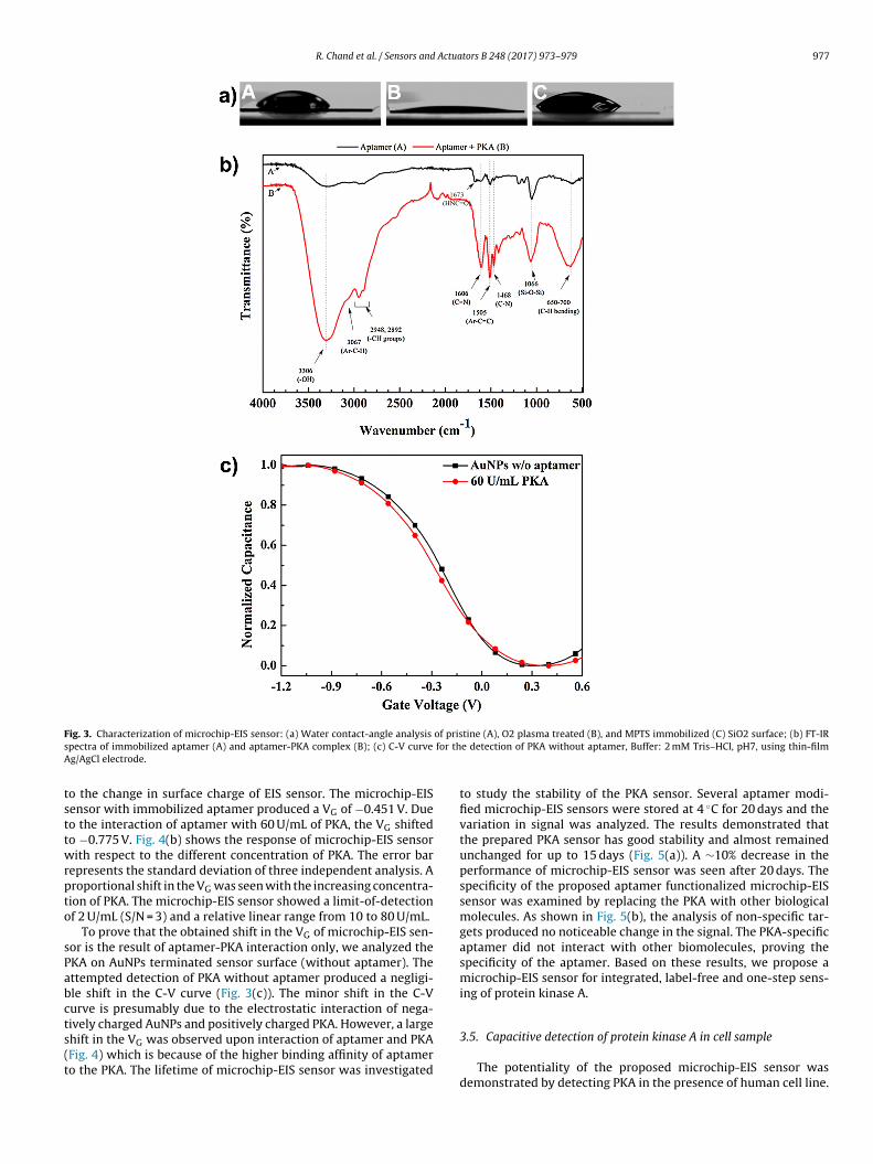
R. Chand et al. / Sensors and Actuators B 248 (2017) 973–979 977
F of priss for thA
tsttwrpto
sPabcts(t
ig. 3. Characterization of microchip-EIS sensor: (a) Water contact-angle analysis
pectra of immobilized aptamer (A) and aptamer-PKA complex (B); (c) C-V curve
g/AgCl electrode.
o the change in surface charge of EIS sensor. The microchip-EISensor with immobilized aptamer produced a VG of −0.451 V. Dueo the interaction of aptamer with 60 U/mL of PKA, the VG shiftedo −0.775 V. Fig. 4(b) shows the response of microchip-EIS sensorith respect to the different concentration of PKA. The error bar
epresents the standard deviation of three independent analysis. Aroportional shift in the VG was seen with the increasing concentra-ion of PKA. The microchip-EIS sensor showed a limit-of-detectionf 2 U/mL (S/N = 3) and a relative linear range from 10 to 80 U/mL.
To prove that the obtained shift in the VG of microchip-EIS sen-or is the result of aptamer-PKA interaction only, we analyzed theKA on AuNPs terminated sensor surface (without aptamer). Thettempted detection of PKA without aptamer produced a negligi-le shift in the C-V curve (Fig. 3(c)). The minor shift in the C-Vurve is presumably due to the electrostatic interaction of nega-ively charged AuNPs and positively charged PKA. However, a large
hift in the VG was observed upon interaction of aptamer and PKAFig. 4) which is because of the higher binding affinity of aptamero the PKA. The lifetime of microchip-EIS sensor was investigatedtine (A), O2 plasma treated (B), and MPTS immobilized (C) SiO2 surface; (b) FT-IRe detection of PKA without aptamer, Buffer: 2 mM Tris–HCl, pH7, using thin-film
to study the stability of the PKA sensor. Several aptamer modi-fied microchip-EIS sensors were stored at 4 ◦C for 20 days and thevariation in signal was analyzed. The results demonstrated thatthe prepared PKA sensor has good stability and almost remainedunchanged for up to 15 days (Fig. 5(a)). A ∼10% decrease in theperformance of microchip-EIS sensor was seen after 20 days. Thespecificity of the proposed aptamer functionalized microchip-EISsensor was examined by replacing the PKA with other biologicalmolecules. As shown in Fig. 5(b), the analysis of non-specific tar-gets produced no noticeable change in the signal. The PKA-specificaptamer did not interact with other biomolecules, proving thespecificity of the aptamer. Based on these results, we propose amicrochip-EIS sensor for integrated, label-free and one-step sens-ing of protein kinase A.
3.5. Capacitive detection of protein kinase A in cell sample
The potentiality of the proposed microchip-EIS sensor wasdemonstrated by detecting PKA in the presence of human cell line.

978 R. Chand et al. / Sensors and Actuators B 248 (2017) 973–979
Fig. 4. The response of microchip-EIS sensor for the detection of PKA w.r.t thin-fiRp
Dmasmou
lm Ag/AgCl electrode: (a) C-V curves of EIS sensor for PKA from 0 to 80 U/mL, (b)elationship between PKA concentration and gate voltage. Buffer: 2 mM Tris–HCl,H7.
U145 cells in conditioned medium were spiked with the reactionixture containing different concentrations of PKA, to simulate
biological sample. For the PKA-aptamer interaction, 10 �L of
piked cell sample was injected and incubated with the aptamer-odified microchip-EIS sensor as discussed earlier. The interactionf aptamer and PKA present in the sample was then analyzedsing C-V curves. Fig. 6 summarizes the analyses, demonstrating
Fig. 5. The stability (a) and specificity (b) of microc
Fig. 6. Detection of PKA in the spiked human cell sample.
good recoveries with respect to the concentrations of spiked PKA.The device showed high signal to noise ratio with EIS sensor pre-cisely distinguishing between the spiked PKA and other interferingmolecules present in the medium. Therefore, the microchip-EISsensor can be used to detect PKA in cell samples or serum frompatients.
4. Conclusion
In conclusion, this work describes a strategy for developmentand integration of a polymeric microchip with EIS sensor for detec-tion of PKA. The proposed sensor was highly sensitive towards thechange in pH. Aptamer immobilized sensor surface facilitated inrapid biosensing. Interaction of aptamer and PKA on microchip-EISsensor produced a shift in the gate voltage. The sensor showed adetection limit of 2 U/mL for PKA. The developed method offersattractive features like one-step detection, integrated analysis,and costless polymeric microchip. The biosensor exhibited a highresponse, low detection limit, and specificity towards the PKA.A polymer-based microchip makes this work beneficial for dis-
posable and point-of-care use. The proposed sensor can also beintegrated with other biosensors for multiplexed analysis.hip-EIS sensor, BSA = Bovine serum albumin.

Actua
A
t
R
[
[
[
[
[
[
[
[
[
[
[
[
[
[[
[
[
[
[
[
[
R. Chand et al. / Sensors and
ppendix A. Supplementary data
Supplementary data associated with this article can be found, inhe online version, at http://dx.doi.org/10.1016/j.snb.2017.02.140.
eferences
[1] R. Chand, D. Han, I.-S. Shin, J.-I. Hong, Y.-S. Kim, Gold nanoparticle enhancedelectrochemical assay for protein kinase activity using a syntheticchemosensor on a microchip, J. Electrochem. Soc. 162 (2015) B89–B93.
[2] A. Tedgui, Z. Mallat, Cytokines in atherosclerosis: pathogenic and regulatorypathways, Physiol. Rev. 86 (2005) 515–581.
[3] H. Wang, M. Li, W. Lin, W. Wang, Z. Zhang, E.R. Rayburn, J. Lu, D. Chen, X. Yue,F. Shen, F. Jiang, J. He, W. Wei, X. Zeng, R. Zhang, Extracellular activity of cyclicAMP-dependent protein kinase as a biomarker for human cancer detection:distribution characteristics in a normal population and cancer patients,Cancer Epidemiol. Biomark. Prev. 16 (2007) 789–795.
[4] O. von Ahsen, U. Bomer, High-throughput screening for kinase inhibitors,ChemBioChem 6 (2005) 481–490.
[5] C. D’Ambrosio, A.M. Salzano, S. Arena, G. Renzone, A. Scaloni, Analyticalmethodologies for the detection and structural characterization ofphosphorylated proteins, J. Chromatogr. B 849 (2007) 163–180.
[6] I.-S. Shin, R. Chand, S.W. Lee, H.-W. Rhee, Y.-S. Kim, J.-I. Hong, Homogeneouselectrochemical assay for protein kinase activity, Anal. Chem. 86 (2014)10992–10995.
[7] X. Xu, X. Liu, Z. Nie, Y. Pan, M. Guo, S. Yao, Label-free fluorescent detection ofprotein kinase activity based on the aggregation behavior of unmodifiedquantum dots, Anal. Chem. 83 (2011) 52–59.
[8] R. Chand, D. Han, Y.-S. Kim, Rapid detection of protein kinase on capacitivesensing platforms, IEEE Trans. Nanobiosci. 15 (2016) 843–848.
[9] M.J. Schöning, Playing around with field-effect sensors on the basis of EISstructures, LAPS and ISFETs, Sensors 5 (2005) 126–138.
10] T.S. Bronder, A. Poghossian, S. Scheja, C. Wu, M. Keusgen, D. Mewes, M.J.Schoning, DNA immobilization and hybridization detection by the intrinsicmolecular charge using capacitive field-effect sensors modified with acharged weak polyelectrolyte layer, ACS Appl. Mater. Interfaces 7 (2015)20068–20075.
11] T.-W. Lina, D. Kekudaa, C.-W. Chua, Label-free detection of DNA using novelorganic-based electrolyte-insulator-semiconductor, Biosens. Bioelectron. 25(2010) 2706–2710.
12] T.-M. Pan, T.-W. Lin, C.-Y. Chen, Label-free detection of rheumatoid factorusing YbYxOy electrolyte-insulator-semiconductor devices, Anal. Chim. Acta891 (2015) 304–311.
13] T.-M. Pan, M.-D. Huang, W.-Y. Lin, M.-H. Wu, A urea biosensor based onpH-sensitive Sm2TiO5 electrolyte–insulator–semiconductor, Anal. Chim. Acta11 (2010) 68–74.
14] Y.-H. Lin, S.-H. Wang, M.-H. Wu, T.-M. Pan, C.-S. Lai, J.-D. Luo, C.-C. Chiou,Integrating solid-state sensor and microfluidic devices for glucose, urea andcreatinine detection based on enzyme-carrying alginate microbeads, Biosens.Bioelectron. 43 (2013) 328–335.
15] B. Veigas, R. Branquinho, J.V. Pinto, P.J. Wojcik, R. Martins, E. Fortunato, P.V.Baptista, Ion sensing (EIS) real-time quantitative monitorization of isothermalDNA amplification, Biosens. Bioelectron. 52 (2014) 50–55.
16] Y.-T. Lin, A. Purwidyantri, J.-D. Luo, C.-C. Chiou, C.-M. Yang, C.-H. Lo, T.-L.Hwang, T.-H. Yen, C.-S. Lai, Programming a nonvolatile memory-like sensorfor KRAS gene sensing and signal enhancement, Biosens. Bioelectron. 79(2016) 63–70.
17] N. Bhalla, M.D. Lorenzo, G. Pula, P. Estrela, Protein phosphorylation analysisbased on proton release detection: potential tools for drug discovery, Biosens.Bioelectron. 54 (2014) 109–114.
18] R. Freeman, R. Gill, I. Willner, Following a protein kinase activity using afield-effect transistor device, Chem. Commun. (2007) 3450–3452.
19] C.-S. Lee, T.H. Kim, A Simple and sensitive Assay for protein kinase a usingsingle-walled carbon nanotube Field-Effect transistor, Bull. Korean Chem. Soc.37 (2016) 1167–1168.
tors B 248 (2017) 973–979 979
20] Y.-H. Lin, C.-H. Chiang, M.-H. Wu, T.-M. Pan, J.-D. Luo, C.-C. Chiou, Solid-statesensor incorporated in microfluidic chip and magnetic-bead enzymeimmobilization approach for creatinine and glucose detection in serum, Appl.Phys. Letts. 99 (2011) 253704.
21] K.-M. Song, S. Lee, C. Ban, Aptamers and their biological applications, Sensors12 (2012) 612–631.
22] J. Turkevich, P.C. Stevenson, J. Hillier, A study of the nucleation and growthprocesses in the synthesis of colloidal gold, Discuss. Faraday Soc. 11 (1951)55–75.
23] P.K. Yuen, V.N. Goral, Lab Chip 10 (2010) 384–387.24] H. Eldar-Finkelman, M. Eisenstein, Peptides targeting protein kinases:
strategies and implications, Curr. Pharm. Des. 15 (2009) 2463–2470.25] D.B. Glass, H.C. Cheng, B.E. Kemp, D.A. Walsh, Differential and common
recognition of the catalytic sites of the cGMP-dependent andcAMP-dependent protein kinases by inhibitory peptides derived from theheat-stable inhibitor protein, J. Biol. Chem. 261 (1986) 12166–12171.
26] L.P. Kozlowski, IPC − Isoelectric Point Calculator, 2016. Available: http://dx.doi.org/10.1101/049841.
27] M. Zayats, Y. Huang, R. Gill, C.-a. Ma, I. Willner, Label-free and reagentlessaptamer-based sensors for small molecules, J. Am. Chem. Soc. 128 (2006)13666–13667.
28] M.C. Rodriguez, A.-N. Kawde, J. Wang, Aptamer biosensor for label-freeimpedance spectroscopy detection of proteins based on recognition-inducedswitching of the surface charge, Chem. Commun. (2005) 4267–4269.
29] Y.H. Tan, J.R. Schallom, N.V. Ganesh, K. Fujikawa, A.V. Demchenko, K.J. Stine,Characterization of protein immobilization on nanoporous gold using atomicforce microscopy and scanning electron microscopy, Nanoscale 3 (2011)3395–3407.
30] J.A.U. Paredes, A. Polini, W. Chrzanowski, Protein-based biointerfaces tocontrol stem cell differentiation, in: D. Hutmacher, W. Chrzanowski (Eds.),Biointerfaces: Where Material Meets Biology, RSC publications, 2014, 2017,pp. 1–29.
Biographies
Rohit Chand: Rohit obtained his Ph.D. in Electronic and Electrical Engineering fromSungkyunkwan University, South Korea and M.Sc. from KIIT University, India. Hehas developed electrochemical and microfluidic biosensors for the detection of can-cer biomarkers. Rohit worked as a post-doctoral fellow and research professor atSungkyunkwan University, South Korea. At present, he is working as a post-doctoralfellow at BioNano Lab, University of Guelph, Canada. His research work involvesmicro-fabrication, surface functionalization, biosensors, and lab-on-a-chip devel-opment.
Dawoon Han: Dawoon is currently a doctoral candidate in the NEMS Lab atSungkyunkwan University, South Korea. She completed her MS degree in Nano Sci-ence and Engineering from Myongji University, South Korea. Her research interestsare in the development of electrochemical, field-effect transistor, and lab-on-a-chipbased biosensors.
Suresh Neethirajan: Prof. Neethirajan is an assistant professor in Biological andBiomedical Engineering program of the University of Guelph with demonstratedresearch excellence and experience in large-scale multi-faceted international col-laborative research projects. He is currently directing the state-of-the-art BioNanoLab and the team leader of the Precision Livestock Biosensor group at Guelph.
Yong-Sang Kim: Prof. Kim is a professor at the School of Electronic and Electri-cal Engineering and head of the Nano-Electronics and Microfluidic Sensors lab ofthe Sungkyunkwan University, South Korea. Prior to this, he was a professor in theDept. of Electrical Engineering and director of Nano-Bio Research Centre at MyongjiUniversity, South Korea. He also served as an associate researcher at Univ. of Cal-
ifornia at Berkeley, U.S.A. Prof. Kim’s research focuses on developing organic andoxide based TFTs for biological detection and use in electrical circuits. He also workson the development of microfluidic platforms for the electrochemical analysis ofbiomolecules and organic solar cells. He has published over 150 papers in severalpeer-reviewed journals.