Segmental bile duct leakage after hepatic resection ... · PDF fileHyeon Sik Kim, et al....
5
Korean J Hepatobiliary Pancreat Surg 2012;16:115-119 Case Report Segmental bile duct leakage after hepatic resection managed with percutaneous ablation by N-butyl cyanoacrylate Hyeon Sik Kim 1 , Tae Hyo Kim 1,3 , Eun Young Yun 1 , Hyun Seok Ham 1 , Hong Jun Kim 1 , Chi-Young Jeong 2 , Hyun Jin Kim 1 , Woon Tae Jung 1 , Ok-Jae Lee 1 , and Soon-Chan Hong 2 Departments of 1 Internal Medicine, 2 Surgery and 3 Institute of Health Science, Gyeongsang National University School of Medicine, Jinju, Korea A biloma is a rare abnormal accumulation of intrahepatic or extrahepatic bile caused by a traumatic or spontaneous rupture of the biliary tree. The reported incidence of postoperative biloma ranges from 4.8% to 7.6%. Biliary drainage is usually important and necessary for the treatment of biloma, but sometimes bile leakage fails to improve despite prolonged conservative drainage. We report a case of postoperative refractory biliary leakage managed with percuta- neous ablation by N-butyl cyanoacrylate. (Korean J Hepatobiliary Pancreat Surg 2012;16:115-119) Key Words: Biloma; Biliary leakage; N-butyl cyanoacrylate Received: June 20, 2012; Revised: July 25, 2012; Accepted: July 30, 2012 Corresponding author: Tae Hyo Kim Department of Internal Medicine and Institute of Health Science, Gyeongsang National University School of Medicine, 90, Chilam-dong, Jinju, 660-702, Korea Tel: +82-55-750-8726, Fax: +82-55-758-9122, E-mail: [email protected] Copyright Ⓒ 2012 by The Korean Association of Hepato-Biliary-Pancreatic Surgery Korean Journal of Hepato-Biliary-Pancreatic Surgery ∙ pISSN: 1738-6349 Fig. 1. Postcontrast abdominal computed tomography shows a large intrahepatic fluid collection at the resected bed. INTRODUCTION A biloma has been reported to occur as a result of bili- ary surgery, endoscopic retrograde cholangiopancreatog- raphy (ERCP) procedures, laparoscopic cholecystectomy, trauma, and occasionally spontaneously. 1,2 The amount of bile leakage from small ducts on the raw cut surface of a liver is usually small and the site of the leakage may close spontaneously, although it may need facilitation by a temporary percutaneous or endoscopic drainage to divert bile from the leakage site. 2,3 But in patients with bile leak- age from a transected, isolated bile duct, surgery should sometimes be performed. 4 We report a case of post- operative refractory segmental bile duct leakage, managed successfully by intrahepatic biliary ablation with N-butyl cyanoacrylate. CASE A 70-year-old man was admitted to our hospital for fur- ther management of a 15 cm-sized abdominal fluid collection. He was diagnosed with hepatocellular carcino- ma and underwent a central lobectomy fifteen months prior. At follow-up, he underwent a CT scan every three months. Fifteen months after surgery, computed tomog- raphy (CT) imaging studies revealed a 15×12 cm-sized fluid collection located on the resected liver surface (Fig. 1). He was admitted for a possible rupture of the biloma. On admission, his vital signs and physical examination
Transcript of Segmental bile duct leakage after hepatic resection ... · PDF fileHyeon Sik Kim, et al....
Korean J Hepatobiliary Pancreat Surg 2012;16:115-119 Case Report
Segmental bile duct leakage after hepatic resection managed withpercutaneous ablation by N-butyl cyanoacrylate
Hyeon Sik Kim1, Tae Hyo Kim1,3, Eun Young Yun1, Hyun Seok Ham1, Hong Jun Kim1, Chi-Young Jeong2, Hyun Jin Kim1, Woon Tae Jung1, Ok-Jae Lee1, and Soon-Chan Hong2
Departments of 1Internal Medicine, 2Surgery and 3Institute of Health Science, Gyeongsang National University School of Medicine, Jinju, Korea
A biloma is a rare abnormal accumulation of intrahepatic or extrahepatic bile caused by a traumatic or spontaneous rupture of the biliary tree. The reported incidence of postoperative biloma ranges from 4.8% to 7.6%. Biliary drainage is usually important and necessary for the treatment of biloma, but sometimes bile leakage fails to improve despite prolonged conservative drainage. We report a case of postoperative refractory biliary leakage managed with percuta-neous ablation by N-butyl cyanoacrylate. (Korean J Hepatobiliary Pancreat Surg 2012;16:115-119)
Key Words: Biloma; Biliary leakage; N-butyl cyanoacrylate
Received: June 20, 2012; Revised: July 25, 2012; Accepted: July 30, 2012Corresponding author: Tae Hyo KimDepartment of Internal Medicine and Institute of Health Science, Gyeongsang National University School of Medicine, 90, Chilam-dong, Jinju,660-702, KoreaTel: +82-55-750-8726, Fax: +82-55-758-9122, E-mail: [email protected]
Copyright Ⓒ 2012 by The Korean Association of Hepato-Biliary-Pancreatic SurgeryKorean Journal of Hepato-Biliary-Pancreatic Surgery ∙ pISSN: 1738-6349
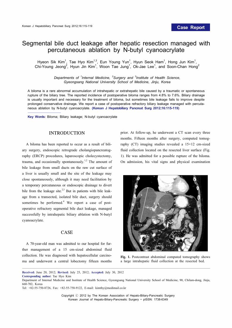
Fig. 1. Postcontrast abdominal computed tomography shows a large intrahepatic fluid collection at the resected bed.
INTRODUCTION
A biloma has been reported to occur as a result of bili-ary surgery, endoscopic retrograde cholangiopancreatog-raphy (ERCP) procedures, laparoscopic cholecystectomy, trauma, and occasionally spontaneously.1,2 The amount of bile leakage from small ducts on the raw cut surface of a liver is usually small and the site of the leakage may close spontaneously, although it may need facilitation by a temporary percutaneous or endoscopic drainage to divert bile from the leakage site.2,3 But in patients with bile leak-age from a transected, isolated bile duct, surgery should sometimes be performed.4 We report a case of post-operative refractory segmental bile duct leakage, managed successfully by intrahepatic biliary ablation with N-butyl cyanoacrylate.
CASE
A 70-year-old man was admitted to our hospital for fur-ther management of a 15 cm-sized abdominal fluid collection. He was diagnosed with hepatocellular carcino-ma and underwent a central lobectomy fifteen months
prior. At follow-up, he underwent a CT scan every three months. Fifteen months after surgery, computed tomog-raphy (CT) imaging studies revealed a 15×12 cm-sized fluid collection located on the resected liver surface (Fig. 1). He was admitted for a possible rupture of the biloma. On admission, his vital signs and physical examination

116 Korean J Hepatobiliary Pancreat Surg Vol. 16, No. 3, August 2012
Fig. 2. Fluoroscopic spot film of a tube cholangiogram dem-onstrating contrast (bile) extravasation (bile leak site; open ar-row) from a dehisced isolated right intrahepatic bile duct seg-ment (arrow). An adjacent biloma is seen with a drain in it.
Fig. 3. Image obtained during an N-butyl cyanoacrylate (glue)injection for ablating a fistula. It shows obliteration of the communication between the biloma and the bile duct.
were normal. The laboratory studies were as follows: leu-cocyte 5,160/mm3, hemoglobin 10.8 g/dl, platelets 139,000/mm3, AST 20 IU/L, ALT 11 IU/L, alkaline phos-phatase 68 IU/L, total bilirubin 0.48 mg/dl, total protein 5.9 g/dl, albumin 3.5 g/dl, BUN/Cr 20.5/0.84 mg/dl, C-re-active protein 1.0 mg/dl, alpha-fetoprotein 1.26 ng/ml, and prothrombin time of 13.4 seconds. The viral marker tests, including HBsAg, anti-HBs Ab and anti-hepatitis C virus (HCV), were all negative.
On the third day after admission, percutaneous drainage (PCD) with a 10-Fr pigtail catheter, under the guidance of an abdominal ultrasound, was performed for diagnosis and treatment. We removed 1,000 ml of fluid that was dark green in color. For a diagnosis, biochemical tests were carried out on the bile, and these tests revealed a total bilirubin of 9.35 mg/dl and a direct bilirubin of 7.45 mg/dl that was thought to be caused by bile leakage. We then performed a right percutaneous transhepatic biliary drainage (PTBD) procedure (8 Fr) to divert bile from the site of the injury. But the cholangiogram image did not demonstrate segmental contrast (bile) leaks. On the sev-enth day after admission, no communication to the biloma was found in a cholangiogram via the right PTBD catheter. Bile continued to drain from the catheter for 1 week, at approximately 20-40 ml daily after PTBD clamping. Because there was no communication between the biloma and the common hepatic duct, the right PTBD was removed.
On the seventeenth day after admission, the patient was discharged with a percutaneous biloma drainage catheter for spontaneous closure of the biliary leakage during out-patient follow-up. However, the patient inadvertently pulled out his external drainage catheter one month later.
He was admitted to the emergency room with abdomi-nal pain, nausea and febrile sensation. A percutaneous flu-id drainage under ultrasonograhic guidance was done again. Approximately 50-100 ml per day of bile continued to drain for 17 days. For bile diversion, a percutaneous right transhepatic biliary drainage procedure (8.5 Fr) was performed. On the seventh and fifteenth day after PCD reinsertion, tubograms were performed and no communi-cation to the bile ducts was found at that time. On sev-enteenth day after PCD reinsertion, the communication between the biloma and the isolated right superior intra-hepatic bile duct was noted via a third tubogram per-formed with a PCD catheter (Fig. 2). For cessation of bile production from that segment and its subsequent atrophy, we did additional selective catheterization (6 Fr) of the actual leaking bile duct. The next day, 2 ml of cyanoacry-late combined with lipiodol was injected into an isolated bile duct as the selective catheter was withdrawn under real-time fluoroscopic control. This immediately achieved fistula closure (Fig. 3). On the nineteenth day after PCD reinsertion, the volume of bile juice decreased markedly and ceased. Then the PTBD was clamped and removed

Hyeon Sik Kim, et al. Segmental bile duct leakage 117
Fig. 4. Follow-up contrast-enhanced computed tomography image demonstrating the hardened N-butyl cyanoacrylate cast-ing the ablated right intrahepatic bile duct. Marked decrease in the size of the biloma is shown.
two days later. The percutaneous catheter was removed after five days and the patient had no complaints or symptoms.
The patient has been carefully monitored every three months since discharge. A CT scan obtained 10 months later showed significant decreased collection in the cavity (Fig. 4). The patient’s recovery was uneventful.
DISCUSSION
A biloma is defined as an encapsulated collection of bile outside the biliary tree. Bile leaks can be classified as either due to biliary discontinuity with biliary spillage into the peritoneal cavity or due to biliary cutaneous fistulae. A biloma is most commonly caused by surgery, percutaneous transhepatic cholangiography (PTC), PTBD, and abdominal trauma.1,2,5 The clinical presentation of a biloma varies greatly from non-specific abdominal pain to biliary sepsis.6 Encapsulation of bile within the omentum and mesentery prevents generalized peritonitis in most cases.7 Abdominal ultrasonography is the first modality to evaluate the nature of a biloma and the underlying pathology. However, an abdominal CT can define the dis-ease, the cause and the relationships with adjacent struc-tures more accurately.8 A differential diagnosis should in-clude hematoma, seroma, liver abscess, cysts, pseudo-cysts, and lymphocele.9 Helpful distinguishing features are
clinical history, anatomy and location of the lesion, CT number, and character of the material obtained by aspiration. Most bilomas have CT numbers of less than 20H, but may be higher when the bile is mixed with blood or exudates.7 Ultrasonography or CT scan of the abdomen is useful in locating a fluid collection but does not in-dicate the site of leakage. Percutaneous aspiration under radiologic guidance can also aid in diagnosis and treatment. Typically, bilomas contain clear, greenish bile, but the bile may be discolored by a secondary infection, exudates, or blood. When necessary, the presence of bilir-ubin may be documented by chemical techniques.
Management of the biloma in a patient includes appro-priate measures such as intravenous hydration and ini-tiation of antibiotic treatment if sepsis is present. Some bilomas, especially those that are small in size and asymp-tomatic, can be followed without intervention, but most cases require treatment.8 A bile leak may persist because of an elevated intrabiliary pressure caused by a distal stone obstruction or simply due to the normal positive in-trabiliary pressure maintained by the Sphincter of Oddi. Thus, it is important to lower the intrabiliary pressure in order to promote closure and healing of the injured bile duct.10
In the past, surgery was the main approach to treatment. Surgery is now performed only in cases with a persistent bile leak or for treatment of an underlying disease. Today, there is a much wider variety of options such as percutaneous catheter drainage and/or endopros-thesis placement, endoscopic sphincterotomy (EST), endo-scopic nasobiliary drainage (ENBD) and endoscopic drainage.2,6,11 However, biliary decompression will not be effective when the leaking ducts do not communicate with the common bile duct. In such cases, bile leak site embo-lization/sclerosis, and leaking biliary segment ablation are useful. This report indicates that N-butyl Cyanoacrylate ablation may be effective in the unfortunate cases of re-fractory biliary leakage after hepatectomy.
There are two scenarios for biliary ablation. The first is an actual bile leak site ablation or embosclerosis to re-duce an aperture or ablate a fistula (block a hole). The second is ablating an entire biliary segment to cease bile production and induce hepatic segmental atrophy (cease bile production).12 The most common biliary segment to be inadvertently transected in hepatic resection (left hemi-
118 Korean J Hepatobiliary Pancreat Surg Vol. 16, No. 3, August 2012
hepatectomy or left lobectomy or laparoscopic injury) is the right posterior bile duct. This is due to its not un-common aberrant anatomy where the right posterior duct drains into the left biliary system.
When approaching from the biliary tract in an acute bile leak setting, ablative materials that may be used in-clude absolute (98-99%) alcohol, acetic acid, and cyanoa-crylate (glue).12 Absolute alcohol requires that the biliary system be purged of anything that would dilute the alcohol. Fifty percent acetic acid is made by combining one-part absolute acetic acid with one-part of contrast. Both 50% acetic acid and absolute alcohol are left to dwell for approximately 5 minutes and then the biliary system is lavaged with saline.3,13 Cyanoacrylate (glue) re-quires the biliary system to be irrigated with a dextrose solution. The cyanoacrylate is combined with ethiodol for fluoroscopic visualization and does not require dwell time.14 In fact, the administering catheter is removed be-fore it can adhere to the glue. The literature is limited re-garding this subject; however, it appears that cyanoacry-late requires fewer sessions (one to two sessions) than the other ablative agents that are required to dwell in the bili-ary system (four to six sessions).3,13,14 It should be noted that, in the acute setting with no fistulous tract formation, ablative substances (acetic acid, alcohol, and cyanoacry-late) may cause intense pain if they come in contact with the peritoneum.12 Unfortunately, biliary leakage is some-times encountered, and it can be complicated when it does occur.4,15,16 An N-butyl cyanoacrylate injection into the bile duct provides a management option in cases in which conservative treatment has failed and a surgical approach is relatively difficult.
Although we have no data how large a leakage could be sealed with the N-butyl cyanoacrylate, we believe that leakages of less than 50 ml/d could be blocked by the procedure. Once the glue seals the fistula completely, the effect is immediate and safe. Cyanoacrylate glue, which polymerizes on contact with biologic tissue, may arrest leaks by plugging fistulous tracts arising from the bile duct lumen. The contrast agent lipiodol is used for in-creasing the viscosity of the mixture applied and for pre-cise radiographic localization.17 In one series of patients with biliary leaks that were sealed with cyanoacrylate, treatment was successful in 7 of 9 cases with a single injection.18 But one article reported a serious complication
of cyanoacrylate where there was ascending venous thrombosis from the middle hepatic vein to the left pul-monary artery and it was removed surgically.19 Other complications are the potential risk of catheter and stent adherence, occlusion, biliary obstruction, and atrophy of the liver.
We reported a case of hepatic resection of hep-atocellular carcinoma that was complicated by an intra-hepatic biloma. Because external biliary drainage con-tinued despite prolonged conservative management, a re-peated tubogram was done to find the site of the bile leakage. A repeated tubogram or fistulogram may be help-ful in assessing the nature of the bile leak because a com-munication may not always be demonstrated until a fistula is established, or any associated intra-abdominal collection has been resolved. We found an isolated bile duct and performed cyanoacrylate ablation of the isolated bile duct.
In summary, intrahepatic biliary ablation with N-butyl cyanoacrylate is effective in controlling bile leak and of-fers an alternative treatment to surgical repair.
REFERENCES
1. Fujiwara H, Yamamoto M, Takahashi M, et al. Spontaneous rup-ture of an intrahepatic bile duct with biloma treated by percuta-neous drainage and endoscopic sphincterotomy. Am J Gastroenterol 1998;93:2282-2284.
2. Jun KH, Cho HM, Chin HM, et al. A nontraumatic rupture of intrahepatic bile duct and perihepatic biloma formation in a patient with choledocholithiasis: a case report. Korean J Hepatobiliary Pancreat Surg 2004;8:46-49.
3. Park JH, Oh JH, Yoon Y, et al. Acetic acid sclerotherapy for treatment of a biliary leak from an isolated bile duct after hepatic surgery. J Vasc Interv Radiol 2005;16:885-888.
4. Ota T, Hirai R, Tsukuda K, et al. Biliary reconstruction with right hepatic lobectomy due to delayed management of laparo-scopic bile duct injuries: a case report. Acta Med Okayama 2004;58:163-167.
5. Mueller PR, Ferrucci JT Jr, Simeone JF, et al. Detection and drain-age of bilomas: special considerations. AJR Am J Roentgenol 1983;140:715-720.
6. Binmoeller KF, Katon RM, Shneidman R. Endoscopic manage-ment of postoperative biliary leaks: review of 77 cases and report of two cases with biloma formation. Am J Gastroenterol 1991;86:227-231.
7. Vazquez JL, Thorsen MK, Dodds WJ, et al. Evaluation and treat-ment of intraabdominal bilomas. AJR Am J Roentgenol 1985;144: 933-938.
8. Lee JH, Suh JI. A case of infected biloma due to spontaneous intrahepatic biliary rupture. Korean J Intern Med 2007;22: 220-224.
9. Akhtar MA, Bandyopadhyay D, Montgomery HD, et al. Spontaneous idiopathic subcapsular biloma. J Hepatobiliary Pancreat Surg 2007;14:579-581.

Hyeon Sik Kim, et al. Segmental bile duct leakage 119
10. Geenen JE, Toouli J, Hogan WJ, et al. Endoscopic sphincter-otomy: follow-up evaluation of effects on the sphincter of Oddi. Gastroenterology 1984;87:754-758.
11. Hartle RJ, McGarrity TJ, Conter RL. Treatment of a giant biloma and bile leak by ERCP stent placement. Am J Gastroenterol 1993;88:2117-2118.
12. Saad WE, Darcy MD. Percutaneous management of biliary leaks: biliary embosclerosis and ablation. Tech Vasc Interv Radiol 2008;11:111-119.
13. Kyokane T, Nagino M, Sano T, et al. Ethanol ablation for seg-mental bile duct leakage after hepatobiliary resection. Surgery 2002;131:111-113.
14. Vu DN, Strub WM, Nguyen PM. Biliary duct ablation with N-bu-tyl cyanoacrylate. J Vasc Interv Radiol 2006;17:63-69.
15. Tanaka S, Hirohashi K, Tanaka H, et al. Incidence and manage-
ment of bile leakage after hepatic resection for malignant hepatic tumors. J Am Coll Surg 2002;195:484-489.
16. Bhattacharjya S, Puleston J, Davidson BR, et al. Outcome of ear-ly endoscopic biliary drainage in the management of bile leaks after hepatic resection. Gastrointest Endosc 2003;57:526-530.
17. Wright G, Jairath V, Reynolds M, et al. Endoscopic glue in-jection for persistent biliary leakage. Gastrointest Endosc 2009;70:1279-1281.
18. Seewald S, Groth S, Sriram PV, et al. Endoscopic treatment of biliary leakage with n-butyl-2 cyanoacrylate. Gastrointest Endosc 2002;56:916-919.
19. Tomazic A, Mlinaric V, Pleskovic A. Ascending venous throm-bosis from the middle hepatic vein to the left pulmonary artery as a complication of endoscopic Histoacryl sealing of a bile leak after blunt liver injury. Endoscopy 2006;38 Suppl 2:E8-9.